Chapter 9 Discovery of Selective and Potent Inhibitors of Palmitoylation Sonia Lobo Planey Additional information is available at the end of the chapter http://dx.doi.org/10.5772/52503 1. Introduction Palmitoylation is a reversible, post-translational modification of a protein through the addi‐ tion of the 16-carbon fatty acid, palmitate, to a cysteine residue. There are two types of pal‐ mitoylation, one called thio- or S-palmitoylation in which palmitate is added to the thiol side chain of a cysteine residue via a labile thioester bond [1]. The other type, N-palmitoyla‐ tion, is the addition of palmitate to an N-terminal cysteine via a stable amide bond [2]. The two forms of palmitoylation are regulated by different families of palmitoyl acyltransferases (PATs)—S-palmitoylation via a family of multi-pass transmembrane proteins called DHHC (Asp-His-His-Cys) proteins [3] and N-palmitoylation via a family of multi-pass transmem‐ brane proteins termed membrane-bound O-acyltransferase [4]. S-palmitoylation, the focus of this chapter, is more common and because of the labile thioester bond, can dynamically regulate protein sorting and function. Palmitoylation increases the lipophilicity of the modified protein often changing its subcel‐ lular distribution in both dramatic and subtle ways. The larger-scale changes occur when cy‐ toplasmic proteins relocate from the cytoplasm to membrane and when integral membrane proteins move from one membrane system to another, such as from the endoplasmic reticu‐ lum (ER) to the plasma membrane (PM). The more subtle changes, in terms of distance, oc‐ cur at the nanoscale level within a membrane. The increase in lipophilicity upon palmitoylation often results in an altered affinity for a particular lipid microenvironment within that membrane [5]. For example, lipid rafts are small islands in membranes with dis‐ tinct lipid compositions that selectively attract or exclude both peripheral (often exclusively by virtue of palmitoylation) and integral membrane palmitoylated proteins. Palmitoylated proteins have affinity for lipid rafts that are rich in cholesterol, while prenylated proteins have little or no affinity for these rafts [5]. Such lipophilicity-driven changes in protein dis‐ © 2013 Planey; licensee InTech. This is an open access article distributed under the terms of the Creative Commons Attribution License (http://creativecommons.org/licenses/by/3.0), which permits unrestricted use, distribution, and reproduction in any medium, provided the original work is properly cited.

Welcome message from author
This document is posted to help you gain knowledge. Please leave a comment to let me know what you think about it! Share it to your friends and learn new things together.
Transcript

Chapter 9
Discovery of Selective and Potent Inhibitors ofPalmitoylation
Sonia Lobo Planey
Additional information is available at the end of the chapter
http://dx.doi.org/10.5772/52503
1. Introduction
Palmitoylation is a reversible, post-translational modification of a protein through the addi‐tion of the 16-carbon fatty acid, palmitate, to a cysteine residue. There are two types of pal‐mitoylation, one called thio- or S-palmitoylation in which palmitate is added to the thiolside chain of a cysteine residue via a labile thioester bond [1]. The other type, N-palmitoyla‐tion, is the addition of palmitate to an N-terminal cysteine via a stable amide bond [2]. Thetwo forms of palmitoylation are regulated by different families of palmitoyl acyltransferases(PATs)—S-palmitoylation via a family of multi-pass transmembrane proteins called DHHC(Asp-His-His-Cys) proteins [3] and N-palmitoylation via a family of multi-pass transmem‐brane proteins termed membrane-bound O-acyltransferase [4]. S-palmitoylation, the focusof this chapter, is more common and because of the labile thioester bond, can dynamicallyregulate protein sorting and function.
Palmitoylation increases the lipophilicity of the modified protein often changing its subcel‐lular distribution in both dramatic and subtle ways. The larger-scale changes occur when cy‐toplasmic proteins relocate from the cytoplasm to membrane and when integral membraneproteins move from one membrane system to another, such as from the endoplasmic reticu‐lum (ER) to the plasma membrane (PM). The more subtle changes, in terms of distance, oc‐cur at the nanoscale level within a membrane. The increase in lipophilicity uponpalmitoylation often results in an altered affinity for a particular lipid microenvironmentwithin that membrane [5]. For example, lipid rafts are small islands in membranes with dis‐tinct lipid compositions that selectively attract or exclude both peripheral (often exclusivelyby virtue of palmitoylation) and integral membrane palmitoylated proteins. Palmitoylatedproteins have affinity for lipid rafts that are rich in cholesterol, while prenylated proteinshave little or no affinity for these rafts [5]. Such lipophilicity-driven changes in protein dis‐
© 2013 Planey; licensee InTech. This is an open access article distributed under the terms of the CreativeCommons Attribution License (http://creativecommons.org/licenses/by/3.0), which permits unrestricted use,distribution, and reproduction in any medium, provided the original work is properly cited.
tribution may alter access of a palmitoylated protein to extracellular ligands (when the pro‐tein moves from the ER to the PM), protein-protein interactions, or the engagement of thepalmitoyl-protein in multi-molecular signaling complexes. The role of palmitoylation as aversatile protein sorting signal, regulating intracellular protein trafficking and targeting tomembrane microdomains has been reviewed recently [6]. Palmitate may be the most com‐mon lipid species to occupy cysteine residues, but it is not the only one. Marilyn Resh andcolleagues identified the lipid moieties resident on the cysteine residue of the N-terminal tailof Src family kinases [7-9]. While for these proteins the cysteine residue near the N-terminusis most frequently palmitoylated, it is also modified by palmitoleate, stearate, or oleate witha frequency that is apparently related to the abundance of palmitate in cells [10]. The phys‐iological differences that result from proteins being modified by these other lipids has notbeen explored extensively; however, given their different physical properties, it seems rea‐sonable that their impact on a protein should be subtly different than palmitate.
Unlike other forms of lipidation such as myristoylation and prenylation, palmitoylation isreversible, by virtue of the labile thioester bond. This allows for dynamic regulation of theprotein’s lipophilicity [11-13]. By contrast, prenyl groups are attached to cysteines by a sta‐ble thioether bond and myristate to glycines by a stable amide bond. It is now apparent thatmany instances of palmitoylation are enzymatically mediated by a family of palmitoyl acyl‐transferases (PATs), whereas the mechanisms for depalmitoylation are poorly understood.Nevertheless, it is known that palmitate cycles on and off of many proteins at variable ratesranging from minutes to days. Such dynamic regulation makes palmitoylation uniqueamong post-translational protein lipid modifications and places it in a category similar tophosphorylation. Discovering the molecular identity of PATs was a pivotal event that dra‐matically accelerated the pace of discovery in the field. Likewise, there has been increasedinterest in palmitoylation partly because many of the genes encoding PATs have beenlinked to human diseases like cancer. With a greater understanding of how palmitate is en‐zymatically attached to proteins, some of the most interesting questions include: What arethe substrate(s) of each PAT?; how does a PAT recognize and palmitoylated a substrate?;how are PATs regulated?; and how is depalmitoylation regulated? The answers to thesequestions are beginning to unfold due to the recent discovery of pharmacological modula‐tors of palmitoylation as well as the development of novel assays and refinement of existingassays. Our ability to understand palmitoylation and its importance to human health anddisease is only as good as the methods we use to test our hypotheses. Thus, the discovery ofpotent and selective inhibitors of palmitoylation as well as the continued development of as‐says with increased sensitivity and selectivity is critical to this venture.
2. Palmitoylation and DHHC proteins
2.1. Molecular identity of palmitoyl acyltransferases (PATs)
It has been known for many years that palmitoylation is a critical regulator of diverse andcomplex signaling networks, but the mechanism responsible for palmitoylation of most pro‐
Drug Discovery252

teins remained a mystery and somewhat controversial until only recently. The apparent ab‐sence of a consensus site for palmitoylation encoded by the sequence of amino acid residuessurrounding palmitoyl cysteines, as well as the difficulty in purifying and identifying theenzymes capable of mediating the reaction, led many to believe that it was autocatalytic.Given these issues and the high reactivity of cysteines and palmitoyl-CoA, especially in invitro protein palmitoylation assays, the possibility was not unreasonable [11, 14, 15]. Manyof the arguments for and against autocatalytic palmitoylation have been reviewed recently[16].Yet, given the prevalence of palmitoylated proteins in parts of cells where signalingevents are so highly concentrated, complex, and regulated, such as the neuronal synapse, itseemed somewhat unreasonable that all regulation of palmitoylation could be left to diffu‐sion—a nagging reality that kept the search for an enzymatic mechanism alive despite thearguments to the contrary. Additionally, there was evidence over the years in support of theidea that these enzymes existed because PAT activity in detergent solubilized protein frac‐tions had been measured using viral glycoproteins [17], p59fyn [18], and H-Ras [19] as sub‐strates among others.
The experiments that conclusively provided the molecular identity of PATs were presented ina series of papers spanning almost a decade. The experimental model organism that ultimate‐ly provided the information was yeast. First, palmitoylation-dependent alleles of yeast RAS2were identified. A genetic screen designed to identify mutations that rendered cells non-via‐ble if Ras2p was not palmitoylated was utilized to identify mutations in two genes- ERF2 andERF4/SHR5 [20, 21]. These mutations resulted in diminished palmitoylation of Ras2p and mis‐localization of GFP-Ras2p (respectively or it takes both mutations to cause both effects [20, 22]).However, it could not be decisively concluded if the mutations in ERF2 and ERF4 were affect‐ing Ras2p palmitoylation directly or indirectly by altering Ras2p trafficking (which could haveprevented an interaction between the palmitoyl acyltransferase and Ras2p).
In collaboration with Maurine Linder, Deschenes and colleagues used an in vitro palmitoyla‐tion assay to show that Erf2p and Erf4p together constituted a Ras2p PAT that used palmito‐yl-CoA as a donor [23]. Erf2p is a ~42-kDa integral membrane protein that is expressed inthe ER. The protein contains the DHHC-CRD (Asp-His-His-Cys-cysteine rich domain), alsoreferred to as the NEW1 or zf-DHHC domain (PF01529), which is found in an extensive fam‐ily of membrane proteins ranging from unicellular eukaryotes to humans [24, 25]. This do‐main is now recognized as the molecular signature for PATs that add palmitate to cysteinesvia a labile thioester bond.
At almost the same time that the Erf2p/Erf4p complex was identified as the Ras2p PAT,Akr1p was identified as a PAT with specificity for Yck2p [26]. An important clue leading tothe relationship between these two proteins came from the fact that mutants in both Ras2pand Yck2P exhibited a reduced rate of pheromone receptor internalization [27, 28]. Akr1pcontains a DHYC-CRD instead of a DHHC-CRD as well as ankyrin repeats not present inErf2p. The DHYC motif present in three yeast proteins (Akr1p, Akr2p and Pfa5) does notappear to occur in the mammalian genome. Akr1p and Akr2p are most closely related to themammalian HIP14 (DHHC17) and HIP14L (DHHC13) which contains the variant DQHC—the only observed mammalian deviation from DHHC [3].
Discovery of Selective and Potent Inhibitors of Palmitoylationhttp://dx.doi.org/10.5772/52503
253
2.2. The ZDHHC family of PATs
The mammalian genome contains at least 23 members of the ZDHHC PAT gene family iden‐tified by the presence of the signature DHHC-cysteine rich domain. Members of the familyhad been identified as being genes of interest (e.g., “REAM” in metastatic cancer [29]) priorto understanding their function. The genomic structure of ZDHHC genes varies widely, in‐cluding the number and differential use of exons that are spliced together to generate themRNA. EC gene analyses (http://genome.ewha.ac.kr/ECgene/) of the mRNAs that encodePATs suggest that all of the genes are alternatively spliced at various sites throughout theprotein coding sequence as well as within untranslated regions. Many of the putative, alter‐natively-spliced exons are predicted to encode small peptides that change the structure ofthe protein in a way that may alter substrate specificity. Similarly, splicing may alter sitesfor other post-translational modifications, such as phosphorylation or glycosylation, all ofwhich may regulate activity, substrate specificity, subcellular distribution, or interactionswith non-substrate proteins. ZDHHC7, for example, alters the use of a 111 bp exon that isdifferentially and specifically expressed in tissues such as placenta, lung, liver, thymus, andsmall intestine [30]. This exon encodes a 37-residue peptide (EKSSDCRP‐SACTVKTGLDPTLVGICGEGTESVQSLLL) within the intracellular loop between trans‐membrane domain 2 (TM2) and TM3 that contains a PKC phosphorylation site. It isconceivable that phosphorylation of this serine changes DHHC7 in such a way that sub‐strate specificity or the rate of palmitate transfer activity is altered. In addition to alternativemRNA splicing, aberrant splicing induced by mutations or single nucleotide polymor‐phisms has been shown to occur in at least two ZDHHC genes. A splice-site mutation inhighly conserved residues of ZDHHC9, a PAT that has been shown to palmitoylate H-Rasand N-Ras [31], has been described in families with X-linked mental retardation (XLMR)[32]. This mutation creates an additional, stronger splice-donor site 140 nt before (towardthe 5’ end) the normal donor site. Usage of the new site results in a mRNA that is frame-shifted and that encodes a truncated protein. Single nucleotide polymorphisms that affectsplicing of ZDHHC8 have also been implicated in schizophrenia [33] (also see: [34-36]).
Hydropathy analyses predict that the PATs encoded by these genes all pass through a mem‐brane multiple times (at least four) and are expressed predominantly in the ER and Golgimembranes [30, 37, 38]. Currently, there is little published data on the numbers of TM do‐mains in any of the PATs with the exception of Akr1p in yeast [39]. Predictions usingTopPred II 1.1 [40] as presented by Ohno and colleagues [30] show that most PATs have aneven number of TM domains with the DHHC-CRD motif in the cytoplasm. However forDHHC13, -16, -11, and -22, the DHHC-CRD motif resides just C-terminal to the first or thirdTM domain. Assuming the N-terminus is cytoplasmic, this places the DHHC-CRD motif ei‐ther in the lumen of the ER (the membrane compartment of residence reported for each byOhno and colleagues 2006) or outside of the cell if expressed on the PM. Given that the envi‐ronment in these two locations is oxidizing in nature [41, 42] and assuming this topologicalmodel is correct, it is possible that the cysteines of the DHHC-CRD motif could form inter-or intra-molecular disulfide bridges rather than being involved in the transfer of palmitate.However, while it is possible that PATs may assume duties in addition to palmitoylation, it
Drug Discovery254

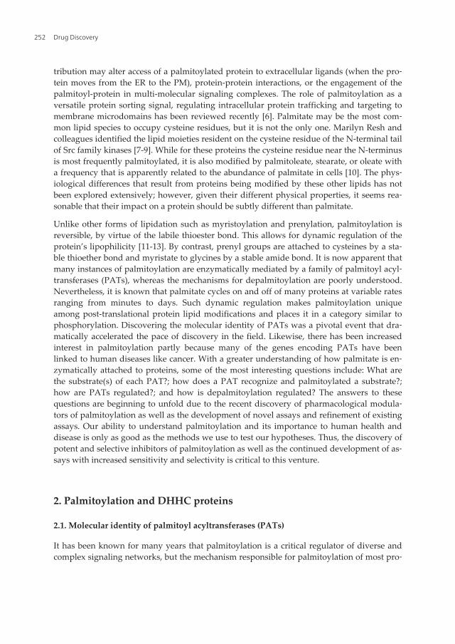
seems somewhat unlikely they would do so in this arrangement as it represents a state inwhich it would be difficult to perform these functions. The highest-scoring predictions ofthe membrane topology using TMpred show that the human protein sequence of DHHC11and -16 should contain four TM domains, DHHC13 eight TM domains, and DHHC22 eitherfour or five TM domains, with each model placing the DHHC-CRD motif inside the cells.There is clear disparity among the predictions generated by the algorithms available and ul‐timately, any of these predictions of topology must be confirmed or disproved by experi‐mental data. In any case, for a member of the PAT family to function as a PAT, the DHHC-CRD motif should probably reside in the cytoplasm (Figure 1A). The regions of the PATproteins that contain the greatest diversity at the amino-acid level are the N- and C-terminalcytoplasmic tails (Figure 1B).
B.
A.
N C
DHHC-CRD
TM1 TM2 TM3 TM4
N-Variable C-Variable
DPG TTxE
DHHC-CRD
lumen
cytoplasm
Figure 1. Predicted general structure of PATs. A) The predicted topology of PATs places the DHHC-CRD domain in thecytoplasm but such predictions must be confirmed experimentally. B) Each PAT is predicted to span the membranefour to six times; each is ~40 kDa with the greatest degree of sequence variability residing in the cytoplasmic N- and C-termini. The DHHC-CRD motif defines PATs. Palmitoylation of the cysteine in the DHHC portion is required for transferof palmitate to a substrate. Most PATs also have a conserved DPG (aspartate-proline-glycine) motif and TTxE (threo‐nine-threonine-asparagine-glutamate) motif, but their role in the function of PATs is not yet known.
In addition to the importance of PAT membrane topology, their membrane system of resi‐dence is likely to be an important aspect of their function. PATs have been localized to ER, Gol‐gi, plasma membrane, endosomes, and the yeast vacuole [30, 43-48]; yet, little is known abouthow these proteins achieve their respective localizations. Immunolocalization of epitope-tag‐ged DHHC proteins has been somewhat inconsistent among various cell types, between labo‐ratories (e.g. DHHC2 [30, 37], and even in our own laboratory (unpublished observations SLP)in terms of within which membrane system a protein resides. Such inconsistencies suggest thatthe cell type, cell cycle, health of the cells, or even the location of the epitope tag may affect sub‐cellular distribution. An interesting exception is DHHC2. DHHC2 has recently been shown totraffic between the PM and intracellular membranes via recycling endosomes [47]. Important‐ly, the C-terminal 68 amino acids of DHHC2 was shown to play an important role in defining
Discovery of Selective and Potent Inhibitors of Palmitoylationhttp://dx.doi.org/10.5772/52503
255

its intracellular localization; however, a defined targeting signal present within this region ofDHHC2 and in other DHHC proteins has yet to be defined.
2.3. PAT genes, palmitoylation and human disease
PATs have already been linked, in varying degrees, to human disease despite their recentdiscovery. At least 7 genes encoding PATs have been implicated in human disorders, in‐cluding ZDHHC8 with schizophrenia [33], ZDHHC17 with Huntington’s disease [49],ZDHHC15 and ZDHHC9 with X-linked mental retardation [32, 50], and ZDHHC2, ZDHHC9,ZDHHC17, and ZDHHC11 with cancer [29, 51-53]; most of the demonstrated and putativeconnections are with cancer.
Overexpression of some PATs has also been shown to alter cancer-related signaling.DHHC17 (HIP14) is oncogenic. DHHC9 and DHHC11 display characteristics that stronglysuggest they also are oncogenic. Overexpression of DHHC17 has the ability to induce colo‐ny formation and anchorage-independent growth in cell culture and tumors in mice [53]. Ithas been shown that these effects occur, at least in part, by DHHC17-mediated palmitoyla‐tion of H-, N-, and K2A- RAS proteins [53]. DHHC9 is strongly upregulated in some adeno‐carcinomas of the gastrointestinal tract at the transcript and protein levels [52] and has alsobeen shown to palmitoylate H- and N- RAS proteins in vitro [31]. ZDHHC11 has a high inci‐dence of additional genomic copies in cases of non-small cell lung cancer and bladder cancerin which it is strongly linked to high-grade, advanced stage and disease progression [51].
Conversely to the behavior of the oncogenic PATs, a failure to express ZDHHC2 results inan increase in metastasis in an in vivo model leading to the suggestion that ZDHHC2 is a tu‐mor/metastasis suppressor [29]. This absence of expression suggests that substrates ofDHHC2 are no longer palmitoylated, and that whatever role palmitoylation had in signalingdownstream from that event has been disrupted. Such is the case of DHHC2, where due to alack of palmitoylation, one of its substrates, CKAP4, is no longer normally palmitoylated.One consequence of this is that CKAP4 no longer traffics efficiently (or at all) to the cell sur‐face where it acts as a receptor for antiproliferative factor (APF) [37] [or presumably its othertwo known ligands, tissue plasminogen activator [54] and surfactant protein A [55]]. With‐out surface expression of CKAP4, APF is unable to initiate a wide range of downstream ef‐fects, including halting cellular proliferation and altering the expression of genes related tothe progression of cancer [44].
CD9 and CD151, both tetraspanin proteins, have also been identified as substrates ofDHHC2 [56]. CD9, which has been suggested to be a tumor suppressor [57, 58], is palmitoy‐lated on multiple cysteines, but which of these are palmitoylated by DHHC2 is not known.Nonetheless, it is clear that suppression of DHHC2-mediated palmitoylation of CD9 in A431cells affects cell behaviors that are consistent with it playing a role in tumor suppression. Inparticular, the cells undergo what appears to be epithelial-mesenchymal transition (EMT)—a process in which epithelial cells lose epithelial morphology and markers and gain a fibro‐blastic morphology during tumor progression [59-61]. It is not yet clear whether this changein cellular behavior was mediated solely by the reduction in CD9 palmitoylation or throughreduced palmitoylation of this and other DHHC2 substrates such as CKAP4. It will be inter‐
Drug Discovery256
esting to learn if a select subset of cysteines of CD9 is palmitoylated by DHHC2 and alsohow decreasing palmitoylation of specific cysteines results in the observed cellular behavior.Several other substrates of DHHC2 have been identified ranging from the neuronal adaptor/scaffold protein PSD95 [62], the SNARE proteins SNAP-23/25 [63], the non-receptor tyrosinekinase Lck [64], and the intracellular signaling proteins Gαi2 [65], GAP43 [62], R7BP [66],and eNOS [48]. Notably, there is no apparent structural similarity between the reported sub‐strates of DHHC2, or even any sequence similarities surrounding the palmitoylated cysteineresidues. Thus, DHHC2 can apparently palmitoylate cysteines located in the N-terminal re‐gions (PSD-95, GAP-43, and Gα), internally in the protein sequence (SNAP-23/25), in the jux‐tamembrane region of transmembrane proteins (CD9, CD151, and CKAP4) and close to anN-terminal myristoylated glycine (Lck and eNOS).
From these examples, it is clear that upsetting the homeostatic balance of protein palmitoy‐lation, in either direction, can have significant and deleterious effects on signaling networks.It is also clear that identification of PAT cognate substrates will provide important informa‐tion concerning the molecular mechanisms underlying the oncogenic nature of the affiliatedsignaling systems as well as reveal important, novel targets for pharmacologic intervention.The development of specific DHHC protein inhibitors would provide vital reagents withwhich to study the physiological and pathophysiological importance of many palmitoylatedproteins and may offer potential for therapeutic development.
2.4. PAT functions in addition to palmitoylation
It is not surprising that a disruption in the homeostatic balance of protein palmitoylation, ineither direction, can have pathophysiological consequences. However, one must remainmindful that palmitoylation may not be the sole function of these proteins. Recently, twoPATs—HIP14 (DHHC17) and HIP14L (DHHC13)—have been shown to mediate the trans‐port of Mg2+ [67]. The first indication that these PATs were involved in Mg2+ regulation wasthat the abundance of their corresponding mRNAs was increased in cells grown in mediumwith reduced Mg2+ concentration. The authors then showed that Mg2+ (but not Ca2+) trans‐port was both electrogenic and voltage dependent, and that the transport required palmitoy‐lation of the PAT. The authors concluded that these two PATs fall into a category ofenzymes called “chanzymes” or ion channels that also have enzymatic activity; a type ofprotein previously represented only by the transient receptor potential melastatin (TRPM)family of transporters [68, 69]. The fact that GODZ (DHHC3) does not appear to mediateMg2+ transport [70] but can mediate the transport of Ca2+ [71] suggests that this is not a gen‐eral property of all PATs. The discovery that these PATs transport Mg2+ was astonishing es‐pecially in light of the fact that the DHHC-CRD motif appears, by sequence and predictedstructure, to be a Zn2+-binding protein; (a divalent cation with an atomic radius similar toMg2+)—not Mg2+. However, Goytain and colleagues also found that HIP14 and HIP14Ltransported Zn2+ with approximately half the efficiency as Mg2+. The role of these and otherPATs in binding to and/or transporting Zn2+ remains to be elucidated, but demonstrates theimportance of not limiting ones view of PAT function (or many other proteins for that mat‐ter) only to palmitoylation.
Discovery of Selective and Potent Inhibitors of Palmitoylationhttp://dx.doi.org/10.5772/52503
257

2.5. Enzymatic mechanisms of palmitoylation
The physical and chemical mechanisms that result in enzymatic palmitoylation have yet tobe defined clearly, but some progress has been made using purified proteins. It has been es‐tablished that mutation of the cysteine in the DHHC motif of all PATs studied to date abol‐ishes autoacylation of the PAT and palmitoylation of the substrate [23, 56, 62, 72]. Thisliterature as well as discussion of potential physical mechanisms for the reaction have beenreviewed recently [3, 73].
3. Palmitoyl-Cysteine prediction
Prior to the discovery of PATs, attempts were made to define stretches of amino acids thatwere preferred for palmitoylation. Palmitoylation near the N-terminus, following myristoy‐lation, is among the predictable places for palmitoylation to occur provided there is one ormore nearby cysteines. Navarro-Lérida et al (2002) fused a myristoylation motif (MGCTLS)to GFP with a short intervening sequence containing cysteines at various locations. Theseauthors found a preference for cysteine palmitoylation at positions 3, 9, 15 and (to a muchlesser degree) 21 residues away from the N-terminal methionine, but intervening residueswere not evaluated. Commonalities in the composition of amino acid residues surroundingpalmitoylated cysteines have been noted among members of the family of yeast amino acidpermeases [74].
As more palmitoylated proteins and specific palmitoyl-cysteines are discovered, the task ofpredicting which adjacent amino acids provide a favorable environment for palmitoylationbecomes easier. Algorithms trained with data from identified palmitoyl cysteines and adja‐cent amino acid residues are now able to provide predictions of the statistical likelihood thata cysteine of interest may be palmitoylated [75-78]. CSS-Palm 2.0, which was designed topredict potential palmitoylation sites, has been published [75]. The algorithm was trained torecognize potential palmitoyl-cysteines using a dataset of 263 experimentally determinedpalmitoylation sites from 109 distinct proteins. Interestingly, CSS-Palm 2.0 also successfullypredicted most (~75%) of the same novel palmitoyl-cysteines in yeast proteins previouslyidentified by Roth. et al [74] as well as palmitoyl-cysteines predicted by Roth et al., to be pal‐mitoylated but not experimentally determined. This rate of success in both cases suggeststhat CSS-PALM 2.0 is more conservative at calling a site, potentially resulting in a greaterrate of false negative results but is reasonably accurate nonetheless. This algorithm shouldprove useful when prioritizing which cysteine(s), often among multiple potential cysteinesof a candidate palmitoyl protein, to analyze experimentally.
Patterns of amino acid residues surrounding palmitoyl-cysteines have emerged from theseanalyses. A diagram of favored residues generated by an early version of CSS-Palm 2.0(NBA-Palm) [76] shows that leucines and additional cysteines are more commonly observedaround palmitoyl-cysteines. The subsequent versions of NBA-palm used significantly im‐proved predictive tests, but the rough sequence of preferred residues remains. An importantaspect that cannot yet be considered when attempting to predict cysteine palmitoylation
Drug Discovery258

with these algorithms is the complexity of the PAT-substrate recognition that is encoded byresidues outside of those that immediately surround the palmitoyl-cysteine; the higher or‐der components of the recognition sites.
3.1. The physical properties of cysteines and thioester bonds
The unique physical and biochemical nature of the thioester bond that links palmitate tocysteine residues is the basis for the design of many recent assays for palmitoylation. Thecysteine residue is among the most nucleophilic entities in a cell [79] and is the most com‐mon site of palmitoylation. Other residues can be modified by palmitate, but their occur‐rence is relatively rare and the bond chemistries are different [2, 80-83]. Palmitoylation canalso occur in other ways, for example, on an amine of an N-terminal cysteine as is the casewith Hedgehog [2, 83, 84], a secreted signaling protein. An example of palmitate modifyingthe weaker –OH nucleophile of threonine occurs on the carboxyl terminus of a spider toxin[81]. The ε-amino group of lysine can also be modified by palmitate linked by an amidebond. This occurs in several secreted proteins including a bacterial toxin [80].
The reactivity of the thiolate anion of cysteine residues makes it a key component in thestructure and function of many proteins by stabilizing higher order structures via disulfidebridges and post-translational modifications like nitrosylation, prenylation, and acylation[85-87]. The high degree of reactivity has also provided a well-characterized, indispensabletarget for modification by synthetic, thiol-reactive ligands, allowing capture and characteri‐zation of proteins [88]. An exceptionally useful application of such thiol-specific chemistry isisotope-coded affinity tags (ICAT) for mass spectrometric determination of relative proteinor peptide abundance among two or more samples [89-91]. With these probes, changes inabundance of identified proteins or peptides are determined by changes in the ratio ofheavy to light-isotope-modified peptides from mixed samples. Combining ICAT technologywith functional genomics methods like siRNA-mediated PAT-gene knockdown is one ofseveral mechanisms that will allow us to identify substrates of PATs [37].
In healthy cells the cytoplasm is generally a reducing environment, meaning that solvent-exposed cysteine side chains are not typically disulfides and thus available to engage in re‐actions with other molecules [92]. The reactivity of a free cysteine depends on the pKa of thecysteine which is a function of the local environment surrounding the residue within thecontext of the whole protein. Unlike other residues with nucleophilic side chains (-OH or –NH2), thiol side chains undergo conjugations, redox, and exchange reactions [85]. Conjuga‐tion reactions (in addition to fatty acylation) include nitric oxide (NO) or S-nitrosylation, re‐active oxygen species (ROS), and reactive nitrogen species (RNS) forming bonds that are notsusceptible to cleavage by hydroxylamine at neutral pH. Hydroxylamine is a reagent usedto selectively remove thioester-linked palmitate [93]. Importantly, we know that hydroxyla‐mine does not perturb disulfides [94], and that it efficiently cleaves thioesters in a quantita‐tive manner [95].
In addition to the linkage of palmitate to cysteines, another thioester bond that occurs incells is the transient association between ubiquitin and the E1, E2, and certain E3 ubiquitina‐tion enzymes [87, 96]. However, these thioester bonds are easily distinguished from the thio‐
Discovery of Selective and Potent Inhibitors of Palmitoylationhttp://dx.doi.org/10.5772/52503
259
ester bond that links palmitate to cysteines by their pKa; the pKa in the case ofpalmitoylation is near neutral pH (~7.4) whereas, for the thioester in the ubiquitin system itis pH 10.5 or greater. This wide differential allows for a high degree of selectivity when us‐ing hydroxylamine to cleave palmitate from proteins on the physical characteristics of ubiq‐uitin-related cysteines. It is highly unlikely that they are ever in a position to bepalmitoylated [97, 98].
Retinoic acid (RA) and RA-CoA have also been shown to be enzymatically attached to cys‐teines via a thioester bond that can be cleaved by hydroxylamine and reducing reagentssuch as βME at neutral pH. The reaction can be inhibited, but not fully, by myristate andpalmitate suggesting that RA competes for the same cysteines as palmitate [99-107]. There issome debate in the RA field about how it binds to proteins, particularly the nuclear RA re‐ceptors, to carry out its signaling functions. RA binding to a hydrophobic cleft is the favoredmechanism; however, there are many effects of RA (e.g. [108, 109] ) that are independent ofRA-receptor binding suggesting that cysteine modification may also have a place in the mo‐lecular mechanism of RA action.
3.2. Mass spectrometric identification of acyl groups that modify cysteines via a thioesterbond
Lipid-modified thiols have been successfully identified using MALDI-TOF (matrix-assistedlaser desorption ionization time-of-flight) mass spectrometry [110]. Using this method, directinformation on the nature of the endogenous lipids on proteins or peptides (revealing interest‐ing variability) can be obtained, whereas most other methods rely on surrogate markers forpalmitate including thiol-reactive probes or radiolabeled palmitate. Using MALDI-TOF massspectrometry, Marilyn Resh and colleagues found that the cysteine in the N-terminal Met-Gly-Cys of Src family kinases and two cysteines near the N-terminus of GAP43 are modified notonly by palmitate but also (and to a lesser degree) by palmitoleate, stearate, or oleate [7, 8].While palmitate appears to be the most common acyl group that forms a thioester bond tomodify internal, cytoplasmic cysteines, it is clearly not the only one. The 16-carbon palmitateacyl group represents the longest chain synthesized by mammalian fatty acid synthase and isapparently the most abundant chain length present in some tissue types [111]. This relativelygreater abundance may underlie the dominance of palmitate as the main acyl group to modi‐fy free thiols by S-acylation. The functional implications of incorporating lipids with shorter orlonger acyl chains and especially those with different degrees of saturation may be that theproteins have different affinities for various lipid microdomains present in membranes. Thespecificity of PATs for chain-lengths different than 16 carbons has not been rigorously de‐fined. However, it is known that acyl groups with differing carbon chain lengths and degreesof saturation can also be incorporated [7, 112].
3.3. PAT/Substrate recognition
Determining the nature of PAT/substrate recognition remains one of the more importanttasks to be undertaken. This is especially true for PATs encoded by genes that have beenlinked to disease. There are two general approaches to defining PAT/substrate relationships:
Drug Discovery260

1. identification of the PAT with specificity for a known palmitoyl protein and
2. identification of an unknown substrate of an individual PAT.
The first of these has been the most common. With this forward approach, each one of the 23PATs is independently co-overexpressed with a known palmitoyl protein; the cells express‐ing the pair are metabolically labeled with 3H-palmitate and the proteins analyzed by SDS-PAGE and fluorography. The incorporation of 3H-palmitate onto the substrate protein inone or more of the co-overexpressions at a level significantly above background suggeststhat a particular PAT is responsible for palmitoylating that known substrate. Similarly, thepresumptive PAT and substrate proteins can be purified and combined with 3H-palmitoyl-CoA in a tube, allowed to react, and the incorporation of 3H-palmitate measured as above.The current level of understanding of PAT/substrate recognition makes it unreasonable toassume that the more closely two PATs are related by sequence homology, the more likelythey should palmitoylate a particular substrate. For this reason, assigning substrate status ofa protein to a single PAT among a select group of tested, more closely-homologous PATs, tothe exclusion of others because they are less homologous, may lead to erroneous exclusions.Similarly, we cannot yet assume that homology among residues surrounding palmitoyl cys‐teines of different proteins is an indication that they are palmitoylated by a particular PAT.The mechanism for molecular recognition is likely to be defined in part by the higher orderstructure (even quaternary as is the case with ERF2p and AKR1p) of the PATs and sub‐strates. The reverse approach, defining unknown substrates of a single PAT can occur with‐out these same biases as has been demonstrated in yeast and in human cells [37, 74].
4. Novel methods to discover and identify PAT/substrate specificity
The chemistry supporting novel assays to study palmitoylation and the reagents that are be‐ing incorporated into them have, for the most part, been known and available for years [88] .Most of the methods that are now being developed to study palmitoylation capitalize onmany years of knowledge and development of cysteine-specific chemistries, developedmainly as methods to purify and/or specifically target proteins and peptides with variousreagents. Many of the reagents that specifically label cysteines have been created as both af‐finity and fluorescent tags, the former for purification and structure determinations [88]andthe latter as cellular reporters of protein abundance, subcellular distribution, protein confor‐mation changes, the formation of the Golgi, and even the concentration of cellular analytesin specific subcellular domains. The following references provide a short list of some of themost clever uses of thiol chemistry [113-119]. Given the wealth of information on the uniquechemistry of the palmitoyl thioester bond and the tools for capturing and characterizing cys‐teines in proteins, it is somewhat surprising that we are only now developing innovative as‐says to increase our understanding of palmitoylation. This recent increase is most likely tiedto the dramatic increase in the utility of mass spectrometry as a proteomic tool. To provide ageneral frame of reference for the recent shift in the types of assays that are being devel‐oped, we will briefly discuss other assays that have been used successfully for a longer peri‐
Discovery of Selective and Potent Inhibitors of Palmitoylationhttp://dx.doi.org/10.5772/52503
261
od of time. These assays are by no means outdated and some continue to be the mostappropriate way to answer specific questions.
4.1. Chemistry and physical properties of palmitoyl cysteines: Reactions and probes
Working with palmitoylated proteins is inherently difficult due to the labile nature of thethioester bond and the increased hydrophobicity of the protein or peptide due to palmitate.On the other hand, the unique physical and chemical properties of thiols, palmitoylated thi‐ols, and the thioester bond make them particularly amenable to modification by highly spe‐cific chemistry and a wide variety of thiol reactive probes.
Reactions of free thiols in the cytoplasm
Thiol modification occurs most commonly in cells by one of two routes: disulfide exchangeor alkylation. Many of the reactive groups that undergo these two reactions are relativelystable in aqueous environments; the reactions are rapid and provide high yields of thioetherand disulfide bonds [88]. Thiols will also react with many amine reactive reagents includingisothiocyanates and succinimidyl esters but lack a high degree of specificity, resulting in un‐stable bonds that are much less useful for routine modification of thiols in proteins. Thiol-specific reagents and chemistry figure strongly into the design and development of novelassays for palmitoylation. Most investigators are limited somewhat to reagents that areavailable from a catalog but, fortunately, there are already many useful reagents available.Among the most useful are thio-reactive chemicals that are linked to another moiety (reac‐tive or reporter) by a spacer arm of variable length and physical characteristics. Such hetero-and homo-bifunctional crosslinking reagents have provided much of the foundation forrecent developments in palmitoylation assays and provide a fairly rich toolbox for future as‐say development.
Chemical moieties that react with palmitoyl-cysteines
Iodoacetamide conjugates are among the most commonly used tools for modifying cysteinethiols. These undergo nucleophilic substitution to form stable thioether bonds at physiologi‐cal pH in aqueous environments. When using iodoacetamide and its conjugates, one shouldremember that depending on the pH of the solution, they can also react with histidine, ly‐sine, and methionine (at pH >1.7) residues and N-terminal amines. However, when used atslightly alkaline pH in the dark and in the absence of reducing reagents, cysteine modifica‐tion will be the exclusive reaction [88]. A good example of iodoacetamide-based probes arethe isotope-coded affinity tags or ICAT [120]. These have proved particularly useful in de‐termining the substrates of DHHC2 [37].
Maleimides are also common constituents of heterobifunctional crosslinking reagents andblocking reagents that target cysteines. They are ~1000 times more specific for cysteine sulf‐hydryls at pH 6.5-7.5, but at higher pH some cross reactivity can occur with amines. Mal‐eimides form stable thioether bonds by adding the sulfhydryl across the double bond ofthe maleimide.
Phenylmercury derivatives react with thiols, including nitrosothiols, under conditions simi‐lar to iodoacetamides and maleimides to form stable mercury-thiol bonds that can be re‐
Drug Discovery262

versed in 0.1N HCl and reducing reagents like dithiothreitol (DTT) but apparently not byTCEP. Phenylmercury derivatives also react faster with thiols than do the commonly usedthiol-reactive N-ethylmaleimide (NEM).
Compounds containing disulfide bonds are able to undergo disulfide exchange reactionswith another thiol by the free thiol attacking the disulfide bond and subsequent formation ofa new mixed thiol. Two examples of useful compounds in this category are Methylmethane‐thiosulfonate (MMTS) and pyridyl disulfide derivitives like biotin HPDP ((N-(6-(Biotinami‐do)hexyl)-3'-(2'-pyridyldithio)-propionamide). MMTS can be used in some cases to blockfree thiols more effectively than NEM, as it is uncharged and thus more likely to modify allfree reactive cysteines. MMTS has been shown not to react with nitrosothiols or existing di‐sulfides [121].
4.2. Metabolic labeling with radiolabeled palmitate
The most common method to identify palmitoyl proteins and to determine the residencehalf-life of palmitate on a specific protein or palmitate turnover (e.g. [122] for a particularlyinteresting example) is to metabolically label cells with radiolabled palmitate. 14C-, 3H- and125I-labeled palmitate have all been used, but 3H-palmitate is most common because it is rela‐tively inexpensive and widely available. However, using 125I -labeled palmitate providessome advantages. In practical terms, the time required for detection is considerably shorter -hours instead of (often) weeks with 3H-palmitate. The γ-irradiation from the 125I is also com‐patible with phosphorimaging technology which is much more rapid and quantitative thandensitometric measurements from films generated by autofluorography (as is used with tri‐tium). The principle downside of using 125I-labeled palmitate is that it is not commerciallyavailable, and the labeling must be done by the investigator. Reviews of the methods usingradiolabeled palmitate and including technical details have been published recently[123-125].
4.3. Fluorescently-labeled peptide substrates for palmitoylation
Fluorescently-labeled peptides that mimic PAT substrates have been used to characterizePAT activity and for the discovery of inhibitors of palmitoylation [123, 126, 127]. The use ofthese peptides over the last several years was reviewed recently [123]. Peptide substrates forpalmitoylation have also been genetically fused to fluorescent proteins and expressed incells. This strategy has been used to determine how palmitoylation affects subcellular traf‐ficking both between and within membranes [124]. Monomeric GFP-based reporters andfluorescence resonance energy transfer proved to be helpful in the identification of lipidrafts with an affinity for palmitate on the inner leaflet of the plasma membrane [5].
4.4. Acyl-biotin exchange: ABE
Most of the novel assays for palmitoylation utilize the same basic foundation first describedfor a palmitoyl protein by Schmidt and colleagues [128] and now most commonly known asacyl-exchange. First, free cysteines are blocked on proteins that have been extracted from
Discovery of Selective and Potent Inhibitors of Palmitoylationhttp://dx.doi.org/10.5772/52503
263
live cells or tissue. Next, palmitates are removed from cysteines by cleavage of the thioesterbond with hydroxylamine (typically 1.0M) at neutral pH. This creates a new set of free thiolsunique in that they were all formerly palmitoylated; ideally, no others should exist. Finally,this new set of formerly-palmitoylated cysteines is modified by any one of the many thiol-specific reagents. The uniqueness of the individual assays that incorporate these steps liesprimarily in the choice of thiol-specific reagents, and this choice depends on what questionsthe investigator wants to answer. There are also variations in the reagents used to block freecysteines in the first step. Both NEM and MMTS have been used in the assays described be‐low but NEM is used most commonly.
Cysteines that are palmitoylated can also be modified by fatty acids other that palmitate [7]including stearate and oleate. The acyl-exhange method cannot yet distinguish between pal‐mitate and the other fatty acids modifying cysteines by a thioester bond. Two additionalpoints that relate to the specificity of this method for palmitoylation are: 1) that it will notreport modification of cysteines by prenyl groups (geranylgeranyl or farnesyl) because theyare attached by a thioether bond that is not susceptible to cleavage by hydroxylamine and 2)it will not report myristoylated proteins because this 14-carbon acyl group is linked to an N-terminal glutamate by an amide bond which is also insensitive to cleavage by hydroxyla‐mine.
The recent development of novel assays using the three-step acyl exchange method to studypalmitoylation in a broader sense was invigorated by two publications describing a newtwist on the method that incorporated the use of radiolabeled NEM assay [129, 130]. Workdescribed in these papers showed that labeling palmitoyl cysteines with radiolabeled NEMresulted in a remarkable 5- to 12-fold increase in sensitivity to detect several known palmito‐yl proteins, including PSD-95 and SNAP-25, when compared to labeling with 3H-palmitate.In addition, the authors demonstrated the utility of the biotinylated, heterobifunctionalcrosslinker, 4-[4′-(maleimidomethyl)cyclohexanecarboxamido] butane (Btn-BMCC), as an ef‐fective tool to capture and purify (using streptavidin-agarose) palmitoylated proteins. In do‐ing so, they also demonstrated the general potential of using the wide variety of existingthiol-specific probes for the development of additional assays for palmitoylation that are be‐ginning to materialize.
4.5. The palmitoyl proteome
The demonstration that one can effectively replace palmitate with a biotin group led to de‐velopment of the first, large-scale, proteomic analysis of palmitoylation [74] in yeast, themodel system in which the molecular identify of PATs was first determined [23, 26]. Thismethod was dubbed “acyl biotin exchange” or ABE and used the same basic three-steps asdescribed above. As the name implies, the proteins were labeled with a thiol-reactive, bioti‐nylated heterobifunctional probe, [6-(Biotinamido)hexyl]-3'-(2'-pyridyldithio)propionamide(HPDP-biotin), with subsequent capture on streptavidin affinity matrix (for a detailed proto‐col see Wan et al. 2007 [131]). It is interesting to note the number of proteins that Roth andcolleagues captured in the negative control samples (Figure 1a;[74]). The degree of overlapamong proteins captured in the experimental and control samples suggests that the step in
Drug Discovery264
which free thiols were blocked with NEM was not quantitative and/or that the wash stepsfollowing binding of biotinylated proteins to the streptavidin matrix were not sufficientlystringent (steps 7 and 16 respectively from Wan et al, 2007 [131]) thereby resulting in the po‐tential for a higher number of false-positive hits. However, issues of signal to noise and lim‐its of sensitivity are by no means unique to this work (avidin-biotin affinity purification isnotoriously difficult); rather they are unavoidable issues faced by all developers of novelstrategies and users of nascent technologies. Incremental improvements in important assayslike this always follow.
One of the key features of all proteomic methods is the system used for detection of specifi‐cally-isolated proteins or peptides. Work by Roth et al. [74] identified proteins by multi-di‐mensional protein identification technology (MudPIT), a high-throughput, tandem massspectrometry (MS/MS)-based proteomic technology [132] [see also [131, 133]]. Compared toother mass spectrometric methods, MudPIT has the potential to identify less abundant pro‐teins with a higher degree of confidence, because multiple peptides of a single protein canbe used to identify a protein of interest. One downside with MudPIT in this case is that thepalmitoyl cysteine(s) cannot be pinpointed, as there may be many candidates among the in‐dividual peptides of a whole protein suspected as being a palmitoyl protein. After demon‐strating the usefulness of this large-scale method for purification and identification ofpalmitoylated proteins, the authors used mutant strains of yeast lacking one or more of theseven yeast PAT proteins to identify substrates of individual PATs. Comparison of the de‐gree of palmitoylation of individual proteins between wild type yeast (a full set of normallypalmitoylated proteins) and those not expressing one or more of the yeast PATs (each with aspecific set of hypo/depalmitoylated proteins) provided the identity of the substrates of indi‐vidual PATs. Together, this work represents a very significant contribution to the identifica‐tion and understanding of the yeast palmitoyl proteome and provided many importantclues about potential homologous PAT-substrate pairs in other systems.
The complexity of palmitoylation is greater in a vertebrate system. With at least 23 genes en‐coding PATs identified in humans, the diversity at the most basic level is at least three-foldgreater than in yeast. When one considers the additional variants encoded by alternativesplicing of PAT mRNAs, the potential diversity increases even more. The greater number ofPATs suggests (but does not prove) that there are also more palmitoylated proteins in mam‐mals. The ability to genetically manipulate mammalian cells is improving but lags behindyeast. Nevertheless, defining the palmitoyl proteome or palmitoylosome and how it is regu‐lated in mammals (humans in particular) is a task of significant importance and interest.Now that the enzymes capable of mediating palmitoylation have been identified, one of themost important questions that we face is which substrates are palmitoylated by each PAT—a question brought sharply into focus when one considers the known connections betweenmutations or deletions in PAT genes and human disease, in particular cancer. DHHC2 is de‐leted in many types of cancer (see above). Its absence is strongly correlated with an increasein the metastatic potential of cancer cells. The simplest inverse corollary in this case is thatpalmitoylated substrates of DHHC2 are responsible for keeping cells from metastasizing.Identification of these substrates and their associated signaling networks using novel assays
Discovery of Selective and Potent Inhibitors of Palmitoylationhttp://dx.doi.org/10.5772/52503
265
for palmitoylation has begun to provide supporting evidence for known mechanisms of can‐cer progression [56] as well as a novel signaling pathway for the regulation of cellular prolif‐eration and metastasis [37].
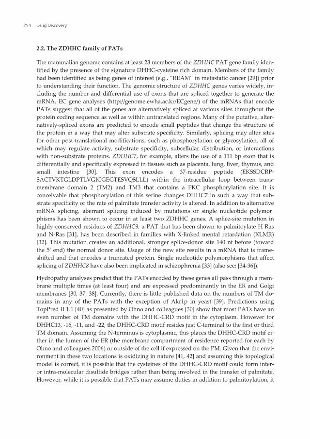
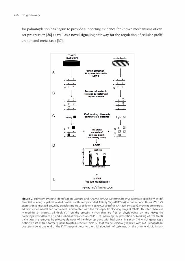
Figure 2. Palmitoyl-cysteine Identification Capture and Analysis (PICA): Determining PAT-substrate specificity by dif‐ferential labeling of palmitoylated proteins with Isotope-coded Affinity Tags (ICAT) (A) In one set of cultures, ZDHHC2expression is knocked down by transfecting HeLa cells with ZDHHC2-specific siRNA (Dharmacon). Proteins are extract‐ed from experimental and control cells and treated with the thiol-specific blocking reagent MMTS. This step chemical‐ly modifies or protects all thiols (“X” on the proteins P1-P3) that are free at physiological pH and leaves thepalmitoylated cysteines (P) undisturbed as depicted on P1-P3. (B) Following the protection or blocking of free thiols,palmitates are removed by selective cleavage of the thioester bond with hydroxylamine at pH 7.4, which generates adistinctive set of free, formerly-palmitoylated, reactive thiols (C) that can be selectively labeled with ICAT reagents. Io‐doacetamide at one end of the ICAT reagent binds to the thiol sidechain of cysteines; on the other end, biotin pro‐
Drug Discovery266

vides a mechanism for affinity purification of thiol-captured peptides on an avidin column. (D) Proteins fromknockdown and control conditions are mixed in equal amounts and digested in-gel with trypsin. ICAT labeled pepti‐des are enriched by avidin affinity and analyzed by LC/MS. A pair of ICAT-labeled peptides is chemically identical andis easily visualized, because they essentially coelute and there is a 9 Da mass difference measured in a scanning massspectrometer. Even if equal amounts of a single protein exist in two different samples, the quantity of protein that iscaptured depends directly on its degree of palmitoylation; if all of a single protein is palmitoylated under one condi‐tion, then all of it will be captured; if only half of this protein is palmitoylated under another condition then the cap‐ture rate of that protein will be half as much, relative to control, making it appear half as abundant. Proteins for whichthere has been no change in palmitoylation (ie, equal capture rates) will yield a heavy:light (H/L) ratio of 1. The degreeto which palmitoylation is diminished will register as a decrease in the H/L ratio (ie, 50% reduction in palmitoylationwill correspond to a H/L ratio of 0.5). A change in the capture rate that results in a change in the post-purificationabundance is measured in the LC/MS phase. (E) Finally, the peptides are further fragmented into their constituentamino acids by MS/MS, enabling identification of the proteins corresponding to the captured peptides.
4.6. Palmitoyl-cysteine Identification Capture and Analysis (PICA): Identification of PATsubstrates and palmitoyl proteins in vertebrates
With the aim of defining PAT-substrate specificity in a living vertebrate system, we devel‐oped a method to identify substrates of specific PATs in mammalian cells and tissues calledPalmitoyl cysteine Isolation Capture and Analysis or PICA [37]. We used this method toidentify CKAP4/p63, a known palmitoyl protein [134] as one substrate of DHHC2 in HeLacells [37]. This method is similar to ABE as described by Roth et al (2006) but was inspired[135] in part by the work of Drisdel and Green (2004) and incorporated several novel fea‐tures that will be discussed below.
The ability of PICA to identify PAT substrates is based on the principle that it quantifies thedifferential frequency of palmitoylation of individual proteins or peptides in control condi‐tions versus conditions in which the function of a single PAT is reduced by siRNA-mediatedgene knockdown. The process to identify substrates of DHHC2 consisted of four basic stepsoutlined in Figure 2. In the first part we generated two distinct pools of palmitoylated pro‐teins, one from control HeLa cells and the other from HeLa cells in which the activity of onePAT (DHHC2) was reduced. These two distinct pools of palmitoylated proteins were thencaptured and compared directly to identify differences in the degree of palmitoylation of in‐dividual proteins between the two pools. To do this, we reduced the expression of ZDDHC2mRNA (and consequently the abundance of the encoded enzyme, DHHC2) in HeLa cells us‐ing siRNA-mediated gene knockdown which resulted in a reduced level of palmitoylationof DHHC2 substrates. Total protein from knockdown and control cells was prepared by firstblocking free thiols with MMTS in the presence of SDS. This was followed by selective expo‐sure of all palmitoyl cysteines by cleavage of the palmitoyl-cysteine thioester bond with1.0M hydroxylamine at neutral pH, thereby generating a unique population of formerly pal‐mitoylated cysteines. Second, we selectively and differentially labeled the exposed, former‐ly-palmitoylated cysteines from knockdown and control cells with biotinylated, thiol-reactive heavy (H) and light (L) ICAT reagents, respectively. Third, we combined equalquantities of the ICAT-labeled protein from ZDHHC2 knockdown and control cells and di‐gested the mixture with trypsin. The resulting H and L ICAT-labeled tryptic peptides werecaptured and purified on an avidin affinity column. Finally, ICAT-labeled, putative, former‐ly-palmitoylated peptides were analyzed by mass spectrometry. Peptides with a reduced
Discovery of Selective and Potent Inhibitors of Palmitoylationhttp://dx.doi.org/10.5772/52503
267
H/L ratio over four independent runs were analyzed further to confirm that the identifiedcysteine was indeed palmitoylated by DHHC2 under physiological conditions. Details of theprotocol and reagents used and outlined in Figure 2 can be found in [37].
There are several unique aspects in the PICA method. First, we used MMTS to block the freethiols in the first step. NEM is used most commonly at this step, but MMTS is more reactiveand smaller than NEM or iodoacetamide, enhancing its ability to modify all free reactivecysteines. Inefficient blocking of free thiols in the first step is one factor that could easilycontribute to false-positive capture of proteins in the purification step. Qualitative evalua‐tion (silver-stained SDS-PAGE) of protein capture in experimental and control (no-hydrox‐ylamine) conditions [Figure 2, [37]] suggests that it may be more efficient than NEM (forcomparison see Figure 1A [74]). However, it may also be that we captured very few proteinsunder control conditions because of a more stringent wash protocol than described by Rothet al [74]. The use of ICAT reagents in PICA allowed us to combine formerly-palmitoylatedpeptides purified from control and experimental cells in the same pool, and subsequently, adirect, simultaneous analysis of palmitoylation in the two pools in a single analytical sam‐ple. We defined a substrate of DHHC2 as one that had a consistently reduced H/L ratio overfour independent PICA runs. This approach provided us with many (the vast majority), con‐venient internal control peptides which are peptides that were not substrates of DHHC2that had unchanged H/L ratios. This approach significantly reduces the potential for identi‐fication of false-positive hits because, if a protein can be falsely labeled by an ICAT, itshould do so with equal efficiency in both the control and experimental cells yielding a pep‐tide with an H/L ratio of ~1. The greater risk with this approach is the failure to identify sub‐strates that exist in low abundance. Using tandem mass spectrometry, we analyzed a sampleof significantly reduced complexity including only ICAT-tagged peptides. As is inherent insuch analyses, the most abundant peptides dominate the report. However, one advantage ofthis approach is that when a peptide is identified, whether it is a substrate of a single PAT ornot, the palmitoyl cysteine(s) is also identified. In the case of CKAP4/p63 (and the majorityof other peptides) there was only a single cysteine, and it was already known to be a site forpalmitoylation [134]. Spectral counting has the potential to positively identify palmitoyl pro‐teins of lower abundance because more than a single peptide from any given protein is fac‐tored into to the identification. There is greater overall coverage (identified peptidefragments of a protein) using this method thereby increasing the confidence level of identifi‐cation. However, the disadvantage inherent in analyzing a complex mixture, including non-palmitoylated peptides by spectral counting, is that identification of the palmitoyl cysteine(in the cases where there are multiple candidate cysteines) must await subsequent and tedi‐ous analyses. The tradeoff between these two complementary approaches in mass spectro‐metric analysis is sensitivity versus specificity. Combining these analyses will provide amuch greater depth of coverage.
4.7. Forward and reverse approaches to assigning PAT-substrate pairs
The first reports that identified PAT-substrate pairings took the reverse approach: start witha known palmitoylated protein then use metabolic labeling with radiolabeled palmitate and
Drug Discovery268
co-overexpression of one PAT and the substrate (for a review see: [72]). Using this method,an increase in the incorporation of radiolabled palmitate on the overexpressed substrate inthe presence of an overexpressed PAT is used to claim specificity. This method is an impor‐tant tool for increasing our understanding of palmitoylation-related phenomena includingconfirmation of putative PAT-substrate pairs identified by other methods. Likewise, whenstarting with a known palmitoyl protein and the intention of identifying the PAT responsi‐ble for its palmitoylation, it remains a useful method. However, we should remember thatjust because overexpression of a PAT can increase the incorporation of palmitate onto a spe‐cific protein does not necessarily mean that it does so in a live cell. Again, problems like thisare not unique to this method and simply reflect our lack of knowledge about where andwhen PATs and their substrates are expressed, the degree of promiscuity among PATs and,how PAT function is regulated.
The potential for specific cysteines to be modified by both palmitate and RA via a thioesterbond is an issue that deserves attention from those of us interested primarily in palmitoyla‐tion for at least two reasons. One is the potential that an exchange between the two modifi‐cations is a physiologically relevant means of regulating signaling and second, thepossibility that proteins identified as being palmitoylated in assays utilizing some form ofABE chemistry are RA-modified instead.
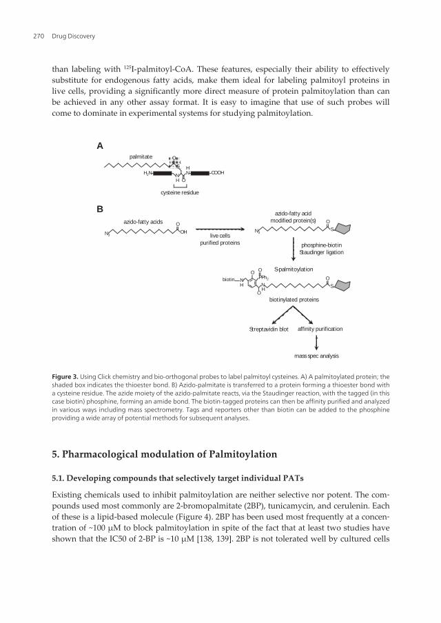
4.8. Labeling palmitoyl proteins with bioorthogonal probes
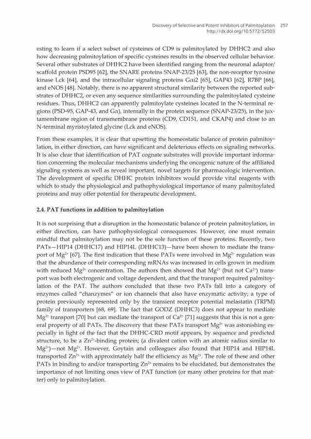
This particularly interesting approach labels cysteines with isosteric, azido-derivitives offatty acids that are able to substitute for fatty acids that occur naturally in cells (Figure3) [112, 136]. Once bound to cysteines, the azido group on the fatty acid is reacted, witha high degree of selectivity, via the Staudinger reaction [137] with (triaryl)phosphines thatare themselves derivatized with Myc, biotin, a fluorophore, or others. Using this meth‐od Hang and colleagues [112] found that ω-azido fatty acids with 12 and 15 carbons canbe efficiently metabolized by mammalian cells and accurately report myristoylation andthio-palmitoylation, respectively.
Work by Kostiuk and colleagues identified palmitoyl proteins from mitochondria us‐ing azido-palmitate [136]. To accomplish this they purified proteins from cellular mito‐chondrial fractions, first by differential centrifugation, then by further purification basedfirst on charge and subsequently by size using chromatographic separation. Labeling ofproteins in this study was outside of a living system presumably leaving only the pos‐sibility of autocatalytic/non-enzymatic palmitoylation. Mass spectrometric analysis of se‐lected bands identified 21 palmitoylated proteins, 19 of which were novel. The majorityof the proteins labeled were metabolic-type proteins unique to the mitochondrion. Thisraises the interesting possibility that the principle mechanism of palmitoylation in thisorganelle is autocatalytic rather than enzymatic, and that, as suggested by the au‐thors, the key role of palmitoylation in the mitochondrion is to inhibit enzymes by pal‐mitoylation of cysteines in the vicinity of the active site.
These so called bio-orthogonal probes have been reported to be nontoxic and very stable un‐der physiological conditions. Importantly, this two-step reaction is rapid and more sensitive
Discovery of Selective and Potent Inhibitors of Palmitoylationhttp://dx.doi.org/10.5772/52503
269
than labeling with 125I-palmitoyl-CoA. These features, especially their ability to effectivelysubstitute for endogenous fatty acids, make them ideal for labeling palmitoyl proteins inlive cells, providing a significantly more direct measure of protein palmitoylation than canbe achieved in any other assay format. It is easy to imagine that use of such probes willcome to dominate in experimental systems for studying palmitoylation.
O
S
ONH2N N
H
H
COOH
palmitate
cysteine residue
A
BO
OHN3
azido-fatty acids
live cellspurified proteins
O
SN3
azido-fatty acidmodified protein(s)
phosphine-biotinStaudinger ligation
S-palmitoylation
O
SNH
O
PPh2
OO
HNbiotin
biotinylated proteins
Streptavidin blot affinity purification
mass spec analysis
Figure 3. Using Click chemistry and bio-orthogonal probes to label palmitoyl cysteines. A) A palmitoylated protein; theshaded box indicates the thioester bond. B) Azido-palmitate is transferred to a protein forming a thioester bond witha cysteine residue. The azide moiety of the azido-palmitate reacts, via the Staudinger reaction, with the tagged (in thiscase biotin) phosphine, forming an amide bond. The biotin-tagged proteins can then be affinity purified and analyzedin various ways including mass spectrometry. Tags and reporters other than biotin can be added to the phosphineproviding a wide array of potential methods for subsequent analyses.
5. Pharmacological modulation of Palmitoylation
5.1. Developing compounds that selectively target individual PATs
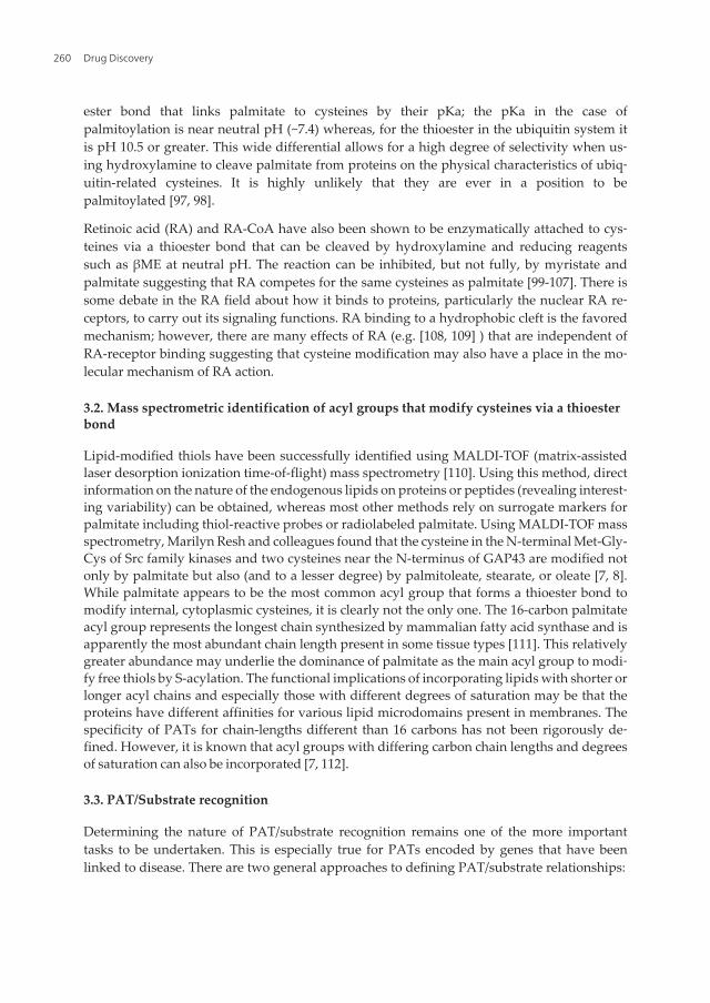
Existing chemicals used to inhibit palmitoylation are neither selective nor potent. The com‐pounds used most commonly are 2-bromopalmitate (2BP), tunicamycin, and cerulenin. Eachof these is a lipid-based molecule (Figure 4). 2BP has been used most frequently at a concen‐tration of ~100 µM to block palmitoylation in spite of the fact that at least two studies haveshown that the IC50 of 2-BP is ~10 µM [138, 139]. 2BP is not tolerated well by cultured cells
Drug Discovery270

and causes death even after a brief exposure to 100 µM. 2BP inhibits several enzymes in‐volved in lipid metabolism, including carnitine palmitoyltransferase 1, fatty acid CoA ligase,glycerol-3-phosphate acyltransferase, and enzymes in the synthesis of triacylglycerol bio‐synthesis [140, 141]. This high degree of promiscuity as well as the toxicity of 2BP renders itnearly useless as a tool to determine anything specific about palmitoylation related signal‐ing issues that equally plague cerulenin and tunicamycin. The uses and effects of these threeinhibitors was reviewed recently [142].
2-bromopalmitate
tunicamycin
cerulenin
Figure 4. Lipid-based inhibitors of palmitoylation.
Smith and colleagues [126] recently screened a compound library in an attempt to identifymore selective and potent inhibitors of palmitoylation, in particular inhibitors of PATs. Thisscreen identified single compounds from five chemical classes (Compounds I-V) that inhib‐ited cellular processes associated with palmitoylation. The assays used in the screens includ‐ed: measuring the in vivo and in vitro growth rate of an NIH/3T3 cell line that overexpressedDHHC17, displacement from the plasma membrane of myristoylated or farnesylated GFP,and in vitro palmitoylation of small, non-complex, myristoylated or farnesylated, synthetic,fluorescent peptides intended to mimic known palmitoylation substrates [123, 126, 127].These assays could not discriminate a direct effect of any compound on any PAT. Theycould only report the activity of compounds that acted at some point (not excluding directPAT inhibition) in any pathway that leads to or affects palmitoylation; compounds like 2BP,cerulenin, and tunicamycin. This assertion was borne out in follow-up studies on the samecompounds [138] (see below). Perhaps the most intriguing finding in this report was thatcompounds I-IV were able to suppress the oncogenic behavior of human cells that overex‐
Discovery of Selective and Potent Inhibitors of Palmitoylationhttp://dx.doi.org/10.5772/52503
271

pressed DHHC17. However, there is no direct evidence to indicate that compounds I-IV ex‐ert these actions via inhibition of DHHC17 or through the palmitoylation of Ras proteins byDHHC17 as was speculated [53, 126]. Nevertheless, since these compounds reduced the invivo growth of tumors from cells overexpressing DHHC17 [126], it would be worth deter‐mining their exact mechanism of action.
Subsequent studies by Linder, Deschenes and colleagues [138] tested four of the five com‐pounds identified by Smith and colleagues and found that they were not selective forDHHC proteins. This report also included a wealth of information defining the mechanismsby which 2BP inhibits palmitoylation. Briefly, only one of the four compounds re-tested,compound V and 2BP, inhibited the activity of any of the four DHHC proteins tested. Nei‐ther compound V nor 2BP was selective for any of the PATs tested, and 2BP was more po‐tent. Both compound V and 2BP blocked autoacylation of the PATs; compound V wasreversible, 2BP was not. Even though compound V was able to inhibit the activity of thefour PATs tested and in the same manner as 2BP, these experiments could not determinewhether compound V also blocks palmitoylation indiscriminately at steps prior to the actualpalmitoylation event, as is the case with 2BP, cerulenin, and tunicamycin.
There would be no compelling reason to begin a drug discovery program to identify inhibi‐tors of just any PAT. Rather one would choose to begin with a PAT that is linked to a dis‐ease state—a situation where misregulated expression or function of that PAT was clearlylinked to a pathological state. As discussed earlier, links between PAT expression (but notyet altered function) have been demonstrated for both neurological disorders and cancer;thus, candidate PATs that would be appropriate targets for drug development exist. Bothoverexpression and absence of PAT expression have been implicated in the development ofcancer. Dampening the activity of an existing PAT is a conceptually and mechanisticallysimpler task than accurately restoring the specific activity of a PAT that is not expressed orabsent. This review is concerned with PAT assays and PAT inhibitors, so we will addressthe case of PAT overexpression in ideal terms as well as the technical issues that surroundthe development and implementation of the assays designed to discover PAT inhibitors.
5.2. Considerations for development of high-throughput screens to discover PATinhibitors
The DHHC motif in PATs defines the active site and is highly conserved in all mammalianPATs [3]. The regions of highest diversity are primarily in the N- and C-termini of the PAT.Mutation of the cysteine in the DHHC motif abolishes PAT autoacylation and palmitoyla‐tion of the substrate, a property of all DHHC proteins studied so far. This high degree ofhomology in the active site sequence among PATs could give the impression that develop‐ing highly specific, active-site inhibitors for palmitoylation will be impossible. However, thissame issue exists with kinases [143, 144], and yet the development of selective and potentactive-site, ATP-competitive inhibitors has been successful (eg, [145]).
The specificity of palmitoylation must be derived in part from the unique physical interac‐tions of individual PATs with their substrates. The sequence of amino acids surrounding asubstrate cysteine partially defines the potential for that cysteine to be palmitoylated. How‐
Drug Discovery272

ever, the physical determinants for substrate recognition will likely extend throughout theaccessible portions of the PAT and substrate as was elegantly demonstrated for DHHC17[146]. Other factors that are likely to regulate palmitoylation are the temporal and spatial as‐pects of PAT and substrate expression.
There are many more palmitoylated proteins than there are PATs; therefore, modulating theactivity of a single PAT, even with complete compound selectivity, will likely yield a changein the palmitoylation of multiple substrates. This conundrum is common to the develop‐ment of highly selective and potent pharmacological modulators of all enzymes that medi‐ate post-translational protein modifications, again kinases being a classic example.
Another challenge is that each PAT traverses the plasma membrane multiple times. A con‐servative guess would suggest that the membrane environment is important for determin‐ing PAT structure and substrate recognition. However, Jennings et al.,[138] demonstratedthat at least four PATs can be purified from a membrane environment and remain enzymati‐cally active. These findings are both remarkable and encouraging evidence that enzyme ac‐tivity-based and drug-binding screens for selective PAT inhibitors can be accomplished withpurified proteins.
5.3. Primary screen for PAT inhibitors
The discovery and refinement of drugs to modulate PAT activity will require the use of mul‐tiple assay types. The initial success of each can only be a matter of speculation at the begin‐ning of the project, and the success of the primary screen will influence the choice of followup assays. However, one unique aspect of palmitoylation suggests a logical starting point.The most dramatic visible change that can occur when a protein is palmitoylated is when itmoves from the cytoplasm to the plasma (or other) membrane. The technology to measuresuch a translocation in living cells using high-throughput microscopy has been demonstrat‐ed [139, 147] and along with many other such morphometric analyses, has become well es‐tablished in drug discovery programs and the basic life sciences [148-150]. This technologyis often referred to as high-content screening (HCS). To develop an assay to identify inhibi‐tors of a single PAT using HCS, it would be ideal to have identified the most clinically rele‐vant, cytosolic substrate of the PAT of interest and to have determined that this substrate ispalmitoylated exclusively by this one PAT or, alternatively, by no other PAT expressed inthe cell type that will be used for the screen. However, biological systems rarely offer idealsituations, and accommodations will inevitably need to be made. The ideal substrate wouldthen be fused to a monomeric fluorescent protein (FP) [5, 151] to generate a fluorescent re‐porter of palmitoylation that localizes primarily or exclusively to the PM. Cells stably ex‐pressing this reporter would then be grown in multi-well imaging plates and exposed to achemical compound library, and the subcellular distribution of the FP-tagged palmitoyla‐tion substrate evaluated by HCS. Compounds that cause redistribution of the fluorescent re‐porter from the PM to the cytoplasm are candidates (or hits) for follow up analyses that willdetermine if they blocked palmitoylation of the reporter by directly inhibiting the PAT ofinterest or indirectly by some other mechanism. Typically, compounds in a large library(tens to hundreds of thousands of compounds) are tested at a single concentration and repli‐
Discovery of Selective and Potent Inhibitors of Palmitoylationhttp://dx.doi.org/10.5772/52503
273

cated, often three times, to increase the confidence of selecting biologically active com‐pounds. But, the relationship of replicates is solely statistical, not pharmacological. Analternative screening method for identifying hits is “titration-based screening” called qHTS[152]. This method, which has been used successfully by Jim Inglese, Doug Auld and Collea‐gues at the NIH Chemical Genomics Center, measures the assay system response to multi‐ple (up to seven), different concentrations of a single compound. The increased density andaccuracy of the data produced by this method can provide many benefits over screening at asingle concentration (for a full description of the merits of qHTS see [152]). Among the mostimportant benefits of screening at multiple concentrations is that it alleviates the problemsof false-negatives and false-positives that plague screens run at a single concentration. Thenominal, additional effort required at the front end of the process is generously compensat‐ed by a subsequent reduction in the effort required to choose which hits to pursue in follow-up assays.
Displacement or translocation of the fluorescent palmitoylation reporter from the PM to thecytoplasm in response to a compound cannot provide evidence that the compound has thiseffect by direct inhibition of a PAT. Secondary screens designed to determine which of thehits works by direct inhibition of PAT activity will be required. One option would be to de‐termine the effects of each hit on the enzymatic activity of the PAT of interest. Jennings et alhave demonstrated that a PAT can be purified from a membrane environment and retain itsenzymatic function i.e., transfer of palmitate to a substrate. The metabolically active form ofpalmitate in a living system is palmitoyl-CoA. Transfer of palmitate to a substrate results inthe liberation of CoA from palmitate, a chemical species that can be measured with accuracyand sensitivity in a high throughput manner (Figure 5) [153].
PalmitoylAcyltransferase
CoA � SH
S
O
Palmitoylated protein
O
N
O N
Protein
Cysteine +
Palmitoyl CoA
CoA � S
O
O
NSH
O N
Protein
Figure 5.
Drug Discovery274
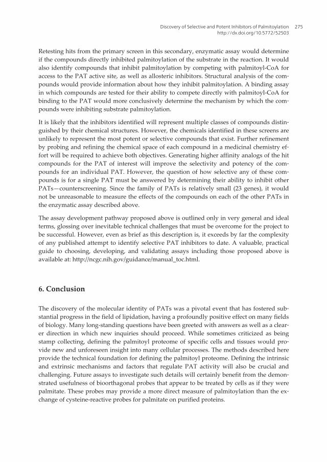
Retesting hits from the primary screen in this secondary, enzymatic assay would determineif the compounds directly inhibited palmitoylation of the substrate in the reaction. It wouldalso identify compounds that inhibit palmitoylation by competing with palmitoyl-CoA foraccess to the PAT active site, as well as allosteric inhibitors. Structural analysis of the com‐pounds would provide information about how they inhibit palmitoylation. A binding assayin which compounds are tested for their ability to compete directly with palmitoyl-CoA forbinding to the PAT would more conclusively determine the mechanism by which the com‐pounds were inhibiting substrate palmitoylation.
It is likely that the inhibitors identified will represent multiple classes of compounds distin‐guished by their chemical structures. However, the chemicals identified in these screens areunlikely to represent the most potent or selective compounds that exist. Further refinementby probing and refining the chemical space of each compound in a medicinal chemistry ef‐fort will be required to achieve both objectives. Generating higher affinity analogs of the hitcompounds for the PAT of interest will improve the selectivity and potency of the com‐pounds for an individual PAT. However, the question of how selective any of these com‐pounds is for a single PAT must be answered by determining their ability to inhibit otherPATs—counterscreening. Since the family of PATs is relatively small (23 genes), it wouldnot be unreasonable to measure the effects of the compounds on each of the other PATs inthe enzymatic assay described above.
The assay development pathway proposed above is outlined only in very general and idealterms, glossing over inevitable technical challenges that must be overcome for the project tobe successful. However, even as brief as this description is, it exceeds by far the complexityof any published attempt to identify selective PAT inhibitors to date. A valuable, practicalguide to choosing, developing, and validating assays including those proposed above isavailable at: http://ncgc.nih.gov/guidance/manual_toc.html.
6. Conclusion
The discovery of the molecular identity of PATs was a pivotal event that has fostered sub‐stantial progress in the field of lipidation, having a profoundly positive effect on many fieldsof biology. Many long-standing questions have been greeted with answers as well as a clear‐er direction in which new inquiries should proceed. While sometimes criticized as beingstamp collecting, defining the palmitoyl proteome of specific cells and tissues would pro‐vide new and unforeseen insight into many cellular processes. The methods described hereprovide the technical foundation for defining the palmitoyl proteome. Defining the intrinsicand extrinsic mechanisms and factors that regulate PAT activity will also be crucial andchallenging. Future assays to investigate such details will certainly benefit from the demon‐strated usefulness of bioorthagonal probes that appear to be treated by cells as if they werepalmitate. These probes may provide a more direct measure of palmitoylation than the ex‐change of cysteine-reactive probes for palmitate on purified proteins.
Discovery of Selective and Potent Inhibitors of Palmitoylationhttp://dx.doi.org/10.5772/52503
275
The number of signaling networks in which palmitoylation plays a pivotal role is large andgrowing. The relationship between PAT gene expression and cancer is perhaps the most evi‐dent as the fraction of PAT genes implicated in metastasis and tumorigenesis is notablyhigh. There is also a remarkable confluence between our increasing understanding of palmi‐toylation and our increasing awareness of the importance of lipid rafts, one of the primaryresidences of palmitoylated proteins in cancer [154, 155]. In instances where there is a rela‐tionship between aberrant expression of PATs and cancer, the critical questions relate to thesubstrates of these PATs and their associated signaling networks. Identification of these sig‐naling networks will potentially provide new therapeutic targets for the prevention or rever‐sal of cancer progression. Given the preponderance of palmitoylated proteins resident in theneuronal synapse (which is itself a lipid raft of sorts), it is clearly another area of researchthat deserves (and has already received) a great deal of attention.
While there has been some progress made in identifying pharmacological modulators ofpalmitoylation [125, 126, 138], there is nothing yet known about how to specifically targetindividual PATs. From a practical standpoint, inhibiting specific PATs may be a simplerprocess than developing specific PAT agonists. The advent of assays with the ability tomeasure changes in the activity of a single PAT along with the identification of PAT-sub‐strate associations should enable further development of new assays to identify specificpharmacological modulators of individual PATs as well as providing important informationon the signaling networks associated with specific PATs.
Our ability to understand palmitoylation and its importance to human health and disease isonly as good as the technological methods we use to make accurate and valid measure‐ments. Our ability to investigate the basic mechanisms of how PATs work, of PAT/substraterelationships, and how palmitoylation affects signaling processes related to disease wouldbe improved significantly by the development of selective and potent pharmacological tools.Until such tools are available, we should be mindful that using compounds such as 2-BP,cerulenin, and tunicamycin may lead to erroneous conclusions. Developing non-lipid, selec‐tive inhibitors that target the PAT active site is feasible. The challenges that exist are concep‐tually similar in some aspects to those faced during the development of selective, small-molecule inhibitors of kinases that do not resemble ATP. Based on current knowledge, themost logical PATs to target first are those for which overexpression is oncogenic. However,the motivation to initiate drug discovery programs on a large scale will probably remain be‐low the required threshold until more conclusive data are available from more sophisticat‐ed, whole-animal experiments that link PATs to oncogenesis.
Author details
Sonia Lobo Planey
Address all correspondence to: [email protected]
The Commonwealth Medical College, Scranton, PA, USA
Drug Discovery276

References
[1] Linder, M. E., & Deschenes, R. J. (2007). Palmitoylation: policing protein stability andtraffic. Nat Rev Mol Cell Biol. Jan, 8(1), 74-84.
[2] Buglino, J. A., & Resh, M. D. Hhat is a palmitoylacyltransferase with specificity forN-palmitoylation of Sonic Hedgehog. J Biol Chem, 283(32), 22076-88.
[3] Mitchell, D. A., Vasudevan, A., Linder, M. E., & Deschenes, R. J. Protein palmitoyla‐tion by a family of DHHC protein S-acyltransferases. J Lipid Res, 47(6), 1118-27.
[4] Hofmann, K. A superfamily of membrane-bound O-acyltransferases with implica‐tions for wnt signaling. Trends Biochem Sci, 25(3), 111-2.
[5] Zacharias, D. A., Violin, J. D., Newton, A. C., & Tsien, R. Y. Partitioning of lipid-modified monomeric GFPs into membrane microdomains of live cells. Science,296(5569), 913-6.
[6] Greaves, J., Prescott, G. R., Gorleku, O. A., & Chamberlain, L. H. The fat controller:roles of palmitoylation in intracellular protein trafficking and targeting to membranemicrodomains (Review). Mol Membr Biol, 26(1), 67-79.
[7] Liang, X., Lu, Y., Neubert, T. A., & Resh, M. D. Mass spectrometric analysis ofGAP-43/neuromodulin reveals the presence of a variety of fatty acylated species. J Bi‐ol Chem, 277(36), 33032-40.
[8] Liang, X., Lu, Y., Wilkes, M., Neubert, T. A., & Resh, M. D. The N-terminal SH4 re‐gion of the Src family kinase Fyn is modified by methylation and heterogeneous fattyacylation: role in membrane targeting, cell adhesion, and spreading. J Biol Chem,279(9), 8133-9.
[9] Liang, X., Nazarian, A., Erdjument-Bromage, H., Bornmann, W., Tempst, P., & Resh,M. D. Heterogeneous fatty acylation of Src family kinases with polyunsaturated fattyacids regulates raft localization and signal transduction. J Biol Chem, 276(33),30987-94.
[10] Jones, T. L., Degtyarev, M. Y., & Backlund, P. S. Jr. The stoichiometry of G alpha(s)palmitoylation in its basal and activated states. Biochemistry, 36(23), 7185-91.
[11] Bizzozero, O. A., Mc Garry, J. F., & Lees, M. B. Autoacylation of myelin proteolipidprotein with acyl coenzyme A. J Biol Chem, 262(28), 13550-7.
[12] Morello, J. P., & Bouvier, M. (1996). Palmitoylation: a post-translational modificationthat regulates signalling from G-protein coupled receptors. Biochem Cell Biol, 74(4),449-57.
[13] Wedegaertner, P. B., & Bourne, H. R. Activation and depalmitoylation of Gs alpha.Cell, 77(7), 1063-70.
Discovery of Selective and Potent Inhibitors of Palmitoylationhttp://dx.doi.org/10.5772/52503
277

[14] Bano, M. C., Jackson, C. S., & Magee, A. I. (1998). Pseudo-enzymatic S-acylation of amyristoylated yes protein tyrosine kinase peptide in vitro may reflect non-enzymaticS-acylation in vivo. Biochem J. Mar 1 , 330(Pt 2), 723-31.
[15] Duncan, J. A., & Gilman, A. G. (1996). Autoacylation of G protein alpha subunits. JBiol Chem. Sep 20; , 271(38), 23594-600.
[16] Dietrich, L. E., & Ungermann, C. (2004). On the mechanism of protein palmitoyla‐tion. EMBO Rep. Nov; , 5(11), 1053-7.
[17] Berger, M., & Schmidt, M. F. Cell-free fatty acid acylation of Semliki Forest viral pol‐ypeptides with microsomal membranes from eukaryotic cells. J Biol Chem, 259(11),7245-52.
[18] Berthiaume, L., & Resh, M.D. Biochemical characterization of a palmitoyl acyltrans‐ferase activity that palmitoylates myristoylated proteins. J Biol Chem, 270(38),22399-405.
[19] Liu, L., Dudler, T., & Gelb, M. H. Purification of a protein palmitoyltransferase thatacts on H-Ras protein and on a C-terminal N-Ras peptide. J Biol Chem, 271(38),23269-76.
[20] Bartels, D. J., Mitchell, D. A., Dong, X., & Deschenes, R. J. Erf2, a novel gene productthat affects the localization and palmitoylation of Ras2 in Saccharomyces cerevisiae.Mol Cell Biol, 19(10), 6775-87.
[21] Mitchell, D. A., Farh, L., Marshall, T. K., & Deschenes, R. J. A polybasic domain al‐lows nonprenylated Ras proteins to function in Saccharomyces cerevisiae. J BiolChem, 269(34), 21540-6.
[22] Jung, V., Chen, L., Hofmann, S. L., Wigler, M., & Powers, S. Mutations in the SHR5gene of Saccharomyces cerevisiae suppress Ras function and block membrane attach‐ment and palmitoylation of Ras proteins. Mol Cell Biol, 15(3), 1333-42.
[23] Lobo, S., Greentree, W. K., Linder, M. E., & Deschenes, R. J. Identification of a Raspalmitoyltransferase in Saccharomyces cerevisiae. J Biol Chem, 277(43), 41268-73.
[24] Putilina, T., Wong, P., & Gentleman, S. (1999). The DHHC domain: a new highly con‐served cysteine-rich motif. Mol Cell Biochem May; , 195(1-2), 219 -26 .
[25] Mesilaty-Gross, S., Reich, A., Motro, B., & Wides, R. The Drosophila STAM gene ho‐molog is in a tight gene cluster, and its expression correlates to that of the adjacentgene ial. Gene, 231(1-2), 173-86.
[26] Roth, A. F., Feng, Y., Chen, L., & Davis, N. G. The yeast DHHC cysteine-rich domainprotein Akr1p is a palmitoyl transferase. J Cell Biol, 159(1), 23-8.
[27] Feng, Y., & Davis, N. G. Akr1p and the type I casein kinases act prior to the ubiquiti‐nation step of yeast endocytosis: Akr1p is required for kinase localization to the plas‐ma membrane. Mol Cell Biol, 20(14), 5350-9.
Drug Discovery278
[28] Givan, S. A., & Sprague, G. F. Jr. The ankyrin repeat-containing protein Akr1p is re‐quired for the endocytosis of yeast pheromone receptors. Mol Biol Cell, 8(7), 1317-27.
[29] Oyama, T., Miyoshi, Y., Koyama, K., Nakagawa, H., Yamori, T., Ito, T., et al. Isolationof a novel gene on 8p21.3-22 whose expression is reduced significantly in human col‐orectal cancers with liver metastasis. Genes Chromosomes Cancer, 29(1), 9-15.
[30] Ohno, Y., Kihara, A., Sano, T., & Igarashi, Y. Intracellular localization and tissue-spe‐cific distribution of human and yeast DHHC cysteine-rich domain-containing pro‐teins. Biochim Biophys Acta, 1761(4), 474-83.
[31] Swarthout, J. T., Lobo, S., Farh, L., Croke, M. R., Greentree, W. K., Deschenes, R. J., etal. DHHC9 and GCP16 constitute a human protein fatty acyltransferase with specif‐icity for H- and N-Ras. J Biol Chem, 280(35), 31141-8.
[32] Raymond, F. L., Tarpey, P. S., Edkins, S., Tofts, C., O’Meara, S., Teague, J., et al. Mu‐tations in ZDHHC9, which encodes a palmitoyltransferase of NRAS and HRAS,cause X-linked mental retardation associated with a marfanoid habitus. Am J HumGenet, 80(5), 982-7.
[33] Mukai, J., Liu, H., Burt, R. A., Swor, D. E., Lai, W. S., Karayiorgou, M., et al. Evidencethat the gene encoding ZDHHC8 contributes to the risk of schizophrenia. Nat Genet,36(7), 725-31.
[34] Demily, C., Legallic, S., Bou, J., Houy-Durand, E., Van Amelsvoort, T., Zinkstok, J., etal. ZDHHC8 single nucleotide polymorphism rs175174 is not associated with psychi‐atric features of the 22q11 deletion syndrome or schizophrenia. Psychiatr Genet, 17(5),311-2.
[35] Glaser, B., Schumacher, J., Williams, H. J., Jamra, R. A., Ianakiev, N., Milev, R., et al.No association between the putative functional ZDHHC8 single nucleotide polymor‐phism rs175174 and schizophrenia in large European samples. Biol Psychiatry, 58(1),78-80.
[36] Saito, S., Ikeda, M., Iwata, N., Suzuki, T., Kitajima, T., Yamanouchi, Y., et al. No asso‐ciation was found between a functional SNP in ZDHHC8 and schizophrenia in a Jap‐anese case-control population. Neurosci Lett, 374(1), 21-4.
[37] Zhang, J., Planey, S. L., Ceballos, C., Stevens, S. M. Jr, Keay, S. K., & Zacharias, D. A.Identification of CKAP4/63as a major substrate of the palmitoyl acyltransferaseDHHC2, a putative tumor suppressor, using a novel proteomics method. Mol CellProteomics, 7(7), 1378-88.
[38] Rocks, O., Gerauer, M., Vartak, N., Koch, S., Huang, Z. P., Pechlivanis, M., et al. Thepalmitoylation machinery is a spatially organizing system for peripheral membraneproteins. Cell, 141(3), 458-71.
[39] Politis, E. G., Roth, A. F., & Davis, N. G. Transmembrane topology of the protein pal‐mitoyl transferase Akr1. J Biol Chem, 280(11), 10156-63.
Discovery of Selective and Potent Inhibitors of Palmitoylationhttp://dx.doi.org/10.5772/52503
279
[40] Claros, M. G., & von, Heijne. G. TopPred II: an improved software for membraneprotein structure predictions. Comput Appl Biosci, 10(6), 685-6.
[41] Freedman, R. B., Dunn, A. D., & Ruddock, L. W. Protein folding: a missing redox linkin the endoplasmic reticulum. Curr Biol, 8(13), R 468-70.
[42] Huppa, J. B., & Ploegh, H. L. The eS-Sence of-SH in the ER. Cell, 92(2), 145-8.
[43] Hou, H., Subramanian, K., La Grassa, T. J., Markgraf, D., Dietrich, L. E., Urban, J., etal. The DHHC protein Pfa3 affects vacuole-associated palmitoylation of the fusionfactor Vac8. Proc Natl Acad Sci U S A, 102(48), 17366-71.
[44] Planey, S. L., Keay, S. K., Zhang, C. O., & Zacharias, D. A. Palmitoylation of cytoske‐leton associated protein 4 by DHHC2 regulates antiproliferative factor-mediated sig‐naling. Mol Biol Cell, 20(5), 1454-63.
[45] Smotrys, J. E., & Linder, M. E. (2004). Palmitoylation of intracellular signaling pro‐teins: regulation and function. Annu Rev Biochem, 73, 559-87.
[46] Lam, K. K., Davey, M., Sun, B., Roth, A. F., Davis, N. G., & Conibear, E. Palmitoyla‐tion by the DHHC protein Pfa4 regulates the ER exit of Chs3. J Cell Biol, 174(1), 19-25.
[47] Greaves, J., Carmichae, J. A., & Chamberlain, L. H. The palmitoyl transferaseDHHC2 targets a dynamic membrane cycling pathway: regulation by a C-terminaldomain. Mol Biol Cell, 22(11), 1887-95.
[48] Fernandez-Hernando, C., Fukata, M., Bernatchez, P. N., Fukata, Y., Lin, M. I., Bredt,D. S., et al. Identification of Golgi-localized acyl transferases that palmitoylate andregulate endothelial nitric oxide synthase. J Cell Biol, 174(3), 369-77.
[49] Yanai, A., Huang, K., Kang, R., Singaraja, R. R., Arstikaitis, P., Gan, L., et al. Palmi‐toylation of huntingtin by HIP14 is essential for its trafficking and function. Nat Neu‐rosci, 9(6), 824-31.
[50] Mansouri, M. R., Marklund, L., Gustavsson, P., Davey, E., Carlsson, B., Larsson, C., etal. Loss of 5p15.33 ZDHHC15 expression in a woman with a balanced translocationt(X;15)(q13.3;cen) and severe mental retardation. Eur J Hum Genet, 13(8), 970-7.
[51] Yamamoto, Y., Chochi, Y., Matsuyama, H., Eguchi, S., Kawauchi, S., Furuya, T., et al.(2007). Gain of is associated with progression of bladder cancer. Oncology, 72(1-2),132-8.
[52] Mansilla, F., Birkenkamp-Demtroder, K., Kruhoffer, M., Sorensen, F. B., Andersen, C.L., Laiho, P., et al. Differential expression of DHHC9 in microsatellite stable and in‐stable human colorectal cancer subgroups. Br J Cancer, 96(12), 1896-903.
[53] Ducker, C. E., Stettler, E. M., French, K. J., Upson, J. J., & Smith, C. D. Huntingtin in‐teracting protein 14 is an oncogenic human protein: palmitoyl acyltransferase. Onco‐gene, 23(57), 9230-7.
[54] Razzaq, T. M., Bass, R., Vines, D. J., Werner, F., Whawell, S. A., & Ellis, V. Functionalregulation of tissue plasminogen activator on the surface of vascular smooth muscle
Drug Discovery280
cells by the type-II transmembrane protein p63(CKAP4). J Biol Chem, 278(43),42679-85.
[55] Gupta, N., Manevich, Y., Kazi, Tao. J. Q., Fisher, A. B., & Bates, S. R. Identificationand characterization of p63 (CKAP4/ERGIC-63/CLIMP-63), a surfactant protein Abinding protein, on type II pneumocytes. Am J Physiol Lung Cell Mol Physiol, 291(3), L436-46.
[56] Sharma, C., Yang, X. H., & Hemler, M. E. DHHC2 Affects Palmitoylation, Stability,and Functions of Tetraspanins CD9 and CD151. Mol Biol Cell.
[57] Ikeyama, S., Koyama, M., Yamaoko, M., Sasada, R., & Miyake, M. Suppression of cellmotility and metastasis by transfection with human motility-related protein (MRP-1/CD9) DNA. J Exp Med, 177(5), 1231-7.
[58] Miyake, M., Inufusa, H., Adachi, M., Ishida, H., Hashida, H., Tokuhara, T., et al. Sup‐pression of pulmonary metastasis using adenovirally motility related protein-1(MRP-1/CD9) gene delivery. Oncogene, 19(46), 5221-6.
[59] Zavadil, J., Bitzer, M., Liang, D., Yang, Y. C., Massimi, A., Kneitz, S., et al. Geneticprograms of epithelial cell plasticity directed by transforming growth factor-beta.Proc Natl Acad Sci U S A, 98(12), 6686-91.
[60] Janda, E., Lehmann, K., Killisch, I., Jechlinger, M., Herzig, M., Downward, J., et al.Ras and TGF[beta] cooperatively regulate epithelial cell plasticity and metastasis:dissection of Ras signaling pathways. J Cell Biol, 156(2), 299-313.
[61] Thiery, J. P. Epithelial-mesenchymal transitions in tumour progression. Nat Rev Can‐cer, 2(6), 442-54.
[62] Fukata, M., Fukata, Y., Adesnik, H., Nicoll, R. A., & Bredt, . Identification of PSD-95palmitoylating enzymes. Neuron, 44(6), 987-96.
[63] Greaves, J., Gorleku, O. A., Salaun, C., & Chamberlain, L. H. Palmitoylation of theSNAP25 protein family: specificity and regulation by DHHC palmitoyl transferases. JBiol Chem, 285(32), 24629-38.
[64] Zeidman, R., Buckland, G., Cebecauer, M., Eissmann, P., Davis, D. M., & Magee, A. I.DHHC2 is a protein S-acyltransferase for Lck. Mol Membr Biol, 28(7-8), 473-86.
[65] Tsutsumi, R., Fukata, Y., Noritake, J., Iwanaga, T., Perez, F., & Fukata, M. Identifica‐tion of G protein alpha subunit-palmitoylating enzyme. Mol Cell Biol, 29(2), 435-47.
[66] Jia, L., Linder, M. E., & Blumer, K. J. Gi/o signaling and the palmitoyltransferaseDHHC2 regulate palmitate cycling and shuttling of RGS7 family-binding protein. JBiol Chem, 286(15), 13695-703.
[67] Goytain, A., Hines, R. M., & Quamme, G. A. Huntingtin-interacting proteins, HIP14and HIP14L, mediate dual functions, palmitoyl acyltransferase and Mg2+ transport.J. Biol Chem, 283(48), 33365-74.
Discovery of Selective and Potent Inhibitors of Palmitoylationhttp://dx.doi.org/10.5772/52503
281

[68] Schlingmann, K. P., & Gudermann, T. A critical role of TRPM channel-kinase for hu‐man magnesium transport. J Physiol, 566(Pt 2), 301 -8 .
[69] Schlingmann, K. P., Waldegger, S., Konrad, M., Chubanov, V., & Gudermann, T.TRPM6 and TRPM7--Gatekeepers of human magnesium metabolism. Biochim BiophysActa, 1772(8), 813-21.
[70] Goytain, A., Hines, R. M., & Quamme, G. A. Huntingtin-interacting proteins, HIP14and HIP14L, mediate dual functions:Palmitoyl acytransferase and Mg2+ transport. JBiol Chem.
[71] Hines, R. M., Kang, R., Goytain, A., & Quamme, G. A. Golgi-specific DHHC zinc fin‐ger protein GODZ mediates membrane Ca2+ transport. J Biol Chem, 285(7), 4621-8.
[72] Fukata, Y., Iwanaga, T., & Fukata, M. Systematic screening for palmitoyl transferaseactivity of the DHHC protein family in mammalian cells. Methods, 40(2), 177-82.
[73] Nadolski, M. J., & Linder, M. E. Protein lipidation. FEBS J, 274(20), 5202-10.
[74] Roth, A. F., Wan, J., Bailey, A. O., Sun, B., Kuchar, J. A., Green, W. N., et al. Globalanalysis of protein palmitoylation in yeast. Cell, 125(5), 1003-13.
[75] Ren, J., Wen, L., Gao, X., Jin, C., Xue, Y., & Yao, X. CSS-Palm 2.0: an updated softwarefor palmitoylation sites prediction. Protein Eng Des Sel.
[76] Xue, Y., Chen, H., Jin, C., Sun, Z., & Yao, X. (2006). NBA-Palm: prediction of palmi‐toylation site implemented in Naive Bayes algorithm. BMC Bioinformatics, 7, 458 .
[77] Zhou, F., Xue, Y., Yao, X., & Xu, Y. (2006). A general user interface for predictionservers of proteins’ post-translational modification sites. Nat Protoc, 1(3), 1318-21.
[78] Zhou, F., Xue, Y., Yao, X., & Xu, Y. CSS-Palm: palmitoylation site prediction with aclustering and scoring strategy (CSS). Bioinformatics, 22(7), 894-6.
[79] Bernstein, L. S., Linder, M. E., & Hepler, J. R. (2004). Analysis of RGS protein palmi‐toylation. Methods Mol Biol, 237, 195-204.
[80] Basar, T., Havlicek, V., Bezouskova, S., Halada, P., Hackett, M., & Sebo, P. The con‐served lysine 860 in the additional fatty-acylation site of Bordetella pertussis adeny‐late cyclase is crucial for toxin function independently of its acylation status. J BiolChem, 274(16), 10777-83.
[81] Branton, W. D., Rudnick, M. S., Zhou, Y., Eccleston, E. D., Fields, G. B., & Bowers, L.D. Fatty acylated toxin structure. Nature, 365(6446), 496-7.
[82] Kleuss, C., & Krause, E. Galpha(s) is palmitoylated at the N-terminal glycine. Embo J,22(4), 826-32.
[83] Pepinsky, R. B., Zeng, C., Wen, D., Rayhorn, P., Baker, D. P., Williams, K. P., et al.Identification of a palmitic acid-modified form of human Sonic hedgehog. J BiolChem, 273(22), 14037-45.
Drug Discovery282

[84] Bijlsma, M. F., Spek, C. A., & Peppelenbosch, M. P. Hedgehog: an unusual signaltransducer. Bioessays, 26(4), 387-94.
[85] Di Simplicio, P., Franconi, F., Frosali, S., & Di Giuseppe, D. Thiolation and nitrosationof cysteines in biological fluids and cells. Amino Acids, 25(3-4), 323 -39 .
[86] Lipton, S. A., Choi, Y. B., Takahashi, H., Zhang, D., Li, W., Godzik, A., et al. Cysteineregulation of protein function--as exemplified by NMDA-receptor modulation.Trends Neurosci, 25(9), 474-80.
[87] Hershko, A., & Ciechanover, A. (1998). The ubiquitin system. Annu Rev Biochem, 67,425-79.
[88] Hermanson, G. T. (1996). Bioconjugate Techniques. 1st ed. San Diego: Academic Press.
[89] Gygi, S. P., Rist, B., Gerber, S. A., Turecek, F., Gelb, M. H., & Aebersold, R. Quantita‐tive analysis of complex protein mixtures using isotope-coded affinity tags. Nat Bio‐technol, 17(10), 994-9.
[90] Schrimpf, S. P., Meskenaite, V., Brunner, E., Rutishauser, D., Walther, P., Eng, J., et al.Proteomic analysis of synaptosomes using isotope-coded affinity tags and mass spec‐trometry. Proteomics, 5(10), 2531-41.
[91] Gygi, S. P., Rist, B., Griffin, T. J., Eng, J., & Aebersold, R. Proteome analysis of low-abundance proteins using multidimensional chromatography and isotope-coded af‐finity tags. J Proteome Res, 1(1), 47-54.
[92] Sitia, R., & Molteni, S. N. Stress, protein (mis)folding, and signaling: the redox con‐nection. Sci STKE [239], pe27.
[93] Munro, A. P., & Williams, D. L. H. (1999). Reactivity of nitrogen nucleophiles to‐wards S-nitrosopenicillamine. Journal of the Chemical Society, Perkin Transactions, 2,1989-93.
[94] Fenton, S. S., & Fahey, R. C. Analysis of biological thiols: determination of thiol com‐ponents of disulfides and thioesters. Anal Biochem, 154(1), 34-42.
[95] Bizzozero, O. A., & Good, L. K. Rapid metabolism of fatty acids covalently bound tomyelin proteolipid protein. J Biol Chem, 266(26), 17092-8.
[96] Passmore, L. A., & Barford, D. Getting into position: the catalytic mechanisms of pro‐tein ubiquitylation. Biochem J, 379(Pt3), 513-25.
[97] Bizzozero, O. A. (1995). Chemical analysis of acylation sites and species. Methods En‐zymol, 250, 361-79.
[98] Tolbert, B. S., Tajc, S. G., Webb, H., Snyder, J., Nielsen, J. E., Miller, B. L., et al. Theactive site cysteine of ubiquitin-conjugating enzymes has a significantly elevatedpKa: functional implications. Biochemistry, 44(50), 16385-91.
[99] Cione, E., Pingitore, A., Perri, M., & Genchi, G. Influence of all-trans-retinoic acid onoxoglutarate carrier via retinoylation reaction. Biochim Biophys Acta, 1791(1), 3-7.
Discovery of Selective and Potent Inhibitors of Palmitoylationhttp://dx.doi.org/10.5772/52503
283

[100] Cione, E., Tucci, P., Chimento, A., Pezzi, V., & Genchi, G. Retinoylation reaction ofproteins in Leydig (TM-3) cells. J Bioenerg Biomembr, 37(1), 43-8.
[101] Cione, E., Tucci, P., Senatore, V., Ioele, G., & Genchi, G. (2005). Binding of all-trans-retinoic acid to MLTC-1 proteins. Mol Cell Biochem Aug; , 276(1-2), 55 -60 .
[102] Kubo, Y., Wada, M., Ohba, T., & Takahashi, N. Formation of retinoylated proteinsfrom retinoyl-CoA in rat tissues. J Biochem, 138(4), 493-500.
[103] Takahashi, N., & Breitman, T. R. Retinoic acid acylation (retinoylation) of a nuclearprotein in the human acute myeloid leukemia cell line HL60. J Biol Chem, 264(9),5159-63.
[104] Takahashi, N., & Breitman, T. R. Retinoylation of HL-60 proteins. Comparison to la‐beling by palmitic and myristic acids. J Biol Chem, 265(31), 19158-62.
[105] Takahashi, N., & Breitman, T. R. Retinoylation of proteins in leukemia, embryonalcarcinoma, and normal kidney cell lines: differences associated with differential re‐sponses to retinoic acid. Arch Biochem Biophys, 285(1), 105-10.
[106] Takahashi, N., Liapi, C., Anderson, W. B., & Breitman, T. R. Retinoylation of thecAMP-binding regulatory subunits of type I and type II cAMP-dependent proteinkinases in HL60 cells. Arch Biochem Biophys, 290(2), 293-302.
[107] Takahashi, N., Jetten, A. M., & Breitman, T. R. Retinoylation of cytokeratins in nor‐mal human epidermal keratinocytes. Biochem Biophys Res Commun, 180(1), 393-400.
[108] Djakoure, C., Guibourdenche, J., Porquet, D., Pagesy, P., Peillon, F., Li, J. Y., et al. Vi‐tamin A and retinoic acid stimulate within minutes cAMP release and growth hor‐mone secretion in human pituitary cells. J Clin Endocrinol Metab, 81(8), 3123-6.
[109] Varani, J., Burmeister, B., Perone, P., Bleavins, M., & Johnson, K. J. All-trans retinoicacid inhibits fluctuations in intracellular Ca2+ resulting from changes in extracellularCa2+. Am J Pathol, 147(3), 718-27.
[110] Hensel, J., Hintz, M., Karas, M., Linder, D., Stahl, B., & Geyer, R. Localization of thepalmitoylation site in the transmembrane protein p12E of Friend murine leukaemiavirus. Eur J Biochem, 232(2), 373-80.
[111] De Mar, J. C., Jr , , & Anderson, R. E. Identification and quantitation of the fatty acidscomposing the CoA ester pool of bovine retina, heart, and liver. J Biol Chem, 272(50),31362-8.
[112] Hang, H. C., Geutjes, E. J., Grotenbreg, G., Pollington, A. M., Bijlmakers, MJ, &Ploegh, HL. Chemical probes for the rapid detection of Fatty-acylated proteins inMammalian cells. J Am Chem Soc, 129(10), 2744-5.
[113] Adams, S. R., Campbell, R. E., Gross, L. A., Martin, B. R., Walkup, G. K., Yao, Y., etal. New biarsenical ligands and tetracysteine motifs for protein labeling in vitro andin vivo: synthesis and biological applications. J Am Chem Soc, 124(21), 6063-76.
Drug Discovery284

[114] Adams, S. R., Lev-Ram, V., & Tsien, R. Y. (1997). A new caged Ca2+, azid-1, is farmore photosensitive than nitrobenzyl- based chelators. Chem Biol, 4(11), 867-78.
[115] Griffin, B. A., Adams, S. R., Jones, J., & Tsien, R. Y. (2000). Fluorescent labeling of re‐combinant proteins in living cells with FlAsH. Methods Enzymol, 327, 565-78.
[116] Nagai, T., Sawano, A., Park, E. S., & Miyawaki, A. (2001). Circularly permuted greenfluorescent proteins engineered to sense Ca2+. Proc Natl Acad Sci U S A, 98(6),3197-202.
[117] Gaietta, G. M., Giepmans, B. N., Deerinck, T. J., Smith, W. B., Ngan, L., Llopis, J., etal. Golgi twins in late mitosis revealed by genetically encoded tags for live cell imag‐ing and correlated electron microscopy. Proc Natl Acad Sci U S A, 103(47), 17777-82.
[118] Martin, B. R., Giepmans, B. N., Adams, S. R., & Tsien, R. Y. Mammalian cell-basedoptimization of the biarsenical-binding tetracysteine motif for improved fluorescenceand affinity. Nat Biotechnol, 23(10), 1308-14.
[119] Ju, W., Morishita, W., Tsui, J., Gaietta, G., Deerinck, T. J., Adams, S. R., et al. Activity-dependent regulation of dendritic synthesis and trafficking of AMPA receptors. NatNeurosci, 7(3), 244-53.
[120] Stults, J. T., & Arnott, D. (2005). Proteomics. Methods Enzymol.; , 402, 245-89.
[121] Kenyon GL, Bruice TW. (1977). Novel sulfhydryl reagents. Methods Enzymol, 47,407-30.
[122] Qanbar, R., & Bouvier, M. Determination of protein-bound palmitate turnover ratesusing a three-compartment model that formally incorporates [3H]palmitate recy‐cling. Biochemistry, 43(38), 12275-88.
[123] Ducker, C. E., Draper, J. M., Xia, Z., & Smith, C. D. In vitro and cellular assays forpalmitoyl acyltransferases using fluorescent lipidated peptides. Methods, 40(2),166-70.
[124] Kenworthy, A. K. Fluorescence-based methods to image palmitoylated proteins.Methods, 40(2), 198-205.
[125] Resh, M. D. Use of analogs and inhibitors to study the functional significance of pro‐tein palmitoylation. Methods, 40(2), 191-7.
[126] Ducker, C. E., Griffel, L. K., Smith, R. A., Keller, S. N., Zhuang, Y., Xia, Z., et al. Dis‐covery and characterization of inhibitors of human palmitoyl acyltransferases. MolCancer Ther, 5(7), 1647-59.
[127] Varner, A. S., Ducker, C. E., Xia, Z., Zhuang, Y., De Vos, M. L., & Smith, C. D. Char‐acterization of human palmitoyl-acyl transferase activity using peptides that mimicdistinct palmitoylation motifs. Biochem J, 373(Pt1), 91-9.
Discovery of Selective and Potent Inhibitors of Palmitoylationhttp://dx.doi.org/10.5772/52503
285

[128] Schmidt, M., Schmidt, M. F., & Rott, R. Chemical identification of cysteine as palmi‐toylation site in a transmembrane protein (Semliki Forest virus E1). J Biol Chem,263(35), 18635-9.
[129] Drisdel, R. C., & Green, W. N. Labeling and quantifying sites of protein palmitoyla‐tion. Biotechniques, 36(2), 276-85.
[130] Drisdel, R. C., Manzana, E., & Green, W. N. The role of palmitoylation in functionalexpression of nicotinic alpha7 receptors. J Neurosci, 24(46), 10502-10.
[131] Wan, J., Roth, A. F., Bailey, A. O., & Davis, N. G. Palmitoylated proteins: purificationand identification. 2(7), 1573-84.
[132] Liu, H., Lin, D., & Yates, J. R. 3rd. Multidimensional separations for protein/peptideanalysis in the post-genomic era. Biotechniques, 32(4), 898 -900, 2 passim.
[133] Roth, A. F., Wan, J., Green, W. N., Yates, J. R., & Davis, N. G. Proteomic identificationof palmitoylated proteins. Methods, 40(2), 135-42.
[134] Schweizer, A., Rohrer, J., Jeno, P., De Maio, A., Buchman, T. G., & Hauri, H. P. A re‐versibly palmitoylated resident protein (63 of an ER-Golgi intermediate compart‐ment is related to a circulatory shock resuscitation protein. J Cell Sci, 104(Pt 3),685-94.
[135] Zhang, J., & Zacharias, D. A. Global Analysis of the Dynamic Palmitoylosome. LipidRafts and Cell Funciton, 23-28, Steamboat Springs, Colorado: Keystone Symposia.
[136] Kostiuk, M. A., Corvi, M. M., Keller, B. O., Plummer, G., Prescher, J. A., Hangauer,M. J., et al. Identification of palmitoylated mitochondrial proteins using a bio-orthog‐onal azido-palmitate analogue. FASEB J, 22(3), 721-32.
[137] Saxon, E., & Bertozzi, C. R. (2000). Cell surface engineering by a modified Staudingerreaction. Science. Mar 17 , 287(5460), 2007-10.
[138] Jennings, B. C., Nadolski, M. J., Ling, Y., Baker, M. B., Harrison, M. L., Deschenes, R.J., et al. 2bromopalmitate and 2-(2-Hydroxy-5-nitro-benzylidene)-benzo[b]thio‐phen-3-one inhibit DHHC-mediated palmitoylation in vitro. J Lipid Res.
[139] Mikic, I., Planey, S., Zhang, J., Ceballos, C., Seron, T., Massenbach, Bv., et al. (2006). Alive-cell, image based approach to understanding the enzymology and pharmacolo‐gy of 2bromopalmitate and palmitoylation. Methods in enzymology, 414.
[140] Chase, J. F., & Tubbs, P. K. Specific inhibition of mitochondrial fatty acid oxidationby2-bromopalmitate and its coenzyme A and carnitine esters. The Biochemical journal,129(1), 55-65.
[141] Coleman, R. A., Rao, P., Fogelsong, R. J., & Bardes, E. S. Bromopalmitoyl-CoA and 2-bromopalmitate: promiscuous inhibitors of membrane-bound enzymes. Biochimica etbiophysica acta, 1125(2), 203-9.
Drug Discovery286

[142] Draper, J. M., & Smith, C. D. (2009). Palmitoyl acyltransferase assays and inhibitors(Review). Mol Membr Biol. Jan , 26(1), 5-13.
[143] Fabian, M. A., Biggs, W. H. 3.r.d, Treiber, . D. K., Atteridge, C. E., Azimioara, M. D.,Benedetti, M. G., et al. A small molecule-kinase interaction map for clinical kinase in‐hibitors. Nature biotechnology, 23(3), 329-36.
[144] Karaman, M. W., Herrgard, S., Treiber, D. K., Gallant, P., Atteridge, C. E., Campbell,B. T., et al. A quantitative analysis of kinase inhibitor selectivity. Nature biotechnology,26(1), 127-32.
[145] Feng, Y., Yin, Y., Weiser, A., Griffin, E., Cameron, Lin. L., et al. Discovery of substi‐tuted 4-(pyrazol-4-yl)-phenylbenzodioxane-2-carboxamides as potent and highly se‐lective Rho kinase (ROCK-II) inhibitors. Journal of medicinal chemistry, 51(21), 6642-5.
[146] Huang, K., Sanders, S., Singaraja, R., Orban, P., Cijsouw, T., Arstikaitis, P., et al. Neu‐ronal palmitoyl acyl transferases exhibit distinct substrate specificity. Faseb J, 23(8),2605-15.
[147] Prigozhina, N. L., Zhong, L., Hunter, E. A., Mikic, I., Callaway, S., Roop, D. R., et al.Plasma membrane assays and three-compartment image cytometry for high contentscreening. Assay and drug development technologies, 5(1), 29-48.
[148] Morelock, M. M., Hunter, E. A., Moran, T. J., Heynen, S., Laris, C., Thieleking, M., etal. Statistics of assay validation in high throughput cell imaging of nuclear factorkappaB nuclear translocation. Assay and drug development technologies, 3(5), 483-99.
[149] Wolff, M., Kredel, S., Wiedenmann, J., Nienhaus, G. U., & Heilker, R. Cell-based as‐says in practice: cell markers from autofluorescent proteins of the GFP-family. Combi‐natorial chemistry & high throughput screening, 11(8), 602-9.
[150] Collins, M. A. Generating’omic knowledge: The Role of Informatics in High ContentScreening. Combinatorial chemistry & high throughput screening.
[151] Shaner, N. C., Campbell, R. E., Steinbach, P. A., Giepmans, B. N., Palmer, A. E., &Tsien, R. Y. Improved monomeric red, orange and yellow fluorescent proteins de‐rived from Discosoma sp. red fluorescent protein. Nature biotechnology, 22(12),1567-72.
[152] Inglese, J., Auld, Jadhav. A., Johnson, R. L., Simeonov, A., Yasgar, A., et al. Quantita‐tive high-throughput screening: a titration-based approach that efficiently identifiesbiological activities in large chemical libraries. Proceedings of the National Academy ofSciences of the United States of America, 103(31), 11473-8.
[153] Chung, C. C., Ohwaki, K., Schneeweis, J. E., Stec, E., Varnerin, J. P., Goudreau, P. N.,et al. A fluorescence-based thiol quantification assay for ultra-high-throughputscreening for inhibitors of coenzyme A production. Assay and drug development tech‐nologies, 6(3), 361-74.
Discovery of Selective and Potent Inhibitors of Palmitoylationhttp://dx.doi.org/10.5772/52503
287

[154] Lazo, P.A. Functional implications of tetraspanin proteins in cancer biology. CancerSci; 98: 1666-77.
[155] Patra, S.K. Dissecting lipid raft facilitated cell signaling pathways in cancer. Biochimi‐ca et biophysica acta; 1785: 182-206.
Drug Discovery288
Related Documents