Discovery and Binding Studies on a Series of Novel Pin1 Ligands Bainan Wu, Michele F. Rega, Jun Wei, Hongbin Yuan, Russell Dahl, Ziming Zhang, and Maurizio Pellecchia * Infectious and Inflammatory Disease Center and Cancer Center, Burnham Institute for Medical Research, 10901 North Torrey Pines Road, La Jolla, CA 92037, USA Abstract Pin1 plays a key role in various biological cellular processes via the recognition of phosphorylated Ser/Thr-Proline motifs. Moreover, high expression levels of Pin1 are correlated to tumorgenesis in some cancer types. Here, we identify a novel series of small molecular weight compounds with a core structure mimicking the phoshorylated serine. The binding affinity and binding mode of the compounds for Pin1 are analyzed via NMR spectroscopy and computational studies. The reported chemical probes and relative binding data to Pin1 represent valuable stepping stones for the validation of Pin1 as target for drug discovery and for eventually the development of possible lead compounds. Keywords comparative molecular field analysis; NMR; Pin1; WW-domain Peptidyl-prolyl cis/trans isomerases (PPI ase ) are enzymes catalyzing proline cis/trans isomerization, a crucial step for protein folding (1–3). To date, three families of highly conserved PPI ase have been identified, namely, cyclophilins (Cyp) (4), FK506-binding proteins (FKBP) (5) and parvulins (6). Unlike Cyp and FKBP, the parvulin family of proteins has a unique feature to selectively recognize a phosphorylated Ser/Thr-Pro motif (p-Ser/Thr-Pro) (7,8). Human Pin1 is the most widely studied protein in the parvulin family. Isomerization by Pin1 modulates a multitude of biological processes, including protein folding and biological activity (9), protein stability (8,10) as well as subcellular localization (11). As phosphorylation is a major regulation mechanism in cell, Pin1 is also involved in cycle regulation, oncogenesis, signal transduction and neurodegeration in Alzheimer’s disease via targeting several key proteins, such as cyclin D1 (12–15), c-jun (16), c-Myc (17), β-catenin (11), p53 (9,18,19) and tau (9). Pin1 is constituted by two domains, an N-terminal WW domain (Pin1 ww ) and a C- terminal catalytic domain (Pin1 cat ), both of which have a specificity for the p-Ser/Thr-Pro motif. Pin1 ww is a member of the WW family proteins (20) and has a much higher binding affinity for the motif than Pin1 cat (21), hence is thought as a docking site mediating the substrate recognition of Pin1. Pin1 cat displays a weaker binding affinity against p-Ser/Thr-Pro motif but possesses a full isomerase activity even without the WW domain (10). Two different crystal structures of Pin1-peptide complexes have been reported (8,21). The first structure of Pin1 containing a Ala-Pro peptide in the catalytic domain (PDB code: 1PIN) exhibits a ‘close’ active *Corresponding author: Maurizio Pellecchia, [email protected]. Supporting Information Additional Supporting Information may be found in the online version of this article: Please note: Wiley-Blackwell is not responsible for the content or functionality of any supporting materials supplied by the authors. Any queries (other than missing material) should be directed to the corresponding author for the article. NIH Public Access Author Manuscript Chem Biol Drug Des. Author manuscript; available in PMC 2010 April 1. Published in final edited form as: Chem Biol Drug Des. 2009 April ; 73(4): 369–379. doi:10.1111/j.1747-0285.2009.00795.x. NIH-PA Author Manuscript NIH-PA Author Manuscript NIH-PA Author Manuscript

Welcome message from author
This document is posted to help you gain knowledge. Please leave a comment to let me know what you think about it! Share it to your friends and learn new things together.
Transcript
Discovery and Binding Studies on a Series of Novel Pin1 Ligands
Bainan Wu, Michele F. Rega, Jun Wei, Hongbin Yuan, Russell Dahl, Ziming Zhang, andMaurizio Pellecchia*Infectious and Inflammatory Disease Center and Cancer Center, Burnham Institute for MedicalResearch, 10901 North Torrey Pines Road, La Jolla, CA 92037, USA
AbstractPin1 plays a key role in various biological cellular processes via the recognition of phosphorylatedSer/Thr-Proline motifs. Moreover, high expression levels of Pin1 are correlated to tumorgenesis insome cancer types. Here, we identify a novel series of small molecular weight compounds with acore structure mimicking the phoshorylated serine. The binding affinity and binding mode of thecompounds for Pin1 are analyzed via NMR spectroscopy and computational studies. The reportedchemical probes and relative binding data to Pin1 represent valuable stepping stones for the validationof Pin1 as target for drug discovery and for eventually the development of possible lead compounds.
Keywordscomparative molecular field analysis; NMR; Pin1; WW-domain
Peptidyl-prolyl cis/trans isomerases (PPIase) are enzymes catalyzing proline cis/transisomerization, a crucial step for protein folding (1–3). To date, three families of highlyconserved PPIase have been identified, namely, cyclophilins (Cyp) (4), FK506-binding proteins(FKBP) (5) and parvulins (6). Unlike Cyp and FKBP, the parvulin family of proteins has aunique feature to selectively recognize a phosphorylated Ser/Thr-Pro motif (p-Ser/Thr-Pro)(7,8).
Human Pin1 is the most widely studied protein in the parvulin family. Isomerization by Pin1modulates a multitude of biological processes, including protein folding and biological activity(9), protein stability (8,10) as well as subcellular localization (11). As phosphorylation is amajor regulation mechanism in cell, Pin1 is also involved in cycle regulation, oncogenesis,signal transduction and neurodegeration in Alzheimer’s disease via targeting several keyproteins, such as cyclin D1 (12–15), c-jun (16), c-Myc (17), β-catenin (11), p53 (9,18,19) andtau (9). Pin1 is constituted by two domains, an N-terminal WW domain (Pin1ww) and a C-terminal catalytic domain (Pin1cat), both of which have a specificity for the p-Ser/Thr-Promotif. Pin1ww is a member of the WW family proteins (20) and has a much higher bindingaffinity for the motif than Pin1cat (21), hence is thought as a docking site mediating the substraterecognition of Pin1. Pin1cat displays a weaker binding affinity against p-Ser/Thr-Pro motif butpossesses a full isomerase activity even without the WW domain (10). Two different crystalstructures of Pin1-peptide complexes have been reported (8,21). The first structure of Pin1containing a Ala-Pro peptide in the catalytic domain (PDB code: 1PIN) exhibits a ‘close’ active
*Corresponding author: Maurizio Pellecchia, [email protected] InformationAdditional Supporting Information may be found in the online version of this article:Please note: Wiley-Blackwell is not responsible for the content or functionality of any supporting materials supplied by the authors. Anyqueries (other than missing material) should be directed to the corresponding author for the article.
NIH Public AccessAuthor ManuscriptChem Biol Drug Des. Author manuscript; available in PMC 2010 April 1.
Published in final edited form as:Chem Biol Drug Des. 2009 April ; 73(4): 369–379. doi:10.1111/j.1747-0285.2009.00795.x.
NIH
-PA Author Manuscript
NIH
-PA Author Manuscript
NIH
-PA Author Manuscript
site conformation, in which a phosphate binding loop β1/α1 (residues 70–83) is foldedbackwards the binding pocket via specific interactions with a phosphate-mimic sulfate ion.The second structure of Pin1 contains a phosphopeptide bound to the WW domain (PDB code:1F8A) in which the loop β1/α1 is positioned away from the catalytic domain thus leading toan ‘open’ conformation in the binding pocket. Moreover, the WW domain twists towards thecatalytic domain to form a deep clamp that constitutes the phosphopeptide docking site. Insolution, two domains of Pin1 are loosely connected (22) but their mobility can be differentlymodulated by different phosphopeptides (23).
The fact that Pin1 is prevalently overexpressed in several human tumors including breast(16), colorectal (24), prostate (25) and thyroid cancers (26) makes Pin1 an attractive therapeutictarget for cancer treatment. To date, relevant Pin1 inhibitors reported are phosphopeptidemimics targeting on Pin1cat at the nanomolar level (27,28). However, poor cell membranepermeability of the peptide mimics limits their application for drug development and targetvalidation (29). Considering its substrate recognition capability, Pin1ww is potentially also avalid target for inhibitor design. So far, the only known Pin1ww inhibitors are pThr-Pro analogs(30).
In this study, we report a novel series of compounds that bind to Pinww. The compound seriesBI-81 was found via a virtual screening approach of a commercial library followed byexperimental binding data using NMR spectroscopy techniques. To explain the differences incompound binding affinities, a three-dimensional quantitative structure activity relationship(3D QSAR) model was established and comparative molecular field analysis (CoMFA) (31)studies were subsequently performed. The generated 3D QSAR model gives insights into thecontribution of various substituents on the binding affinity of the compounds and providesinformation in designing and predicting the binding ability of novel derivatives combined withdocking studies. These studies resulted in the identification of compound 29 which, possessinga comparable binding affinity but a higher stability and membrane permeability to the knownphosphopeptide, represents a valid stepping stone for further validation studies and eventuallyhit to lead optimizations.
Materials and MethodsProtein expression and production
The gene coding for the human Pin1 was amplified via PCR and subcloned into pET28α vectorusing the Nde I and EcoR I clone sites. The resulting proteins contain an extra His-taque(GSSHHHHHHSSGLVPRGSH) on the N-terminus. The protein was expressed in theEscherichia coli strain BL21(DE3) and purified using Ni2+ affinity chromatography. Theuniformly N15-labeled Pin1 was produced by growing the bacteria in M9 minimal mediacontaining 15NH4Cl as the sole nitrogen source. The NMR samples were dissolved in 50 mMpotassium phosphate buffer (pH 6.5) containing 5 mM Deuterium–dithioerythritol, 0.03%NaN3 and 90%/10% (H2O/D2O).
NMR spectroscopyNMR spectra were acquired on a 600 MHz Bruker Avance spectrometer equipped with eithera TXI probe and z-shielded gradient coils or a TCI cryoprobe. All NMR data were processedand analyzed using TOPSPIN2.0 (Bruker Biospin Corp, Billerica, MA, USA) and SPARKY(32). 2D [15N, 1H]-HSQC experiments were acquired using 32 scans with 2048 and 128complex data points in the 1H and 15N dimensions at 300 K. 3D 15N-NOESY experimentswere acquired using 16 scans with 2048 × 96 × 128 data points. Compound binding wasdetected at 27 °C by comparing the 2D [15N, 1H]-HSQC spectra of 100 μM Pin1 in the absence
Wu et al. Page 2
Chem Biol Drug Des. Author manuscript; available in PMC 2010 April 1.
NIH
-PA Author Manuscript
NIH
-PA Author Manuscript
NIH
-PA Author Manuscript
and presence of compounds at mole ratios of 5:1 and 20:1. The chemical shift changes werecalculated using the following equation (33):
Dissociation equilibrium constants (Kd) of compounds against Pin1 were determined bymonitoring the protein chemical shift perturbations as function of compound concentration.For instance, equivalent amounts of BI-81 compounds were added to a 100 μM sample of Pin1to yield 1:1 and 1:2 stoichiometries of protein/ligand concentration. Titration analysis was doneby fitting chemical shift data into the equation:
where X is the mole ratio of ligand/protein, K represents Kd, Y is the observed chemical shiftperturbation value at each titration point and P is the maximum chemical shift perturbationvalue of the fully complexed protein (30).
3D QSAR model and CoMFA analysisA library of BI-81 containing 39 1,2,4-triazole analogs was built for the following QSARstudies. The BI-81 compounds were divided into a training set (32 compounds) for generatinga 3D QSAR model and a test set (seven compounds) for validating the quality of the model.Selection of the training set and test set compounds was done based on the structural diversityand wide range of activity such that the test set compounds represent a range of biologicalactivity similar to that of the training set. The activity of compounds was represented by theinduced chemical shift perturbation value of Trp34NE in Pin1, which was then converted tolog value and used as dependent variables in the 3D QSAR calculations.
As the spatial alignment of compounds in 3D QSAR study is one of the most sensitive anddetermining factors in obtaining a reliable model, we manually aligned the structures of BI-81compounds. First, we adopted the docked conformation of compound 29 as a template afterenergy minimization using the standard Tripos force field. Second, we sketched the structuresof the remaining compounds based on the template using SYBYL 7.0 (Tripos, St Louis, MO,USA) and fitted the atoms of 1,2,4-triazole core structure into that of the template. Thestructural energy minimization was performed using standard Tripos force field and Gasteiger–Huckel partial charges calculation with an energy gradient convergence criterion of 0.05 kcal/mol.
For the CoMFA studies, the regression analysis was carried out using the full cross-validatedpartial least-squares (PLS) method (leave-one-out) using standard options after the compoundstructure alignment. The final model was developed via non-cross-validated conventionalanalysis with the optimum number of components equal to 5 that yields the highest q2.
Steric and electrostatic interactions were calculated using an sp3 carbon atom as steric probeand a +1 charge as electrostatic probe with the Tripos force field. The CoMFA grid spacingwas 2.0 Å in the x, y and z directions. The default value of 30 kcal/mol was set as the maximumsteric and electrostatic energy cutoff.
Wu et al. Page 3
Chem Biol Drug Des. Author manuscript; available in PMC 2010 April 1.
NIH
-PA Author Manuscript
NIH
-PA Author Manuscript
NIH
-PA Author Manuscript
Docking studiesThe virtual screening model of Pin1 was prepared using the crystal structure of Pin1 in complexwith phosphopeptide (PDB code 1F8A). A library of 420 000 compounds (Chembridge Corp,San Diego, CA, USA) was docked into this model using the program GOLD version 2.1 (CCDCSoftware Ltd, Cambridge, UK) and ranked using Gold-score. The compound binding site wasdefined within a 10 Å radius around the binding sites of phosphopeptide. Standard defaultparameter settings were used. In virtual screening study, 10 genetic algorithm (GA) steps wereused for each compound. The docking of the best compound 29 was also carried out usingGOLD program under the same parameters used in virtual screening except that GA value wasset 500 and the best 50 structures were selected.
Plasma stability assayTest compound solution was incubated (20 μM final concentration) with fresh rat plasma at37 °C. The reactions were terminated at 0, 30 and 60 min by the addition of two volumes ofmethanol containing internal standard. Following protein precipitation and centrifugation, thesamples were analyzed by LC-MS. The percentage of parent compound remaining at eachtime-point relative to the 0 min sample is calculated from peak area ratios in relation to theinternal standard. Compounds were run in duplicate with a positive control known to bedegraded in plasma.
Parallel artificial membrane permeation assay (PAMPA) (34)A 96-well microtiter plate (Millipore, #MSSACCEPTOR; Billerica, MA, USA) wascompletely filled with aqueous buffer solution (pH 7.2) and covered with a microtiter filterplate(Millipore, #MAP-BMN310) to create a sort of sandwich construction. The hydrophobic filtermaterial was impregnated with a lipid solution (Avanti Polar Lipids) in chloroform and theorganic solvent was allowed to completely evaporate. Permeation studies were started by thetransfer of 200 μL of a 100 μM test compound solution on top of the filterplate. In generalphosphate pH 7.2 buffer was used. The maximum DMSO content of the stock solutions was<5%. In parallel, an equilibrium solution lacking a membrane was prepared using the exactconcentrations and specifications but lacking the membrane. The concentrations of the acceptorand equilibrium solutions were determined using the Shimadzu LCMS-2010EV and AUCmethods. The permeation of a compound through the membrane layer is described by thepercentage permeation (% flux). The flux values were calculated considering the concentrationof the acceptor compartment after 8 h and that of a reference well with the same concentrationcontaining no membrane barrier.
Results and DiscussionIdentification of small molecular weight compounds binding to Pin1
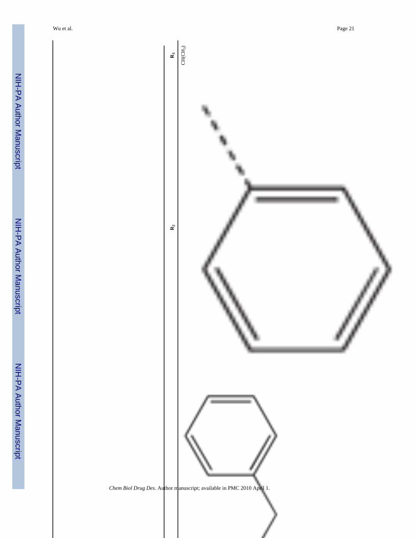
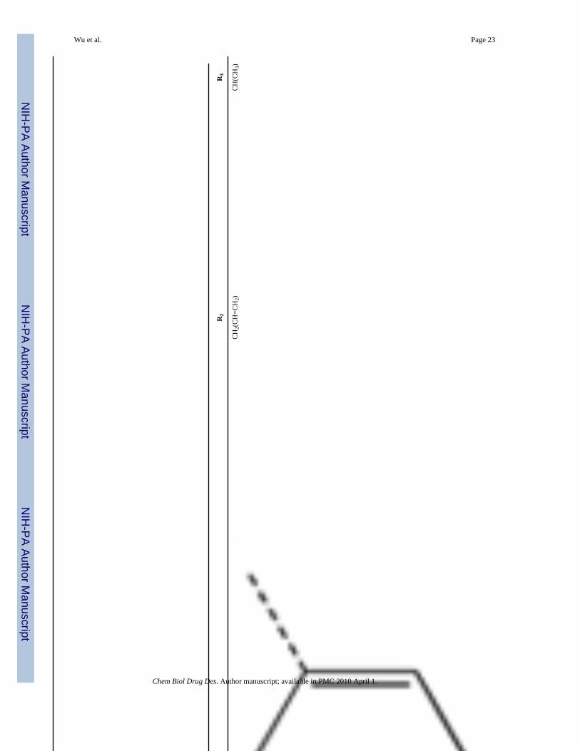
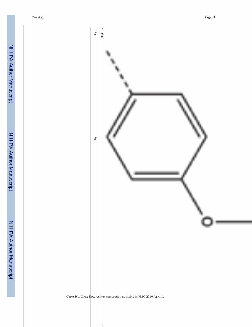
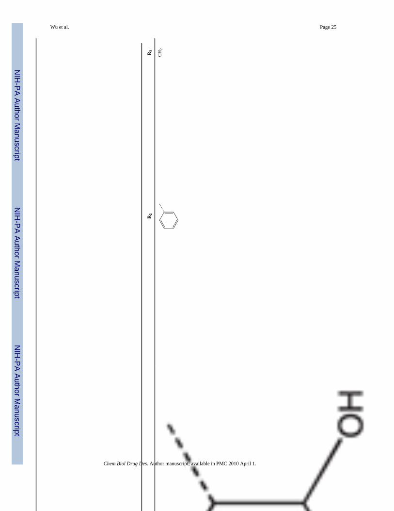
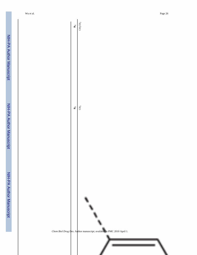
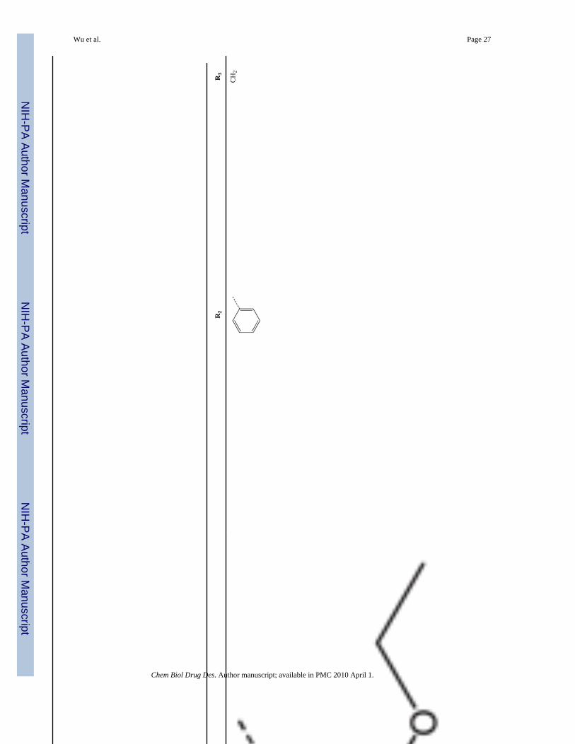
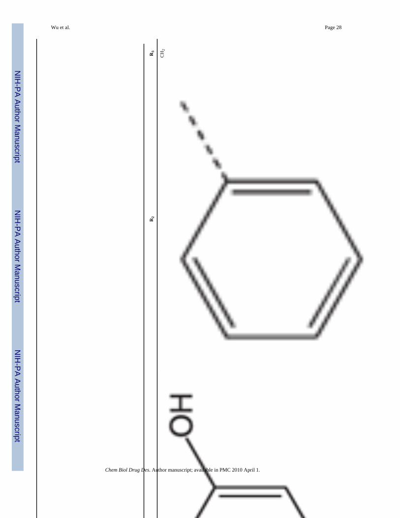
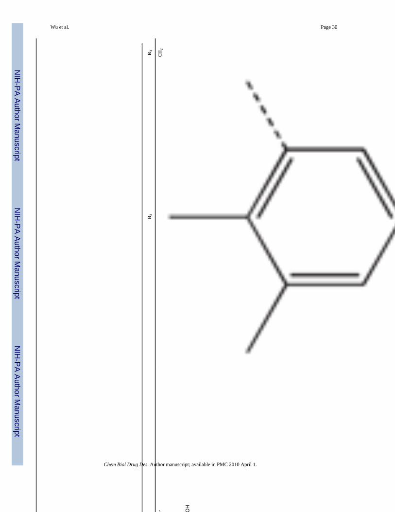
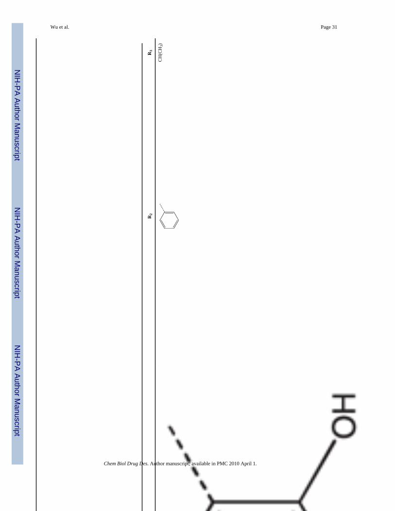
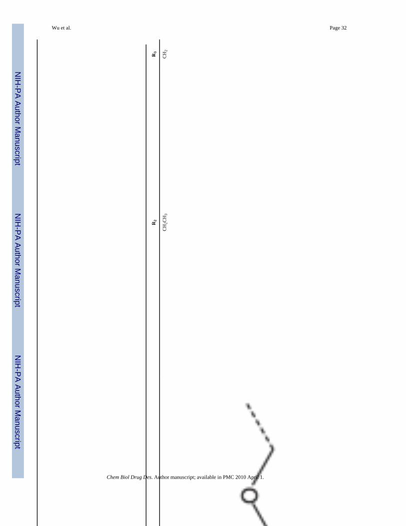
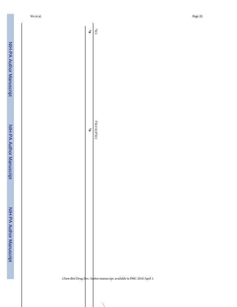
To identify low molecular weight compounds binding to Pin1, a library of ~420 000 compounds(Chembridge Corp) was screened in silico against Pin1 using GOLD (35,36). From the 200top ranking hits according to goldscore, we found a series that has a core structure of 1,2,4-triazole with an extended acidic arm presenting a putative phosphoserine mimic (Table 1).Although it is well known that Pin1 recognizes p-Ser/Thr-Pro motif, early studies also indicatedthat using a glutamate rather than p-Ser/Thr group before the critical proline is well toleratedin Pin1 enzyme activity assays (37,38). Furthermore, latter studies on the interactions betweenPin1 and amyloid peptides (38,39) indicated that the side chain of a glutamate residue is alsorecognized by Pin1ww. These results suggest the possibility of using an acidic group in thedesign of p-Ser/Thr-Pro mimics. Hence, nearly 110 commercially available analogs of theinitially selected virtual hits were found with similarity above 70%, 39 of which were thenselected (library BI-81) for experimental verification.
Wu et al. Page 4
Chem Biol Drug Des. Author manuscript; available in PMC 2010 April 1.
NIH
-PA Author Manuscript
NIH
-PA Author Manuscript
NIH
-PA Author Manuscript
The ability of BI-81 compounds to interact with Pin1 was determined via NMR chemical shiftperturbation experiments. The chemical shift perturbation experiment is a widely usedapproach to detect protein–ligand interactions that is detected by observing cross-peak shiftsin 2D [15N, 1H]-HSQC spectra of the target recorded after the addition of test ligands (33,40–42). Therefore, the 2D [ 15N, 1H]-HSQC spectra of 100 μM Pin1 mixed with each of BI-81compounds at various concentrations (500 μM to 4 mM) were recorded. When the HSQCspectra of Pin1 in the presence of BI-81 compounds were superimposed on the spectrum ofPin1 alone (Figure 1A), significant shifts of selected cross-peak were observed indicative ofspecific binding. To further delineate the site of binding for the hits, the induced chemical shiftperturbation values of four effective BI-81 compounds were mapped on the primary sequenceof Pin1 (Figure 1B). Consistent to our virtual screening strategy, most of the significant cross-peak movements caused by the hits (Δppm > 0.1) correspond to residues located the WWdomain. The two most significantly affected cross peaks are from the side chain amide of Trp34(Trp34HE) and the backbone amide of Arg17. Considering their highly structural similarity,it is not surprising that BI-81 compounds exhibit similar binding behaviors and inducequalitatively similar perturbation in the HSQC spectrum of Pin1 binding. Hence, differencesin the observed perturbation values are indicative of the relative binding affinities of individualcompounds against Pin1. These measurements can therefore be used to monitor improvementin binding affinity of subsequent compounds.
CoMFA analysis of BI-81 compoundsTo elucidate and rationalize the structural features of BI-81 compounds required for bindingto Pin1, we built a 3D QSAR model and performed CoMFA analyses. The initial compoundactivity data were obtained by testing three compounds, 7, 11 and 29, which induced a relativelylarge chemical shift perturbation on Trp34HE (Figure 2A). Compound dissociation constants(Kd) for the three compounds were determined via a 2D HSQC titration method (Figure 2B,C).The Kd values of the three compounds are 2.5 mM, 1 mM and 300 μM respectively. Therefore,the three compounds can be ranked as 29 > 11 > 7 according to the binding affinities from highto low. However, using the 2D HSQC titration to determine Kd value is a time-consuming andprotein-consuming task to be performed for all BI-81 compounds. Comparing the chemicalshift perturbation values of 7, 11 and 29 with the measured Kd values indicates that the chemicalshift values at a given protein/ligand ratio can be used to estimate relative Kd values. Hence,the binding affinity data can be estimated by measurements of the compound induced chemicalshift perturbation of Trp34HE at a given ligand to protein ratio.
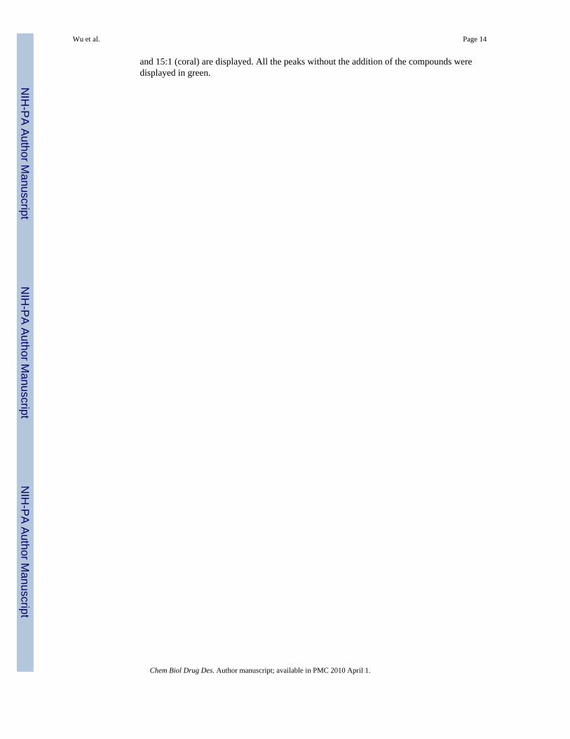
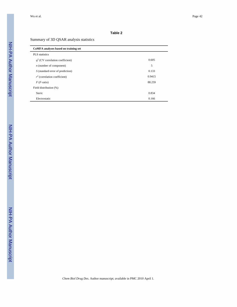
By using these values, we performed a CoMFA analysis to dissect the contribution of eachsubstituent to the observed binding affinities. Of the total 39 BI-81 compounds, 32 wererandomly selected as a training set to build a CoMFA model and the remaining seven wereselected as a test set for model validation. As the spatial alignment of compounds in 3D QSARstudy is one of the most sensitive and determining factors in obtaining a reliable model, wemanually aligned the energy minimized conformers of BI-81 compounds via fitting the fiveatoms in 1,2,4-trizole ring (Figure 3A). After the alignment, the leave-one-out PLS analysiswas carried out and yielded a cross-validation q2 value of 0.606 with five components. As thecross-validated correlation coefficient (q2) is used as a measure of reliability of prediction, theq2 value above 0.6 suggests that our model should posses a reliable predictive ability.Subsequently, internal non-cross-validated PLS regression was carried out using the previouslyobtained optimum number of components and yielded a regression coefficient (r 2) of 0.946.The r2 value represents the goodness of 3D QSAR model. The statistical parameters associatedwith the CoMFA analysis are reported in Table 2.
To evaluate the predictive ability of this model, we subsequently calculated the chemical shiftperturbation values of the compounds in the test set. The experimental values versus predicted
Wu et al. Page 5
Chem Biol Drug Des. Author manuscript; available in PMC 2010 April 1.
NIH
-PA Author Manuscript
NIH
-PA Author Manuscript
NIH
-PA Author Manuscript
values of BI-81 compounds in the training set and test set are plotted in Figure 3B. As shown,the predicted values from the constructed 3D QSAR model are in very good agreement withthe experimental data, again confirming the good predictive ability of the derived QSAR model.
To further utilize and evaluate this model, the affinities of a total of 64 commercially availableanalogs (application set) were predicted. Based on the built 3D QSAR model, the chemicalshift perturbation values of the application set were predicted and displayed in Figure 3B. Inthe application set, two compounds (47 and 48) were predicted having the highest chemicalshift perturbation values close to the best compound, 29 in the training set, suggesting that theyare expected to have a similar or even higher binding affinities for Pin1 compared withcompound 29. The actual Kd value of compounds 47 and 48 were measured via 2D HSQCtitrations. In agreement with the predictions from the 3D QSAR model, the Kd values ofcompounds 47 and 48 were 326 and 423 μM respectively (Figure 3C), hence close to the valueobtained for compound 29. Therefore, the predictive ability of the derived 3D QSAR modelwas proved to be successful. As a result, this model should be useful in identifying more potentcompounds and in guiding further medical chemistry efforts.
Visualization of CoMFA mapsTo obtain a pictorial view of the information included in the 3D QSAR model, CoMFA contourmaps were generated. Contour maps display how the variation of steric or electrostaticproperties in the structural features of molecules contained in the training set lead to increaseor decrease activity. Figure 4A,B show the contour maps of CoMFA steric and electrostaticfields, respectively. For convenience, all the main positions in Figure 4 were labeled and shownwith the compound 29. As indicated from Figure 4A, the large green contour around thenaphthalene ring indicates that bulky groups are favored for activity in this area. This illustrateswhy the compounds of large substituents in this area, such as compounds 29, 32 and 33 aremore potent than the compounds of small R1 substituents such as compounds 11 and 12. Onthe other hand, as most of R1 substituents contain linker connecting hydrophilic heads to theC5 on the core structure, the linkers play key roles for increasing activity via delivering thebulky group to the proper position (green contour). This explains why compound 11 has astronger binding affinity than compound 4 even though they have same R1 substituent. Theyellow contours labeled 2′ and 3′ in Figure 4A represent sterically disfavored areas, whichmean that the addition of bulky modifications near these regions will decrease activity. Thecloseness of the green and yellow contours suggests tight hit for the R1 substituent. Consistentwith this observation, some compounds with a long chain modification on R1 hydrophobichead exhibit weak binding affinities for Pin1 (data not shown).
Red CoMFA contour maps represent electronegative favorite regions. As shown in Figure 4B,contours 4′ and 5′ correspond to the acidic group on compound 29. In fact, compounds lackingthe acidic head linked to 1,2,4-triazole cannot appreciably bind to Pin1. The red contour 6′corresponds to the nitro group in compound 3 and 7 which illustrates why these compoundshave higher binding affinities than compounds 4 and 5. Blue contours in Figure 4B representelectronegative disfavorable regions. Overlaying BI-81 compounds indicates that the position7′ corresponds to the R1 substituents without linkers. However, compared with the steric fields,electrostatic fields only contribute 17% to the overall activity. Moreover, the compoundswithout linkers display very weak binding affinity. In conclusion, the information included inthe contour maps provides a visual guide that could be useful for future medicinal chemistryoptimizations to increase potency. For instance, compound activity can be improved viaincreasing the hydrophilic property on R1 substituents or modifying R1 with moreelectronegative groups.
Wu et al. Page 6
Chem Biol Drug Des. Author manuscript; available in PMC 2010 April 1.
NIH
-PA Author Manuscript
NIH
-PA Author Manuscript
NIH
-PA Author Manuscript
Binding mode of BI81 compounds to Pin1To further ascertain the binding mode of compound 29 to Pin1, we performed docking studiesusing GOLD. The binding orientation of compound 29 in the docking complex is shown inFigure 4C,D as the same orientation as that in the Figure 4A,B. In the docking complex, theacidic extension of compound 29 functions as a hook to interact with the side chain of Arg14via two electrostatic interactions. The importance of the acidic hook is supported by the factthat the other 1,2,4-triazole analogs without this acidic hook cannot interact with Pin1 (datanot shown). The naphthalene substituent of compound 29 is located in the hydrophobic patchof the WW domain and stabilized via hydrophobic interactions with the side chain of Trp34.The 1,2,4-triazole functioning as a linker to connect two functional important components, theacidic group and the naphthalene ring, is located in the narrow tunnel under Arg17 andstabilized via hydrogen bonds with the backbone and side chain of Arg17. The short R2substituent pointing towards the wall of the hydrophobic pocket seems to be stabilized viahydrophobic interactions with the side chain of Tyr23. These important residues includingArg14, Arg17, Tyr23 and Trp34 are indicated in Figure 4C,D. To compare whether this bindingmode is consistent with the CoMFA and docking studies, the QSAR model was loaded intothe protein surface by fitting the orientations of compound 29. As indicated in Figure 4C,E,compound 29 displays very similar orientations within the contour maps which accuratelydescribe the overall properties of the binding surface of the WW domain. This parallel providesfurther confidence in the predictive ability of the 3D QSAR model.
To further verify the docking results, we performed further NMR studies with compound 29.Figure 5 displays the 1D spectra of compound 29 (200 μM) recorded in presence and absenceof Pin1 (20 μM). Apart from peaks from position 9 and 10 in the R2 substituent that are invisiblebecause of the overlap with the water resonance (data not shown), the NMR spectrum nicelycorresponds with the structure of the compound. After the addition of Pin1, the peaks fromnaphthalene are those that within the observable resonances of compound 29 experience themost significant shifts (Figure 5). The fact that both, the peaks from the naphthalene ofcompound 29 and those of Trp34HE of Pin1 experience the most significant chemical shiftperturbation upon complexation, would suggest the two moieties possibly directly interact witheach other. To verify this hypothesis, we also collected 3D 15N-resolved [H 1, H 1]-NOESYspectra of Pin1 in the absence and presence of compound 29. After the addition of compound29, significant NOE cross-peaks changes nearby the Trp34 have been observed and additionalpeaks corresponding to the naphthalene of compound 29 have been detected (Figure S1). Thesedata and evidences support a direct interaction between the naphthalene of compound 29 andthe indole of Trp34 as anticipated by the docking studies.
A crystal structure of Pin1 in complex with a phoshorylated peptide (CTD phosphopeptide:YpSPTpSPS) binding in the WW domain was recently reported (21). Three important factorsfunction as main forces to maintain the stability of the complex include hydrophobicinteractions between CTD phosphopeptide and the side chains of Trp34 and Tyr23, andhydrogen bonds between two phosphates from the peptide and the side chains of Arg14 andArg17. Comparing the binding orientation of compound 29 in the docking complex and CTDphosphopeptide orientation in the crystal structure as shown in Figure 4F indicates thatcompound 29 mimics CTD phosphopeptide in its interaction with Pin1ww via the same threemain relations. First, the large hydrophobic pocket composed of the side chain of Trp34 in theWW domain corresponds to the green contour which makes it possible to accommodate thebulky naphthalene group in compound 29. Second, the red contours 1′ and 2′ correspond to theside chain of Arg14 forming electrostatic interactions with the acidic hook in compound 29.Third, the core structure of 1,2,4-triazole forms hydrogen bonds with the side chain andbackbone amine of Arg17.
Wu et al. Page 7
Chem Biol Drug Des. Author manuscript; available in PMC 2010 April 1.
NIH
-PA Author Manuscript
NIH
-PA Author Manuscript
NIH
-PA Author Manuscript
Comparing the docking complex structure with the CoMFA contour maps discloses newperspectives for improvement of binding affinities. It has been reported that to bind Pin1, thepeptides of four residues or longer (43) have significantly increased binding affinities. Thereported crystal structure also indicated that except for three main interactions, other possibleinteractions can take place between the tail of the peptide and the upper wall of the pocket.NMR studies of a bivalent peptide and Pin1 interactions (44) indicated that, aside from residues17 and 34, the region from residues 27–31 also experienced apparent chemical shiftperturbation in the HSQC spectra upon the addition of the bivalent peptide. Residues 27–31constitute the wall of the hydrophobic pocket corresponding to the position of the R2substituent in our CoMFA analysis and docking model. Because of lack of sufficient diversityon R2 substituent in the commercial compounds, contributions of R2 to the activity could notbe further exploited in this study. However, these observations should provide valid insightsfor the design of compounds to be synthesized.
Comparison with the known peptide inhibitorInteractions between Pin1 and peptides have been reported in the previous studies and theKd values for the peptides were determined via various approaches. Hence, we selected andsynthesized Pintide and its Kd value was measured under the same approach and conditionsused in this study. We repeated 2D HSQC titration experiments and calculated a Kd value of323 μM for this peptide (data not shown), which is similar to the reported value of ~200 to 400μM (23). Likewise, the locations of chemical shift perturbations induced by Pintide are similarto those induced by compound 29, e.g. strong chemical shift perturbations in the WW domainand weak chemical shift perturbations in the catalytic domain. According to the previous study,the strong chemical shift perturbations derives from the direct interactions between Pintide andthe WW domain while the weak chemical shift perturbations in the catalytic domain resultsfrom the composite effects including the direct weak interaction an indirect conformationalchanges of the catalytic domain. Therefore, compound 29 displays a similar binding affinitythan Pintide.
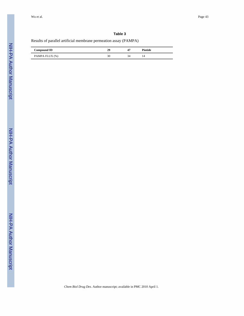
Some potent peptide mimic and small molecular weight inhibitors of Pin1 have also beenrecently reported (27,28,30,44). However, as for peptide mimics, the permeability and stabilityissues limit their applications as chemical probes for target validation. To evaluate the stabilityand permeability of our compounds, we performed molecular stability and membranepermeability assays. As shown in Figure 6, Pintide is rapidly degraded in plasma after 30 minwhile compounds 29 and 47 exhibit fourfolds greater stability after 60 min. Likewise,compounds 29 and 47 exhibit two times higher permeability than Pintide (30% versus 14%,Table 3). The reported chemical probes and relative binding data to Pin1 represent valuablestepping stones for the validation of Pin1 as target for drug discovery and for eventually thedevelopment of possible lead compounds.
ConclusionsA novel scaffold of Pin1 inhibitor presenting a putative phosphoserine mimic was found basedon a virtual screening approach. Subsequently, an expanded library of compounds wasassembled by collecting the commercial available analogs. The binding affinities and bindingmode of these compounds against Pin1 were investigated via NMR chemical shift perturbationexperiments. Compound 29 exhibited an effective dissociation constant of 300 μM, similar tothat of the reported phosphorylated peptide. Moreover, compound 29 possesses a higherstability and cell membrane permeation than the peptide. Furthermore, the CoMFA analysiswas carried out to further highlight the contributions of each substituent in the testedcompounds to their binding affinity. Combined with docking studies and NMR data, theresulting 3D QSAR model provides an invaluable tool to evaluate the binding affinity of
Wu et al. Page 8
Chem Biol Drug Des. Author manuscript; available in PMC 2010 April 1.
NIH
-PA Author Manuscript
NIH
-PA Author Manuscript
NIH
-PA Author Manuscript
additional compounds. With the employment of all of these results, further design, synthesisand activity evaluation will be performed in our laboratory.
Supplementary MaterialRefer to Web version on PubMed Central for supplementary material.
AcknowledgmentsThis work was supported in part by NIH grants and HL082574, AI059572 (to M.P.).
References1. Gothel SF, Marahiel MA. Peptidyl-prolyl cis–trans isomerases, a superfamily of ubiquitous folding
catalysts. Cell Mol Life Sci 1999;55:423–436. [PubMed: 10228556]2. Schmid FX, Mayr LM, Mucke M, Schonbrunner ER. Prolyl isomerases: role in protein folding. Adv
Protein Chem 1993;44:25–66. [PubMed: 8317297]3. Shaw PE. Peptidyl-prolyl isomerases: a new twist to transcription. EMBO Rep 2002;3:521–526.
[PubMed: 12052773]4. Handschumacher RE, Harding MW, Rice J, Drugge RJ, Speicher DW. Cyclophilin: a specific cytosolic
binding protein for cyclosporin A. Science 1984;226:544–547. [PubMed: 6238408]5. Harding MW, Galat A, Uehling DE, Schreiber SL. A receptor for the immunosuppressant FK506 is a
cis–trans peptidyl-prolyl isomerase. Nature 1989;341:758–760. [PubMed: 2477715]6. Rahfeld JU, Schierhorn A, Mann K, Fischer G. A novel peptidyl-prolyl cis/trans isomerase from
Escherichia coli. FEBS Lett 1994;343:65–69. [PubMed: 8163020]7. Lu KP, Hanes SD, Hunter T. A human peptidyl-prolyl isomerase essential for regulation of mitosis.
Nature 1996;380:544–547. [PubMed: 8606777]8. Ranganathan R, Lu KP, Hunter T, Noel JP. Structural and functional analysis of the mitotic rotamase
Pin1 suggests substrate recognition is phosphorylation dependent. Cell 1997;89:875–886. [PubMed:9200606]
9. Zheng H, You H, Zhou XZ, Murray SA, Uchida T, Wulf G, Gu L, Tang X, Lu KP, Xiao ZX. Theprolyl isomerase Pin1 is a regulator of p53 in genotoxic response. Nature 2002;419:849–853. [PubMed:12397361]
10. Zhou XZ, Kops O, Werner A, Lu PJ, Shen M, Stoller G, Kullertz G, Stark M, Fischer G, Lu KP.Pin1-dependent prolyl isomerization regulates dephosphorylation of Cdc25C and tau proteins. MolCell 2000;6:873–883. [PubMed: 11090625]
11. Ryo A, Nakamura M, Wulf G, Liou YC, Lu KP. Pin1 regulates turnover and subcellular localizationof beta-catenin by inhibiting its interaction with APC. Nat Cell Biol 2001;3:793–801. [PubMed:11533658]
12. Lu KP, Liou YC, Vincent I. Proline-directed phosphorylation and isomerization in mitotic regulationand in Alzheimer’s disease. BioEssays 2003;25:174–181. [PubMed: 12539244]
13. Lee MS, Kao SC, Lemere CA, Xia W, Tseng HC, Zhou Y, Neve R, Ahlijanian MK, Tsai LH. APPprocessing is regulated by cytoplasmic phosphorylation. J Cell Biol 2003;163:83–95. [PubMed:14557249]
14. Liou YC, Ryo A, Huang HK, Lu PJ, Bronson R, Fujimori F, Uchida T, Hunter T, Lu KP. Loss ofPin1 function in the mouse causes phenotypes resembling cyclin D1-null phenotypes. Proc Natl AcadSci USA 2002;99:1335–1340. [PubMed: 11805292]
15. Ryo A, Liou YC, Wulf G, Nakamura M, Lee SW, Lu KP. PIN1 is an E2F target gene essential forNeu/Ras-induced transformation of mammary epithelial cells. Mol Cell Biol 2002;22:5281–5295.[PubMed: 12101225]
16. Wulf GM, Ryo A, Wulf GG, Lee SW, Niu T, Petkova V, Lu KP. Pin1 is overexpressed in breastcancer and cooperates with Ras signaling in increasing the transcriptional activity of c-Jun towardscyclin D1. EMBO J 2001;20:3459–3472. [PubMed: 11432833]
Wu et al. Page 9
Chem Biol Drug Des. Author manuscript; available in PMC 2010 April 1.
NIH
-PA Author Manuscript
NIH
-PA Author Manuscript
NIH
-PA Author Manuscript
17. Dominguez-Sola D, Dalla-Favera R. PINning down the c-Myc oncoprotein. Nat Cell Biol2004;6:288–289. [PubMed: 15057241]
18. Ryan KM, Vousden KH. Cancer: pinning a change on p53. Nature 2002;419:795–797. [PubMed:12397340]
19. Zacchi P, Gostissa M, Uchida T, Salvagno C, Avolio F, Volinia S, Ronai Z, Blandino G, SchneiderC, Del Sal G. The prolyl isomerase Pin1 reveals a mechanism to control p53 functions after genotoxicinsults. Nature 2002;419:853–857. [PubMed: 12397362]
20. Wintjens R, Wieruszeski JM, Drobecq H, Rousselot-Pailley P, Buee L, Lippens G, Landrieu I. 1HNMR study on the binding of Pin1 Trp-Trp domain with phosphothreonine peptides. J Biol Chem2001;276:25150–25156. [PubMed: 11313338]
21. Verdecia MA, Bowman ME, Lu KP, Hunter T, Noel JP. Structural basis for phosphoserine-prolinerecognition by group IV WW domains. Nat Struct Biol 2000;7:639–643. [PubMed: 10932246]
22. Bayer E, Goettsch S, Mueller JW, Griewel B, Guiberman E, Mayr LM, Bayer P. Structural analysisof the mitotic regulator hPin1 in solution: insights into domain architecture and substrate binding. JBiol Chem 2003;278:26183–26193. [PubMed: 12721297]
23. Jacobs DM, Saxena K, Vogtherr M, Bernado P, Pons M, Fiebig KM. Peptide binding induces largescale changes in inter-domain mobility in human Pin1. J Biol Chem 2003;278:26174–26182.[PubMed: 12686540]
24. Kim CJ, Cho YG, Park YG, Nam SW, Kim SY, Lee SH, Yoo NJ, Lee JY, Park WS. Pin1overexpression in colorectal cancer and its correlation with aberrant beta-catenin expression. WorldJ Gastroenterol 2005;11:5006–5009. [PubMed: 16124054]
25. Ayala G, Wang D, Wulf G, Frolov A, Li R, Sowadski J, Wheeler TM, Lu KP, Bao L. The prolylisomerase Pin1 is a novel prognostic marker in human prostate cancer. Cancer Res 2003;63:6244–6251. [PubMed: 14559810]
26. Nakashima M, et al. Cyclin D1 overexpression in thyroid tumours from a radio-contaminated areaand its correlation with Pin1 and aberrant beta-catenin expression. J Pathol 2004;202:446–455.[PubMed: 15095272]
27. Wildemann D, Erdmann F, Alvarez BH, Stoller G, Zhou XZ, Fanghanel J, Schutkowski M, Lu KP,Fischer G. Nanomolar inhibitors of the peptidyl prolyl cis/trans isomerase Pin1 from combinatorialpeptide libraries. J Med Chem 2006;49:2147–2150. [PubMed: 16570909]
28. Zhang Y, Daum S, Wildemann D, Zhou XZ, Verdecia MA, Bowman ME, Lucke C, Hunter T, LuKP, Fischer G, Noel JP. Structural basis for high-affinity peptide inhibition of human Pin1. ACSChem Biol 2007;2:320–328. [PubMed: 17518432]
29. Lu KP, Zhou XZ. The prolyl isomerase PIN1: a pivotal new twist in phosphorylation signalling anddisease. Nat Rev Mol Cell Biol 2007;8:904–916. [PubMed: 17878917]
30. Smet C, Duckert JF, Wieruszeski JM, Landrieu I, Buee L, Lippens G, Deprez B. Control of protein-protein interactions: structure-based discovery of low molecular weight inhibitors of the interactionsbetween Pin1 WW domain and phosphopeptides. J Med Chem 2005;48:4815–4823. [PubMed:16033261]
31. Cramer RD III, Patterson DE, Bunce JD. Comparative molecular field analysis (CoMFA). 1. Effectof shape on binding of steriods to carrier proteins. J Am Chem Soc 1988;110:5959–5967.
32. Goddard, TD.; Kneller, DG. SPARKY 3. San Francisco: University of California; 2008.33. Farmer BT II, Constantine KL, Goldfarb V, Friedrichs MS, Wittekind M, Yanchunas J Jr, Robertson
JG, Mueller L. Localizing the NADP+ binding site on the MurB enzyme by NMR. Nat Struct Biol1996;3:995–997. [PubMed: 8946851]
34. Seydel, JK.; Wiese, M. Drug–Membrane Interactions: Analysis, Drug Distribution, Modeling.Weinheim: Wiley-VCH; 2002.
35. Jones G, Willett P, Glen RC. Molecular recognition of receptor sites using a genetic algorithm witha description of desolvation. J Mol Biol 1995;245:43–53. [PubMed: 7823319]
36. Jones G, Willett P, Glen RC, Leach AR, Taylor R. Development and validation of a genetic algorithmfor flexible docking. J Mol Biol 1997;267:727–748. [PubMed: 9126849]
37. Yaffe MB, Schutkowski M, Shen M, Zhou XZ, Stukenberg PT, Rahfeld JU, Xu J, Kuang J, KirschnerMW, Fischer G, Cantley LC, Lu KP. Sequence-specific and phosphorylation-dependent proline
Wu et al. Page 10
Chem Biol Drug Des. Author manuscript; available in PMC 2010 April 1.
NIH
-PA Author Manuscript
NIH
-PA Author Manuscript
NIH
-PA Author Manuscript
isomerization: a potential mitotic regulatory mechanism. Science 1997;278:1957–1960. [PubMed:9395400]
38. Ramelot TA, Nicholson LK. Phosphorylation-induced structural changes in the amyloid precursorprotein cytoplasmic tail detected by NMR. J Mol Biol 2001;307:871–884. [PubMed: 11273707]
39. Pastorino L, Sun A, Lu PJ, Zhou XZ, Balastik M, Finn G, Wulf G, Lim J, Li SH, Li X, Xia W,Nicholson LK, Lu KP. The prolyl isomerase Pin1 regulates amyloid precursor protein processingand amyloid-beta production. Nature 2006;440:528–534. [PubMed: 16554819]
40. Lian LY, Barsukov IL, Derrick JP, Roberts GC. Mapping the interactions between streptococcalprotein G and the Fab fragment of IgG in solution. Nat Struct Biol 1994;1:355–357. [PubMed:7664045]
41. Williamson RA, Carr MD, Frenkiel TA, Feeney J, Freedman RB. Mapping the binding site for matrixmetalloproteinase on the N-terminal domain of the tissue inhibitor of metalloproteinases-2 by NMRchemical shift perturbation. Biochemistry 1997;36:13882–13889. [PubMed: 9374866]
42. Shuker SB, Hajduk PJ, Meadows RP, Fesik SW. Discovering high-affinity ligands for proteins: SARby NMR. Science 1996;274:1531–1534. [PubMed: 8929414]
43. Zhang Y, Fussel S, Reimer U, Schutkowski M, Fischer G. Substrate-based design of reversible Pin1inhibitors. Biochemistry 2002;41:11868–11877. [PubMed: 12269831]
44. Daum S, Lucke C, Wildemann D, Schiene-Fischer C. On the benefit of bivalency in peptide ligand/pin1 interactions. J Mol Biol 2007;374(1):147–161. [PubMed: 17931657]
Wu et al. Page 11
Chem Biol Drug Des. Author manuscript; available in PMC 2010 April 1.
NIH
-PA Author Manuscript
NIH
-PA Author Manuscript
NIH
-PA Author Manuscript
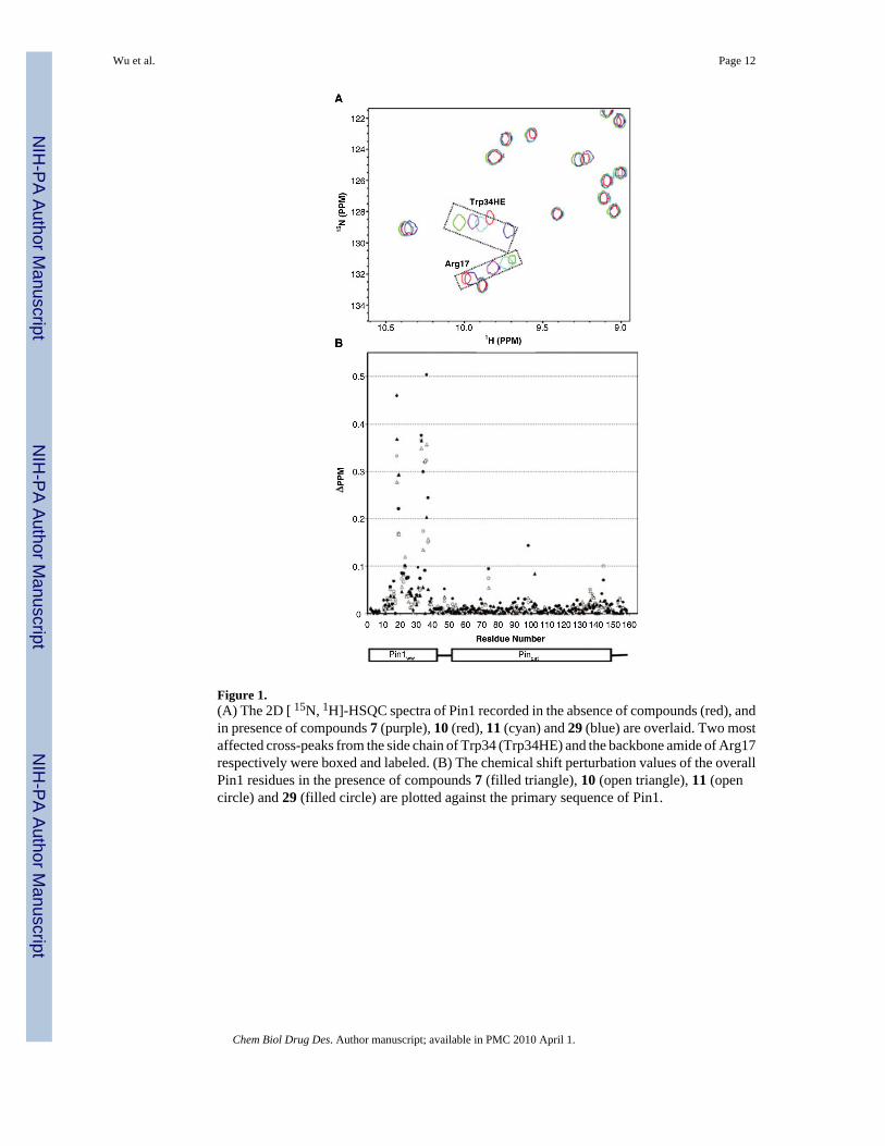
Figure 1.(A) The 2D [ 15N, 1H]-HSQC spectra of Pin1 recorded in the absence of compounds (red), andin presence of compounds 7 (purple), 10 (red), 11 (cyan) and 29 (blue) are overlaid. Two mostaffected cross-peaks from the side chain of Trp34 (Trp34HE) and the backbone amide of Arg17respectively were boxed and labeled. (B) The chemical shift perturbation values of the overallPin1 residues in the presence of compounds 7 (filled triangle), 10 (open triangle), 11 (opencircle) and 29 (filled circle) are plotted against the primary sequence of Pin1.
Wu et al. Page 12
Chem Biol Drug Des. Author manuscript; available in PMC 2010 April 1.
NIH
-PA Author Manuscript
NIH
-PA Author Manuscript
NIH
-PA Author Manuscript
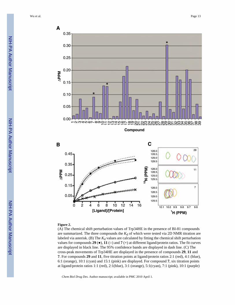
Figure 2.(A) The chemical shift perturbation values of Trp34HE in the presence of BI-81 compoundsare summarized. The three compounds the Kd of which were tested via 2D NMR titration arelabeled via asterisk. (B) The Kd values are calculated by fitting the chemical shift perturbationvalues for compounds 29 (●), 11 (○) and 7 (×) at different ligand/protein ratios. The fit curvesare displayed in black line. The 95% confidence bands are displayed in dash line. (C) Thecross-peak movements of Trp34HE are displayed in the presence of compounds 29, 11 and7. For compounds 29 and 11, five titration points at ligand/protein ratios 2:1 (red), 4:1 (blue),6:1 (orange), 10:1 (cyan) and 15:1 (pink) are displayed. For compound 7, six titration pointsat ligand/protein ratios 1:1 (red), 2:1(blue), 3:1 (orange), 5:1(cyan), 7:1 (pink), 10:1 (purple)
Wu et al. Page 13
Chem Biol Drug Des. Author manuscript; available in PMC 2010 April 1.
NIH
-PA Author Manuscript
NIH
-PA Author Manuscript
NIH
-PA Author Manuscript
and 15:1 (coral) are displayed. All the peaks without the addition of the compounds weredisplayed in green.
Wu et al. Page 14
Chem Biol Drug Des. Author manuscript; available in PMC 2010 April 1.
NIH
-PA Author Manuscript
NIH
-PA Author Manuscript
NIH
-PA Author Manuscript
Figure 3.(A) Alignment of the BI-81 compounds in the training set by fitting the atoms in 1,2,4-triazolering. (B) Plots of the calculated versus experimental chemical shift perturbation values ofTrp34HE. The values of compounds in the training set, test set and application set are displayedin black circle, red circle and yellow dot, respectively. (C) The Kd values for the compounds,compounds 47 and 48 are calculated by fitting the titration points. The structures of compounds47 and 48 are displayed as insert. The dashed lines represent 95% confidence fitting.
Wu et al. Page 15
Chem Biol Drug Des. Author manuscript; available in PMC 2010 April 1.
NIH
-PA Author Manuscript
NIH
-PA Author Manuscript
NIH
-PA Author Manuscript
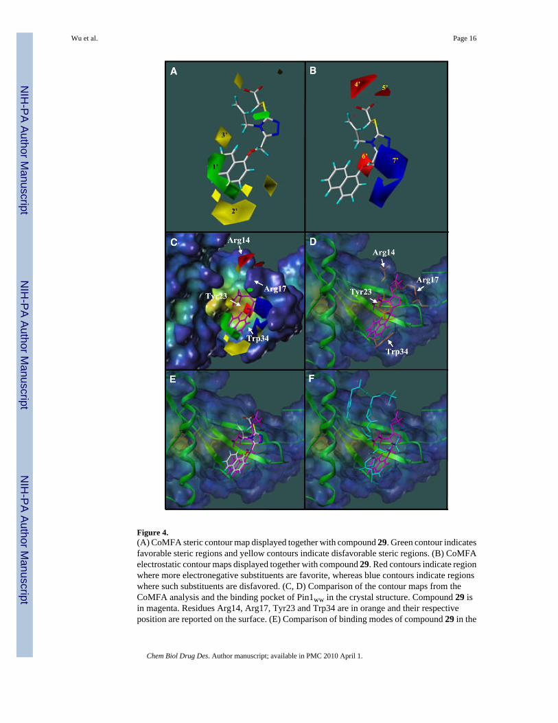
Figure 4.(A) CoMFA steric contour map displayed together with compound 29. Green contour indicatesfavorable steric regions and yellow contours indicate disfavorable steric regions. (B) CoMFAelectrostatic contour maps displayed together with compound 29. Red contours indicate regionwhere more electronegative substituents are favorite, whereas blue contours indicate regionswhere such substituents are disfavored. (C, D) Comparison of the contour maps from theCoMFA analysis and the binding pocket of Pin1ww in the crystal structure. Compound 29 isin magenta. Residues Arg14, Arg17, Tyr23 and Trp34 are in orange and their respectiveposition are reported on the surface. (E) Comparison of binding modes of compound 29 in the
Wu et al. Page 16
Chem Biol Drug Des. Author manuscript; available in PMC 2010 April 1.
NIH
-PA Author Manuscript
NIH
-PA Author Manuscript
NIH
-PA Author Manuscript
docking complex (magenta) and the CoMFA model. (F) Comparison of binding modes ofdocked compound 29 and the CTD phosphopeptide in the crystal structure (cyan).
Wu et al. Page 17
Chem Biol Drug Des. Author manuscript; available in PMC 2010 April 1.
NIH
-PA Author Manuscript
NIH
-PA Author Manuscript
NIH
-PA Author Manuscript
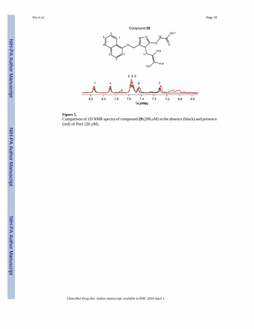
Figure 5.Comparison of 1D NMR spectra of compound 29 (200 μM) in the absence (black) and presence(red) of Pin1 (20 μM).
Wu et al. Page 18
Chem Biol Drug Des. Author manuscript; available in PMC 2010 April 1.
NIH
-PA Author Manuscript
NIH
-PA Author Manuscript
NIH
-PA Author Manuscript
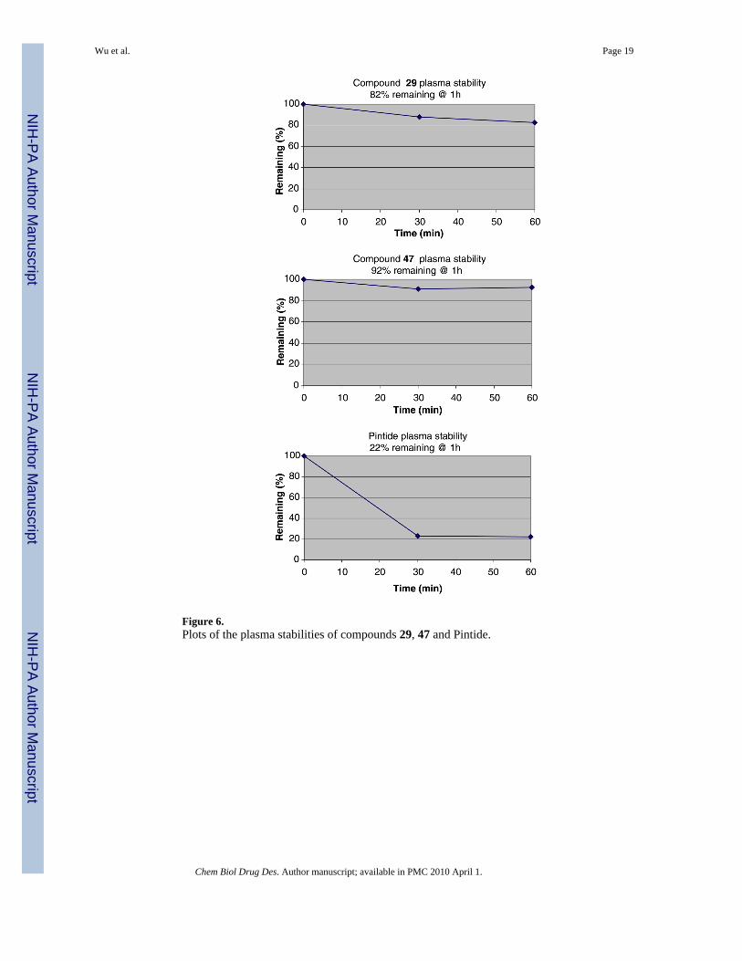
Figure 6.Plots of the plasma stabilities of compounds 29, 47 and Pintide.
Wu et al. Page 19
Chem Biol Drug Des. Author manuscript; available in PMC 2010 April 1.
NIH
-PA Author Manuscript
NIH
-PA Author Manuscript
NIH
-PA Author Manuscript
NIH
-PA Author Manuscript
NIH
-PA Author Manuscript
NIH
-PA Author Manuscript
Wu et al. Page 20
Tabl
e 1
Chem Biol Drug Des. Author manuscript; available in PMC 2010 April 1.
NIH
-PA Author Manuscript
NIH
-PA Author Manuscript
NIH
-PA Author Manuscript
Wu et al. Page 21
Stru
ctur
es o
f BI-
81 c
ompo
unds
IDR
1R
2R
3ID
R1
R2
R3
1aC
H2
21C
H(C
H3)
Chem Biol Drug Des. Author manuscript; available in PMC 2010 April 1.

NIH
-PA Author Manuscript
NIH
-PA Author Manuscript
NIH
-PA Author Manuscript
Wu et al. Page 22
IDR
1R
2R
3ID
R1
R2
R3
2C
H2
22C
H2(
CH
=CH
2)C
H2
Chem Biol Drug Des. Author manuscript; available in PMC 2010 April 1.
NIH
-PA Author Manuscript
NIH
-PA Author Manuscript
NIH
-PA Author Manuscript
Wu et al. Page 23
IDR
1R
2R
3ID
R1
R2
R3
3C
H2(
CH
=CH
2)C
H2
23C
H2(
CH
=CH
2)C
H(C
H3)
Chem Biol Drug Des. Author manuscript; available in PMC 2010 April 1.
NIH
-PA Author Manuscript
NIH
-PA Author Manuscript
NIH
-PA Author Manuscript
Wu et al. Page 24
IDR
1R
2R
3ID
R1
R2
R3
4C
H2(
CC
H3=
CH
2)C
H2
24C
H2C
H2
Chem Biol Drug Des. Author manuscript; available in PMC 2010 April 1.
NIH
-PA Author Manuscript
NIH
-PA Author Manuscript
NIH
-PA Author Manuscript
Wu et al. Page 25
IDR
1R
2R
3ID
R1
R2
R3
5 a
CH
2(C
H=C
H2)
CH
225
CH
2
Chem Biol Drug Des. Author manuscript; available in PMC 2010 April 1.
NIH
-PA Author Manuscript
NIH
-PA Author Manuscript
NIH
-PA Author Manuscript
Wu et al. Page 26
IDR
1R
2R
3ID
R1
R2
R3
6C
H2
26C
H3
CH
2CH
2
Chem Biol Drug Des. Author manuscript; available in PMC 2010 April 1.
NIH
-PA Author Manuscript
NIH
-PA Author Manuscript
NIH
-PA Author Manuscript
Wu et al. Page 27
IDR
1R
2R
3ID
R1
R2
R3
7C
H2(
CC
H3=
CH
2)C
H2
27C
H2
Chem Biol Drug Des. Author manuscript; available in PMC 2010 April 1.
NIH
-PA Author Manuscript
NIH
-PA Author Manuscript
NIH
-PA Author Manuscript
Wu et al. Page 28
IDR
1R
2R
3ID
R1
R2
R3
8C
H2(
CC
H3=
CH
2)C
H2
28C
H2
Chem Biol Drug Des. Author manuscript; available in PMC 2010 April 1.

NIH
-PA Author Manuscript
NIH
-PA Author Manuscript
NIH
-PA Author Manuscript
Wu et al. Page 29
IDR
1R
2R
3ID
R1
R2
R3
9C
H2(
CH
=CH
2)C
H2
29C
H2(
CH
=CH
2)C
H2
Chem Biol Drug Des. Author manuscript; available in PMC 2010 April 1.
NIH
-PA Author Manuscript
NIH
-PA Author Manuscript
NIH
-PA Author Manuscript
Wu et al. Page 30
IDR
1R
2R
3ID
R1
R2
R3
10C
H2(
CH
=CH
2)C
H2
30 a
CH
2
Chem Biol Drug Des. Author manuscript; available in PMC 2010 April 1.
NIH
-PA Author Manuscript
NIH
-PA Author Manuscript
NIH
-PA Author Manuscript
Wu et al. Page 31
IDR
1R
2R
3ID
R1
R2
R3
11 a
CH
2(C
CH
3=C
H2)
CH
231
CH
(CH
3)
Chem Biol Drug Des. Author manuscript; available in PMC 2010 April 1.
NIH
-PA Author Manuscript
NIH
-PA Author Manuscript
NIH
-PA Author Manuscript
Wu et al. Page 32
IDR
1R
2R
3ID
R1
R2
R3
12C
H2(
CH
=CH
2)C
H2
32C
H2C
H3
CH
2
Chem Biol Drug Des. Author manuscript; available in PMC 2010 April 1.
NIH
-PA Author Manuscript
NIH
-PA Author Manuscript
NIH
-PA Author Manuscript
Wu et al. Page 33
IDR
1R
2R
3ID
R1
R2
R3
13C
H2
33 a
CH
2(C
H=C
H2)
CH
2
Chem Biol Drug Des. Author manuscript; available in PMC 2010 April 1.

NIH
-PA Author Manuscript
NIH
-PA Author Manuscript
NIH
-PA Author Manuscript
Wu et al. Page 34
IDR
1R
2R
3ID
R1
R2
R3
14C
H2(
CH
=CH
2)C
H2
34C
H2(
CH
=CH
2)C
H2
Chem Biol Drug Des. Author manuscript; available in PMC 2010 April 1.
NIH
-PA Author Manuscript
NIH
-PA Author Manuscript
NIH
-PA Author Manuscript
Wu et al. Page 35
IDR
1R
2R
3ID
R1
R2
R3
15C
H2C
H2
35C
H2
Chem Biol Drug Des. Author manuscript; available in PMC 2010 April 1.
NIH
-PA Author Manuscript
NIH
-PA Author Manuscript
NIH
-PA Author Manuscript
Wu et al. Page 36
IDR
1R
2R
3ID
R1
R2
R3
16C
H(C
H3)
36C
H2
Chem Biol Drug Des. Author manuscript; available in PMC 2010 April 1.
NIH
-PA Author Manuscript
NIH
-PA Author Manuscript
NIH
-PA Author Manuscript
Wu et al. Page 37
IDR
1R
2R
3ID
R1
R2
R3
17C
H(C
H3)
37C
H2
Chem Biol Drug Des. Author manuscript; available in PMC 2010 April 1.
NIH
-PA Author Manuscript
NIH
-PA Author Manuscript
NIH
-PA Author Manuscript
Wu et al. Page 38
IDR
1R
2R
3ID
R1
R2
R3
18 a
CH
238
CH
2
Chem Biol Drug Des. Author manuscript; available in PMC 2010 April 1.

NIH
-PA Author Manuscript
NIH
-PA Author Manuscript
NIH
-PA Author Manuscript
Wu et al. Page 39
IDR
1R
2R
3ID
R1
R2
R3
19C
H2
39C
H2
Chem Biol Drug Des. Author manuscript; available in PMC 2010 April 1.
NIH
-PA Author Manuscript
NIH
-PA Author Manuscript
NIH
-PA Author Manuscript
Wu et al. Page 40
IDR
1R
2R
3ID
R1
R2
R3
20C
H2(
CH
=CH
2)C
H2
Chem Biol Drug Des. Author manuscript; available in PMC 2010 April 1.
NIH
-PA Author Manuscript
NIH
-PA Author Manuscript
NIH
-PA Author Manuscript
Wu et al. Page 41a C
ompo
unds
in th
e tra
inin
g se
t.
Chem Biol Drug Des. Author manuscript; available in PMC 2010 April 1.
NIH
-PA Author Manuscript
NIH
-PA Author Manuscript
NIH
-PA Author Manuscript
Wu et al. Page 42
Table 2
Summary of 3D QSAR analysis statistics
CoMFA analyses based on training set
PLS statistics
q2 (CV correlation coefficient) 0.605
n (number of component) 5
S (standard error of prediction) 0.133
r2 (correlation coefficient) 0.9415
F (F-ratio) 88.259
Field distribution (%)
Steric 0.834
Electrostatic 0.166
Chem Biol Drug Des. Author manuscript; available in PMC 2010 April 1.
NIH
-PA Author Manuscript
NIH
-PA Author Manuscript
NIH
-PA Author Manuscript
Wu et al. Page 43
Table 3
Results of parallel artificial membrane permeation assay (PAMPA)
Compound ID 29 47 Pintide
PAMPA FLUX (%) 30 34 14
Chem Biol Drug Des. Author manuscript; available in PMC 2010 April 1.
Related Documents