1 Ultrasound Evaluation of the Hip Jon A. Jacobson, M.D. Professor of Radiology Director, Division of Musculoskeletal Radiology University of Michigan Disclosures: • Consultant: Bioclinica • Book Royalties: Elsevier • Advisory Board: GE, Philips Note: all images from the textbook Fundamentals of Musculoskeletal Ultrasound are copyrighted by Elsevier Inc. Pathology: • Joint abnormalities • Bursal pathology • Muscle and tendon injury • Snapping hip syndrome • Miscellaneous pathology Hip: anterior recess • Anterior and posterior layers – Fibrous tissue + minute layer of synovium – Hyperechoic – Each 2 - 4 mm thick Radiology 1999; 210:499 Hip: anterior recess • Anterior +posterior layers – Fibrous tissue + minute layer of synovium – Hyperechoic – Each 2 - 4 mm thick Radiology 1999; 210:499 Anterior Posterior Femur Hip Effusion: • Separation of anterior and posterior layers 1 • Capsule distention at femoral neck > 7 mm or difference of 1 mm from opposite side 2 • Extension & abduction improves visualization 3 • Do not internally rotate hip: capsule thickens 1 Radiology 1999; 210:449 2 Scand J Rheumatology 1989; 18:113 3 Acta Radiologica 1997; 38:867

Welcome message from author
This document is posted to help you gain knowledge. Please leave a comment to let me know what you think about it! Share it to your friends and learn new things together.
Transcript

1
Ultrasound Evaluation of the Hip
Jon A. Jacobson, M.D.
Professor of Radiology
Director, Division of Musculoskeletal Radiology
University of Michigan
Disclosures:
• Consultant: Bioclinica
• Book Royalties: Elsevier
• Advisory Board: GE, Philips
Note: all images from the textbook Fundamentals of Musculoskeletal Ultrasound are copyrighted
by Elsevier Inc.
Pathology:
• Joint abnormalities
• Bursal pathology
• Muscle and tendon injury
• Snapping hip syndrome
• Miscellaneous pathology
Hip: anterior recess
• Anterior and posterior layers– Fibrous tissue + minute layer of synovium
– Hyperechoic
– Each 2 - 4 mm thickRadiology 1999; 210:499
Hip: anterior recess
• Anterior +posterior layers
– Fibrous tissue + minute layer of synovium
– Hyperechoic
– Each 2 - 4 mm thick
Radiology 1999; 210:499
Anterior
PosteriorFemur
Hip Effusion:
• Separation of anterior and posterior layers1
• Capsule distention at femoral neck > 7 mm or difference of 1 mm from opposite side2
• Extension & abduction improves visualization3
• Do not internally rotate hip: capsule thickens
1Radiology 1999; 210:4492Scand J Rheumatology 1989; 18:113
3Acta Radiologica 1997; 38:867
2
Hip Joint: septic effusion
Long Axis
NeckFH *
*
**
Hip Effusion: misconception
• It is incorrect to assume that joint fluid may not be seen anterior due to gravity
• Native hip: joint fluid distributes around femoral neck
• In no cases was fluid only seen posterior
• Exception: after hip surgery
Moss et al. Radiology 1998; 208:43
Hip Effusion:
• Cannot predict infection by ultrasound
• Negative power color Doppler does not exclude infection*
• Guided aspiration
Head
Neck
*
*
* AJR 1998; 206:731
Joint injection
• Anterior recess
• In plane
• Transducer: – Parallel to femoral neck
– Consider curvilinear
• Needle: distal to proximal
• 97% accuracy1
F1Smith J. J Ultrasound Med 2009; 28:329
Joint Injection
• Femoral neck target
• Preferred over aiming for femoral head
• Allows higher injection volumes
• Less extra-articular contrast
From Kantarci F et al. Skeletal Radiol2013; 42:37.
Pigmented Villonodular Synovitis
Head
Erosion

3
Hip Labrum
• Normal:– Hyperechoic, triangular
• Degeneration: hypoechoic• Tear:
– Anechoic cleft– Most common anterior– Possible paralabral cyst– Sensitivity 82%,
specificity 60%*
Femoral Head
Acetab
Labral Tear
*Jin W et al. J Ultrasound Med 2012; 31:439
Chondrocalcinosis
Labral Tear and Paralabral Cyst
Courtesy of D. Fessell, Ann Arbor, MI
Femoroacetabular Impingement:
• Pincer-type: deep acetabulum
• Cam-type
– Broad irregular femoral neck
– Possible cortical irregularity at US
• Associated with anterior labrum tear
• Consider dynamic evaluation
Radiology 2005; 236:588
CAM Impingement
Courtesy of M. van Holsbeeck, Detroit, MI
Note: labral tear (yellow arrow) and osseous bump (white arrow)
Femoroacetabular Impingement
Sagittal-oblique
Hip Arthroplasty:
• Prosthesis identifiable
• May use sonography to guide hip aspiration
• Most useful: non-communicating abscess, bursitis, incision infection

4
Total Hip Arthroplasty:
• Metal components demonstrate posterior reverberation
• Artifact occurs deep to prosthesis away from fluid collection (unlike MRI, CT)
AcetH Neck
Femur
Hip Arthroplasty:
• Pseudocapsule distention:> 3.2 mm: suspect infection*
• Extra-articular fluid collection:– Suspect infection– Not visualized with
arthrography if non-communication
*AJR 1994; 163:381
Neck
Head
A
> 3.2 mm
Hip Arthroplasty: infection
Superior Inferior
Sagittal
Native Femur
Hip Arthroplasty: infection
Coronal Radiograph
Femur
Metal-on-Metal Arthroplasty: pseudotumor
Anterior
Cup
Neck
Troch
Cup
Lateral
Pathology:
• Joint abnormalities
• Bursal pathology
• Muscle and tendon injury
• Snapping hip syndrome
• Miscellaneous pathology

5
Trochanteric Pain Syndrome:
• Most commonly caused by gluteus minimus and medius tendon abnormalities1
• Trochanteric bursitis: uncommon
– 20% of symptomatic patients2
– Not actually inflamed3
– Not associated with pain4
1Eur Rad 2007; 17:17722Long SS et al. AJR 2013; 201:1083
3Clin Rheumatol 2008; 14:824Skeletal Radiol 2008; 37:903
Greater Trochanter
Pfirrmann et al. Radiology 2001; 221:469
FACETS: AF = anterior; LF = lateral; SPF = superoposterior; PF = posterior
Greater Trochanter
AFLF
Gluteus MinimusGluteus Medius
PF
Glut Max
Trochanteric Bursa
Subgluteus Medius Bursa
Subgluteus Minimus
Bursa
TFL
AF: anterior facetLF: lateral facet
PF: posterior facet
ITB
Gmax
GminGmed
ITB
Note: ITB is formed by fascia from gluteus maximus and tensor fascia latae
Trochanteric Bursitis
Transverse Coronal
PF
Trochanteric Bursitis
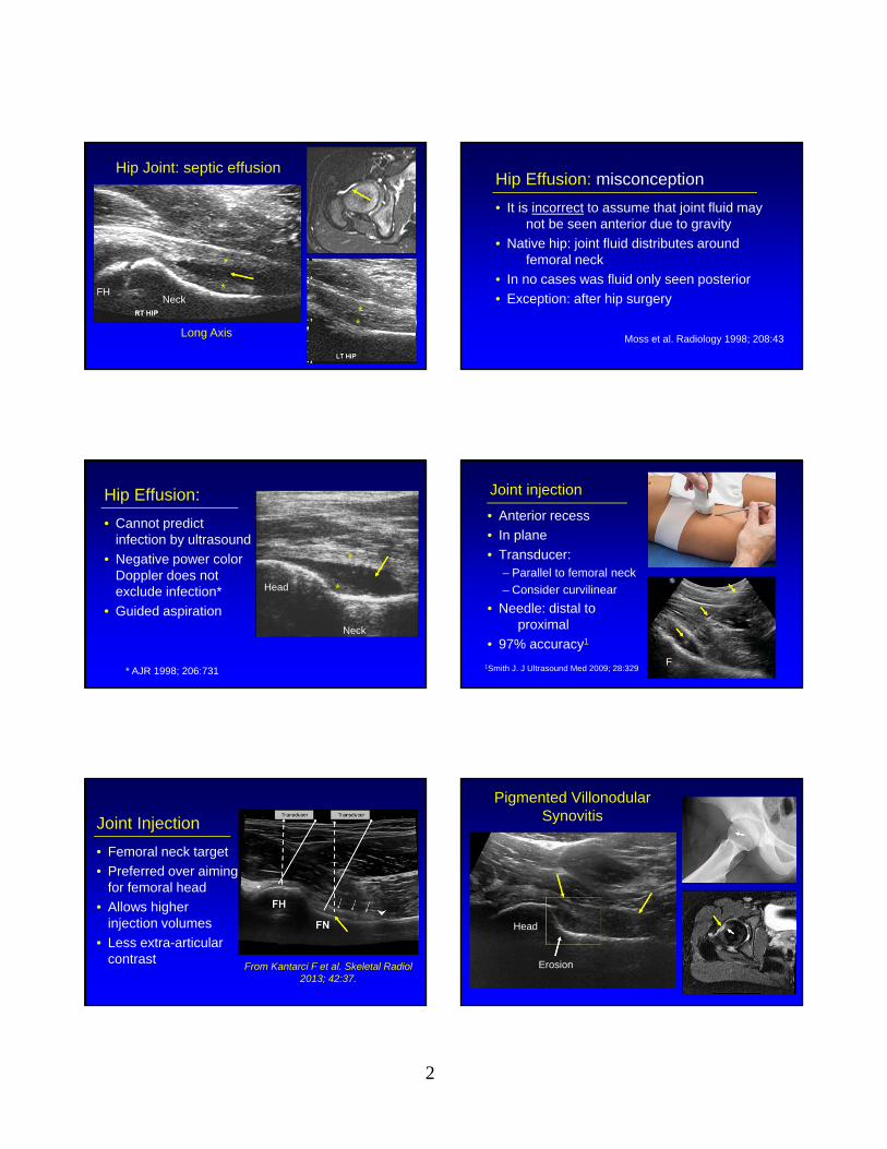
Iliopsoas Bursa:
• Hip joint communication in 10%– Increased with hip joint pathology
• May extend cephalad into abdomen• May be mistaken for abscess:
– Look for hip joint communication
Radiology 1995; 197:853
6
Iliopsoas Bursal Fluid
Femoral Head
IPIP
Short Axis
Long Axis
Iliopsoas Bursa
• Oblique-axial plane:– Superior to femoral head– Lateral to medial– Inject between tendon and
ilium
• Pain relief = successful iliopsoas surgical release
IliumI
Blankenbaker DG. Skeletal Radiol 2006; 35: 565
Iliopsoas Bursa
• Needle placed between iliopsoas tendon and ilium
• Fills iliopsoas bursa• There is no peritendinous
spaced deep to iliopsoas tendon
From: Dauffenbach J et al. J Ultrasound Med 2014; 33:405
Pathology:
• Joint abnormalities
• Bursal pathology
• Muscle and tendon injury
• Snapping hip syndrome
• Miscellaneous pathology
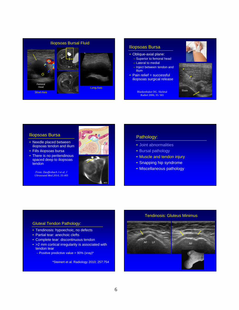
Gluteal Tendon Pathology:
• Tendinosis: hypoechoic, no defects• Partial tear: anechoic clefts• Complete tear: discontinuous tendon• >2 mm cortical irregularity is associated with
tendon tear– Positive predictive value = 90% (xray)*
*Steinert et al. Radiology 2010; 257:754
Tendinosis: Gluteus Minimus
AF LF AF
7
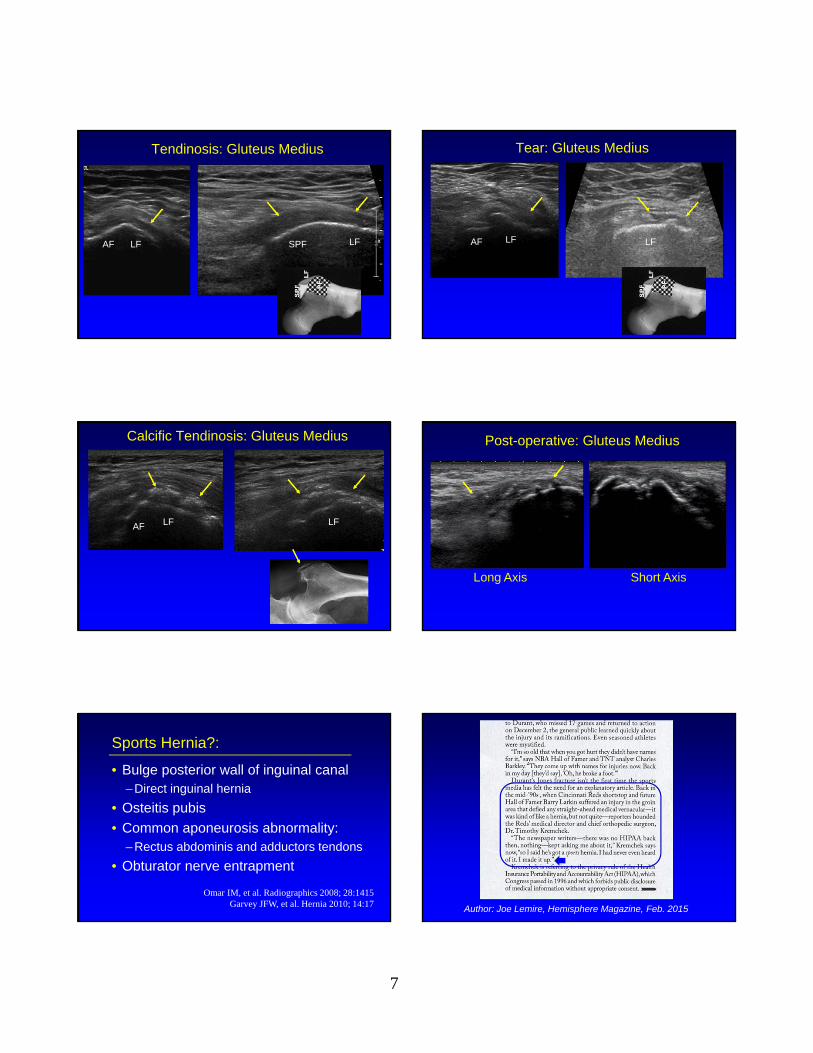
Tendinosis: Gluteus Medius
AF LF LFSPF
Tear: Gluteus Medius
AF LF LF
Calcific Tendinosis: Gluteus Medius
AF LF LF
Post-operative: Gluteus Medius
AF LF LFSPF
Short AxisLong Axis
Sports Hernia?:
• Bulge posterior wall of inguinal canal– Direct inguinal hernia
• Osteitis pubis
• Common aponeurosis abnormality:– Rectus abdominis and adductors tendons
• Obturator nerve entrapment
Omar IM, et al. Radiographics 2008; 28:1415Garvey JFW, et al. Hernia 2010; 14:17 Author: Joe Lemire, Hemisphere Magazine, Feb. 2015

8
Rectus Abdominis + Adductor: “Sports Hernia”
Note: common aponeurosis
From: RadioGraphics 2008; 28:1415
Rectus Abdominis / Adductor Tendinosis:
“Sports Hernia”
Pubis
Pubis
Rectus Abdominus
Adductor Longus
Adductor Longus
Pathology:
• Joint abnormalities
• Bursal pathology
• Muscle and tendon injury
• Snapping hip syndrome
• Miscellaneous pathology
Snapping Hip Syndrome
• Painful snap with hip motion
• Intraarticular
• Extraarticular:– Anterior: iliopsoas tendon
– Lateral: iliotibial tract or gluteus maximus
Iliopsoas Complex
Short Axis
Femoral Head
AIIS
Pubis
Ilium
B
A
A
From: Guillin R. et al. Eur Rad 2009; 19:995
Red: psoas majorOrange: medial iliacus fibersPurple: lateral iliacus fibers
Snapping Hip Syndrome: iliopsoas
• Image long axis to inguinal ligament superior to femoral head
• Extension of flexed abducted and externally rotated hip
• Abrupt movement of iliopsoas as iliacus muscle interposed between tendon and bone moves
Deslandes et al. AJR 2008; 190:576
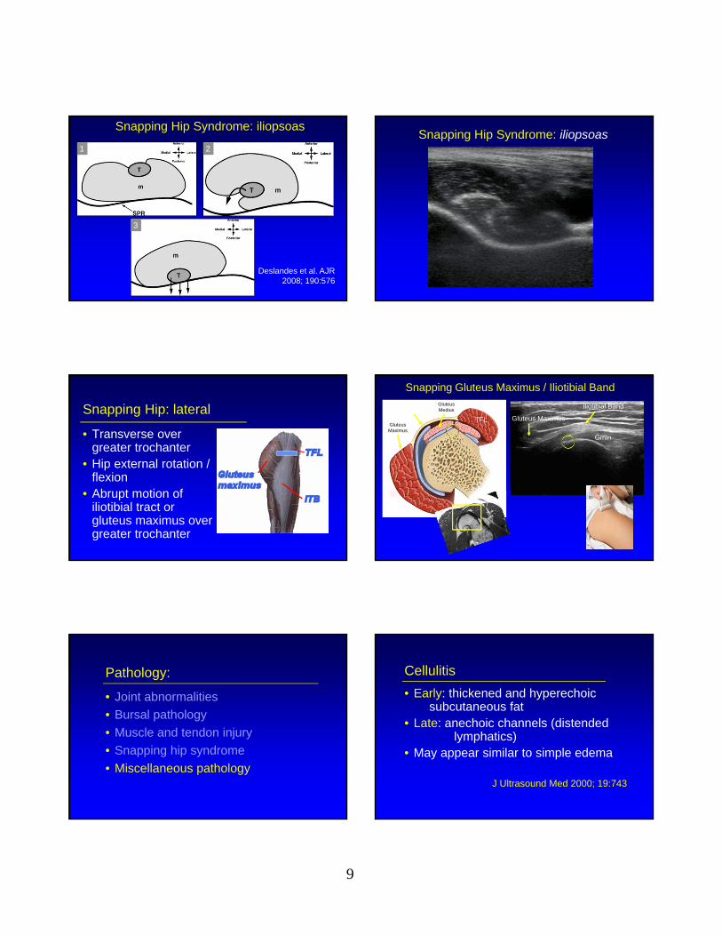
9
Snapping Hip Syndrome: iliopsoas
Deslandes et al. AJR 2008; 190:576
1 2
3
Snapping Hip Syndrome: iliopsoas
Snapping Hip: lateral
• Transverse over greater trochanter
• Hip external rotation / flexion
• Abrupt motion of iliotibial tract or gluteus maximus over greater trochanter
Snapping Gluteus Maximus / Iliotibial Band
Iliotibial Band
Gluteus MaximusGluteus Maximus
TFL
Gluteus Medius
Gmin
Pathology:
• Joint abnormalities
• Bursal pathology
• Muscle and tendon injury
• Snapping hip syndrome
• Miscellaneous pathology
Cellulitis
• Early: thickened and hyperechoic subcutaneous fat
• Late: anechoic channels (distended lymphatics)
• May appear similar to simple edema
J Ultrasound Med 2000; 19:743
10
Cellulitis: acuteCellulitis: chronic
Coronal Coronal T2w
Soft Tissue Abscess:
• Anechoic or hypoechoic– Less likely hyperechoic
• Posterior acoustic enhancement• Swirling of contents with transducer
pressure• Hyperemia
AJR 1996; 166:149
Gluteus Muscle: abscess
Axial T1w post-gadolinium
A
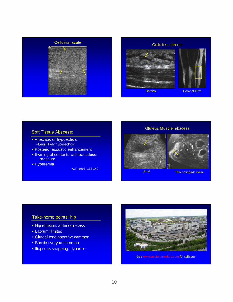
Take-home points: hip
• Hip effusion: anterior recess
• Labrum: limited
• Gluteal tendinopathy: common
• Bursitis: very uncommon
• Iliopsoas snapping: dynamic
See www.jacobsonmskus.com for syllabus
Related Documents