Dirofilariasis in Argentina: Historical review and first report of Dirofilaria immitis in a natural mosquito population Darı ´o Vezzani a,b, * , Diego F. Eiras c , Cristina Wisnivesky a,b a Unidad de Ecologı ´a de Reservorios y Vectores de Para ´sitos, Dto. de Ecologı ´a, Gene ´tica y Evolucio ´n, Facultad de Cs. Exactas y Naturales, Universidad de Buenos Aires, Ciudad Universitaria, Pabello ´n 2, 48 piso, (C1428EHA) Buenos Aires, Argentina b Consejo Nacional de Investigaciones Cientı ´cas y Te ´cnicas (CONICET), Argentina c Laboratorio DIAP (Diagno ´stico en Animales Pequen ˜os), Inca 109 (B1836BBC), Llavallol, Buenos Aires, Argentina Received 13 September 2005; received in revised form 28 October 2005; accepted 28 October 2005 Abstract Argentina is one of the four South American countries where the presence of Dirofilaria immitis is currently confirmed. The objective of this study was to review information on dirofilariasis in the country, and to report our recent findings on mosquito vectors. Since the first report of dogs with unidentified microfilariae in 1926, D. immitis was found in seven provinces and canine prevalence ranged 0–71% at local scale. National prevalence was 8% by the end of the 1980s and current information is available only for Buenos Aires Province. Four pulmonary human infections of D. immitis and one subcutaneous of Dirofilaria sp. were documented. The common coati was the only wild host found, and natural infection in mosquitoes was not previously reported in the country. In our recent mosquito survey in Greater Buenos Aires, we captured and dissected 2380 mosquitoes belonging to 20 species. According to a minimum temperature of 14 8C, the potential transmission period (PTP) for D. immitis in Buenos Aires covers 6 months, and the most favourable period (mean temperature above 20 8C) takes place from the middle of November to the beginning of April. To identify potential vectors of the parasite, we assessed weekly abundances of mosquito species during those PTP estimated previously. We found two specimens of Culex pipiens and one of Aedes aegypti carrying non-infective stages of D. immitis. These two highly anthropophilic mosquitoes may enhance the role of D. immitis as zoonotic agent in temperate Argentina. # 2005 Elsevier B.V. All rights reserved. Keywords: Dirofilaria; Canine heartworm; South America; Argentina; Mosquito vector; Culex pipiens; Aedes aegypti www.elsevier.com/locate/vetpar Veterinary Parasitology 136 (2006) 259–273 * Corresponding author. Tel.: +54 11 4576 3384; fax: +54 11 4576 3354. E-mail addresses: [email protected], vzztato@fibertel.com.ar (D. Vezzani). 0304-4017/$ – see front matter # 2005 Elsevier B.V. All rights reserved. doi:10.1016/j.vetpar.2005.10.026

Welcome message from author
This document is posted to help you gain knowledge. Please leave a comment to let me know what you think about it! Share it to your friends and learn new things together.
Transcript
Dirofilariasis in Argentina: Historical review and first report
of Dirofilaria immitis in a natural mosquito population
Darıo Vezzani a,b,*, Diego F. Eiras c, Cristina Wisnivesky a,b
aUnidad de Ecologıa de Reservorios y Vectores de Parasitos, Dto. de Ecologıa, Genetica y Evolucion,
Facultad de Cs. Exactas y Naturales, Universidad de Buenos Aires, Ciudad Universitaria,
Pabellon 2, 48 piso, (C1428EHA) Buenos Aires, ArgentinabConsejo Nacional de Investigaciones Cientıcas y Tecnicas (CONICET), Argentina
c Laboratorio DIAP (Diagnostico en Animales Pequenos), Inca 109 (B1836BBC), Llavallol,
Buenos Aires, Argentina
Received 13 September 2005; received in revised form 28 October 2005; accepted 28 October 2005
Abstract
Argentina is one of the four South American countries where the presence of Dirofilaria immitis is currently confirmed. The
objective of this study was to review information on dirofilariasis in the country, and to report our recent findings on mosquito
vectors. Since the first report of dogs with unidentified microfilariae in 1926,D. immitiswas found in seven provinces and canine
prevalence ranged 0–71% at local scale. National prevalencewas 8% by the end of the 1980s and current information is available
only for Buenos Aires Province. Four pulmonary human infections of D. immitis and one subcutaneous of Dirofilaria sp. were
documented. The common coati was the only wild host found, and natural infection in mosquitoes was not previously reported in
the country. In our recent mosquito survey in Greater Buenos Aires, we captured and dissected 2380 mosquitoes belonging to 20
species. According to a minimum temperature of 14 8C, the potential transmission period (PTP) for D. immitis in Buenos Aires
covers 6 months, and the most favourable period (mean temperature above 20 8C) takes place from the middle of November to
the beginning of April. To identify potential vectors of the parasite, we assessed weekly abundances of mosquito species during
those PTP estimated previously. We found two specimens of Culex pipiens and one of Aedes aegypti carrying non-infective
stages of D. immitis. These two highly anthropophilic mosquitoes may enhance the role of D. immitis as zoonotic agent in
temperate Argentina.
# 2005 Elsevier B.V. All rights reserved.
Keywords: Dirofilaria; Canine heartworm; South America; Argentina; Mosquito vector; Culex pipiens; Aedes aegypti
www.elsevier.com/locate/vetpar
Veterinary Parasitology 136 (2006) 259–273
* Corresponding author. Tel.: +54 11 4576 3384; fax: +54 11 4576 3354.
E-mail addresses: [email protected], [email protected] (D. Vezzani).
0304-4017/$ – see front matter # 2005 Elsevier B.V. All rights reserved.
doi:10.1016/j.vetpar.2005.10.026
D. Vezzani et al. / Veterinary Parasitology 136 (2006) 259–273260
1. Introduction
Dirofilariasis is a disease caused by filarial worms
of the genus Dirofilaria transmitted by mosquitoes.
This genus consists of 27 valid species, and 15 species
of questionable validity (Canestri Trotti et al., 1997).
Definitive hosts are mammals, mainly primates and
carnivores, and adult worms occur in subcutaneous
tissues or in the heart. The intermediate hosts
and vectors are usually mosquitoes (D. ursi is
transmitted by simuliids), and larval development
generally takes place in the Malpighian tubules (D.
corynodes develops in the fat body) (Anderson, 2000).
Although dirofilariasis was originally considered a
disease of strict veterinary importance, it has
been recognized as an emerging zoonosis by several
authors (Robinson et al., 1977; Simon et al., 1991;
Pampiglione et al., 2001; Pampiglione and Rivasi,
2001). Humans are dead-end hosts, and over one
thousand cases (295 pulmonary and 780 subcuta-
neous/ocular) were reported throughout the world
(Simon et al., 2005).
Two Dirofilaria species, D. repens and D. immitis,
are of special interest to humans because of their
harmful effect on our company pets (dogs and cats)
and potential zoonotic role (Orihel and Eberhard,
1998; Genchi et al., 2005a). The former is only present
in the OldWorld (Europe, Asia and Africa), where it is
the etiologic agent of most subcutaneous/ocular
human cases, whereas D. immitis is cosmopolitan
and responsible for human pulmonary dirofilariasis
(Soulsby, 1987; Canestri Trotti et al., 1997; Simon
et al., 2005). In regard to canine dirofilariasis, D.
repens and D. immitis have been studied exhaustively,
showing great regional and local variations in their
prevalences worldwide; e.g. see Genchi et al. (2005b)
for a recent review of canine dirofilariasis in Europe,
and Labarthe and Guerrero (2005) in South America
and Mexico.
In Argentina, many studies about this disease have
been conducted since the first finding of microfilariae
in the peripheral blood of dogs from northwest
provinces in 1926. Unfortunately, the bulk of
information is scattered through journals and proceed-
ings of local interest, thus remaining unknown to the
international scientific community. This study sum-
marized information available from Argentina on
canine and human dirofilariasis, and on D. immitis
wild hosts and mosquito vectors. We also included
results of our recent studies on mosquitoes aiming at
identifying potential vectors of D. immitis in the
country.
2. A historical perspective of dirofilariasis in
Argentina
2.1. The regional context
From the thirteen South American countries,
current information on the presence or absence of
dogs infested with D. immitis is available only for
Argentina, Brazil, Peru, Colombia, and Chile
(Labarthe and Guerrero, 2005). There are some
records of canine dirofilariasis in Venezuela, Surinam,
Paraguay, and Guiana before 1980, and to our
knowledge, no data on the disease have been reported
in Bolivia, Ecuador, French Guiana, and Uruguay.
Since the first South American report of canine
dirofilariasis in 1875, most surveys on canine heart-
worm have been conducted in Brazil. Labarthe and
Guerrero (2005), who recently summarized the large
amount of information on canine prevalence published
in this country, showed a decreasing trend in
prevalence from 7.9% in 1998 to 2% in 2001, being
local values as high as 52.5% in the southeast region.
In Peru, the parasite was first documented in 1945
(Acuna and Chavez, 2002), and recent studies by
ELISA reported a canine prevalence ranging between
0 and 12.8% (Acuna and Chavez, 2002; Bravo et al.,
2002; Chipana et al., 2002; Adrianzen et al., 2003). In
Colombia, the first records of canine filariasis due to
D. immitis were reported in 1964 and 1967, with 1
case/400 dogs and 8 cases/109 dogs, respectively
(Little et al., 1968), and the overall prevalence
registered by 1990 was 4.8–8.4% (Labarthe and
Guerrero, 2005). In Chile, D. immitis was not found in
two studies conducted during 1976–1979 and 1994,
among a total of 1281 dogs surveyed (Alcaıno et al.,
1984, 1995). Thus, Chile remains as the only South
American country where D. immitis was searched but
not found.
In Venezuela, Surinam, Guiana, and Paraguay there
is no information on canine dirofilariasis for the last two
decades, but some historical records suggest the
presence of D. immitis in these countries. In this sense,
D. Vezzani et al. / Veterinary Parasitology 136 (2006) 259–273 261
Venezuela represents the most notorious example, with
at least eight reports ofD. immitis from itsfirst finding in
1934 through 1970, covering seven of the twenty-three
estates; these studies, summarized by D’Alessandro
(1971), revealed a canine prevalence of 4–29%. In
Surinam, dogs infected withD. immitiswere registered
in 1920, 1938, and 1956 (Panday et al., 1981). Rep and
Heinemann (1976) estimated a prevalence of 22%
among 124 street dogs from seven localities, and a few
years later Panday et al. (1981) recorded a prevalence of
26% (n = 521) in the Capital City. In Guiana, Orihel
(1964) found a prevalence of 14.1% among 2135
inspected dogs, with values along the rural coast higher
than in urban areas. Finally, in Paraguay, Masi Pallares
et al. (1967) reported 8D. immitis-infected dogs among
200 necropsies of street animals.
Feline dirofilariasis in South America was reported
only in Venezuela and Brazil (Labarthe and Guerrero,
2005). Generally, infected cats are amicrofilaremic
and their prevalence parallels that in dogs but at a
lower rate; for example, in Rio de Janeiro feline
prevalence was 20 times lower than canine prevalence
(Labarthe et al., 1997). On this basis, it is highly
probable that veterinarian practitioners would not
detect infected cats in other countries where canine
dirofilariasis occurs.
The first case of human pulmonary dirofilariasis in
the world was reported by De Magalhaes in 1887 from
a child of Rio de Janeiro, Brazil (Shah, 1999). Since
then, 50 new cases were documented in Brazil, mainly
from Rio de Janeiro, Sao Paulo, and Florianopolis
(Milanez de Campos et al., 1997; Cavallazzi et al.,
2002; Rodrigues-Silva et al., 2004). In the remaining
South American countries, human cases of pulmonary
dirofilariasis due to Dirofilaria spp. have rarely been
reported: one case from Venezuela (Salfelder et al.,
1976), one from Colombia (Beaver et al., 1990), and a
few from Argentina compiled in the present study. In
addition, Vieira et al. (2000) detected human
dirofilariasis in different geographical and climatic
areas of Colombia by means of ELISA test. Human
infections with D. immitis are found wherever the
parasite is enzootic (Orihel and Eberhard, 1998), and
therefore the disease is likely to remain undiagnosed
in several South American countries, as is the case
with feline dirofilariasis.
In regard to research on mosquito vectors of
Dirofilaria in South America, Brazil has the lead over
the other countries, just like in other issues concerning
dirofilariasis. At least eight mosquito species were
found infected with D. immitis larvae in this country.
The occurrence of last stage L3 was recorded in natural
populations of Aedes taeniorhynchus, Ochlerotatus
scapularis, and Culex quinquefasciatus (Lourenco de
Oliveira and Deane, 1995; Labarthe et al., 1998; Ahid
and Lourenco de Oliveira, 1999). Experimental
infections using brazilian mosquito strains were
successful in Oc. fluviatilis, Oc. scapularis, Cx.
quinquefasciatus, and Ae. aegypti (Kasai and Wil-
liams, 1986; Macedo et al., 1998; Brito et al., 1999;
Ahid et al., 2000; Serrao et al., 2001). On the other
hand, Cx. declarator, Cx. saltanensis, and Wyeomyia
bourrouli were found naturally infected, with non-
infective L1–L2 larvae only (Labarthe et al., 1998).
Except for Brazil and Argentina, no information on D.
immitis vectors is available in the continent, and data
collected from the latter country are provided in
Section 2.5.
2.2. Canine heartworm
In Argentina, there were only verbal references
about dogs infected with D. immitis until 1926. In this
year, Mazza and Rosenbusch (1926) documented
microfilariae in the peripheral blood of 34.5% dogs
(n = 55) from northwest provinces of the country
(Salta, Jujuy, and Tucuman; Fig. 1a). No adult worms
were found in the four infected dogs that were
necropsied, and based on length measures of the
microfilariae and the relative location of some
reference points (Giemsa stain method), they con-
cluded that the parasite did not match with D. immitis
or D. repens. In the same year, two surveys conducted
in Buenos Aires City and its outskirts revealed dogs
hosting unidentified microfilariae (Mazza et al., 1926;
Bacigalupo, 1941). In this area, blood microfilariae
were again detected in four out of twenty dogs
(Antequeda, 1929). Two years later, Mazza and
Romana (1931) found two infected dogs in the north
of Santa Fe Province, one harbouring D. immitis and
D. repens (microfilariae and adults of both species),
and the otherD. repens (microfilariae and adults). This
reference is considered for all subsequent researchers
as the first citation of D. immitis in the country; the
finding of D. repens will be discussed later. Between
1941 and 1949, 13 microfilaremic dogs from Buenos
D. Vezzani et al. / Veterinary Parasitology 136 (2006) 259–273262
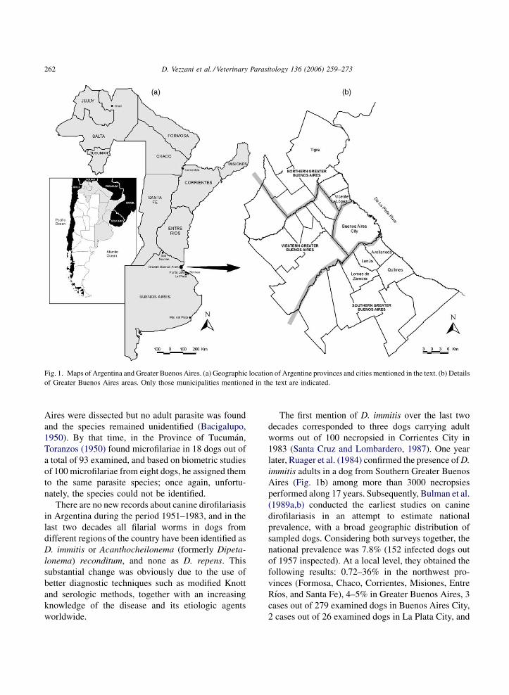
Fig. 1. Maps of Argentina and Greater Buenos Aires. (a) Geographic location of Argentine provinces and cities mentioned in the text. (b) Details
of Greater Buenos Aires areas. Only those municipalities mentioned in the text are indicated.
Aires were dissected but no adult parasite was found
and the species remained unidentified (Bacigalupo,
1950). By that time, in the Province of Tucuman,
Toranzos (1950) found microfilariae in 18 dogs out of
a total of 93 examined, and based on biometric studies
of 100 microfilariae from eight dogs, he assigned them
to the same parasite species; once again, unfortu-
nately, the species could not be identified.
There are no new records about canine dirofilariasis
in Argentina during the period 1951–1983, and in the
last two decades all filarial worms in dogs from
different regions of the country have been identified as
D. immitis or Acanthocheilonema (formerly Dipeta-
lonema) reconditum, and none as D. repens. This
substantial change was obviously due to the use of
better diagnostic techniques such as modified Knott
and serologic methods, together with an increasing
knowledge of the disease and its etiologic agents
worldwide.
The first mention of D. immitis over the last two
decades corresponded to three dogs carrying adult
worms out of 100 necropsied in Corrientes City in
1983 (Santa Cruz and Lombardero, 1987). One year
later, Ruager et al. (1984) confirmed the presence ofD.
immitis adults in a dog from Southern Greater Buenos
Aires (Fig. 1b) among more than 3000 necropsies
performed along 17 years. Subsequently, Bulman et al.
(1989a,b) conducted the earliest studies on canine
dirofilariasis in an attempt to estimate national
prevalence, with a broad geographic distribution of
sampled dogs. Considering both surveys together, the
national prevalence was 7.8% (152 infected dogs out
of 1957 inspected). At a local level, they obtained the
following results: 0.72–36% in the northwest pro-
vinces (Formosa, Chaco, Corrientes, Misiones, Entre
Rıos, and Santa Fe), 4–5% in Greater Buenos Aires, 3
cases out of 279 examined dogs in Buenos Aires City,
2 cases out of 26 examined dogs in La Plata City, and
D. Vezzani et al. / Veterinary Parasitology 136 (2006) 259–273 263
0% in 30 dogs from Mar del Plata City (Fig. 1a).
Almost simultaneously, a study carried out on the
northern border of the country (Formosa Province)
showed a canine prevalence of 41.1% (189/460), and
revealed a gradient of prevalence inversely related to
the degree of urbanisation (urban: 34%, suburban:
44%, rural: 74%) (Mancebo et al., 1992). In addition,
this was the first research in Argentina to evaluate D.
immitis microfilarial periodicity in dogs, showing an
abundance peak between 19 and 24 h.
From 1992 onwards, all studies dealing with canine
prevalencewere conducted exclusively inBuenosAires
Province. In Greater Buenos Aires (including Buenos
Aires City), none of the 320 examined dogs had blood
microfilariae (Lightowler et al., 1992). In Northern
Greater Buenos Aires (Municipality of Tigre), four out
of twenty dogs were infected with D. immitis
microfilariae (Rosa et al., 1994). In Southern Greater
Buenos Aires, local prevalences at municipality scale
were highly variable, with values of 8.7% in Lanus and
Lomas de Zamora (Labbe et al., 1995), 36% in Lanus,
and 41–60% in Avellaneda (Meyer and Milanta, 1997)
(Fig. 1b). In two cities located at the south of Greater
Buenos Aires, prevalences were 12.7% (Berisso,
n = 94) and 3.3% (La Plata, n = 61) (Arias et al.,
1994). Recently, Rosa et al. (2002), who evaluated
prevalence by ELISA in dogs of Buenos Aires City and
its surroundings, detected infection in the northern (17/
96 = 17.7%) and southern areas (23/98 = 23.5%),while
dogs from western areas (n = 417) and Buenos Aires
City (n = 171) were uninfected. Finally, Notarnicola
(2004) reported an overall prevalence of 2.3% among
265 dogs from two municipalities of Southern Greater
Buenos Aires and southern cities of Berisso and La
Plata (Fig. 1b).
In regard toD. repens, it is widely accepted that this
species is present only in the Old World (Canestri
Trotti et al., 1997; Pampiglione et al., 2001). There are
two historical references on this species in Argentina
(Mazza and Romana, 1931; Bacigalupo, 1950).
However, the record of 1950 was invalidated by
Bulman et al. (1989a), who suggested that the
described species was A. reconditum according to
morphological characteristics. Likewise, the record of
1931 may involve specimens of A. reconditum or wild
Dirofilaria species.
A. reconditum, transmitted by fleas, is another
subcutaneous parasite of dogs (Newton and Wright,
1957). In Argentina it was first identified by 1947 in
dogs of the Pampean District, probably within Buenos
Aires Province (Roveda and Ringuelet, 1947). Bulman
et al. (1989a) detected 2 infected dogs of a total of 162
examined in Buenos Aires City, and Rosa et al. (1994)
6 out of 20 in the Municipality of Tigre (Province of
Buenos Aires). In the north of the country, there is a
single report from Formosa Province by Mancebo
et al. (1992), showing a prevalence of 2.4% (n = 460).
In this study, microfilariae showed periodicity in the
peripheral blood with an abundance peak between 12
and 18 h.
According to the information given above, La Plata
is the southernmost city withD. immitis-infected dogs,
and nearby areas would represent the southern limit of
dirofilariasis in Argentina. This disease is probably
absent southwards due to temperature constraints for
the development of the parasite in the mosquito vector.
Despite the large amount of available information, it is
difficult to assess the overall canine prevalence in
Argentina. The value of 7.8% obtained when
considering the two surveys of Bulman et al.
(1989a,b) would be the most reliable estimation of
national prevalence, at least at the end of the 1980s.
The highest prevalences documented in the country
were 71% for a rural environment (Mancebo et al.,
1992) and 60% for an urban one (Meyer and Milanta,
1997), but the number of examined dogs was not
specified. There was great heterogeneity of preva-
lences in Greater Buenos Aires (e.g. 0, 5, 8.7, 17.7,
23.5, 36, 41, 57, 60%). This could be partially
explained by differences in environmental and urban
characteristics among municipalities that may affect
vector abundances and dog densities. However, some
methodological issues such as the low number of
sampled dogs should also be considered.
2.3. Human dirofilariasis
The first two cases of human dirofilariasis in
Argentina were documented just 11 years ago. These
were 54- and 60-year-old men residing in Buenos
Aires with a solitary pulmonary nodule due to D.
immitis (Caballer et al., 1994). These two patients had
travelled to countries in South or North America, and
it could not be ascertained if infection was acquired in
Argentina. The third case was a woman of 61 years
living in San Nicolas City (Fig. 1a), at the north of
D. Vezzani et al. / Veterinary Parasitology 136 (2006) 259–273264
Buenos Aires Province, who suffered from subcuta-
neous nodules in thorax and head due toDirofilaria sp.
(Abuin et al., 1998). The fourth case was a man aged
53 years from Buenos Aires (Barcat et al., 1999), and
the last one was a man aged 35 years from Corrientes
City (Fig. 1a) (Riache et al., 2001), both with a solitary
pulmonary nodule.
In summary, there have been only five reports of
human dirofilariasis in the country, one of them being
recorded out of Buenos Aires Province. These findings
could be due to the actual occurrence of more human
cases, or to a higher level of awareness on pulmonary
diseases and more frequent medical attention near De
La Plata River (central-east) than in northeast areas of
the country. In the Old World, the increased incidence
of human dirofilariasis was partially attributed to a
higher availability of information that raised aware-
ness about dirofilariasis (Simon et al., 2005). Taking
into account that (a) access to information by patients
and physicians is easier in Buenos Aires than in the
rest of the country and (b) canine prevalence in Buenos
Aires is similar to or even lower than that in northern
provinces, it can be assumed that the higher number of
human cases detected in the Federal District and its
surroundings is due to a higher awareness on medical
issues.
Although pulmonary dirofilariasis does not pose a
significant threat to humans, it is important for the
differential diagnosis of other well-defined pulmonary
lesions such as tuberculosis, fungal infections,
carcinoma, and hamartoma (Narine et al., 1999). This
parasitosis is likely to be underdiagnosed worldwide,
and in some areas seroprevalence in humans correlates
to a certain extent with the prevalence in the canine
population (Muro et al., 1999; Simon et al., 2005;
Theis, 2005). From this viewpoint, human dirofilar-
iasis in Argentina would be highly underdiagnosed on
account of the canine prevalences reported.
2.4. Other hosts of D. immitis
Considering the lack of information about canine
dirofilariasis in wide areas of Argentina (e.g. central
and northwest provinces) and the few current data
outside Buenos Aires Province, it is not surprising that
only two reports about wild hosts of D. immitis were
published in the country. The first one involved a coati
(Nasua sp.) from Oran (Salta Province) harbouring
microfilariae and adults of D. nasuae, a supposed new
Dirofilaria species (Mazza, 1926), currently consid-
ered as D. immitis (Canestri Trotti et al., 1997). The
second study, conducted in Formosa byMancebo et al.
(1992), comprised 57 individuals of 9 mammal
species examined for microfilariae (by modified
Knott’s method) and adult worms (by necropsy).
Animals analysed were as follows: 16 Dasypus
novemcinctus (nine-banded armadillo), 15 Nasua
solitaria (common coati), 13 Lutreolina crassicaudata
(thick-tailed opossum), 5 Cavia aparea (guinea pig), 3
Cerdocyon thous (crab-eating fox), 2 Procyon lotor
(raccoon), 1 Didelphis albiventris (white-eared opos-
sum), 1 Didelphis sp., and 1 mouse. Six individuals of
N. solitariawere carrying adult D. immitis, and five of
them microfilariae.
To our knowledge, there are no documented cases
of feline dirofilariasis in the country.
2.5. Vectors of D. immitis
There is little information on Dirofilaria vectors in
Argentina, as is the case for wild and human hosts. In
the decade of the 1940s, Bacigalupo (1941, 1945)
performed a series of experiments to study the
development of microfilariae commonly found among
dogs from Buenos Aires in somemosquito species, but
without knowing the filarial species he was dealing
with. Using infected dogs as microfilariae source, the
author infected successfully three mosquito species,
namely Mansonia titillans, Oc. albifasciatus, and
Psorophora cyanescens. In these species, microfilar-
iae reached the L3 infective stage in 12 days ranging
1–1.2 mm in length. The daily measures reported by
Bacigalupo mismatched with those by Taylor (1960)
for D. immitis infecting Ae. aegypti at 26 8C and 80%
RH, but the former author did not specify the
experimental conditions used. Bacigalupo (1941) also
referred to an attempt to infect Cx. pipiens, but none of
the mosquitoes fed on the microfilaremic dog. In
addition, he did not find any filarial larvae among over
400 mosquitoes captured along the river coast of
Buenos Aires.
In the period 1989–1991, some comprehensive
surveys of mosquitoes for the assessment of their
natural enemies were conducted in Punta Lara
(Buenos Aires Province), a natural reserve located
in an unurbanised area characterised by marginal
D. Vezzani et al. / Veterinary Parasitology 136 (2006) 259–273 265
forest. Among the parasite species recorded in female
mosquitoes, larvae of Onchocercidae were found in
the hemocoele of Oc. albifasciatus, Oc. crinifer, Cx.
dolosus, and Ps. ferox (Garcıa et al., 1994; Campos
et al., 1995; Macia et al., 1995). Onchocercidae larvae
were also observed in the Malpighian tubules of a few
Oc. crinifer specimens (Macia et al., 1995); these
could belong to some wild Dirofilaria species.
Recently, Notarnicola (2004) captured in the
Municipality of Quilmes and in La Plata City female
mosquitoes of Oc. albifasciatus, Cx. pipiens, Ps.
albigens, Ae. aegypti, and Oc. crinifer. All 412
mosquitoes dissected were negative for filarial forms.
The same author performed two experiments to infect
Cx. pipiens using a microfilaremic dog as infection
source. In the first trial (Cx. pipiens laboratory strain)
mosquitoes failed to feed, and in the second one (field
population) none of the engorged mosquitoes became
infected.
Until the present work, natural infection with D.
immitis in mosquitoes has not been reported from
Argentina. Among the eight potential vectors
described from Brazil, Ae. aegypti, Oc. fluviatilis,
Oc. scapularis, Cx. quinquefasciatus, and Cx. salt-
anensis are present in Argentina (Campos and Macia,
1998); on this basis, these species could be considered
a priori as potential vectors of D. immitis in the
country. Experimental research undertaken by Baci-
galupo (1941, 1945) suggests that Ma. titillans, Oc.
albifasciatus, and Ps. cyanescens could also be
regarded as potential vectors.
3. Mosquito survey in Greater Buenos Aires,
Argentina
We evaluated the occurrence of potential mosquito
vectors ofD. immitis in Buenos Aires according to two
different approaches. The first one dealt with the
finding of mosquitoes infected with parasite larvae.
The second approach was to identify mosquito species
being highly abundant during the theoretically
favourable period for the development of D. immitis
within the vector (potential transmission period). A
climate that provides adequate temperature to allow
maturation of ingested microfilariae to infective larvae
within the vector is a pivotal prerequisite for heart-
worm transmission to occur (McCall et al., 2004).
Consequently, climate dictates the seasonal occur-
rence of heartworm transmission in temperate
latitudes, and the temperature threshold below which
development will not proceed is approximately 14 8C(Genchi et al., 2005b).
3.1. Materials and methods
3.1.1. Study area
Greater Buenos Aires is the most crowded urban
centre of Argentina. This megalopolis of 11 million
inhabitants includes the Federal District (Buenos
Aires City; 348350S 588290W) and a surrounding urban
belt composed of 24 municipalities (INDEC, 2003).
The climate is temperate humid with four seasons,
mean annual rainfall is 1076 mm and mean annual
temperature is 17.4 8C (IGM, 1998).
Mosquito surveys were carried out in Northern
Greater Buenos Aires (municipalities of Vicente
Lopez and Tigre), Southern Greater Buenos Aires
(municipalities of Quilmes and Lomas de Zamora),
and Buenos Aires City (Fig. 1b). Although the whole
Greater Buenos Aires is an urban area, the landscape
pattern in northern and southern municipalities ranges
from suburban to highly urbanised small centres,
whereas the Federal District is almost entirely
urbanised.
3.1.2. Methodology
Mosquito captures were performed at least weekly
between December 2003–May 2004 and September
2004–May 2005, and in the time band from 7 a.m. to
midnight. Places visited were private gardens, inside
of houses, public pavements, an university campus,
and a public trail along the river. To increase the
chance of finding infected mosquitoes, all capture sites
were inhabited by dogs in a radius smaller than 100 m.
Specimens were collected with oral aspirators and
sweep nets by disturbing vegetation and nearby
places. The collection included mosquitoes in a
host-seeking behaviour and those resting on natural
or man-made structures. Collections of resting
mosquitoes usually provide more representative
samples of the population as a whole than most other
methods, with the additional advantage of catching
blood-fed and gravid females (Service, 1976).
The mosquitoes were transported alive to the
laboratory and maintained at 5 8C until taxonomic
D. Vezzani et al. / Veterinary Parasitology 136 (2006) 259–273266
Table 1
Number of mosquitoes captured in Greater Buenos Aires (Argen-
tina) during the period January 2004–May 2005
Species Northern
Buenos Aires
Southern
Buenos Aires
Buenos
Aires City
Total
Oc. albifasciatus 125 216 263 604
Cx. pipiens 56 281 261 598
Ae. aegypti 4 253 138 395
Cx. maxi 45 121 46 212
Cx. apicinus 0 114 49 163
Oc. crinifer 60 54 3 117
Oc. scapularis 13 71 24 108
Ma. titillans 1 0 101 102
Cx. eduardoi 0 14 11 25
Cx. dolosus 2 15 0 17
Ps. ferox 9 1 1 11
Cx. lahillei 0 3 6 9
Cx. chidesteri 0 2 4 6
Cx. bidens 0 2 1 3
Cx. acharistus 0 2 0 2
Ps. albigenu 1 1 0 2
Ps. varinervis 0 1 1 2
Is. paranensis 0 0 2 2
Ur. pulcherrima 0 0 1 1
An. albitarsis 0 0 1 1
Total 327 1151 902 2380
identification and dissection within 48 h; dead
mosquitoes were removed. The specimens were
anesthetized with ethanol vapour and identified by
using complementarily the keys to Argentine mos-
quitoes (Darsie, 1985), Buenos Aires mosquitoes
(Rossi et al., 2002), and neotropical mosquitoes
(Forattini, 2002). Considering that the two principal
species of the Cx. pipiens complex, i.e. Cx. pipiens
s.str. and Cx. quinquefasciatus, are sympatric in
Buenos Aires (Mitchell and Darsie, 1985; Rossi, 2000;
Forattini, 2002) and that intermediate forms can be
found where their ranges overlap (Almiron et al.,
1995), these species were not distinguished from each
other and were referred to as Cx. pipiens.
Immediately after mosquito identification, each
specimen was dissected. The head, thorax, Mal-
pighian tubules and alimentary tract, and abdomen
remnant were placed separately in a saline droplet.
Compression was accomplished by pressing over
with a coverslip to detect nematode larvae under
light microscope. Larvae found were identified
considering the following criteria adopted by
Lourenco de Oliveira and Deane (1995) and Labarthe
et al. (1998): (a) general morphological character-
istics of filarioids described by Taylor (1960), (b)
only species of the genus Dirofilaria are known to
develop within Malpighian tubules, (c) D. immitis is
the only species belonging to genus Dirofilaria
reported in the study area, and (d) the study area is an
active D. immitis transmission focus. Larval stages
were classified according to length and width
measures, and to the specific location within the
mosquito (Taylor, 1960).
3.1.3. Data analysis
Considering the thermal threshold of 14 8C, weestimated the potential transmission period (PTP) of
D. immitis in Buenos Aires. We calculated the weekly
mean and minimum temperatures throughout the year
using daily mean and minimum temperatures of 2002,
2003, 2004, and first semester of 2005 (National
Meteorology Service). The period estimated by mean
temperature (PTPmean14) represents the part of the
year when parasite larvae can develop at least during
some hours a day. The period estimated by minimum
temperature (PTPmin14) represents the part of the
year when parasite larvae can develop during the
whole day. In addition, we defined the most favourable
period based on a mean temperature above 20 8C(PTPmean20).
To assess potential mosquito vectors, we described
the weekly presence and relative abundance of
mosquito species captured during those PTP pre-
viously estimated.
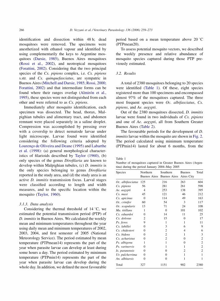
3.2. Results
A total of 2380 mosquitoes belonging to 20 species
were identified (Table 1). Of these, eight species
registered more than 100 specimens and encompassed
almost 97% of the mosquitoes captured. The three
most frequent species were Oc. albifasciatus, Cx.
pipiens, and Ae. aegypti.
Out of the 2380 mosquitoes dissected, D. immitis
larvae were found in two individuals of Cx. pipiens
and one of Ae. aegypti, all from Southern Greater
Buenos Aires (Table 2).
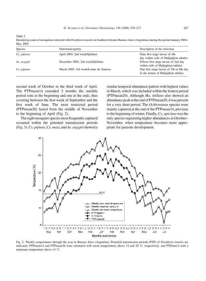
The favourable periods for the development of D.
immitis larvae within the mosquito are shown in Fig. 2.
The period calculated using minimum temperature
(PTPmin14) lasted for about 6 months, from the
D. Vezzani et al. / Veterinary Parasitology 136 (2006) 259–273 267
Table 2
Detailed account of mosquitoes infected withDirofilaria immitis in Southern Greater Buenos Aires (Argentina) during the period January 2004–
May 2005
Species Date/municipality Description of the infection
Cx. pipiens April 2004, 2nd week/Quilmes Nine first stage larvae of 4th
day within cells of Malpighian tubules
Ae. aegypti December 2004, 2nd week/Quilmes Fifteen first stage larvae of 2nd day
within cells of Malpighian tubules
Cx. pipiens March 2005, 3rd week/Lomas de Zamora One first stage larvae of 7th or 8th day
in the lumen of Malpighian tubules
second week of October to the third week of April.
The PTPmean14 extended 2 months the suitable
period (one at the beginning and one at the end), thus
covering between the first week of September and the
first week of June. The most restricted period
(PTPmean20) lasted from the middle of November
to the beginning of April (Fig. 2).
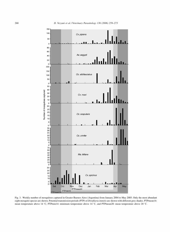
Theeightmosquito speciesmost frequently captured
occurred within the potential transmission periods
(Fig. 3).Cx. pipiens,Cx.maxi, andAe. aegypti showed a
Fig. 2. Weekly temperatures through the year in Buenos Aires (Argentin
indicated. PTPmean14 and PTPmean20 were estimated with mean temp
minimum temperature above 14 8C.
similar temporal abundance pattern with highest values
in March, which was included within the hottest period
(PTPmean20). Although Ma. titillans also showed an
abundancepeakat the endofPTPmean20, itwaspresent
for a very short period. The Ochlerotatus species were
mainly captured at the end of the PTPmean14, previous
to the beginning of winter. Finally,Cx. apicinuswas the
only species registering higher abundances in October–
November, when temperature becomes more appro-
priate for parasite development.
a). Potential transmission periods (PTP) of Dirofilaria immitis are
eratures above 14 and 20 8C, respectively, and PTPmin14 with a
D. Vezzani et al. / Veterinary Parasitology 136 (2006) 259–273268
Fig. 3. Weekly number of mosquitoes captured in Greater Buenos Aires (Argentina) from January 2004 to May 2005. Only the most abundant
eight mosquito species are shown. Potential transmission periods (PTP) ofDirofilaria immitis are shown with different grey shades. PTPmean14:
mean temperature above 14 8C, PTPmin14: minimum temperature above 14 8C, and PTPmean20: mean temperature above 20 8C.
D. Vezzani et al. / Veterinary Parasitology 136 (2006) 259–273 269
3.3. Discussion
To our knowledge, this is the first report of D.
immitis infection in natural mosquito populations from
Argentina. The finding of Cx. pipiens and Ae. aegypti
hosting the parasite confirms that both mosquito
species can acquire the infection under natural
conditions. However, their role as true vectors in
Buenos Aires remains unclear because only non-
infective L1 larvae were found.
According to the temperature analysis, the period
of potential transmission based on the thermal
threshold of 14 8C (PTPmin14) may indicate that
the canine population of Buenos Aires is at risk of
acquiring heartworms during 6 months. The most
favourable period for larvae development (i.e. mean
temperature above 20 8C) takes place from the middle
of November to early April. On the other hand,
temperature seems to be suitable for some hours a day
during 1 month before and 1 month after PTPmin14.
Nevertheless, mosquitoes would not live enough to
allow parasite maturity to the infective stage because
larval development is retarded. Based on these results,
a preventive treatment such as monthly prophylactic
doses of ivermectin should be administered to dogs at
least from October to April. It is also worthwhile
mentioning that in the Northern Hemisphere, the
peaks of heartworm transmission are observed in July
and August and, like for Buenos Aires, transmission
would be limited to 6 months near the 37th parallel
(McCall et al., 2004).
Among the mosquito species found, Ae. aegypti,
Cx. pipiens complex, and Cx. maxi, were highly
abundant during the PTPmean20. The two former
species have been reported as potential vectors of D.
immitis in Brazil (see the end of Section 2.1). Cx. maxi
was found to feed exclusively on chickens and is not
considered of medical importance (Mitchell et al.,
1987; Almiron and Brewer, 1995; Almiron and
Harbach, 1996). Cx. apicinus was the only species
showing a high abundance at the beginning of the
PTPmean20, but it is also known to feed on birds
(Almiron and Brewer, 1995). Although two Ochler-
otatus species, Oc. albifasciatus and Oc. scapularis,
could be considered a priori as potential vectors (see
Section 2.5), in Buenos Aires they were abundant at
the end of PTPmean14, when temperature became
unsuitable for parasite development. Finally, Ma.
titillans could be seen as a potential vector because of
its occurrence within the PTPmean20, but it was
restricted to a short period and was mainly caught in a
trail along the river, instead of in typical domestic
environments as a backyard.
The 20 mosquito species found have already been
registered in the Province of Buenos Aires (Rossi,
2000). Despite the fact that our mosquito survey was
not specifically designed to evaluate species abun-
dance or seasonal patterns of adult populations of
mosquitoes, previous studies performed in Buenos
Aires support our observations. For example, see
Campos and Macia (1996) and Vezzani et al. (2004)
for Ae. aegypti, Macia et al. (1995) for Oc.
albifasciatus, and Garcıa et al. (1995) forMa. titillans.
Some important issues of Cx. pipiens complex and
Ae. aegypti in Buenos Aires can be highlighted: (a)
they were some of the most abundant mosquito
species, (b) their abundances during the PTPmean20
were the highest, and (c) they harboured L1 larvae.
Furthermore, natural populations of Cx. quinquefas-
ciatus have been found carrying infective L3 larvae in
Brazil (Labarthe et al., 1998), and natural transmission
of D. immitis by Ae. aegypti was demonstrated
(Hendrix et al., 1986). For the reasons mentioned
above, these species are the most likely candidates for
transmitting D. immitis in Buenos Aires. Both species
breed in man-made containers and frequently feed on
humans (Rossi and Almiron, 2004), and their high
anthropophily enhances the role of this parasite as
zoonotic agent in temperate Argentina.
4. Conclusions and future perspectives
The leading position of Brazil on dirofilariasis in
South America has been stressed throughout the
present work. To perform a future comprehensive
analysis of this disease at a regional scale, it is
necessary to evaluate the presence of D. immitis in
countries providing only historical records (Vene-
zuela, Surinam, Paraguay, and Guiana), as well as in
those where information is not available (Bolivia,
Uruguay, Ecuador, and French Guyana).
In Argentina, canine heartworm is widespread from
temperate cities in Buenos Aires Province to
subtropical cities close to the northern border of the
country, covering at least seven provinces. The
D. Vezzani et al. / Veterinary Parasitology 136 (2006) 259–273270
national prevalencewas approximately 8%at the end of
the 80s, and local values range between 0 and 71%.
Indeed, no data were recorded from Northwest
Argentina since the finding of dogs harbouring
unidentified microfilariae, more than 50 years ago.
Some crucial questions are pending on canine
dirofilariasis at country scale. First, which is the
epidemiological situation in the centre and northwest of
the country? And second, which is the current national
prevalence? Answering these questions will allow to
determine if canine heartworm shows a temporal trend,
either downward or upward, in Argentina.
Knowledge on human dirofilariasis, as well as on
wild hosts and mosquito vectors of D. immitis is
certainly at an early stage in Argentina. Potential D.
immitis vectors should be evaluated in different areas
because actual vectors can vary throughout the country
due to climate heterogeneity. Different vector control
strategies should be used depending on the breeding
sites of mosquito species (e.g. artificial containers,
temporary pools, swamps). It is also necessary to
expand the knowledge of wild mammals serving as
hosts of Dirofilaria species; the common coati is the
only one found infected in the country so far. Despite
the fact that at least six wild Dirofilaria species have
been reported from South America (Canestri Trotti
et al., 1997), there is no information about Dirofilaria
species other than D. immitis in Argentina.
Human pulmonary dirofilariasis due to D. immitis
is known to exist in Argentina based on the few but
well documented cases since 1994, and therefore
clinicians, radiologists and pathologists should be
aware of this zoonotic infection. In the last few years,
several authors mainly fromUnited States and Europe,
provided strong evidence supporting the highly
underdiagnosed condition of human dirofilariasis,
and assumed that seroprevalence in humans could
correlate with canine prevalence. In this sense, human
seroprevalence surveys conducted in endemic areas of
canine heartworm in Argentina would weigh up how
frequent is the contact between humans and infective
larvae of D. immitis.
Acknowledgements
To the houses’ owners for allowing mosquito
surveys, to Silvia Pietrokovsky for her helpful
comments, and to Stella Maris Velazquez for
providing indispensable literature.
References
Abuin, J., Maulen, S., Navarro, L., Lloveras, S., Orduna, T., Martino,
O., Bellegarde, E., Roodt, A., 1998. Nodulos subcutaneos por
Dirofilaria spp. In: Primer Congreso Latinoamericano de Enfer-
medades Emergentes, Buenos Aires, p. 209.
Acuna, P., Chavez, A., 2002. Determinacion de la prevalencia de
Dirofilaria immitis en los distritos de San Martın de Porres,
Rımac y Cercado de Lima. Rev. Inv. Vet. Peru 13, 108–110.
Adrianzen, G., Chavez, A., Casas, E., Olga, L., 2003. Seropreva-
lencia de la dirofilariosis y ehrlichiosis canina en tres distritos de
Lima. Rev. Inv. Vet. Peru 14, 43–48.
Ahid, S., Lourenco de Oliveira, R., 1999. Mosquitoes potential
vectors of canine heartworm in the Northeast Region from
Brazil. Rev. Saude Pub. 33, 560–565.
Ahid, S., Vasconcelos, P., Lourenco de Oliveira, R., 2000. Vector
competence of Culex quinquefasciatus Say from different
regions of Brazil to Dirofilaria immitis. Mem. Inst. Oswaldo
Cruz 95, 769–775.
Alcaıno, H., Gorman, T., Puelma, M., 1984. Canine filariasis in
Chile. Arch. Med. Vet. 16, 67–73.
Alcaıno, H., Monasterio, X., Gorman, T., 1995. Filariidosis canina
en la V region de Chile. Parasitol. al Dıa 19, 143–145.
Almiron, W., Brewer, M., 1995. Host preference of Culicidae
(Diptera) collected in central Argentina. Rev. Saude Pub. 29,
108–114.
Almiron, W., Harbach, R., 1996. Taxonomy and biology of Culex
(Culex)maxiDyar (Diptera: Culicidae) in South America. Mem.
Inst. Oswaldo Cruz 91, 579–588.
Almiron, W., Humeres, S., Gardenal, C., 1995. Distribution and
hybridization between Culex pipiens and Culex quinquefascia-
tus (Diptera: Culicidae) in Argentina. Mem. Inst. Oswaldo Cruz
90, 469–473.
Anderson, R., 2000. 2nd ed. Nematode Parasites of Vertebrates:
Their Development and Transmission, 2nd ed., CAB Interna-
tional, 672 pp.
Antequeda, E.A., 1929. Investigaciones parasitarias en los perros de
Buenos Aires. Soc. Arg. Patol. Reg. Norte (58 Reunion) 1020–1029.
Arias, M., Klima, L., Stanchi, N., 1994. Frecuencia de microfilar-
emia en caninos en la ciudad de Berisso, La Plata y Ensenada por
tres metodos de laboratorio. Pet’s 10, 229–238.
Bacigalupo, J., 1941. La evolucion en el Taeniorhynchus titillans
Walker, de la microfilaria de nuestros perros. La Semana Med.
48, 718–723.
Bacigalupo, J., 1945. Importancia de algunos de nuestros mosquitos
en la transmision de la filaria del perro en la Argentina. Puerto
Rico J. Pub. Health Trop. Med. 3–13.
Bacigalupo, J., 1950. Hallazgo de Dirofilaria acutiuscula (Morin,
1858) en un perro del Tigre. Rev. Soc. Arg. Biol. 26, 332–334.
Barcat, J., Isidoro, R., Alume, H., 1999. Dirofilariasis pulmonar.
Medicina (Bs. As.) 59, 218–220.
D. Vezzani et al. / Veterinary Parasitology 136 (2006) 259–273 271
Beaver, P., Orihel, T., Leonard, G., 1990. Pulmonary dirofilariasis:
restudy of worms reported gravid. Am. J. Trop. Med. Hyg. 43,
167–169.
Bravo, R., Chavez, A., Casas, E., Suarez, F., 2002. Dirofilariosis
canina en los distritos colindantes con la ribera del Rıo Lurın.
Rev. Inv. Vet. Peru 13, 80–83.
Brito, A., Fontes, G., Rocha, E., Rocha, D., Regis, L., 1999.
Development of Dirofilaria immitis (Leidy) in Aedes aegypti
(L.) and Culex quinquefasciatus (Say) from Maceio, Alagoas,
Brazil. Mem. Inst. Oswaldo Cruz 94, 575–576.
Bulman, G., Gonzales, G., Santamarıa, P., Pampillo, F., Ambrustolo,
R., Fiel, C., 1989a. Prevalencia de Dirofilaria immitis (Leidy,
1856) mediante el test de Knott modificado, en 1043 canes
domesticos de la Mesopotamia Gran Buenos Aires y Capital
Federal (Argentina). Vet. Arg. 6, 144–151.
Bulman, G., Gonzales, G., Mancebo, O., Santamarıa, P., Ambrus-
tolo, R., Guerrero, J., 1989b. Prevalencia de Dirofilaria immitis
(Leidy, 1856) mediante el test de Knott modificado y un
inmunoensayo enzimatico, en 914 canes domesticos de la
Mesopotamia Gran Buenos Aires y Capital Federal (Argentina).
Vet. Arg. 6, 668–675.
Caballer, B., Perez, N., Elsner, B., Esteva, H., Eychremendy, E.,
1994. Dirofilariasis pulmonar: presentacion de dos casos. Rev.
Arg. Cirug. 67, 172–174.
Campos, R.E., Macia, A., 1996. Observaciones biologicas de una
poblacion natural de Aedes aegypti (Diptera: Culicidae) en la
Provincia de Buenos Aires, Argentina. Rev. Soc. Entomol.
Argent. 55, 67–72.
Campos, R., Macia, A., 1998. Culicidae. In: Morrone, J.J., Cos-
caron, S. (Eds.), Biodiversidad de Artropodos Argentinos. Edi-
ciones SUR, La Plata, pp. 291–303.
Campos, R., Macia, A., Garcıa, J., 1995. Seasonality of Psorophora
spp. populations (Diptera: Culicidae) and survey of parasites and
pathogens in Buenos Aires Province, Argentina. Acta Entomol.
Chil. 19, 113–121.
Canestri Trotti, G., Pampiglione, S., Rivasi, F., 1997. The species of
the genus Dirofilaria Railliet & Henry, 1911. Parasitology 39,
369–374.
Cavallazzi, R., Cavallazzi, A., Souza, I., Cardoso, J., 2002. Diro-
filariose pulmonar humana: relato de sete casos. J. Pneumol. 28,
100–102.
Chipana, C., Chavez, A., Casas, E., Suarez, F., 2002. Estudio de la
dirofilariosis canina en la ribera del Rıo Chillon, Lima. Rev. Inv.
Vet. Peru 13, 72–76.
D’Alessandro, A., 1971. Prevalencia de Dirofilaria immitis (Leidy
1856) en perros de caza del Estado Aragua. Rev.Med. Vet. Paras.
24, 109–130.
Darsie, J., 1985. Mosquitoes of Argentina. Part I, keys for identi-
fication of adult females and fourth stage larvae in English and
Spanish (Diptera Culicidae) Mosq. Syst. 17, 153–253.
Forattini, O.P., 2002. Culicidologia medica. Identificacao, Biologia,
Epidemiologia, vol. 2. Editora da Universidade de Sao Paulo,
Sao Paulo, 860 pp.
Garcıa, J., Campos, R., Macia, A., 1995. Observaciones ecologicas
sobreMansonia indubitans yMa. titillans (Diptera: Culicidae) y
sus enemigos naturales en Punta Lara, Argentina. Rev. Soc.
Entomol. Argent. 54, 43–50.
Garcıa, J., Campos, R., Macia, A., 1994. Prospeccion de enemigos
naturales de Culicidae (Diptera) de la selva marginal de Punta
Lara (Prov. de Buenos Aires, Republica Argentina) Rev. Acad.
Colomb. Cienc. Exactas Fıs. Nat. 19, 209–215.
Genchi, C., Simon, F., Kramer, L., 2005a. Dirofilariosis in humans:
is it a real zoonotic concern? In: Proceedings of the 30th World
Congress of the World Small Animal Association. available
online at http://www.wsave2005.com.
Genchi, C., Rinaldi, L., Cascone, C., Mortarino, M., Cringoli, G.,
2005b. Is heartworm disease really spreading in Europe? Vet.
Parasitol. 133, 137–148.
Hendrix, C., Brunner, C., Bellamy, L., 1986. Natural transmission of
Dirofilaria immitis byAedes aegypti. J. Am.Mosq. Contr. Assoc.
2, 48–51.
IGM, 1998. Atlas Geografico de la Republica Argentina. Instituto
Geografico Militar, Buenos Aires, 95 pp.
INDEC, 2003.
?
Que es el Gran Buenos Aires? Instituto Nacional de
Estadısticas y Censos (INDEC), Ministerio de Economıa y
Produccion, Republica Argentina, 12 pp.
Kasai, N., Williams, P., 1986. Experimental infection of Aedes
fluviatilis (Lutz, 1904) by Dirofilaria immitis (Leidy, 1856).
Rev. Bras. Biol. 46, 277–283.
Labarthe, N., Guerrero, J., 2005. Epidemiology of heartworm: what
is happening in South America and Mexico? Vet. Parasitol. 133,
149–156.
Labarthe, N., Ferreira, A., Guerrero, J., Newcomb, K., Paes de
Almeida, E., 1997. Survey of Dirofilaria immitis (Leidy, 1856)
in random source cats in metropolitan Rio de Janeiro, Brazil,
with descriptions of lesions. Vet. Parasitol. 71, 301–306.
Labarthe, N., Serrao, M., Melo, Y., Oliveira, S., Lourenco de
Oliveira, R., 1998. Potential vectors of Dirofilaria immitis
(Leidy, 1856) in Itacoatiara, oceanic region of Niteroi Munici-
pality, State of Rio de Janeiro. Brazil. Mem. Inst. Oswaldo Cruz
93, 425–432.
Labbe, J., Castronuovo, A., Labbe, C., Velasquez, J., 1995. Morbi-
lidad de Dirofilaria immitis en canes de los partidos de Lanus y
Lomas de Zamora. In: 18 Congreso Latinoamericano de Zoo-
nosis. p. 187.
Lightowler, C., Siri, F., Bokenhans, R., Mercado, M., 1992. Inves-
tigacion de la prevalencia de Dirofilaria immitis en caninos de
Capital Federal y conurbano bonaerense. Pet’s 8, 9–12.
Little, M., Vaughn, J., Williams, L., 1968. Canine filariasis in
Colombia. J. Parasitol. 54, 634–635.
Lourenco de Oliveira, R., Deane, L., 1995. Presumed Dirofilaria
immitis infections in wild-caught Aedes taeniorhynchus and
Aedes scapularis in Rio de Janeiro. Brazil. Mem. Inst. Oswaldo
Cruz 90, 387–388.
Macedo, F., Labarthe, N., Lourenco de Oliveira, R., 1998. Suscept-
ibility of Aedes scapularis (Rondani, 1848) to Dirofilaria immi-
tis (Leidy, 1856), an emerging zoonosis. Mem. Inst. Oswaldo
Cruz 93, 435–437.
Macia, A., Garcıa, J., Campos, R., 1995. Bionomıa de Aedes
albifasciatus y Ae. crinifer (Diptera: Culicidae) y sus enemigos
naturales en Punta Lara, Buenos Aires. Neotropica 41, 43–50.
Mancebo, O., Russo, A., Bulman, G., Carabajal, L., Villavicencio de
Mancebo, V., 1992. Dirofilaria immitis: Caracterısticas, preva-
lencia y diagnostico de la dirofilariasis en la poblacion canina en
D. Vezzani et al. / Veterinary Parasitology 136 (2006) 259–273272
areas urbanas, suburbanas y rurales de la Prov. de Formosa
(Argentina) y descripcion de la enfermedad en el coatı comun
(Nasua solitaria) Pet’s 8, 95–117.
Masi Pallares, R., Rojas, E., Usher, C., Vergara, G., 1967. Helmin-
tiosis en perros de Asuncion (Paraguay). Rev. Parag. Microb. 2,
67–70.
Mazza, S., 1926. Sobre un filarıdeo del corazon de coatı (Dirofilaria
nasuae n. sp.) Bol. Inst. Clin. Quir. 2, 273–279.
Mazza, S., Rosenbusch, F., 1926. Sobre una microfilaria sp. de los
perros del norte de laRepublica.Bol. Inst. Clin.Quir. 11, 130–132.
Mazza, S., Rosenbusch, F., Antequeda, E.A., 1926. Segunda nota
preliminar sobre la microfilaria sp. de los perros del Norte. Soc.
Arg. Patol. Reg. Norte (28 Reunion) 29.Mazza, S., Romana, C., 1931. Comprobacion de Dirofilaria immitis
Leidy, 1859 y Dirofilaria repens, Railliet y Henry, 1911 en
perros del Chaco santafesino. Soc. Arg. Patol. Reg. Norte (78Reunion) 1024–1031.
McCall, J., Guerrero, J., Genchi, C., Kramer, L., 2004. Recent
advances in heartworm disease. Vet. Parasitol. 125, 105–130.
Meyer, P., Milanta, G., 1997. Evolucion explosiva de la filariasis
canina en Argentina. Perıodo 1982–1995. Pet’s 13, 224–225.
Milanez de Campos, J., Barbas, C., Filomeno, L., Fernandez, A.,
Minamoto, H., Barbas Filho, J., Jatene, F., 1997. Human pul-
monary dirofilariasis. Analysis of 24 cases from Sao Paulo,
Brazil. Chest 112, 729–733.
Mitchell, C., Darsie, J.R., 1985. Mosquitoes of Argentina. Part II,
geographic distribution and bibliography (Diptera, Culicidae)
Mosq. Syst. 17, 279–360.
Mitchell, C., Monath, T., Sabattini, M., Christensen, H., Darsie, J.,
Jakob, W., Daffner, J., 1987. Host-feeding patterns of Argentine
mosquitoes (Diptera: Culicidae) collected during and after an
epizootic of western equine encephalitis. J. Med. Entomol. 24,
260–267.
Muro, A., Genchi, C., Cordero, M., Simon, F., 1999. Human
dirofilariasis in the European Union. Parasitol. Today 15,
386–389.
Narine, K., Brennan, B., Gilfillan, I., Hodge, A., 1999. Pulmonary
presentation of Dirofilaria immitis (canine heartworm) in man.
Europ. J. Cardio-thoracic. Surg. 16, 475–477.
Newton, W., Wright, W., 1957. The ocurrence of a dog filariid other
than Dirofilaria immitis in the United States. J. Parasitol. 42,
246–256.
Notarnicola, J., 2004. Taxonomıa y biologıa de las filarias de
animales silvestres y de importancia sanitaria en la Republica
Argentina. Doctoral Thesis. Universidad Nacional de La Plata,
La Plata, 187 pp.
Orihel, T., 1964. Canine filariasis in British Guiana. J. Parasitol. 50
(Suppl. 33).
Orihel, T., Eberhard, M., 1998. Zoonotic filariasis. Clin. Microb.
Rev. 11, 366–381.
Pampiglione, S., Rivasi, F., 2001. Dirofilariasis. In: Service, M.W.
(Ed.), The Encyclopedia of Arthropod-transmitted Infections.
CABI Publishing, pp. 143–150.
Pampiglione, S., Rivasi, F., Angeli, G., Boldorini, R., Incensati, R.,
Pastormerlo, M., Pavesi, M., Ramponi, A., 2001. Dirofilariasis
due toDirofilaria repens in Italy, an emergent zoonosis: report of
60 new cases. Histopathology 38, 344–354.
Panday, R., Lieuw, A., Joe, R., Moll, K., Oemrawsingh, I., 1981.
Dirofilaria in dogs of Surinam. Vet. Quart. 3, 25–30.
Rep, B., Heinemann, D., 1976. Changes in hookworm distribution in
Suriman. Trop. Geogr. Med. 28, 104–110.
Riache, R., Godoy, R., Godoy, D., Del Curto, O., 2001. Dirofilariasis
immitis pulmonar: Presentacion de un caso y revision de la
bibliografıa. Comunicaciones Cientıficas y Tecnologicas de la
Universidad Nacional del Nordeste, Corrientes, available online
at http://www1.unne.edu.ar/cyt/2001/3-Medicas/M-017.pdf.
Robinson, N., Chavez, C., Conn, J., 1977. Pulmonary dirofilariasis
in man. A case report and review of the literature. J. Thorac.
Cardiovasc. Surg. 74, 403–408.
Rodrigues-Silva, R., Alcantara Guerra, R., Barbosa de Almeida, F.,
Machado-Silva, J., Darci de Paiva, D., 2004. Human pulmonary
dirofilariasis at Rio de Janeiro, Brazil: a case report. Rev. Soc.
Bras. Med. Trop. 37, 56–59.
Rosa, A., Ribicich, M., Betti, A., Kistermann, J., Cardillo, N., Basso,
N., Hallu, R., 2002. Prevalence of canine dirofilariosis in the
City of Buenos Aires and its outskirts (Argentina). Vet. Parasitol.
109, 261–264.
Rosa, A., Ribicich, M., Miguez, M., Cattaneo, M., 1994. Presencia
de Dirofilaria immitis en caninos del Arroyo Estudiante y
Felicaria (Tigre) Pcia de Bs As. In: VII Congreso Argentino
de Ciencias Veterinarias, Buenos Aires, p. 303.
Rossi, G., Almiron, W., 2004. Clave ilustrada para la identificacion
de larvas de mosquitos de interes sanitario encontradas en
criaderos artificiales en la Argentina. Serie Enfermedades Trans-
misibles, Fundacion Mundo Sano, Buenos Aires, 53 pp.
Rossi, G., Mariluis, J., Schnack, J., Spinelli, G., 2002. Dıpteros
Vectores (Culicidae y Calliphoridae) de la Provincia de Buenos
Aires. Secretarıa de Polıtica Ambiental y Universidad de la
Plata, Buenos Aires, 46 pp.
Rossi, G., 2000. Las especies de mosquitos (Diptera: Culicidae) de
la Provincia de Buenos Aires, Argentina. Rev. Soc. Entomol.
Argent. 59, 141–145.
Roveda, R., Ringuelet, R., 1947. Lista de los parasitos de los
animales domesticos en la Argentina. Gaceta Vet. 10, 67–
78.
Ruager, J., Idiart, J., Led, J., 1984.Dirofilaria immitis en un perro de
los alrededores de la Ciudad de Buenos Aires. Vet. Arg. 1, 256–
260.
Salfelder, K., Davila de Arriaga, A., Rujano, J., 1976. Caso humano
de dirofilariasis pulmonar. Acta Med. Venez. 23, 87–90.
Santa Cruz, A., Lombardero, O., 1987. Resultados parasitologicos
de 100 necropsias de perros callejeros de la ciudad de Corrientes.
Rev. Med. Vet. (Bs. As.) 68, 262–265.
Serrao, M., Labarthe, N., Lourenco de Oliveira, R., 2001. Vectorial
competence of Aedes aegypti (Linnaeus 1762) Rio de Janeiro
strains to Dirofilaria immitis (Leidy 1856). Mem. Inst. Oswaldo
Cruz 96, 593–598.
Service, M.W., 1976. Mosquito Ecology. Field Sampling Methods.
Applied Science Publishers Ltd, London, 583 pp.
Shah, M., 1999. Human pulmonary dirofilariasis: review of the
literature. Southern Med. J. 92, 276–279.
Simon, F., Muro, A., Cordero, M., Martin, J., 1991. A seroepide-
miologic survey of human dirofilariosis in Western Spain. Trop.
Med. Parasitol. 42, 106–108.

D. Vezzani et al. / Veterinary Parasitology 136 (2006) 259–273 273
Simon, F., Lopez-Belmonte, J., Marcos-Atxutegi, C., Morchon, R.,
Martın-Pacho, J., 2005. What is happening outside North Amer-
ica regarding human dirofilariasis? Vet. Parasitol. 133, 181–189.
Soulsby, E.J.L., 1987. Parasitologıa y enfermedades parasitarias en
los animales domesticos. Nueva Interamericana, Mexico D.F.,
823 pp.
Taylor, E., 1960. The development of Dirofilaria immitis in the
mosquito Aedes aegypti. J. Helminthol. 34, 27–38.
Theis, J., 2005. Public health aspects of dirofilariasis in the United
States. Vet. Parasitol. 133, 157–180.
Toranzos, L., 1950. Contribucion al estudio biometrico de micro-
filarias de perros de Tucuman. An. Inst. Med. Reg. 3, 39–44.
Vezzani, D., Velazquez, S., Schweigmann, N., 2004. Seasonal
pattern of abundance of Aedes aegypti (Diptera: Culicidae) in
Buenos Aires City, Argentina. Mem. Inst. Oswaldo Cruz 99,
351–356.
Vieira, C., Montoya, M., Agudelo, S., Velez, I., Simon, F., 2000.
Human antibody response to a 56-kDa purified excretory/secre-
tory product of Dirofilaria immitis. Trop. Med. Int. Health 5,
855–859.
Related Documents