Dioxygen Activation at Non-Heme Diiron Centers: Oxidation of a Proximal Residue in the I100W Variant of Toluene/o-Xylene Monooxygenase Hydroxylase † Leslie J. Murray 1 , Ricardo García-Serres 2 , Michael S. McCormick 1 , Roman Davydov 3 , Sunil Naik 2 , Sun-Hee Kim 3 , Brian M. Hoffman 3,* , Boi Hanh Huynh 2,* , and Stephen J. Lippard 1,* 1 Department of Chemistry, Massachusetts Institute of Technology Cambridge, MA 02139 2 Department of Physics, Emory University, Atlanta, GA 30322 3 Department of Chemistry, Northwestern University, Evanston, IL 60208 Abstract At its carboxylate-bridged diiron active site, the hydroxylase component of toluene/o-xylene monooxygenase activates dioxygen for subsequent arene hydroxylation. In an I100W variant of this enzyme, we characterized the formation and decay of two species formed by addition of dioxygen to the diiron(II) form by rapid-freeze quench (RFQ) EPR, Mössbauer, and ENDOR spectroscopy. The dependence of the formation and decay rates of this mixed-valent transient on pH and the presence of phenol, propylene, or acetylene, was investigated by double-mixing stopped-flow optical spectroscopy. Modification of the α-subunit of the hydroxylase after reaction of the reduced protein with dioxygen-saturated buffer was investigated by tryptic digestion coupled mass spectrometry. From these investigations, we conclude that (i) a diiron(III,IV)–W • transient, kinetically-linked to a preceding diiron(III) intermediate, arises from the one-electron oxidation of W100; (ii) the tryptophan radical is deprotonated; (iii) rapid exchange of either a terminal water or hydroxide ion with water occurs at the ferric ion in the diiron(III,IV) cluster; and (iv) the diiron(III,IV) core and W • decay to the diiron(III) product by a common mechanism. No transient radical was observed by stopped-flow optical spectroscopy for reactions of the reduced hydroxylase variants I100Y, L208F, and F205W with dioxygen. The absence of such species, and the deprotonated state of the tryptophanyl radical in the diiron(III,IV)–W • transient, allow for a conservative estimate of the reduction potential of the diiron(III) intermediate as lying between 1.1 and 1.3 V. We also describe the X-ray crystal structure of the I100W variant of ToMOH. Keywords toluene monooxygenase; carboxylate-bridged diiron proteins; oxygen activation; hydrocarbon oxidation Metal-activated dioxygen species are capable of oxidizing a broad range of substrates (1–4). In synthetic systems, these units are generated in a solvent that is inert to oxidation. The peptide matrix surrounding the active site protects reactive intermediates in protein systems. Enzymes † This study was supported by NIH Grants GM32134 (to SJL), GM47295 (to BHH), and HL13531 (to BMH). LJM was supported in part by a fellowship from the Martin Society (M.I.T.). * Authors to whom correspondence should be addressed: Email: [email protected]. Phone: 617-253-1892. Fax: 617-258-8150. Email: [email protected]. Phone: 404-727-4295. Fax: 404-727-0873. Email: [email protected]. Phone: 847-491-3104. Fax: 847-491-7713. NIH Public Access Author Manuscript Biochemistry. Author manuscript; available in PMC 2008 December 25. Published in final edited form as: Biochemistry. 2007 December 25; 46(51): 14795–14809. NIH-PA Author Manuscript NIH-PA Author Manuscript NIH-PA Author Manuscript

Welcome message from author
This document is posted to help you gain knowledge. Please leave a comment to let me know what you think about it! Share it to your friends and learn new things together.
Transcript
Dioxygen Activation at Non-Heme Diiron Centers: Oxidation of aProximal Residue in the I100W Variant of Toluene/o-XyleneMonooxygenase Hydroxylase†
Leslie J. Murray1, Ricardo García-Serres2, Michael S. McCormick1, Roman Davydov3, SunilNaik2, Sun-Hee Kim3, Brian M. Hoffman3,*, Boi Hanh Huynh2,*, and Stephen J. Lippard1,*
1 Department of Chemistry, Massachusetts Institute of Technology Cambridge, MA 02139
2 Department of Physics, Emory University, Atlanta, GA 30322
3 Department of Chemistry, Northwestern University, Evanston, IL 60208
AbstractAt its carboxylate-bridged diiron active site, the hydroxylase component of toluene/o-xylenemonooxygenase activates dioxygen for subsequent arene hydroxylation. In an I100W variant of thisenzyme, we characterized the formation and decay of two species formed by addition of dioxygento the diiron(II) form by rapid-freeze quench (RFQ) EPR, Mössbauer, and ENDOR spectroscopy.The dependence of the formation and decay rates of this mixed-valent transient on pH and thepresence of phenol, propylene, or acetylene, was investigated by double-mixing stopped-flow opticalspectroscopy. Modification of the α-subunit of the hydroxylase after reaction of the reduced proteinwith dioxygen-saturated buffer was investigated by tryptic digestion coupled mass spectrometry.From these investigations, we conclude that (i) a diiron(III,IV)–W• transient, kinetically-linked to apreceding diiron(III) intermediate, arises from the one-electron oxidation of W100; (ii) the tryptophanradical is deprotonated; (iii) rapid exchange of either a terminal water or hydroxide ion with wateroccurs at the ferric ion in the diiron(III,IV) cluster; and (iv) the diiron(III,IV) core and W• decay tothe diiron(III) product by a common mechanism. No transient radical was observed by stopped-flowoptical spectroscopy for reactions of the reduced hydroxylase variants I100Y, L208F, and F205Wwith dioxygen. The absence of such species, and the deprotonated state of the tryptophanyl radicalin the diiron(III,IV)–W• transient, allow for a conservative estimate of the reduction potential of thediiron(III) intermediate as lying between 1.1 and 1.3 V. We also describe the X-ray crystal structureof the I100W variant of ToMOH.
Keywordstoluene monooxygenase; carboxylate-bridged diiron proteins; oxygen activation; hydrocarbonoxidation
Metal-activated dioxygen species are capable of oxidizing a broad range of substrates (1–4).In synthetic systems, these units are generated in a solvent that is inert to oxidation. The peptidematrix surrounding the active site protects reactive intermediates in protein systems. Enzymes
†This study was supported by NIH Grants GM32134 (to SJL), GM47295 (to BHH), and HL13531 (to BMH). LJM was supported inpart by a fellowship from the Martin Society (M.I.T.).* Authors to whom correspondence should be addressed: Email: [email protected]. Phone: 617-253-1892. Fax: 617-258-8150. Email:[email protected]. Phone: 404-727-4295. Fax: 404-727-0873. Email: [email protected]. Phone: 847-491-3104. Fax:847-491-7713.
NIH Public AccessAuthor ManuscriptBiochemistry. Author manuscript; available in PMC 2008 December 25.
Published in final edited form as:Biochemistry. 2007 December 25; 46(51): 14795–14809.
NIH
-PA Author Manuscript
NIH
-PA Author Manuscript
NIH
-PA Author Manuscript
are able to coordinate reduction of an active metal center and dioxygen activation with substratebinding, thereby assuring that reactive metal-oxygen units are generated only when thesubstrate is available (5,6). The active sites in carboxylate-bridged diiron (CBDI) proteins arehoused within a four-helix bundle and shielded by the protein framework (7–9). As aconsequence, reactive intermediates such as Q, an oxo-bridged diiron(IV) center, in MMOHand X, a mixed-valent diiron(III,IV) cluster, in RNR-R2 can accumulate and be characterizedby a number of spectroscopic methods (10,11).
The primary coordination spheres of the diiron centers in the CBDI enzyme superfamily areremarkably similar, yet the high-valent intermediates Q and X are observed only in MMOHand RNR-R2, respectively (12). Although the capacity to form these iron(IV)-containing unitsis reserved for these two enzymes, proteins in the superfamily family activate dioxygen byanalogous mechanisms. The reduced diiron(II) core reacts rapidly with O2 to generate a peroxo-bridged diiron(III) species as the first observable intermediate. No other oxygenatedintermediates are observed in Ft and Δ9D, and the peroxodiiron(III) unit is detectable in Δ9Donly when the substrate-carrier protein conjugate is bound to the desaturase. This behavior hasbeen ascribed to stearoyl-ACP blocking access to the active site via a large substrate channel(13). To date, no oxygenated intermediates have been reported for reactions of diiron(II) ordiiron(III) forms of rubrerythrin with either dioxygen or hydrogen peroxide, respectively. Fromits crystal structure, the active site of rubrerythrin appears to be more accessible to solvent andbuffer components than those of other enzymes in the family (14). The accumulation of reactiveoxygenated iron(III) and iron(IV) intermediates would therefore appear to be correlated withaccessibility of the diiron center to potential quenching moieties in the medium.
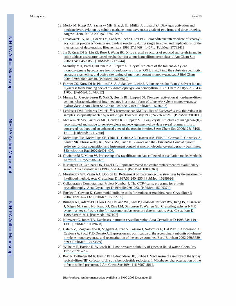
At the beginning of the present study, no transient species with UV-visible absorption bandshad been detected kinetically for the reaction of reduced ToMOH with dioxygen. In the crystalstructure of the native enzyme, a large channel for substrate access and/or product egress,extending from the protein surface to the active site pocket, was identified (15). Site-directedmutagenesis studies of residues within the channel of ToMOH were therefore undertaken toevaluate the possibility that reactive intermediates might accumulate if solvent or buffercomponents were occluded from the active site in this system. Residues I100, F198, F205, andH96 form a hydrophobic portal to the active site at the end of the solvent-accessible channelin ToMOH (Figure 1). Of these residues, I100 is especially noteworthy. The analogous residuein MMOH, L110, has been proposed to function as a gate for substrate entry to, and/or productegress from, the active site during catalysis (7). Residue L98 in hemerythrin might perform asimilar function, controlling solvent access to the diiron core (16). We therefore wonderedwhether mutation of I100 would sufficiently isolate the active site pocket from the channel toshield the dimetallic center from buffer components or solvent, allowing accumulation ofreactive intermediates to observable levels. Residue F205, which is on the opposite side of thechannel from I100, and L208, which is farther from the active site pocket, were also selectedas targets for mutagenesis (Figure 2). We substituted I100 with tyrosine and tryptophan, L208with phenylalanine, and F205 with tryptophan. Modeling structures of these varianthydroxylases using the native crystal structure suggested that they would afford maximalclosure of the channel without disrupting the protein fold of the surrounding matrix.
Preliminary investigations of reaction of ToMOH I100W with dioxygen were reportedpreviously (17). From this initial study, dioxygen activation at the diiron(II) core wasdiscovered to produce an optically tranparent diiron(III) intermediate that could be identifiedby Mössbauer spectroscopy. This intermediate oxidizes the nearby W100 to form a mixed-valent diiron(III,IV) unit coupled to a protein-based tryptophanyl radical. This speciessubsequently decays to the resting, oxidized diiron(III) state. The process by which the diiron(III) intermediate oxidizes the nearby W residue, and the subsequent decay of the mixed-valentcenter and the protein-based radical, have been investigated in considerably detail and form
Murray et al. Page 2
Biochemistry. Author manuscript; available in PMC 2008 December 25.
NIH
-PA Author Manuscript
NIH
-PA Author Manuscript
NIH
-PA Author Manuscript

the basis for this report. The reactivity of other channel-blocking mutations, I100Y, L208F,and F205W was also investigated under pre-steady-state and steady-state conditions. Weinclude these results included because they provide additional insight into the nature of theimportant diiron(III) transient.
Experimental MethodsGeneral Considerations
Plasmids containing the genes for the ToMO components were supplied by the laboratory ofProfessor Alberto Di Donato, Naples, Italy. Recombinant expression and preparation of theToMO component proteins were carried out as described elsewhere (15). ToMOH variantproteins were prepared by the same protocol as used for the native enzyme. The iron content,measured by the ferrozine assay, ranged from 4.2 to 4.6 iron atoms per ToMOH dimer for allsamples. Isotopically enriched ToMOH I100W protein for Mössbauer and ENDORspectroscopy was obtained by expression in LeMaster’s media containing 57FeCl3 ortryptophan-d5, selectively labeled on the indole ring (Cambridge Isotope Labs, Andover, MA)(18). The solution of 57FeCl3 was prepared by dissolving 57Fe powder (96.7% isotopic purity,Advanced Materials Technologies Ltd., Nes-Ziona, Israel) in concentrated hydrochloric acid.Deuterium oxide was purchased from Cambridge Isotope Labs and all other reagents wereacquired from Aldrich Chemical Company. HPLC experiments were carried out with a Vydacprotein & peptide C18 column connected to a Waters 600s controller and a Waters 2487 dualwavelength absorbance detector. Optical absorption spectra were recorded with an HP8452diode-array spectrophotometer.
Crystallization and Data CollectionCrystallization conditions for ToMOH I100W were as published (19). Purified ToMOH I100Wprotein was exchanged into buffer containing 10 mM MES, pH 7.5, and 10% (v/v) glycerol toa final concentration of 50 μM. A 2 μL aliquot of this solution was mixed with 1 μL of a nativeToMOH micro seed stock solution containing 2.3 M (NH4)2SO4, 100 mM HEPES, pH 7.5,and 2% (v/v) PEG 400, as well as 1 μL of precipitant solution. The precipitant solutioncontained 2.0–3.0 M (NH4)2SO4, 100 mM HEPES, pH 7.5, and 2% (v/v) PEG 400. Cryogenicsolution for data collection contained the precipitant solution with added 20% (v/v) glycerol.X-ray diffraction data were collected at the SSRL on beam line 9-2 using the BLU-ICE datacollection suite (20). Crystal annealing was conducted as described for 1.85 Å native ToMOH,using a 5.0-s pause in the cryo stream flow (19). Diffraction data were integrated and scaledin HKL2000 (21).
Structure Determination and RefinementPhasing of the native ToMOH data was accomplished by using EPMR and 1.85Å nativeToMOH coordinates (PDB code 2INC) in which all non-protein atoms and the side chains ofthe iron coordinating ligands and residues I100, T201, N202, Q228, S232, and R233 removedas a starting model (15,22). Subsequent models were built in Coot and refined using REFMAC5in CCP4 and CNS (23–25). Simulated annealing composite omit maps were generated in CNS(26). MSDchem ideal coordinates as well as CNS topology and parameter files for glycerol,PEG 400, and MOPS heterocompounds were obtained from the HIC-UP database (residuecodes GOL, P6G, and MPO, respectively) (27).
Site Directed Mutagenesis of the ToMOH α-SubunitThe mutations were introduced by site directed mutagenesis on the parent pET-22b(+)/touBEAplasmid with an MJ Research MiniCycler using DNA polymerase pfU Turbo, dNTPs, andreaction buffer (Stratagene, La Jolla, CA) according to the manufacturer’s protocol. The
Murray et al. Page 3
Biochemistry. Author manuscript; available in PMC 2008 December 25.
NIH
-PA Author Manuscript
NIH
-PA Author Manuscript
NIH
-PA Author Manuscript
sequences for oligonucleotides (Invitrogen, Carlsbad, CA) used as primers are given in TableS1. Primers for the mutation I100W were as reported previously (17). PCR products weretransformed into E. coli XL-1 Blue supercompetent cells (Strategene, La Jolla, CA) by heat-shock as described by the manufacturer and grown overnight on LB-Agar plates containingampicillin (300 μg/mL). Five colonies from each plate were picked and grown in 5 mL cultures(LB media, 300 μg ampicillin/mL) for 20 h. Cells were pelleted at 3500 rpm for 15 min andthe plasmids were isolated with a Qiagen Mini-Prep kit. Isolated plasmids were submitted forsequencing in the forward and reverse directions to the Biopolymers core facility in the Centerfor Cancer Research (M.I.T.).
Steady-State Activity Assays and Product Determinations for ToMOH VariantsA colorimetric assay was used to detect catechol that formed during steady-state hydroxylationof phenol by these variant hydoxylases (28). Catechol-2,3-dioxygenase cleaves catechol toform 2-hydroxymuconic semialdehyde, which can be monitored by measuring the absorptionat 410 nm (ε = 1260 M−1cm−1). Assays were conducted at 25 °C in 0.1 M Tris/HCl pH 7.5 ina final volume of 1 mL. Reaction mixtures contained 0.15 μM ToMOH, 10 μM ToMOD, 4μM ToMOC, 30 nM ToMOF, and saturating amounts of catechol-2,3-dioxygenase. Steady-state hydroxylation of phenol (1 mM) was initiated with NADH to a final concentration of 1mM.
To determine if the regiospecificity of hydroxylation was altered by these mutations, productsof steady-state turnover for phenol were identified by HPLC. The ToMO protein componentconcentrations were 0.3 μM ToMOH, 2 μM ToMOD, 4 μM ToMOC, and 30 nM ToMOF in0.1 M Tris/HCl pH 7.5 (150 μL). Assay solutions contained 1 mM phenol, and were initiatedwith NADH to a final concentration of 2 mM. Reaction mixtures were incubated at 25 °C for15 min, quenched with 50 μL TFA, centrifuged for 10 min at 14000 × g, frozen in liquidnitrogen, and stored at 20 °C. Samples were thawed and 100 μL of the supernatant was injectedon to the Vydac column. HPLC conditions for separation of hydroxylated products from phenolwere 0% buffer B for 7 min, 0% to 40% B for 1 min (linear gradient), 40% to 100% for 7 min(linear gradient), and 100% B for 3 min (A: 1% acetonitrile, 98.8% ddH2O, 0.2% TFA; B:49.9% acetonitrile, 49.9% ddH2O, 0.2% TFA). Absorption at 280 nm was monitored with timefor all samples. Retention times for catechol, resorcinol, and hydroquinone were determinedunder these conditions.
Stopped-Flow Optical SpectroscopyA HiTech DX2 stopped flow instrument was made anaerobic by treatment with an anaerobicsolution of sodium dithionite (> 4 mM). This solution was allowed to stand in the drive syringesand flow lines for at least 15 min to ensure complete scavenging of dioxygen. The instrumentwas then flushed with anaerobic 25 mM MOPS buffer at the appropriate pH immediately priorto use. Solutions containing the hydroxylase and regulatory protein were made anaerobic bycycles of vacuum gas exchange with nitrogen and transferred to a Vacuum AtmospheresMO-20 anaerobic chamber, where they were reduced with excess sodium dithionite in thepresence of methyl viologen. The reduced protein was dialyzed (8000 MWCO) twice against25 mM MOPS at specific pH values. Samples were then loaded either into tonometers orHamilton gas-tight sample-lock syringes and loaded into the anaerobic stopped-flowinstrument and mixed against O2-saturated 25 mM MOPS buffer at a specific pH. The softwarepackages KinetAsyst 3.14 (HiTech Scientific, UK) or Kaleidagraph 3.5 (Synergy Software,Reading, PA) was used to fit the time dependence of the optical data.
Reaction of ToMOHred I100Y, F205W, and L208F with O2—Concentrations of thevariant hydroxylases after mixing varied from 25 μM to 150 μM. Reaction mixtures containedthree equivalents of ToMOD to one equivalent of ToMOH. The reduced hydroxylase pre-
Murray et al. Page 4
Biochemistry. Author manuscript; available in PMC 2008 December 25.
NIH
-PA Author Manuscript
NIH
-PA Author Manuscript
NIH
-PA Author Manuscript
complexed with the regulatory protein was dialyzed against buffer, pH 7.0, and allowed toreact with dioxygen-saturated buffer of the same pH. The temperature of the stopped-flowinstrument was maintained at 4.0 ± 0.1 °C. Multi-wavelength data were collected between 350and 750 nm with a xenon arc lamp and a diode array detector.
Solvent Kinetic Isotope Effect (SKIE) for Formation and Decay of the ToMOHI100W Transient—After reduction, protein mixtures were dialyzed anaerobically againsteither 25 mM MOPS buffer pD 6.61 in D2O or pH 7.0 in H2O and allowed to react withdioxygen. The reaction was carried out over the range 4.0 and 36.0 °C. The concentration ofToMOH I100W:3ToMOD varied from 25 to 60 μM in the optical cell. The optical tracescollected in single-wavelength mode at 500 nm were fit to an A→B→C model to determinethe formation and decay rate constants.
Effect of pH on the Reaction of ToMOHred I100W with O2—In these experiments, thepH of the reaction mixture was adjusted either by using double-mixing mode to set the pH afterdialysis and prior to oxygenation in the second push, or in single-mixing mode with dialysiscarried out using buffers at specific pH values. For double-mixing experiments, theconcentrations of the hydroxylase and regulatory protein after mixing were 94 μM and 282μM, respectively. The reduced protein was dialyzed against buffer with a pH value of 6.6. Inthe first push, the solution of the hydroxylase and regulatory protein was mixed with anaerobicbuffer, pH 6.6 or 8.0, and allowed to age for 1 min. In the second push, the aged protein solutionwas allowed to react with oxygenated buffer, pH 6.6 or 7.2, and data were collected in multi-and single-wavelength modes. The instrument temperature was maintained at 4.0 °C. Forsingle-mixing experiments, the concentrations of the hydroxylase and regulatory protein in theoptical cell were 18 μM and 45 μM, respectively. After reduction, the first dialysis was carriedout against 25 mM MOPS pH 7.0. The pH of the buffer was 6.5, 7.0, or 7.5 for the seconddialysis. The reduced anaerobic samples were mixed at 4.0 °C with oxygenated 25 mM MOPSbuffer at the appropriate pH values. Data were collected at 500 nm for 100 to 200 s.
Effect of Substrates on the Decay Rate of the ToMOH I100W Transient—Thisdouble-mixing experiment was carried out at 4.0 °C monitoring the absorption 500 nm. Theconcentration of ToMOH I100W:3ToMOD after mixing was 25 μM. Reduced ToMOHI100W:3ToMOD was dialyzed against buffer at pH 7.0, and loaded into the anaerobic stopped-flow instrument. In the first push, the reduced hydroxylase was reacted with dioxygen-saturatedbuffer (pH 7.0) for 3.9 s. The aged solution was subsequently mixed with 25 mM MOPS pH7.0 containing phenol, acetylene, or propylene. The concentration of phenol after mixingranged from 25 μM to 5.2 mM. For acetylene and propylene, an aliquot (10 mL) of buffer wassparged with the gas for 1.5 h to obtain saturated solutions of the substrate (7.7 mM or 42.4mM, respectively) (29). Optical data were collected for 10 s to 200 s and fit to either one ortwo exponential functions.
Rapid-Freeze Quench Sample PreparationProtein solutions of ToMOH I100W:3ToMOD were reduced and dialyzed against 25 mMMOPS buffer pH 7.0 as described for stopped-flow experiments. Reduced mixtures wereloaded into gas-tight syringes for an Update Instruments 1000 ram drive system connected toa model 705A computer controller. The RFQ instrumentation has been described in detailelsewhere (30). Reduced protein was mixed with dioxygen-saturated buffer, and allowed toage for reaction times between 0.03 s and 900 s, after which the protein solution was quenchedin isopentane at −140 °C. The frozen protein solutions were packed into X-band EPR tubesand Mössbauer sample cups. Samples for ENDOR spectroscopy were quenched 4 s aftermixing with dioxygen-saturated buffer, and packed into Q-band EPR tubes. All RFQ sampleswere stored in liquid nitrogen until spectra were acquired.
Murray et al. Page 5
Biochemistry. Author manuscript; available in PMC 2008 December 25.
NIH
-PA Author Manuscript
NIH
-PA Author Manuscript
NIH
-PA Author Manuscript
Mössbauer SpectroscopyToMOH I100W isotopically enriched with 57Fe was used to generate RFQ samples forMössbauer spectroscopy. The concentration of ToMOH I100W:3ToMOD after mixing was290 μM. Mössbauer spectra were recorded at 4.2 K in an applied magnetic field of either 50mT or between 1 T and 8 T applied parallel to the γ-beam on instrumentation describedelsewhere (30). The zero velocity refers to the centroid of a room temperature spectrum of anFe foil.
X-Band EPR SpectroscopySamples were generated with ToMOH I100W that contained either 56Fe or 57Fe. Theconcentration of ToMOH I100W:3ToMOD in the quenched reaction mixtures was 198 μM.EPR spectra at g = 2.00 were recorded at 30 K with the following parameters: power = 0.02mW; frequency = 9.65 GHz; modulation frequency = 100 kHz; modulation amplitude = 5 G;gain = 6.3 × 10⁴. EPR spectra at g = 16 were recorded at 8 K with the following parameters inparallel mode: power = 20 mW; frequency = 9.39 GHz; modulation frequency = 100 kHz;modulation amplitude = 10 G; gain = 6.3 ×10⁴.
ENDOR SpectroscopySamples were prepared either with 25 mM MOPS in D2O (pD 6.6) or H2O (pH 7.0), and thehydroxylase contained either natural abundance 56Fe or was isotopically enriched with 57Fe.For samples prepared with D2O-containing buffers, the reduced protein was dialyzed againstdeuterated buffers. The concentration of ToMOH I100W:3ToMOD in ENDOR samples rangedfrom 250 μM to 350 μM. 1H- and 2H-Mims ENDOR spectra were recorded on instrumentationdescribed elsewhere (31).
Enzymatic Digestion and Mass Spectrometry of ToMOH I100WReduced ToMOH I100W was prepared as described above for the stopped-flow experiments.A 10-μL control aliquot of the ToMOH I100W:3ToMOD mixture was removed prior to makingthe protein anaerobic. The remainder of the protein was reduced with excess sodium dithioniteand dialyzed under anaerobic conditions against 25 mM MOPS pH 7.0. The reduced proteinwas mixed with buffer oxygenated with either natural abundance 16O2 or enriched 18O2 (95%,Cambridge Isotope Labs, Andover, MA) in the stopped-flow instrument. The polypeptidechains of reacted and unreacted oxidized protein solutions were separated on a 4–20% Tris/HCl SDS-PAGE gel run at 200 V for 45 min. The band corresponding to the γ-subunit wasexcised and digested with trypsin according to the manufacturer’s protocol (New EnglandBiolabs, Ipswich, MA). Positive ion MALDI-TOF mass spectrometry of digests was carriedout with a Voyager DE-STR MALDI-TOF mass spectrometer (Applied Biosystems, FosterCity, CA), installed in the Biopolymers Core Facility of the M.I.T. Center for Cancer Research.The instrument was operated in reflector mode with an accelerating potential of 20 kV andmass resolution of at least 1:10000. The MALDI matrix was α-cyano-4-hydroxybenzoic acid.Protein digest samples were mixed with a 10 mg/mL matrix solution in a 1:1 ratio and weredeposited on the MALDI plate. Mass spectra were obtained using a nitrogen UV laser (337nm). Each mass spectrum is the average of 50 laser shots. The mass spectrometer was calibratedas per the manufacturer’s standard operating protocols, with a mixture of peptides of knownmass that were spotted on the MALDI plate adjacent to the sample. Mass spectra wereprocessed using the Applied Biosystems Data Explorer software.
LC-MS analyses of the protein digests were carried out using a Tempo nano HPLC system(Applied Biosystems, Foster City, CA) coupled on-line to a QSTAR Elite quadrupole-time-of-flight tandem mass spectrometer (MDS Sciex/Applied Biosystems, Foster City, CA),installed in the Proteomics Core Facility of the M.I.T. Center for Cancer Research. The mass
Murray et al. Page 6
Biochemistry. Author manuscript; available in PMC 2008 December 25.
NIH
-PA Author Manuscript
NIH
-PA Author Manuscript
NIH
-PA Author Manuscript

spectrometer was calibrated as per the manufacturer’s standard operating protocols, with thefragment ions from a peptide of known sequence. Separation of proteolytic peptides was carriedout on a C18 capillary HPLC column (Michrom Bioresources, Auburn, CA) and a water-acetonitrile (with 0.1% formic acid) solvent gradient at a flow rate of 300 nL/min. Mass spectraldata were acquired and processed with the Applied Biosystems Analyst QS software. Dataacquisition was performed using the software “Information Dependent Acquisition” mode witheach MS scan, where peptide ion m/z values were measured, followed by four MS/MS scans,where fragment ion spectra of the four most abundant precursor ions were acquired. Atemporary exclusion list was generated by the software after each MS/MS data acquisition tominimize the generation of duplicate MS/MS spectra from the same precursor ion.
ResultsX-Ray Crystal Structure of ToMOH I100W
ToMOH I100W crystallization time and morphology matched that published for native andMn(II)-ToMOH (19). The best-diffracting I100W crystal yielded a 2.1-Å resolution data set,which was used for the structure determination. Successful phasing followed by multiplerounds of model fitting and refinements afforded the current structure with refinement statisticsshown in Table S2. The diffraction data exhibit a moderately low overall completeness of84.7%, but there are high I/σ ratios and redundancy counts in all resolution shells. The globalfolds of the α-, β-, and γ-subunits of ToMOH I100W are within error the same as those for thenative protein, except for predicted differences in the channel interior, with a Cα-to-Cβ r.m.s.dof 0.183 Å. The indole ring of the W100 side chain was modeled in two different rotamericform as required by the observed electron density. One position orients the plane of the indolering almost parallel to the Fe-Fe vector (Figure 3, Position A), whereas the second positionsCζ3 through Cε3 of the indole side chain toward the diiron active site (Figure 3, Position B).The two rotamers were assigned equal occupancies of 50%, which led to average refinedDebye-Waller factors for the two W100 side chains of 43.1 Å2 for Position A and 40.7 Å2 forPosition B; the average B-value for all other atoms in the α-subunit is 50.4 Å3. Both positionsof the W100 side chain block direct access to the active site, as can be seen from van der Waalssurface renderings of the channel interior (Figure 4). Distances between the indole ring andthe active site iron atoms range from 6.1 to 12.1 Å, with average values of 10.6 and 8.2 Å forW100 positions A and B, respectively (Table S3). The shortest distance is that between Cζ3(Position B) and Fe1 at 6.1 Å, and the longest is that between Cζ2 (Position A) and Fe2 at 12.1Å.
Flanking the W100 side chain are two molecules of glycerol, presumably derived from thepurification or cryo buffers used in sample preparation. These glycerol molecules, togetherwith the W100 indole ring, occupy the space containing hexaethylene glycol encountered inthe recent crystal structures of ToMOH (19). One of these two glycerol molecules resides inthe active site cavity and, in a manner similar to that observed for ethanol in MMOH,asymmetrically bridges the active site iron atoms, replacing the hydroxide ion typically foundsyn to the ligating histidine residues (Figure 3) (32). With exception of this {μ-−OCH(CH2OH)2} bridging glycerol anion, the diiron center in ToMOH I100W is otherwise identicalto that in native ToMOHox (15,19). The B-factors for atoms in the coordinated and fullyoccupied glycerol molecule average 42.7 Å2.
Steady-State Product Distribution and Activity of ToMOH Variants with Phenol as SubstrateThe steady-state specific activities of the ToMOH variants for phenol were lower than thatobserved for wild-type hydroxylase as determined by the coupled assay with catechol-2,3-dioxygenase. The I100W variant had a specific activity of 80 mU/mg compared to 1250 mU/mg for the wild-type protein. ToMOH I100Y, L208F, and F205W showed no activity under
Murray et al. Page 7
Biochemistry. Author manuscript; available in PMC 2008 December 25.
NIH
-PA Author Manuscript
NIH
-PA Author Manuscript
NIH
-PA Author Manuscript
the conditions of this assay. Catechol, resorcinol, hydroquinone, and phenol are well separatedby the method described in the Experimental Section, with retention times of 13, 11, 6.5, and15 min, respectively. The peak corresponding to the enzymatic product had a retention timecoincident with that of catechol (Figure S1). No peaks corresponding to resorcinol orhydroquinone were observed. Product analyses for the variant hydroxylases revealed catecholto be the only product formed. A trace amount of catechol was observed in assay mixtures forToMOH I100Y, F205W, and L208F.
Stopped-Flow Optical Study of the Reaction of ToMOH Variants with O2-Saturated BufferNo transient absorption bands between 350 and 750 nm were observed after mixing solutionsof chemically reduced hydroxylase variants I100Y, F205W, or L208F in complex withToMOD against dioxygen-saturated buffer.
Reaction of Reduced ToMOH I100W with O2 Monitored by Mössbauer SpectroscopyAn initial spectroscopic characterization of the dioxygen activation reaction by the reducedI100W ToMOH variant revealed two transient species, an EPR-silent, optically transparentdiiron(III) intermediate that exhibits Mössbauer spectra characteristic of ferric iron (δ= 0.54mm/s and ΔEQ = 0.67 mm/s) and a spin-coupled diiron(III,IV)–W• chromophore that displaysan absorption band at 500 nm, an EPR signal in the g = 2.0 region, and Mössbauer spectralcharacters indicative of a mixed-valent diiron(III,IV) cluster (17). The magnetic fielddependence of the Mössbauer spectrum of the diiron(III,IV)–W• intermediate confirmed adipolar spin-spin interaction between the diiron(III,IV) cluster and W•. To gain further insightinto dioxygen activation by reduced ToMOH I100W variant and to obtain time evolutionprofiles of Fe species generated in the reaction, Mössbauer and EPR spectra of RFQ samplesfreeze-quenched during the reaction were collected and analyzed.
Figure 5 shows Mössbauer spectra of reduced protein before (A) and after (B-F) mixing withO2. Because the parameters of these iron intermediates had been determined previously (17),decomposition of the spectra into components corresponding to different Fe species waspossible (Figure 5, colored lines). Percentages of Fe-absorption corresponding to speciesgenerated at different time points, including others not depicted here, were thereby obtained.On the basis of the Fe/protein ratio determined for the freeze-quenched samples, 3.88 Fe atoms/ToMOH dimer, these relative percentages were converted to accumulation amounts of diironcluster/protomer for the diiron species at various time points. The results (diamonds) arepresented in Figure 6, which shows clearly the formation and decay of the various diironspecies. Before mixing with O2, the reduced protein sample contains mainly diiron(II) clusters,0.96 diiron clusters/ToMOH protomer, the spectrum of which can be modeled as twounresolved quadrupole doublets (Figure 5A, green lines) with parameters given in the figurecaption. After mixing with O2-saturated buffer, approximately 40% of the diiron(II) sites reactrapidly to form the diiron(III) intermediate (Figure 5, red lines) with a rate constant of ~ 18s−1. Accumulation of the diiron(III) intermediate reaches a maximum of 0.32 clusters/protomerat 0.14 s and then decays at a rate of ~ 1.1 s−1 (Figure 6, red line and diamonds). The decay ofthe diiron(III) intermediate parallels the formation of the diiron(III,IV)–W• species (Figure 6,blue line and diamonds), while the unreacted diiron(II) sites stay relatively stable (Figure 6,green line and diamonds) during this decay phase of the diiron(III) intermediate. This resultestablishes unambiguously that the diiron(III) transient is a true precursor to diiron(III,IV)–W•. The data also reveal that the diiron(III,IV)–W• species reaches a maximum accumulationof 0.33 clusters/protomer at 3.5 s and decays with a rate constant of ~ 0.08 s−1 to generate thediiron(III) product (Figure 6, orange line). The formation and decay rates of the optically silentdiiron(III,IV) cluster are linked to that of the optically active W•, because the rates of the former,determined from Mössbauer spectroscopy, agree with those of the latter, determined fromoptical studies (17, also, see Table 1). Slow oxidation (~ 0.01 s−1) of residual unreacted diiron
Murray et al. Page 8
Biochemistry. Author manuscript; available in PMC 2008 December 25.
NIH
-PA Author Manuscript
NIH
-PA Author Manuscript
NIH
-PA Author Manuscript

(II) species accompanies the second phase formation of the diiron(III) product and thegeneration of other minor, unidentified ferric species. A previously unreported, minoroxidation product (Figure 5, cyan lines) can detected as forming around 1 to 2 s after mixingwith O2, after 5 s its accumulation reaches a constant value of ~ 8% of the total iron in thesamples. Its Mössbauer parameters (δ= 0.56 mm/s and ΔEQ = 1.77 mm/s) and diamagnetism,revealed by high-field Mössbauer measurements, indicate a diiron(III) cluster. Its kineticprofile suggests that it is a final product. A definitive identification cannot be made, however,due to the small amount of material that accumulates.
Reaction of Reduced ToMOH I100W with O2 Monitored by EPR SpectroscopyAs reported previously (17), two EPR signals were observed during the reaction of the reducedI100W variant hydroxylase with dioxygen, a signal at g = 16, corresponding to diiron(II)centers, and a transient g = 2.0 signal, associated with the diiron(III,IV)–W• intermediate. Theg = 16 signal decays in a biphasic manner. The first phase is rapid and could not be simulatedaccurately with the data collected. This initial decay is complete by ~ 0.17 s, which is consistentwith the decay kinetics of the rapidly interacting diiron(II) sites observed in the Mössbauermeasurements (vide supra). The second phase was slower and incomplete by 150 s. Figure 7displays EPR spectra of selected freeze-quenched samples in the g = 2.0 region, showing therise and fall of the transient EPR signal. The intensity of this transient signal appeared anddecayed with rate constants of 0.77 s−1 and 0.15 s−1, maximizing at approximately 4 s (Figure6, ⋄ and −). The rate constants for formation and decay agree with those reported from thestopped-flow optical and Mössbauer experiments, confirming that this EPR active speciescorresponds to the spin coupled mixed-valent diiron(III,IV)–W• intermediate (Table 1). Thetime-dependent EPR data indicate that the fast-reacting diiron(II) protein and the mixed-valentspecies are not kinetically linked because the rate of decay of the former is much faster thanthe rate of formation of the latter. This result indicates the presence of an intervening EPR-silent species, namely, the diiron(III) intermediate observed in the Mössbauer spectra of RFQsamples quenched between 0.03 s and 4 s, described above.
Prior to carrying out the 1H and 2H-Mims ENDOR measurements, X-band EPR spectra wererecorded on samples quenched 4 s after mixing solutions of ToMOHred I100W:3ToMOD withdioxygen-saturated buffer. The g = 2.0 signal has two contributions, as previously mentioned,one from the diiron(III,IV) cluster and the other from the tryptophan radical. The radical signalis visible at temperatures up to ~ 77 K, whereas the diiron signal is visible only below ~ 40 K(Figure S4). The peak width of the mixed-valent diiron(III,IV)–W° transient is smaller thanthat reported elsewhere for tryptophan cation and neutral radicals (33).
Kinetic Isotope Effects for Formation and Decay of the Diiron(III,IV)–W• SpeciesBoth the rates of formation, kf, and decay, kd, were sensitive to the hydrogen isotopeconcentration with normal isotope effects, kH > kD, observed over the examined temperaturerange (Table S4). The ratio of kH/kD decreased with increasing temperature from 2.51 to 1.97for kf and from 3.2 to 1.72 for kd. From Arrhenius plots, the formation activation energy isgreater in deuterated, 15.6 ± 0.5 kcal/mol, than in protic, 13.5 ± 0.1 kcal/mol, solvent asexpected from the ratio of kH/kD (Figure S2). The solvent kinetic isotope effect (SKIE) for theformation process is weakly dependent on temperature and contrasts with the stronger, non-linear dependence for the decay process. The magnitude and sensitivity to temperature of theSKIEs indicate that hydrogen atom transfer or tunneling does not occur in the transition stateduring the reaction of the diiron(III) intermediate with W100, or for subsequent decay of themixed-valent diiron(III,IV)–W• species.
Murray et al. Page 9
Biochemistry. Author manuscript; available in PMC 2008 December 25.
NIH
-PA Author Manuscript
NIH
-PA Author Manuscript
NIH
-PA Author Manuscript

Effect of pH on Reaction of ToMOH I100W with O2-Saturated BufferAs the pH increased from 6.5 and 7.5, the rates of oxidation of W100 by the diiron(III)intermediate and decay of the diiron(III,IV)–W• species also increased (Table S5). From datacollected in diode array mode, the absorption maximum of the tryptophanyl radical wasunchanged over the examined pH range. To explain the pH dependence, we propose a modelin which a rate-limiting deprotonation precedes a fast oxidation reaction. The pH dependencedata agree reasonably well with this model to give a calculated proton-independent electrontransfer rate of 1.22 s−1 (Figure 8). The decay rate of the transient is also sensitive to pH,following a similar trend as observed for the formation rate. Values of kd increase almost two-fold with an order of magnitude decrease in proton concentration. The data were adequatelyfit with the model applied to the formation rate, rate-limiting deprotonation followed byoxidation, yielding a rate constant of 0.12 s−1 for the rapid second step of the reaction (Figure8).
Effect of Substrates on the Decay Rate of the ToMOH I100W TransientThe presence of phenol increased the decay rate constant of the tryptophanyl radical from 0.054s−1 to 13.7 ± 0.4 s−1. The concentration dependence of kd was modeled with a saturation bindingmodel applied to the reaction of oxygenated intermediates in MMOH with alternativesubstrates, where a substrate-enzyme complex forms prior to reaction (eq 1) (34). From thisanalysis, the rate constant for reaction with phenol, krxn, and the substrate-enzyme dissociationconstant, Kd, are 20 ± 2 s−1 and 2.3 ± 0.5 mM, respectively (Figure 9). In double-mixingexperiments employing phenol, the absorbance over all wavelengths increased after decay ofthe transient. This growth in absorption was modeled as an independent exponential functionin all data for which phenol was the substrate. The decay rate of W• is also accelerated in thepresence of propylene. The observed decay rate constants are 0.239 ± 0.003 s−1 and 2.8 ± 0.1s−1 for propylene concentrations of 19 μM and 3.8 mM, respectively. Addition of acetylene tothe tryptophanyl radical had little influence on the decay process, increasing the rate constantfrom 0.079 ± 0.002 s−1 to 0.114 ± 0.006 s−1 (Figure S3).
1H- and 2H-ENDOR Spectra of the Mixed Valent Diiron(III,IV)–W• CoupleTo confirm that the protein-based radical resides on a tryptophan residue, 1,2H-ENDOR spectrawere recorded of this transient in H2O/D2O buffers using enzymes that contained either naturalabundance tryptophan or the tryptophan selectively deuterated on the indole ring. Althoughthe radical EPR signal overlaps that of the diiron center, the spectrum of the latter issubstantially broader. Signals from the two can therefore be distinguished by their fielddependence as well as by their different relaxation properties.
The 1H–CW ENDOR spectra collected at fields associated with the radical for samplescontaining natural abundance tryptophan residues are dominated by signals from the Cβ protonsof the residue, with strong hyperfine couplings of ~ 20 MHz, and by those from the indole ring,with smaller couplings (Figure S5). Additional features near νH have been assigned to proteinmatrix protons in other systems (35). To determine that the radical is centered at tryptophan,we collected 2H-Mims ENDOR spectra from natural-abundance and selectively deuterated,tryptophan(d5-indole), hydroxylase.
The sample prepared with isotopically enriched tryptophan shows 2H-Mims ENDOR signalswith hyperfine couplings corresponding to AH > 10 MHz when the field of observation is wherethe radical signal is strongest (Figure 10). These signals arise from the aromatic protons of theindole ring because they are absent at fields outside the EPR envelope of the radical signal inthe natural-abundance sample. The signals unequivocally confirm that the protein-basedradical resides on a tryptophan residue.
Murray et al. Page 10
Biochemistry. Author manuscript; available in PMC 2008 December 25.
NIH
-PA Author Manuscript
NIH
-PA Author Manuscript
NIH
-PA Author Manuscript
1H–CW ENDOR spectra collected at the field corresponding to the maximum intensity of theradical are similar for the diiron(III,IV)–W• transient generated with unlabelled protein indeuterated and protic buffers, but such comparisons are difficult because of strong signals fromnon-exchangeable protons (Figure S5). As a result, the presence of potentially exchangeableprotons was investigated by 2H Mims ENDOR measurements. 2H-Mims ENDOR signalscorresponding to 1H coupling of AH ~ 4–8 MHz were observed in samples generated indeuterated buffer (Figure 11). Because these signals are observed at fields across the EPRenvelope of the diiron center, and outside that of the tryptophanyl radical, they can be assignedto the diiron center (compare Figures 10 and 11). By analogy to the diiron centers ofintermediate X and MMOHmv, for which ENDOR signals from terminal water molecules onthe iron(III) ion correspond to species with AH ~ 7–9 MHz, the signals observed in the diiron(III,IV)–W• transient can be assigned to a terminal water molecule or hydroxide ion on theferric ion (35, 36).
If the tryptophanyl radical were protonated at the indole nitrogen atom, one would expect anadditional 2H signal, with AH > 10 MHz, at fields associated with this radical (37–41). Nosuch 2H signals are observed, however (Figure 11). The absence of an exchangeable protonassociated with the protein radical is consistent with our interpretation of the stopped-flowoptical data, where a band with λmax of 500 nm indicated a deprotonated tryptophanyl radical.
Tryptic Digestion and Mass Spectrometric Analyses of ToMOH I100W and its OxidationProduct
The most intense ion envelopes in the MALDI-TOF(+) spectra are between 700 and 2500 m/z for the in-gel tryptic digested α-subunit of as-isolated and O2-reacted ToMOHred I100W. Theexpected tryptic peptide containingW100, 85ADPGWVSTMQLHFGAWALEEYAASTAEAR113, has a predicted monoisotopicmass of 3165.5 Da for the [M+H]+ parent ion. The ion envelope at m/z = 3166 in the as-isolatedsample is well separated from other ions in the spectrum and is assigned to this 29mer peptidecontaining W100 (Figure 12). Two additional envelopes of lower intensity are present at m/zvalues of 3182 and 3198. The relative intensities of these latter two envelopes in the reactedsample increase markedly, with that at m/z = 3182 becoming the most intense. The envelopesat m/z = 3182 and 3198 were unchanged in MALDI-TOF spectra of digestion products ofreduced ToMOH I100W reacted with 18O2-saturated buffer.
The tryptic peptide of interest also contains other residues sensitive to oxidation, such asmethionine and histidine. To determine the decay pathway chemistry for the W• species, weattempted to identify the specific residue that is oxidized. Fragmention of the tryptic peptidewas carried out by ESI(+)-MS/MS. Ions resulting from the [M+H]3+ ion were assigned torespective b and y peptides by considering the fragment ion mass and the peak-to-peakseparation within the ion envelope (Figure S6) (42). We could not identify the ionscorresponding to every possible fragment because the peak intensities were below our detectionthreshold. From the ions that were isolated, the b ions limit the site of modification to liebetween S91 and E104, a region that includes two other possible sites of oxidation, M93 andH96. The y fragment ions further narrowed the possible region of oxidation, excluding M93.Probable sites of oxidation are therefore limited to H96 and W100.
DiscussionMixed-Valent Diiron(III,IV)–W• Species as an Entry to Identifying Oxygenated Intermediatesin ToMOH
Dioxygen reacts rapidly, kobs ~ 18 s−1, with reduced diiron(II) ToMOH I100W preincubatedwith ToMOD to yield a diiron(III) intermediate that was only disclosed because it generated
Murray et al. Page 11
Biochemistry. Author manuscript; available in PMC 2008 December 25.
NIH
-PA Author Manuscript
NIH
-PA Author Manuscript
NIH
-PA Author Manuscript
the tryptophan neutral radical chromophore. One-electron reduction of this diiron(III) transientby W100 gives rise to a mixed-valent diiron(III,IV) center. The peroxodiiron(III) species inRNR-R2 reacts in a similar manner. Oxidation of W48 and protonation of one of the oxygenatoms in the peroxo-adduct of RNR-R2 facilitates O–O bond cleavage to form intermediate X(43,44). The proposed mechanism for dioxygen activation in cytochrome P450 enzymesrequires protonation of the distal oxygen atoms in the peroxoiron(III) intermediate by aconserved threonine-aspartic acid pair (6,31). Residue T201 in the active site cavity of ToMOH,which is strongly conserved among the BMMs, could function in a proton shuttle pathway tothe diiron core. In MMOH, this threonine is proposed either to deliver protons to theperoxodiiron(III) species directly or, more plausibly, by strategically holding a hydronium ionfor proton transfer during reduction of the oxidized diiron(III) center (45,46). Conversion ofthe diiron(III) intermediate in ToMOH to the mixed-valent diiron center could proceed by ananalogous pathway as that in the heme systems and RNR-R2. In the I100W variant, T201 couldhelp to provide a crucial proton to facilitate cleavage of the O–O bond to form the mixed-valenttransient. Protons would be consumed during the oxidative phase of the ToMOH catalyticcycle, which would contrast with MMOH where protons are proposed to be required duringreduction of the oxidized diiron(III) core (47). In the native system, T201 may be importantfor steady-state catalysis if substrate radical generation is a pathway for arene hydroxylation.Pre-steady-state studies of a series of variants at this position would be valuable in discerningits possible role in the mechanism of substrate oxidation.
An investigation of T201 variants of T4MOH demonstrated that this residue does not affectsteady-state catalysis (48). For an observable effect under steady-state conditions, however,T201 must be involved in the rate-determining step. Product release is believed to be rate-limiting for hydroxylation by MMOH (49). The products catechol and phenol can bind to thediiron center in ToMOHox isolated after purification or following single-turnover experiments(50). Lack of knowledge of the rate-determining step under steady-state catalysis prevents usfrom making a meaningful comparison between the earlier steady-state and current pre-steady-state analyses.
Values of δ and ΔEQ for the mixed-valent diiron(III,IV) transient in ToMOH I100W arecomparable to those of the Fe(III)Fe(IV) centers of intermediate X in RNR-R2 and QX inMMOH (43,51). In addition, the 2H-ENDOR spectra of the ToMOH diiron(III,IV) speciesprepared in deuterated buffer suggest that an exchangeable proton-containing species iscoordinated to the iron(III) ion. The observed hyperfine couplings for this protonated ligandare within the range of those reported for terminal water molecules on the ferric centers inMMOHmv and intermediate X (36,44). This terminal hydroxide ion or water molecule mayarise by a mechanism similar to that proposed for RNR-R2 (44). Protonation-aided cleavageof the O–O bond in the diiron(III) species would yield a water molecule that coordinates to theferric ion. Despite the differences between the diiron(III) intermediate in ToMOH and thoseof other CBDI enzymes, the ability to form high-valent transients with similar spectroscopicparameters might arise from the homologous primary coordination spheres that these enzymesshare.
Our preliminary assignment of the protein-based radical as W• was based on stopped-flowoptical experiments. In these studies, a λmax was measured to be 500 nm, within the previouslyreported range for tryptophanyl radicals (52–54). Since the species (λmax 500 nm) was observedonly in the tryptophan variant, this specific residue seemed to be critical for transientformation. 1H- and 2H-Mims ENDOR spectra of this species containing unlabeled andselectively deuterated tryptophan residues firmly establish the presence of a tryptophanylradical. Samples made in protic and deuterated buffers displayed similar 1H-Mims ENDORspectra, providing evidence that the tryptophanyl radical is deprotonated, confirming
Murray et al. Page 12
Biochemistry. Author manuscript; available in PMC 2008 December 25.
NIH
-PA Author Manuscript
NIH
-PA Author Manuscript
NIH
-PA Author Manuscript
unequivocally our initial assignment that the optically active intermediate observed in stopped-flow studies is a neutral tryptophanyl radical.
The redox potentials of the aromatic side chains of tyrosine and tryptophan are pH dependentwith lower proton concentrations favoring oxidation (52,54). In the present study, we observedthe deprotonated form of the radical, the λmax value of which was invariant across the examinedpH range of 6.5 to 7.5. We were unable to access pH values near the reported pKa values forthe N–H group of the indole ring and the cationic indolyl radical because of protein instability(54,55). Nonetheless, deprotonation of the indole nitrogen is tightly coupled to electronabstraction because the tryptophan cation radical is not observed. We exclude mechanismssuch as hydrogen atom transfer from W100 to the diiron center or a proton tunneling eventduring the rate-determing step for formation and decay of the diiron(III,IV)–W• transientbecause of the small magnitude and temperature dependence of the kinetic isotope effects. Inenzymes where hydrogen-atom transfer or proton tunneling are proposed to occur, the isotopeeffects range from 3 to > 100 and are temperature independent (56,57). The isotope effectsobserved here are < 3.2 for either rate constant and temperature dependent, both of which areatypical of hydrogen-atom transfer or tunneling.
Disruption of electron transfer pathways from W48 and Y122 to oxygenated diironintermediates in RNR-R2 results in oxidation of phenylalanine and tyrosine residues near thedimetallic center (58–60). Models of the I100Y mutation estimate the distance between thediiron center and this tyrosine at 7 to 10Å, positioning this residue for possible oxidation(61). Absorption bands corresponding to tyrosyl radicals or iron-catecholate species were notobserved during or after the reaction of ToMOHred I100Y with dioxygen. Absence of thesebands suggests that, if oxidation of the tyrosine residue were to occur, a stable radical speciesis not formed and the L-dopa product is unable to coordinate to the diiron center. Alternatively,the diiron(III) intermediate may not be a strong enough oxidant to abstract an electron fromY100. This limitation may arise from the redox potential of tyrosine or from other parametersthat affect ET, such as the distance.
As mentioned in the Results Section, a model whereby a slow reversible equilibrium precedesfast oxidation provides the most satisfactory explanation for the pH dependence data.Deprotonation of the tryptophan residue, the diiron(III) intermediate, or an amino acid side-chain required to accept the proton from the tryptophan residue could limit the oxidation rate.The tryptophan cation radical is not observed in our optical studies, implying that proton lossmust occur upon oxidation of this residue. Deprotonation of the indole nitrogen before electronabstraction by the diiron(III) intermediate is expected to be unfavorable because the pKa valuefor this proton is estimated to be 17 (55). Oxidation of W100 prior to proton loss would yielda transient tryptophan cation radical, the pKa of which is 3.7 (54). Deprotonation of the radicalcation is therefore predicted to be facile within the examined pH range. Proton loss from theindole ring could limit the oxidation rate if it is required to occur prior to this reaction.Alternatively, if loss occurs after oxidation, the rate limiting deprotonation event might involveeither the diiron(III) intermediate or nearby amino acid residues.
A Novel Diiron(III) Intermediate Oxidizes W100The mechanism for arene hydroxylation by the native hydroxylase could proceed by eitherone- or two-electron oxidation pathways. If the reaction occurs by initial electron abstractionfrom substrate, the hydroxylation mechanism could involve generation of a species similar tothe mixed-valent diiron(III,IV)–W• center in ToMOH I100W. However, oxidation of thearomatic substrate by electrophilic attack on the π-system would be comparable to the reactionsof MMOHperoxo with electron-rich substrates, bypassing stable radical intermediates (62).
Murray et al. Page 13
Biochemistry. Author manuscript; available in PMC 2008 December 25.
NIH
-PA Author Manuscript
NIH
-PA Author Manuscript
NIH
-PA Author Manuscript

Both radical- and cation-derived products were observed for oxidation of RCS probes byT4MO, implying that one- and two-electron oxidation mechanisms might occur in this system(63). Formation of both of these products could reflect two competing mechanisms for substrateoxidation depending upon the ease of approach of these unnatural substrates to the diiron site.If substrate is bound close to the reactive intermediate, then oxidation might proceed by hydrideabstraction to generate a substrate cation. Ring-opening of the substrate would give rise to theobserved cation-derived product. On the other hand, if the hydrocarbon were not able toapproach the oxidizing intermediate, electron abstraction may predominate to generatesubstrate-radical intermediates. If the diiron(III) species is the only iron-based transient formedduring dioxygen activation in the native hydroxylase, the preference for electron-richsubstrates, such as the indole ring of W100 or aromatic compounds, by this transient in ToMOHwould be similar to the observed reactivity of MMOHperoxo and the peroxodiiron(III) speciesin RNR-R2. The formation of both radical- and cation-derived RCS products could arise fromtwo distinct intermediates that react with these substrates. In MMOH, Q is proposed to carryout one-electron oxidations and MMOHperoxo by hydride abstraction and epoxidationmechanisms (62). The diiron(III) intermediate observed in ToMOH I100W is the only observedprecursor to the mixed-valent diiron(III,IV) center, yet we cannot exclude formation of a short-lived oxo-bridged diiron(IV) intermediate. Formation of such a Q-type intermediate wouldfacilitate electron abstraction pathways and the diiron(III) intermediate would allow for two-electron oxidation mechanisms. We recently reported a detailed examination of dioxygenactivation in the native system where a similar diiron(III) intermediate forms and is kineticallycompetent for substrate hydroxylation (50).
The Mössbauer parameters and lack of optical absorption features in the near-IR region for thediiron(III) transient species are unique among intermediates at this oxidation level in the CBDIenzyme family (4). Specifically, values for λmax near 700 nm, δ > 0.6 mm/s, and ΔEQ > 1 mm/s are characteristic parameters of μ-1,2-peroxodiiron(III) clusters in synthetic and enzymesystems (12). This intermediate is EPR–silent, like other peroxo-bridged diiron(III) clusters.We tentatively assign this intermediate as a peroxodiiron(III) species based on the similaritiesof the proposed reactivity to other peroxo-intermediates and by analogy to the mechanism ofdioxygen activation at synthetic and enzymatic CBDI centers.
The differences between the Mössbauer parameters for this intermediate and otherperoxodiiron(III) species might arise from an alternate binding geometry of the peroxidemoiety to the diiron core. Binding modes, such as μ-η 1:η 2- and μ-1,1-, of peroxide fragmentsat diiron(III) centers are proposed to occur during formation of MMOHperoxo and X (64–66).The peroxide fragment here might adopt such a conformation. The protonation state of theperoxide ion may also differ. Hydroperoxoiron(III) intermediates reported in heme systemsare proposed to be electrophilic oxidants, similar to the observed reactivity of peroxodiiron(III) centers (6,67). Structural investigations of this intermediate in the native system byENDOR and XAS would provide insight into the structure of this species.
The redox potential of the diiron(III) intermediate can be estimated by Marcus theory (eq 2).The variable kET is the electron transfer rate, k0 is the characteristic frequency of the nuclei,usually assigned a value of 1013, R is the gas constant, and T is the temperature in K (68). Theterm β, fixed at 1.1 Å−1, is related to the nature of the intervening medium between the redoxpartners. The distance between the tryptophan residue and the diiron center, r, was determinedfrom the crystal structure to be 6.5 Å, and r0 is the contact distance, which is generally set to3 Å. Reorganization energies, λ, of 1.0 and 0.1 V were used to calculate a range for the reductionpotential of the diiron(III) intermediate. The driving force, ΔG, is the sum of the reductionpotentials for the forward reaction. Since we did not observe a transient tyrosyl radical in theI100Y variant, the reduction potential of the diiron(III) intermediate might lie between that ofY and W. Depending on whether deprotonation of the indole ring occurs before or after electron
Murray et al. Page 14
Biochemistry. Author manuscript; available in PMC 2008 December 25.
NIH
-PA Author Manuscript
NIH
-PA Author Manuscript
NIH
-PA Author Manuscript
abstraction, the potential may be further limited to between those of the W•/W− and the W•+/W half reactions. To obtain a conservative estimate of the oxidizing power of the diiron(III)intermediate, we assumed that proton loss must occur prior to electron transfer because theenergetic cost is less for oxidation of the deprotonated versus neutral indole ring. The foregoinganalysis was used to estimate the potential for the Fe2
III/Fe2III,IV half reaction, assuming that
the W•/W− couple is 0.73 V, which corresponds to deprotonation of 50% of the tryptophanresidues (54). To negate the inhibitory effect of protons on the oxidation rate, the calculatedproton-independent oxidation rate of 1.22 s−1 determined from the variable pH data was usedfor kET. With these assumptions, we estimate the reduction potential of the intermediate to liebetween 1.1 and 1.3 V versus NHE. This range is close to the reduction potentials of oxidantssuch as hydrogen peroxide (0.878 V), manganese dioxide (1.224 V), and chromate (1.35 V).The latter two oxidants, MnO2 and CrO4
−, are commonly employed for the conversion ofhydroxyl groups to either carbonyl or carboxylate functionalities. The peroxodiiron(III)intermediate in MMOH is proposed to carry out similar oxidation reactions, albeit via hydrideabstraction (62).
Mechanism of Dioxygen Activation and W100 OxidationScheme 1 depicts our proposed mechanism of formation for the mixed-valent diiron(III,IV)–W• transient. Dioxygen binding to the reduced diiron(II) enzyme would yield a peroxodiiron(III) intermediate, two possible geometries for which are shown. Electron abstraction fromW100 by the diiron(III) center is accompanied by protonation-aided cleavage of the O–O bondto form the mixed-valent diiron(III,IV)–W• transient. The structure of the mixed-valent diironcore is similar to that proposed for intermediate X, where the oxygen atoms derived fromdioxygen (filled circles in Scheme 1) become an oxo-bridge and either a terminal hydroxideion or water molecule.
The diiron(III,IV) cluster and W• radical decay at the same rate as measured by Mössbauer,EPR, and optical spectroscopy. The two S = ½ centers are therefore likely to share a commonmechanism to restore the oxidized diiron(III) cluster and quench the protein radical. In reactedToMOH I100W, the installed tryptophan appears to be oxidized, as evidence by the 16 Daincrease in mass of the tryptic peptide containing W100 and the fragmentation peptides thereof.In the crystal structure of ToMOH I100W, the indole ring adopts conformations in which theC4–C7 vector is oriented either away from or toward the active site. We predict either C5 orC6 to be the site of oxidation, based on the distances between these two carbon atoms and thediiron core in the oxidized crystal structure. Oxidation of W• arising from attack by dioxygenis unlikely, since the mass of the predicted products would be 32 Da greater than the parent[M+H]+ ion of the unmodified peptide, in disagreement with the experimental results.Dioxygen attack on W• gives rise to a transient tryptophan peroxyl radical, which results incleavage of the pyrrole ring (69). The peak at m/z = 3198 in the MALDI-TOF spectra is ofweak intensity and is attributed to further oxidation of the W100 from multiple turnovers ofthe hydroxylase, instead of oxidation of the indolyl radical by O2. In addition, such a pathwaywould not explain the simultaneous decay of the mixed-valent diiron(III,IV) center. Instead,we propose a radical recombination mechanism, where an oxygen atom species on the diiron(III,IV) cluster is transferred to the indolyl radical (Scheme 2).
The terminal hydroxide or water molecule must be exchangeable to explain the insensitivityof the mass of the tryptic peptide and its fragments containing W100 to the isotopic content ofthe dioxygen source. 2H-ENDOR spectra of the transient in deuterated buffers identified anexchangeable species with hyperfine coupling constants similar to those reported for terminalwater ligands on the ferric ion in X and MMOHmv (35,36). The exchange rate of this labilespecies must be significantly faster than the rate of the decay of the transient species to preventincorporation of 18O-atoms into the tryptic peptide. Rapid exchange of oxygen atom ligands
Murray et al. Page 15
Biochemistry. Author manuscript; available in PMC 2008 December 25.
NIH
-PA Author Manuscript
NIH
-PA Author Manuscript
NIH
-PA Author Manuscript
at high-valent iron centers has been reported for bridging and terminal ligands in intermediateX and terminal oxo-groups at mononuclear iron centers (44,70,71). Hydroxyl radical transferfrom the diiron center to W• followed by rearomatization of the indole ring gives rise to anoxo-bridged diiron(III) cluster and a hydroxyindole side chain at W100. Addition of water tothe diiron core reforms the di(μ-hydroxo)diiron(III) resting state.
The decay reaction is sensitive to proton concentration. The active site of ToMOH is chargeneutral, and depending on the ligand type and binding geometries in the diiron(III,IV) transient,the terminal ligand could be either a bound water or hydroxide ion. If the terminal species is abound water molecule, deprotonation might be required prior to hydroxyl radical transfer.Alternatively, the protonation state of residues within the active site cavity or channel nearW100 may be important for stabilizing the diiron(III,IV) or W•. Deprotonation of these residuescould favor decay of these transient species. More detailed structural information on thetransient species formed during dioxygen activation in the native and I100W variant of thehydroxylase is required to determine the source of this proton dependence.
Potential substrates for BMMs, such as propylene and phenol, accelerate decay of thetryptophanyl radical in stopped-flow optical studies. In the steady state, the activity of ToMOHI100W is more than an order of magnitude lower than that of the wild type enzyme, possiblydue to retardation of substrate access or product egress in the variant hydroxylase. The abilityof phenol and propylene to accelerate the decay rate is interesting. Phenol could quench thetryptophanyl radical by hydrogen atom transfer from the O–H group to form a phenoxyl radical.Similar reactivity is observed in small molecule chemistry where analogues of phenol are usedas radical scavengers. Neutral and cationic tryptophanyl radicals reportedly abstract hydrogenatoms from the hydroxyl group of p-methoxyphenol at rates exceeding 105 s−1 (72). Noabsorption bands were observed at 410 nm during our double-mixing optical experiments,which indicates that any phenoxyl radical formed by hydrogen-atom abstraction must be short-lived. To explain the sensitivity of the decay rate on propylene and acetylene, we consider thebond dissociation energies of the weakest C–H bonds in these molecules. The BDE of thephenolic O–H bond is similar to that of the methyl sp3 C–H bond in propylene, ~ 87 kcal/mol(73,74). Acetylene, by comparison, has a C–H BDE almost 50 kcal/mol greater than that ofphenol or propylene. The tryptophanyl radical is capable of abstracting a hydrogen atom fromthe weak O–H bond in phenol and C–H bond in propylene, accelerating the decay of thistransient. Because the same reaction with acetylene requires more energy, the decay rate is notappreciably perturbed upon mixing the radical with this substrate.
The reaction of phenol with the neutral tryptophan radical is a pathway distinct fromhydroxylation of to yield catechol under steady state conditions where the diiron centers aredistributed into populations of reduced, diiron(III) intermediate, diiron(III,IV)–W• transient,and diiron(III) product species. The presence of W100 effectively out-competes the phenolicsubstrate for the diiron(III) intermediate, if substrate binding occurs after dioxygen activation.Substrate could conceivably bind to the active site in either the reduced state, prior to dioxygenbinding and activation, or in the oxidized state after oxidation of W100 or phenol. The reducedcapacity of the I100W hydroxylase to oxidize phenol reflects the retarded access of substrateinto the active site pocket afforded by the mutation. Considering the location of residue 100 inthe ToMOH α-subunit, separating the active site pocket from the rest of the channel and cavity2, it is reasonable to propose that it may serve to gate substrate entry, product egress, or solventaccess to the diiron center as proposed for L110 in MMOH and L98 in hemerythrin (16,32).Evidence supporting this proposal is illustrated by the two conformations of the indole sidechain in the crystal structure of I100W, which allow different levels of access to the diironcenter of ToMOH.
Murray et al. Page 16
Biochemistry. Author manuscript; available in PMC 2008 December 25.
NIH
-PA Author Manuscript
NIH
-PA Author Manuscript
NIH
-PA Author Manuscript

ConclusionsThe reaction of diiron(II) ToMOH I100W with dioxygen yields a diiron(III) intermediate,which subsequently abstracts an electron from W100 to form a chromophoric mixed-valentdiiron(III,IV)–W• species. This coupled species could decay by transfer of an O-atom from thediiron core to the protein-based radical. The one-electron redox chemistry afforded by thediiron(III) intermediate resembles that of the peroxodiiron(III) intermediate in RNR-R2. Noother high-valent diiron species were observed, suggesting that oxidation of hydrocarbons inthis system occurs at the diiron(III) level. This diiron(III) intermediate is spectroscopicallydifferent from that of μ-1,2-peroxodiiron(III) clusters in CBDI enzymes and model compounds.These differences suggest that this intermediate in ToMOH may have an alternate bindinggeometry or protonation state of the dioxygen-derived fragment. The oxidation of W100 toform deprotonated tryptophanyl radical has allowed us to estimate the reduction potential ofthe diiron(III) intermediate as 1.1–1.3 V. The mixed-valent diiron(III,IV) center hasspectroscopic parameters similar to those of intermediates X and QX, although the diiron(III)precursors differ spectroscopically.
Supplementary MaterialRefer to Web version on PubMed Central for supplementary material.
Acknowledgements
X-ray data were collected at the Stanford Synchrotron Radiation Laboratory (SSRL), which is funded by theDepartment of Energy (BES, BER) and the National Institutes of Health (NCRR, NIGMS). Crystallographiccoordinates for ToMOH I100W have been deposited into the RCSB Protein Databank with accession code 2RDB.Crystallographic figures were generated using PyMOL (75). We thank Dr. Ioannis Papayannopoulos (Center forCancer Research, M.I.T.) for collection, acquisition, and analysis of the MS data.
ABBREVIATIONSCBDI
carboxylate-bridged diiron
Δ9D stearoyl-ACP Δ9 desaturase
Ft ferritin
MMOH hydroxylase component of methane monooxygenase
ToMO toluene/o-xylene monooxygenase
ToMOC Rieske component of ToMO
ToMOD regulatory component of ToMO
ToMOF NADH oxidoreductase component of ToMO
ToMOH hydroxylase component of ToMO
Murray et al. Page 17
Biochemistry. Author manuscript; available in PMC 2008 December 25.
NIH
-PA Author Manuscript
NIH
-PA Author Manuscript
NIH
-PA Author Manuscript

Hmv diiron(II,III) form of the hydroxylase
Hperoxo peroxodiiron(III) intermediate of the hydroxylase
Hred diiron(II) form of the hydroxylase
Q di(μ-oxo)diiron(IV) transient of MMOH
RNR-R2 ribonucleotide reductase R2 subunit (Class I)
SSRL Stanford Synchrotron Radiation Laboratories
X mixed-valent diiron(III,IV) intermediate formed in RNR-R2
References1. Song WJ, Seo MS, George SD, Ohta T, Song R, Kang MJ, Tosha T, Kitagawa T, Solomon EI, Nam
W. Synthesis, characterization, and reactivities of manganese(V)-oxo porphyrin complexes. J AmChem Soc 2007;129:1268–1277. [PubMed: 17263410]
2. Nam W. High-valent iron(IV)–oxo complexes of heme and non-heme ligands in oxygenation reactions.Acc Chem Res 2007;40:522–531. [PubMed: 17469792]
3. Mirica LM, Vance M, Jackson Rudd D, Hedman B, Hodgson KO, Solomon EI, Stack DP. Tyrosinasereactivity in a model complex: an alternative hydroxylation mechanism. Science 2005;308:1890–1892.[PubMed: 15976297]
4. Lippard SJ. Hydroxylation of C-H bonds at carboxylate-bridged diiron centres. Philos Trans R Soc A2005;363:861–877.
5. Sinnecker S, Svensen N, Barr EW, Ye S, Bollinger JM Jr, Neese F, Krebs C. Spectroscopic andcomputational evaluation of the structure of the high-spin Fe(IV)-oxo intermediates in taurine: α-ketoglutarate dioxygenase from Escherichia coli and its His99Ala ligand variant. J Am Chem Soc2007;129:6168–6179. [PubMed: 17451240]
6. Denisov IG, Makris TM, Sligar SG, Schlichting I. Structure and chemistry of cytochrome P450. ChemRev 2005;105:2253–2277. [PubMed: 15941214]
7. Rosenzweig AC, Brandstetter H, Whittington DA, Nordlund P, Lippard SJ, Frederick CA. Crystalstructures of the methane monooxygenase hydroxylase from Methylococcus capsulatus (Bath):implications for substrate gating and component interactions. Proteins 1997;29:141–152. [PubMed:9329079]
8. Logan DT, Su X-D, Åberg A, Regnström K, Hajdu J, Eklund H, Nordlund P. Crystal structure ofreduced protein R2 of ribonucleotide reductase: the structural basis for oxygen activation at a dinucleariron site. Structure 1996;4:1053–1064. [PubMed: 8805591]
9. Lindqvist Y, Huang W, Schneider G, Shanklin J. Crystal structure of Δ9 stearoyl-acyl carrier proteindesaturase from castor seed and its relationship to other di-iron proteins. EMBO J 1996;15:4081–4092.[PubMed: 8861937]
10. Liu KE, Wang D, Huynh BH, Edmondson DE, Salifoglou A, Lippard SJ. Spectroscopic detection ofintermediates in the reaction of dioxygen with the reduced methane monooxygenase hydroxylasefrom Methylococcus capsulatus (Bath). J Am Chem Soc 1994;116:7465–7466.
11. Sturgeon BE, Burdi D, Chen S, Huynh BH, Edmondson DE, Stubbe J, Hoffman BM. Reconsiderationof X, the diiron intermediate formed during cofactor assembly in E. coli ribonucleotide reductase. JAm Chem Soc 1996;118:7551–7557.
Murray et al. Page 18
Biochemistry. Author manuscript; available in PMC 2008 December 25.
NIH
-PA Author Manuscript
NIH
-PA Author Manuscript
NIH
-PA Author Manuscript
12. Merkx M, Kopp DA, Sazinsky MH, Blazyk JL, Müller J, Lippard SJ. Dioxygen activation andmethane hydroxylation by soluble methane monooxygenase: a tale of two irons and three proteins.Angew Chem, Int Ed 2001;40:2782–2807.
13. Broadwater JA, Ai J, Loehr TM, Sanders-Loehr J, Fox BG. Peroxodiferric intermediate of stearoyl-acyl carrier protein Δ9 desaturase: oxidase reactivity during single turnover and implications for themechanism of desaturation. Biochemistry 1998;37:14664–14671. [PubMed: 9778341]
14. Jin S, Kurtz DJ Jr, Liu ZJ, Rose J, Wang BC. X-ray crystal structures of reduced rubrerythrin and itsazide adduct: a structure-based mechanism for a non-heme diiron peroxidase. J Am Chem Soc2002;124:9845–9855. [PubMed: 12175244]
15. Sazinsky MH, Bard J, DiDonato A, Lippard SJ. Crystal structure of the toluene/o-Xylenemonooxygenase hydroxylase from Pseudomonas stutzeri OX1: insight into the substrate specificity,substrate channeling, and active site tuning of multicomponent monooxygenases. J Biol Chem2004;279:30600–30610. [PubMed: 15096510]
16. Farmer CS, Kurtz DJ Jr, Phillips RS, Ai J, Sanders-Loehr J. A leucine residue “gates” solvent but notO2 access to the binding pocket of Phascolopsis gouldii hemerythrin. J Biol Chem 2000;275:17043–17050. [PubMed: 10748012]
17. Murray LJ, García-Serres R, Naik S, Huynh BH, Lippard SJ. Dioxygen activation at non-heme diironcenters: characterization of intermediates in a mutant form of toluene/o-xylene monooxygenasehydroxylase. J Am Chem Soc 2006;128:7458–7459. [PubMed: 16756297]
18. LeMaster DM, Richards FM. 1H-15N heteronuclear NMR studies of Escherichia coli thioredoxin insamples isotopically labeled by residue type. Biochemistry 1985;24:7263–7268. [PubMed: 3910099]
19. McCormick MS, Sazinsky MH, Condon KL, Lippard SJ. X-ray crystal structures of manganese(II)-reconstituted and native toluene/o-xylene monooxygenase hydroxylase reveal rotamer shifts inconserved residues and an enhanced view of the protein interior. J Am Chem Soc 2006;128:15108–15110. [PubMed: 17117860]
20. McPhillips TM, McPhillips SE, Chiu HJ, Cohen AE, Deacon AM, Ellis PJ, Garman E, Gonzalez A,Sauter NK, Phizackerley RP, Soltis SM, Kuhn PJ. Blu-Ice and the Distributed Control System:software for data acquisition and instrument control at macromolecular crystallography beamlines.J Synchrotron Rad 2002;9:401–406.
21. Otwinowski Z, Minor W. Processing of x-ray diffraction data collected in oscillation mode. MethodsEnzymol 1997;276:307–326.
22. Kissinger CR, Gehlhaar DK, Fogel DB. Rapid automated molecular replacement by evolutionarysearch. Acta Crystallogr D 1999;55:484–491. [PubMed: 10089360]
23. Murshudov GN, Vagin AA, Dodson EJ. Refinement of macromolecular structures by the maximum-likelihood method. Acta Crystallogr D 1997;53:240–255. [PubMed: 15299926]
24. Collaborative Computational Project Number 4. The CCP4 suite: programs for proteincrystallography. Acta Crystallogr D 1994;50:760–763. [PubMed: 15299374]
25. Emsley P, Cowtan K. Coot: model-building tools for molecular graphics. Acta Crystallogr D2004;60:2126–2132. [PubMed: 15572765]
26. Brünger AT, Adams PD, Clore GM, DeLano WL, Gros P, Grosse-Kunstleve RW, Jiang JS, KuszewskiJ, Nilges M, Pannu NS, Read RJ, Rice LM, Simonson T, Warren GL. Crystallography & NMRsystem: a new software suite for macromolecular structure determination. Acta Crystallogr D1998;54:905–921. [PubMed: 9757107]
27. Kleywegt G, Jones TA. Databases in protein crystallography. Acta Crystallogr D 1998;54:1119–1131. [PubMed: 10089488]
28. Cafaro V, Scognamiglio R, Viggiani A, Izzo V, Passaro I, Notomista E, Dal Piaz F, Amoresano A,Casbarra A, Pucci P, DiDonato A. Expression and purification of the recombinant subunits of toluene/o-xylene monooxygenase and reconstitution of the active complex. Eur J Biochem 2002;269:5689–5699. [PubMed: 12423369]
29. Wilhelm E, Battino R, Wilcock RJ. Low-pressure solubility of gases in liquid water. Chem Rev1977;77:219–262.
30. Ravi N, Bollinger JM Jr, Huynh BH, Edmondson DE, Stubbe J. Mechanism of assembly of the tyrosylradical-diiron(III) cofactor of E. coli ribonucleotide reductase. 1 Mössbauer characterization of thediferric radical precursor. J Am Chem Soc 1994;116:8007–8014.
Murray et al. Page 19
Biochemistry. Author manuscript; available in PMC 2008 December 25.
NIH
-PA Author Manuscript
NIH
-PA Author Manuscript
NIH
-PA Author Manuscript
31. Davydov R, Perera R, Jin S, Yang TC, Bryson TA, Sono M, Dawson JH, Hoffman BM. Substratemodulation of the properties and reactivity of the oxy-ferrous and hydroperoxo-ferric intermediatesof cytochrome P450cam as shown by cryoreduction-EPR/ENDOR spectroscopy. J Am Chem Soc2005;127:1403–1413. [PubMed: 15686372]
32. Whittington DA, Sazinsky MH, Lippard SJ. X-ray crystal structure of alcohol products bound at theactive site of soluble methane monooxygenase hydroxylase. J Am Chem Soc 2001;123:1794–1795.[PubMed: 11456795]
33. Lendzian F. Structure and interactions of amino acid radicals in class I ribonucleotide reductasestudied by ENDOR and high-field EPR spectroscopy. Biochim Biophys Acta 2005;1707:67–90.[PubMed: 15721607]
34. Ambundo EA, Friesner RA, Lippard SJ. Reactions of methane monooxygenase intermediate Q withderivatized methanes. J Am Chem Soc 2002;124:8770–8771. [PubMed: 12137510]
35. Willems JP, Lee HI, Burdi D, Doan PE, Stubbe J, Hoffman BM. Identification of the protonatedoxygenic ligands of ribonucleotide reductase intermediate X by Q-Band 1,2H CW and pulsedENDOR. J Am Chem Soc 1997;119:9816–9824.
36. Smoukov SK, Kopp DA, Valentine AM, Davydov R, Lippard SJ, Hoffman BM. Product binding tothe diiron(III) and mixed-valence diiron centers of methane monooxygenase hydroxylase studied by1,2H and 19F ENDOR spectroscopy. J Am Chem Soc 2002;124:2657–2663. [PubMed: 11890816]
37. Himo F, Eriksson LA. Theoretical study of model tryptophan radicals and radical cations: comparisonwith experimental data of DNA photolyase, cytochrome c peroxidase, and ribonucleotide reductase.J Phys Chem B 1997;101:9811–9819.
38. Huyett JE, Doan PE, Gurbiel R, Houseman ALP, Sivaraja M, Goodin DB, Hoffman BM. CompoundES of cytochrome c peroxidase contains a Trp π-cation radical: characterization by CW and pulsedQ-band ENDOR spectroscopy. J Am Chem Soc 1995;117:9033–9041.
39. Walden SE, Wheeler RA. Distinguishing features of indolyl radical and radical cation: implicationsfor tryptophan radical studies. J Phys Chem 1996;100:1530–1535.
40. Rigby SEJ, Jünemann S, Rich PR, Heathcote P. Reaction of bovine cytochrome c oxidase withhydrogen peroxide produces a tryptophan cation radical and a porphyrin cation radical. Biochemistry2000;39:5921–5928. [PubMed: 10821663]
41. O’Malley PJ, Ellson DA. The calculation of 1H, 13C, 14N isotropic and anisotropic hyperfineinteractions for the 3-methyl indole cation and neutral radicals using hybrid density functionalmethods models for in vivo tryptophan-based radicals. Chem Phys Lett 1996;260:492–498.
42. Papayannopoulos IA. The interpretation of collision-induced dissociation tandem mass spectra ofpeptides. Mass Spectrom Rev 1995;14:49–73.
43. Krebs C, Chen S, Baldwin J, Ley BA, Patel U, Edmondson DE, Huynh BH, Bollinger JM Jr.Mechanism of rapid electron transfer during oxygen activation in the R2 subunit of Escherichiacoli ribonucleotide reductase 2 Evidence for and consequences of blocked electron transfer in theW48F variant. J Am Chem Soc 2000;122:12207–12219.
44. Burdi D, Willems JP, Riggs-Gelasco P, Antholine WE, Stubbe J, Hoffman BM. The core structureof X generated in the assembly of the diiron cluster of ribonucleotide reductase: 17O2 and H2
17O. JAm Chem Soc 1998;120:12910–12919.
45. Lee S-K, Lipscomb JD. Oxygen activation catalyzed by methane monooxygenase hydroxylasecomponent: proton delivery during the O-O bond cleavage steps. Biochemistry 1999;38:4423–4432.[PubMed: 10194363]
46. Lovell T, Li J, Noodleman L. Energetics of oxidized and reduced methane monooxygenase activesite clusters in the protein environment. Inorg Chem 2001;40:5267–5278. [PubMed: 11559091]
47. Murray LJ, Lippard SJ. Substrate trafficking and dioxygen activation in bacterial multicomponentmonooxygenases. Acc Chem Res 2007;40:466–474. [PubMed: 17518435]
48. Pikus JD, Mitchell KH, Studts JM, McClay K, Steffan RJ, Fox BG. Threonine 201 in the diironenzyme toluene 4-monooxygenase is not required for catalysis. Biochemistry 2000;39:791–799.[PubMed: 10651645]
49. Lipscomb JD. Biochemistry of soluble methane monooxygenase. Annu Rev Microbiol 1994;48:371–399. [PubMed: 7826011]
Murray et al. Page 20
Biochemistry. Author manuscript; available in PMC 2008 December 25.
NIH
-PA Author Manuscript
NIH
-PA Author Manuscript
NIH
-PA Author Manuscript
50. Murray LJ, Naik SG, Ortillo DO, García-Serres R, Lee JK, Huynh BH, Lippard SJ. Characterizationof the arene-oxidizing intermediate in ToMOH as a diiron(III) species. 2007submitted
51. Valentine AM, Tavares P, Pereira AS, Davydov R, Krebs C, Hoffman BM, Huynh BH, Lippard SJ.Generation of a mixed-valent Fe(III)Fe(IV) form of intermediate Q in the reaction cycle of solublemethane monooxyenase, an analogue of intermediate X in ribonucleotide reductase R2 assembly. JAm Chem Soc 1998;120:2190–2191.
52. Reece SY, Stubbe J, Nocera DG. pH dependence of charge transfer between tryptophan and tyrosinein dipeptides. Biochim Biophys Acta 2005;1706:232–238. [PubMed: 15694351]
53. Aubert C, Vos MH, Mathis P, Eker APM, Brettel K. Intraprotein radical transfer duringphotoactivation of DNA photolyase. Nature 2000;405:586–590. [PubMed: 10850720]
54. Tommos C, Skalicky JJ, Pilloud DL, Wand AJ, Dutton PL. De novo proteins as models of radicalenzymes. Biochemistry 1999;38:9495–9507. [PubMed: 10413527]
55. Yagil G. The proton dissociation constant of pyrrole, indole, and related compounds. Tetrahedron1967;23:2855–2861. [PubMed: 6047529]
56. Aikens J, Sligar SG. Kinetic solvent isotope effects during oxygen activation by cytochromeP-450cam. J Am Chem Soc 1994;116:1143–1144.
57. Kohen A. Kinetic isotope effects as probes for hydrogen tunneling, coupled motion and dynamicscontributions to enzyme catalysis. Prog React Kinet Mech 2003;28:119–156.
58. Baldwin J, Voegtli WC, Khidekel N, Moënne-Loccoz P, Krebs C, Pereira AS, Ley BA, Huynh BH,Loehr TM, Riggs-Gelasco PJ, Rosenzweig AC, Bollinger JM Jr. Rational reprogramming of the R2subunit of Escherichia coli ribonucleotide reductase into a self-hydroxylating monooxygenase. J AmChem Soc 2001;123:7017–7030. [PubMed: 11459480]
59. Parkin SE, Chen S, Ley BA, Mangravite L, Edmondson DE, Huynh BH, Bollinger JM Jr. Electroninjection through a specific pathway determines the outcome of oxygen activation at the diiron clusterin the F208Y mutant of Escherichia coli ribonucleotide reductase protein R2. Biochemistry1998;37:1124–1130. [PubMed: 9454605]
60. Ormö M, deMaré F, Regnström K, Åberg A, Sahlin M, Ling J, Loehr TM, Sanders-Loehr J, SjöbergBM. Engineering of the iron site in ribonucleotide reductase to a self-hydroxylating monooxygenase.J Biol Chem 1992;267:8711–8714. [PubMed: 1577712]
61. McCormick MS, Lippard SJ. unpublished results62. Beauvais LG, Lippard SJ. Reactions of the peroxo intermediate of soluble methane monooxygenase
with ethers. J Am Chem Soc 2005;127:7370–7378. [PubMed: 15898785]63. Moe LA, Hu Z, Deng D, Austin RN, Groves JT, Fox BG. Remarkable aliphatic hydroxylation by the
diiron enzyme toluene 4-monooxygenase in reactions with radical or cation diagnostic probesnorcarane, 1,1-dimethylcyclopropane, and 1,1-diethylcyclopropane. Biochemistry 2004;43:15688–15701. [PubMed: 15595825]
64. Gherman BF, Baik MH, Lippard SJ, Friesner RA. Dioxygen activation in methane monooxygenase:a theoretical study. J Am Chem Soc 2004;126:2978–2990. [PubMed: 14995216]
65. Baldwin J, Krebs C, Saleh L, Stelling M, Huynh BH, Bollinger JM Jr, Riggs-Gelasco PJ. Structuralcharacterization of the peroxodiiron(III) intermediate generated during oxygen activation by theW48A/D84E variant of ribonucleotide reductase. Biochemistry 2003;42:13269–13279. [PubMed:14609338]
66. Brunold TC, Tamura N, Kitajima N, Moro-oka Y, Solomon EI. Spectroscopic study of [Fe2(O2)(OBz)2{HB(pz′)3}2]: nature of the μ-1,2 peroxide-Fe(III) bond and its possible relevance to O2activation by non-heme iron enzymes. J Am Chem Soc 1998;120:5674–5690.
67. Nam W, Ryu YO, Song WJ. Oxidizing intermediates in cytochrome P450 model reactions. J BiolInorg Chem 2004;9:654–660. [PubMed: 15365902]
68. Blazyk JL, Gassner GT, Lippard SJ. Intermolecular electron-transfer reactions in soluble methanemonooxygenase: a role for hysteresis in protein function. J Am Chem Soc 2005;127:17364–17376.[PubMed: 16332086]
69. Candeias LP, Wardman P, Mason RP. The reaction of oxygen with radicals from oxidation oftryptophan and indole-3-acetic acid. Biophys Chem 1997;67:229–237. [PubMed: 9397527]references cited therein
Murray et al. Page 21
Biochemistry. Author manuscript; available in PMC 2008 December 25.
NIH
-PA Author Manuscript
NIH
-PA Author Manuscript
NIH
-PA Author Manuscript

70. Dorovska-Taran V, Posthumus MA, Boeren S, Boersma MG, Teunis CJ, Rietjens IMCM, Veeger C.Oxygen exchange with water in heme-oxo intermediates during H2O2-driven oxygen incorporationin aromatic hydrocarbons catalyzed by microperoxidase-8. Eur J Biochem 1998;253:659–668.[PubMed: 9654063]
71. Seo MS, In JH, Kim SO, Oh NY, Hong J, Kim J, Que L Jr, Nam W. Direct evidence for oxygen-atomexchange between nonheme oxoiron(IV) complexes and isotopically labeled water. Angew Chem,Int Ed 2004;43:2417–2420.
72. Jovanovic SV, Steenken S, Simic MG. Kinetics and energetics of one-electron-transfer reactionsinvolving tryptophan neutral and cation radicals. J Phys Chem 1991;95:684–687.
73. Blanksby SJ, Ellison GB. Bond dissociation energies of organic molecules. Acc Chem Res2003;36:255–263. [PubMed: 12693923]
74. Silva G, Chen CC, Bozzelli JW. Bond dissociation energy of the phenol O-H bond from ab initiocalculations. Chem Phys Lett 2006;424:42–45.
75. DeLano, WL. DeLano Scientific; San Carlos, CA: 2002.
Murray et al. Page 22
Biochemistry. Author manuscript; available in PMC 2008 December 25.
NIH
-PA Author Manuscript
NIH
-PA Author Manuscript
NIH
-PA Author Manuscript
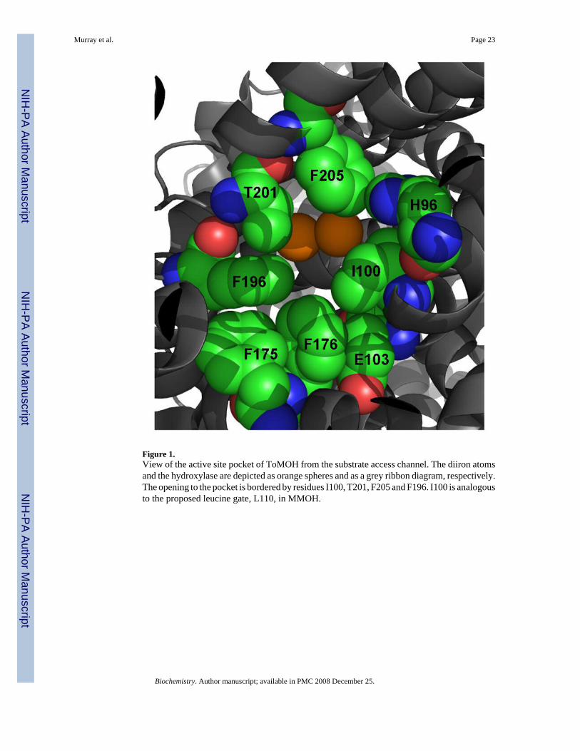
Figure 1.View of the active site pocket of ToMOH from the substrate access channel. The diiron atomsand the hydroxylase are depicted as orange spheres and as a grey ribbon diagram, respectively.The opening to the pocket is bordered by residues I100, T201, F205 and F196. I100 is analogousto the proposed leucine gate, L110, in MMOH.
Murray et al. Page 23
Biochemistry. Author manuscript; available in PMC 2008 December 25.
NIH
-PA Author Manuscript
NIH
-PA Author Manuscript
NIH
-PA Author Manuscript
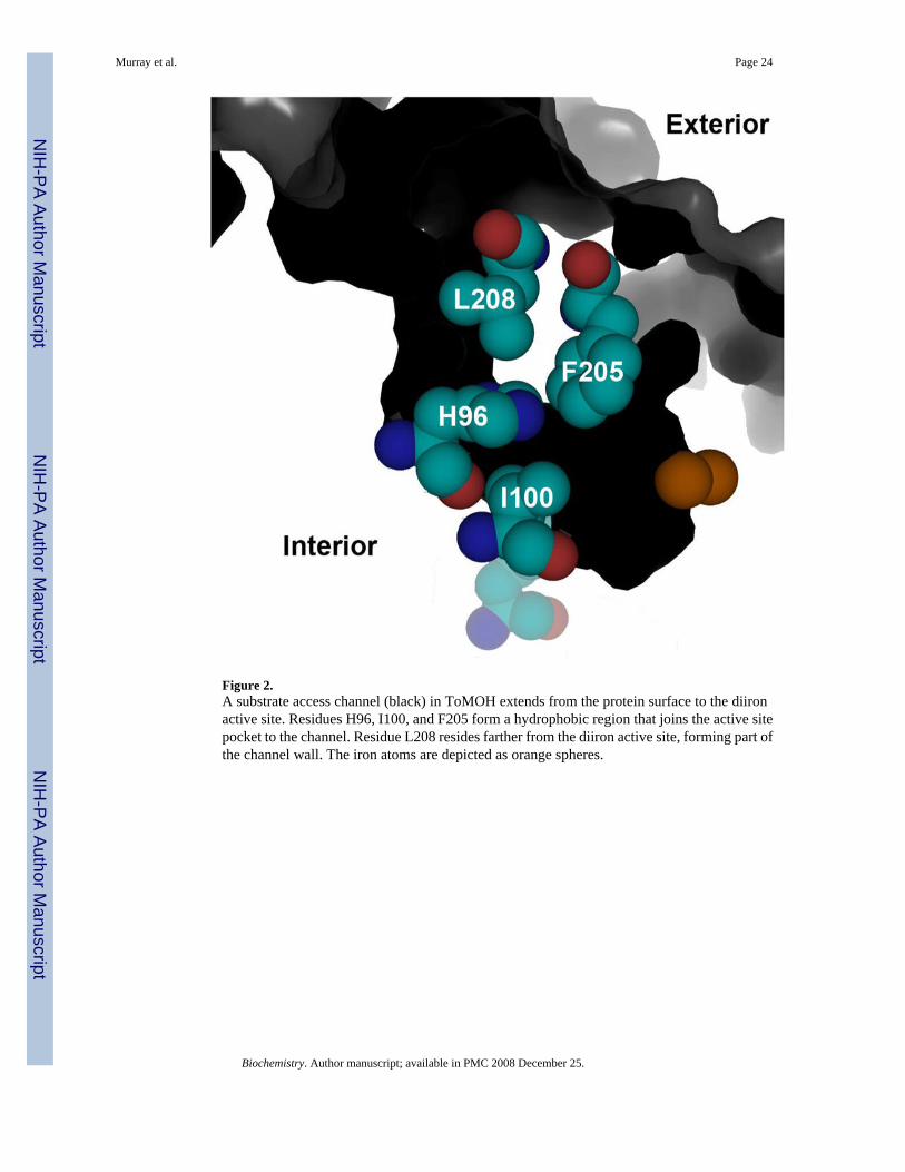
Figure 2.A substrate access channel (black) in ToMOH extends from the protein surface to the diironactive site. Residues H96, I100, and F205 form a hydrophobic region that joins the active sitepocket to the channel. Residue L208 resides farther from the diiron active site, forming part ofthe channel wall. The iron atoms are depicted as orange spheres.
Murray et al. Page 24
Biochemistry. Author manuscript; available in PMC 2008 December 25.
NIH
-PA Author Manuscript
NIH
-PA Author Manuscript
NIH
-PA Author Manuscript

Figure 3.Conformations adopted by W100 in the crystal structure. The aromatic side-chain occupiestwo positions with populations of 50% for positions A and position B. The indole ring in A isoriented parallel to the diiron center whereas Cζ3 to Cε3 are pointed towards the diiron centerin second conformation. The inset shows the labeling scheme referred to for the atoms in theindole ring.
Murray et al. Page 25
Biochemistry. Author manuscript; available in PMC 2008 December 25.
NIH
-PA Author Manuscript
NIH
-PA Author Manuscript
NIH
-PA Author Manuscript

Figure 4.Diiron center blocking effect of W100 in ToMOH I100W (Bottom) in comparison to MMOH(Top, left) and native ToMOH (Top, right). Amino acids in the region of the active site pocketand iron atoms are shown as spheres in grey (carbon), red (oxygen), blue (nitrogen), and green(iron). The two partially occupied positions of the W100 side chain in the variant ToMOHstructure are highlighted in yellow.
Murray et al. Page 26
Biochemistry. Author manuscript; available in PMC 2008 December 25.
NIH
-PA Author Manuscript
NIH
-PA Author Manuscript
NIH
-PA Author Manuscript
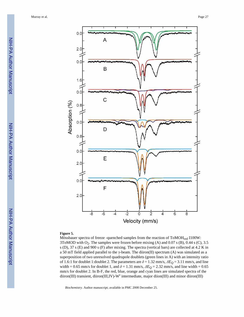
Figure 5.Mössbauer spectra of freeze -quenched samples from the reaction of ToMOHred I100W:3ToMOD with O2. The samples were frozen before mixing (A) and 0.07 s (B), 0.44 s (C), 3.5s (D), 37 s (E) and 900 s (F) after mixing. The spectra (vertical bars) are collected at 4.2 K ina 50 mT field applied parallel to the γ-beam. The diiron(II) spectrum (A) was simulated as asuperposition of two unresolved quadrupole doublets (green lines in A) with an intensity ratioof 1.6:1 for doublet 1:doublet 2. The parameters are δ = 1.32 mm/s, ΔEQ = 3.11 mm/s, and linewidth = 0.65 mm/s for doublet 1, and δ = 1.31 mm/s, ΔEQ = 2.32 mm/s, and line width = 0.65mm/s for doublet 2. In B-F, the red, blue, orange and cyan lines are simulated spectra of thediiron(III) transient, diiron(III,IV)-W• intermediate, major diion(III) and minor diiron(III)
Murray et al. Page 27
Biochemistry. Author manuscript; available in PMC 2008 December 25.
NIH
-PA Author Manuscript
NIH
-PA Author Manuscript
NIH
-PA Author Manuscript
products, respectively. The spectra of the diiron(III) transient ( ) and diiron(III,IV)-W•
intermediate ( ) are simulated with parameters reported previously (17). The spectrum of themajor diiron(III) product ( ) is modeled with a single quadrupole doublet with δ = 0.51 mm/s, ΔEQ = 0.84 mm/s, and line width = 0.35 mm/s. The spectrum of the minor diiron(III) product( ) is simulated with δ = 0.56 mm/s, ΔEQ = 1.77 mm/s, and line width = 0.33 mm/s. Forclarity, the diiron(II) spectral component is not shown specifically in B-F. The simulatedspectra are plotted at the following absorption intensity: red, 29% and 27% in B and C,respectively; blue, 16% and 34% in C and D, respectively; orange, 14%, 60% and 65% in D,E, and F, respectively; cyan, 3%, 8% and 8% in D, E, and F, respectively. The black linesoverlaid with the experimental spectra are composite spectra including the diiron(II) and allother species mentioned above. Approximately 25% of the total Fe in F appears in the form ofbroad, featureless absorptions, indicative of ill-defined paramagnetic ferric species. Wetherefore did not include this iron in the composite spectra.
Murray et al. Page 28
Biochemistry. Author manuscript; available in PMC 2008 December 25.
NIH
-PA Author Manuscript
NIH
-PA Author Manuscript
NIH
-PA Author Manuscript
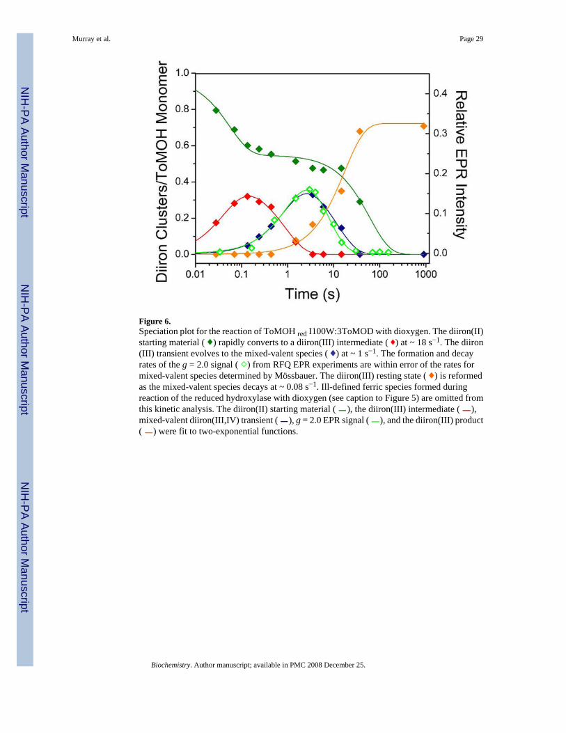
Figure 6.Speciation plot for the reaction of ToMOH red I100W:3ToMOD with dioxygen. The diiron(II)starting material ( ) rapidly converts to a diiron(III) intermediate ( ) at ~ 18 s−1. The diiron(III) transient evolves to the mixed-valent species ( ) at ~ 1 s−1. The formation and decayrates of the g = 2.0 signal ( ) from RFQ EPR experiments are within error of the rates formixed-valent species determined by Mössbauer. The diiron(III) resting state ( ) is reformedas the mixed-valent species decays at ~ 0.08 s−1. Ill-defined ferric species formed duringreaction of the reduced hydroxylase with dioxygen (see caption to Figure 5) are omitted fromthis kinetic analysis. The diiron(II) starting material ( ), the diiron(III) intermediate ( ),mixed-valent diiron(III,IV) transient ( ), g = 2.0 EPR signal ( ), and the diiron(III) product( ) were fit to two-exponential functions.
Murray et al. Page 29
Biochemistry. Author manuscript; available in PMC 2008 December 25.
NIH
-PA Author Manuscript
NIH
-PA Author Manuscript
NIH
-PA Author Manuscript
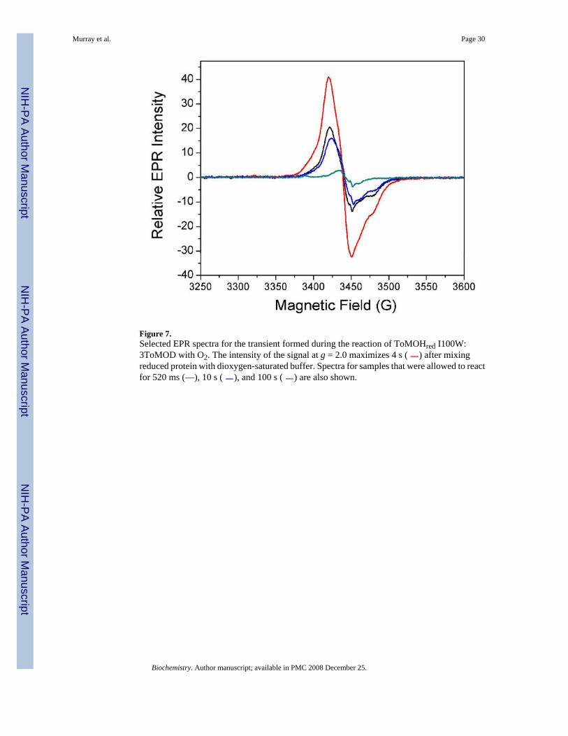
Figure 7.Selected EPR spectra for the transient formed during the reaction of ToMOHred I100W:3ToMOD with O2. The intensity of the signal at g = 2.0 maximizes 4 s ( ) after mixingreduced protein with dioxygen-saturated buffer. Spectra for samples that were allowed to reactfor 520 ms (—), 10 s ( ), and 100 s ( ) are also shown.
Murray et al. Page 30
Biochemistry. Author manuscript; available in PMC 2008 December 25.
NIH
-PA Author Manuscript
NIH
-PA Author Manuscript
NIH
-PA Author Manuscript

Figure 8.Effect of proton concentration on formation and decay of the diiron(III,IV)–W• species.Increasing pH increases kf and kd of the ToMOH I100W transient. The rate constants bothincrease with decreasing [H+], with kf (▲) and kd (●) increasing ~ 2-fold between pH 6.5 and7.5. The y-axis scales differ above and below the break.
Murray et al. Page 31
Biochemistry. Author manuscript; available in PMC 2008 December 25.
NIH
-PA Author Manuscript
NIH
-PA Author Manuscript
NIH
-PA Author Manuscript
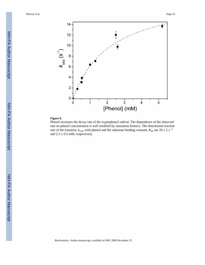
Figure 9.Phenol increases the decay rate of the tryptophanyl radical. The dependence of the observedrate on phenol concentration is well modeled by saturation kinetics. The determined reactionrate of the transient, krxn, with phenol and the substrate binding constant, Kd, are 20 ± 2 s−1
and 2.3 ± 0.5 mM, respectively.
Murray et al. Page 32
Biochemistry. Author manuscript; available in PMC 2008 December 25.
NIH
-PA Author Manuscript
NIH
-PA Author Manuscript
NIH
-PA Author Manuscript
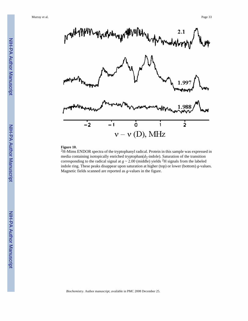
Figure 10.2H-Mims ENDOR spectra of the tryptophanyl radical. Protein in this sample was expressed inmedia containing isotopically enriched tryptophan(d5-indole). Saturation of the transitioncorresponding to the radical signal at g ~ 2.00 (middle) yields 2H signals from the labeledindole ring. These peaks disappear upon saturation at higher (top) or lower (bottom) g-values.Magnetic fields scanned are reported as g-values in the figure.
Murray et al. Page 33
Biochemistry. Author manuscript; available in PMC 2008 December 25.
NIH
-PA Author Manuscript
NIH
-PA Author Manuscript
NIH
-PA Author Manuscript
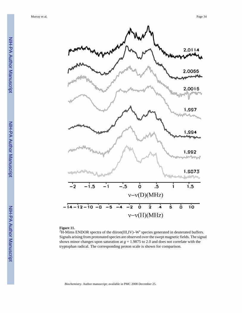
Figure 11.2H-Mims ENDOR spectra of the diiron(III,IV)–W• species generated in deuterated buffers.Signals arising from protonated species are observed over the swept magnetic fields. The signalshows minor changes upon saturation at g = 1.9875 to 2.0 and does not correlate with thetryptophan radical. The corresponding proton scale is shown for comparison.
Murray et al. Page 34
Biochemistry. Author manuscript; available in PMC 2008 December 25.
NIH
-PA Author Manuscript
NIH
-PA Author Manuscript
NIH
-PA Author Manuscript

Figure 12.MALDI-TOF spectra of tryptic peptides for the α-subunit of O2-reacted (top) and as-isolated(bottom) ToMOH I100W. The mass of the [M+H]+ ion at 3166 m/z agrees with the predictedmass for the 29mer peptide. Two additional envelopes of 16 and 32 Da higher mass are alsopresent. The relative intensity of these three envelopes changes in the reacted sample, with [M+H]+ less abundant than [M+H]++16.
Murray et al. Page 35
Biochemistry. Author manuscript; available in PMC 2008 December 25.
NIH
-PA Author Manuscript
NIH
-PA Author Manuscript
NIH
-PA Author Manuscript
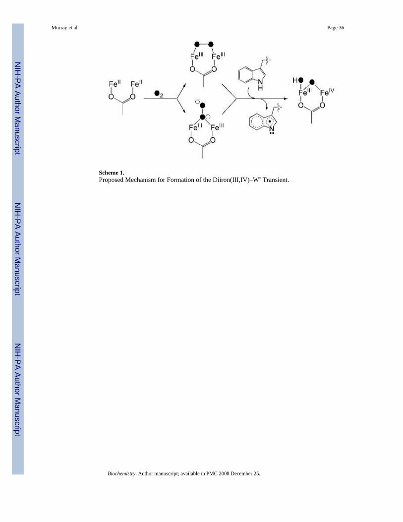
Scheme 1.Proposed Mechanism for Formation of the Diiron(III,IV)–W• Transient.
Murray et al. Page 36
Biochemistry. Author manuscript; available in PMC 2008 December 25.
NIH
-PA Author Manuscript
NIH
-PA Author Manuscript
NIH
-PA Author Manuscript

Scheme 2.Proposed Mechanism for Decay of the Diiron(III,IV)–W• Transient.
Murray et al. Page 37
Biochemistry. Author manuscript; available in PMC 2008 December 25.
NIH
-PA Author Manuscript
NIH
-PA Author Manuscript
NIH
-PA Author Manuscript

NIH
-PA Author Manuscript
NIH
-PA Author Manuscript
NIH
-PA Author Manuscript
Murray et al. Page 38
Table 1Formation and Decay Rate Constants of the Mixed-Valent Diiron(III,IV)-W• Species Measured by Optical, Mössbauer,and EPR Spectroscopy
Rate Constant Optical Mössbauer EPR
kf (s−1) 0.804 (1) 1.1 0.77
kd (s−1) 0.054 (2) 0.08 0.15
Biochemistry. Author manuscript; available in PMC 2008 December 25.
Related Documents