Published Ahead of Print 27 September 2006. 2006, 80(23):11486. DOI: 10.1128/JVI.00324-06. J. Virol. Connors Mican, Julia A. Metcalf, Christiana Iyasere and Mark Lempicki, Claire W. Hallahan, Mary McLaughlin, JoAnn M. M. Manion, Jun Yang, Joseph W. Adelsberger, Richard A. John C. Tilton, Alison J. Johnson, Marlise R. Luskin, Maura Mediated by Type I Interferons Immunodeficiency Virus Viremia Is Proinflammatory Cytokines during Human Diminished Production of Monocyte http://jvi.asm.org/content/80/23/11486 Updated information and services can be found at: These include: SUPPLEMENTAL MATERIAL Supplemental material REFERENCES http://jvi.asm.org/content/80/23/11486#ref-list-1 at: This article cites 57 articles, 33 of which can be accessed free CONTENT ALERTS more» articles cite this article), Receive: RSS Feeds, eTOCs, free email alerts (when new http://journals.asm.org/site/misc/reprints.xhtml Information about commercial reprint orders: http://journals.asm.org/site/subscriptions/ To subscribe to to another ASM Journal go to: on February 19, 2014 by guest http://jvi.asm.org/ Downloaded from on February 19, 2014 by guest http://jvi.asm.org/ Downloaded from

Welcome message from author
This document is posted to help you gain knowledge. Please leave a comment to let me know what you think about it! Share it to your friends and learn new things together.
Transcript
Published Ahead of Print 27 September 2006. 2006, 80(23):11486. DOI: 10.1128/JVI.00324-06. J. Virol.
ConnorsMican, Julia A. Metcalf, Christiana Iyasere and Mark Lempicki, Claire W. Hallahan, Mary McLaughlin, JoAnn M.M. Manion, Jun Yang, Joseph W. Adelsberger, Richard A. John C. Tilton, Alison J. Johnson, Marlise R. Luskin, Maura Mediated by Type I InterferonsImmunodeficiency Virus Viremia IsProinflammatory Cytokines during Human Diminished Production of Monocyte
http://jvi.asm.org/content/80/23/11486Updated information and services can be found at:
These include:
SUPPLEMENTAL MATERIAL Supplemental material
REFERENCEShttp://jvi.asm.org/content/80/23/11486#ref-list-1at:
This article cites 57 articles, 33 of which can be accessed free
CONTENT ALERTS more»articles cite this article),
Receive: RSS Feeds, eTOCs, free email alerts (when new
http://journals.asm.org/site/misc/reprints.xhtmlInformation about commercial reprint orders: http://journals.asm.org/site/subscriptions/To subscribe to to another ASM Journal go to:
on February 19, 2014 by guest
http://jvi.asm.org/
Dow
nloaded from
on February 19, 2014 by guest
http://jvi.asm.org/
Dow
nloaded from
JOURNAL OF VIROLOGY, Dec. 2006, p. 11486–11497 Vol. 80, No. 230022-538X/06/$08.00�0 doi:10.1128/JVI.00324-06Copyright © 2006, American Society for Microbiology. All Rights Reserved.
Diminished Production of Monocyte Proinflammatory Cytokinesduring Human Immunodeficiency Virus Viremia Is
Mediated by Type I Interferons�†John C. Tilton,1 Alison J. Johnson,1 Marlise R. Luskin,1 Maura M. Manion,1 Jun Yang,2
Joseph W. Adelsberger,2 Richard A. Lempicki,2 Claire W. Hallahan,1 Mary McLaughlin,1JoAnn M. Mican,1 Julia A. Metcalf,1 Christiana Iyasere,1 and Mark Connors1*
Laboratory of Immunoregulation, National Institute of Allergy and Infectious Diseases, National Institutes of Health,Bethesda, Maryland,1 and Clinical Services Program, SAIC-Frederick Inc., Frederick, Maryland2
Received 14 February 2006/Accepted 29 August 2006
The effect of human immunodeficiency virus (HIV) infection and high-level HIV replication on the functionof monocytes was investigated. HIV-positive patients had elevated levels of spontaneous production of some orall of the monocyte proinflammatory cytokines measured (interleukin-1� [IL-1�], IL-6, and tumor necrosisfactor alpha [TNF-�]) compared to uninfected controls. In patients on therapy with high frequencies ofmonocytes producing proinflammatory cytokines, this frequency was diminished in the context of viremiaduring an interruption of therapy. Diminished production of proinflammatory cytokines during viremia wasrestored by culture with autologous CD4� T cells or monocytes from an on-therapy time point or lipopoly-saccharide (LPS). Microarray analysis demonstrated that diminished monocyte production of proinflamma-tory cytokines was correlated with elevated type I interferon-stimulated gene transcripts. The addition ofexogenous alpha 2A interferon diminished the spontaneous production of IL-1�, IL-6, and TNF-� but did notaffect responses to LPS, recapitulating the changes observed for HIV-viremic patients. These results suggestthat monocyte function is diminished during high-level HIV viremia and that this effect is mediated by chronicstimulation by type I interferons. This effect on monocytes during viremia may play a role in diminished innateor adaptive immune system functions in HIV-infected patients. In addition, the restoration of these functionsmay also play a role in some immune reconstitution syndromes observed during initiation of therapy.
One of the hallmarks of human immunodeficiency virus type1 (HIV-1) infection is generalized immune system activation,although the mechanisms by which this is induced remain in-completely understood (reviewed in references 2 and 22). InHIV-infected patients, increased immune system activation istypically characterized by increased turnover of CD4� andCD8� T cells (24, 29, 40, 46) and increased expression ofactivation markers, such as CD38 (16, 31, 39) and HLA-DR(15, 26, 47), on immune system cells. The consequences ofchronic activation on the function of cells of the immune sys-tem are not well defined and are likely important in the patho-genesis of HIV-1, the opportunistic infections and malignan-cies associated with HIV disease, and HIV-specific immunity.
Over the past several years, an increasingly detailed pictureof the effects of HIV viremia on the immune system hasemerged. It has become clear that HIV viremia has effects ona wide variety of cells involved in innate or adaptive immuneresponses and mediates these effects through a number ofdifferent direct or indirect mechanisms. HIV viremia causes anexpansion of HIV-specific CD4� and CD8� T cells (17, 37, 38,43, 49, 56). Other direct effects have also been described,
including binding of viral products to non-antigen-specific re-ceptors, such as envelope protein binding to CCR5 (55) orHIV RNA binding to toll-like receptor (TLR) 7 or 8 (5, 23).Effects of HIV viremia on immune system cells, such as thosemediated by proinflammatory cytokines (reviewed in refer-ences 8 and 10) or cell-contact-dependent effects on naturalkiller (NK) cells (1, 36), might also be indirect. Although thedetails of many of these effects remain to be further defined, itappears clear that viremia has some direct or indirect effect onnearly every immune system cell type.
In the present study, we report an effect of HIV infectionand high-level HIV viremia on the function of monocytes.HIV-positive patients (on or off therapy) had elevated levels ofspontaneous production of some or all of the monocyte proin-flammatory cytokines measured (interleukin-1� [IL-1�], IL-6,and tumor necrosis factor alpha [TNF-�]) compared to unin-fected controls. In patients on therapy with high frequencies ofmonocytes producing proinflammatory cytokines, this fre-quency was diminished in the context of viremia during aninterruption of therapy. Unexpectedly, HIV-1-infected pa-tients on antiretroviral therapy with effective control of viralreplication had markedly elevated levels of monocytes pro-ducing these proinflammatory cytokines, even comparedwith uninfected controls. Diminished frequencies of monocytesproducing proinflammatory cytokines during active HIV-1 repli-cation were tightly correlated with increased type I interferon(IFN)-stimulated gene expression and monocyte HLA-DR ex-pression. Monocyte cytokine production was partially restoredby cell-cell contact with autologous monocytes or CD4� T cells
* Corresponding author. Mailing address: LIR, NIAID, NIH, Bldg10, Rm 11B-09, 10 Center Dr., MSC 1876, Bethesda, MD 20892-1876.Phone: (301) 496-8057. Fax: (301) 402-0070. E-mail: [email protected].
† Supplemental material for this article may be found at http://jvi.asm.org/.
� Published ahead of print on 27 September 2006.
11486
on February 19, 2014 by guest
http://jvi.asm.org/
Dow
nloaded from
from time points when the patients were on therapy. Treat-ment of peripheral blood mononuclear cells (PBMC) withrecombinant IFN-�2A in vitro caused diminished productionof monocyte proinflammatory cytokines, recapitulating the ob-servations in patients off therapy. These findings suggest apotent effect of in vivo production of type I IFN on monocytefunction. In addition, these findings likely have important im-plications for the pathogenesis of HIV-1, for opportunisticinfections, and in reconstitution syndromes associated withimproved immunologic function following control of HIV-1replication.
MATERIALS AND METHODS
Study population. HIV-1 infection in study participants was documented byHIV-1/2 immunoassay. All HIV-1-infected subjects signed informed consent andparticipated in protocols approved by a National Institute of Allergy and Infec-tious Diseases (NIAID) investigational review board. An additional 22 HIV-1-seronegative, healthy volunteers from the National Institutes of Health donorapheresis clinic were recruited into a control cohort.
Storage of samples. PBMC were freshly isolated from peripheral blood orapheresis donor packs by sodium diatrizoate-Ficoll density centrifugation (withlymphocyte separation medium [ICN Biomedicals, Aurora, OH]). PBMC werecryopreserved in cell culture freezing medium-dimethyl sulfoxide (Gibco, GrandIsland, NY) by using a Cryomed controlled-rate freezer (ThermoForma,Waltham, MA) and were stored at �140°C.
Multiplex supernatant cytokine assay (Luminex). Cytokine concentrations ofIL-1�, IL-2, IL-4, IL-5, IL-6, IL-8, IL-10, IL-12p70, granulocyte-macrophagecolony-stimulating factor, TNF-�, and IFN-� were measured in the supernatantsof stimulated and unstimulated PBMC by using the human cytokine LINCOplexmultiplex bead array (Linco Research, St. Charles, MO). Briefly, incubations andwashes were performed in 1.2-�m filter membrane 96-well plates (Millipore,Bedford, MA). After the final wash, beads in the 96-well plate were resuspendedin 150 �l of Luminex sheath fluid and were loaded onto the Luminex 200 system.Raw data (mean fluorescence intensities) from the beads were analyzed with theStatistical Ligand Immunoassay Analysis (StatLIA) software (Brendan Technol-ogies, Grosse Pointe Farms, MI) in order to obtain concentration values. Allsamples were run in duplicate, and results were obtained two or three times.
Intracellular cytokine staining. Cryopreserved samples were thawed and ali-quoted at 2 � 106 to 4 � 106 cells per stimulation tube under the followingconditions: medium alone, 5 �g/ml aldrithiol-2-inactivated HIV-1ADA-M, HIV-1JR-FL, or HIV-1MN (provided by Jeffrey Lifson of the AIDS Vaccine Program,SAIC, Frederick, MD), 100 ng/ml Escherichia coli lipopolysaccharide (LPS)(InvivoGen, San Diego, CA), or 100 U/ml IFN-�2A (PBL Biomedical Labora-tories, Piscataway, NJ). Cell stimulation, fixation, permeabilization, staining, andflow cytometry were conducted as previously described (14). Briefly, cells wereplaced in an incubator at 37°C and 5% CO2, and after 2 h, brefeldin A (SigmaAldrich, St. Louis, MO) was added to the medium at a final concentration of 10�g/ml to inhibit cytokine secretion. Following an additional 8-h incubation, cellswere labeled with anti-CD14 phycoerythrin (PE) antibody and viability stain(Viaprobe Violet; Molecular Probes, Eugene, OR) and were then fixed in 2%paraformaldehyde (Sigma Aldrich). The cells were permeabilized and blockedovernight in a buffer containing saponin (Sigma Aldrich) and 5% milk. Intracel-lular staining was performed using anti-CD3 fluorescein isothiocyanate (FITC)and anti-CD4 peridinin chlorophyll protein and anti-IL-1�, anti-IL-6, or anti-TNF-� allophycocyanin (Becton Dickinson, San Jose, CA). Data were collectedwith a FACSAria three-laser cytometer. Between 25,000 and 250,000 CD14�
events were collected and analyzed using FlowJo software (TreeStar Inc.,Ashland, OR).
Mixing experiments, in which autologous PBMC of patients on therapy and offtherapy were cultured together, were performed as follows: cryopreserved sam-ples were prepared and the cells from one time point were labeled for 8 min in0.625 �M CFSE [5 (and 6)-carboxyfluorescein diacetate succinimidyl ester] (Mo-lecular Probes). Labeled PBMC were then washed and mixed with unlabeledPBMC at a 1:1 ratio. Purified CD4� T cells and monocytes were obtained by useof the CD4� T-Cell Isolation Kit II and the Monocyte Isolation Kit, respectively(Miltenyi Biotec, Bergish Gladbach, Germany), with �90% purity. These cellswere subsequently labeled with CFSE as described above and cocultured withautologous PBMC of patients off therapy at various concentrations. Transwellexperiments were performed by coculturing autologous PBMC of patients onand off therapy in 24-well plates, separated by a 0.4-�m transwell insert (Costar;
Corning Inc., Corning, NY). For all mixing and transwell experiments, the incu-bation, fixation, staining, and data collection were performed as described above.
Microarray analysis. Peripheral blood CD14� cells were isolated by fluores-cence-activated cell sorting, using a FACSVantage SE flow cytometer (BectonDickinson). Frozen peripheral blood mononuclear cells were thawed and lym-phocytes were stained according to the manufacturer’s instructions with themonoclonal antibodies CD3 FITC (Becton Dickinson), CD4 PC5 (BeckmanCoulter, Miami, FL), and CD14 PE (Becton Dickinson). Sorting for CD14�
monocytes was performed using a quadrant gate on CD3 FITC� CD14 PE�
cells. CD14� monocyte purity was determined by flow cytometry to be �95%.One-half to two micrograms of total RNA was isolated from 1 million to 2million CD14� sorted cells. Target RNA was labeled as described in the Af-fymetrix 2003 technical note “GeneChip Eukaryotic Small Sample Target La-beling Assay Version II” and hybridized to Affymetrix U133A GeneChips fol-lowing the manufacturer’s recommended protocol. Chip-to-chip normalizationand gene expression index were performed using Affymetrix MAS5.0 algorithms.
Expression data for CD14� cells from patients on therapy and off therapy wereobtained from eight patients (16 samples total). A two-way analysis of variancewas performed using PartekPro software with therapy (on therapy or off therapy)as the main effect and patients as a random effect. Differentially expressed geneswere selected based on the following criteria: a P value of �0.05, an absolutemean expression level difference between groups greater than 30, and an abso-lute mean expression change (n-fold) between groups greater than 1.5. A total of784 genes were selected. In a separate analysis, 1,382 genes with expression levelscorrelating to the fraction of monocytes staining positive for at least one of theproinflammatory cytokines (IL-1�, IL-6, or TNF-�) were also identified (r, �0.62or ��0.62; P, 0.01; and maximum to minimum expression level difference,�40 or ratio �2 for a given gene). Functional annotation and biological categoryenrichment were performed with the DAVID knowledge base (11).
Statistical analysis. Because the data were not normally distributed, the per-centages of cytokine-producing monocytes for independent groups were com-pared by the Wilcoxon two-sample test. In cases in which longitudinal data wereanalyzed, paired data were compared by the Wilcoxon signed-rank test. Mediansare reported. Significance of correlations was determined by the Spearman rankmethod. The Bonferroni method was used to adjust P values for multiple testing.
Microarray data accession number. Microarray results have been deposited inthe Gene Expression Omnibus database under accession number GSE5220.
RESULTS
Frequencies of monocytes producing proinflammatory cyto-kines are elevated in patients on antiretroviral therapy andare diminished during treatment interruption. The conse-quences of active HIV infection on cytokine production wereexamined by measuring supernatant cytokine concentrations inunstimulated PBMC cultures from patients undergoing anti-retroviral treatment interruptions. Using a multiplex cytokineassay that simultaneously measured IL-1�, IL-2, IL-4, IL-5,IL-6, IL-8, IL-10, IL-12p70, granulocyte-monocyte colony-stimulating factor, TNF-�, and IFN-�, we found that three offive patients examined showed marked elevations in the proin-flammatory cytokines IL-1�, IL-6, and TNF-� in the PBMCcultures from the on-therapy time point compared with thecorresponding off-therapy time point (data not shown). Inthese patients with elevated proinflammatory cytokines, flow-cytometric intracellular cytokine staining determined that�90% of the cells producing IL-1�, IL-6, and TNF-� wereCD14� monocytes and revealed that there were marked re-ductions in the percentages of monocytes that produced thesecytokines during treatment interruption (Fig. 1A).
Spontaneous monocyte proinflammatory cytokine produc-tion was subsequently analyzed by intracellular cytokine stain-ing in patients undergoing scheduled treatment interruption(Fig. 1B). The clinical data for these patients are shown inTable 1. In agreement with the supernatant cytokine data, theintracellular cytokine staining revealed that a majority of
VOL. 80, 2006 INHIBITION OF MONOCYTE FUNCTION BY TYPE I IFN 11487
on February 19, 2014 by guest
http://jvi.asm.org/
Dow
nloaded from
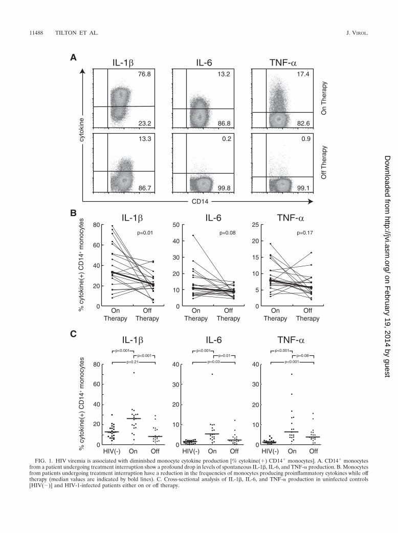
FIG. 1. HIV viremia is associated with diminished monocyte cytokine production [% cytokine(�) CD14� monocytes]. A. CD14� monocytesfrom a patient undergoing treatment interruption show a profound drop in levels of spontaneous IL-1�, IL-6, and TNF-� production. B. Monocytesfrom patients undergoing treatment interruption have a reduction in the frequencies of monocytes producing proinflammatory cytokines while offtherapy (median values are indicated by bold lines). C. Cross-sectional analysis of IL-1�, IL-6, and TNF-� production in uninfected controls[HIV(�)] and HIV-1-infected patients either on or off therapy.
11488 TILTON ET AL. J. VIROL.
on February 19, 2014 by guest
http://jvi.asm.org/
Dow
nloaded from
patients undergoing treatment interruption had reduced fre-quencies of monocytes producing IL-1�, IL-6, and TNF-�.Overall, the patients studied longitudinally had diminished me-dian frequencies of monocytes producing IL-1� (P 0.01)when off therapy, although changes in IL-6 (P 0.08) andTNF-� (P 0.17) did not reach statistical significance. De-creases in proinflammatory cytokine production were not cor-related with peak viral load, the number of days off therapy, oractivation of CD8� T cells based upon CD38 expression(Spearman’s rank correlation-adjusted P values of �0.5 foreach comparison). This cohort included a number of patientswith a relatively low production of these cytokines at both timepoints. For these patients, we did not observe large decreasesgiven the low baseline cytokine production. High frequenciesof monocytes producing cytokines in patients on therapy werestrongly correlated with greater decreases in IL-1� (r �0.84,P 0.001), IL-6 (r �0.91, P 0.001), and TNF-� (r �0.89, P 0.001) when antiretroviral therapy was stopped.This suggested that the effect of viremia was more readilydetected in patients with high frequencies of cells producingthese cytokines at the on-therapy time point. In addition, therewere strong correlations among the percentages of monocytesproducing IL-1�, IL-6, and TNF-� (P was �0.001 and theSpearman rank correlation was �0.81 for all comparisons),and only one patient showed discordant changes in cytokineproduction during therapy interruption. This suggested that, inpatients for whom a drop in proinflammatory cytokine produc-tion was observed, these changes were not isolated to onecytokine but reflected a more global effect of viremia on mono-cyte function.
To further define the effect of viremia on monocyte cytokineproduction in larger numbers of patients, cross-sectional
groups of healthy controls, HIV-infected patients on therapyand patients off therapy were also studied (Fig. 1C). Surpris-ingly, compared with uninfected controls, patients who were ontherapy demonstrated markedly increased frequencies ofmonocytes producing IL-1�, IL-6, and TNF-� (IL-1�, median26.0% in patients on therapy versus 12.9% in controls, P 0.001; IL-6, 5.5% versus 1.7%, P 0.001; TNF-�, 6.6% versus1.4%, P 0.001). When 61 samples from patients on variouscombinations of antiretroviral therapy were analyzed, changesin monocyte cytokine production were not attributable to anyof the following antiretrovirals: abacavir, didanosine, efavirenz,indinavir, lamivudine, lopinavir, nelfinavir, nevirapine, ritona-vir, stavudine, tenofovir, or zidovudine. HIV-infected patientswho were off therapy also had elevations in frequencies ofmonocytes producing IL-6 (2.4%, P 0.03) and TNF-� (3.9%,P 0.001), but not IL-1� (8.4%, P 0.21), compared withcontrols. In the cross-sectional analyses, patients on therapyhad elevated monocyte production of IL-1� (P 0.001) andIL-6 (P 0.01) compared with patients off therapy, butchanges in TNF-� production did not reach statistical signifi-cance (P 0.08).
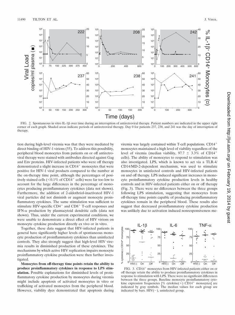
The effect of prolonged suppression of viral replication orprolonged low-level viremia on spontaneous monocyte cyto-kine production was also examined. Results from six patientsfrom whom more prolonged follow-up longitudinal sampleswere available are shown in Fig. 2. Interruption of therapyresulted in an initial decline in production of monocyte proin-flammatory cytokines in each case. Reinstitution of therapyresulted in a rebound and subsequent decline in cytokine pro-duction. In general, this kinetic is similar to one recently de-scribed following initiation of therapy (7).
One possibility for the observed changes in monocyte func-
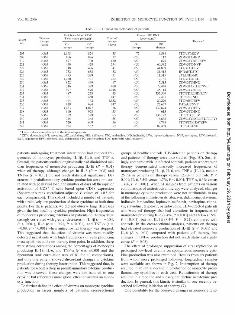
TABLE 1. Clinical characteristics of patients
Patientno.
Time ontherapy(days)
Peripheral blood CD4�
T-cell count (cells/�l)a Time offtherapy(days)
Plasma HIV RNA(copy eq/ml)a
Therapyb
Ontherapy
Offtherapy
Ontherapy
Offtherapy
203 �365 1,193 824 35 72 6,584 3TC/d4T/SQV208 �365 666 896 37 50 112 ZDV/3TC/IND219 �365 677 700 180 50 876 ZDV/3TC/ddI/EFV221 �365 449 428 554 50 60,582 ZDV/3TC/NVP222 �365 718 496 41 50 44,839 d4T/3TC/EFV224 �365 752 652 52 50 31,413 IND/d4T/3TC225 �365 493 389 31 50 11,515 d4T/IND/ddC229 �365 1,230 791 252 50 2,185 d4T/3TC/NEL230 �365 822 469 97 50 7,515 ZDV/3TC/IND231 �365 516 329 400 50 72,448 ZDV/3TC/TDF/NVP232 �365 997 576 1,048 50 35,114 ZDV/3TC/NEL233 �365 487 258 43 50 335,500 3TC/TDF/IND/RTV234 �365 701 450 447 50 7,491 3TC/ddI/IND235 �365 494 162 1,652 50 20,220 3TC/ABC/EFV236 �365 928 684 247 50 23,637 D4T/ddI/NVP237 �365 1,433 1,077 56 50 139,874 ZDV/3TC/EFV238 �365 690 928 55 65 613 ZDV/3TC/IND239 �365 759 579 63 50 136,192 TDF/3TC/EFV240 �365 745 362 55 50 16,638 ZDV/3TC/ABC/TDF/LPVr241 �365 720 609 56 50 9,738 3TC/FTC/TDF/EFV242 �365 934 562 62 50 87,489 3TC/d4T/IND
a Listed values were obtained at the time of apheresis.b ZDV, zidovudine; d4T, stavudine; ddC, zalcitabine; NEL, nelfinavir; 3TC, lamivudine; IND, indinavir; LPVr, lopinavir/ritonavir; NVP, nevirapine; RTV, ritonavir;
SQV, saquinavir; EFV, efavirenz; ddI, didanosine; FTC, emtricitabine; TDF, tenofovir; ABC, abacavir.
VOL. 80, 2006 INHIBITION OF MONOCYTE FUNCTION BY TYPE I IFN 11489
on February 19, 2014 by guest
http://jvi.asm.org/
Dow
nloaded from
tion during high-level viremia was that they were mediated bydirect binding of HIV-1 virions (55). To address this possibility,peripheral blood monocytes from patients on or off antiretro-viral therapy were stained with antibodies directed against Gagand Env proteins. HIV-infected patients who were off therapydemonstrated a slight increase in CD14� monocytes that werepositive for HIV-1 viral products compared to the number atthe on-therapy time point, although the percentages of posi-tively stained cells (0.1% of CD14� cells) were far too low toaccount for the large differences in the percentage of mono-cytes producing proinflammatory cytokines (data not shown).Furthermore, the addition of 2-aldothriol-inactivated HIV-1viral particles did not diminish or augment monocyte proin-flammatory cytokines. The same stimulation was sufficient tostimulate HIV-specific CD4� and CD8� T-cell responses andIFN-� production by plasmacytoid dendritic cells (data notshown). Thus, under the current experimental conditions, wewere unable to demonstrate a direct effect of HIV virions onmonocyte cytokine production directly ex vivo or in vitro.
Together, these data suggest that HIV-infected patients ingeneral have significantly higher levels of spontaneous mono-cyte production of proinflammatory cytokines than uninfectedcontrols. They also strongly suggest that high-level HIV vire-mia results in diminished production of these cytokines. Themechanisms by which active HIV replication impairs monocyteproinflammatory cytokine production were then further inves-tigated.
Monocytes from off-therapy time points retain the ability toproduce proinflammatory cytokines in response to LPS stim-ulation. Possible explanations for diminished levels of proin-flammatory cytokine production by monocytes during viremiamight include apoptosis of activated monocytes in vitro ortrafficking of activated monocytes from the peripheral blood.However, viability dyes demonstrated that apoptosis during
viremia was largely contained within T-cell populations. CD14�
monocytes maintained a high level of viability regardless of thelevel of viremia (median viability, 97.7 � 3.3% of CD14�
cells). The ability of monocytes to respond to stimulation wasalso investigated. LPS, which is known to act via a TLR-4/CD14/MD-2-dependent mechanism, was used to stimulatemonocytes in uninfected controls and HIV-infected patientson and off therapy. LPS induced significant increases in mono-cyte proinflammatory cytokine production levels in healthycontrols and in HIV-infected patients either on or off therapy(Fig. 3). There were no differences between the three groupsfollowing LPS stimulation, suggesting that monocytes fromoff-therapy time points capable of producing proinflammatorycytokines remain in the peripheral blood. These results alsosuggest that decreased proinflammatory cytokine productionwas unlikely due to activation induced nonresponsiveness me-
FIG. 2. Spontaneous in vitro IL-1� over time during an interruption of antiretroviral therapy. Patient numbers are indicated in the upper rightcorner of each graph. Shaded areas indicate periods of antiretroviral therapy. Day 0 for patients 237, 238, and 241 was the day of interruption oftherapy.
FIG. 3. CD14� monocytes from HIV-infected patients either on oroff therapy retain the ability to produce proinflammatory cytokines inresponse to stimulation with LPS. There were no significant differencesbetween the three groups. Baseline monocyte proinflammatory cyto-kine expression frequencies [% cytokine(�) CD14� monocytes] areindicated by gray symbols. The median values for each group areindicated by bars. HIV(�), uninfected group.
11490 TILTON ET AL. J. VIROL.
on February 19, 2014 by guest
http://jvi.asm.org/
Dow
nloaded from
diated by TLR-4/CD14 stimulation in vivo, since productionwas readily overcome by LPS stimulation in vitro.
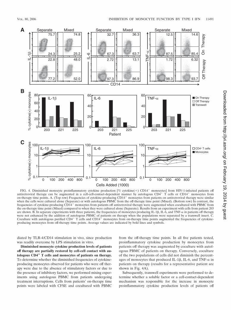
Diminished monocyte cytokine production levels of patientsoff therapy are partially restored by cell-cell contact with au-tologous CD4� T cells and monocytes of patients on therapy.To determine whether the diminished frequencies of cytokine-producing monocytes observed for patients who were off ther-apy were due to the absence of stimulatory factors or due tothe presence of inhibitory factors, we performed mixing exper-iments using autologous PBMC from patients undergoingtreatment interruptions. Cells from patients’ on-therapy timepoints were labeled with CFSE and cocultured with PBMC
from the off-therapy time points. In all five patients tested,proinflammatory cytokine production by monocytes frompatients off therapy was augmented by coculture with autol-ogous PBMC of patients on therapy. Conversely, cocultureof the two populations of cells did not diminish the percent-ages of monocytes that produced IL-1�, IL-6, and TNF-� inpatients on therapy (results for a representative patient areshown in Fig. 4A).
Subsequently, transwell experiments were performed to de-termine whether a soluble factor or a cell-contact-dependentmechanism was responsible for the increase in monocyteproinflammatory cytokine production levels of patients off
FIG. 4. Diminished monocyte proinflammatory cytokine production [% cytokine(�) CD14� monocytes] from HIV-1-infected patients offantiretroviral therapy can be augmented in a cell-cell-contact-dependent manner by autologous CD4� T cells or CD14� monocytes fromon-therapy time points. A. (Top row) Frequencies of cytokine-producing CD14� monocytes from patients on antiretroviral therapy were similarwhen the cells were cultured alone (Separate) or with autologous PBMC from the off-therapy time point (Mixed). (Bottom row) In contrast, thefrequencies of cytokine-producing CD14� monocytes from patients off antiretroviral therapy were augmented when cocultured with PBMC fromthe on-therapy time point (Mixed) compared to when they were cultured alone (Separate). Results from an experiment with cells from patient 203are shown. B. In separate experiments with three patients, the frequencies of monocytes producing IL-1�, IL-6, and TNF-� in patients off therapywere not enhanced by the addition of autologous PBMC of patients on therapy when the populations were separated by a transwell insert. C.Coculture with autologous purified CD4� T cells and CD14� monocytes from on-therapy time points augmented the frequencies of cytokine-producing monocytes from off-therapy time points. Average values are indicated by bold lines and symbols.
VOL. 80, 2006 INHIBITION OF MONOCYTE FUNCTION BY TYPE I IFN 11491
on February 19, 2014 by guest
http://jvi.asm.org/
Dow
nloaded from
therapy following mixing with autologous PBMC from patientson therapy. For all three patients tested, preventing cell-cellcontact blocked the increased frequencies of monocytes pro-ducing proinflammatory cytokines observed in the mixing cul-tures (Fig. 4B).
The cells responsible for the increase in the frequencies ofmonocytes producing proinflammatory cytokines were also ex-amined by magnetic bead purification and coculture experi-ments. Purified autologous CD14� monocytes and CD4� Tcells of patients on therapy were both able to augment proin-flammatory cytokine production by monocytes of patients offtherapy (Fig. 4C). In each experiment, CD4� T cells werebetter able to recover monocyte cytokine production over arange of dilutions. Although these were highly enriched pop-ulations (purity, �90%), we cannot formally rule out the pos-sibility that the effect of the addition of monocytes may be dueto very small numbers of contaminating CD4� T cells. Takentogether, these data suggest that recovery of cytokine produc-tion by monocytes from viremic patients can be mediated byautologous CD4� T cells or monocytes through a cell-contact-dependent mechanism.
Alterations in proinflammatory cytokine production bymonocytes were inversely associated with type I IFN-stimu-lated gene regulation. Gene expression profiling using mi-croarrays was performed on purified CD14� monocytes iso-lated from on-therapy and off-therapy time points in eightpatients undergoing treatment interruption. This cohort in-cluded patients with a wide range of monocyte proinflamma-tory cytokine responses to treatment interruption, includingseveral patients with large decreases, as well as several patientswith no changes or slight increases, in cytokine production.Differentially expressed genes were selected based on an ab-solute mean expression level difference between the groupsgreater than 30, a P value of �0.05, and an absolute meanexpression change (n-fold) between groups greater than 1.5,resulting in 784 differentially expressed genes (see Table S1 inthe supplemental material). A group of interferon-inducedgenes were found to be highly enriched (P 8.4 � 10�8) in thelist of genes up-regulated during plasma viremia followingtreatment interruption (Fig. 5A). These data indicate that un-restricted viral replication is strongly correlated with type IIFN-stimulated gene activation in CD14� monocytes.
The microarray data were subsequently analyzed to deter-mine whether individual genes that correlated with changes inthe frequencies of monocytes producing proinflammatory cy-tokines in patients on or off antiretroviral therapy could beidentified. A functional analysis of the 1,382 genes correlatingwith monocyte cytokine levels (correlated with at least onemonocyte cytokine with an r value of �0.62 or ��0.62 and aP value of �0.01; see Table S2 in the supplemental material)indicated that many of these genes were involved in type Iinterferon responses, NF-�B, mitogen-activated protein ki-nase, and Jun signaling pathways, general immune activation,immune down-regulation, protein degradation, protein secre-tion, and apoptosis (Fig. 5B). These data indicate that inter-ruption of antiretroviral therapy is associated with alterationsin monocyte gene expression across multiple functional cate-gories, suggesting that diminished proinflammatory cytokineexpression is part of a global effect of HIV viremia uponmonocytes.
The elevations of type I interferon-stimulated gene expres-sion in CD14� monocytes from patients off antiretroviral ther-apy suggest that these cells may be activated in vivo and thatactivation of monocytes may be linked to the diminished abilityof these cells to produce proinflammatory cytokines. CD14�
monocytes from 10 patients undergoing treatment interruptionwere assayed for surface expression of the activation markerHLA-DR, and increased HLA-DR expression was stronglycorrelated with decreased IL-1� (r �0.94, P 0.001), IL-6(r �0.87, P 0.004), and TNF-� (r �0.94, P 0.001)production levels (data not shown). The inverse correlations ofboth interferon-stimulated gene expression and HLA-DR sur-face expression with the production of monocyte proinflam-matory cytokines strongly suggests that the diminished cyto-kine production is tightly linked to the activation of monocytesin the context of HIV viremia.
The mRNA expression levels of IL-1�, IL-6, and TNF-� inthe eight patients undergoing treatment interruption were alsoexamined by microarrays in order to determine whether thereduced protein expression at the off-therapy time point cor-related with reduced cytokine mRNA levels. The microarrayanalysis revealed that IL-1� and TNF-� mRNA levels were notdifferent between patients who were on or off therapy. IL-6mRNA levels were actually higher in patients when they wereoff therapy at a time when protein levels were diminished. Theexpression levels of the mRNAs for IL-1�, IL-6, and TNF-� allsuggest that the diminished cytokine protein levels of patientsoff therapy are not due to decreases in mRNA levels but ratherthat a posttranscriptional regulatory mechanism, most likelyoperating at the level of mRNA translation or protein stability,is responsible for the changes in spontaneous monocyte cyto-kine production. This possibility is potentially supported by theinverse correlation between monocyte proinflammatory cyto-kine protein levels and the increased expression of genes in-volved in ubiquitination and proteasomal degradation path-ways (see Table S2 in the supplemental material).
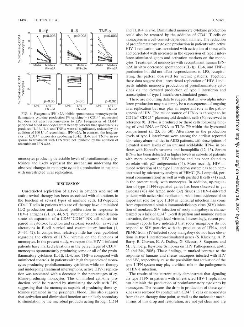
Treatment of CD14� monocytes with exogenous IFN-�2Arecapitulates the changes in proinflammatory cytokines ob-served in HIV-infected patients off therapy. The inverse rela-tionship between type I interferon-stimulated gene expressionand monocyte proinflammatory cytokine expression raised thepossibility that exposure to type I interferons in vivo mightdirectly influence the cytokine production observed ex vivo.Exposure of monocytes to type I interferons induces matura-tion into dendritic cells over 24 to 48 h; however, its effects onbaseline and LPS-induced proinflammatory cytokine produc-tion are not well characterized. CD14� monocytes fromhealthy patients were incubated with recombinant IFN-�2Aand cultured with or without LPS stimulation. The frequenciesof monocytes producing detectable baseline levels of IL-1�,IL-6, and TNF-� were significantly reduced by treatment withIFN-�2A (P 0.003 for all three cytokines) (Fig. 6). However,IFN-�2A did not reduce the percentages of monocytes pro-ducing IL-1�, IL-6, and TNF-� in response to LPS stimulation(P values of �0.30 for all cytokines). This pattern of reducedbaseline proinflammatory cytokine production with retainedresponsiveness to LPS stimulation following treatment withIFN-�2A was similar to that observed for HIV-infected pa-tients off antiretroviral therapy. These results indicate thatexposure to type I interferons can reduce the frequency of
11492 TILTON ET AL. J. VIROL.
on February 19, 2014 by guest
http://jvi.asm.org/
Dow
nloaded from
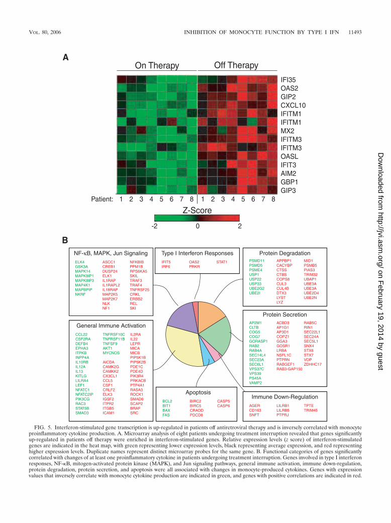
FIG. 5. Interferon-stimulated gene transcription is up-regulated in patients off antiretroviral therapy and is inversely correlated with monocyteproinflammatory cytokine production. A. Microarray analysis of eight patients undergoing treatment interruption revealed that genes significantlyup-regulated in patients off therapy were enriched in interferon-stimulated genes. Relative expression levels (z score) of interferon-stimulatedgenes are indicated in the heat map, with green representing lower expression levels, black representing average expression, and red representinghigher expression levels. Duplicate names represent distinct microarray probes for the same gene. B. Functional categories of genes significantlycorrelated with changes of at least one proinflammatory cytokine in patients undergoing treatment interruption. Genes involved in type I interferonresponses, NF-�B, mitogen-activated protein kinase (MAPK), and Jun signaling pathways, general immune activation, immune down-regulation,protein degradation, protein secretion, and apoptosis were all associated with changes in monocyte-produced cytokines. Genes with expressionvalues that inversely correlate with monocyte cytokine production are indicated in green, and genes with positive correlations are indicated in red.
VOL. 80, 2006 INHIBITION OF MONOCYTE FUNCTION BY TYPE I IFN 11493
on February 19, 2014 by guest
http://jvi.asm.org/
Dow
nloaded from
monocytes producing detectable levels of proinflammatory cy-tokines and likely represent the mechanism underlying theobserved changes in monocyte cytokine production in patientswith unrestricted viral replication.
DISCUSSION
Unrestricted replication of HIV-1 in patients who are offantiretroviral therapy has been associated with alterations inthe function of several types of immune cells. HIV-specificCD4� T cells in patients who are off therapy have diminishedIL-2 production and reduced proliferation in response toHIV-1 antigens (21, 27, 44, 57). Viremic patients also demon-strate an expansion of a CD56�CD16� NK cell subset im-paired in cytotoxic function and cytokine secretion, as well asalterations in B-cell survival and costimulatory function (1,34–36, 42). In comparison, relatively little has been publishedregarding the effects of HIV-1 viremia on the functions ofmonocytes. In the present study, we report that HIV-1-infectedpatients have marked elevations in the percentages of CD14�
monocytes spontaneously producing some or all of the proin-flammatory cytokines IL-1�, IL-6, and TNF-� compared withuninfected controls. In patients with high frequencies of mono-cytes producing proinflammatory cytokines while on therapyand undergoing treatment interruptions, active HIV-1 replica-tion was associated with a decrease in the percentages of cy-tokine-producing monocytes. This diminished cytokine pro-duction could be restored by stimulating the cells with LPS,suggesting that the monocytes capable of producing these cy-tokines remained in the peripheral blood. This also suggeststhat activation and diminished function are unlikely secondaryto stimulation by the microbial products acting through CD14
and TLR-4 in vivo. Diminished monocyte cytokine productioncould also be restored by the addition of CD4� T cells ormonocytes in a cell-contact-dependent manner. The reductionof proinflammatory cytokine production in patients with activeHIV-1 replication was associated with activation of these cellsand correlated with increases in the expression of type I inter-feron-stimulated genes and activation markers on the mono-cytes. Treatment of monocytes with recombinant human IFN-�2A in vitro decreased spontaneous IL-1�, IL-6, and TNF-�production but did not affect responsiveness to LPS, recapitu-lating the pattern observed for viremic patients. Together,these data suggest that unrestricted replication of HIV-1 indi-rectly inhibits monocyte production of proinflammatory cyto-kines via the elevated production of type I interferon andtranscription of type I interferon-stimulated genes.
There are mounting data to suggest that in vivo alpha inter-feron production may not simply be a consequence of ongoingviral replication but may play an important role in the patho-genesis of HIV. The major source of IFN-� is thought to beCD11c� CD123� plasmacytoid dendritic cells (50; reviewed inreference 9). IFN-� is produced by these cells following bind-ing of viral RNA or DNA to TLRs 7/9 within the lysosomalcompartment (5, 23, 30, 50). Alterations in the productionlevels of type I interferons were among the earliest reportedlaboratory abnormalities in AIDS patients, with descriptions ofelevated serum levels of an unusual acid-labile IFN-� in pa-tients with Kaposi’s sarcoma and hemophilia (12, 13). SerumIFN-� has been detected in higher levels in subsets of patientswith more advanced HIV infection and has been found tocorrelate with p24 antigenemia (54). More recently, HIV-in-duced activation of the type I interferon system has been dem-onstrated by microarray analysis of PBMC (R. Lempicki, per-sonal communication) as well as with purified B cells (41) and,in the present study, with monocytes. In addition, up-regula-tion of type I IFN-regulated genes has been observed in gutmucosal (48) and lymph node (32) tissues in HIV-1-infectedpatients with active viral replication. Additional evidence of animportant role for type I IFN in lentiviral infection has comefrom experimental simian immunodeficiency virus (SIV) infec-tion of monkeys. SIV infection of sooty mangabeys is charac-terized by a lack of CD4� T-cell depletion and immune systemactivation, despite high-level viremia. Interestingly, recent pre-liminary reports have indicated that sooty mangabeys do notrespond to SIV particles with the production of IFN-�, andPBMC from SIV-infected sooty mangabeys do not have eleva-tions in type I interferon-stimulated genes (S. Klucking, A. P.Barry, R. Chavan, K. A. Dalbey, G. Silvestri, S. Staprans, andM. Feinberg, Keystone Symposia on HIV Pathogenesis, abstr.22 and 244, 2005). These findings, in marked contrast to theresponse of humans and rhesus macaques infected with HIVand SIV, respectively, raise the possibility that activation of thetype I IFN system may play a critical role in the pathogenesisof HIV-1 infection.
The results of the current study demonstrate that signalingvia type I IFN in patients with unrestricted HIV-1 replicationcan diminish the production of proinflammatory cytokines bymonocytes. The reasons the drop in production of these cyto-kines was restored by contact with CD4� T cells or monocytesfrom the on-therapy time point, as well as the molecular mech-anisms of this drop and restoration, are not yet clear and are
FIG. 6. Exogenous IFN-�2A inhibits spontaneous monocyte proin-flammatory cytokine production [% cytokine(�) CD14� monocytes]but does not affect responsiveness to LPS. Frequencies of CD14�
peripheral blood monocytes from healthy patients that spontaneouslyproduced IL-1�, IL-6, and TNF-� were all significantly reduced by theaddition of 100 U of recombinant IFN-�2A. In contrast, the frequen-cies of CD14� monocytes producing IL-1�, IL-6, and TNF-� in re-sponse to treatment with LPS were not inhibited by the addition ofrecombinant IFN-�2A.
11494 TILTON ET AL. J. VIROL.
on February 19, 2014 by guest
http://jvi.asm.org/
Dow
nloaded from
the subject of ongoing work. Such an effect is potentially me-diated by a number of cell surface molecules known to regulatemonocyte cytokine production (19). It should be noted thatIFN-� has been shown to inhibit production of IL-1�, TNF-�,and IL-6 in vitro in at least one report in tumor cell lines (25).In addition, IFN-� has been used to diminish inflammationassociated with Behcet’s syndrome (20). However, the conse-quences of prolonged stimulation of the type I interferon sys-tem in chronic viral infections in vivo, such as HIV, are not wellcharacterized. In acute infections and in vitro assays, signalingvia IFN-�/� has been implicated in the establishment of anti-viral states within target cells, activation of innate immunecells, and modulation of adaptive immune responses. Persis-tent overactivation of the type I interferon pathway may affectall of these broad groups of antiviral immune defenses. Inprimary human cells, the findings of the present study areconsistent with, and extend, observations in one recent reportdemonstrating that monocytes from patients with active HIV-1replication have diminished up-regulation of costimulatorymolecules and maturation into dendritic cells, suggesting thatactivation of the type I IFN system in vivo may have detrimen-tal consequences on monocyte function (28). It is possible thatthese alterations in the innate immune system negatively im-pact adaptive immune responses and diminish the ability of theimmune system to control HIV-1 replication and opportunisticinfections associated with HIV disease.
Conversely, the results of the present study may also beinterpreted in the context of a number of clinical observationssuggesting improved monocyte function in HIV-infected pa-tients during antiretroviral therapy. Following the initiation ofantiretroviral therapy, some patients experience an immunereconstitution and inflammatory syndrome (IRIS), a conditionin which some patients starting antiretroviral therapy experi-ence severe clinical symptoms resulting from an inflammatoryresponses to non-HIV pathogens (33, 52). Of these pathogens,the vast majority are caused by pathogens for which monocytesare thought to play a critical role in containment, such asMycobacterium tuberculosis, Mycobacterium avium, or somefungi. In this setting, the median time to onset of IRISs is 3weeks, well before there were large increases in CD4� T cellsin the peripheral blood in most patients. In approximately 10%of patients, these syndromes occur in the absence of increasesin CD4� T-cell numbers in the peripheral blood. IRIS has alsobeen associated with elevations of serum (51, 53) or sponta-neous in vitro production of proinflammatory cytokines (7),suggesting that improvements in monocyte and macrophagefunction following control of HIV replication may play a rolein the development of this syndrome. Although it is specula-tive, it is reasonable to suggest that a reversal of HIV-inducedinhibition of monocyte function may underlie some forms ofIRISs. If so, it is possible that therapies that modulate theeffects of monocyte proinflammatory cytokines or their pro-duction, such as treatments with the TNF-� antagonist etan-ercept or alpha interferon, may have a role in treatment ofthese syndromes.
Taken in the context of prior work, the results of the presentstudy also suggest that the interrelationship of HIV viremiaand proinflammatory cytokine production is more complexthan previously appreciated. Increases in serum levels of thesecytokines have only intermittently been observed in the earliest
stages of HIV infection, similar to the case for patients in thepresent study compared to uninfected controls. However, in-creases have been better documented for patients with ad-vanced disease (3, 4, 18, 54). These data are, in general, con-sistent with those of the present study. It should be noted thatmonocytes are not the only source of TNF-�. The majority ofHIV-specific CD8� T cells produce TNF-� and would be ex-pected to be present at high levels during viremia and rapidlydecline during effective therapy (6, 17). However, the effects ofantiretroviral therapy on serum levels of these cytokines havebeen less well studied. Increases in serum levels of some proin-flammatory cytokines following therapy of patients with IRIShave been documented, as noted above.
In the present study, it was somewhat surprising that even inthe absence of disease, patients on antiretroviral therapy hadthe highest frequency of monocytes producing proinflamma-tory cytokines. A relatively wide range of frequencies of mono-cytes producing these cytokines was observed. This variationmight be attributable to viral or host genetics of the responsein monocytes or plasmacytoid dendritic cells and efficacy of orcompliance with the antiretroviral regimen. Typically, in pa-tients on antiretroviral therapy, nearly all immunological re-sponses measured are equal to or approximate uninfected con-trols. However, it should be noted that even in the context ofeffective antiretroviral therapy with 50 copies of HIV RNAper ml of plasma, there is evidence of ongoing viral replication.When highly sensitive, real-time PCR methods are used, pa-tients are observed to typically maintain a mode of 10 copies ofHIV RNA per ml of plasma (45). In addition, there is evidenceof ongoing immune system activation in such patients. Basedupon expression array experiments, there is evidence of ongo-ing immune system activation in treated patients with 50copies of HIV RNA per ml of plasma compared to uninfectedcontrols (R. Lempicki, personal communication). It is mostlikely that the increases in proinflammatory monocyte cyto-kines observed for treated patients in the current study arerelated to HIV replication and not due to stimulation by-products of opportunistic pathogens, given that this is a healthycohort with relatively high CD4� T-cell counts. The observa-tions from the present study, taken in the context of priorwork, are most consistent with the view that during therapy,there is ongoing, most likely indirect activation of monocytesresulting from low-level HIV replication. In patients off anti-retroviral therapy, there is ongoing monocyte activation butdiminished function in the context of high-level viremia at leastin part mediated by the chronic effects of high levels of type IIFNs. These functions may be rapidly restored in the context ofinitiation of antiretroviral therapy and may play a role in im-mune reconstitution syndromes.
Over the past several years, an increasingly detailed pictureof the widespread nature of HIV-induced immune system ac-tivation has emerged. Improved tools for measuring immuneresponses in a more quantitative and reproducible manner,coupled with the ability to alter levels of HIV replicationthrough antiretroviral therapy, are permitting a greater analy-sis of the cells involved, as well as primary and secondaryeffects of viral products on the immune system and the mech-anisms involved, than was previously available. Further defini-tion of these mechanisms may provide information that is
VOL. 80, 2006 INHIBITION OF MONOCYTE FUNCTION BY TYPE I IFN 11495
on February 19, 2014 by guest
http://jvi.asm.org/
Dow
nloaded from
important for modulation of responses to non-HIV pathogens,adaptive responses to HIV, or pathogenesis of HIV disease.
ACKNOWLEDGMENTS
This project has been funded in whole or in part with federal fundsfrom the National Cancer Institute, National Institutes of Health,under contract NO1-CO-12400.
The content of this publication does not necessarily reflect the viewsor policies of the Department of Health and Human Services, nor doesmention of trade names, commercial products, or organizations implyendorsement by the U.S. Government.
REFERENCES
1. Alter, G., N. Teigen, B. T. Davis, M. M. Addo, T. J. Suscovich, M. T. Waring,H. Streeck, M. N. Johnston, K. D. Staller, M. T. Zaman, X. G. Yu, M.Lichterfeld, N. Basgoz, E. S. Rosenberg, and M. Altfeld. 2005. Sequentialderegulation of NK cell subset distribution and function starting in acuteHIV-1 infection. Blood 106:3366–3369.
2. Appay, V., F. Boutboul, and B. Autran. 2005. The HIV infection and immuneactivation: “to fight and burn”. Curr. Infect. Dis. Rep. 7:473–479.
3. Aziz, N., P. Nishanian, and J. L. Fahey. 1998. Levels of cytokines andimmune activation markers in plasma in human immunodeficiency virusinfection: quality control procedures. Clin. Diagn. Lab. Immunol. 5:755–761.
4. Bedini, J. L., F. Garcia, J. M. Miro, E. Aznar, J. Serrano, L. Lozano, J.Mallolas, X. Filella, J. Vidal, X. Latorre, J. M. Gatell, A. M. Ballesta, and E.Soriano. 1998. Serum levels of beta2-microglobulin, neopterin, TNF-alphaand soluble receptors of TNF-alpha and interleukin-2 in intravenous drugabusers according to HIV-1 status and stage of the HIV-1 infection. Clin.Microbiol. Infect. 4:4–10.
5. Beignon, A. S., K. McKenna, M. Skoberne, O. Manches, I. DaSilva, D. G.Kavanagh, M. Larsson, R. J. Gorelick, J. D. Lifson, and N. Bhardwaj. 2005.Endocytosis of HIV-1 activates plasmacytoid dendritic cells via Toll-likereceptor-viral RNA interactions. J. Clin. Investig. 115:3265–3275.
6. Betts, M. R., M. C. Nason, S. M. West, S. C. De Rosa, S. A. Migueles, J.Abraham, M. M. Lederman, J. M. Benito, P. A. Goepfert, M. Connors, M.Roederer, and R. A. Koup. 2006. HIV nonprogressors preferentially main-tain highly functional HIV-specific CD8� T cells. Blood 107:4781–4789.
7. Bourgarit, A., G. Carcelain, V. Martinez, C. Lascoux, V. Delcey, B. Gicquel,E. Vicaut, P. H. Lagrange, D. Sereni, and B. Autran. 2006. Explosion oftuberculin-specific Th1-responses induces immune restoration syndrome intuberculosis and HIV co-infected patients. AIDS 20:F1–F7.
8. Cohen, O. J., A. Kinter, and A. S. Fauci. 1997. Host factors in the patho-genesis of HIV disease. Immunol. Rev. 159:31–48.
9. Colonna, M., G. Trinchieri, and Y. J. Liu. 2004. Plasmacytoid dendritic cellsin immunity. Nat. Immunol. 5:1219–1226.
10. Connolly, N. C., S. A. Riddler, and C. R. Rinaldo. 2005. Proinflammatorycytokines in HIV disease—a review and rationale for new therapeutic ap-proaches. AIDS Rev. 7:168–180.
11. Dennis, G., Jr., B. T. Sherman, D. A. Hosack, J. Yang, W. Gao, H. C. Lane,and R. A. Lempicki. 2003. DAVID: Database for Annotation, Visualization,and Integrated Discovery. Genome Biol. 4:P3.
12. DeStefano, E., R. M. Friedman, A. E. Friedman-Kien, J. J. Goedert, D.Henriksen, O. T. Preble, J. A. Sonnabend, and J. Vilcek. 1982. Acid-labilehuman leukocyte interferon in homosexual men with Kaposi’s sarcoma andlymphadenopathy. J. Infect. Dis. 146:451–459.
13. Eyster, M. E., J. J. Goedert, M. C. Poon, and O. T. Preble. 1983. Acid-labilealpha interferon. A possible preclinical marker for the acquired immunode-ficiency syndrome in hemophilia. N. Engl. J. Med. 309:583–586.
14. Gea-Banacloche, J. C., S. A. Migueles, L. Martino, W. L. Shupert, A. C.McNeil, M. S. Sabbaghian, L. Ehler, C. Prussin, R. Stevens, L. Lambert, J.Altman, C. W. Hallahan, J. C. Lopez Bernaldo de Quiros, and M. Connors.2000. Maintenance of large numbers of virus specific CD8� T cells in HIVinfected progressors and long term nonprogressors. J. Immunol. 165:1082–1092.
15. Giorgi, J. V., and R. Detels. 1989. T-cell subset alterations in HIV-infectedhomosexual men: NIAID multicenter AIDS cohort study. Clin. Immunol.Immunopathol. 52:10–18.
16. Giorgi, J. V., H. N. Ho, K. Hirji, C. C. Chou, L. E. Hultin, S. O’Rourke, L.Park, J. B. Margolick, J. Ferbas, J. P. Phair, et al. 1994. CD8� lymphocyteactivation at human immunodeficiency virus type 1 seroconversion: devel-opment of HLA-DR� CD38- CD8� cells is associated with subsequentstable CD4� cell levels. J. Infect. Dis. 170:775–781.
17. Gray, C. M., J. Lawrence, J. M. Schapiro, J. D. Altman, M. A. Winters, M.Crompton, M. Loi, S. K. Kundu, M. M. Davis, and T. C. Merigan. 1999.Frequency of class I HLA-restricted anti-HIV CD8� T cells in individualsreceiving highly active antiretroviral therapy (HAART). J. Immunol. 162:1780–1788.
18. Gurram, M., N. Chirmule, X. P. Wang, N. Ponugoti, and S. Pahwa. 1994.Increased spontaneous secretion of interleukin 6 and tumor necrosis factor
alpha by peripheral blood lymphocytes of human immunodeficiency virus-infected children. Pediatr. Infect. Dis. J. 13:496–501.
19. Hamerman, J. A., and L. L. Lanier. 2006. Inhibition of immune responses byITAM-bearing receptors. Sci. STKE 2006:re1. [Online.]
20. Hamuryudan, V., F. Moral, S. Yurdakul, C. Mat, Y. Tuzun, Y. Ozyazgan, H.Direskeneli, T. Akoglu, and H. Yazici. 1994. Systemic interferon alpha 2btreatment in Behcet’s syndrome. J. Rheumatol. 21:1098–1100.
21. Harari, A., S. Petitpierre, F. Vallelian, and G. Pantaleo. 2004. Skewedrepresentation of functionally distinct populations of virus-specific CD4 Tcells in HIV-1-infected subjects with progressive disease: changes after an-tiretroviral therapy. Blood 103:966–972.
22. Hazenberg, M. D., S. A. Otto, B. H. van Benthem, M. T. Roos, R. A.Coutinho, J. M. Lange, D. Hamann, M. Prins, and F. Miedema. 2003.Persistent immune activation in HIV-1 infection is associated with progres-sion to AIDS. AIDS 17:1881–1888.
23. Heil, F., H. Hemmi, H. Hochrein, F. Ampenberger, C. Kirschning, S. Akira,G. Lipford, H. Wagner, and S. Bauer. 2004. Species-specific recognition ofsingle-stranded RNA via toll-like receptor 7 and 8. Science 303:1526–1529.
24. Hellerstein, M. K., R. A. Hoh, M. B. Hanley, D. Cesar, D. Lee, R. A. Neese,and J. M. McCune. 2003. Subpopulations of long-lived and short-lived T cellsin advanced HIV-1 infection. J. Clin. Investig. 112:956–966.
25. Heslop, H. E., A. C. Bianchi, F. T. Cordingley, M. Turner, W. Chandima,C. P. De Mel, A. V. Hoffbrand, and M. K. Brenner. 1990. Effects of interferonalpha on autocrine growth factor loops in B lymphoproliferative disorders. J.Exp. Med. 172:1729–1734.
26. Ho, H. N., L. E. Hultin, R. T. Mitsuyasu, J. L. Matud, M. A. Hausner, D.Bockstoce, C. C. Chou, S. O’Rourke, J. M. Taylor, and J. V. Giorgi. 1993.Circulating HIV-specific CD8� cytotoxic T cells express CD38 andHLA-DR antigens. J. Immunol. 150:3070–3079.
27. Iyasere, C., J. C. Tilton, A. J. Johnson, S. Younes, B. Yassine-Diab, R. P.Sekaly, W. W. Kwok, S. A. Migueles, A. C. Laborico, W. L. Shupert, C. W.Hallahan, R. T. Davey, Jr., M. Dybul, S. Vogel, J. Metcalf, and M. Connors.2003. Diminished proliferation of human immunodeficiency virus-specificCD4� T cells is associated with diminished interleukin-2 (IL-2) productionand is recovered by exogenous IL-2. J. Virol. 77:10900–10909.
28. Jiang, W., M. M. Lederman, J. R. Salkowitz, B. Rodriguez, C. V. Harding,and S. F. Sieg. 2005. Impaired monocyte maturation in response to CpGoligodeoxynucleotide is related to viral RNA levels in human immunodefi-ciency virus disease and is at least partially mediated by deficiencies inalpha/beta interferon responsiveness and production. J. Virol. 79:4109–4119.
29. Kovacs, J. A., R. A. Lempicki, I. A. Sidorov, J. W. Adelsberger, B. Herpin,J. A. Metcalf, I. Sereti, M. A. Polis, R. T. Davey, J. Tavel, J. Falloon, R.Stevens, L. Lambert, R. Dewar, D. J. Schwartzentruber, M. R. Anver, M. W.Baseler, H. Masur, D. S. Dimitrov, and H. C. Lane. 2001. Identification ofdynamically distinct subpopulations of T lymphocytes that are differentiallyaffected by HIV. J. Exp. Med. 194:1731–1741.
30. Latz, E., A. Schoenemeyer, A. Visintin, K. A. Fitzgerald, B. G. Monks, C. F.Knetter, E. Lien, N. J. Nilsen, T. Espevik, and D. T. Golenbock. 2004. TLR9signals after translocating from the ER to CpG DNA in the lysosome. Nat.Immunol. 5:190–198.
31. Lenkei, R., G. Bratt, V. Holmberg, K. Muirhead, and E. Sandstrom. 1998.Indicators of T-cell activation: correlation between quantitative CD38 ex-pression and soluble CD8 levels in asymptomatic HIV� individuals andhealthy controls. Cytometry 33:115–122.
32. Li, Q., T. Schacker, J. Carlis, G. Beilman, P. Nguyen, and A. T. Haase. 2004.Functional genomic analysis of the response of HIV-1-infected lymphatictissue to antiretroviral therapy. J. Infect. Dis. 189:572–582.
33. Lipman, M., and R. Breen. 2006. Immune reconstitution inflammatory syn-drome in HIV. Curr. Opin. Infect. Dis. 19:20–25.
34. Malaspina, A., S. Moir, S. Kottilil, C. W. Hallahan, L. A. Ehler, S. Liu, M. A.Planta, T. W. Chun, and A. S. Fauci. 2003. Deleterious effect of HIV-1plasma viremia on B cell costimulatory function. J. Immunol. 170:5965–5972.
35. Malaspina, A., S. Moir, S. M. Orsega, J. Vasquez, N. J. Miller, E. T.Donoghue, S. Kottilil, M. Gezmu, D. Follmann, G. M. Vodeiko, R. A.Levandowski, J. M. Mican, and A. S. Fauci. 2005. Compromised B cellresponses to influenza vaccination in HIV-infected individuals. J. Infect. Dis.191:1442–1450.
36. Mavilio, D., G. Lombardo, J. Benjamin, D. Kim, D. Follman, E. Marcenaro,M. A. O’Shea, A. Kinter, C. Kovacs, A. Moretta, and A. S. Fauci. 2005.Characterization of CD56-/CD16� natural killer (NK) cells: a highly dys-functional NK subset expanded in HIV-infected viremic individuals. Proc.Natl. Acad. Sci. USA 102:2886–2891.
37. McNeil, A. C., W. L. Shupert, C. A. Iyasere, C. W. Hallahan, J. Mican, R. T.Davey, Jr., and M. Connors. 2001. High-level HIV-1 viremia suppresses viralantigen-specific CD4� T cell proliferation. Proc. Natl. Acad. Sci. USA98:13878–13883.
38. Migueles, S. A., A. C. Laborico, W. L. Shupert, M. S. Sabbaghian, R. Rabin,C. W. Hallahan, D. Van Baarle, S. Kostense, F. Miedema, M. McLaughlin,L. Ehler, J. Metcalf, S. Liu, and M. Connors. 2002. HIV-specific CD8� Tcell proliferation is coupled to perforin expression and is maintained innonprogressors. Nat. Immunol. 3:1061–1068.
39. Mocroft, A., M. Bofill, M. Lipman, E. Medina, N. Borthwick, A. Timms, L.
11496 TILTON ET AL. J. VIROL.
on February 19, 2014 by guest
http://jvi.asm.org/
Dow
nloaded from
Batista, M. Winter, C. A. Sabin, M. Johnson, C. A. Lee, A. Phillips, and G.Janossy. 1997. CD8�,CD38� lymphocyte percent: a useful immunologicalmarker for monitoring HIV-1-infected patients. J. Acquir. Immune Defic.Syndr. Hum. Retrovirol. 14:158–162.
40. Mohri, H., A. S. Perelson, K. Tung, R. M. Ribeiro, B. Ramratnam, M.Markowitz, R. Kost, A. Hurley, L. Weinberger, D. Cesar, M. K. Hellerstein,and D. D. Ho. 2001. Increased turnover of T lymphocytes in HIV-1 infectionand its reduction by antiretroviral therapy. J. Exp. Med. 194:1277–1287.
41. Moir, S., A. Malaspina, O. K. Pickeral, E. T. Donoghue, J. Vasquez, N. J.Miller, S. R. Krishnan, M. A. Planta, J. F. Turney, J. S. Justement, S.Kottilil, M. Dybul, J. M. Mican, C. Kovacs, T. W. Chun, C. E. Birse, andA. S. Fauci. 2004. Decreased survival of B cells of HIV-viremic patientsmediated by altered expression of receptors of the TNF superfamily. J. Exp.Med. 200:587–599.
42. Moir, S., K. M. Ogwaro, A. Malaspina, J. Vasquez, E. T. Donoghue, C. W.Hallahan, S. Liu, L. A. Ehler, M. A. Planta, S. Kottilil, T. W. Chun, and A. S.Fauci. 2003. Perturbations in B cell responsiveness to CD4� T cell help inHIV-infected individuals. Proc. Natl. Acad. Sci. USA 100:6057–6062.
43. Palmer, B. E., E. Boritz, N. Blyveis, and C. C. Wilson. 2002. Discordancebetween frequency of human immunodeficiency virus type 1 (HIV-1)-spe-cific gamma interferon-producing CD4� T cells and HIV-1-specific lympho-proliferation in HIV-1-infected subjects with active viral replication. J. Virol.76:5925–5936.
44. Palmer, B. E., E. Boritz, and C. C. Wilson. 2004. Effects of sustained HIV-1plasma viremia on HIV-1 Gag-specific CD4� T cell maturation and func-tion. J. Immunol. 172:3337–3347.
45. Palmer, S., A. P. Wiegand, F. Maldarelli, H. Bazmi, J. M. Mican, M. Polis,R. L. Dewar, A. Planta, S. Liu, J. A. Metcalf, J. W. Mellors, and J. M. Coffin.2003. New real-time reverse transcriptase-initiated PCR assay with single-copy sensitivity for human immunodeficiency virus type 1 RNA in plasma.J. Clin. Microbiol. 41:4531–4536.
46. Sachsenberg, N., A. S. Perelson, S. Yerly, G. A. Schockmel, D. Leduc, B.Hirschel, and L. Perrin. 1998. Turnover of CD4� and CD8� T lymphocytesin HIV-1 infection as measured by Ki-67 antigen. J. Exp. Med. 187:1295–1303.
47. Salazar-Gonzalez, J. F., D. J. Moody, J. V. Giorgi, O. Martinez-Maza, R. T.Mitsuyasu, and J. L. Fahey. 1985. Reduced ecto-5 -nucleotidase activity andenhanced OKT10 and HLA-DR expression on CD8 (T suppressor/cytotoxic)
lymphocytes in the acquired immune deficiency syndrome: evidence of CD8cell immaturity. J. Immunol. 135:1778–1785.
48. Sankaran, S., M. Guadalupe, E. Reay, M. D. George, J. Flamm, T.Prindiville, and S. Dandekar. 2005. Gut mucosal T cell responses and geneexpression correlate with protection against disease in long-term HIV-1-infected nonprogressors. Proc. Natl. Acad. Sci. USA 102:9860–9865.
49. Scott, Z. A., C. M. Beaumier, M. Sharkey, M. Stevenson, and K. Luzuriaga.2003. HIV-1 replication increases HIV-specific CD4(�) T cell frequenciesbut limits proliferative capacity in chronically infected children. J. Immunol.170:5786–5792.
50. Siegal, F. P., N. Kadowaki, M. Shodell, P. A. Fitzgerald-Bocarsly, K. Shah,S. Ho, S. Antonenko, and Y. J. Liu. 1999. The nature of the principal type 1interferon-producing cells in human blood. Science 284:1835–1837.
51. Stone, S. F., P. Price, J. Brochier, and M. A. French. 2001. Plasma bioavail-able interleukin-6 is elevated in human immunodeficiency virus-infectedpatients who experience herpesvirus-associated immune restoration diseaseafter start of highly active antiretroviral therapy. J. Infect. Dis. 184:1073–1077.
52. Stone, S. F., P. Price, and M. A. French. 2004. Immune restoration disease:a consequence of dysregulated immune responses after HAART. Curr. HIVRes. 2:235–242.
53. Stone, S. F., P. Price, N. M. Keane, R. J. Murray, and M. A. French. 2002.Levels of IL-6 and soluble IL-6 receptor are increased in HIV patients witha history of immune restoration disease after HAART. HIV Med. 3:21–27.
54. von Sydow, M., A. Sonnerborg, H. Gaines, and O. Strannegard. 1991.Interferon-alpha and tumor necrosis factor-alpha in serum of patients invarious stages of HIV-1 infection. AIDS Res. Hum. Retrovir. 7:375–380.
55. Weissman, D., R. L. Rabin, J. Arthos, A. Rubbert, M. Dybul, R. Swofford, S.Venkatesan, J. M. Farber, and A. S. Fauci. 1997. Macrophage-tropic HIVand SIV envelope proteins induce a signal through the CCR5 chemokinereceptor. Nature 389:981–985.
56. Wilson, J. D., N. Imami, A. Watkins, J. Gill, P. Hay, B. Gazzard, M. Westby,and F. M. Gotch. 2000. Loss of CD4� T cell proliferative ability but not lossof human immunodeficiency virus type 1 specificity equates with progressionto disease. J. Infect. Dis. 182:792–798.
57. Younes, S. A., B. Yassine-Diab, A. R. Dumont, M. R. Boulassel, Z. Grossman,J. P. Routy, and R. P. Sekaly. 2003. HIV-1 viremia prevents the establishmentof interleukin 2-producing HIV-specific memory CD4� T cells endowedwith proliferative capacity. J. Exp. Med. 198:1909–1922.
VOL. 80, 2006 INHIBITION OF MONOCYTE FUNCTION BY TYPE I IFN 11497
on February 19, 2014 by guest
http://jvi.asm.org/
Dow
nloaded from
Related Documents