Digestive System Histology and Models

Digestive System Histology and Models
Jan 02, 2016
Digestive System Histology and Models. Glands and Tonsils on the Models. Submandibular gland Sublingual gland Parotid gland Palatine tonsil Pharyngeal tonsil. Glands. Parotid gland Submandibular. Glands. Sublingual Submandibular. Pharyngeal tonsil. Palatine tonsil. Stomach. - PowerPoint PPT Presentation
Welcome message from author
This document is posted to help you gain knowledge. Please leave a comment to let me know what you think about it! Share it to your friends and learn new things together.
Transcript

Digestive System Histology and Models
Glands and Tonsils on the Models
• Submandibular gland
• Sublingual gland
• Parotid gland
• Palatine tonsil
• Pharyngeal tonsil

Glands
• Parotid gland
• Submandibular

Glands
Sublingual
Submandibular
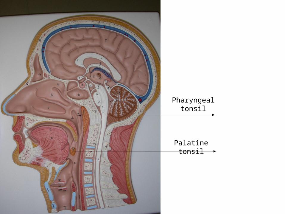
Pharyngeal tonsil
Palatine tonsil
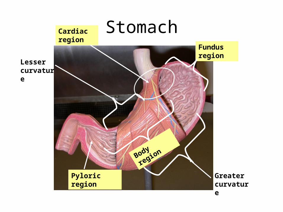
StomachFundus region
Pyloric region
Cardiac region
Greater curvature
Lesser curvature
Body region

Stomach
Pyloric sphincter
Duodenum
RugaePlicae circularis

PANCREAS AND SPLEEN MODELS
• Kidney• Gall bladder• Pancreas• spleen (function is to remove foreign antigens
and aging RBC’s) • duodenum• plicae circularis
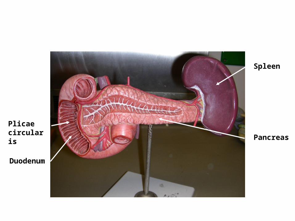
Duodenum
Pancreas
Spleen
Plicae circularis

Spleen
Kidney
Pancreas
Duodenum
Gall bladder
Plicae circularis

GI MODEL• Stomach• Pancreas• Spleen• Pyloric sphincter • circular folds (known as plicae circularis; these are macroscopic folds in the
mucosa)• ascending colon• transverse colon• descending colon• tenia coli• Jejunum (middle part of small intestine)• Ileum• Cecum• Ileocecal valve• Appendix• Rectum• NOTE: Plicae circularis, microvilli, and villi all increase the surface area of
the small intestinal lining

GI Model

Pancreas
Duodenum
Pyloric sphincter
Plicae circularis
Stomach
Spleen

Ascending colon
Descending colon
Transverse colon
Tenia coli
Jejunum
Rectum
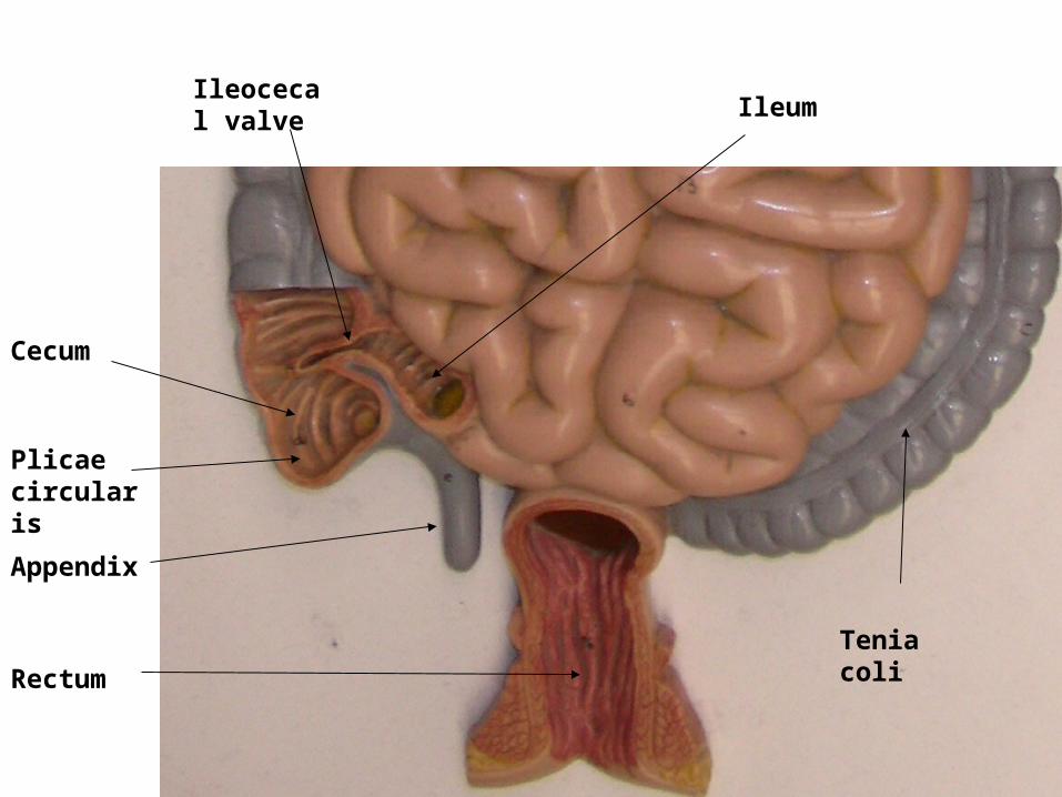
Ileum
Cecum
Ileocecal valve
Appendix
Tenia coli
Rectum
Plicae circularis

INTESTINE MODEL• Stomach• Pancreas• Spleen• Pyloric sphincter • circular folds (known as plicae circularis; these are macroscopic folds in the
mucosa)• ascending colon• transverse colon• descending colon• tenia coli• Jejunum (middle part of small intestine)• Ileum• Cecum• Ileocecal valve• Appendix• Rectum• NOTE: Plicae circularis, microvilli, and villi all increase the surface area of
the small intestinal lining

Greater OmentumTenia coli
Cecum
PancreasDuodenum
Jejunum
Transverse colon

Tenia coli
Mesentery
Pancreas
Common bile duct
Duodenum
Appendix
CecumSigmoid colon (first half)
Ascending colon
Descending colon

Other half of the Sigmoid colon

Liver Models
• gall bladder
• cystic duct
• common bile duct
• falciform ligament
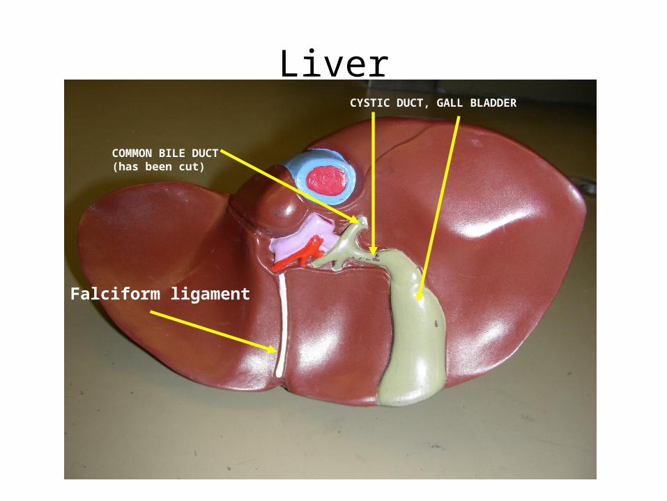
LiverCYSTIC DUCT, GALL BLADDER
COMMON BILE DUCT (has been cut)
Falciform ligament
SLIDES
• Salivary gland
• Tongue– taste bud
• Small Intestine– Villi– Intestinal crypt
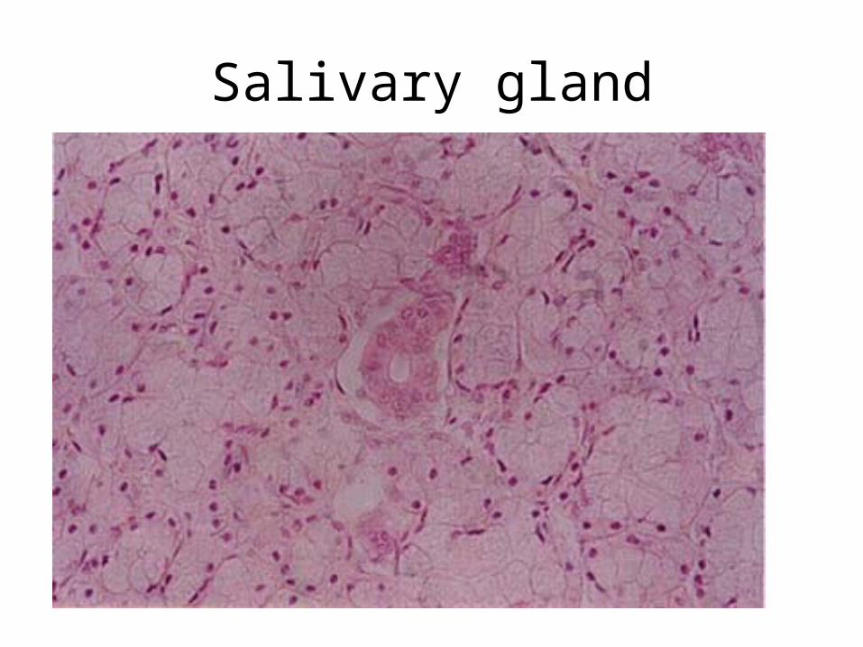
Salivary gland

Salivary gland

TongueTaste buds
SLIDES
• Small intestine – Villi, microvilli, and plicae circularis: (Serve to
increase the surface area of the small intestine)
– Intestinal crypts
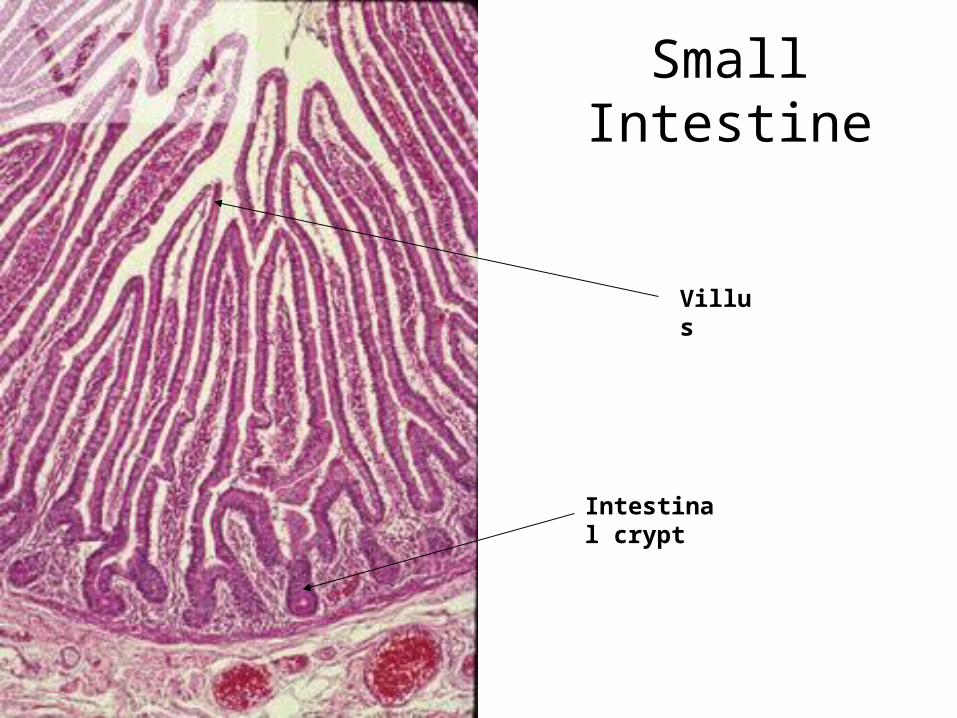
Small Intestine
Villus
Intestinal crypt

SLIDES
• Liver: the functional unit of the liver is the lobule.– central vein
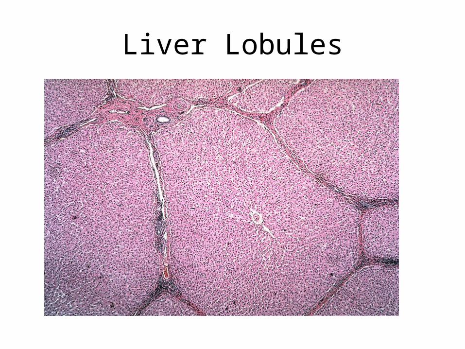
Liver Lobules

Liver Lobule
• Central vein
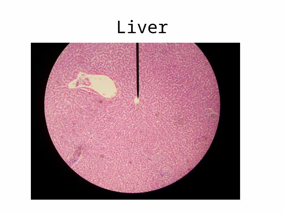
Liver

SLIDES
• Spleen: removes foreign antigens and aging RBC's– Red pulp– White pulp
• Pancreas– Islets– Acini
– Be able to tell the difference between liver, spleen, pancreas, and kidney slides!

Spleen

Spleen
Red Pulp
White pulp

PancreasIslet of Langerhans (secretes insulin)

Pancreas
Acinar cells (secrete enzymes)
Islet of Langerhans (secretes insulin)

Glomerulus
TubuleKidney

Pancreas Kidney
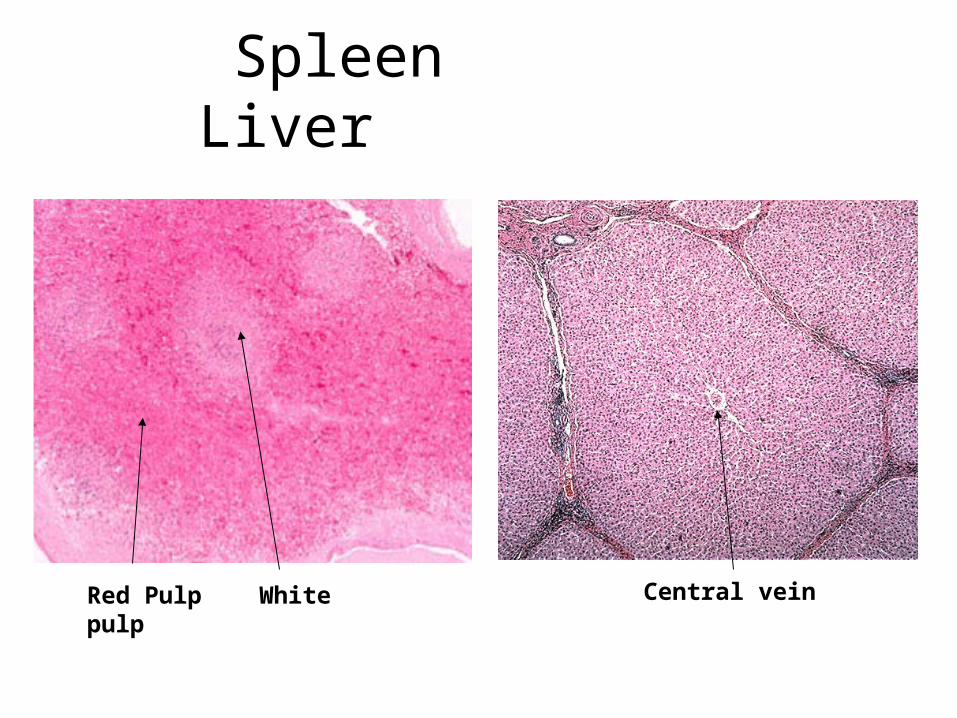
Spleen Liver
Red Pulp White pulp Central vein
Related Documents