Research Paper Diffusion Studies of Nanometer Polymersomes Across Tissue Engineered Human Oral Mucosa Vanessa Hearnden, 1,2 Hannah Lomas, 1 Sheila MacNeil, 1 Martin Thornhill, 2 Craig Murdoch, 2 Andrew Lewis, 3 Jeppe Madsen, 4 Adam Blanazs, 4 Steve Armes, 4 and Giuseppe Battaglia 1,5 Received August 15, 2008; accepted March 19, 2009; published online April 22, 2009 Purpose. To measure the diffusion of nanometer polymersomes through tissue engineered human oral mucosa. Methods. In vitro models of full thickness tissue engineered oral mucosa (TEOM) were used to assess the penetration properties of two chemically different polymersomes comprising two of block copolymers, PMPC-PDPA and PEO-PDPA. These copolymers self-assemble into membrane-enclosed vesicular structures. Polymersomes were conjugated with fluorescent rhodamine in order to track polymersome diffusion. Imaging and quantification of the diffusion properties were assessed by confocal laser scanning microscopy (CLSM). Results. TEOM is morphologically similar to natural oral mucosa. Using CLSM, both formulations were detectable in the TEOM within 6 h and after 48 h both penetrated up to 80 μm into the TEOM. Diffusion of PMPC-PDPA polymersomes was widespread across the epithelium with intra-epithelial uptake, while PEO-PDPA polymersomes also diffused into the epithelium. Conclusions. CLSM was found to be an effective and versatile method for analysing the level of diffusion of polymersomes into TEOM. The penetration and retention of PMPC-PDPA and PEO-PDPA polymersomes means they may have potential for intra-epithelial drug delivery and/or trans-epithelial delivery of therapeutic agents. KEY WORDS: confocal laser scanning microscopy; diffusion; epithelium; oral mucosa; polymersome. INTRODUCTION Historically, most in vitro biological research has relied on two-dimensional monolayer cell culture studies. Whilst these can provide valuable information regarding cell behav- iour and responses, they are poor at replicating the three- dimensional behaviour of tissues and are not always clinically relevant or representative of the in vivo situation. Further- more, because of differences in the cell microenvironment, cells grown in three-dimensional tissue engineered models behave differently than those cultured as monolayers (reviewed in (1,2)). Three-dimensional tissue engineered models of human tissue that accurately reflect the in vivo situation are a very valuable tool for studies of toxicity and diffusion (3,4). In this study we used a full thickness tissue engineered in vitro model of the oral mucosa. These models comprise de-epidermised acellular human dermis repopulated with laboratory-expanded primary human oral epithelial cells and fibroblasts (5). Selvaratnam et al. have compared the permeability of a TEOM, similar to the one used here, with normal human buccal mucosa from adult necropsies (6) and found that the permeability constant, as assessed using permeation of tritated water, was very similar in the model and intact oral mucosa. The TEOM used in this study has been developed for clinical applications (7) and has been used successfully in the clinic to replace scarred tissue in the urethra with nearly 3 years follow-up (8), confirming the physiological relevance of this tissue. Polymersomes are macromolecular aggregates formed in aqueous solution from amphiphilic block copolymers. Their design is based on a concept found in many biological systems, most clearly in cell membranes. Amphiphilic molecules containing a hydrophobic portion and a hydrophilic portion self-assemble when in contact with water to achieve the most entropically favourable configuration. The configuration of the structure depends on the concentration of the molecules and the hydrophilic/hydrophobic balance (9,10,12,13). In this study 0724-8741/09/0700-1718/0 # 2009 Springer Science + Business Media, LLC 1718 Pharmaceutical Research, Vol. 26, No. 7, July 2009 ( # 2009) DOI: 10.1007/s11095-009-9882-6 1 Biomaterials and Tissue Engineering Group, Department of Engi- neering Materials, Kroto Research Institute, North Campus, Uni- versity of Sheffield, Broad Lane, Sheffield, S3 7HQ, UK. 2 Department of Oral & Maxillofacial Medicine & Surgery, School of Clinical Dentistry, University of Sheffield, Sheffield, UK. 3 Biocompatibles UK Ltd., Farnham, Surrey, UK. 4 Department of Chemistry, University of Sheffield, Brook Hill, Sheffield, S3 7HF, UK. 5 To whom correspondence should be addressed. (e-mail: g.battaglia@ sheffield.ac.uk) ABBREVIATIONS: ALI, Air liquid interface; ATRP, Atomic transfer radical polymerisation; CLSM, Confocal laser scanning microscopy; DED, De-epithelialized dermis; PEO 23 -PDPA 15 , Poly(ethylene oxide)- poly(2-(diisopropylamino)ethyl methacrylate); PMPC 25 -PDPA 70 , 2- (Methacryloyloxy)ethyl phosphorylcholine)-poly(2-(diisopropylamino) ethyl methacrylate; THF, Tetrahydrofuran; TEM, Transmission electron microscopy; TEOM, Tissue engineered oral mucosa.

Welcome message from author
This document is posted to help you gain knowledge. Please leave a comment to let me know what you think about it! Share it to your friends and learn new things together.
Transcript

Research Paper
Diffusion Studies of Nanometer Polymersomes Across Tissue EngineeredHuman Oral Mucosa
Vanessa Hearnden,1,2 Hannah Lomas,1 Sheila MacNeil,1 Martin Thornhill,2 Craig Murdoch,2 Andrew Lewis,3
Jeppe Madsen,4 Adam Blanazs,4 Steve Armes,4 and Giuseppe Battaglia1,5
Received August 15, 2008; accepted March 19, 2009; published online April 22, 2009
Purpose. To measure the diffusion of nanometer polymersomes through tissue engineered human oralmucosa.Methods. In vitro models of full thickness tissue engineered oral mucosa (TEOM) were used to assess thepenetration properties of two chemically different polymersomes comprising two of block copolymers,PMPC-PDPA and PEO-PDPA. These copolymers self-assemble into membrane-enclosed vesicularstructures. Polymersomes were conjugated with fluorescent rhodamine in order to track polymersomediffusion. Imaging and quantification of the diffusion properties were assessed by confocal laser scanningmicroscopy (CLSM).Results. TEOM is morphologically similar to natural oral mucosa. Using CLSM, both formulations weredetectable in the TEOM within 6 h and after 48 h both penetrated up to 80 μm into the TEOM. Diffusionof PMPC-PDPA polymersomes was widespread across the epithelium with intra-epithelial uptake, whilePEO-PDPA polymersomes also diffused into the epithelium.Conclusions. CLSM was found to be an effective and versatile method for analysing the level of diffusionof polymersomes into TEOM. The penetration and retention of PMPC-PDPA and PEO-PDPApolymersomes means they may have potential for intra-epithelial drug delivery and/or trans-epithelialdelivery of therapeutic agents.
KEY WORDS: confocal laser scanning microscopy; diffusion; epithelium; oral mucosa; polymersome.
INTRODUCTION
Historically, most in vitro biological research has reliedon two-dimensional monolayer cell culture studies. Whilstthese can provide valuable information regarding cell behav-iour and responses, they are poor at replicating the three-dimensional behaviour of tissues and are not always clinicallyrelevant or representative of the in vivo situation. Further-more, because of differences in the cell microenvironment,cells grown in three-dimensional tissue engineered models
behave differently than those cultured as monolayers(reviewed in (1,2)). Three-dimensional tissue engineeredmodels of human tissue that accurately reflect the in vivosituation are a very valuable tool for studies of toxicity anddiffusion (3,4). In this study we used a full thickness tissueengineered in vitro model of the oral mucosa. These modelscomprise de-epidermised acellular human dermis repopulatedwith laboratory-expanded primary human oral epithelial cellsand fibroblasts (5). Selvaratnam et al. have compared thepermeability of a TEOM, similar to the one used here, withnormal human buccal mucosa from adult necropsies (6) andfound that the permeability constant, as assessed usingpermeation of tritated water, was very similar in the modeland intact oral mucosa. The TEOM used in this study hasbeen developed for clinical applications (7) and has beenused successfully in the clinic to replace scarred tissue in theurethra with nearly 3 years follow-up (8), confirming thephysiological relevance of this tissue.
Polymersomes are macromolecular aggregates formed inaqueous solution from amphiphilic block copolymers. Theirdesign is based on a concept found in many biological systems,most clearly in cell membranes. Amphiphilic moleculescontaining a hydrophobic portion and a hydrophilic portionself-assemble when in contact with water to achieve the mostentropically favourable configuration. The configuration of thestructure depends on the concentration of the molecules andthe hydrophilic/hydrophobic balance (9,10,12,13). In this study
0724-8741/09/0700-1718/0 # 2009 Springer Science + Business Media, LLC 1718
Pharmaceutical Research, Vol. 26, No. 7, July 2009 (# 2009)DOI: 10.1007/s11095-009-9882-6
1 Biomaterials and Tissue Engineering Group, Department of Engi-neering Materials, Kroto Research Institute, North Campus, Uni-versity of Sheffield, Broad Lane, Sheffield, S3 7HQ, UK.
2Department of Oral & Maxillofacial Medicine & Surgery, School ofClinical Dentistry, University of Sheffield, Sheffield, UK.
3 Biocompatibles UK Ltd., Farnham, Surrey, UK.4 Department of Chemistry, University of Sheffield, Brook Hill,Sheffield, S3 7HF, UK.
5 To whom correspondence should be addressed. (e-mail: [email protected])
ABBREVIATIONS: ALI, Air liquid interface; ATRP, Atomic transferradical polymerisation; CLSM, Confocal laser scanning microscopy;DED, De-epithelialized dermis; PEO23-PDPA15, Poly(ethylene oxide)-poly(2-(diisopropylamino)ethyl methacrylate); PMPC25-PDPA70, 2-(Methacryloyloxy)ethyl phosphorylcholine)-poly(2-(diisopropylamino)ethyl methacrylate; THF, Tetrahydrofuran; TEM, Transmissionelectron microscopy; TEOM, Tissue engineered oral mucosa.

we used membrane-enclosed spherical vesicular structures,known as polymersomes.
The first membrane-enclosed structures designed in alaboratory were liposomes made from naturally-derivedphospholipids (14). Liposomes have now been developedfor use as drug carriers for hydrophobic drugs as well ascarriers of DNA for transfection (15). Unfortunately, al-though liposomes are fairly biocompatible they exhibit lowcirculation times in the bloodstream and have poor stability(16). To increase circulation times, stealth liposomes coatedwith poly(ethylene glycol) (PEG) were developed to reducedetection from the body (17). However, the release profile ofmaterials encapsulated into these liposomes is hard tocontrol; some material leaks out quickly while other materialnever gets released (18–20). Moreover, relying on naturallyoccurring phospholipids restricts opportunities to adaptliposome properties into more efficient delivery vehicles.The difficulties experienced with liposomes led to thegeneration of polymersomes; polymer vesicles created viathe self-assembly of amphiphilic block copolymers (12). Bymaking these membrane-enclosed structures from syntheticamphiphiles the stability of the structure is improved andtheir properties can be tailored to meet their desiredapplication (13). In addition, polymersomes comprise rela-tively high molecular weight polymer chains compared tophospholipids and these show a high level of entanglementwithin the bilayer membrane, improving stability compared toliposomes (21). Polymersome membranes can also be tentimes less permeable to water than phospholipid bilayersmaking them far less leaky and more retentive as drugdelivery vectors than liposomes (12,22). Both liposomalbilayer membranes and polymersome membranes have theability to remodel and heal if damaged due to the hydropho-bic effect (23).
Polymersomes are capable of encapsulating both hydro-phobic molecules, such as many cancer drugs, and hydrophilicmolecules such as DNA and proteins. Materials thus farencapsulated into polymersomes include DNA for transfec-tion (24,25), anticancer drugs for intracellular delivery (20),haemoglobin for blood substitutes (26,27) and contrast agentsfor in vivo imaging (28). Antibodies have also been conju-gated to polymersomes for targeted delivery (29).
Major advances in polymer chemistry have opened upmany possibilities for block copolymer formulations andtherefore numerous polymersome structures (30). The deg-radation of polymersomes can be adjusted using differentpolymer formulations. pH sensitive polymersomes (10),oxidative species (31) and biodegradable polymers all de-grade within the body in response to different stimuli(26,32,33).
Delivery of drugs into or across the oral mucosa isdifficult because of the relative impermeability of the oralepithelium. Delivery of drugs into the epithelium (intra-epithelial delivery), is important for the treatment of manymucosal diseases such as squamous cell carcinoma (34).Alternatively, trans-mucosal drug delivery offers the possibil-ity to deliver drugs quickly and effectively into the circulationwithout the need for injection and avoiding the first-passmetabolism of orally-delivered treatments. Trans-epithelialdelivery has been the focus of much research (35). Polymer-somes have the potential to provide a delivery system which
is able to deliver intra-epithelial and trans-epithelial thera-pies. To achieve an effective delivery system it is essential tohave a reproducible in vitro test system that allows us to studyand track polymersome penetration and retention within thedifferent compartments of the oral mucosa.
Here, using confocal laser scanning microscopy, weinvestigate the diffusion properties of polymersomes synthes-ised from two block copolymers (2-(methacryloyloxy)ethylphosphorylcholine)-poly(2-(diisopropylamino)ethyl methac-rylate) (PMPC25-PDPA70) and poly(ethylene oxide)-poly(2-(diisopropylamino)ethyl methacrylate) (PEO23-PDPA15)across TEOM, with long-term view of developing a targetedintra- and trans-epithelial drug delivery systems. To the bestof our knowledge, this is the first study to examinepenetration of nanomaterials through a tissue engineeredepithelia.
MATERIALS AND METHODS
2-(Methacryloyloxy)ethyl phosphorylcholine (MPC;>99%) was provided by Biocompatibles UK Ltd. 2-(Diiso-propylamino)ethyl methacrylate (DPA; Scientific PolymerProducts, USA) was passed through the column supplied bythe manufacturer to remove inhibitor. Copper(I) bromide(CuBr; 99.999%), copper(I) chloride (CuCl; 99.995%), 2,2′-bipyridine (bpy, 99%), 2-bromoisobutyl bromide, triethyl-amine, methanol, tetrahydrofuran (THF) and isopropanolwere purchased from Sigma-Aldrich UK (Poole, Dorset, UK)and were used as received. Column chromatography gradesilica gel 60 (0.063–0.200 mm) used for removal of the atomtransfer radical polymerisation (ATRP) copper catalyst waspurchased from E. Merck (Darmstadt, Germany). Monohy-droxy-capped poly(ethylene oxide) was purchased fromFluka (Poole, Dorset, UK) and freeze-dried before use toremove water. Regenerated Cellulose (RC) dialysis tubing(Spectra Por® 6, molecular weight cut-off 3.5 kDa) waspurchased from Spectrum Labs (Rancho Dominguez, CA,USA). 2-(N-Morpholino)ethyl 2-bromo-2-methylpropanoate(ME-Br) initiator and poly(ethylene oxide) macro-initiator(PEO-Br) were synthesized according to a previouslyreported procedure (36).
PMPC25-PDPA70 Copolymer Synthesis
PMPC25-PDPA70 copolymer was synthesized by ATRPas reported elsewhere (Fig. 1a) (10). Briefly, a Schlenk flaskwith a magnetic stir bar and a rubber septum was chargedwith CuBr (25.6 mg, 0.178 mmol) and MPC (1.32 g,4.46 mmol). ME-Br initiator (50.0 mg, 0.178 mmol) and bpyligand (55.8 mg, 0.358 mmol) were dissolved in methanol(2 ml), and this solution was deoxygenated by bubbling N2 for30 min before being injected into the flask using a syringe.The [MPC]:[ME-Br]:[CuBr]:[bpy] relative molar ratios were25:1:1:2. The polymerization was conducted under a nitrogenatmosphere at 20°C. After 65 min, a mixture of deoxygenatedDPA (2.67 g, 12.5 mmol) and methanol (3 ml) was injectedinto the flask. After 48 h, the reaction solution was diluted byaddition of 200 ml isopropanol and then passed through asilica column to remove the spent Cu catalyst.
1719Polymersome Diffusion Across Human Oral Mucosa
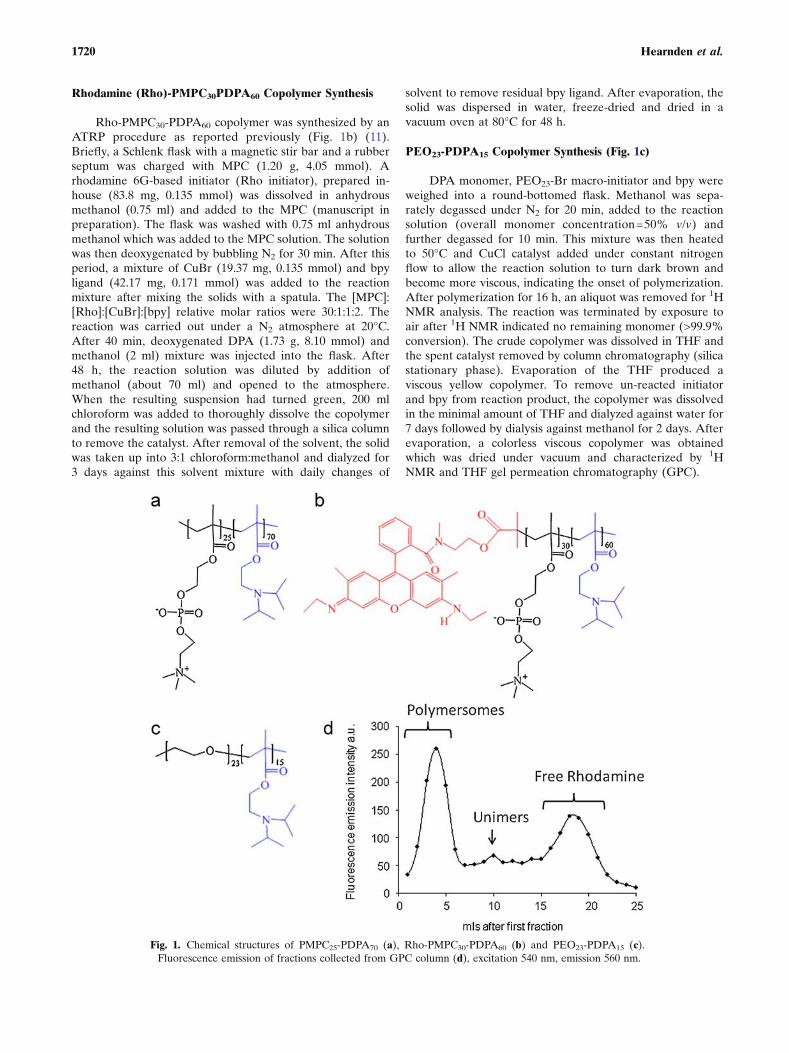
Rhodamine (Rho)-PMPC30PDPA60 Copolymer Synthesis
Rho-PMPC30-PDPA60 copolymer was synthesized by anATRP procedure as reported previously (Fig. 1b) (11).Briefly, a Schlenk flask with a magnetic stir bar and a rubberseptum was charged with MPC (1.20 g, 4.05 mmol). Arhodamine 6G-based initiator (Rho initiator), prepared in-house (83.8 mg, 0.135 mmol) was dissolved in anhydrousmethanol (0.75 ml) and added to the MPC (manuscript inpreparation). The flask was washed with 0.75 ml anhydrousmethanol which was added to the MPC solution. The solutionwas then deoxygenated by bubbling N2 for 30 min. After thisperiod, a mixture of CuBr (19.37 mg, 0.135 mmol) and bpyligand (42.17 mg, 0.171 mmol) was added to the reactionmixture after mixing the solids with a spatula. The [MPC]:[Rho]:[CuBr]:[bpy] relative molar ratios were 30:1:1:2. Thereaction was carried out under a N2 atmosphere at 20°C.After 40 min, deoxygenated DPA (1.73 g, 8.10 mmol) andmethanol (2 ml) mixture was injected into the flask. After48 h, the reaction solution was diluted by addition ofmethanol (about 70 ml) and opened to the atmosphere.When the resulting suspension had turned green, 200 mlchloroform was added to thoroughly dissolve the copolymerand the resulting solution was passed through a silica columnto remove the catalyst. After removal of the solvent, the solidwas taken up into 3:1 chloroform:methanol and dialyzed for3 days against this solvent mixture with daily changes of
solvent to remove residual bpy ligand. After evaporation, thesolid was dispersed in water, freeze-dried and dried in avacuum oven at 80°C for 48 h.
PEO23-PDPA15 Copolymer Synthesis (Fig. 1c)
DPA monomer, PEO23-Br macro-initiator and bpy wereweighed into a round-bottomed flask. Methanol was sepa-rately degassed under N2 for 20 min, added to the reactionsolution (overall monomer concentration=50% v/v) andfurther degassed for 10 min. This mixture was then heatedto 50°C and CuCl catalyst added under constant nitrogenflow to allow the reaction solution to turn dark brown andbecome more viscous, indicating the onset of polymerization.After polymerization for 16 h, an aliquot was removed for 1HNMR analysis. The reaction was terminated by exposure toair after 1H NMR indicated no remaining monomer (>99.9%conversion). The crude copolymer was dissolved in THF andthe spent catalyst removed by column chromatography (silicastationary phase). Evaporation of the THF produced aviscous yellow copolymer. To remove un-reacted initiatorand bpy from reaction product, the copolymer was dissolvedin the minimal amount of THF and dialyzed against water for7 days followed by dialysis against methanol for 2 days. Afterevaporation, a colorless viscous copolymer was obtainedwhich was dried under vacuum and characterized by 1HNMR and THF gel permeation chromatography (GPC).
Fig. 1. Chemical structures of PMPC25-PDPA70 (a), Rho-PMPC30-PDPA60 (b) and PEO23-PDPA15 (c).Fluorescence emission of fractions collected from GPC column (d), excitation 540 nm, emission 560 nm.
1720 Hearnden et al.

Production of Polymersomes
PMPC25-PDPA70 or PEO23-PDPA15 (890 nM) weredissolved in a glass vial in 2:1 chloroform:methanol solution.For rhodamine-labelled samples, 5% (v/v) dissolved rhoda-mine-labelled polymer (Rho-PMPC30PDPA60) was added tothe polymer solution. A copolymer film was formed byevaporating the solvent overnight in a vacuum oven at 50°C. The film was then rehydrated using 2 ml pH 2 PBS(100 mM). Once the film dissolved the pH was increased to7.4. This solution was sonicated for 5 min (Sonicor Instru-ments Corporation, NY, USA) and then purified by gelpermeation chromatography using a sepharose 4B sizeexclusion column to extract the fraction containing vesiclesand remove any remaining free rhodamine dye.
Removal of Free Rhodamine
One ml fractions from the GPC column were collected,diluted 1 in 10 with PBS and the fluorescence of 1 ml of thissolution measured using a fluorescence spectrophotometerwith excitation set at 540 nm and emission at 560 nm (VarianCary Eclipse, CA, USA). Polymersomes were contained infractions 2 ml to 5 ml (Fig. 1d). The subsequent fractions(>5 ml) contained unimers and free rhodamine and werediscarded.
Transmission Electron Microscopy (TEM) Analysisof Polymersomes
For TEM analysis, samples were mounted onto pre-carbon-coated copper grids. Grids were first glow dischargedfor 40 s, and then submerged into the polymersome solution(10 mg/ml for both polymersome solutions) for 60 s. The gridswere then blotted dry and submerged into a phosphotungsticacid solution (0.75% w/v, made up using distilled water) for20 s, before being blotted dry and briefly dried under vacuum.Imaging was performed on a Philips CM100 instrumentoperating at 80 kV equipped with a Gatan 1 k CCD camera.
Cell Culture
Normal oral keratinocytes (NOK) and fibroblasts (NOF)were isolated from oral mucosal biopsies obtained fromconsenting patients during oral surgical procedures. Thebiopsies were incubated overnight at 4°C in 0.1% w/v Difcotrypsin solution supplemented with 100 IU/ml penicillin,100 mg/ml streptomycin and 0.625 μg/ml amphotericin B.The epithelium was then peeled from the connective tissuecomponent. Keratinocytes were gently scraped from theunderside of the epithelium and the top side of the connectivetissue layer using a scalpel. Keratinocytes were culturedaccording to the method of Rheinwald and Green (37).Briefly, cells were cultured on an irradiated mouse fibroblast(i3T3) feeder layer in Green’s media composed of Dulbecco’smodified Eagle’s medium (DMEM) and Hams F12 mediumin a 3:1 (v/v) ratio, supplemented with 10% (v/v) foetal calfserum (FCS), 100 IU/ml penicillin, 100 mg/ml streptomycinand 0.625 μg/ml amphotericin B, 0.1 μM cholera toxin, 10 ng/ml epidermal growth factor (EGF), 0.4 μg/ml hydrocortisone,0.18 mM adenine, 5 μg/ml insulin, 5 μg/ml transferrin, 2 mM
glutamine and 0.2 μM triiodothyronine. The media used toculture the NOK was removed and replenished every 3–4 days until the culture flasks were 80% confluent. To passagethe cells i3T3 cells were detached using 0.02% EDTAsolution for 5 min before removal of NOK from the flasksusing trypsin/0.02% EDTA solution.
NOF were isolated from the connective tissue of oralbiopsies. The connective tissue was finely minced andincubated at 37°C in a 5% CO2 humidified incubatorovernight in 10 ml of 0.5% collagenase A. The isolatedfibroblasts were cultured in DMEM supplemented with 10%(v/v) FCS, 100 IU/ml penicillin, 100 mg/ml streptomycin and0.625 μg/ml amphotericin B. Keratinocytes were used up topassage 3 and fibroblasts between passage 3 and 8.
Preparation of Sterilized De-Epithelized Dermis (DED)
Skin obtained from consenting donors was stored at 4°Cin DMEM supplemented with 10% (v/v) FCS, 100 IU/mlpenicillin, 100 mg/ml streptomycin and 0.625 μg/ml ampho-tericin B for 7 days. DED was de-cellularized in 1 M sodiumchloride for at least 8 h. This method removes the epidermisbut retains basement membrane proteins such as collagen IVand laminin, which aids the subsequent attachment of NOKsin the 3D culture.
Culture of 3D Tissue Engineered Oral Mucosa
TEOM was generated using a previously establishedmethod (7) with the exception that the DED was notsterilised for these in vitro studies. DED was cut into 2 cm×2 cm squares and placed into six well plates submerged inGreen’s media. Chamfered surgical stainless steel rings withan internal diameter of 6 mm were pushed onto the DED toprovide a liquid tight seal. One milliliter of cell suspensioncontaining 5×10⁵ NOKs and 2.5×10⁵ NOFs in Green’s mediawas added into the ring. Green’s media was also addedoutside the ring to stop the cell solution leaking out (Fig. 2,step 1). After 2 days half the media inside the ring wasremoved and replenished with fresh media. On day 3 DEDwith cells attached was brought to an air-liquid interface(ALI) using a stainless steel grid. The underside of the modelwas in contact with Green’s media while the top was exposedto the air to encourage epithelial stratification (Fig. 2, step 2).Models were cultured for 10–14 days at the ALI. Onehundred μl of rhodamine-labelled polymersome was addedto the top surface of the model in a plastic ring and incubatedfor up to 48 h (Fig. 2, step 3). Before imaging the models werecarefully washed three times in PBS and left submerged inPBS for laser scanning confocal imaging.
Confocal Laser Scanning Microscopy (CLSM)
CLSM imaging was done with the samples submerged inPBS using an Acroplan ×40 dipping lens (Carl Zeiss, Jena,Germany) on an upright confocal microscope (Zeiss LSM 510Meta, Jena, Germany). CLSM was used to determine thespectra of autofluorescence emitted from the TEOM andemission spectra of samples exposed to rhodamine-labelledpolymersomes. Samples were excited at 488 nm or 543 nmand emission was detected every 10.45 nm. All measurements
1721Polymersome Diffusion Across Human Oral Mucosa

were taken using the same excitation intensity and detectorgain. Excitation at 488 nm, emission>505 nm detects autofluor-escence from the collagen and other proteins in the sample.Excitation at 543 nm detects rhodamine. As the rhodamine iscovalently attached in the Rho-PMPC-PDPA polymer, thismethod of detection enables tracking of the polymersomes.Three dimensional images of 200 μm×200 μm×100–200 μmwere obtained using the z-stack function. These images aremade up of 50–100 images 2 μm apart.
Histological Analysis
For histological analysis TEOM were fixed in 3%paraformaldehyde for 24 h, processed and embedded inparaffin wax. Four micrometer sections were cut, de-waxed,stained with haematoxylin and eosin and mounted on slidesfor analysis.
RESULTS
Characterization of TEOM
Fig. 3 shows histological analysis of (a) a normal oralmucosa biopsy and (b) the TEOM. This is a full thicknessmodel as it contains both a stratified epithelium and aconnective tissue component containing fibroblasts. Bothimages show the presence of a well attached epithelial layerindicating a good epithelial/connective tissue junction, highlyprolific basal epithelial cells and differentiating cells in theupper layers of the epithelium. In addition, they both have adense fibrous connective tissue containing fibroblasts. Thismodel is representative of normal stratified squamous oralepithelium (38).
In Fig. 4 we show the emission spectra of the oral mucosamodel when excited at 488 nm and 543 nm. Both spectra weretaken using the same settings and the same excitationintensity. There is a broad emission of autofluorescence fromthe TEOM when excited at 488 nm (Fig. 4a) but very littlewhen excited at 543 nm (Fig. 4b). The autofluorescencecomes predominantly from the extra-cellular matrix (ECM)proteins (39). To avoid autofluorescence as far as possible weused rhodamine 6G to label the polymersomes, since thisgroup exhibits maximum fluorescence intensity at 543 nm.
Quantification and Characterization of PolymersomePenetration into Oral Mucosa
Transmission electron micrographs (TEM) showmembrane-enclosed spherical structures with diameters ofaround 100 nm for both formulations (Fig. 5a, b).
After exposure of TEOM to rhodamine labelled poly-mersomes the emission spectra arising from 543 nm excitationchanges dramatically, demonstrating the presence of polymer-somes (Fig. 6a). Fig. 6b shows a confocal image of TEOMexcited at 488 nm and 543 nm after 48 h exposure topolymersomes while Fig. 6c shows the control model withoutpolymersome exposure. Combining both excitation channelsgives a composed image. A negative control of unlabelledpolymersomes also showed no fluorescence in the 543 nmchannel (data not shown). Here we have tracked the diffusionof polymersomes across the oral epithelium using confocallaser scanning microscopy (CLSM).
The diffusion of both PEO23-PDPA15 and PMPC25-PDPA70 polymersomes into the TEOM is time-dependent.In Fig. 7 the z–x plane shows a cross section through the fullthickness model. After 6 h exposure to rhodamine-labelledpolymersomes synthesized from either polymer, rhodamine(red) fluorescence can be seen in the most superficialepithelial layers of the model (Fig. 7b, e). The position ofthe polymersomes with respect to the epithelial/connectivetissue interface can be judged from the relative positions of
Fig. 2. Schematic diagram of the method used to culture tissueengineered full thickness oral mucosa. Primary oral keratinocytes andoral fibroblasts are seeded onto a DED scaffold (step 1). After cellattachment the DED is raised to an air–liquid interface to encourageepithelial stratification using a stainless steel grid (step 2). Afterculturing for 10–14 days at the air-liquid interface, polymersomes areadded to the top surface of the models (step 3).
1722 Hearnden et al.

the red fluorescence of the polymersomes and the blueautofluorescence of the connective tissue. Due to the highECM protein content of the connective tissue, the autofluor-escence of the connective tissue is more intense than from theepithelium, which appears relatively dark. Fig. 7c, f and gclearly show a layer of rhodamine fluorescence in thesuperficial cells of the epithelium, above a darker region(the basal layers of epithelium) which lies above the highlyautofluorescent connective tissue. After 30 h there is morewidespread uptake of polymersomes by the superficialepithelial cells for the PMPC25-PDPA70 polymersomes thanthe PEO23-PDPA15 (Fig. 7c, f). After 48 h exposure toPMPC25-PDPA70 it appears that all cells within the epitheli-um contain polymersomes (Fig. 7d) and display high levels offluorescence. There is no dark region between the upperepithelium and connective tissue component in Fig. 7d,suggesting polymersomes have diffused to the epithelial/connective tissue interface and have been internalized bybasal, as well as more superficial, epithelial cells. The TEOMexposed to PEO23-PDPA15 expressed high levels of polymer-some uptake and fluorescence in the upper layers of theepithelium, but less uptake and fluorescence in the deeper
layers of the epithelium compared to PMPC25-PDPA70
polymersomes (Fig. 7g). The PEO23-PDPA15 polymersomediffusion pattern is less uniform and appears patchy. Howev-er, this may be due to differences in fluorescence intensitybetween the two polymersome formulations.
Confocal images were also used to obtain quantitativeinformation about the penetration of the two polymersomeformulations into the TEOM. Zmax was calculated from thetop of the sample to the deepest point of rhodaminefluorescence with higher intensity than the control (Fig. 8a).The depth of penetration was very similar for the two types ofpolymersome.
DISCUSSION
The aim of this study was to explore the use of TEOM asa convenient surrogate for normal oral mucosa to examinethe penetration of two novel forms of polymersomes throughoral mucosa using CLSM. These polymersomes are beingdeveloped to deliver drugs, proteins or genes into tissues forfuture clinical use. Information on their penetration intotissues is clearly an important part of their development.
Fig. 4. Emission spectra of TEOM without polymersomes when excited with 488 nm and 543 nm lasers. z–xsection of TEOM excited at 488 nm (a), and at 543 nm (b). Scale bar 50 μm.
Fig. 3. Haematoxylin and eosin stained sections of an oral mucosa biopsy from a healthy patient (a) andTEOM cultured for 10 days at air liquid interface (b). Scale bar 200 μm.
1723Polymersome Diffusion Across Human Oral Mucosa

This study showed time-dependent penetration of poly-mersomes into the epithelium of a TEOM model over 48 h.The TEOM used in this study closely resembles normal oralmucosa with epithelial cell differentiation and stratification,minimal keratinization of superficial epithelial layers and awell attached epithelium on a collagenous connective tissuecontaining fibroblasts (38). MacKenzie et al. demonstratedthat fibroblasts and subepithelial connective tissue influencethe formation of epithelia (40). Factors released by fibroblastsin the connective tissue affect keratinocyte differentiation andproliferation (41). Culturing the model at an air–liquidinterface encourages the epithelial cells to differentiate,stratify and organize themselves to imitate natural oralmucosa (Fig. 3).
To successfully image the diffusion of polymersomesthrough the TEOM, autofluorescence of the TEOM neededto be taken into account. By measuring the fluorescenceemission across a range of wavelengths a lambda stack wasobtained. This shows the autofluorescence has a broademission when excited at 488 nm but this is negligible whenexcited with a 543 nm laser (Fig. 4). Therefore, labelling thepolymersomes with rhodamine 6G (maximally excited at543 nm) allowed us to track their diffusion throughout themodel using CLSM. The autofluorescence from 488 nmexcitation is useful to judge the spatial location of the
polymersomes. Using CLSM we could clearly track thepenetration of both PMPC25-PDPA70 and PEO23-PDPA15
rhodamine-labelled polymers over time. The PMPC25-PDPA70 polymersomes appear to penetrate the epitheliummove more quickly and over a more widespread area thanthe PEO23-PDPA15 polymersomes. After 48 h the fluores-cence generated by the PMPC25-PDPA70 polymersomesfollows the contours of the basal epithelial cells which residealong the basement membrane, strongly suggesting that thesepolymersomes diffuse as far as the basement membrane. ThePEO23-PDPA15 polymersomes did not appear to diffuse asmuch as the PMPC25-PDPA70 polymersomes, although spotsof fluorescence could still be observed deep within theepithelium with this polymer.
The hydrophobic portion of both polymers is the PDPAblock. This component is pH-sensitive; below its pKa of 6.4the PDPA block is hydrophilic. When the block copolymer isabove its pKa the PDPA chain becomes hydrophobic, whichdrives the self-assembly of polymersomes. This hydrophobicblock becomes shielded from the aqueous solution by thePMPC or PEO block when in the polymersome configura-tion. PEO, also known as PEG, is a biocompatible polymerwhich exhibits very low protein adsorption; it is non-immunogenic and non-antigenic (42). Therefore PEO23-PDPA15 polymersomes are unable to bind to cell membrane
Fig. 6. Emission spectra of tissue engineered oral mucosa after exposure to rhodamine-labelled polymer-somes excited at 543 nm (a). z–x section of TEOM exposed to PMPC25-PDPA70 polymersomes for 48 h (b),z–x section of TEOM with no polymersome exposure (c). Blue=488 nm excitation. Red=543 nm excitation.Scale bar 50 μm.
Fig. 5. TEM images of PEO23-PDPA15 (a) and PMPC25-PDPA70 (b).
1724 Hearnden et al.

proteins, limiting the amount of polymersomes internalizedby the cells (43). PMPC is also non-fouling to proteins whenexpressed at a surface. However, PMPC25-PDPA70 polymer-somes bind more strongly to cell membranes when compared
to PEO23-PDPA15 increasing the amount of polymersomesinternalized (25,43). The behaviours seen from the twodifferent polymersome preparations may both be clinicallyuseful.
Fig. 7. Tissue engineered models were exposed to rhodamine-labelled PMPC25-PDPA70 or PEO23-PDPA15
polymersomes for 6, 30 or 48 h. (a) shows control sample, no exposure to polymersomes. CLSM images ofTEOM exposed to PMPC25-PDPA70 for 6 h (b), 30 h (c) or 48 h (d). TEOM exposed to PEO23-PDPA15
polymersomes for 6 h (e), 30 h (f) or 48 h (g). Images on the left show x–y and x–z sections. Images on theright are three-dimensional projections of these models, approximately 200μm × 200μm × 100μm. Scale bar50 μm.
1725Polymersome Diffusion Across Human Oral Mucosa

The oral epithelium provides a defensive barrier pre-venting unwanted materials from entering the body andretaining fluid within the mucosa (38). This barrier alsoprevents many therapeutic agents from crossing the epitheli-um. For diseases of the oral mucosa such as lichen planus andsquamous cell carcinoma, which affect the basal cells of theepithelium, intra-epithelial drug delivery is desirable (34,44).Topical delivery is an important method for delivering drugsat high concentration into diseased oral mucosa whilstlimiting any systemic toxicity or side effects. Currently, onlya limited range of drugs such as topical steroid preparationscan be delivered across the epithelial permeability barrier andinto basal epithelial cells. Developing drug delivery systemsthat can carry a wider array of therapeutic agents across theepithelial permeability barrier, e.g. biological agents andgenes, and delivering them with high efficiency into basalkeratinocytes, would open up a broad spectrum of potentiallymore effective therapeutic tools for treating many oralmucosal diseases.
For other diseases, widespread systemic delivery of drugsis desirable. For convenience, this is achieved whereverpossible by oral administration. Unfortunately, this is notpossible with many novel therapeutic agents, particularlybiological compounds such as peptides and antibodies (45). Ifdelivered orally, these compounds get degraded by enzymeswithin the digestive system. As a result, such drugs often haveto be given parenterally i.e. by injection. This severely limitsthe usefulness of these drugs, particularly for the treatment ofchronic and less severe diseases where they would otherwiserevolutionize treatment. Trans-epithelial delivery offers theprospect of a more practical and effective method of
delivering such drugs if the permeability barrier can beovercome without damaging the therapeutic agent or disrupt-ing the epithelium (35,46). For trans-epithelial delivery, avector that crosses the epithelium and is not retained byepithelial cells but delivers its contents into the connectivetissue or circulation is highly desirable. Current examples ofeffective trans-epithelial drug delivery in the oral mucosainclude the delivery of Diazepam to treat patients with statusepilepticus and the delivery of glyceryl trinitrate for the reliefof episodes of angina (35).
The permeability barrier of TEOM is very similar to thatof normal human oral mucosa (6). This permeability barrier isdue to the supra-basal cells (Fig. 3), also known as spinouscells. These cells begin to differentiate from the stem cell-likebasal cells as they move towards the surface of the epitheliawhere they form strong intercellular desmosomal junctionsand release sphingolipids into the surrounding intercellularspaces to produce a barrier which is impermeable to watersoluble molecules (47).
There are two methods of diffusion across the epithelialpermeability barrier. From the current data we are unable todetermine which path is taken by our polymersomes.However, there are two possible routes: (1) the intercellularpathway where material passes through lipid-rich domainsaround the cells and (2) the trans-cellular pathway wherematerial passes in and out of the cells in each layer (34). Tightjunctions which are, found in the superficial layer of theepithelia, force material to pass through the cells. Thesejunctions are not sufficiently widespread enough in the oralepithelium to completely prevent material using the intercel-lular pathway but are more widespread in the skin (47). Thepolymersome membranes are highly deformable, enablingthem to pass through gaps between cells in tissues such as theoral epithelium that are smaller than their own diameter. Thedeformability of polymersomes has been demonstrated (12)using micropipette aspiration of giant (20–50 μm) polymer-somes. The diffusion of polymersomes across the oral epithe-lium may not be driven by a concentration gradient but rathervia a hydration gradient across the different layers (48). In theepithelium the hydration gradient pulls the carriers throughthe relatively dehydrated keratinized layer until they reach theviable epithelium, where there is a higher level of hydration.These rather bulky carriers may then be pushed through thelower layers of the epithelium by more carriers, which aredrawn into the epithelium by the hydration gradient (48).
Both polymersome formulations are able to penetrateinto the oral epithelium, demonstrating they can travel acrossthe permeability barrier of our TEOM. Further research isneeded to determine by which mechanism these polymer-somes pass across the epithelium. However, the method oftracking the polymersomes described in this paper is animportant first step.
CONCLUSION
The TEOM model combined with CLSM is an effectiveand versatile model to monitor the diffusion and penetrationof polymersomes into oral mucosa. Confocal microscopy canbe used to both image the diffusion and to quantify the depthof penetration. PMPC25-PDPA70 and PEO23-PDPA15 poly-mersomes are both able to diffuse through the tissue
Fig. 8. Depth of penetration (zmax) for PMPC25-PDPA70 polymer-somes into the model as measured by analysing the x–z and y–zprojections (a). Depth of penetration over time for the two differentpolymersome formulations (b).
1726 Hearnden et al.

engineered epithelia in a time-dependent manner. The depthof penetration of the two polymers is very similar. Bothformulations may have clinical potential for the intra- and/ortrans-epithelial delivery of therapeutic agents.
ACKNOWLEDGMENTS
We would like to thank Dr. Anthony Bullock, Dr. HelenColley, Mr. Tom Smart and Ms. Marzia Massignani for theirhelp and guidance. This work was supported by funding fromEPSRC (DTA PhD studentship to Vanessa Hearnden) andBiocompatibles UK Ltd.
REFERENCES
1. MacNeil S. Progress and opportunities for tissue-engineeredskin. Nature 2007;445:874–80. doi:10.1038/nature05664.
2. Moharamzadeh K, Brook IM, Van Noort R, Scutt AM,Thornhill MH. Tissue-engineered oral mucosa: a review ofthe scientific literature. J Dent Res. 2007;86:115–24. doi:10.1177/154405910708600203.
3. Schmalz G. Materials science: biological aspects. J Dent Res.2002;81:660–3. doi:10.1177/154405910208101001.
4. Schmalz G, Schuster U, Koch A, Scheweikl H. Cytotoxicity oflow pH dentin-bonding agents in a dentin barrier test in vitro. JEndod. 2002;28:188–92. doi:10.1097/00004770-200203000-00011.
5. Chakrabarty KH, DawsonRA, Harris P, Layton C, BabuM, GouldL, Phillips J, Leigh I, Green C, Freedlander E, Mac Neil S.Development of autologous human dermal–epidermal compositesbased on sterilized human allodermis for clinical use. Br JDermatol. 1999;141:811–23. doi:10.1046/j.1365-2133.1999.03153.x.
6. Selvaratnam L, Cruchley AT, Navsaria H, Wertz PW, Hagi-PavliEP, Leigh IM, Squier CA, Williams DM. Permeability barrierproperties of oral keratinocyte cultures: a model of intact humanoral mucosa. Oral Dis. 2001;7:252–8. doi:10.1034/j.1601-0825.2001.70409.x.
7. Bhargava S, Chapple CR, Bullock AJ, Layton C, MacNeil S.Tissue-engineered buccal mucosa for substitution urethroplasty.BJU Int. 2004;93:807–11. doi:10.1111/j.1464-410X.2003.04723.x.
8. Bhargava S, Patterson JM, Inman RD, MacNeil S, Chapple CR.Tissue-engineered buccalmucosa urethroplasty—clinical outcomes.Eur Urol. 2008;53:1263–71. doi:10.1016/j.eururo.2008.01.061.
9. Smart T, Lomas H, Massignani M, Flores-Merino MV, Perez LR,BattagliaG. Block copolymer nanostructures. Nanotoday 2008;3:1–9.
10. Du J, Tang Y, Lewis AL, Armes SP. pH-sensitive vesicles basedon a biocompatible zwitterionic diblock copolymer. J Am ChemSoc. 2005;127:17982–3. doi:10.1021/ja056514l.
11. Madsen J. PhD thesis, University of Sheffield; 200912. Discher BM, Won Y, Ege DS, Lee JCM, Bates FS, Discher DE,
Hammer DA. Polymersomes: tough vesicles made from diblockcopolymers. Science 1999;284:1143–6. doi:10.1126/science.284.5417.1143.
13. Discher DE, Eisenberg A. Polymer vesicles. Science 2002;297:967–73. doi:10.1126/science.1074972.
14. Bangham AD. A correlation between surface charge andcoagulant action of phospholipids. Nature 1961;192:1197–8.doi:10.1038/1921197a0.
15. Lasic DD, Papahadjopoulos D. Medical applications of lip-osomes. Amsterdam: Elsevier; 1998.
16. Photos PJ, Bacakova L, Discher B, Bates FS, Discher DE.Polymer vesicles in vivo: correlations with PEG molecularweight. J Control Release 2003;90:323–34. doi:10.1016/S0168-3659(03)00201-3.
17. Lasic DD. Sterically stabilized vesicles. Angew Chem Int Ed.1994;33:1685–98. doi:10.1002/anie.199416851.
18. Lasic DD. Recent developments in medical applications ofliposomes: sterically stabilized liposomes in cancer therapy andgene delivery in vivo. J Control Release 1997;48:203–22.doi:10.1016/S0168-3659(97)00045-X.
19. Duncan R. The dawning era of polymer therapeutics. Nat RevDrug Discov. 2003;2:347–60. doi:10.1038/nrd1088.
20. Discher DE, Ortiz V, Srinivas G, Klein ML, Kim Y, Christian D,Cai S, Photos P, Ahmed F. Emerging applications of polymer-somes in delivery: from molecular dynamics to shrinkage oftumors. Prog Polym Sci. 2007;32:838–57. doi:10.1016/j.progpolymsci.2007.05.011.
21. Battaglia G, Ryan A. Bilayers and interdigitation in blockcopolymer vesicles. J Am Chem Soc. 2005;127:8757–64.doi:10.1021/ja050742y.
22. Battaglia G, Ryan AJ, Tomas S. Polymeric vesicle permeability:a facile chemical assay. Langmuir 2006;22:4910–3. doi:10.1021/la060354p.
23. Aranda-Espinoza H, Bermudez H, Bates FS, Discher DE.Electromechanical limits of polymersomes. Phys Rev Lett.2001;87:208301. doi:10.1103/PhysRevLett.87.208301.
24. Lomas H, Canton I, MacNeil S, Du J, Armes SPA, Ryan AJ,Lewis AL, Battaglia G. Biomimetic pH sensitive polymersomesfor efficient DNA encapsulation and delivery. Adv Mater.2007;19:4238–43. doi:10.1002/adma.200700941.
25. Lomas H, Massignani M, Abdullah KA, Canton I, Lo Presti C,MacNeil S, Du J, Blanazs A, Madsen J, Armes SP, Lewis AL,Battaglia G. Non-cytotoxic polymer vesicles for rapid andefficient intracellular delivery. Faraday Discuss 2008;139:143.doi:10.1039/b717431d.
26. Rameez S, Alosta H, Palmer AF. Biocompatible and biodegrad-able polymersome encapsulated hemoglobin: a potential oxygencarrier. Bioconjugate Chem. 2008;19:1025–32. doi:10.1021/bc700465v.
27. Arifin DR, Palmer AF. Polymersome encapsulated hemoglobin:a novel type of oxygen carrier. Biomacromolecules 2005;6:2172–81. doi:10.1021/bm0501454.
28. Ghoroghchian PP, Frail PR, Susumu K, Blessington D, BrannanAK, Bates FS, Chance B, Hammer DA, Therien MJ. Near-infrared-emissive polymersomes: self-assembled soft matter forin vivo optical imaging. PNAS 2005;102:2922–7. doi:10.1073/pnas.0409394102.
29. Lin JJ, Ghoroghchian PP, Zhang Y, Hammer DA. Adhesion ofantibody-functionalized polymersomes. Langmuir 2006;22:3975–9. doi:10.1021/la052445c.
30. Matyjaszewski K, Spanswick J. Controlled/living radical poly-merization. Mater Today 2005;8:26–33. doi:10.1016/S1369-7021(05)00745-5.
31. Cerritelli S, Velluto D, Hubbell JA. PEG-SS-PPS: reduction-sensitive disulfide block copolymer vesicles for intracellular drugdelivery. Biomacromolecules 2007;8:1966–72. doi:10.1021/bm070085x.
32. Meng F, Engbers GHM, Feijen J. Biodegradable polymersomesas a basis for artificial cells: encapsulation, release and targeting.J Control Release 2005;101:187–98. doi:10.1016/j.jconrel.2004.09.026.
33. Ben-Haim N, Broz P, Marsch S, Meier W, Hunziker P. Cell-specific integration of artificial organelles based on functional-ized polymer vesicles. Nano Letters 2008;8:1368–73. doi:10.1021/nl080105g.
34. Sood S, Shiff SJ, YangCS, ChenX. Selection of topically applied non-steroidal anti-inflammatory drugs for oral cancer chemoprevention.Oral Oncol. 2005;41:562–7. doi:10.1016/j.oraloncology.2005.01.003.
35. Zhang H, Zhang J, Streisand JB. Oral mucosal drug delivery:clinical pharmacokinetics and therapeutic applications. ClinPharmacokinet. 2002;41:661–80. doi:10.2165/00003088-200241090-00003.
36. Robinson KL, Weaver JVM, Armes SP, Diaz Marti E, MeldrumF. Synthesis of controlled-structure sulfate-based copolymers viaatom transfer radical polymerisation and their use as crystalhabit modifiers for BaSO4. J Mater Chem. 2002;12:890–6.doi:10.1039/b200348c.
37. Rheinwald JG, Green H. Serial cultivation of strains of humanepidermal keratinocytes: the formation of keratinizing coloniesfrom single cells. Cell 1975;6:331–43. doi:10.1016/S0092-8674(75)80001-8.
38. Nanci A. Ten Cate’s oral histology—development, structure andfunction. St. Louis: Mosby; 2003.
39. Schenke-Layland K, Riemann I, Damour O, Stock UA, König K.Two-photon microscopes and in vivo multiphoton tomographs—
1727Polymersome Diffusion Across Human Oral Mucosa

powerful diagnostic tools for tissue engineering and drug delivery.Adv Drug Deliv Rev. 2006;58:878–96. doi:10.1016/j .addr.2006.07.004.
40. Mackenzie IC, Fusenig NE. Regeneration of organized epithelialstructure. J Invest Dermatol. 1983;81:189s–94s. doi:10.1111/1523-1747.ep12541093.
41. Costea DE, Loro LL, Dimba EAO, Vintermyr OK, JohannessenAC. Crucial effects of fibroblasts and keratinocyte growth factor onmorphogenesis of reconstituted human oral epithelium. J InvestDermatol. 2003;121:1479–86. doi:10.1111/j.1523-1747.2003.12616.x.
42. Alcantar NA, Aydil ES, Israelachvili JN. Polyethylene glycol-coated biocompatible surfaces. J Biomed Mater Res. 2000;51:343–51. doi:10.1002/1097-4636(20000905)51:3<343::AID-JBM7>3.0.CO;2-D.
43. Massignani M, Blanazs A, Madsen J, Armes SP, Lewis AL,Battaglia G. Engineering polymeric nanovectors for effectiveand rapid cellular delivery. In preparation (2008).
44. Campisi G, Giandalia G, De Caro V, Di Liberto C, Aricò P,Giannola LI. A new delivery system of clobetasol-17-propionate
(lipid-loaded microspheres) compared with a conventionalformulation (lipophilic ointment in a hydrophilic phase) intopical treatment of atrophic/erosive oral lichen planus. A PhaseIV, randomized, observer-blinded, parallel group clinical trial.Br J Dermatol. 2004;150:984–90. doi:10.1111/j.1365-2133.2004.05943.x.
45. Blanchette J, Kavimandan N, Peppas NA. Principles of trans-mucosal delivery of therapeutic agents. Biomed Pharmacother.2004;58:142–51. doi:10.1016/j.biopha.2004.01.006.
46. Guy RH. Current status and future prospects of transdermaldrug delivery. Pharm Res. 1996;13:1765–9. doi:10.1023/A:1016060403438.
47. Shimono M, Clementi F. Intercellular junctions of oral epitheli-um. I. Studies with freeze-fracture and tracing methods ofnormal rat keratinized oral epithelium. J Ultrastruct Res.1976;56:121–36. doi:10.1016/S0022-5320(76)80145-1.
48. Cevc G, Gebauer D. Hydration-driven transport of deformablelipid vesicles through fine pores and the skin barrier. Biophys J.2003;84:1010–24. doi:10.1016/S0006-3495(03)74917-0.
1728 Hearnden et al.
Related Documents