Proc. Nati. Acad. Sci. USA Vol. 87, pp. 8670-8674, November 1990 Cell Biology Differentiation of F9 embryonal carcinoma cells induced by the c-jun and activated c-Ha-ras oncogenes (transcription factor AP1/gene regulation/retinoic acid/endoderm) YUKO YAMAGUCHI-IWAI*, MASANOBU SATAKE*, YOTA MURAKAMI*, MASAHARU SAKAItt, MASAMI MURAMATSUt, AND YOSHIAKI ITO*§ *Department of Viral Oncology, Institute for Virus Research, Kyoto University, Shogoin, Sakyo-ku, Kyoto 606, Japan; tDepartment of Biochemistry, Faculty of Medicine, The University of Tokyo, Hongo, Bunkyo-ku, Tokyo 113, Japan; and tDepartment of Biochemistry, Hokkaido University School of Medicine, Kita-ku, Sapporo 060, Japan Communicated by Peter K. Vogt, August 6, 1990 ABSTRACT The activated c-Ha-ras oncogene induced APi-site DNA-binding activity in F9 cells. This induction appeared to be due, at least in part, to the induction of c-jun transcription. Both activated c-Ha-ras and c-jun induced the differentiation of F9 cells to endoderm-like cells. Thus, AP1 appears to play a key role in the initial stage of F9 cell differentiation. The nuclear protooncogene c-jun was originally identified as a cellular homologue of the transforming gene of an avian retrovirus, avian sarcoma virus 17 (1). The product of the gene, Jun protein, forms a heterodimer with Fos, the product of the c-fos nuclear protooncogene and its family, to become a transcriptional activator protein, AP1, which recognizes and binds to an AP1 consensus sequence (reviewed in refs. 2 and 3). When quiescent cells are stimulated by serum growth factors, very rapid and transient expression of c-fos and c-jun is observed. AP1 is also known to respond to a tumor promoter, phorbol 12-myristate 13-acetate (PMA), and to enhance gene expression (4, 5). Therefore, AP1 is consid- ered to play a key role in switching on the gene expression that ultimately leads to DNA replication and cell division. More recently, AP1 has been implicated as a regulator of DNA replication (6). Here we present evidence that AP1 is also involved in the initial stage of cell differentiation. The murine embryonal carcinoma cell line F9 has been used widely to study the mechanism of gene regulation in the process of cell differentiation (7), since F9 cells can be induced to differentiate to endoderm-like cells by retinoic acid and cAMP (8). AP1 is undetectable in F9 cells, whereas it is easily detectable after differentiation of the cells (9). The expression of both c-jun and c-fos is very low in F9 cells (10). An interesting question is whether AP1 becomes detectable as a consequence of differentiation or whether AP1 is re- quired for cell differentiation. Muller and Wagner reported in 1984 (11) that the overexpression of c-fos in F9 cells induced F9 cell differentiation. However, only a small fraction of the cells expressing c-fos underwent differentiation, regardless of the levels of c-fos expression (12). Moreover, the pheno- type of the differentiated cells was somewhat different from that of the cells induced to differentiate by retinoic acid (11). The activated Ha-ras oncogene [Ha-ras(Val-12)] and PMA stimulate the DNA binding of murine AP1, PEBP1 (13), or PEA1 (14) in lymphoid cells and in fibroblasts (15, 16). PMA superinduces the transcriptional activating function of AP1 in the presence of protein synthesis inhibitors by posttransla- tional modification of the proteins (17). On the other hand, the mechanism of the induction of AP1 by Ha-ras(Val-12) has not been studied in detail. Here we show that Ha-ras(Val-12) induces c-jun expres- sion and the AP1 DNA-binding activity in F9 cells. This finding allowed us to analyze whether AP1 would induce F9 cell differentiation. MATERIALS AND METHODS Cell Culture. F9 cells were cultured in Dulbecco's modified Eagle's medium (DMEM) supplemented with 10%6 (vol/vol) fetal bovine serum at 370C in humidified 10% C02/90% air. Plasmids. The chloramphenicol acetyltransferase (CAT) gene constructs used were pA10CAT2 derivatives, p(WA)2- CAT and p(MlA)2-CAT through p(M4A)2-CAT, in which the CAT gene is regulated by the simian virus 40 (SV40) early promoter under the control of dimers of the polyomavirus A element or a mutated A element (16). pT24c3 is a recombinant plasmid harboring Ha-ras(Val-12) (18). pHfARJ101, in which rat c-jun derived from pRJ101 (19) is regulated by the human 8-actin enhancer and promoter, was constructed by inserting a 2010-base-pair (bp) EcoRI fragment of rat c-jun cDNA into the HindIII-BamHI sites of the mammalian expression vector pHPAPr-1 (20). pHfBARJ101(tr) is a frame- shift mutant of pHl3ARJ101 in which 2 bp were inserted at the Acc I site in the c-jun coding region by restriction enzyme digestion at this site, filling the sticky ends with appropriate nucleotides, and religation. This insertion resulted in the truncation of the c-jun product. pSV2neo contains the neo- mycin-resistance gene driven by the SV40 enhancer and promoter (21). DNA Transfection and CAT Assays. F9 cells were plated at 106 per 100-mm dish 5 hr before transfection. Each CAT construct (2.5 ug) was cotransfected into cells with 17.5 pxg of pT24c3 or pBR322 by calcium phosphate coprecipitation. Twelve hours later, cells were treated with 15% (vol/vol) glycerol in DMEM for 1 min and harvested 36 hr after transfection. CAT assays were performed as described (16). Transfections were performed at least three times with two different preparations of plasmid DNA. Preparation of Whole Cell Extract and Mobility-Shift Assay. F9 cells were transfected with pT24c3 (20 jig) and pSV2neo (1 ,g). Selection with G418 (400 ,ug/ml) was started 24 hr later and cells were cultured for 4 days. Surviving cells were collected and a whole cell extract was prepared (22). The polyomavirus A element cloned into the BamHI site of the pUC13 polylinker was excised by cleavage at EcoRI and HindIII sites and the 5' ends were labeled with 32p. The binding reaction of the probe with the cell extract and subsequent polyacrylamide gel electrophoresis were per- formed as described (13). Abbreviations: PMA, phorbol 12-myristate 13-acetate; CAT, chlor- amphenicol acetyltransferase. §To whom reprint requests should be addressed. 8670 The publication costs of this article were defrayed in part by page charge payment. This article must therefore be hereby marked "advertisement" in accordance with 18 U.S.C. §1734 solely to indicate this fact.

Welcome message from author
This document is posted to help you gain knowledge. Please leave a comment to let me know what you think about it! Share it to your friends and learn new things together.
Transcript

Proc. Nati. Acad. Sci. USAVol. 87, pp. 8670-8674, November 1990Cell Biology
Differentiation of F9 embryonal carcinoma cells induced by thec-jun and activated c-Ha-ras oncogenes
(transcription factor AP1/gene regulation/retinoic acid/endoderm)
YUKO YAMAGUCHI-IWAI*, MASANOBU SATAKE*, YOTA MURAKAMI*, MASAHARU SAKAItt,MASAMI MURAMATSUt, AND YOSHIAKI ITO*§*Department of Viral Oncology, Institute for Virus Research, Kyoto University, Shogoin, Sakyo-ku, Kyoto 606, Japan; tDepartment of Biochemistry, Facultyof Medicine, The University of Tokyo, Hongo, Bunkyo-ku, Tokyo 113, Japan; and tDepartment of Biochemistry, Hokkaido University School of Medicine,Kita-ku, Sapporo 060, Japan
Communicated by Peter K. Vogt, August 6, 1990
ABSTRACT The activated c-Ha-ras oncogene inducedAPi-site DNA-binding activity in F9 cells. This inductionappeared to be due, at least in part, to the induction of c-juntranscription. Both activated c-Ha-ras and c-jun induced thedifferentiation of F9 cells to endoderm-like cells. Thus, AP1appears to play a key role in the initial stage of F9 celldifferentiation.
The nuclear protooncogene c-jun was originally identified asa cellular homologue of the transforming gene of an avianretrovirus, avian sarcoma virus 17 (1). The product of thegene, Jun protein, forms a heterodimer with Fos, the productof the c-fos nuclear protooncogene and its family, to becomea transcriptional activator protein, AP1, which recognizesand binds to an AP1 consensus sequence (reviewed in refs.2 and 3). When quiescent cells are stimulated by serumgrowth factors, very rapid and transient expression of c-fosand c-jun is observed. AP1 is also known to respond to atumor promoter, phorbol 12-myristate 13-acetate (PMA), andto enhance gene expression (4, 5). Therefore, AP1 is consid-ered to play a key role in switching on the gene expressionthat ultimately leads to DNA replication and cell division.More recently, AP1 has been implicated as a regulator ofDNA replication (6). Here we present evidence that AP1 isalso involved in the initial stage of cell differentiation.The murine embryonal carcinoma cell line F9 has been
used widely to study the mechanism ofgene regulation in theprocess of cell differentiation (7), since F9 cells can beinduced to differentiate to endoderm-like cells by retinoicacid and cAMP (8). AP1 is undetectable in F9 cells, whereasit is easily detectable after differentiation of the cells (9). Theexpression of both c-jun and c-fos is very low in F9 cells (10).An interesting question is whether AP1 becomes detectableas a consequence of differentiation or whether AP1 is re-quired for cell differentiation. Muller and Wagner reported in1984 (11) that the overexpression of c-fos in F9 cells inducedF9 cell differentiation. However, only a small fraction of thecells expressing c-fos underwent differentiation, regardlessof the levels of c-fos expression (12). Moreover, the pheno-type of the differentiated cells was somewhat different fromthat of the cells induced to differentiate by retinoic acid (11).The activated Ha-ras oncogene [Ha-ras(Val-12)] and PMA
stimulate the DNA binding of murine AP1, PEBP1 (13), orPEA1 (14) in lymphoid cells and in fibroblasts (15, 16). PMAsuperinduces the transcriptional activating function ofAP1 inthe presence of protein synthesis inhibitors by posttransla-tional modification of the proteins (17). On the other hand,the mechanism of the induction ofAP1 by Ha-ras(Val-12) hasnot been studied in detail.
Here we show that Ha-ras(Val-12) induces c-jun expres-sion and the AP1 DNA-binding activity in F9 cells. Thisfinding allowed us to analyze whether AP1 would induce F9cell differentiation.
MATERIALS AND METHODSCell Culture. F9 cells were cultured in Dulbecco's modified
Eagle's medium (DMEM) supplemented with 10%6 (vol/vol)fetal bovine serum at 370C in humidified 10% C02/90% air.
Plasmids. The chloramphenicol acetyltransferase (CAT)gene constructs used were pA10CAT2 derivatives, p(WA)2-CAT and p(MlA)2-CAT through p(M4A)2-CAT, in which theCAT gene is regulated by the simian virus 40 (SV40) earlypromoter under the control of dimers of the polyomavirus Aelement or a mutatedA element (16). pT24c3 is a recombinantplasmid harboring Ha-ras(Val-12) (18). pHfARJ101, inwhich rat c-jun derived from pRJ101 (19) is regulated by thehuman 8-actin enhancer and promoter, was constructed byinserting a 2010-base-pair (bp) EcoRI fragment of rat c-juncDNA into the HindIII-BamHI sites of the mammalianexpression vector pHPAPr-1 (20). pHfBARJ101(tr) is a frame-shift mutant ofpHl3ARJ101 in which 2 bp were inserted at theAcc I site in the c-jun coding region by restriction enzymedigestion at this site, filling the sticky ends with appropriatenucleotides, and religation. This insertion resulted in thetruncation of the c-jun product. pSV2neo contains the neo-mycin-resistance gene driven by the SV40 enhancer andpromoter (21).DNA Transfection and CAT Assays. F9 cells were plated at
106 per 100-mm dish 5 hr before transfection. Each CATconstruct (2.5 ug) was cotransfected into cells with 17.5 pxgof pT24c3 or pBR322 by calcium phosphate coprecipitation.Twelve hours later, cells were treated with 15% (vol/vol)glycerol in DMEM for 1 min and harvested 36 hr aftertransfection. CAT assays were performed as described (16).Transfections were performed at least three times with twodifferent preparations of plasmid DNA.
Preparation of Whole Cell Extract and Mobility-Shift Assay.F9 cells were transfected with pT24c3 (20 jig) and pSV2neo(1 ,g). Selection with G418 (400 ,ug/ml) was started 24 hr laterand cells were cultured for 4 days. Surviving cells werecollected and a whole cell extract was prepared (22). Thepolyomavirus A element cloned into the BamHI site of thepUC13 polylinker was excised by cleavage at EcoRI andHindIII sites and the 5' ends were labeled with 32p. Thebinding reaction of the probe with the cell extract andsubsequent polyacrylamide gel electrophoresis were per-formed as described (13).
Abbreviations: PMA, phorbol 12-myristate 13-acetate; CAT, chlor-amphenicol acetyltransferase.§To whom reprint requests should be addressed.
8670
The publication costs of this article were defrayed in part by page chargepayment. This article must therefore be hereby marked "advertisement"in accordance with 18 U.S.C. §1734 solely to indicate this fact.

Proc. Natl. Acad. Sci. USA 87 (1990) 8671
Phase-Contrast Microscopy. Plasmid DNA (20 ,ug) ofpH/3ARJ101, pHBARJ1O1(tr), or pT24c3 was cotransfectedwith 1 gg of pSV2neo into F9 cells. Cells were incubated withG418 and photographs were taken at a magnification of 100after 6 days of selection.Immunofluorescent Staining. F9 cells cultured on glass
coverslips were cotransfected with pHf3ARJ101, pHf3-ARJ101(tr), or pT24c3 together with pSV2neo, and G418-resistant cells were selected. Surviving cells were fixed with3.7% (vol/vol) formaldehyde in phosphate-buffered saline(PBS) for 20 min at room temperature. In the case of stainingof intracellular antigens, the cells were permeabilized with0.1% (vol/vol) Nonidet P-40 in PBS for 10 min after fixation.The cells were incubated for 40 min at 370C with the firstantibody (mouse monoclonal anti-SSEA-1 IgM, rabbit anti-mouse laminin IgG fraction, or rat monoclonal anti-Hsp47IgG) and then incubated with rhodamine-conjugated goatantibody against mouse IgM, rabbit IgG, or rat IgG (Cooper-Cappel). The coverslips were mounted on glass slides andphotomicrographs were taken with a x40 objective.Plasminogen Activator Assay. F9 cells were cotransfected
with pH,3ARJ101, pH/3ARJ101(tr), or pT24c3 together withpSV2neo in 35-mm dishes. After 48 hr of incubation withG418, cells were washed twice with PBS and covered with 1.5ml of prewarmed (45°C) DMEM containing 0.75% Bactopurified agar (Difco), 2.5% skim milk (Difco), and 0.2 unit ofbovine plasminogen (Daiichi Chemical, Tokyo). The plateswere incubated for 24 hr at 37°C and photographed.RNA Preparation and Northern Blot Analysis. Total RNA
was prepared (16) from F9 cells 20 hr after transfection.Poly(A)+ RNA was selected by oligo(dT)-cellulose columnchromatography. RNA was electrophoresed in 1% agarosegels with 0.66 M formaldehyde and transferred onto nitro-cellulose or Zeta-Probe (Bio-Rad) filters in 20x SSC (1x is0.15 M NaCl/0.015 M sodium citrate, pH 7). Hybridizationwas carried out for 18 hr at 42°C in 50% (vol/vol) formamide/0.1% Ficoll/0.1% polyvinylpyrrolidone/0.1% bovine serumalbumin/5x SSC/50 mM Tris-HCI, pH 7.4/0.1% SDS con-taining sheared salmon sperm DNA at 100 ,ug/ml (for nitro-cellulose filters) or in 0.5 M NaHPO4, pH 7.2/1 mMEDTA/7% SDS (for Zeta-Probe). To probe the RNA derivedfrom the c-Ha-ras, c-jun, or f-actin gene, a 6.5-kbp BamHIfragment of pT24c3 (18), a 2.0-kbp EcoRI fragment ofpRJ101(19), or a 0.8-kbp Pst I-Kpn I fragment ofpAL41 that harborsmouse skeletal muscle f-actin cDNA (23), respectively, wasused. Hybridized filters were washed twice in 2x SSC/0.5%SDS for 30 min at room temperature and twice in 0.1 xSSC/0.5% SDS for 30 min at 43°C.
RESULTSInduction of the AP1-Site DNA-Binding Activity by Ha-
ras(Val-12). The DNA-binding activity of murine AP1,PEBP1 (13), or PEA1 (14) can be enhanced by transientlyexpressed Ha-ras(Val-12) (13, 15). To analyze this enhance-ment in detail, Ha-ras(Val-12) was transfected into F9 cells inwhich AP1/PEBP1 was undetectable (9). The AP1/PEBP1DNA-binding activity was monitored using the A element ofthe polyomavirus enhancer as a probe (Fig. 1). TheA elementrepresents one of the two cores of the enhancer and containsthe three sequence motifs recognized by PEBP1 (PEA1),PEBP2/3 (PEA2), and PEBP5 (PEA3) (16). Since there is noneed to distinguish PEBP1 (PEA1) from AP1 for the discus-sion in the present study, the term AP1 will be used through-out the rest of this paper to indicate the factor(s) that bindsto the AP1 consensus sequence, including PEBP1 (PEA1).When the CAT construct that was under the control of theA-element dimer was transfected, the CAT activity becamedetectable only after cotransfection with Ha-ras(Val-12) (Fig.1, lanes 1 and 2). Introduction of pairwise mutations in the
Ln PEBP 5 PEBP1(AP1) PEBP2,3 LCAGGAAGTGACTAACT.GACCGCAG
M3A ---C T - --- ---- --+ !- -M4A - -- --C -I -._ i
MA-A G--- TM2A - - --- C-- G --
(WA)2 (M3A)2 (M4A)2 (M1A)2 (M2A)- pAiACAT:;-- r- r~ --li -| --
c-Ha-ras - + - + - + - +-
* 9
% acetylated 0.1 5.7 0.5 1.2 0.1 0.2 0.1 0.3 0.3 45 0.1 0.3L 3 4 D 6J i 8 9 10
FIG. 1. Effect of transiently expressed Ha-ras(Val-12) on theA-element activity of polyomavirus enhancer in F9 cells. (Upper)Sequences of the wild-type (WA) and four kinds (M1A-M4A) ofmutated A element are shown in the upper panel. Boxes indicate thebinding sites of PEBP1(AP1) and PEBP2/3 (13). The binding site ofPEBP5 is underlined (41). (Lower) CAT activity in cell extracts.Each CAT construct under the control of the A-element dimer wascotransfected into F9 cells with Ha-ras(Val-12) (even-numberedlanes) or with control plasmid pBR322 (odd-numbered lanes). CATactivity is expressed as the percentage of acetylated chloramphen-icol.
AP1 or PEBP5 binding sites virtually abolished the responseof the A element to Ha-ras(Val-12) (lanes 3-8). Therefore,Ha-ras(Val-12) was able to induce the AP1 as well as thePEBP5 DNA-binding activity in F9 cells. This induction wasa relatively early response, observed as early as 24 hr aftertransfection (data not shown). The induction of PEBP5binding activity suggests that the target of the activation byHa-ras(Val-12) was multiple. The characteristics of PEBP5have been reported (41). Mutations in the PEBP2/3 siteincreased the response to Ha-ras(Val-12) nearly 10-fold(compare lanes 2 and 10). This is related to the derepressionresponse observed by Wasylyk et al. (24) and has beendiscussed fully elsewhere (25). The backbone vector con-taining no insert did not respond to cotransfected Ha-ras(Val-12) (lanes 11 and 12).The induction of the AP1 DNA-binding activity by Ha-
ras(Val-12) was also examined by mobility-shift assay.Whole cell extract was prepared from F9 cells that werecotransfected with pSV2neo and Ha-ras(Val-12) and selectedwith G418 for 4 days. The extract was incubated with a32P-labeled A-element probe and analyzed by mobility-shiftassay (Fig. 2, lanes 3-10). The slowly migrating band repre-sents AP1, since its formation was specifically abolished byinclusion of excess amounts of the AM oligonucleotide as acompetitor in the binding reaction but was not affected by theinclusion of the AE oligonucleotide. The AM and AE oligo-nucleotides represent the AP1 and PEBP2/3 binding sites,respectively (Fig. 2 Lower). The formation of the bandindicated with the asterisk was abolished by the AE oligo-nucleotide, suggesting that this band is related to PEBP2/3.On the other hand, the cell extract prepared from F9 cellstransfected only with pSV2neo did not give rise to a discern-ible band (lane 1). This indicates that the expression ofHa-ras(Val-12) in F9 cells induced the AP1-site DNA-bindingactivity. Since G418 selection was used to enrich the trans-fected cell population, the above experiment could not beperformed much earlier than 4 days after transfection.
Cell Biology: Yamaguchi-lwai et al.

8672 Cell Biology: Yamaguchi-Iwai et al.
c~omrjetitor : At: AMv.: CL
:-f% ::,c=' 6 Css OD ;= __LI' -- - in, _
X ( ;x. -- > V X..
|2 ( A ~~~~~P1
a4** A * .EB
1 4 5 6 7 8
.rBOA CAGGAAGTGACTAACTGACCGCAGG"CCT TCACTGAT -GACTGGCGT C
AM
Al
FIG. 2. Mobility-shift assay of factors expressed in Ha-ras(Val-12)-transfected F9 cells. Whole cell extracts from pSV2neo- (lane 1)or Ha-ras(Val-12)- (lanes 3-10) transfected F9 cells were incubatedwith 32P-labeled A-element (WA) probe before electrophoresis. AMor AE oligonucleotide (sequences indicated below the autoradio-gram) were included in the reaction mixture at 50-, 100-, or 1000-foldmolar excess. Two boxes within the AM and AE fragments indicatethe minimum binding sites of PEBP1 (AP1) and PEBP2/3, respec-tively. The nuclear extract from murine NIH 3T3 fibroblasts wasused in lane 2 and the position of PEBP1 (AP1) is indicated at right.The band marked by the asterisk may represent PEBP2/3 (see text).
Induction of c-jun Gene Expression by Ha-ras(Val-12). Toanalyze the mechanism of induction of AP1 activity byHa-ras(Val-12), poly(A)+ RNA was prepared from F9 cells 20hr after transfection with Ha-ras(Val-12). Northern analysiswith c-jun-specific 32P-labeled DNA revealed that the amountof the 2.7-kilobase (kb) c-jun-specific transcript was signifi-cantly higher in Ha-ras(Val-12) transfectants than in controlcells (Fig. 3). By densitometric scanning of the autoradio-gram, the increase in the c-jun transcript was calculated to be2.4-fold after normalization using an internal control ofp8-actin transcript. The efficiency of transfection was found tobe 1% by transfecting a parallel culture with the lacZ genedriven by the long terminal repeat (LTR) promoter of theRous sarcoma virus and subsequently counting the number ofcells stained blue after the addition of 5-bromo-4-chloro-3-indolyl 13-D-galactoside. By taking this transfection efficiencyinto consideration, the overall increase of c-jun expression byHa-ras(Val-12) was estimated to be 140-fold. The c-fos tran-
F 9
c Ha-ras 4 --
C LiJl 4j
,,)tarfin
I~tr_ A
FIG. 3. Induction of endogenousc-jun expression by Ha-ras(Val-12) in
2S F9 cells. RNA was extracted fromnontransfected F9 cells (lane 2) orfrom cells transfected with Ha-ras(Val-12) (lane 1). Five micrograms
18 of poly(A)+ RNA, recovered by pass-ing 250 Ag of total RNA through an
oligo(dT)-cellulose column twice,was electrophoresed through an aga-rose gel. The blotted filter was hy-bridized with a 32P-labeled c-junprobe (Upper) and rehybridized witha mouse B-actin probe (Lower). Posi-tions of 18S and 28S rRNA are shownas size markers.
script could not be detected under the conditions used. Thisresult suggests that the induction of the AP1 binding activityin F9 cells by Ha-ras(Val-12) was due, at least in part, to theincrease of c-jun expression.
Morphological Changes of F9 Cells Induced by Ha-ras(Val-12) and c-jun. Since AP1 was detectable in F9 cells afterdifferentiation by retinoic acid (9), it was of interest todetermine the long-term effect of Ha-ras(Val-12) on F9 cells.When F9 cells were cotransfected with Ha-ras(Val-12) andpSV2neo and selected for G418 resistance for 4-6 days,practically all the surviving cells were morphologically dif-ferent from the parental F9 cells. The majority of the cellsformed colonies and were multiplying, although they seemedto stop growing after several cycles of division. Thesegrowing cells were flattened polygonal cells, similar in mor-phology to retinoic acid-treated F9 cells (Fig. 4A, c).The induction of AP1 activity by Ha-ras(Val-12) appeared
to precede the morphological differentiation of F9 cells(24-36 hr vs. 4-6 days). Hence, a causative role ofAP1 in thisdifferentiation was envisaged. As mentioned earlier, theintroduction of c-fos into F9 cells induced differentiation butonly partially (12). Therefore, we tested the effect of c-jun.The morphological differentiation of c-jun-transfected F9cells was very similar to that of Ha-ras(Val-12)-transfectedF9 cells (Fig. 4A, a). As a control, we also transfected aplasmid that has the potential to express a truncated form ofthe Jun protein that lacks the DNA-binding domain. Themorphology of such F9 cells (Fig. 4A, b) was indistinguish-able from that of parental F9 cells. We also observed nomorphological change in F9 cells when the nonactivatedc-Ha-ras protooncogene was introduced (data not shown).Northern analysis of the RNAs extracted from the c-jun- or
mutant c-jun-transfected cells 20 hr after transfection re-vealed the expected 2.0-kb c-jun-specific RNA (Fig. 5, lanes2 and 3). The amounts of each transcript detected werecomparable, indicating that the wild-type and the mutatedc-jun genes were transfected and expressed at about the sameefficiency. Since relatively small amounts of poly(A)+ RNAwere loaded on the gel compared to that used in Fig. 3, theendogenous mouse c-jun transcript was not detected underthese conditions (lane 1). Therefore, the morphological dif-ferentiation of F9 cells was likely to be due to the function ofJun. In the case of Ha-ras(Val-12)-transfected cells, a 1.2-kbc-Ha-ras-specific RNA was detected, which was the ex-pected size for the transcript from the transfected plasmid(lane 5). The endogenous c-Ha-ras transcript was not de-tected under the conditions used (lane 4).
Characterization of F9 Cells Induced to Differentiate byHa-ras(Val-12) and c-jun. The stage-specific embryonic anti-gen SSEA-1 (26) is a known stem-cell marker present in F9cells, but it disappears when cells are induced to differentiateby retinoic acid. The presence of SSEA-1 was examined bythe indirect immunofluorescence method. SSEA-1 was de-tected on the surface of F9 cells or on those expressingmutated c-jun (Fig. 4B, lb) but was completely absent fromHa-ras(Val-12)-(lc) or c-jun-(la) transfected cells.The expression of several differentiation marker proteins
including laminin (27), plasminogen activator (8), and a heatshock protein, Hsp47, which is associated with collagen typeIV (28), were also examined. Cytoplasmic laminin (Fig. 4B,2a and 2c) and Hsp47 (3a and 3c) were detectable by indirectimmunofluorescence in cells transfected with c-jun or Ha-ras(Val-12) but were undetectable in cells transfected withmutated c-jun (2b and 3b). Plasminogen activator was simi-larly induced in c-jun or Ha-ras(Val-12)-transfected cells(Fig. 4C, a and c) but not in cells transfected with the mutatedc-jun (b), as judged by the transparent area generated byproteolysis of casein in overlaid milk. Expression of Ha-ras(Val-12) or c-jun in F9 cells did not preferentially select adifferentiated subpopulation of cells that existed in the orig-
Proc. Natl. Acad. Sci. USA 87 (1990)
.--
Proc. Nati. Acad. Sci. USA 87 (1990) 8673
- c-jurl C-JLur! 'tr, leo c-Ha-ras
c-jun (tr) c-Ha-ras
-28S
.i.
:;
- 28S
---18S
a b c
C-JUnl (tr) c-Ha -ras
4 51 2 3
2
3
b
C
c-jun c-jun (tr) c-Ha-ras
a b c
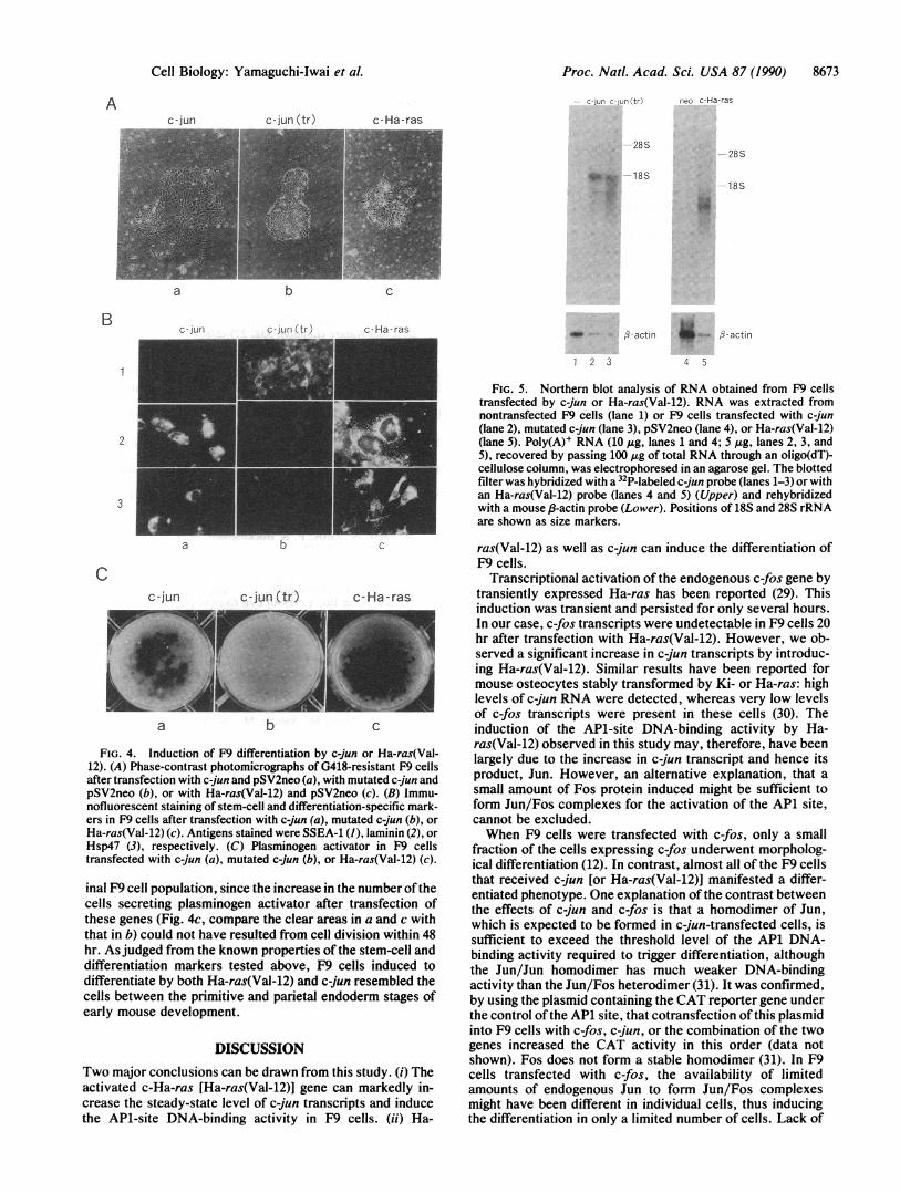
FIG. 4. Induction of F9 differentiation by c-jun or Ha-ras(Val-12). (A) Phase-contrast photomicrographs of G418-resistant F9 cellsafter transfection with c-jun and pSV2neo (a), with mutated c-jun andpSV2neo (b), or with Ha-ras(Val-12) and pSV2neo (c). (B) Immu-nofluorescent staining of stem-cell and differentiation-specific mark-ers in F9 cells after transfection with c-jun (a), mutated c-jun (b), orHa-ras(Val-12) (c). Antigens stained were SSEA-1 (1), laminin (2), orHsp47 (3), respectively. (C) Plasminogen activator in F9 cellstransfected with c-jun (a), mutated c-jun (b), or Ha-ras(Val-12) (c).
inal F9 cell population, since the increase in the number ofthecells secreting plasminogen activator after transfection ofthese genes (Fig. 4c, compare the clear areas in a and c withthat in b) could not have resulted from cell division within 48hr. As judged from the known properties of the stem-cell anddifferentiation markers tested above, F9 cells induced todifferentiate by both Ha-ras(Val-12) and c-jun resembled thecells between the primitive and parietal endoderm stages ofearly mouse development.
DISCUSSIONTwo major conclusions can be drawn from this study. (i) Theactivated c-Ha-ras [Ha-ras(Val-12)] gene can markedly in-crease the steady-state level of c-jun transcripts and inducethe AP1-site DNA-binding activity in F9 cells. (ii) Ha-
FIG. 5. Northern blot analysis of RNA obtained from F9 cellstransfected by c-jun or Ha-ras(Val-12). RNA was extracted fromnontransfected F9 cells (lane 1) or F9 cells transfected with c-jun(lane 2), mutated c-jun (lane 3), pSV2neo (lane 4), or Ha-ras(Val-12)(lane 5). Poly(A)' RNA (10 Mtg, lanes 1 and 4; 5 ,tg, lanes 2, 3, and5), recovered by passing 100 Ag of total RNA through an oligo(dT)-cellulose column, was electrophoresed in an agarose gel. The blottedfilter was hybridized with a 32P-labeled c-jun probe (lanes 1-3) or withan Ha-ras(Val-12) probe (lanes 4 and 5) (Upper) and rehybridizedwith a mouse f3-actin probe (Lower). Positions of 18S and 28S rRNAare shown as size markers.
ras(Val-12) as well as c-jun can induce the differentiation ofF9 cells.
Transcriptional activation of the endogenous c-fos gene bytransiently expressed Ha-ras has been reported (29). Thisinduction was transient and persisted for only several hours.In our case, c-fos transcripts were undetectable in F9 cells 20hr after transfection with Ha-ras(Val-12). However, we ob-served a significant increase in c-jun transcripts by introduc-ing Ha-ras(Val-12). Similar results have been reported formouse osteocytes stably transformed by Ki- or Ha-ras: highlevels of c-jun RNA were detected, whereas very low levelsof c-fos transcripts were present in these cells (30). Theinduction of the APi-site DNA-binding activity by Ha-ras(Val-12) observed in this study may, therefore, have beenlargely due to the increase in c-jun transcript and hence itsproduct, Jun. However, an alternative explanation, that asmall amount of Fos protein induced might be sufficient toform Jun/Fos complexes for the activation of the AP1 site,cannot be excluded.When F9 cells were transfected with c-fos, only a small
fraction of the cells expressing c-fos underwent morpholog-ical differentiation (12). In contrast, almost all of the F9 cellsthat received c-jun [or Ha-ras(Val-12)] manifested a differ-entiated phenotype. One explanation of the contrast betweenthe effects of c-jun and c-fos is that a homodimer of Jun,which is expected to be formed in c-jun-transfected cells, issufficient to exceed the threshold level of the APi DNA-binding activity required to trigger differentiation, althoughthe Jun/Jun homodimer has much weaker DNA-bindingactivity than the Jun/Fos heterodimer (31). It was confirmed,by using the plasmid containing the CAT reporter gene underthe control ofthe AP1 site, that cotransfection of this plasmidinto F9 cells with c-fos, c-jun, or the combination of the twogenes increased the CAT activity in this order (data notshown). Fos does not form a stable homodimer (31). In F9cells transfected with c-fos, the availability of limitedamounts of endogenous Jun to form Jun/Fos complexesmight have been different in individual cells, thus inducingthe differentiation in only a limited number of cells. Lack of
Ac-jun
B c-jun
Cell Biology: Yamaguchi-lwai et al.
16'g.id
dw i-actin 4 I,--.- I;-actin

8674 Cell Biology: Yamaguchi-Iwai et al.
laminin expression in c-fos-transfected F9 cells (11) might bedue to low levels of Fos/Jun complexes, even if they areformed, in the cells. Regardless of the mechanism, ourobservation, together with that of Muller and Wagner (11)demonstrates that AP1 can act as an inducer of F9 celldifferentiation.An important question to be answered is whether AP1 is
involved when retinoic acid induces F9 cell differentiation.We used a strong promoter to express c-jun in the presentstudy. How faithfully our results represent the physiologicalprocesses induced by retinoic acid would have to be carefullyevaluated. Within the limits of the present study, however,the phenotype of the cells induced to differentiate by c-jun issimilar to that after retinoic acid induction. Adenovirus ElAprotein can also induce the partial differentiation of F9 cells(32). It is necessary to examine whether the ElA geneinduces c-jun expression.The expression of c-myc rapidly decreases as F9 cells
differentiate. Since antisense c-myc RNA induces F9 differ-entiation, c-myc appears to have an active role in maintainingF9 cells in an undifferentiated state (33). It is interesting thatthe silencer found in the regulatory region of human c-mycharbors the AP1 binding site to which a factor containing Fosbinds (34). The role ofAP1 might be to inhibit the expressionof c-myc in this case.Neuronal differentiation of a pheochromocytoma cell line,
PC12, can be induced by nerve growth factor (35) or byintroducing activated Ha-ras into the cells (36, 37). There aresome apparent similarities in gene regulation between F9 andPC12 cells during differentiation. Treatment of PC12 cellswith nerve growth factor transiently induces the expressionof both c-fos and c-jun (38). The induction of PC12 differen-tiation by Ha-ras(Val-12) also appears to be mediated byAP1, since the transcriptional activating function ofAP1 canbe induced by Ha-ras in the cells (39). In this case, thetranscription of c-fos is augmented by Ha-ras(Val-12) and theactivation occurs through the serum-responsive element inthe c-fos regulatory region (39). When both F9 and PC12differentiation are taken into consideration, it appears thatAP1 is involved in triggering cell differentiation at multiplelevels of the developmental hierarchy.
It must be noted, however, that AP1 may antagonize theprocess of cell differentiation in a different situation. Intro-duction of c-fos or activated ras has been reported to sup-press the MyoDi-induced differentiation of C2 fibroblastsinto myoblasts (40).AP1 has been shown to play an essential role in growth
regulation and oncogenesis (2, 3). The present study andthose from other laboratories suggest that it has an importantrole in developmental regulation as well.
We thank Drs. K. Nagata and R. Kannagi (Kyoto University) forgifts of anti-Hsp47 and anti-SSEA-1 antibodies, respectively. Wethank Dr. Y. Ogiso (Medical College of Kyoto Prefecture) for adviceon plasminogen activator assay. This work was supported in part bya Grant-in-Aid for Cancer Research (subject no. 63010036) and by aGrant-in-Aid for Scientific Research (subject no. 63440027) from theMinistry of Education, Science, and Culture, Japan.
1. Maki, Y., Bos, T. J., Davis, C., Starbuck, M. & Vogt, P. K.(1987) Proc. Natl. Acad. Sci. USA 84, 2848-2852.
2. Vogt, P. K. & Tjian, R. (1988) Oncogene 3, 3-7.3. Curran, T. & Franza, B. R., Jr. (1988) Cell 55, 395-397.4. Angel, P., Imagawa, M., Chiu, R., Stein, B., Imbra, R. J.,
Rahmsdorf, H. J., Jonat, C., Herrlich, P. & Karin, M. (1987)Cell 49, 729-739.
5. Lee, W., Mitchell, P. & Tjian, R. (1987) Cell 49, 741-752.6. Murakami, Y., Asano, M., Satake, M. & Ito, Y. (1990) Onco-
gene 5, 5-13.7. Martin, G. R. (1980) Science 209, 768-776.8. Strickland, S. & Mahdavi, V. (1978) Cell 15, 393-403.9. Kryszke, M.-H., Piette, J. & Yaniv, M. (1987) Nature (London)
328, 254-256.10. Chiu, R., Angel, P. & Karin, M. (1989) Cell 59, 979-986.11. Muller, R. & Wagner, E. F. (1984) Nature (London) 311,
438-442.12. Ruther, U., Wagner, E. F. & Muller, R. (1985) EMBO J. 4,
1775-1781.13. Satake, M., lbaraki, T. & Ito, Y. (1988) Oncogene 3, 69-78.14. Piette, J. & Yaniv, M. (1987) EMBO J. 6, 1331-1337.15. Imler, J. L., Schatz, C., Wasylyk, C., Chatton, B. & Wasylyk,
B. (1988) Nature (London) 332, 275-278.16. Yamaguchi, Y., Satake, M. & Ito, Y. (1989) J. Virol. 63,
1040-1048.17. Imbra, R. J. & Karin, M. (1986) Nature (London) 323, 555-558.18. Santos, E., Tronick, S. R., Aaronson, S. A., Pulciani, S. &
Barbacid, M. (1982) Nature (London) 298, 343-347.19. Sakai, M., Okuda, A., Hatayama, I., Sato, K., Nishi, S. &
Muramatsu, M. (1989) Cancer Res. 49, 5633-5637.20. Gunning, P., Leavitt, J., Muscat, G., Ng, S.-Y. & Kedes, L.
(1987) Proc. Natl. Acad. Sci. USA 84, 4831-4835.21. Southern, P. J. & Berg, P. (1982) J. Mol. Appl. Genet. 1,
327-341.22. Reichel, R., Kovesdi, 1. & Nevins, J. R. (1987) Cell 48,
501-506.23. Minty, A. J., Caravatti, M., Robert, B., Cohen, A., Daubas, P.,
Weydert, A., Gros, F. & Buckingham, M. E. (1981) J. Biol.Chem. 256, 1008-1014.
24. Wasylyk, B., Imler, J. L., Chatton, B., Schatz, C. & Wasylyk,C. (1988) Proc. Natl. Acad. Sci. USA 85, 7952-7956.
25. Furukawa, K., Yamaguchi, Y., Ogawa, E., Shigesada, K.,Satake, M. & Ito, Y. (1990) Cell Growth Differ. 1, 135-147.
26. Solter, D. & Knowles, B. B. (1978) Proc. Nati. Acad. Sci. USA75, 5565-5569.
27. Kahan, B. & Adamson, E. D. (1983) Cold Spring Harbor Conf.Cell Prolif. 10, 131-141.
28. Saga, S., Nagata, K., Chen, W.-T. & Yamada, K. M. (1987) J.Cell Biol. 105, 517-527.
29. Schonthal, A., Herrlich, P., Rahmsdorf, H. J. & Ponta, H.(1988) Cell 54, 325-334.
30. Nose, K., Itami, M., Satake, M., Ito, Y. & Kuroki, T. (1989)Mol. Carcinogen. 2, 208-216.
31. Halazonetis, T. D., Georgopoulos, K., Greenberg, M. E. &Leder, P. (1988) Cell 55, 917-924.
32. Montano, X. & Lane, D. P. (1987) Mol. Cell. Biol. 7, 1782-1790.
33. Griep, A. E. & Westphal, H. (1988) Proc. Natl. Acad. Sci. USA85, 6806-6810.
34. Hay, N., Takimoto, M. & Bishop, J. M. (1989) Genes Dev. 3,293-303.
35. Greene, L. A. & Tischler, A. S. (1976) Proc. NatI. Acad. Sci.USA 73, 2424-2428.
36. Noda, M., Ko, M., Ogura, A., Liu, D.-G., Amano, T., Takano,T. & Ikawa, Y. (1985) Nature (London) 318, 73-75.
37. Bar-Sagi, D. & Feramisco, J. R. (1985) Cell 42, 841-848.38. Wu, B.-Y., Fodor, E. J. B., Edwards, R. H. & Rutter, W. J.
(1989) J. Biol. Chem. 264, 9000-9003.39. Sassone-Corsi, P., Der, C. J. & Verma, 1. M. (1989) Mol. Cell.
Biol. 9, 3174-3183.40. Lassar, A. B., Thayer, M. J., Overell, R. W. & Weintraub, H.
(1989) Cell 58, 659-667.41. Asano, M., Murakami, Y., Furukawa, K., Yamaguchi-Iwai,
Y., Satake, M. & Ito, Y. (1990) J. Virol., in press.
Proc. Natl. Acad. Sci. USA 87 (1990)
Related Documents