Differentiation and Transplantation of Human Induced Pluripotent Stem Cell-derived Hepatocyte-like Cells Samira Asgari & Mohsen Moslem & Kamran Bagheri-Lankarani & Behshad Pournasr & Maryam Miryounesi & Hossein Baharvand # Springer Science+Business Media, LLC 2011 Abstract The generation of human induced pluripotent stem cells (hiPSCs) with a high differentiation potential provided a new source for hepatocyte generation not only for drug discovery and in vitro disease models, but also for cell replacement therapy. However, the reported hiPSC- derived hepatocyte-like cells (HLCs) were not well charac- terized and their transplantation, as the most promising clue of cell function was not reported. Here, we performed a growth factor-mediated differentiation of functional HLCs from hiPSCs and evaluated their potential for recovery of a carbon tetrachloride (CCl 4 )-injured mouse liver following transplantation. The hiPSC-derived hepatic lineage cells expressed hepatocyte-specific markers, showed glycogen and lipid storage activity, secretion of albumin (ALB), alpha-fetoprotein (AFP), urea, and CYP450 metabolic activity in addition to low-density lipoprotein (LDL) and indocyanin green (ICG) uptake. Similar results were observed with human embryonic stem cell (hESC)-derived HLCs. The transplantation of hiPSC-HLCs into a CCl 4 - injured liver showed incorporation of the hiPSC-HLCs into the mouse liver which resulted in a significant enhancement in total serum ALB after 1 week. A reduction of total serum LDH and bilirubin was seen when compared with the control and sham groups 1 and 5 weeks post- transplantation. Additionally, we detected human serum ALB and ALB-positive transplanted cells in both the host serum and livers, respectively, which showed functional integration of transplanted cells within the mouse livers. Therefore, our results have opened up a proof of concept that functional HLCs can be generated from hiPSCs, thus improving the general condition of a CCl 4 -injured mouse liver after their transplantation. These results may bring new insights in the clinical applications of hiPSCs once safety issues are overcome. Keywords Human induced pluripotent stem cells . Human embryonic stem cells . Hepatocytes . Transplantation Introduction From the first human liver transplantation in 1992 [1] until now, orthotopic liver transplantation (OLT) has been the only effective cure for end stage liver patients as well as many genetic liver disorders [2]. However in addition to the risk of surgery itself, there are two major problems with Samira Asgari and Mohsen Moslem are contributed equally in this work. Electronic supplementary material The online version of this article (doi:10.1007/s12015-011-9330-y) contains supplementary material, which is available to authorized users. S. Asgari : M. Moslem : B. Pournasr : M. Miryounesi : H. Baharvand (*) Department of Stem Cells and Developmental Biology, Cell Science Research Center, Royan Institute for Stem Cell Biology and Technology, ACECR, P.O. Box 19395–4644, Tehran, Iran e-mail: [email protected] S. Asgari Department of Biotechnology, College of Science, University of Tehran, Tehran, Iran K. Bagheri-Lankarani Health Policy Research Center, Shiraz University of Medical Sciences, Shiraz, Iran H. Baharvand Department of Developmental Biology, University of Science and Culture, ACECR, Tehran, Iran Stem Cell Rev and Rep DOI 10.1007/s12015-011-9330-y

Welcome message from author
This document is posted to help you gain knowledge. Please leave a comment to let me know what you think about it! Share it to your friends and learn new things together.
Transcript

Differentiation and Transplantation of Human InducedPluripotent Stem Cell-derived Hepatocyte-like Cells
Samira Asgari & Mohsen Moslem &
Kamran Bagheri-Lankarani & Behshad Pournasr &
Maryam Miryounesi & Hossein Baharvand
# Springer Science+Business Media, LLC 2011
Abstract The generation of human induced pluripotentstem cells (hiPSCs) with a high differentiation potentialprovided a new source for hepatocyte generation not onlyfor drug discovery and in vitro disease models, but also forcell replacement therapy. However, the reported hiPSC-derived hepatocyte-like cells (HLCs) were not well charac-terized and their transplantation, as the most promising clueof cell function was not reported. Here, we performed agrowth factor-mediated differentiation of functional HLCsfrom hiPSCs and evaluated their potential for recovery of acarbon tetrachloride (CCl4)-injured mouse liver following
transplantation. The hiPSC-derived hepatic lineage cellsexpressed hepatocyte-specific markers, showed glycogenand lipid storage activity, secretion of albumin (ALB),alpha-fetoprotein (AFP), urea, and CYP450 metabolicactivity in addition to low-density lipoprotein (LDL) andindocyanin green (ICG) uptake. Similar results wereobserved with human embryonic stem cell (hESC)-derivedHLCs. The transplantation of hiPSC-HLCs into a CCl4-injured liver showed incorporation of the hiPSC-HLCs intothe mouse liver which resulted in a significant enhancementin total serum ALB after 1 week. A reduction of total serumLDH and bilirubin was seen when compared with thecontrol and sham groups 1 and 5 weeks post-transplantation. Additionally, we detected human serumALB and ALB-positive transplanted cells in both the hostserum and livers, respectively, which showed functionalintegration of transplanted cells within the mouse livers.Therefore, our results have opened up a proof of conceptthat functional HLCs can be generated from hiPSCs, thusimproving the general condition of a CCl4-injured mouseliver after their transplantation. These results may bringnew insights in the clinical applications of hiPSCs oncesafety issues are overcome.
Keywords Human induced pluripotent stem cells . Humanembryonic stem cells . Hepatocytes . Transplantation
Introduction
From the first human liver transplantation in 1992 [1] untilnow, orthotopic liver transplantation (OLT) has been theonly effective cure for end stage liver patients as well asmany genetic liver disorders [2]. However in addition to therisk of surgery itself, there are two major problems with
Samira Asgari and Mohsen Moslem are contributed equally in thiswork.
Electronic supplementary material The online version of this article(doi:10.1007/s12015-011-9330-y) contains supplementary material,which is available to authorized users.
S. Asgari :M. Moslem : B. Pournasr :M. Miryounesi :H. Baharvand (*)Department of Stem Cells and Developmental Biology,Cell Science Research Center, Royan Institute for StemCell Biology and Technology, ACECR,P.O. Box 19395–4644, Tehran, Irane-mail: [email protected]
S. AsgariDepartment of Biotechnology, College of Science,University of Tehran,Tehran, Iran
K. Bagheri-LankaraniHealth Policy Research Center,Shiraz University of Medical Sciences,Shiraz, Iran
H. BaharvandDepartment of Developmental Biology,University of Science and Culture, ACECR,Tehran, Iran
Stem Cell Rev and RepDOI 10.1007/s12015-011-9330-y

OLT: lack of a suitable donor and the risk of organ rejection[3]. Therefore, alternative therapeutic approaches, such ascell therapy, are needed to overcome these problems.Hepatocyte transplantation has been shown to be effectiveand minimally invasive, but there is a worldwide shortagefor human livers [1, 4]. Many other cell sources have beenproposed to generate or replace human hepatocytes [5, 6].The discovery of human induced pluripotent stem cells(hiPSCs) [7, 8] have raised hopes for curing diseases withpoor prognoses. Rather than their use in regenerativemedicine, hiPSCs or their derivatives can serve as goodtools in drug toxicity and drug discovery. In fact, they canprovide us with a “disease in culture” [5].
The wide differentiation potential of these cells providesan invaluable potential for contributing to the field ofregenerative medicine. hiPSCs avoid the ethical concernssurrounding human embryonic stem cell (hESC) researchand development, therefore these cells can serve as anunlimited cell source for advancement of patient-specificpluripotent stem cell lines and the generation of linessuitable for clinical applications. It has been demonstratedthat hepatocyte-like cells (HLCs) can be produced fromhiPSCs [9–13], using protocols similar to human embry-onic stem cells (hESCs). The efficacy of these approacheshas been demonstrated using induced pluripotent stem cellsof diverse origins. However, the reported hiPSC-HLCshave not been well characterized when compared withhESC-HLCs and cell transplantation, as the most promisingclue of cell function was not performed. Here we used athree-step, developmental-based protocol for efficient der-ivation of HLCs from hiPSCs and hESCs. The differenti-ated cells were subsequently evaluated for mRNA andprotein hallmarks at each step and functional assays wereperformed at the last stage. Finally, we transplanted ourterminally differentiated cells into a carbon tetrachloride(CCl4)-injured mouse liver. Histopathology results showedsuccessful incorporation of transplanted cells into micelivers and according to serological data there was asignificant improvement in the conditions of the mice.These data may shed light on future clinical applications forhiPSC-HLCs.
Materials and Methods
Culture and Hepatic Lineage Differentiation of HumanInduced Pluripotent Stem Cells
Royan hiPSC1 [14] at passages 40–50 and the hESC line,Royan H5 [15] at passages 110–120, were used in theseexperiments. Cells were expanded and passaged underfeeder-free culture conditions in hESC medium thatcontained 100 ng/ml basic fibroblast growth factor (bFGF)
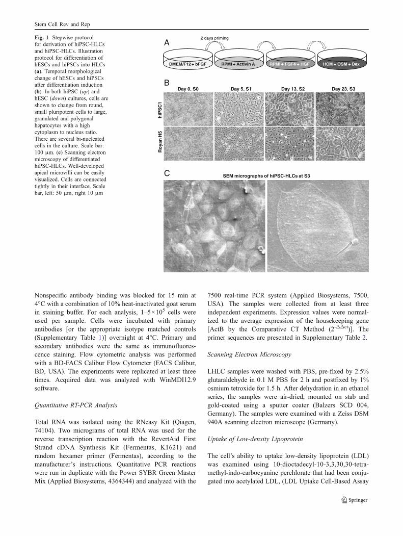
as previously described [16]. We changed the mediumevery other day, until day seven. The hepatic lineagedifferentiation procedure is outlined in Fig. 1a using astepwise, three stage protocol based on liver development.
Briefly, five day-old colonies were fed with RPMI1640(Invitrogen, 51800–035) supplemented with 1 mM nonessen-tial amino acids (Invitrogen, 11140–050), 2 mM L-glutamine(Invitrogen, 25030–164) and 0.1 mM β-mercaptoethanol(Sigma-Aldrich, M7522). The knockout serum replacement(KOSR, Invitrogen, 10828–028) concentration was reducedfrom 20% to 10% and then to 0%within the following 2 days,of which this step was meant to prime cells for endodermformation. Media was changed to RPMI1640 supplementedwith 100 ng/ml Activin A (R & D Systems, Inc., 338-AC) for3 days to induce a definitive endoderm lineage (Stage 1). Theinsulin-transferrin-selenite (ITS, Invitrogen, 41400–045) con-centration was 0% for the first 24 h, then increased to 0.5%and 1.5% for the next 2 days after priming. Cells were thencultured for 8 days in the same basal media that contained10% KOSR supplemented with 10 ng/ml FGF-4 (R & DSystems, Inc., 235-F4) and 10 ng/ml HGF (R & D Systems,Inc., 294-HG; Stage 2). During the last 2 days of Stage 2, 50%RPMI and 50% HCM were used. Cell differentiation wasfollowed by culture for ten additional days in HCM (Lonza,Inc., CC-3198) that contained 10% KOSR, 1 mM nonessen-tial amino acids, L-glutamine, 0.1 μM dexamethasone (Dex,Sigma-Aldrich, D-2915), and 10 ng/ml oncostatin M (OSM,R & D Systems, Inc., 295OM; Stage 3).
Evaluation of Differentiated Hepatic-lineage Cells
Immunofluorescence Staining
Cells were fixed in 4% paraformaldehyde for 20 min,permeabilized with 0.1% Triton X-100 for 10 min andblocked in 10% goat serum (SAFC Bioscience, 12306 C) inphosphate buffered saline (PBS, pH=7.4) for 1 h at 37°C.Cells were incubated with primary antibodies (SupplementaryTable 1) overnight at 4°C, after which they were washed andincubated with PE/FITC-conjugated secondary antibodies for1 h at 37°C. Nuclei were counterstained with DAPI. Thecells were analyzed with a fluorescent microscope (IX71,Olympus, Japan).
Flow Cytometry Analysis
Hepatic lineage directed differentiated cells underwentdissociation in 0.05% trypsin-EDTA (Invitrogen, 25200–072). Then, cells were washed twice in PBS (supplementedwith 1% heat-inactivated FBS, 0.1% sodium azide and2 mM EDTA) and then fixed in 4% paraformaldehyde for15 min. For permeabilization, Triton X-100 [0.1% (v/v),Merck, 11869] was used for 10 min at room temperature.
Stem Cell Rev and Rep
Nonspecific antibody binding was blocked for 15 min at4°C with a combination of 10% heat-inactivated goat serumin staining buffer. For each analysis, 1–5×10⁵ cells wereused per sample. Cells were incubated with primaryantibodies [or the appropriate isotype matched controls(Supplementary Table 1)] overnight at 4°C. Primary andsecondary antibodies were the same as immunofluores-cence staining. Flow cytometric analysis was performedwith a BD-FACS Calibur Flow Cytometer (FACS Calibur,BD, USA). The experiments were replicated at least threetimes. Acquired data was analyzed with WinMDI12.9software.
Quantitative RT-PCR Analysis
Total RNA was isolated using the RNeasy Kit (Qiagen,74104). Two micrograms of total RNA was used for thereverse transcription reaction with the RevertAid FirstStrand cDNA Synthesis Kit (Fermentas, K1621) andrandom hexamer primer (Fermentas), according to themanufacturer’s instructions. Quantitative PCR reactionswere run in duplicate with the Power SYBR Green MasterMix (Applied Biosystems, 4364344) and analyzed with the
7500 real-time PCR system (Applied Biosystems, 7500,USA). The samples were collected from at least threeindependent experiments. Expression values were normal-ized to the average expression of the housekeeping gene[ActB by the Comparative CT Method (2-ΔΔct)]. Theprimer sequences are presented in Supplementary Table 2.
Scanning Electron Microscopy
LHLC samples were washed with PBS, pre-fixed by 2.5%glutaraldehyde in 0.1 M PBS for 2 h and postfixed by 1%osmium tetroxide for 1.5 h. After dehydration in an ethanolseries, the samples were air-dried, mounted on stab andgold-coated using a sputter coater (Balzers SCD 004,Germany). The samples were examined with a Zeiss DSM940A scanning electron microscope (Germany).
Uptake of Low-density Lipoprotein
The cell’s ability to uptake low-density lipoprotein (LDL)was examined using 10-dioctadecyl-10-3,3,30,30-tetra-methyl-indo-carbocyanine perchlorate that had been conju-gated into acetylated LDL, (LDL Uptake Cell-Based Assay
DMEM/F12 + bFGF RPMI + Activin A RPMI + FGF4 + HGF HCM + OSM + Dex
2 days primingA
Day 0, S0 Day 5, S1 Day 13, S2 Day 23, S3
hiP
SC
1R
oya
nH
5
B
SEM micrographs of hiPSC-HLCs at S3C
Fig. 1 Stepwise protocolfor derivation of hiPSC-HLCsand hiPSC-HLCs. Illustrationprotocol for differentiation ofhESCs and hiPSCs into HLCs(a). Temporal morphologicalchange of hESCs and hiPSCsafter differentiation induction(b). In both hiPSC (up) andhESC (down) cultures, cells areshown to change from round,small pluripotent cells to large,granulated and polygonalhepatocytes with a highcytoplasm to nucleus ratio.There are several bi-nucleatedcells in the culture. Scale bar:100 μm. (c) Scanning electronmicroscopy of differentiatedhiPSC-HLCs. Well-developedapical microvilli can be easilyvisualized. Cells are connectedtightly in their interface. Scalebar, left: 50 μm, right 10 μm
Stem Cell Rev and Rep

Kit, Cayman Chemical, Item Number 10011125). Theassay was performed according to the manufacturer’sinstructions. Cells were visualized using a fluorescentmicroscope (IX71, Olympus, Japan).
Cytochrome P450 Activity and Inducibility
Cytochrome P450-dependent pentoxyresorufin o-dealkylaseactivity (PROD) was evaluated using pentoxyresorufin sub-strates. Pentoxyresorufin is o-dealkylated by cytochromeP450 and changes a nonfluorescent compound into resorufin,a fluorescent compound [17]. To evaluate the inducibility ofcytochrome P450, day 23 differentiated cells were exposedto sodium phenobarbital for 3 days and subsequentlywashed. Next, an incubation mixture that contained 7-pentoxyresorufin substrate (Sigma-Aldrich, P9049) anddicumarol (Sigma-Aldrich, M1390) in HBSS was addedand plates incubated at 37°C in a 5% CO2 incubator for30 min. Nuclei were counterstained with DAPI and in situassessment and detection of resorufin was performed byfluorescent microscopy (IX71, Olympus, Japan). The percentof positive cells of three independent experiments before andafter induction for each line were counted.
Periodic Acid-Schiff Staining
Glycogen storage of hiPSC-derived HLCs was evaluated byPeriodic acid-Schiff (PAS) staining at day 23. Culturedishes that contained the cells were fixed with 4%paraformaldehyde, oxidized in 1% periodic acid for 5 minand washed. Cells were then treated with Schiff’s reagentfor 15 min with subsequent color development in dH2O for5–10 min followed by assessment with a light microscope(IX50, Olympus, Japan).
Indocyanine Green Uptake and Release
Differentiated cells at day 23 were incubated with indoc-yanin green (ICG, CardioGreen, Sigma-Aldrich, 12633) inbasal medium for 1 h at 37°C. Uptake of ICG was detectedwith light microscopy (IX50, Olympus, Japan). ICGelimination from the positive cells was verified 6 h later.
Secretion Analysis
Forty-eight hour conditioned media obtained from the fullydifferentiated hiPS cells was collected at day 23 and stored at−20°C until assayed. The conditioned media were assayed foralpha-fetoprotein (AFP) secretion using a Chemilumines-cence Immunoassay Kit (Pishtaz-Teb, Iran); for ALB secre-tion, a mouse ALBELISAKit (Bethyl, E99-134); and for ureasecretion, a Colorimetric Assay Kit (Pars Azmun, Iran)according to the manufacturers’ recommendations. Secretion
was normalized to the total cell number. Samples werecollected from at least three independent experiments.
Oil Red Staining
Cells were assessed for lipid vesicles storage by oil redstaining. Differentiated cells were fixed with 4% parafor-maldehyde and then incubated for one h with oil red.Afterwards, cells were washed and analyzed with a lightmicroscope (IX50, Olympus, Japan).
Cell Preparation for Transplantation
Differentiated cells at stage 3 or day 23 were prepared forcell transplantation. Cells that detached from the plate bytrypsin/EDTA treatment were counted and labeled using thePKH67 Fluorescent Cell Linker Kit (Sigma-Aldrich,PKH67GL) according to the manufacturer’s recommendationand suspended in PBS.
Animal Experimental Model
Four groups of nine mice each were divided into thefollowing groups: normal, sham, control and transplanted.For the first and fifth week analyses, 4 and 5 animals pergroup were used. Liver fibrosis was induced in 8 week-oldmale NMRI mice by intraperitoneal injections of 1.0 ml/kgdose of CCl4 twice weekly for 4 weeks. The normal groupreceived no CCl4. After 4 weeks and eight injections ofCCl4 in the transplanted group, 1 × 10⁶ PKH-labeled cellswere infused slowly via the tail vein. The sham group, onlyreceived a PBS (200 μl) injection via the tail vein. Again,the toxin was administered following transplantation toinduce a high selective pressure for transplanted cells toengraft and differentiate [18]. All animals in the control,sham and transplanted groups received intraperitonealinjections of cyclosporin (Sandimmune, Novartis Pharma-ceuticals, Switzerland) at a dose of 20 mg/kg/24 h perinjection, beginning 5 days prior to transplantation andcontinuing after transplantation. At the end of the proce-dure, animals were sacrificed under deep anesthesia andtheir blood serum collected. Additionally, liver sampleswere taken for histopathologic analysis as well as cellengraftment and immunohistofluorescence evaluations. Allin vivo experiments were performed at the end of 1 and5 weeks post cell transplantation.
Liver Histopathology
Liver tissues were processed per standard protocol forparaffin embedding and sectioned into 6 μm sections.The sections were stained with masson trichrome (MT)according to standard protocol.
Stem Cell Rev and Rep

To analyze the extent of liver fibrosis, randomly pickedfields of MT sections were captured from each animal usinga 10× objective lens. Image analysis and percentages offibrotic areas were measured with Image J software(National Institutes of Health). A total of 30 random fieldsper group were analyzed independently and the percentagesof these fibrotic areas in the livers were calculated bydividing the total blue/green-colored area by the total areasof the livers [19, 20].
Engrafted Cells into the Liver
Recipient liver tissues were fixed in 4% paraformalde-hyde and then soaked in 30% sucrose for 24 h at 4°C.Liver tissues were embedded in Tissue-Tek OCTcompound and 8 μm thick sections were taken bycryostat at −20°C. The sections were mounted on glassslides and nuclei staining were performed using 4′,6-Diamidino-2-phenylindole dihydrochloride (DAPI, SigmaD-8417). The specimens were examined with a fluorescentmicroscope (BX51, Olympus) and randomly picked fields ofsections were captured from each animal, using a20× objective lens.
Immunofluorescence for Transplanted Cells
To evaluate hepatocyte differentiation of engrafted cellsin the recipient livers, human ALB was used as amarker of transplanted cells. Cryosections of livertissues from mice in the cell therapy groups were takenas described above. The sections were rinsed withdH2O, blocked with rabbit serum and then incubated withprimary antibody goat anti-ALB (Santa Cruz, 1:200, sc-46293) overnight at 4°C. After rinsing, sections wereincubated with secondary antibody rabbit anti-goat IgG-Texas Red (Santa Cruz, cs-3919, 1:200) for 1 h at 37°C.Double positive cells (PKH+/ALB+) in the liver werequantified using a fluorescent microscope (BX51, Olympus).A total of ten different areas per liver section wereindependently analyzed. The percentages of doublepositive cells in comparison to the total cells and incomparison to all PKH+cells of the fields wereexamined [21] 5 weeks after cell transplantation.
Serum Albumin, LDH and Bilirubin Measurements
Serologic analysis was performed using commercialkits (all from Pars Azmun, Iran) to measure serumalbumin (ALB) levels by the bromocresol greenmethod; serum lactate dehydrogenase enzyme activitiesby the DGKC method; and serum bilirubin levels bythe photometric DCA method, according to the manu-facturer’s instructions.
Statistical Analysis
Results were expressed as mean±standard deviation (SD).Functional analyses of HLCs in vitro were done with theindependent sample t-test. One-way repeated measuresanalysis of variance (ANOVA) followed by the Tukey posthoc test multiple group comparison analyzed group differ-ences of the in vivo resultant data. The mean difference wassignificant at the p<0.05 level. In the case of quantificationby Image J software, a total of 30 fields for each group wasassayed (fibrosis and cell count).
Results
Generation of HLCs from hESCs and hiPSCs
hESC and hiPSC colonies were cultured in feeder-freeconditions at a high concentration of bFGF on Matrigel(Fig. 1a). Cells in these colonies were compact andexpressed high nucleus to cytoplasm ratios as well asprominent nucleoli (Fig. 1b). Our stepwise protocol wascomposed of the following: cell priming and definitiveendoderm specification, hepatic induction and expansion,and hepatic maturation (Fig. 1a). During these stages, wecompared hiPSC1 with the previously established RoyanH5 hESC line, which has been proven to have the ability ofdifferentiation toward a hepatic lineage. The resultsindicated that hiPSC1 also has the ability to differentiateinto HLCs. At day five of the hESC culture, we replacedhESC medium with RPMI for 2 days supplemented with10% KOSR, which was then decreased to 0%. Althoughthis treatment caused no obvious morphological changes itseemed to protect cells against activin-induced cell death[22], medium replacement or KOSR removal and increasedcell viability up to 50% (data not shown) over the next3 days. Cells were subsequently treated with activin A for3 days in the absence of serum or serum replacement. Forthe next 7 days, cells were placed in RPMI that contained10% KOSR supplemented with FGF-4 and HGF.
Morphologically, the cells gradually transformed intoless dense, flatter cells with prominent nuclei (day 5,Stage 1) and then to a spiky shape (day 13, Stage 2,Fig. 1b). Finally, the medium was changed to HCMmedium supplemented with OSM and Dex for anadditional 10 days. Cells exhibited the characteristichepatocyte morphology of a polygonal shape, distinctround nuclei and large cytoplasm that contained variousgranules (Stage 3, Fig. 1b). Bi-nucleated cells were readilyvisible in Stage 3.
The hiPSC-LHCs at Stage 3 were also examined byscanning electron microscopy (Fig. 1c). Ultrastructuralanalysis showed that the apical surface of the differentiated
Stem Cell Rev and Rep

cells contained numerous microvilli. HLCs were polygonaland in contact with one another.
To confirm that the differentiated hESCs and hiPSCsdeveloped into endoderm and then hepatocytes, weperformed qRT-PCR analysis on the cells at variousdifferentiation stages with primers specific to genes ofthe hepatic lineages (Fig. 2). The expression levels ofendodermal marker genes (SOX17 and FOXA2) for bothcell lines were at their highest level in Stage 1, whichsuggested the induction of an endodermal fate at thisstage. While the early endodermal marker genes weregradually down-regulated during additional differentia-tion, expression of the hepatocyte gene, HNF4α was up-regulated through days 5 to 23 (Fig. 2). The expressions ofmature hepatic marker genes, ALB, G6P, and CYP3A4peaked at day 23 (Fig. 2). In total, these expressions wereindicative of the transition from an endodermal to ahepatic fate.
To further determine the lineage information along thecourse of differentiation, we performed immunofluores-cence staining on hiPSCs colonies at different differentia-tion stages (Fig. 3a). We observed that expression of thepluripotency marker, OCT4, was down-regulated by theinitiation of differentiation and undetectable at Stage 3,whereas cells which expressed SOX17, CXCR4 andFOXA2 were mainly detected in Stage 1, all of whichconfirmed qRT-PCR results (Fig. 3a). The early hepaticcells that expressed AFP and CK18 began at Stage 2 andcontinued to Stage 3, respectively (Fig. 3a). ALB expres-sion followed the same pattern, whereas AAT and CYP1A1were only detected in Stage 3 (Fig. 3a). Few SOX7-positivecells were detected at Stage 1, which indicated thesepopulations failed to differentiate to extra-embryonicendodermal cells.
To further confirm the above results, we performed aquantitative protein expression assay. Cells at each stagewere examined for expression of protein hallmarks. Flowcytometry analysis showed that differentiated cells on dayfive from hESC and hiPSC expressed both SOX17 andCXCR4 as markers of definitive endoderm (55% and 87%in hESC and hiPSC derivatives, respectively; Fig. 3b).Cells were then examined for ALB expression at days 13and 23 (Fig. 3d-e). ALB was expressed in day 13differentiated cells as a marker for hepatic induction fromdefinitive endoderm [hESCs (20%) and hiPSCs (30%)] andfurther increased in day 23 differentiated cells as the proteinhallmark of mature hepatocytes [hECSs (45%) and hiPSCs(74%)].
Despite the expression data, hiPSC-HLCs shouldhave the capability to perform hepatic functions forfurther use in the clinic and industry settings. Therefore,we next examined the functional properties of hESC/hiPSC-derived differentiated cells. Conditioned mediapreviously incubated with differentiating cells at Stages 1, 2and 3 were collected for hepatocyte functional tests, whichincluded ALB and AFP synthesis and ureagenesis (Fig. 4b-d).Both generated HLCs exhibited ALB (p<0.05), AFP (p<0.01), and urea synthesis (p<0.01) that significantly in-creased in parallel with the duration of hepatic differentia-tion, which indicated increased hepatic maturation over thecourse of additional culture.
These HLCs at Stage 3 also exhibited other character-istics of hepatocytes that included LDL conjugated toDyLight™549and ICG uptake (Fig. 4a). Accumulation oflipid droplets and glycogen storage activity were observedby oil red and PAS staining, respectively (Fig. 4a).Additionally, hESC/hiPSC-HLCs exhibited CYP450 meta-bolic activity as demonstrated by PROD activity before and
020406080
100120140160180200
hiP
SC
1
Roy
an H
5
PLC
hiP
SC
1
Roy
an H
5
PLC
hiP
SC
1
Roy
an H
5
PLC
hiP
SC
1
Roy
an H
5
PLC
hiP
SC
1
Roy
an H
5
PLC
hiP
SC
1
Roy
an H
5
PLC
SOX17 FOXA2 HNF4A ALB G6P CYP3A4
250
350
450
550
Rel
ativ
e m
RN
A e
xpre
ssio
n
Fig. 2 Gene expressionanalysis. Relative mRNAexpression pattern of hiPSCsand hESCs in the endoderm(S1), hepatic induction (S2)and hepatic maturation (S3)have been examined duringdifferentiation. PLC: positivecontrol. S: stage
Stem Cell Rev and Rep

hiPSC1 Royan H5
74 ± 3 % 45 ± 8 %
ALB, S3
S0 S1 S2 S3F
OX
A 2
CK
18
SO
X 7
OC
T4
AL
BA
FP
SO
X 1
7C
XC
R 4
AA
TC
YP
1A1
30 ± 3 % 20 ± 5 %
ALB, S2
87 ± 4 %
SOX17
CX
CR
4
55 ± 7 %
SOX17
CX
CR
4
S1
A B
C
D
Fig. 3 Protein expression analysis by immunofluorescence staining.(a) Expression pattern of pluripotency (OCT4), endoderm (SOX7,SOX17, CXCR4, FOXA2), hepatic induction (CK18, AFP, ALB) andhepatic maturation (AAT, CYP1A1) proteins during the differentiationtime span. The results indicate down-regulation of pluripotency andendoderm markers and up-regulation of hepatic maturation markersduring this time. Scale bar: 100 μm. Quantitative protein expression of
(double SOX17+/CXCR4+) in endoderm (b, S1), and ALB in hepaticinduction (c, S2), and ALB in hepatic maturation (d, S3). The numberof double positive cells for S1 and single positive cells for S2 and S3are shown on the panels. The data shows efficient induction ofdefinitive endoderm followed by induction of hepatic fate (ALBexpression) and further maturation (increase in ALB expression level)
Stem Cell Rev and Rep

after phenobarbital induction (Fig. 4e). About 50% of thecells were able to perform the function in each cell line andthe number of positive cells increased up to 1.5 fold afterphenobarbital induction. These results showed that hiPSC-HLCs were functional in vitro.
Analysis of qRT-PCR, flow cytometry and functionalresults showed that the expression levels of hepatic genesand their functions varied among the HLCs of hiPSCs andhESCs (Figs. 2, 3 and 4), which indicated that pluripotentstem cells responded differently to exogenous stimulation.
Transplantation of HLCs into a Mouse Modelof a CCl4-injured Liver
To evaluate in vivo functionality and the therapeuticpotential of our generated hiPSC-HLCs, we compared theanimal survival rate, total LDH, total bilirubin, and mouseand human ALB levels in blood serum from CCl4-injuredmice in the control, sham and transplant groups. Addition-
ally, we examined liver sections from all groups for thepercent of fibrotic area plus the amount of homing and cellfate of the transplanted cells, 5 weeks after transplantation(Figs. 5, 6 and 7).
Figure 5a shows all hiPSC-HLCs and normal micesurvived till week five, whereas only three animals survivedin the control and sham groups by this time. Investigationof histology results demonstrated that liver fibrosis in-creased significantly in CCl4-treated mice when comparedwith normal mice, with thick bundles of collagen thatsurrounded the lobules which lead to fibrous septa(Fig. 5b). Evaluation of liver sections stained with Mason-trichorom by Image J software 1 and 5 weeks post-celltransplantation showed signs of decreased fibrosis whencompared with the control and sham groups, but was notsignificant (Fig. 5c).
Serum analyses in mice who received HLCs, showed asignificant reduction of serum total LDH and total bilirubinwhen compared with the control and sham groups
ALB
050
100150200250300350
S1 S2 S3
ng
/ml/5
0000
0 ce
lls/4
8h
AFP
0
2
4
6
8
10
12
S1 S2 S3
µg
/ml/
5000
00 c
ells
/48h
0
5
10
15
20
25
S1 S2 S3
mg
/dl/5
0000
0 ce
lls/4
8h Urea
0
20
40
60
80
100
Before ind Aftre ind
Po
siti
ve c
ells
(%
) PROD
hiPSC1 Royan H5
PR
OD
P
AS
sta
inin
g
IC
G-u
pta
keL
DL
-up
take
A B
C
D
E
Oil
red
sta
inin
g
Fig. 4 Functional analysis ofhiPSC/hESC-HLCs. (a) Quali-tative functions of the cells inthe hepatic maturation phase(S3) including LDL-uptake, oilred staining, ICG-uptake, PASstaining and PROD activity. Theresults indicate in vitro func-tionality of the cells. Scale bar:100 μm. Quantitative functionsincluding (b) ALB, (c) AFP and(d) urea secretion during thetime span of differentiation and(e) PROD activity of hiPSC/hESC-HLCs before and afterphenobarbital induction. Quan-titative functional analysisshows increase in secretionability of the cells during thetime span of differentiation.PROD activity has increasedabout 1.5 times in response tophenobarbital induction
Stem Cell Rev and Rep

(p<0.05). There was an increase in total ALB (p<0.05)followed by a decrease 1 and 5 weeks post-celltransplantation (Fig. 6).
Before transplantation, we labeled cells with PKH toassess homing of the transplanted cells. Subsequently liversections were examined by fluorescent microscope, whichdemonstrated incorporation of the cells into the host livers.Transplanted cells were detected in the background of themouse liver by PKH fluorescence (Fig. 7a). The absolutenumbers of PKH-positive cells in recipients’ livers werecounted 5 weeks post-cell transplantation. Within the
B
A
Normal Control
Sham Transplanted
00.5
11.5
22.5
33.5
Week 1 Week 5 Week 1 Week 5 Week 1 Week 5 Week 1 Week 5
Normal Control Sham Transplanted
Fibrotic area (%)
a a
C
0
1
2
3
4
5
6
-5 -4 -3 -2 -1 0 1 2 3 4 5 6Num
ber
of a
nim
als
surv
ivin
g
Weeks
hiPSC-HLCs Transplantation
Fig. 5 Evaluation of transplantation of hiPSC-HLCs, 5 weeks post-transplantation for recovery of the CCl4 injured model. The survivalcurve of injured mice who underwent intravenous cell transplantation(a). All animals received Cyclosporine 5 days before cell transplan-tation and CCl4 administration started 4 weeks prior to transplantationand continued until the end of the survey (b). Representativephotomicrographs of masson trichrome-stained mouse livers indifferent experimental groups, fibrous area stained green-blue. (c)Analysis of fibrotic percent by Image J software. a: p<0.05 vs. othergroups
0
0.3
0.6
0.9
1.2
1.5
1.8
Week 1 Week 5 Week 1 Week 5 Week 1 Week 5 Week 1 Week 5
Normal Control Sham Transplanted
Total Bilirubin (mg/dL)
a a
0
1
2
3
4
5
Week 1 Week 5 Week 1 Week 5 Week 1 Week 5 Week 1 Week 5
Normal Control Sham Transplanted
Total Albumin (g/dL) a
0
500
1000
1500
2000
2500
Week 1 Week 5 Week 1 Week 5 Week 1 Week 5 Week 1 Week 5
Normal Control Sham Transplanted
Total LDH (U/I)
aa
a a
aa
Fig. 6 Serological analyses of blood serum in different animal groupsduring 1 and 5 weeks post-treatment. Total levels of LDH andbilirubin decreased 1 and 5 weeks after cell transplantation. Totalalbumin increased 1 week post cell transplantation; however, itreduced after 5 weeks. a: p<0.05 vs. other groups
Stem Cell Rev and Rep

recipient livers, there were 2.0±0.7% of transplanted cellsvisualized (67 PKH+cells out of 3358 counted cells,Fig. 7b). To further explore their fate, transplanted cellswere evaluated in corresponding serial sections. Double
PKH+/ALB+ labeled cells were used as markers offunctional activity of the transplanted cells (Fig. 7a). Albeitrare, 0.3% of total cells were ALB+and 18% (12 out of 67)were double PKH+/ALB+, and were located in the
PK
HA
LB
/ DA
PI
Mer
ge
Magnification
0
0.5
1
1.5
2
2.5
PKH+ cells/Total cells
(PKH & ALB)+ cells/PKH+ cells
(PKH & ALB)+ cells/Total cells
15
18
21
% of positive cells
A
B
200 µm 100 µm
100 µm
100 µm200 µm
200 µm
50 µm
50 µm
50 µm
Fig. 7 The engraftment of hiPSCs-HLCs in recipient livers withfunctional integration in the CCl4 injured mouse model 5 weeks post-transplantation. (a) Cells were labeled with PKH (green color) prior totransplantation. After 5 weeks, transplanted cells were detected in themouse host liver by fluorescence microscopy. The nuclei were
counterstained with DAPI (blue color) to calculate homing cells.Liver sections were stained with an anti-ALB antibody. Picturesshown are representative for different experiments. (b) Quantificationof engrafted cells
Stem Cell Rev and Rep

parenchyma and perivenous regions of the livers of allrecipients (Fig. 7b). Additionally, human ALB was detectedat one (30.5±2.0 mg/dl) and five (36.33±4.10 mg/dl)weeks post-transplantation in the serum of transplantedanimals. These data suggested a functional integration ofthe transplanted cells within the mouse livers.
Additionally, we did not find tumor in the hiPSC-HLCstransplanted recipients during our study.
Discussion
Access to a pure terminally differentiated hepatocytepopulation with therapeutic potential for transplantation toliver failure patients has been the objective of severalstudies. Here we demonstrated the hepatic lineage differ-entiation potential of hiPSCs following stepwise applicationof growth factors and removal of serum in comparison withhESCs, as well as further transplantation of hiPSC-HLCs toa CCL4-injured mouse model. Based on our knowledge,this is the first report of hiPSC-HLCs administration in ananimal model of liver injury.
According to our results, the examined hiPSC linefollowed the same differentiation pattern as the hESC line,and this result agreed with other hESCs [for review see [5]]and hiPSC studies [9–13]. HLCs from both pluripotentstem cell types exhibited a polygonal epithelial morphologyand large cytoplasm that contained various granules andexpressed hepatocyte-specific genes at both the mRNA andprotein levels. Additionally, as some of the knownfunctions of mature hepatocytes, the differentiated cellswere able to secrete ALB, AFP, and urea into the medium.The differentiated HLCs also showed cytochrome p450activity, uptake of organic anion ICG and LDL in afluorescence form (LDL conjugated to DyLight™549).Glycogen and lipid storage, two other characteristics ofhepatocytes, were visible on both types as detected by PASand oil red staining, respectively. However, it seemed thathiPSC1 exhibited a differentiation propensity into hepaticlineage. A marked propensity to differentiate into specificlineages was exhibited in various hESC lines [23] andrecently in hiPSCs [24]. This needs further investigationand repeating the experiment with increased hiPSC andhESC cell lines.
Although clinical applications of hiPSC-HLCs still seemto be far from reach, the availability of these cells andrelative ease of derivation of hepatocytes from these cellsallows for the opportunity to test drugs on a range ofgenetic backgrounds. Moreover, generation of disease-specific hiPSC-HLCs can provide scientists with an invitro model of liver disorder for testing new drugs orstudying the mechanism of such disorders. Rashid et al.[25] have reported the generation of hiPSCs with α1-
antitrypsin deficiency, familial hypercholesterolemia andglycogen storage disease type 1a. Those cells demonstratedkey pathological characteristics of the diseases, such asaggregation of misfolded α1-antitrypsin in the endoplasmicreticulum, deficient LDL receptor–mediated cholesteroluptake and increased lipid and glycogen accumulation.Recently, we have produced liver-specific patient hiPSCswith tyrosinemia, glycogen storage disease, progressivefamilial hereditary cholestasis, and from two siblings withCrigler-Najjar syndrome that had the potential to differen-tiate into functional HLCs [26].
This study also assessed the therapeutic effects ofhiPSC-HLCs in the CCL4-injured mouse liver model over5 weeks post-cell transplantation. When compared with thecontrol and sham groups, the transplanted group had abetter survival rate, and reduced levels of LDH andbilirubin. The significant difference in serologic markersbetween the cell transplanted group and control/shamgroups was possibly due to the recovery potential of HLCs.Transplanted cells could re-establish the altered liverarchitecture and possibly stop additional cell death whichcan be detected by restored LDH and bilirubin levels. Onthe other hand, mouse ALB increased during the first weekpost-cell transplantation. This was a possible effect ofHLCs on mice hepatocytes, which enabled them to increasecapabilities such as ALB secretion. However, this was not asteady effect that continued until the end of 5 weeks, whichmay have been due to long-term CCL4 exposure in theanimals and its negative effects on transplanted cells.Human ALB was also detected in serum 1 and 5 weeksafter HLC transplantation, which has shown that HLCscould secret ALB in vivo and be active in their new niche.CCl4-treated mice had more fibrotic areas when comparedto the normal group. The histological improvement was notsignificant in the HLC transplanted group 1 or 5 weekspost-cell transplantation. However, HLC transplantationameliorated collagen excess and improved liver histologicarchitecture when CCL4 was removed, which was the maincause of the matrix imbalance. Several PKH+/ALB+(18%)cells showed survival and integration of engrafted HLCsinto the host tissue at 5 weeks following transplantation andassistance with recovery of liver function. It has beenshown that transplanted hESC-HLCs improved recovery inliver failure animal models [27–29]. Recently, it has beenalso demonstrated that mouse iPSC-derived hepatocyteswere able to regenerate the liver after transplantation and ina two-third partial hepatectomy in mice with fumarylace-toacetate hydrolase deficiency [29].
In conclusion, our results open up a proof of concept thatfunctional HLCs can be generated from hiPSCs andimprove the fibrotic mouse model after their transplanta-tion. Further research of this nature and investigation of thein vivo functionality of hiPSC-HLCs will bring new
Stem Cell Rev and Rep

insights into the clinical applications of these cells inregenerative and/or individualized medicine. However,further investigations for producing safe iPSCs, moreefficient differentiation protocols, long term analysis andlarger animal models are necessary.
Acknowledgements This study was funded by a grant providedfrom Royan Institute and the Iranian Council of Stem CellTechnology. We also thank Mehran Rezaei, Arefeh Ghodsizadeh,Zahra Farzaneh and Abbass Piriayei for their critical comments.Additionally, we would like to thank the Electron Microscopy Lab atthe University of Tehran.
Conflict of interest None of the authors have any conflicts ofinterest to disclose and all authors support submission to this journal.
References
1. Mazaris, E. M., Roussos, C. T., & Papalois, V. E. (2005).Hepatocyte transplantation: a review of worldwide clinicaldevelopments and experiences. Experimental and Clinical Trans-plantation, 3, 306–315.
2. Miro, J. M., Laguno, M., Moreno, A., & Rimola, A. (2006).Management of end stage liver disease (ESLD): what is thecurrent role of orthotopic liver transplantation (OLT)? Journal ofHepatology, 44, S140–145.
3. Lorenzini, S., Isidori, A., Catani, L., et al. (2008). Stem cellmobilization and collection in patients with liver cirrhosis.Alimentary Pharmacology and Therapeutics, 27, 932–939.
4. Lee, S. W., Wang, X., Chowdhury, N. R., & Roy-Chowdhury, J.(2004). Hepatocyte transplantation: state of the art and strategiesfor overcoming existing hurdles. Annals of Hepatology, 3, 48–53.
5. Asgari, S., Pournasr, B., Salekdeh, G. H., Ghodsizadeh, A., Ott,M., & Baharvand, H. (2010). Induced pluripotent stem cells: anew era for hepatology. Journal of Hepatology, 53, 738–751.
6. Pournasr, B., Farzaneh, Z., Shahsanvani, M., & Baharvand, H.(2010). Liver development and In vitro differentiation ofembryonic stem cells to hepatocytes. Yakhteh Medical Journal(Cell Journal), 11, 348–373.
7. Takahashi, K., Tanabe, K., Ohnuki, M., et al. (2007). Induction ofpluripotent stem cells from adult human fibroblasts by definedfactors. Cell, 131, 861–872.
8. Yu, J., Vodyanik, M. A., Smuga-Otto, K., et al. (2007). Inducedpluripotent stem cell lines derived from human somatic cells.Science, 318, 1917–1920.
9. Liu, H., Ye, Z., Kim, Y., Sharkis, S., & Jang, Y. Y. (2010).Generation of endoderm-derived human induced pluripotent stemcells from primary hepatocytes. Hepatology, 51, 1810–1819.
10. Touboul, T., Hannan, N. R., Corbineau, S., et al. (2010).Generation of functional hepatocytes from human embryonicstem cells under chemically defined conditions that recapitulateliver development. Hepatology, 51, 1754–1765.
11. Si-Tayeb, K., Noto, F. K., Nagaoka, M., et al. (2010). Highlyefficient generation of human hepatocyte-like cells from inducedpluripotent stem cells. Hepatology, 51, 297–305.
12. Song, Z., Cai, J., Liu, Y., et al. (2009). Efficient generation ofhepatocyte-like cells from human induced pluripotent stem cells.Cell Research, 19, 1233–1242.
13. Sullivan, G. J., Hay, D. C., Park, I. H., et al. (2010). Generation offunctional human hepatic endoderm from human induced plurip-otent stem cells. Hepatology, 51, 329–335.
14. Totonchi, M., Taei, A., Seifinejad, A., et al. (2010). Feeder- andserum-free establishment and expansion of human inducedpluripotent stem cells. International Journal of DevelopmentalBiology, 54, 877–886.
15. Baharvand, H., Ashtiani, S. K., Taee, A., et al. (2006). Generationof new human embryonic stem cell lines with diploid and triploidkaryotypes. Development, Growth & Differentiation, 48, 117–128.
16. Pakzad, M., Totonchi, M., Taei, A., Seifinejad, A., Hassani, S. N.,& Baharvand, H. (2010). Presence of a ROCK inhibitor inextracellular matrix supports more undifferentiated growth offeeder-free human embryonic and induced pluripotent stem cellsupon passaging. Stem Cell Reviews, 6, 96–107.
17. Tzanakakis, E. S., Hsiao, C. C., Matsushita, T., Remmel, R. P., &Hu, W. S. (2001). Probing enhanced cytochrome P450 2B1/2activity in rat hepatocyte spheroids through confocal laserscanning microscopy. Cell Transplantation, 10, 329–342.
18. Piryaei, A., Valojerdi, M. R., Shahsavani, M., & Baharvand, H.(2011). Differentiation of bone marrow-derived mesenchymalstem cells into hepatocyte-like cells on nanofibers and theirtransplantation into a carbon tetrachloride-induced liver fibrosismodel. Stem Cell Reviews, 7, 103–118.
19. Marumoto, Y., Terai, S., Urata, Y., et al. (2008). Continuous highexpression of XBP1 and GRP78 is important for the survival ofbone marrow cells in CCl4-treated cirrhotic liver. Biochemical andBiophysical Research Communications, 367, 546–552.
20. Sakaida, I., Terai, S., Yamamoto, N., et al. (2004). Transplantationof bone marrow cells reduces CCl4-induced liver fibrosis in mice.Hepatology, 40, 1304–1311.
21. Terai, S., Sakaida, I., Yamamoto, N., et al. (2003). An in vivo modelfor monitoring trans-differentiation of bone marrow cells intofunctional hepatocytes. Journal of Biochemistry, 134, 551–558.
22. Mfopou, J. K., & Bouwens, L. (2008). Hedgehog signals inpancreatic differentiation from embryonic stem cells: revisiting theneglected. Differentiation, 76, 107–117.
23. Osafune, K., Caron, L., Borowiak, M., et al. (2008). Markeddifferences in differentiation propensity among human embryonicstem cell lines. Nature Biotechnology, 26, 313–315.
24. Kim, D. S., Lee, J. S., Leem, J. W., et al. (2010). Robustenhancement of neural differentiation from human ES and iPScells regardless of their innate difference in differentiationpropensity. Stem Cell Reviews, 6, 270–281.
25. Rashid, S. T., Corbineau, S., Hannan, N., et al. (2010). Modelinginherited metabolic disorders of the liver using human inducedpluripotent stem cells. The Journal of Clinical Investigation, 120,3127–3136.
26. Ghodsizadeh, A., Taei, A., Totonchi, M., et al. (2010). Generationof liver disease-specific induced pluripotent stem cells along withefficient differentiation to functional hepatocyte-like cells. StemCell Reviews, 6, 622–632.
27. Basma, H., Soto-Gutierrez, A., Yannam, G. R., et al. (2009).Differentiation and transplantation of human embryonic stem cell-derived hepatocytes. Gastroenterology, 136, 990–999.
28. Agarwal, S., Holton, K. L., & Lanza, R. (2008). Efficientdifferentiation of functional hepatocytes from human embryonicstem cells. Stem Cells, 26, 1117–1127.
29. Espejel, S., Roll, G. R., McLaughlin, K. J., et al. (2010). Inducedpluripotent stem cell-derived hepatocytes have the functional andproliferative capabilities needed for liver regeneration in mice.The Journal of Clinical Investigation, 120, 3120–3126.
Stem Cell Rev and Rep
Related Documents