RESEARCH Open Access Differential proteomic analysis of synovial fluid from rheumatoid arthritis and osteoarthritis patients Lavanya Balakrishnan 1,2 , Mitali Bhattacharjee 1,3 , Sartaj Ahmad 1,4 , Raja Sekhar Nirujogi 1,5 , Santosh Renuse 1,3 , Yashwanth Subbannayya 1,6 , Arivusudar Marimuthu 1 , Srinivas M Srikanth 1,5 , Rajesh Raju 1 , Mukesh Dhillon 7 , Navjyot Kaur 7 , Ramesh Jois 8 , Vivek Vasudev 9 , YL Ramachandra 2 , Nandini A Sahasrabuddhe 1 , TS Keshava Prasad 1,3,4 , Sujatha Mohan 10 , Harsha Gowda 1 , Subramanian Shankar 7* and Akhilesh Pandey 11,12,13,14* Abstract Background: Rheumatoid arthritis and osteoarthritis are two common musculoskeletal disorders that affect the joints. Despite high prevalence rates, etiological factors involved in these disorders remain largely unknown. Dissecting the molecular aspects of these disorders will significantly contribute to improving their diagnosis and clinical management. In order to identify proteins that are differentially expressed between these two conditions, a quantitative proteomic profiling of synovial fluid obtained from rheumatoid arthritis and osteoarthritis patients was carried out by using iTRAQ labeling followed by high resolution mass spectrometry analysis. Results: We have identified 575 proteins out of which 135 proteins were found to be differentially expressed by ≥3-fold in the synovial fluid of rheumatoid arthritis and osteoarthritis patients. Proteins not previously reported to be associated with rheumatoid arthritis including, coronin-1A (CORO1A), fibrinogen like-2 (FGL2), and macrophage capping protein (CAPG) were found to be upregulated in rheumatoid arthritis. Proteins such as CD5 molecule-like protein (CD5L), soluble scavenger receptor cysteine-rich domain-containing protein (SSC5D), and TTK protein kinase (TTK) were found to be upregulated in the synovial fluid of osteoarthritis patients. We confirmed the upregulation of CAPG in rheumatoid arthritis synovial fluid by multiple reaction monitoring assay as well as by Western blot. Pathway analysis of differentially expressed proteins revealed a significant enrichment of genes involved in glycolytic pathway in rheumatoid arthritis. Conclusions: We report here the largest identification of proteins from the synovial fluid of rheumatoid arthritis and osteoarthritis patients using a quantitative proteomics approach. The novel proteins identified from our study needs to be explored further for their role in the disease pathogenesis of rheumatoid arthritis and osteoarthritis. Sartaj Ahmad and Raja Sekhar Nirujogi contributed equally to this article. Keywords: Arthritis, Joint inflammation, Cartilage degradation, Extracellular matrix * Correspondence: [email protected]; [email protected] 7 Department of Internal Medicine, Armed Forces Medical College, Pune 411040, India 11 McKusick-Nathans Institute of Genetic Medicine, Johns Hopkins University School of Medicine, Baltimore, MD 21205, USA Full list of author information is available at the end of the article CLINICAL PROTEOMICS © 2014 Balakrishnan et al.; licensee BioMed Central Ltd. This is an open access article distributed under the terms of the Creative Commons Attribution License (http://creativecommons.org/licenses/by/2.0), which permits unrestricted use, distribution, and reproduction in any medium, provided the original work is properly cited. Balakrishnan et al. Clinical Proteomics 2014, 11:1 http://www.clinicalproteomicsjournal.com/content/11/1/1

Welcome message from author
This document is posted to help you gain knowledge. Please leave a comment to let me know what you think about it! Share it to your friends and learn new things together.
Transcript
CLINICALPROTEOMICS
Balakrishnan et al. Clinical Proteomics 2014, 11:1http://www.clinicalproteomicsjournal.com/content/11/1/1
RESEARCH Open Access
Differential proteomic analysis of synovial fluidfrom rheumatoid arthritis and osteoarthritispatientsLavanya Balakrishnan1,2, Mitali Bhattacharjee1,3, Sartaj Ahmad1,4, Raja Sekhar Nirujogi1,5, Santosh Renuse1,3,Yashwanth Subbannayya1,6, Arivusudar Marimuthu1, Srinivas M Srikanth1,5, Rajesh Raju1, Mukesh Dhillon7,Navjyot Kaur7, Ramesh Jois8, Vivek Vasudev9, YL Ramachandra2, Nandini A Sahasrabuddhe1, TS Keshava Prasad1,3,4,Sujatha Mohan10, Harsha Gowda1, Subramanian Shankar7* and Akhilesh Pandey11,12,13,14*
Abstract
Background: Rheumatoid arthritis and osteoarthritis are two common musculoskeletal disorders that affect thejoints. Despite high prevalence rates, etiological factors involved in these disorders remain largely unknown.Dissecting the molecular aspects of these disorders will significantly contribute to improving their diagnosis andclinical management. In order to identify proteins that are differentially expressed between these two conditions, aquantitative proteomic profiling of synovial fluid obtained from rheumatoid arthritis and osteoarthritis patients wascarried out by using iTRAQ labeling followed by high resolution mass spectrometry analysis.
Results: We have identified 575 proteins out of which 135 proteins were found to be differentially expressed by≥3-fold in the synovial fluid of rheumatoid arthritis and osteoarthritis patients. Proteins not previously reported tobe associated with rheumatoid arthritis including, coronin-1A (CORO1A), fibrinogen like-2 (FGL2), and macrophagecapping protein (CAPG) were found to be upregulated in rheumatoid arthritis. Proteins such as CD5 molecule-likeprotein (CD5L), soluble scavenger receptor cysteine-rich domain-containing protein (SSC5D), and TTK protein kinase(TTK) were found to be upregulated in the synovial fluid of osteoarthritis patients. We confirmed the upregulationof CAPG in rheumatoid arthritis synovial fluid by multiple reaction monitoring assay as well as by Western blot.Pathway analysis of differentially expressed proteins revealed a significant enrichment of genes involved inglycolytic pathway in rheumatoid arthritis.
Conclusions: We report here the largest identification of proteins from the synovial fluid of rheumatoid arthritisand osteoarthritis patients using a quantitative proteomics approach. The novel proteins identified from our studyneeds to be explored further for their role in the disease pathogenesis of rheumatoid arthritis and osteoarthritis.Sartaj Ahmad and Raja Sekhar Nirujogi contributed equally to this article.
Keywords: Arthritis, Joint inflammation, Cartilage degradation, Extracellular matrix
* Correspondence: [email protected]; [email protected] of Internal Medicine, Armed Forces Medical College, Pune411040, India11McKusick-Nathans Institute of Genetic Medicine, Johns Hopkins UniversitySchool of Medicine, Baltimore, MD 21205, USAFull list of author information is available at the end of the article
© 2014 Balakrishnan et al.; licensee BioMed Central Ltd. This is an open access article distributed under the terms of theCreative Commons Attribution License (http://creativecommons.org/licenses/by/2.0), which permits unrestricted use,distribution, and reproduction in any medium, provided the original work is properly cited.
Balakrishnan et al. Clinical Proteomics 2014, 11:1 Page 2 of 14http://www.clinicalproteomicsjournal.com/content/11/1/1
BackgroundRheumatoid arthritis (RA) is a common systemic auto-immune disorder. Around 0.5-1% of the adult popula-tion is affected with RA in the developed countries with5–50 per 100,000 new cases reported annually [1]. RA ischaracterised by persistent inflammation of the synovialmembrane and pannus formation that results in jointdamage and loss of function [1,2]. Genetic, environmen-tal and stochastic factors act together to contribute tothe pathogenesis of RA [3]. Osteoarthritis (OA) is a pro-gressive disorder characterized by the degradation of thecartilage, osteophyte formation, mild to moderate syn-ovial inflammation, narrowing of the joint space andsubchondral sclerosis [4,5]. It is one of the most preva-lent musculoskeletal diseases that lead to disability in~40% of the adults over 70 years [5].Despite significant advances towards the understand-
ing of the pathophysiology of RA and OA, early diagno-sis and therapeutic intervention remain a challenge [6].RA is usually diagnosed based on clinical symptoms andthe presence of antibodies against rheumatoid factor(RF) and cyclic citrullinated peptides (CCPs) in serum[7]. Although RF has been traditionally used as a bio-marker for RA, it lacks specificity as it is also detectedin the sera of several other autoimmune disorders andinfectious diseases as well as in the healthy elderly popu-lation (10-30%) [7,8]. Antibodies to CCPs have beenshown to be involved in the development of auto-immune arthritis [9]. Their specificity for RA has beenreported to be higher than RF. However the diagnosticsensitivity of antibodies to CCPs has been estimated tobe slightly lower than that of RF [10]. In addition toanti-CCPs, anti-filaggrin (AFA) and anti-Sa antibodieshave also been demonstrated to have high specificity forRA but with a sensitivity of less than 50% [11]. Assess-ment of the pathological changes in the OA joint ismainly carried out using radiography, which is the goldstandard for diagnostic purposes [12]. However, it haspoor sensitivity as the radiographic evidence is obtainedonly when the articular cartilage has degraded signifi-cantly. Thus, this does not provide an account of the ex-tent of disease progression [12]. Biomarkers that areuseful for early diagnosis or for predicting the outcome/progression of OA are not currently available [13]. Thus,there is a need for continued discovery efforts to identifynovel biomarkers with the desired sensitivity and specifi-city for RA and OA.Mass spectrometry-based approaches have provided
an impetus for biomarker discovery for a wide range ofdiseases. Studying the synovial fluid proteome is benefi-cial in arthritis as it is in proximity to the site of diseaseactivity as well as it provides a snapshot of the mostrelevant compartment throughout disease progression.Additionally, alterations in the joint cavity due to injury
or disease may be directly reflected in the compositionof synovial fluid and could be correlated to disease se-verity and progression [14]. In a previous proteomicstudy, surface enhanced laser desorption ionization(SELDI) mass spectrometry was used to identify proteinsthat were present in RA synovial fluid but not in OA[15]. Subsequently, in another proteomic study, 2-D gelelectrophoresis followed by MALDI-TOF analysis wascarried out to identify synovial fluid and plasma proteinsthat could differentiate between RA and reactive arthritis(ReA) or OA. S100A9, S100A12 and serum amyloid A(SAA) were found to be present only in RA but not inReA and OA in that study [16]. A label free quantitativeanalysis of RA and OA synovial fluid proteome byMateos et al., resulted in the identification of 135 pro-teins using MALDI-TOF/TOF mass spectrometer [17].Proteins involved in complement activation, inflamma-tion and immune response were found to be relativelymore abundant in RA and those that participated in theextracellular matrix formation and remodeling weremore abundant in OA synovial fluid [17]. An immuno-proteomics study has also been reported to identifyautoantigens in RA. In this study, the expression ofvimentin, gelsolin, alpha-2-HS-glycoprotein, glial fibril-lary acidic protein and alpha-1-B glycoprotein was foundto be reported to be higher in RA synovial fluid thanthat of OA [18]. Most of the studies carried out thus farhave adopted a label free quantitation approach andemployed low resolution mass spectrometers to identifythe proteins differentially expressed between RA andOA.Isobaric Tags for Relative and Absolute Quantification
(iTRAQ) is a chemical labeling method which uses iso-baric tags with reporter ion group that react with theprimary amine groups present in the peptides [19].iTRAQ-based quantitative proteomics approach coupledto mass spectrometry has been widely used to identifybiomarkers for several diseases including cancer [20–24],meningitis [25] and rabies [26]. Here, we describe aniTRAQ-based strategy to relatively quantitate the syn-ovial fluid proteins from RA and OA. We employed aniTRAQ-based labeling strategy coupled with LTQ-Orbitrap Velos mass spectrometer to identify proteinsdifferentially expressed between RA and OA. The differ-entially expressed proteins obtained from the studymight increase our knowledge pertaining to the mech-anism of pathogenesis of RA and OA.
Results and discussionWe employed a quantitative proteomics approach usingiTRAQ labeling to identify the differentially expressedproteins in RA compared to OA. A schematic workflowillustrating the steps employed in this study is shown inFigure 1.
OA synovial fluid
RA synovial fluid
Protein isolation
Depletion by Multiple Affinity Removal System Human 14
In-solution trypsin digestion
iTRAQ labeling
116 117
Strong cation exchange chromatography
LC-MS/MS analysis
Database search using Sequest and Mascot
Validation by Multiple Reaction Monitoring and Western blot
Figure 1 A schematic workflow illustrating the steps involvedin the differential analysis of RA and OA synovial fluidproteome. Proteins from RA and OA synovial fluid were extractedand depleted to remove the 14 most abundant proteins usingmultiple affinity removal system, Human-14. The depleted proteinfrom RA and OA were then digested with trypsin and labeled withiTRAQ reagents, 117 and 116 respectively. The labeled samples werepooled and subjected to fractionation using strong cation exchangechromatography. The fractions were then analyzed on a LTQ-OrbitrapVelos mass spectrometer. The MS/MS data obtained was searchedagainst Human RefSeq 50 database using Sequest and Mascot searchalgorithms. Validation of the iTRAQ quantitation data was carried outusing multiple reaction monitoring and Western blot.
Balakrishnan et al. Clinical Proteomics 2014, 11:1 Page 3 of 14http://www.clinicalproteomicsjournal.com/content/11/1/1
Quantitative proteomic analysis of the synovial fluidproteome of RA and OAA total of 30,226 MS/MS spectra acquired from the LC-MS/MS analysis of 20 SCX fractions resulted in the iden-tification of 3,488 unique peptides corresponding to 575proteins. Out of these 575 proteins, 135 were differentiallyexpressed between RA and OA by ≥3-fold. A complete listof all the identified peptides along with their correspond-ing proteins is provided in Additional file 1: Table S1.
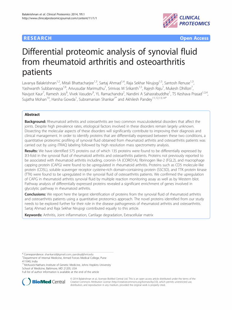
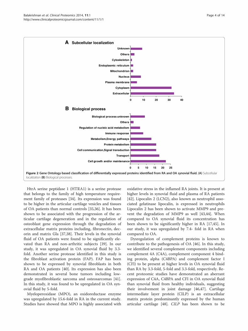
Bioinformatics analysisSubcellular localization and biological process basedclassification for the differentially expressed proteins wasperformed using HPRD [27,28] (http://www.hprd.org)that contains information on human proteins manuallycurated from the published literature. Signal peptide anddomain analysis was also carried out using the data fromHPRD. Classification based on the subcellular localization(Figure. 2A) revealed that 34% of the identified proteinswere extracellular. Biological process-based (Figure. 2B)categorization showed that a majority of the proteins wereinvolved in cell growth and/or maintenance (22%) and cellcommunication or signal transduction (18%). Out of 575identified proteins, 273 possess a signal peptide, 24 have atransmembrane domain and 73 have both a signal peptideand a transmembrane domain.
Differentially expressed proteins between RA and OAOut of the 135 differentially expressed proteins, 92 werefound to be upregulated and 43 were downregulated inRA synovial fluid ≥3-fold when compared to OA. Severalproteins that were earlier reported to be associated withRA and OA were identified in this study confirming thevalidity of our approach.
Proteins previously reported to be associated with RAand OAS100 family of calcium binding proteins, S100A8, S100A9and S100A12, have been shown to act as proinflammatorymediators in various autoimmune disorders includingarthritis [29]. Baillet et al. have shown in a proteomicstudy that these S100 proteins were present at higher con-centration in RA synovial fluid than OA synovial fluid andcould serve as markers to discriminate between RA andother inflammatory arthritis [30]. In this study, S100A8,S100A9 and S100A12, were upregulated in the synovialfluid of RA patients by 9.1-fold, 29-fold and 33.4-fold,respectively.Matrix metalloproteinases (MMPs) are zinc dependent
extracellular proteolytic enzymes known to play a majorrole in tissue remodelling under both physiological andpathological conditions [31]. We identified two MMPs,MMP8 and MMP9, to be more abundant in RA synovialfluid (14.4-fold and 3.4-fold, respectively). The levels ofthese enzymes have already been shown to be elevatedin both synovial fluid and sera of RA patients when com-pared to OA [32]. We also identified a neutrophil elastaseinhibitor, serpin peptidase inhibitor, clade B member 1(SERPINB1) that was upregulated (4.1-fold) in RA synovialfluid. Serpinb1 knock-out mice have been used as a modelto study the role of neutrophil elastase in cellular homeo-stasis and inflammation [33]. In a comparative proteomicstudy between RA and OA, SERPINB1 was detected inRA synovial fluid but not in OA synovial fluid [17].
A Subcellular localization
B Biological process
0 10 20 30
Extracellular
Cytoplasm
Plasma membrane
Nucleus
Mitochondrion
Endoplasmic reticulum
Cytoskeleton
Others
40
Unknown
0 5 10 15 20 25
Cell growth and/or maintenance
Transport
Cell communication,Signal transduction
Protein metabolism
Metabolism,Energy pathways
Immune response
Regulation of nucleic acid metabolism
Others
Biological process unknown
Figure 2 Gene Ontology-based classification of differentially expressed proteins identified from RA and OA synovial fluid. (A) Subcellularlocalization (B) Biological processes.
Balakrishnan et al. Clinical Proteomics 2014, 11:1 Page 4 of 14http://www.clinicalproteomicsjournal.com/content/11/1/1
HtrA serine peptidase 1 (HTRA1) is a serine proteasethat belongs to the family of high temperature require-ment family of proteases [34]. Its expression was foundto be higher in the articular cartilage vesicles and tissuesof OA patients than normal controls [35,36]. It has beenshown to be associated with the progression of the ar-ticular cartilage degeneration and in the regulation ofosteoblast gene expression through the degradation ofextracellular matrix proteins including, fibronectin, dec-orin and matrix Gla [37,38]. Their levels in the synovialfluid of OA patients were found to be significantly ele-vated than RA and non-arthritic subjects [39]. In ourstudy, it was upregulated in OA synovial fluid by 3.3-fold. Another serine protease identified in this study isthe fibroblast activation protein (FAP). FAP has beenshown to be expressed by synovial fibroblasts in bothRA and OA patients [40]. Its expression has also beendemonstrated in several bone tumors including low-grade myofibroblastic sarcoma and osteosarcomas [41].In this study, it was found to be upregulated in OA syn-ovial fluid by 5-fold.Myeloperoxidase (MPO), an oxidoreductase enzyme
was upregulated by 15.6-fold in RA in the current study.Studies have showed that MPO is highly associated with
oxidative stress in the inflamed RA joints. It is present athigher levels in synovial fluid and plasma of RA patients[42]. Lipocalin 2 (LCN2), also known as neutrophil asso-ciated gelatinase lipocalin, is expressed in neutrophilsLipocalin 2 has been shown to activate MMP9 and pre-vent the degradation of MMP9 as well [43,44]. Whencompared to OA synovial fluid its concentration hasbeen shown to be significantly higher in RA [17,45]. Inour study, it was upregulated by 7.4- fold in RA whencompared to OA.Dysregulation of complement proteins is known to
contribute to the pathogenesis of OA [46]. In this study,we identified several complement components includingcomplement 4A (C4A), complement component 4 bind-ing protein, alpha (C4BPA) and complement factor I(CFI) to be present at higher levels in OA synovial fluidthan RA by 3.3-fold, 5-fold and 3.3-fold, respectively. Re-cent proteomic studies have demonstrated an aberrantexpression of C4A, C4BPA and CFI in OA synovial fluidthan synovial fluid from healthy individuals, suggestingtheir involvement in joint damage [46,47]. Cartilageintermediate layer protein (CILP) is an extracellularmatrix protein predominantly expressed by the humanarticular cartilage [48]. CILP has been shown to be
Balakrishnan et al. Clinical Proteomics 2014, 11:1 Page 5 of 14http://www.clinicalproteomicsjournal.com/content/11/1/1
associated with the progression of OA [49]. It acts as anautoantigen and contributes to the development ofchronic synovitis in OA [50]. Its presence in the healthyand OA synovial fluid has been already demonstrated ina proteomic study [51]. It has been found to be upregu-lated by 3.3-fold in OA synovial fluid in our study.
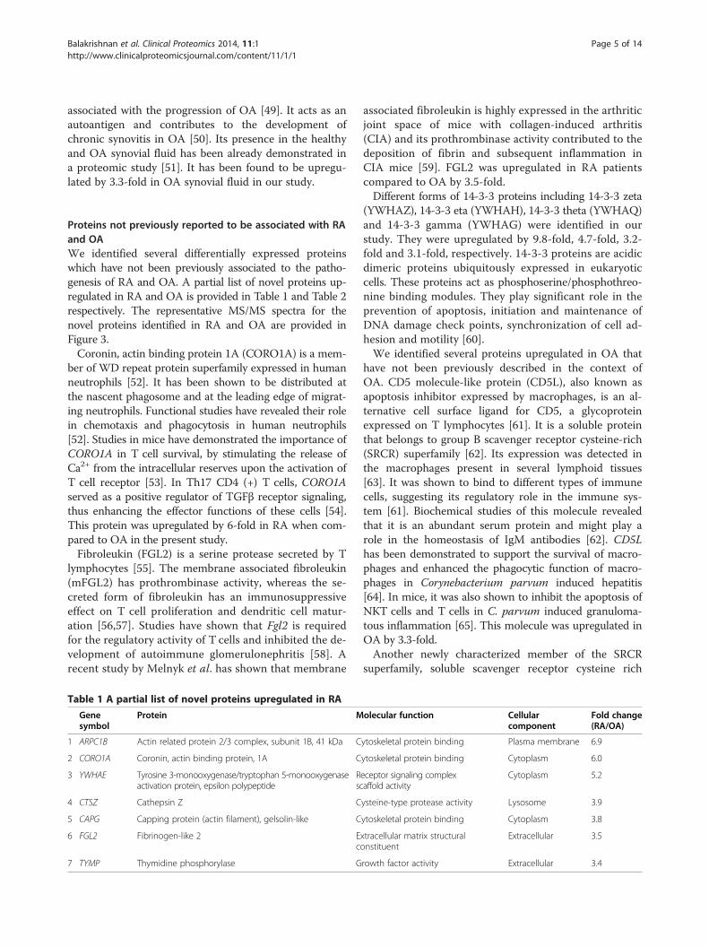
Proteins not previously reported to be associated with RAand OAWe identified several differentially expressed proteinswhich have not been previously associated to the patho-genesis of RA and OA. A partial list of novel proteins up-regulated in RA and OA is provided in Table 1 and Table 2respectively. The representative MS/MS spectra for thenovel proteins identified in RA and OA are provided inFigure 3.Coronin, actin binding protein 1A (CORO1A) is a mem-
ber of WD repeat protein superfamily expressed in humanneutrophils [52]. It has been shown to be distributed atthe nascent phagosome and at the leading edge of migrat-ing neutrophils. Functional studies have revealed their rolein chemotaxis and phagocytosis in human neutrophils[52]. Studies in mice have demonstrated the importance ofCORO1A in T cell survival, by stimulating the release ofCa2+ from the intracellular reserves upon the activation ofT cell receptor [53]. In Th17 CD4 (+) T cells, CORO1Aserved as a positive regulator of TGFβ receptor signaling,thus enhancing the effector functions of these cells [54].This protein was upregulated by 6-fold in RA when com-pared to OA in the present study.Fibroleukin (FGL2) is a serine protease secreted by T
lymphocytes [55]. The membrane associated fibroleukin(mFGL2) has prothrombinase activity, whereas the se-creted form of fibroleukin has an immunosuppressiveeffect on T cell proliferation and dendritic cell matur-ation [56,57]. Studies have shown that Fgl2 is requiredfor the regulatory activity of T cells and inhibited the de-velopment of autoimmune glomerulonephritis [58]. Arecent study by Melnyk et al. has shown that membrane
Table 1 A partial list of novel proteins upregulated in RA
Genesymbol
Protein M
1 ARPC1B Actin related protein 2/3 complex, subunit 1B, 41 kDa C
2 CORO1A Coronin, actin binding protein, 1A C
3 YWHAE Tyrosine 3-monooxygenase/tryptophan 5-monooxygenaseactivation protein, epsilon polypeptide
Rsc
4 CTSZ Cathepsin Z C
5 CAPG Capping protein (actin filament), gelsolin-like C
6 FGL2 Fibrinogen-like 2 Ec
7 TYMP Thymidine phosphorylase G
associated fibroleukin is highly expressed in the arthriticjoint space of mice with collagen-induced arthritis(CIA) and its prothrombinase activity contributed to thedeposition of fibrin and subsequent inflammation inCIA mice [59]. FGL2 was upregulated in RA patientscompared to OA by 3.5-fold.Different forms of 14-3-3 proteins including 14-3-3 zeta
(YWHAZ), 14-3-3 eta (YWHAH), 14-3-3 theta (YWHAQ)and 14-3-3 gamma (YWHAG) were identified in ourstudy. They were upregulated by 9.8-fold, 4.7-fold, 3.2-fold and 3.1-fold, respectively. 14-3-3 proteins are acidicdimeric proteins ubiquitously expressed in eukaryoticcells. These proteins act as phosphoserine/phosphothreo-nine binding modules. They play significant role in theprevention of apoptosis, initiation and maintenance ofDNA damage check points, synchronization of cell ad-hesion and motility [60].We identified several proteins upregulated in OA that
have not been previously described in the context ofOA. CD5 molecule-like protein (CD5L), also known asapoptosis inhibitor expressed by macrophages, is an al-ternative cell surface ligand for CD5, a glycoproteinexpressed on T lymphocytes [61]. It is a soluble proteinthat belongs to group B scavenger receptor cysteine-rich(SRCR) superfamily [62]. Its expression was detected inthe macrophages present in several lymphoid tissues[63]. It was shown to bind to different types of immunecells, suggesting its regulatory role in the immune sys-tem [61]. Biochemical studies of this molecule revealedthat it is an abundant serum protein and might play arole in the homeostasis of IgM antibodies [62]. CD5Lhas been demonstrated to support the survival of macro-phages and enhanced the phagocytic function of macro-phages in Corynebacterium parvum induced hepatitis[64]. In mice, it was also shown to inhibit the apoptosis ofNKT cells and T cells in C. parvum induced granuloma-tous inflammation [65]. This molecule was upregulated inOA by 3.3-fold.Another newly characterized member of the SRCR
superfamily, soluble scavenger receptor cysteine rich
olecular function Cellularcomponent
Fold change(RA/OA)
ytoskeletal protein binding Plasma membrane 6.9
ytoskeletal protein binding Cytoplasm 6.0
eceptor signaling complexaffold activity
Cytoplasm 5.2
ysteine-type protease activity Lysosome 3.9
ytoskeletal protein binding Cytoplasm 3.8
xtracellular matrix structuralonstituent
Extracellular 3.5
rowth factor activity Extracellular 3.4
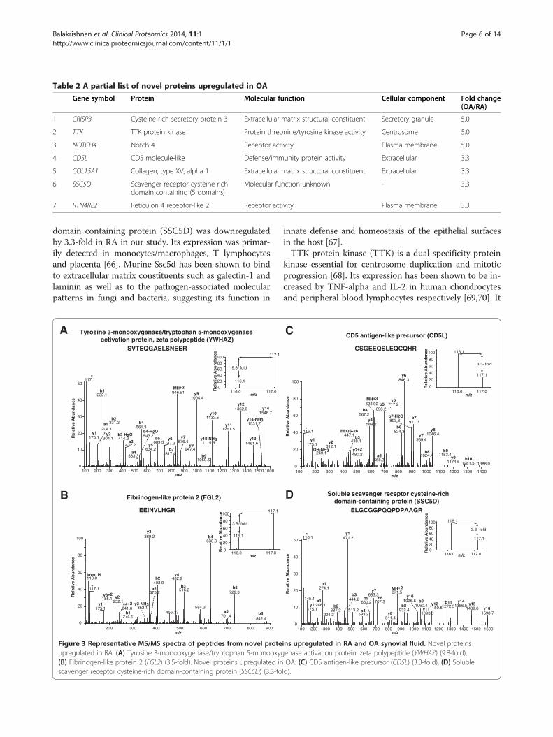
Table 2 A partial list of novel proteins upregulated in OA
Gene symbol Protein Molecular function Cellular component Fold change(OA/RA)
1 CRISP3 Cysteine-rich secretory protein 3 Extracellular matrix structural constituent Secretory granule 5.0
2 TTK TTK protein kinase Protein threonine/tyrosine kinase activity Centrosome 5.0
3 NOTCH4 Notch 4 Receptor activity Plasma membrane 5.0
4 CD5L CD5 molecule-like Defense/immunity protein activity Extracellular 3.3
5 COL15A1 Collagen, type XV, alpha 1 Extracellular matrix structural constituent Extracellular 3.3
6 SSC5D Scavenger receptor cysteine richdomain containing (5 domains)
Molecular function unknown - 3.3
7 RTN4RL2 Reticulon 4 receptor-like 2 Receptor activity Plasma membrane 3.3
Balakrishnan et al. Clinical Proteomics 2014, 11:1 Page 6 of 14http://www.clinicalproteomicsjournal.com/content/11/1/1
domain containing protein (SSC5D) was downregulatedby 3.3-fold in RA in our study. Its expression was primar-ily detected in monocytes/macrophages, T lymphocytesand placenta [66]. Murine Ssc5d has been shown to bindto extracellular matrix constituents such as galectin-1 andlaminin as well as to the pathogen-associated molecularpatterns in fungi and bacteria, suggesting its function in
116.0 117.0m/z
0
20
40
60
80
100
Rel
ativ
e A
bu
nd
ance 117.1
116.1
SVTEQGAELSNEER
100 200 300 400 500 600 700 800 900 1000 1100 1200 1300 1400 1500 1600m/z
0
10
20
30
40
50
Rel
ativ
e A
bu
nd
ance
117.1
846.91232.1
1004.4
1362.61548.7
1132.5331.2
561.3 1261.5
304.1175.1 414.2689.3 1115.5747.3 1461.6432.2
947.4634.2
876.4543.2
817.4
1531.7
533.31059.5
y1
*
204.1a1
b1
y2
b2
b3-H2O
b3
a4
b4-H2O
b4
y5
b5 y6
b7
MH+2
y7
y8
y9
b9
y10-NH3
y10
y11
y12
y13
y14-NH3
y14
EEINVLHGR
Fibrinogen-like protein 2 (FGL2)
116.0 117.0m/z
0
20
40
60
80
100
Rel
ativ
e A
bu
nd
ance
117.1
116.1
200 300 400 500 600 700 800m/z
0
20
40
60
80
100
Rel
ativ
e A
bu
nd
ance
369.2630.3
482.2110.0403.9
117.1 516.2729.3
185.1232.1
584.3175.1 241.6
375.2
352.1
274.1 701.4456.33
842.4
900
Imm. H
y1
y3+2y2
y4+2
b1
y3-NH3
y3
a2
b2y4
b3
b4
a5
b5
b6
*
Tyrosine 3-monooxygenase/tryptophan 5-monooxygenase activation protein, zeta polypeptide (YWHAZ)
A
B
9.8- fold
3.5- fold
Figure 3 Representative MS/MS spectra of peptides from novel proteupregulated in RA: (A) Tyrosine 3-monooxygenase/tryptophan 5-monooxyg(B) Fibrinogen-like protein 2 (FGL2) (3.5-fold). Novel proteins upregulated inscavenger receptor cysteine-rich domain-containing protein (SSC5D) (3.3-fo
innate defense and homeostasis of the epithelial surfacesin the host [67].TTK protein kinase (TTK) is a dual specificity protein
kinase essential for centrosome duplication and mitoticprogression [68]. Its expression has been shown to be in-creased by TNF-alpha and IL-2 in human chondrocytesand peripheral blood lymphocytes respectively [69,70]. It
116.0 117.0m/z
0
20
40
60
80
100
Rel
ativ
e A
bu
nd
ance 116.1
117.1
CSGEEQSLEQCQHR
CD5 antigen-like precursor (CD5L)
200 400 600 800 1000 1200 1400m/z
0
20
40
60
80
100
Rel
ativ
e A
bu
nd
ance
846.3
717.2623.92
567.2
911.3
116.1 824.31046.4447.1
959.4175.1 312.1
249.1 480.2 1024.4 1153.4668.2 1174.5 1281.5 1388.0
y1 y2b3
438.1
b4 696.2
y5b5
b6
b7
y6
y8
b9
y9 b10
QH-NH3
EEQS-28
MH+3
a5
b7-H2O 893.3
y7
b8y7+2
589.2y4
100 300 500 700 900 1100 1300
*
116.0 117.0m/z020406080100
Rel
ativ
e A
bu
nd
ance
116.1
117.1
ELGCGGPQQPDPAAGR
Soluble scavenger receptor cysteine-richdomain-containing protein (SSC5D)
100 200 300 400 500 600 700 800 900 1000 1100 1200 1300 1400 1500 1600m/z
0
10
20
30
40
50
Rel
ativ
e A
bu
nd
ance
471.2116.1
274.1871.5
683.3145.1
707.3246.1 1356.51272.51060.4
1469.6
444.2
1150.5175.1 932.4
650.2
1598.7593.2291.2513.2
811.41093.5
*
y1a1
b1
387.2b2
b3
y5
b4
b5
y7
b6
y8
MH+2
b81036.5
y10b9
y11y12 b11 y14
y15y16
C
D
3.3- fold
3.3- fold
ins upregulated in RA and OA synovial fluid. Novel proteinsenase activation protein, zeta polypeptide (YWHAZ) (9.8-fold),OA: (C) CD5 antigen-like precursor (CD5L) (3.3-fold), (D) Solubleld).
Balakrishnan et al. Clinical Proteomics 2014, 11:1 Page 7 of 14http://www.clinicalproteomicsjournal.com/content/11/1/1
was found to be upregulated in OA by 5-fold. In addition,we have also found proteins including, THAP domain-containing protein 4, (THAP4) reticulon 4 receptor-like 2(RTN4RL2) and leucine rich repeat protein 1 (LRR1) to beupregulated by more than 3-fold in OA.Although the above-mentioned proteins have been ob-
served to be differentially expressed in the synovial fluidof RA and OA patients, it is possible that they are notreally associated with the disease but rather contributedby either individual variation or disease conditions otherthan arthritis in these patients. This is why, it is neces-sary to perform validation studies in a larger cohort ofsamples. In addition, it will be useful to determine ifthese proteins are detected in synovial or cartilage tis-sues to understand their association with RA and OAmore fully.
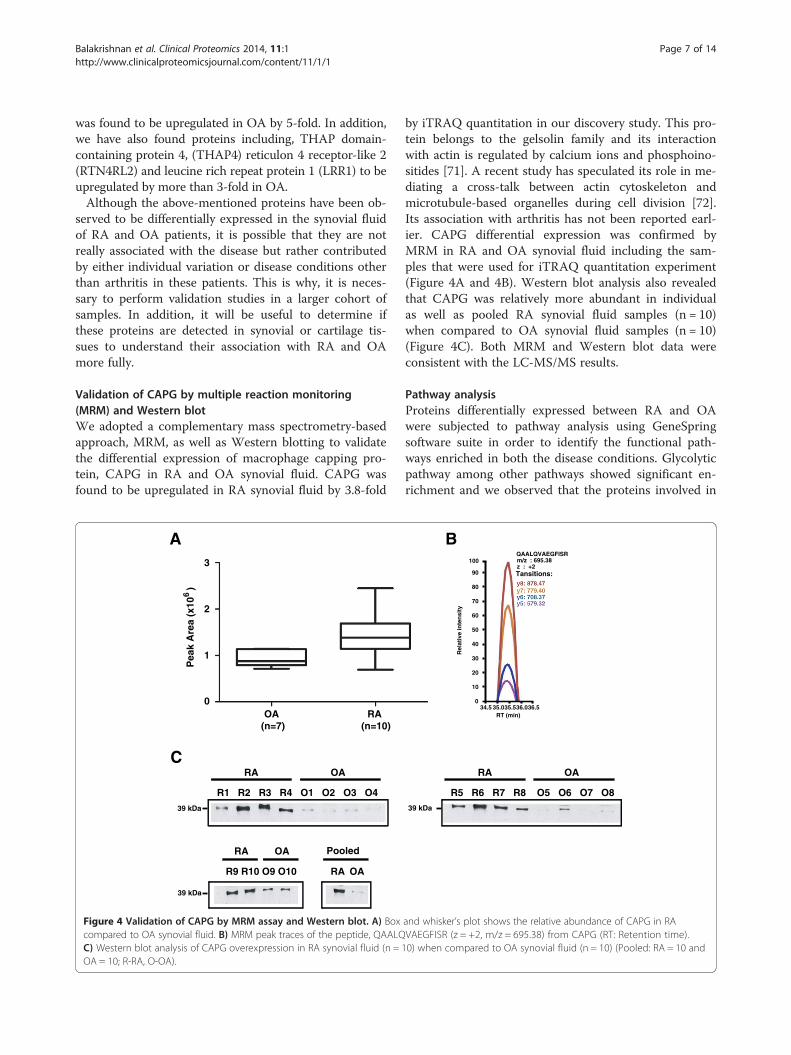
Validation of CAPG by multiple reaction monitoring(MRM) and Western blotWe adopted a complementary mass spectrometry-basedapproach, MRM, as well as Western blotting to validatethe differential expression of macrophage capping pro-tein, CAPG in RA and OA synovial fluid. CAPG wasfound to be upregulated in RA synovial fluid by 3.8-fold
A
OA (n=7)
RA (n=10)
RA OA
R1 R2 R3 R4 O1 O2 O3 O4
R9 R10 O9 O10
PooledOARA
RA OA
C
0
1
2
3
Pea
k A
rea
(x10
)
6
39 kDa
39 kDa
Figure 4 Validation of CAPG by MRM assay and Western blot. A) Boxcompared to OA synovial fluid. B) MRM peak traces of the peptide, QAALQC) Western blot analysis of CAPG overexpression in RA synovial fluid (n = 1OA = 10; R-RA, O-OA).
by iTRAQ quantitation in our discovery study. This pro-tein belongs to the gelsolin family and its interactionwith actin is regulated by calcium ions and phosphoino-sitides [71]. A recent study has speculated its role in me-diating a cross-talk between actin cytoskeleton andmicrotubule-based organelles during cell division [72].Its association with arthritis has not been reported earl-ier. CAPG differential expression was confirmed byMRM in RA and OA synovial fluid including the sam-ples that were used for iTRAQ quantitation experiment(Figure 4A and 4B). Western blot analysis also revealedthat CAPG was relatively more abundant in individualas well as pooled RA synovial fluid samples (n = 10)when compared to OA synovial fluid samples (n = 10)(Figure 4C). Both MRM and Western blot data wereconsistent with the LC-MS/MS results.
Pathway analysisProteins differentially expressed between RA and OAwere subjected to pathway analysis using GeneSpringsoftware suite in order to identify the functional path-ways enriched in both the disease conditions. Glycolyticpathway among other pathways showed significant en-richment and we observed that the proteins involved in
34.5 35.035.536.0 36.5
10
0
20
30
40
50
60
70
80
90
100
RT (min)
QAALQVAEGFISR m/z : 695.38 z : +2 Tansitions:
B
R5 R6 R7 R8 O5 O6 O7 O8
RA OA
Rel
ativ
e in
ten
sity
39 kDa
and whisker’s plot shows the relative abundance of CAPG in RAVAEGFISR (z = +2, m/z = 695.38) from CAPG (RT: Retention time).0) when compared to OA synovial fluid (n = 10) (Pooled: RA = 10 and
Balakrishnan et al. Clinical Proteomics 2014, 11:1 Page 8 of 14http://www.clinicalproteomicsjournal.com/content/11/1/1
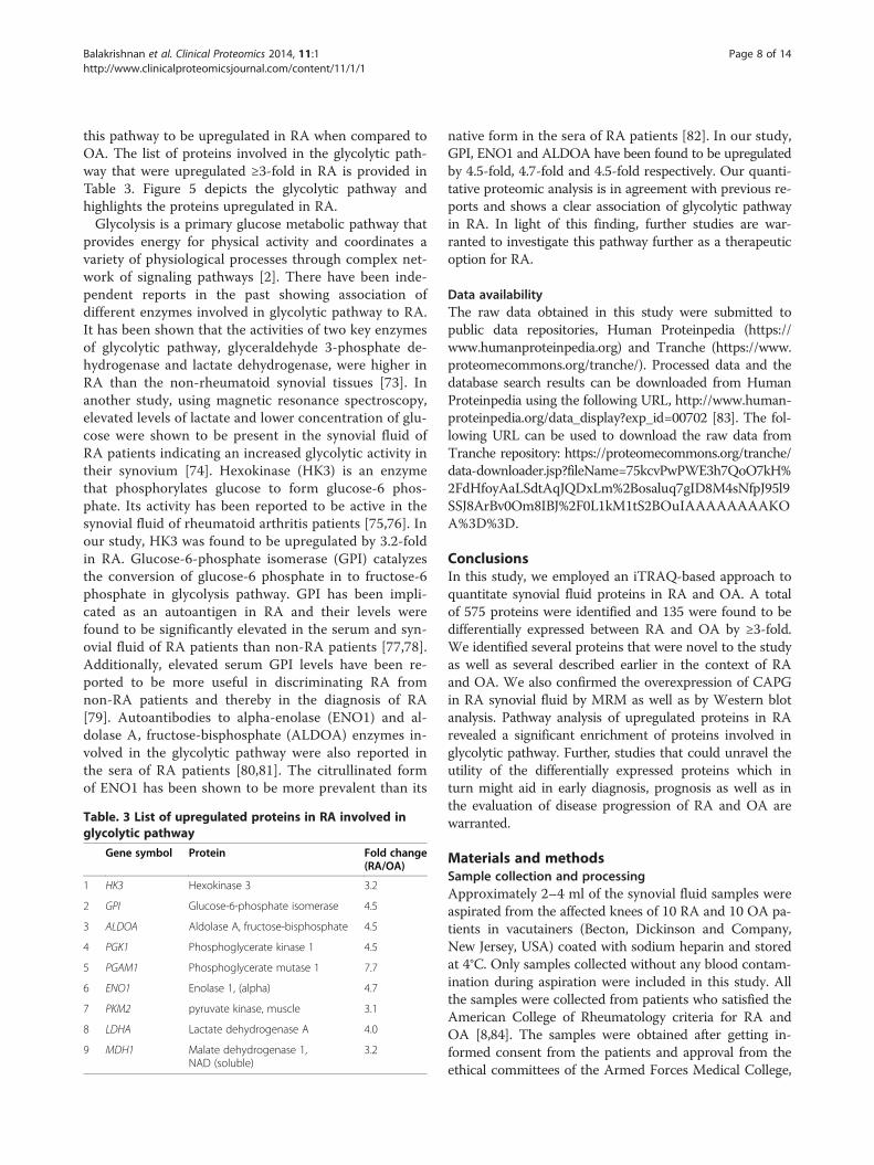
this pathway to be upregulated in RA when compared toOA. The list of proteins involved in the glycolytic path-way that were upregulated ≥3-fold in RA is provided inTable 3. Figure 5 depicts the glycolytic pathway andhighlights the proteins upregulated in RA.Glycolysis is a primary glucose metabolic pathway that
provides energy for physical activity and coordinates avariety of physiological processes through complex net-work of signaling pathways [2]. There have been inde-pendent reports in the past showing association ofdifferent enzymes involved in glycolytic pathway to RA.It has been shown that the activities of two key enzymesof glycolytic pathway, glyceraldehyde 3-phosphate de-hydrogenase and lactate dehydrogenase, were higher inRA than the non-rheumatoid synovial tissues [73]. Inanother study, using magnetic resonance spectroscopy,elevated levels of lactate and lower concentration of glu-cose were shown to be present in the synovial fluid ofRA patients indicating an increased glycolytic activity intheir synovium [74]. Hexokinase (HK3) is an enzymethat phosphorylates glucose to form glucose-6 phos-phate. Its activity has been reported to be active in thesynovial fluid of rheumatoid arthritis patients [75,76]. Inour study, HK3 was found to be upregulated by 3.2-foldin RA. Glucose-6-phosphate isomerase (GPI) catalyzesthe conversion of glucose-6 phosphate in to fructose-6phosphate in glycolysis pathway. GPI has been impli-cated as an autoantigen in RA and their levels werefound to be significantly elevated in the serum and syn-ovial fluid of RA patients than non-RA patients [77,78].Additionally, elevated serum GPI levels have been re-ported to be more useful in discriminating RA fromnon-RA patients and thereby in the diagnosis of RA[79]. Autoantibodies to alpha-enolase (ENO1) and al-dolase A, fructose-bisphosphate (ALDOA) enzymes in-volved in the glycolytic pathway were also reported inthe sera of RA patients [80,81]. The citrullinated formof ENO1 has been shown to be more prevalent than its
Table. 3 List of upregulated proteins in RA involved inglycolytic pathway
Gene symbol Protein Fold change(RA/OA)
1 HK3 Hexokinase 3 3.2
2 GPI Glucose-6-phosphate isomerase 4.5
3 ALDOA Aldolase A, fructose-bisphosphate 4.5
4 PGK1 Phosphoglycerate kinase 1 4.5
5 PGAM1 Phosphoglycerate mutase 1 7.7
6 ENO1 Enolase 1, (alpha) 4.7
7 PKM2 pyruvate kinase, muscle 3.1
8 LDHA Lactate dehydrogenase A 4.0
9 MDH1 Malate dehydrogenase 1,NAD (soluble)
3.2
native form in the sera of RA patients [82]. In our study,GPI, ENO1 and ALDOA have been found to be upregulatedby 4.5-fold, 4.7-fold and 4.5-fold respectively. Our quanti-tative proteomic analysis is in agreement with previous re-ports and shows a clear association of glycolytic pathwayin RA. In light of this finding, further studies are war-ranted to investigate this pathway further as a therapeuticoption for RA.
Data availabilityThe raw data obtained in this study were submitted topublic data repositories, Human Proteinpedia (https://www.humanproteinpedia.org) and Tranche (https://www.proteomecommons.org/tranche/). Processed data and thedatabase search results can be downloaded from HumanProteinpedia using the following URL, http://www.human-proteinpedia.org/data_display?exp_id=00702 [83]. The fol-lowing URL can be used to download the raw data fromTranche repository: https://proteomecommons.org/tranche/data-downloader.jsp?fileName=75kcvPwPWE3h7QoO7kH%2FdHfoyAaLSdtAqJQDxLm%2Bosaluq7gID8M4sNfpJ95l9SSJ8ArBv0Om8IBJ%2F0L1kM1tS2BOuIAAAAAAAAKOA%3D%3D.
ConclusionsIn this study, we employed an iTRAQ-based approach toquantitate synovial fluid proteins in RA and OA. A totalof 575 proteins were identified and 135 were found to bedifferentially expressed between RA and OA by ≥3-fold.We identified several proteins that were novel to the studyas well as several described earlier in the context of RAand OA. We also confirmed the overexpression of CAPGin RA synovial fluid by MRM as well as by Western blotanalysis. Pathway analysis of upregulated proteins in RArevealed a significant enrichment of proteins involved inglycolytic pathway. Further, studies that could unravel theutility of the differentially expressed proteins which inturn might aid in early diagnosis, prognosis as well as inthe evaluation of disease progression of RA and OA arewarranted.
Materials and methodsSample collection and processingApproximately 2–4 ml of the synovial fluid samples wereaspirated from the affected knees of 10 RA and 10 OA pa-tients in vacutainers (Becton, Dickinson and Company,New Jersey, USA) coated with sodium heparin and storedat 4°C. Only samples collected without any blood contam-ination during aspiration were included in this study. Allthe samples were collected from patients who satisfied theAmerican College of Rheumatology criteria for RA andOA [8,84]. The samples were obtained after getting in-formed consent from the patients and approval from theethical committees of the Armed Forces Medical College,
Mitochondrion
Cytosol
Figure 5 Glycolytic pathway enriched from GeneSpring analysis. Proteins upregulated in RA are highlighted in the glycolysis pathway image.
Balakrishnan et al. Clinical Proteomics 2014, 11:1 Page 9 of 14http://www.clinicalproteomicsjournal.com/content/11/1/1
Balakrishnan et al. Clinical Proteomics 2014, 11:1 Page 10 of 14http://www.clinicalproteomicsjournal.com/content/11/1/1
Pune, India, Fortis Hospitals, Bangalore, India andCommand Air Force Hospital, Bangalore, India. Sam-ples were collected from RA and OA patients with anaverage age of 52 and 65 years, respectively. The sam-ples were centrifuged at 1,500 g at room temperaturefor 15 minutes and the supernatants were filtered byusing 0.22 μm filters (Millipore, Ireland). The filteredsamples were stored at -80°C until further analysis.
Depletion of synovial fluid proteome and iTRAQ labelingProtein estimation of the synovial fluid samples was car-ried out using Lowry’s method [85]. Five samples each ofOA and RA synovial fluid were pooled separately. Thepooled samples were then depleted to remove the 14 mostabundant proteins (Albumin, haptoglobin, transferrin,IgA, IgG, alpha 1-antitrypsin, alpha 2-antitrypsin, alpha 1-acid glycoprotein, apolipoprotein A1, apolipoprotein A2,complement C3, IgM, transthyretin and fibrinogen) byusing Human 14 multiple affinity removal spin cartridge(Agilent Technologies, Santa Clara, California, USA). Thedepleted protein was then washed and concentrated using3 kDa MWCO filters (Amicon, Millipore, Ireland). Ap-proximately 65 μg equivalent of the depleted synovial fluidprotein from each group was subjected to trypsin diges-tion and iTRAQ labeling as described earlier [21]. Briefly,denaturation of the protein was done using 2% SDSfollowed by reduction and alkylation with reducing agenttris (2-carboxyethyl) phosphine (TCEP) and cysteineblocking agent, methyl methanethiosulfonate (MMTS), re-spectively. Subsequently the samples were digested withsequencing grade trypsin (Promega, Madison, WI, USA)at 37°C overnight. The tryptic peptides from each groupwere then labeled with 4-plex iTRAQ reagents (iTRAQReagents Multiplex kit, Applied Biosystems, California,USA) as per the manufacturer’s instructions. OA and RAsynovial fluid derived tryptic peptides were labeled with116 and 117 iTRAQ labels respectively. The labeled pep-tides were then pooled, vacuum-dried and reconstitutedin 10 mM KH2PO4, 20% acetonitrile (pH 2.8) (solvent A)and fractionated by strong cation exchange (SCX)chromatography.
Strong cation exchange (SCX)-based fractionationSCX-based fractionation was carried out as describedearlier [20]. In brief, the tryptic peptides were fractionatedon a PolySULFOETHYL A column (PolyLC, Columbia,MD, USA) with 200 Å, 5 μm, 200 × 2.1 mm dimensions,using an Agilent’s 1200 HPLC-system (Agilent Technolo-gies, Santa Clara, California , USA). A linear gradient ofincreasing solvent B (350 mM KCl in solvent A, pH 2.8)at a flow rate of 200 μl/min over a period of 70 min wasused for fractionation. Peptide fractions were collectedusing an automated fraction collector. 20 fractions were ob-tained after fractionation. All the fractions were completely
dried and reconstituted in 0.1% trifluoroacetic acid to befurther desalted using stage-tips packed with C18 material[86]. Desalted fractions were dried in speedvac and recon-stituted in 10 μl of 0.1% TFA prior to LC-MS/MS analysis.
LC-MS/MS analysisTandem mass spectrometric analysis of the iTRAQ la-beled peptides were carried out using LTQ-Orbitrap Velosmass spectrometer (Thermo Scientific, Bremen, Germany)interfaced with Easy nanoLC II (previously Proxeon,Thermo Scientific, Bremen, Germany). The LC systemconsisted of an enrichment column (3 cm× 75 μ) packedwith Magic AQ C18 material, 5 μ particle size with 100 Åpore size) and an analytical column (10 cm× 75 μ), MagicAQ C18 material 5 μ particle size, 100 Å pore size) packedusing pressure injection cell at 800 psi. Electrosprayionization source was fitted with an emitter tip 8 μm(New Objective, Woburn, MA) and maintained at 2000 Vion spray voltage. Peptide samples were loaded onto anenrichment column in 0.1% formic acid, 5% ACN for15 min and peptide separation carried out using a lineargradient of 7-35% solvent B (90% ACN in 0.1% formicacid) for 60 minutes at a constant flow rate of 350 nl/min.Data was acquired using Xcalibur 2.1 (Thermo Scientific,Bremen, Germany). The MS spectra were acquired in adata-dependent manner in the m/z range of 350 to 1800and survey scans were acquired in Orbitrap mass analyzerat a mass resolution of 60,000 at 400 m/z. The MS/MSdata was acquired in Orbitrap mass analyzer at a reso-lution of 15,000 at 400 m/z by targeting top 20 mostabundant ions for fragmentation using higher energycollisional dissociation activation at 39% normalisedcollision energy. Single and unassigned charge state pre-cursor ions were rejected. The dynamic exclusion op-tion was enabled during data acquisition with exclusionduration of 60 seconds. Lock mass option was enabledfor real time calibration using polycyclodimethylsilox-ane (m/z, 415.12) ions [87].
Data analysisProteome Discoverer Beta Version 1.3 (Thermo FisherScientific Inc., Bremen, Germany) was used for databasesearches. A precursor mass range of 350–8000 Da and asignal to noise of 1.5 were used. A combined Mascot(Mascot version 2.2, Matrix Science) and SEQUESTsearch was done using the Proteome Discoverer suite(Version 1.3.339, Thermo Scientific, Bremen, Germany)against the NCBI Human RefSeq database 50 containing33,947 entries with known contaminants. Search param-eters included trypsin as the enzyme with maximum 1missed cleavage allowed; oxidation of methionine wasset as a dynamic modification while alkylation at cysteineand iTRAQ modification at N-terminus of the peptideand lysine were set as static modifications. Precursor and
Balakrishnan et al. Clinical Proteomics 2014, 11:1 Page 11 of 14http://www.clinicalproteomicsjournal.com/content/11/1/1
fragment mass tolerance were set to 20 ppm and 0.1 Da,respectively. Peptide and protein data were fetched usinghigh peptide confidence and rank one peptide match fil-ters. Reporter ion quantitation node was used for relativeexpression pattern of proteins based on the relative inten-sities of reporter ions for the corresponding peptides. Theraw data obtained was searched against decoy database tocalculate 1% false discovery rate cut-off score [88]. Spectrathat matched to the contaminants and those that did notpass the 1% FDR threshold were not considered foranalysis.
Multiple reaction monitoring (MRM)MRM assays [89] were designed to validate the differen-tially expressed protein, CAPG in RA and OA synovialfluid. Skyline version 2.1 [90] was used for method devel-opment and optimization. The target peptide selected forCAPG was QAALQVAEGFISR (z = +2, m/z = 695.38) andtop four transitions monitored included, y8+→ 878.47,y7+→ 779.40, y6+→ 708.37 and y5+→ 579.32. Thesamples were subjected to in-solution digestion as describedearlier [91]. All samples were analyzed in triplicate on TSQQuantum Ultra (Thermo Scientific, San Jose, CA) interfacedwith Easy nanoLC II (previously Proxeon, Thermo Scientific,Bremen, Germany). The peptides were enriched on a trapcolumn (5 μm, 75 μm×2 cm.) with 0.1% formic acid and5% ACN for 5 minutes and separated on an analyticalcolumn (3 μm, 75 μm × 10 cm) with an increasing lin-ear gradient from 5-35% of solvent B (90% ACN in0.1% formic acid) for 60 min at a constant flow rateof 300 nl/min. Both columns were packed in-houseusing Magic AQ C18 material (Michrom Bioresources).Spray voltage of 2.5 kV was applied and ion transfer tubewas maintained at 275°C. The data was acquired with Q1and Q3 set at resolution of 0.4 and 0.7 respectively. Thecollision energy for each transition was optimized with thehelp of Skyline based on the preliminary results [90].
Western blot analysis40 μg of protein from each individual samples of RA (n =10) and OA (n = 10) synovial fluid was used for Westernblot analysis. The primary antibody used was a rabbit anti-human polyclonal antibody for CAPG (ProteintechGroup, Inc, Chicago, USA). SDS-PAGE gels were electro-blotted on nitrocellulose membrane (Whatman Inc.,Maine, USA) at 250 mA for 2 hours using TE-70 ECLsemi-dry transfer unit (GE Healthcare, Pittsburgh, USA).The membranes were then blocked with 5% non-fat drymilk and washed with phosphate buffered saline contain-ing 0.05% tween (PBST). The membranes were incubatedwith primary antibody for 2 hours, washed with PBST andincubated at room temperature for 1 hour in diluted(1:2500) anti-rabbit IgG antibody conjugated with horse-radish peroxidase (GE Healthcare, UK).
Bioinformatics analysisGene Ontology (GO)-based [92] analysis was done usingHuman Protein Reference Database (HPRD) (http://www.hprd.org), which is a GO compliant database [27,28]. Datapertaining to sub-cellular localization, biological processes,domains and motif information associated with the identi-fied proteins were obtained from HPRD.
GeneSpring AnalysisPathway Architect module from GeneSpring GX12 wasused to identify significantly enriched pathways. Using‘single experiment analysis’ tool in the pathway architectmodule the differentially expressed list of genes wassearched against publicly available pathways.
Additional file
Additional file 1: Table S1. List of proteins identified from OA and RAsynovial fluid along with the corresponding peptide sequences, coverage,total number of peptides, unique peptides, PSMs, 117/116 (RA/OA) ratio,117/116 variability, modifications, Xcorr, IonScore, charge, MH+, deltamass (ppm), retention time (RT).
AbbreviationsRA: Rheumatoid arthritis; OA: Osteoarthritis; iTRAQ: isobaric tags for relativeand absolute quantitation; SCX: Strong cation exchange; MMPs: Matrixmetalloproteinases; CAPG: Macrophage capping protein.
Competing interestsThe authors declare that they have no competing interests.
Author’s contributionAP, SS, SM and HG participated in the conception and study design. LB andMB collected the samples and performed the experiments. RSN, SR, MBcarried out fractionation and mass spectrometry analysis of the samples. LB,NS, SA and SR were involved in validation experiments. LB prepared themanuscript. LB and YS prepared the manuscript figures. LB, AM, SMS and RRwere involved in data analysis and interpretation. VV, MD and NK edited themanuscript. RJ, YLR, TSKP, NS, HG, SM and AP critically read and revised themanuscript. All the authors have read and approved the final manuscript.
AcknowledgementsWe thank the Department of Biotechnology (DBT), Government of India forresearch support to the Institute of Bioinformatics, Bangalore. We also thankThermo Scientific for access to instrumentation. Sartaj Ahmad is a recipientof Junior Research Fellowship from University Grants Commission (UGC),Government of India. Raja Sekhar Nirujogi is a recipient of Senior ResearchFellowship award from Council of Scientific and Industrial Research (CSIR),Government of India. Yashwanth Subbannayya and Santosh Renuse arerecipients of Senior Research Fellowship from University Grants Commission(UGC), Government of India. Srinivas M. Srikanth is a recipient of JuniorResearch Fellowship award from University Grants Commission (UGC), India.T. S. Keshava Prasad is supported by a research grant on “Development ofInfrastructure and a Computational Framework for Analysis of ProteomicData” from DBT, Government of India. Harsha Gowda is a WellcomeTrust/DBT India Alliance Early Career Fellow.
Author details1Institute of Bioinformatics, International Technology Park, Bangalore 560066,India. 2Department of Biotechnology, Kuvempu University, Shankaraghatta577451, India. 3Amrita School of Biotechnology, Amrita VishwaVidyapeetham, Kollam 690525, India. 4Manipal University, Madhava Nagar,Manipal 576104, India. 5Centre for Excellence in Bioinformatics, School of LifeSciences, Pondicherry University, Puducherry 605014, India. 6Rajiv GandhiUniversity of Health Sciences, Bangalore 560041, India. 7Department of
Balakrishnan et al. Clinical Proteomics 2014, 11:1 Page 12 of 14http://www.clinicalproteomicsjournal.com/content/11/1/1
Internal Medicine, Armed Forces Medical College, Pune 411040, India.8Department of Rheumatology, Fortis Hospitals, Bangalore 560076, India.9Department of Rheumatology, Command Airforce Hospital, Bangalore560008, India. 10Laboratory for Integrated Bioinformatics, RIKEN Center forIntegrative Medical Sciences (IMS-RCAI), Yokohama Institute, Kanagawa230-0045, Japan. 11McKusick-Nathans Institute of Genetic Medicine, JohnsHopkins University School of Medicine, Baltimore, MD 21205, USA.12Department of Oncology, Johns Hopkins University School of Medicine,Baltimore, MD 21205, USA. 13Department of Pathology, Johns HopkinsUniversity School of Medicine, Baltimore, MD 21205, USA. 14Department ofBiological Chemistry, Johns Hopkins University School of Medicine, Baltimore,MD 21205, USA.
Received: 30 April 2013 Accepted: 10 December 2013Published: 6 January 2014
References1. Scott DL, Wolfe F, Huizinga TW: Rheumatoid arthritis. Lancet 2010,
376:1094–1108.2. Chang X, Wei C: Glycolysis and rheumatoid arthritis. Int J Rheum Dis 2011,
14:217–222.3. Klareskog L, Catrina AI, Paget S: Rheumatoid arthritis. Lancet 2009,
373:659–672.4. Rousseau J, Garnero P: Biological markers in osteoarthritis. Bone 2012,
51:265–277.5. Attur M, Krasnokutsky-Samuels S, Samuels J, Abramson SB: Prognostic
biomarkers in osteoarthritis. Curr Opin Rheumatol 2013, 25:136–144.6. Da Mota LM, Dos Santos Neto LL, De Carvalho JF: Autoantibodies and
other serological markers in rheumatoid arthritis: predictors of diseaseactivity? Clin Rheumatol 2009, 28:1127–1134.
7. Van Boekel MA, Vossenaar ER, van den Hoogen FH, Van Venrooij WJ:Autoantibody systems in rheumatoid arthritis: specificity, sensitivity anddiagnostic value. Arthritis Res 2002, 4:87–93.
8. Arnett FC, Edworthy SM, Bloch DA, McShane DJ, Fries JF, Cooper NS, HealeyLA, Kaplan SR, Liang MH, Luthra HS, et al: The American RheumatismAssociation 1987 revised criteria for the classification of rheumatoidarthritis. Arthritis Rheum 1988, 31:315–324.
9. Kuhn KA, Kulik L, Tomooka B, Braschler KJ, Arend WP, Robinson WH, Holers VM:Antibodies against citrullinated proteins enhance tissue injury inexperimental autoimmune arthritis. J Clin Invest 2006, 116:961–973.
10. Lee DM, Schur PH: Clinical utility of the anti-CCP assay in patients withrheumatic diseases. Ann Rheum Dis 2003, 62:870–874.
11. Goldbach-Mansky R, Lee J, McCoy A, Hoxworth J, Yarboro C, Smolen JS,Steiner G, Rosen A, Zhang C, Menard HA, et al: Rheumatoid arthritisassociated autoantibodies in patients with synovitis of recent onset.Arthritis Res 2000, 2:236–243.
12. Rousseau JC, Delmas PD: Biological markers in osteoarthritis. Nat Clin PractRheumatol 2007, 3:346–356.
13. Mobasheri A: Osteoarthritis year 2012 in review: biomarkers. OsteoarthritisCartilage 2012, 20:1451–1464.
14. Hui AY, McCarty WJ, Masuda K, Firestein GS, Sah RL: A systems biologyapproach to synovial joint lubrication in health, injury, and disease. WileyInterdiscip Rev Syst Biol Med 2012, 4:15–37.
15. Uchida T, Fukawa A, Uchida M, Fujita K, Saito K: Application of a novelprotein biochip technology for detection and identification ofrheumatoid arthritis biomarkers in synovial fluid. J Proteome Res 2002,1:495–499.
16. Sinz A, Bantscheff M, Mikkat S, Ringel B, Drynda S, Kekow J, Thiesen HJ,Glocker MO: Mass spectrometric proteome analyses of synovial fluidsand plasmas from patients suffering from rheumatoid arthritis andcomparison to reactive arthritis or osteoarthritis. Electrophoresis 2002,23:3445–3456.
17. Mateos J, Lourido L, Fernandez-Puente P, Calamia V, Fernandez-Lopez C,Oreiro N, Ruiz-Romero C, Blanco FJ: Differential protein profiling ofsynovial fluid from rheumatoid arthritis and osteoarthritis patients usingLC-MALDI TOF/TOF. J Proteomics 2012, 75:2869–2878.
18. Biswas S, Sharma S, Saroha A, Bhakuni DS, Malhotra R, Zahur M, Oellerich M,Das HR, Asif AR: Identification of novel autoantigen in the synovial fluidof rheumatoid arthritis patients using an immunoproteomics approach.PLoS One 2013, 8:e56246.
19. Ross PL, Huang YN, Marchese JN, Williamson B, Parker K, Hattan S,Khainovski N, Pillai S, Dey S, Daniels S, et al: Multiplexed proteinquantitation in Saccharomyces cerevisiae using amine-reactive isobarictagging reagents. Mol Cell Proteomics 2004, 3:1154–1169.
20. Chaerkady R, Harsha HC, Nalli A, Gucek M, Vivekanandan P, Akhtar J, ColeRN, Simmers J, Schulick RD, Singh S, et al: A quantitative proteomicapproach for identification of potential biomarkers in hepatocellularcarcinoma. J Proteome Res 2008, 7:4289–4298.
21. Pawar H, Kashyap MK, Sahasrabuddhe NA, Renuse S, Harsha HC, Kumar P,Sharma J, Kandasamy K, Marimuthu A, Nair B, et al: Quantitative tissueproteomics of esophageal squamous cell carcinoma for novel biomarkerdiscovery. Cancer Biol Ther 2011, 12:510–522.
22. Polisetty RV, Gautam P, Sharma R, Harsha HC, Nair SC, Gupta MK, Uppin MS,Challa S, Puligopu AK, Ankathi P: LC-MS/MS analysis of differentiallyexpressed glioblastoma membrane proteome reveals altered calciumsignaling and other protein groups of regulatory functions. Mol CellProteomics 2012, 11. M111 013565.
23. Gautam P, Nair SC, Gupta MK, Sharma R, Polisetty RV, Uppin MS, Sundaram C,Puligopu AK, Ankathi P, Purohit AK, et al: Proteins with altered levels in plasmafrom glioblastoma patients as revealed by iTRAQ-based quantitativeproteomic analysis. PLoS One 2012, 7:e46153.
24. Kristjansdottir B, Levan K, Partheen K, Carlsohn E, Sundfeldt K: Potentialtumor biomarkers identified in ovarian cyst fluid by quantitativeproteomic analysis, iTRAQ. Clin Proteomics 2013, 10:4.
25. Kumar GS, Venugopal AK, Mahadevan A, Renuse S, Harsha HC,Sahasrabuddhe NA, Pawar H, Sharma R, Kumar P, Rajagopalan S, et al:Quantitative proteomics for identifying biomarkers for tuberculousmeningitis. Clin Proteomics 2012, 9:12.
26. Venugopal AK, Ghantasala SS, Selvan LD, Mahadevan A, Renuse S, Kumar P,Pawar H, Sahasrabhuddhe NA, Suja MS, Ramachandra YL, et al:Quantitative proteomics for identifying biomarkers for rabies. ClinProteomics 2013, 10:3.
27. Keshava Prasad TS, Goel R, Kandasamy K, Keerthikumar S, Kumar S,Mathivanan S, Telikicherla D, Raju R, Shafreen B, Venugopal A, et al: HumanProtein reference database–2009 update. Nucleic Acids Res 2009,37:D767–D772.
28. Prasad TS, Kandasamy K, Pandey A: Human protein reference databaseand human proteinpedia as discovery tools for systems biology. MethodsMol Biol 2009, 577:67–79.
29. Foell D, Roth J: Proinflammatory S100 proteins in arthritis andautoimmune disease. Arthritis Rheum 2004, 50:3762–3771.
30. Baillet A, Trocme C, Berthier S, Arlotto M, Grange L, Chenau J, Quetant S,Seve M, Berger F, Juvin R, et al: Synovial fluid proteomic fingerprint:S100A8, S100A9 and S100A12 proteins discriminate rheumatoid arthritisfrom other inflammatory joint diseases. Rheumatology (Oxford) 2010,49:671–682.
31. Murphy G, Knauper V, Atkinson S, Butler G, English W, Hutton M, Stracke J,Clark I: Matrix metalloproteinases in arthritic disease. Arthritis Res 2002,4(Suppl 3):S39–S49.
32. Tchetverikov I, Ronday HK, Van El B, Kiers GH, Verzijl N, TeKoppele JM,Huizinga TW, DeGroot J, Hanemaaijer R: MMP profile in paired serum andsynovial fluid samples of patients with rheumatoid arthritis. Ann RheumDis 2004, 63:881–883.
33. Benarafa C: The SerpinB1 knockout mouse a model for studyingneutrophil protease regulation in homeostasis and inflammation.Methods Enzymol 2011, 499:135–148.
34. Clausen T, Southan C, Ehrmann M: The HtrA family of proteases: implicationsfor protein composition and cell fate. Mol Cell 2002, 10:443–455.
35. Rosenthal AK, Gohr CM, Ninomiya J, Wakim BT: Proteomic analysis ofarticular cartilage vesicles from normal and osteoarthritic cartilage.Arthritis Rheum 2011, 63:401–411.
36. Wu J, Liu W, Bemis A, Wang E, Qiu Y, Morris EA, Flannery CR, Yang Z:Comparative proteomic characterization of articular cartilage tissue fromnormal donors and patients with osteoarthritis. Arthritis Rheum 2007,56:3675–3684.
37. Polur I, Lee PL, Servais JM, Xu L, Li Y: Role of HTRA1, a serine protease, inthe progression of articular cartilage degeneration. Histol Histopathol2010, 25:599–608.
38. Hadfield KD, Rock CF, Inkson CA, Dallas SL, Sudre L, Wallis GA, Boot-Handford RP,Canfield AE: HtrA1 inhibits mineral deposition by osteoblasts: requirementfor the protease and PDZ domains. J Biol Chem 2008, 283:5928–5938.
Balakrishnan et al. Clinical Proteomics 2014, 11:1 Page 13 of 14http://www.clinicalproteomicsjournal.com/content/11/1/1
39. Grau S, Richards PJ, Kerr B, Hughes C, Caterson B, Williams AS, Junker U,Jones SA, Clausen T, Ehrmann M: The role of human HtrA1 in arthriticdisease. J Biol Chem 2006, 281:6124–6129.
40. Bauer S, Jendro MC, Wadle A, Kleber S, Stenner F, Dinser R, Reich A, Faccin E,Godde S, Dinges H, et al: Fibroblast activation protein is expressed byrheumatoid myofibroblast-like synoviocytes. Arthritis Res Ther 2006, 8:R171.
41. Dohi O, Ohtani H, Hatori M, Sato E, Hosaka M, Nagura H, Itoi E, Kokubun S:Histogenesis-specific expression of fibroblast activation protein anddipeptidylpeptidase-IV in human bone and soft tissue tumours.Histopathology 2009, 55:432–440.
42. Stamp LK, Khalilova I, Tarr JM, Senthilmohan R, Turner R, Haigh RC, WinyardPG, Kettle AJ: Myeloperoxidase and oxidative stress in rheumatoidarthritis. Rheumatology (Oxford) 2012, 51:1796–1803.
43. Tschesche H, Zolzer V, Triebel S, Bartsch S: The human neutrophil lipocalinsupports the allosteric activation of matrix metalloproteinases. Eur JBiochem 2001, 268:1918–1928.
44. Gupta K, Shukla M, Cowland JB, Malemud CJ, Haqqi TM: Neutrophilgelatinase-associated lipocalin is expressed in osteoarthritis and forms acomplex with matrix metalloproteinase 9. Arthritis Rheum 2007,56:3326–3335.
45. Katano M, Okamoto K, Arito M, Kawakami Y, Kurokawa MS, Suematsu N,Shimada S, Nakamura H, Xiang Y, Masuko K, et al: Implication ofgranulocyte-macrophage colony-stimulating factor induced neutrophilgelatinase-associated lipocalin in pathogenesis of rheumatoid arthritisrevealed by proteome analysis. Arthritis Res Ther 2009, 11:R3.
46. Wang Q, Rozelle AL, Lepus CM, Scanzello CR, Song JJ, Larsen DM, Crish JF,Bebek G, Ritter SY, Lindstrom TM, et al: Identification of a central role forcomplement in osteoarthritis. Nat Med 2011, 17:1674–1679.
47. Ritter SY, Subbaiah R, Bebek G, Crish J, Scanzello CR, Krastins B, Sarracino D,Lopez MF, Crow MK, Aigner T, et al: Proteomic analysis of synovial fluidfrom the osteoarthritic knee: comparison with transcriptome analyses ofjoint tissues. Arthritis Rheum 2013, 65:981–992.
48. Lorenzo P, Neame P, Sommarin Y, Heinegard D: Cloning and deducedamino acid sequence of a novel cartilage protein (CILP) identifies aproform including a nucleotide pyrophosphohydrolase. J Biol Chem 1998,273:23469–23475.
49. Valdes AM, Hart DJ, Jones KA, Surdulescu G, Swarbrick P, Doyle DV, Schafer AJ,Spector TD: Association study of candidate genes for the prevalence andprogression of knee osteoarthritis. Arthritis Rheum 2004, 50:2497–2507.
50. Tsuruha J, Masuko-Hongo K, Kato T, Sakata M, Nakamura H, Nishioka K:Implication of cartilage intermediate layer protein in cartilage destructionin subsets of patients with osteoarthritis and rheumatoid arthritis. ArthritisRheum 2001, 44:838–845.
51. Gobezie R, Kho A, Krastins B, Sarracino DA, Thornhill TS, Chase M, Millett PJ,Lee DM: High abundance synovial fluid proteome: distinct profiles inhealth and osteoarthritis. Arthritis Res Ther 2007, 9:R36.
52. Yan M, Di Ciano-Oliveira C, Grinstein S, Trimble WS: Coronin function isrequired for chemotaxis and phagocytosis in human neutrophils.J Immunol 2007, 178:5769–5778.
53. Mueller P, Massner J, Jayachandran R, Combaluzier B, Albrecht I, Gatfield J,Blum C, Ceredig R, Rodewald HR, Rolink AG, Pieters J: Regulation of T cellsurvival through coronin-1-mediated generation of inositol-1,4,5-tris-phosphate and calcium mobilization after T cell receptor triggering. NatImmunol 2008, 9:424–431.
54. Kaminski S, Hermann-Kleiter N, Meisel M, Thuille N, Cronin S, Hara H, Fresser F,Penninger JM, Baier G: Coronin 1A is an essential regulator of the TGFbetareceptor/SMAD3 signaling pathway in Th17 CD4(+) T cells. J Autoimmun2011, 37:198–208.
55. Marazzi S, Blum S, Hartmann R, Gundersen D, Schreyer M, Argraves S,Von Fliedner V, Pytela R, Ruegg C: Characterization of human fibroleukin, afibrinogen-like protein secreted by T lymphocytes. J Immunol 1998,161:138–147.
56. Chan CW, Kay LS, Khadaroo RG, Chan MW, Lakatoo S, Young KJ, Zhang L,Gorczynski RM, Cattral M, Rotstein O, Levy GA: Soluble fibrinogen-likeprotein 2/fibroleukin exhibits immunosuppressive properties: suppressingT cell proliferation and inhibiting maturation of bone marrow-deriveddendritic cells. J Immunol 2003, 170:4036–4044.
57. Yuwaraj S, Ding J, Liu M, Marsden PA, Levy GA: Genomic characterization,localization, and functional expression of FGL2, the human geneencoding fibroleukin: a novel human procoagulant. Genomics 2001,71:330–338.
58. Shalev I, Liu H, Koscik C, Bartczak A, Javadi M, Wong KM, Maknojia A, He W,Liu MF, Diao J, et al: Targeted deletion of fgl2 leads to impairedregulatory T cell activity and development of autoimmuneglomerulonephritis. J Immunol 2008, 180:249–260.
59. Melnyk MC, Shalev I, Zhang J, Bartczak A, Gorczynski RM, Selzner N, Inman R,Marsden PA, Phillips MJ, Clark DA, Levy GA: The prothrombinase activity ofFGL2 contributes to the pathogenesis of experimental arthritis. Scand JRheumatol 2011, 40:269–278.
60. Wilker E, Yaffe MB: 14–3–3 Proteins–a focus on cancer and humandisease. J Mol Cell Cardiol 2004, 37:633–642.
61. Calvo J, Places L, Padilla O, Vila JM, Vives J, Bowen MA, Lozano F:Interaction of recombinant and natural soluble CD5 forms with analternative cell surface ligand. Eur J Immunol 1999, 29:2119–2129.
62. Sarrias MR, Padilla O, Monreal Y, Carrascal M, Abian J, Vives J, Yelamos J,Lozano F: Biochemical characterization of recombinant and circulatinghuman Spalpha. Tissue Antigens 2004, 63:335–344.
63. Gebe JA, Kiener PA, Ring HZ, Li X, Francke U, Aruffo A: Molecular cloning,mapping to human chromosome 1 q21-q23, and cell binding characteristicsof Spalpha, a new member of the scavenger receptor cysteine-rich (SRCR)family of proteins. J Biol Chem 1997, 272:6151–6158.
64. Haruta I, Kato Y, Hashimoto E, Minjares C, Kennedy S, Uto H, Yamauchi K,Kobayashi M, Yusa S, Muller U, et al: Association of AIM, a novel apoptosisinhibitory factor, with hepatitis via supporting macrophage survival andenhancing phagocytotic function of macrophages. J Biol Chem 2001,276:22910–22914.
65. Kuwata K, Watanabe H, Jiang SY, Yamamoto T, Tomiyama-Miyaji C, Abo T,Miyazaki T, Naito M: AIM inhibits apoptosis of T cells and NKT cells inCorynebacterium-induced granuloma formation in mice. Am J Pathol2003, 162:837–847.
66. Goncalves CM, Castro MA, Henriques T, Oliveira MI, Pinheiro HC, Oliveira C,Sreenu VB, Evans EJ, Davis SJ, Moreira A, Carmo AM: Molecular cloning andanalysis of SSc5D, a new member of the scavenger receptor cysteine-rich superfamily. Mol Immunol 2009, 46:2585–2596.
67. Miro-Julia C, Rosello S, Martinez VG, Fink DR, Escoda-Ferran C, Padilla O,Vazquez-Echeverria C, Espinal-Marin P, Pujades C, Garcia-Pardo A, et al:Molecular and functional characterization of mouse S5D-SRCRB: a newgroup B member of the scavenger receptor cysteine-rich superfamily.J Immunol 2011, 186:2344–2354.
68. Fisk HA, Mattison CP, Winey M: Human Mps1 protein kinase is requiredfor centrosome duplication and normal mitotic progression. Proc NatlAcad Sci USA 2003, 100:14875–14880.
69. Ah-Kim H, Zhang X, Islam S, Sofi JI, Glickberg Y, Malemud CJ,Moskowitz RW, Haqqi TM: Tumour necrosis factor alpha enhances theexpression of hydroxyl lyase, cytoplasmic antiproteinase-2 and a dualspecificity kinase TTK in human chondrocyte-like cells. Cytokine 2000,12:142–150.
70. Schmandt R, Hill M, Amendola A, Mills GB, Hogg D: IL-2-inducedexpression of TTK, a serine, threonine, tyrosine kinase, correlates withcell cycle progression. J Immunol 1994, 152:96–105.
71. Yu FX, Johnston PA, Sudhof TC, Yin HL: gCap39, a calcium ion- andpolyphosphoinositide-regulated actin capping protein. Science 1990,250:1413–1415.
72. Hubert T, Van Impe K, Vandekerckhove J, Gettemans J: The actin-cappingprotein CapG localizes to microtubule-dependent organelles during thecell cycle. Biochem Biophys Res Commun 2009, 380:166–170.
73. Henderson B, Bitensky L, Chayen J: Glycolytic activity in human synoviallining cells in rheumatoid arthritis. Ann Rheum Dis 1979, 38:63–67.
74. Ciurtin C, Cojocaru VM, Miron IM, Preda F, Milicescu M, Bojinca M, Costan O,Nicolescu A, Deleanu C, Kovacs E, Stoica V: Correlation between differentcomponents of synovial fluid and pathogenesis of rheumatic diseases.Rom J Intern Med 2006, 44:171–181.
75. Zborovskaia IA: Serum hexokinase isoenzymes in rheumatoid arthritis.Sov Med 1983, 12:6–11.
76. Logvinenko Iu B, Lokshina EG, Shepotinovskii VI, Chernikova LM: Diagnosticrole of determination of hexokinase activity in the synovial fluid of kneejoints. Lab Delo 1982, 4:212–214.
77. Schaller M, Burton DR, Ditzel HJ: Autoantibodies to GPI in rheumatoidarthritis: linkage between an animal model and human disease. NatImmunol 2001, 2:746–753.
78. Schaller M, Stohl W, Benoit V, Tan SM, Johansen L, Ditzel HJ: Patients withinflammatory arthritic diseases harbor elevated serum and synovial fluid

Balakrishnan et al. Clinical Proteomics 2014, 11:1 Page 14 of 14http://www.clinicalproteomicsjournal.com/content/11/1/1
levels of free and immune-complexed glucose-6-phosphate isomerase(G6PI). Biochem Biophys Res Commun 2006, 349:838–845.
79. Fan LY, Zong M, Wang Q, Yang L, Sun LS, Ye Q, Ding YY, Ma JW: Diagnosticvalue of glucose-6-phosphate isomerase in rheumatoid arthritis.Clin Chim Acta 2010, 411:2049–2053.
80. Saulot V, Vittecoq O, Charlionet R, Fardellone P, Lange C, Marvin L, Machour N,Le Loet X, Gilbert D, Tron F: Presence of autoantibodies to the glycolyticenzyme alpha-enolase in sera from patients with early rheumatoid arthritis.Arthritis Rheum 2002, 46:1196–1201.
81. Ukaji F, Kitajima I, Kubo T, Shimizu C, Nakajima T, Maruyama I: Serum samplesof patients with rheumatoid arthritis contain a specific autoantibody to“denatured” aldolase A in the osteoblast-like cell line, MG-63. Ann RheumDis 1999, 58:169–174.
82. Kinloch A, Tatzer V, Wait R, Peston D, Lundberg K, Donatien P, Moyes D,Taylor PC, Venables PJ: Identification of citrullinated alpha-enolase as acandidate autoantigen in rheumatoid arthritis. Arthritis Res Ther 2005,7:R1421–R1429.
83. Kandasamy K, Keerthikumar S, Goel R, Mathivanan S, Patankar N, Shafreen B,Renuse S, Pawar H, Ramachandra YL, Acharya PK, et al: Humanproteinpedia: a unified discovery resource for proteomics research.Nucleic Acids Res 2009, 37:D773–D781.
84. Altman R, Asch E, Bloch D, Bole G, Borenstein D, Brandt K, Christy W, CookeTD, Greenwald R, Hochberg M, et al: Development of criteria for theclassification and reporting of osteoarthritis. Classification ofosteoarthritis of the knee. Diagnostic and Therapeutic CriteriaCommittee of the American Rheumatism Association. Arthritis Rheum1986, 29:1039–1049.
85. Lowry OH, Rosebrough NJ, Farr AL, Randall RJ: Protein measurement withthe Folin phenol reagent. J Biol Chem 1951, 193:265–275.
86. Rappsilber J, Mann M, Ishihama Y: Protocol for micro-purification, enrichment,pre-fractionation and storage of peptides for proteomics using StageTips.Nat Protoc 2007, 2:1896–1906.
87. Olsen JV, De Godoy LM, Li G, Macek B, Mortensen P, Pesch R, Makarov A,Lange O, Horning S, Mann M: Parts per million mass accuracy on anOrbitrap mass spectrometer via lock mass injection into a C-trap.Mol Cell Proteomics 2005, 4:2010–2021.
88. Kandasamy K, Pandey A, Molina H: Evaluation of several MS/MS searchalgorithms for analysis of spectra derived from electron transferdissociation experiments. Anal Chem 2009, 81:7170–7180.
89. Lange V, Picotti P, Domon B, Aebersold R: Selected reaction monitoringfor quantitative proteomics: a tutorial. Mol Syst Biol 2008, 4:222.
90. MacLean B, Tomazela DM, Shulman N, Chambers M, Finney GL, Frewen B,Kern R, Tabb DL, Liebler DC, MacCoss MJ: Skyline: an open sourcedocument editor for creating and analyzing targeted proteomicsexperiments. Bioinformatics 2010, 26:966–968.
91. Barbhuiya MA, Sahasrabuddhe NA, Pinto SM, Muthusamy B, Singh TD,Nanjappa V, Keerthikumar S, Delanghe B, Harsha HC, Chaerkady R, et al:Comprehensive proteomic analysis of human bile. Proteomics 2011,11:4443–4453.
92. Ashburner M, Ball CA, Blake JA, Botstein D, Butler H, Cherry JM, Davis AP,Dolinski K, Dwight SS, Eppig JT, et al: Gene ontology: tool for theunification of biology. The gene ontology consortium. Nat Genet 2000,25:25–29.
doi:10.1186/1559-0275-11-1Cite this article as: Balakrishnan et al.: Differential proteomic analysis ofsynovial fluid from rheumatoid arthritis and osteoarthritis patients.Clinical Proteomics 2014 11:1.
Submit your next manuscript to BioMed Centraland take full advantage of:
• Convenient online submission
• Thorough peer review
• No space constraints or color figure charges
• Immediate publication on acceptance
• Inclusion in PubMed, CAS, Scopus and Google Scholar
• Research which is freely available for redistribution
Submit your manuscript at www.biomedcentral.com/submit
Related Documents