PSYCHIATRY CLINICAL TRIAL ARTICLE published: 07 October 2013 doi: 10.3389/fpsyt.2013.00124 Differential pharmacological effects on brain reactivity and plasticity in Alzheimer’s disease Anna-Katharine Brem 1 , Natasha J. Atkinson 1 , Erica E. Seligson 1 and Alvaro Pascual-Leone 1,2 * 1 Berenson-Allen Center for Noninvasive Brain Stimulation, Department of Neurology, Beth Israel Deaconess Medical Center, Harvard Medical School, Boston, MA, USA 2 Institut Universitari de Neurorehabilitació Guttmann, Universidad Autónoma de Barcelona, Badalona, Spain Edited by: Niels Hansen, Ruhr University Bochum, Germany Reviewed by: Isidro Ferrer, University of Barcelona, Spain Giacomo Koch, Santa Lucia IRCCS, Italy *Correspondence: Alvaro Pascual-Leone, Department of Neurology, Berenson-Allen Center for Noninvasive Brain Stimulation, Division of Cognitive Neurology, Beth Israel Deaconess Medical Center, Harvard Medical School, 330 Brookline Avenue, Boston, MA 02215, USA e-mail: [email protected] Acetylcholinesterase inhibitors (AChEIs) are the most commonly prescribed monothera- peutic medications for Alzheimer’s disease (AD). However, their underlying neurophysio- logical effects remain largely unknown.We investigated the effects of monotherapy (AChEI) and combination therapy (AChEI and memantine) on brain reactivity and plasticity. Patients treated with monotherapy (AChEI) (N = 7) were compared to patients receiving combina- tion therapy (COM) (N = 9) and a group of age-matched, healthy controls (HCs) (N = 13). Cortical reactivity and plasticity of the motor cortex were examined using transcranial mag- netic stimulation. Cognitive functions were assessed with the cognitive subscale of the Alzheimer Disease Assessment Scale-Cognitive Subscale (ADAS-Cog), activities of daily living (ADLs) with the ADCS-ADL. In addition we assessed the degree of brain atrophy by measuring brain-scalp distances in seven different brain areas. Patient groups differed in resting motor threshold and brain atrophy, with COM showing a lower motor threshold but less atrophy than AChEI. COM showed similar plasticity effects as the HC group, while plasticity was reduced in AChEI. Long-interval intracortical inhibition (LICI) was impaired in both patient groups when compared to HC. ADAS-Cog scores were positively corre- lated with LICI measures and with brain atrophy, specifically in the left inferior parietal cortex. AD patients treated with mono- or combination-therapy show distinct neurophysi- ological patterns. Further studies should investigate whether these measures might serve as biomarkers of treatment response and whether they could guide other therapeutic interventions. Keywords: Alzheimer’s disease, transcranial magnetic stimulation, brain plasticity, pharmacological therapy, acetylcholinesterase inhibitors, memantine INTRODUCTION Alzheimer’s Disease (AD) is the most common form of dementia and its treatment one of the most pressing medical issues. Current therapeutic options are limited and underlying neurophysiologi- cal mechanisms are still insufficiently understood. Available drugs mainly target cholinergic and glutamatergic pathways. The degeneration of cholinergic neurons in the basal fore- brain is thought to extensively contribute to the cognitive decline characteristic of AD (1). This led to the development of acetyl- cholinesterase inhibitors (AChEIs), which slow down the break- down of acetylcholine (ACh). The resulting increase of ACh in affected forebrain regions is thought to improve cognitive func- tions (2, 3). AChEIs currently in use for patients with mild to mod- erate AD include donepezil (Aricept®), rivastigmine (Exelon®), and galantamine (Razadyne®), all of which show similar efficacy and side-effect profiles (4, 5). The N -methyl-d-aspartate (NMDA) receptor antagonist memantine (Namenda™) is traditionally used in moderate to severe AD. At first sight it seems contradictory that blocking NMDA receptors, which play a crucial role in synaptic plasticity and therefore learning and memory, should prove efficacious in AD. Yet, bearing in mind the importance of physiological balance, both hyper- as well as hypoactivity of NMDA receptors leads to dysfunctional effects (6). As NMDA receptors are hyperactive in AD, memantine is used to re-balance glutamatergic pathways and therefore reduce neurotoxic processes. Clinical effects of both AChEI and memantine have been exten- sively studied and include temporary and moderate improve- ments in activities of daily living (ADLs), cognitive, and behav- ioral functions (4, 7–9), and lead to a reduction of pathological encephalographic rhythms (10). Several pharmacological studies have shown inconsistent results regarding the benefits of combining cholinesterase inhibitors with memantine. Tariot et al. (11) found beneficial effects of combination therapy, while consecutive studies showed no additional benefit when memantine was added to a regime of cholinesterase inhibitors (2, 12). Transcranial magnetic stimulation (TMS) is a valuable tool to investigate the neurophysiological effects of drugs. Previous stud- ies have examined various TMS-measures in medicated healthy subjects and in AD patients (13). In healthy subjects, memantine reduces intracortical facilitation (ICF) and enhances short-interval intracortical inhibition (SICI) in the motor cortex (MC) (14), and results in reduced MC plasticity when administered over 8 days www.frontiersin.org October 2013 |Volume 4 | Article 124 | 1

Welcome message from author
This document is posted to help you gain knowledge. Please leave a comment to let me know what you think about it! Share it to your friends and learn new things together.
Transcript
PSYCHIATRYCLINICALTRIAL ARTICLE
published: 07 October 2013doi: 10.3389/fpsyt.2013.00124
Differential pharmacological effects on brain reactivity andplasticity in Alzheimer’s diseaseAnna-Katharine Brem1, Natasha J. Atkinson1, Erica E. Seligson1 and Alvaro Pascual-Leone1,2*1 Berenson-Allen Center for Noninvasive Brain Stimulation, Department of Neurology, Beth Israel Deaconess Medical Center, Harvard Medical School, Boston,
MA, USA2 Institut Universitari de Neurorehabilitació Guttmann, Universidad Autónoma de Barcelona, Badalona, Spain
Edited by:Niels Hansen, Ruhr UniversityBochum, Germany
Reviewed by:Isidro Ferrer, University of Barcelona,SpainGiacomo Koch, Santa Lucia IRCCS,Italy
*Correspondence:Alvaro Pascual-Leone, Department ofNeurology, Berenson-Allen Center forNoninvasive Brain Stimulation,Division of Cognitive Neurology, BethIsrael Deaconess Medical Center,Harvard Medical School, 330Brookline Avenue, Boston, MA02215, USAe-mail: [email protected]
Acetylcholinesterase inhibitors (AChEIs) are the most commonly prescribed monothera-
peutic medications for Alzheimer’s disease (AD). However, their underlying neurophysio-
logical effects remain largely unknown.We investigated the effects of monotherapy (AChEI)
and combination therapy (AChEI and memantine) on brain reactivity and plasticity. Patients
treated with monotherapy (AChEI) (N =7) were compared to patients receiving combina-
tion therapy (COM) (N =9) and a group of age-matched, healthy controls (HCs) (N =13).
Cortical reactivity and plasticity of the motor cortex were examined using transcranial mag-
netic stimulation. Cognitive functions were assessed with the cognitive subscale of the
Alzheimer Disease Assessment Scale-Cognitive Subscale (ADAS-Cog), activities of daily
living (ADLs) with the ADCS-ADL. In addition we assessed the degree of brain atrophy by measuring brain-scalp distances in seven different brain areas. Patient groups differed in
resting motor threshold and brain atrophy, with COM showing a lower motor threshold but
less atrophy than AChEI. COM showed similar plasticity effects as the HC group, while
plasticity was reduced in AChEI. Long-interval intracortical inhibition (LICI) was impaired
in both patient groups when compared to HC. ADAS-Cog scores were positively corre-
lated with LICI measures and with brain atrophy, specifically in the left inferior parietal
cortex. AD patients treated with mono- or combination-therapy show distinct neurophysi-
ological patterns. Further studies should investigate whether these measures might serve
as biomarkers of treatment response and whether they could guide other therapeutic
interventions.
Keywords: Alzheimer’s disease, transcranial magnetic stimulation, brain plasticity, pharmacological therapy,acetylcholinesterase inhibitors, memantine
INTRODUCTIONAlzheimer’s Disease (AD) is the most common form of dementiaand its treatment one of the most pressing medical issues. Currenttherapeutic options are limited and underlying neurophysiologi-cal mechanisms are still insufficiently understood. Available drugsmainly target cholinergic and glutamatergic pathways.
The degeneration of cholinergic neurons in the basal fore-brain is thought to extensively contribute to the cognitive declinecharacteristic of AD (1). This led to the development of acetyl-cholinesterase inhibitors (AChEIs), which slow down the break-down of acetylcholine (ACh). The resulting increase of ACh inaffected forebrain regions is thought to improve cognitive func-tions (2, 3). AChEIs currently in use for patients with mild to mod-erate AD include donepezil (Aricept®), rivastigmine (Exelon®),and galantamine (Razadyne®), all of which show similar efficacyand side-effect profiles (4, 5).
The N -methyl-d-aspartate (NMDA) receptor antagonistmemantine (Namenda™) is traditionally used in moderate tosevere AD. At first sight it seems contradictory that blockingNMDA receptors, which play a crucial role in synaptic plasticityand therefore learning and memory, should prove efficacious inAD. Yet, bearing in mind the importance of physiological balance,
both hyper- as well as hypoactivity of NMDA receptors leads todysfunctional effects (6). As NMDA receptors are hyperactive inAD, memantine is used to re-balance glutamatergic pathways andtherefore reduce neurotoxic processes.
Clinical effects of both AChEI and memantine have been exten-sively studied and include temporary and moderate improve-ments in activities of daily living (ADLs), cognitive, and behav-ioral functions (4, 7–9), and lead to a reduction of pathologicalencephalographic rhythms (10).
Several pharmacological studies have shown inconsistentresults regarding the benefits of combining cholinesteraseinhibitors with memantine. Tariot et al. (11) found beneficialeffects of combination therapy, while consecutive studies showedno additional benefit when memantine was added to a regime ofcholinesterase inhibitors (2, 12).
Transcranial magnetic stimulation (TMS) is a valuable tool toinvestigate the neurophysiological effects of drugs. Previous stud-ies have examined various TMS-measures in medicated healthysubjects and in AD patients (13). In healthy subjects, memantinereduces intracortical facilitation (ICF) and enhances short-intervalintracortical inhibition (SICI) in the motor cortex (MC) (14), andresults in reduced MC plasticity when administered over 8 days
www.frontiersin.org October 2013 | Volume 4 | Article 124 | 1

Brem et al. Pharmacological effects in AD
(15). This effect is likewise observed with the NMDA antago-nist dextromethorphan (16). Memantine furthermore abolishesinhibitory effects of continuous theta burst stimulation (cTBS)and facilitatory effects of intermittent theta burst stimulation(iTBS) on brain plasticity (17). AD patients treated with meman-tine have not yet been investigated with TMS. AChEI given tohealthy subjects lead to reduced SICI and increased ICF and haveno effect on motor threshold and excitability (18). Contradictoryresults have been found for the effect of AChEI on MC excitabilityin AD patients. While one study found improvements in SICI afteradministration of donepezil (19), another study did not find anyreversal in the progressive increase of excitability during 1 year oftreatment (20).
Rivastigmine furthermore leads to improvements of shortlatency afferent inhibition (SAI), which is associated to long-termtreatment response (21). SAI is the most prominent TMS-measureimpaired in AD (13, 21–23). It is mediated through GABA-ergicand cholinergic circuits (24), the latter of which plays a domi-nant role in AD pathology. SAI is assessed by coupling TMS andperipheral nerve stimulation of the contralateral wrist and hasinhibitory interactions with long-interval intracortical inhibition(LICI) (25). To our knowledge, however, no study has investigatedLICI in AD patients.
In order to understand the possible mechanisms of action ofdifferent pharmacological regimes, we evaluated MC excitabilityand plasticity, ADLs, cognitive functions, and brain atrophy in two
groups of patients receiving either AChEI monotherapy or AChEIcombined with memantine. Findings were contrasted against anage-matched healthy control (HC) group on no medications.
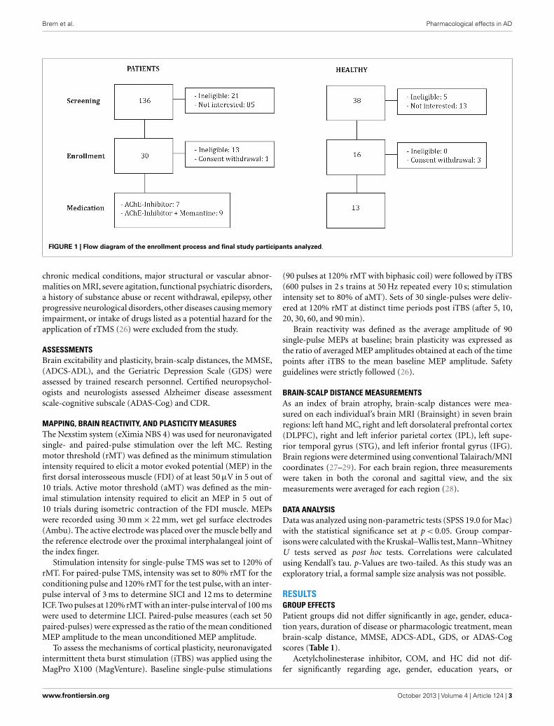
MATERIALS AND METHODSSUBJECTS AND INCLUSION/EXCLUSION CRITERIAWe investigated 16 patients diagnosed with mild to moderate AD(DSM IV, NINCDS-ADRDA): 7 were stable on AChEI (Table 1;Figure 1), and 9 were treated with combination therapy (AChEIand memantine). Patients were eligible if they had a score of1 on the Clinical Dementia Rating scale (CDR), and attainedbetween 18 and 24 points on the Mini-Mental State Examination(MMSE). Patients were recruited from other studies investigatingAD patients (ClinicalTrials.gov NCT01504958) in comparison toolder HCs. Patients were grouped according to psychopharmaco-logical treatment. All experimenters were blinded with regards topharmacological treatment.
Alzheimer’s disease groups were compared to a group of 13age-matched HCs with normal neurological and cognitive examsand a score of 0 on the CDR. The local Institutional Review Boardapproved the study protocol, and all participants or their legallyauthorized representatives gave written informed consent prior tostudy onset. The study visits took place at the Berenson-AllenCenter for Non-invasive Brain Stimulation and the Harvard-Thorndike Clinical Research Center at Beth Israel DeaconessMedical Center in Boston, MA, USA. Patients with unstable or
Table 1 | Demographic, pharmacological, neuropsychological, morphometric, and neurophysiological features of study participants.
AChEI and memantine
(N =9) mean±SD
AChEI (N =7)
mean±SD
p Value Healthy subjects (N =13)
mean±SD
p Value
Age (years) 71.78±3.73 68.00±7.55 0.351 67.762±6.05 0.278
Gender Six female, three male Five female, two male 1.000 Seven female, six male 0.700
Education (years) 15.33±3.97 17.71±3.90 0.470 15.77±2.17 0.659
Duration of disease (months) 38.00±34.90 16.43±15.51 0.071 – –
Duration of treatment (months) 33.00±34.76 15.57±16.26 0.244 – –
Brain-scalp distance mean (mm) 16.94±2.52 19.35±2.93 0.114 15.78±2.33 0.014
Brain-scalp distance left MC (mm) 15.67±2.02 19.38±3.78 0.042 14.47±1.80 0.007
Brain-scalp distance left IPL (mm) 21.98±5.35 26.39±6.51 0.125 18.03±2.82 0.023
rMT 36.83±6.53 50.38±8.16 0.001 45.26±11.98 0.012
aMT 39.96±6.07 48.43±10.39 0.055 44.89±8.01 0.112
MMSE 20.56±2.65 23.43±1.62 0.055 29.46±0.88 0.000
ADCS-ADL 68.57±5.22 72.29±6.47 0.209 74.62±3.59 0.043
GDS 2.22±2.54 1.71±2.06 0.918 0.69±1.11 0.202
ADAS-Cog 28.21±11.02 18.09±6.25 0.091 4.13±2.13 0.000
SICI 0.71±0.47 0.52±0.43 0.368 0.55±0.69 0.360
ICF 1.27±0.61 1.68±1.25 0.758 1.54±0.63 0.614
LICI 0.41±0.52 0.98±1.12 0.470 0.10±0.20 0.018
MC reactivity (µV) 1626±1418 1596±1558 0.758 17.71±3.90 0.907
Plasticity at T5 1.02±0.30 0.78±0.31 0.081 1.45±0.79 0.034
Mean plasticity 1.06±0.41 0.85±0.31 0.232 1.34±0.52 0.046
MC, motor cortex; IPL, inferior parietal lobule; rMT/aMT, resting/active motor threshold; MMSE, mini-mental state examination; ADCS-ADL, Alzheimer’s disease
cooperative study-activities of daily living inventory; GDS, geriatric depression scale; ADAS-Cog, Alzheimer’s disease assessment scale-cognitive subscale; SICI,
short-interval intracortical inhibition; ICF, intracortical facilitation; LICI, long-interval intracortical inhibition; p-values are two-tailed. Mann–Whitney U was used to
compare patient groups, Kruskal–Wallis was used to compare all three groups. Group values are presented as mean± standard deviation (SD). Bold font indicates
significant p-values.
Frontiers in Psychiatry | Neurodegeneration October 2013 | Volume 4 | Article 124 | 2
Brem et al. Pharmacological effects in AD
FIGURE 1 | Flow diagram of the enrollment process and final study participants analyzed.
chronic medical conditions, major structural or vascular abnor-malities on MRI, severe agitation, functional psychiatric disorders,a history of substance abuse or recent withdrawal, epilepsy, otherprogressive neurological disorders, other diseases causing memoryimpairment, or intake of drugs listed as a potential hazard for theapplication of rTMS (26) were excluded from the study.
ASSESSMENTSBrain excitability and plasticity, brain-scalp distances, the MMSE,(ADCS-ADL), and the Geriatric Depression Scale (GDS) wereassessed by trained research personnel. Certified neuropsychol-ogists and neurologists assessed Alzheimer disease assessmentscale-cognitive subscale (ADAS-Cog) and CDR.
MAPPING, BRAIN REACTIVITY, AND PLASTICITY MEASURESThe Nexstim system (eXimia NBS 4) was used for neuronavigatedsingle- and paired-pulse stimulation over the left MC. Restingmotor threshold (rMT) was defined as the minimum stimulationintensity required to elicit a motor evoked potential (MEP) in thefirst dorsal interosseous muscle (FDI) of at least 50 µV in 5 out of10 trials. Active motor threshold (aMT) was defined as the min-imal stimulation intensity required to elicit an MEP in 5 out of10 trials during isometric contraction of the FDI muscle. MEPswere recorded using 30 mm× 22 mm, wet gel surface electrodes(Ambu). The active electrode was placed over the muscle belly andthe reference electrode over the proximal interphalangeal joint ofthe index finger.
Stimulation intensity for single-pulse TMS was set to 120% ofrMT. For paired-pulse TMS, intensity was set to 80% rMT for theconditioning pulse and 120% rMT for the test pulse, with an inter-pulse interval of 3 ms to determine SICI and 12 ms to determineICF. Two pulses at 120% rMT with an inter-pulse interval of 100 mswere used to determine LICI. Paired-pulse measures (each set 50paired-pulses) were expressed as the ratio of the mean conditionedMEP amplitude to the mean unconditioned MEP amplitude.
To assess the mechanisms of cortical plasticity, neuronavigatedintermittent theta burst stimulation (iTBS) was applied using theMagPro X100 (MagVenture). Baseline single-pulse stimulations
(90 pulses at 120% rMT with biphasic coil) were followed by iTBS(600 pulses in 2 s trains at 50 Hz repeated every 10 s; stimulationintensity set to 80% of aMT). Sets of 30 single-pulses were deliv-ered at 120% rMT at distinct time periods post iTBS (after 5, 10,20, 30, 60, and 90 min).
Brain reactivity was defined as the average amplitude of 90single-pulse MEPs at baseline; brain plasticity was expressed asthe ratio of averaged MEP amplitudes obtained at each of the timepoints after iTBS to the mean baseline MEP amplitude. Safetyguidelines were strictly followed (26).
BRAIN-SCALP DISTANCE MEASUREMENTSAs an index of brain atrophy, brain-scalp distances were mea-sured on each individual’s brain MRI (Brainsight) in seven brainregions: left hand MC, right and left dorsolateral prefrontal cortex(DLPFC), right and left inferior parietal cortex (IPL), left supe-rior temporal gyrus (STG), and left inferior frontal gyrus (IFG).Brain regions were determined using conventional Talairach/MNIcoordinates (27–29). For each brain region, three measurementswere taken in both the coronal and sagittal view, and the sixmeasurements were averaged for each region (28).
DATA ANALYSISData was analyzed using non-parametric tests (SPSS 19.0 for Mac)with the statistical significance set at p < 0.05. Group compar-isons were calculated with the Kruskal–Wallis test, Mann–WhitneyU tests served as post hoc tests. Correlations were calculatedusing Kendall’s tau. p-Values are two-tailed. As this study was anexploratory trial, a formal sample size analysis was not possible.
RESULTSGROUP EFFECTSPatient groups did not differ significantly in age, gender, educa-tion years, duration of disease or pharmacologic treatment, meanbrain-scalp distance, MMSE, ADCS-ADL, GDS, or ADAS-Cogscores (Table 1).
Acetylcholinesterase inhibitor, COM, and HC did not dif-fer significantly regarding age, gender, education years, or
www.frontiersin.org October 2013 | Volume 4 | Article 124 | 3
Brem et al. Pharmacological effects in AD
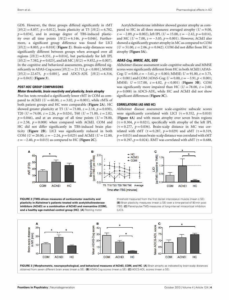
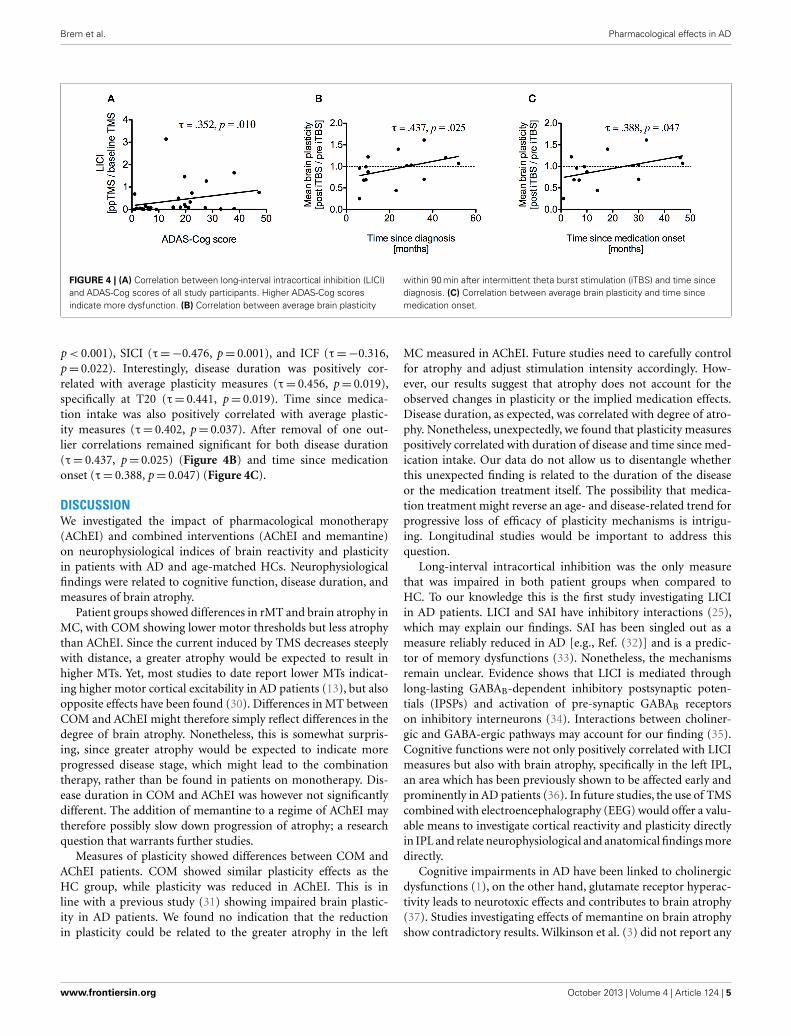
GDS. However, the three groups differed significantly in rMT[H (2)= 8.857, p= 0.012], brain plasticity at T5 [H (2)= 6.782,p= 0.034], and in average degree of TBS-induced plastic-ity over all time points [H (2)= 6.146, p= 0.046]. Further-more, a significant group difference was found for LICI[H (2)= 8.003, p= 0.018] (Figure 2). Brain-scalp distances weresignificantly different between groups when averaged over allregions [H (2)= 8.551, p= 0.014], but particularly for left IPL[H (2)= 7.582, p= 0.023], and left MC [H (2)= 9.852, p= 0.007].In the cognitive and behavioral assessments, groups differed sig-nificantly in ADAS-Cog scores [H (2)= 21.713, p < 0.001], MMSE[H (2)= 22.675, p < 0.001], and ADCS-ADL [H (2)= 6.316,p= 0.043] (Figure 3).
POST HOC GROUP COMPARISONSMotor thresholds, brain reactivity and plasticity, brain atrophyPost hoc tests revealed a significantly lower rMT in COM as com-pared to AChEI (U = 60.00, z = 3.02, p= 0.001), while rMTs ofboth patient groups and HC were comparable (Figure 2A). HCshowed greater plasticity at T5 (U = 73.00, z = 2.18, p= 0.030),T20 (U = 74.00, z = 2.26, p= 0.024), T60 (U = 71.00, z = 2.02,p= 0.046), and at an average of all time points (U = 78.00,z = 2.58, p= 0.008) when compared with AChEI. COM andHC did not differ significantly in TBS-induced brain plas-ticity (Figure 2B). LICI was significantly reduced in bothCOM (U = 20.00, z =−2.24, p= 0.025) and AChEI (U = 12.00,z =−2.40, p= 0.015) as compared to HC (Figure 2C).
Acetylcholinesterase inhibitor showed greater atrophy as com-pared to HC in all three measures: averaged atrophy (U = 9.00,z =−2.89, p= 0.002), left IPL (U = 15.00, z =−2.42, p= 0.014),and MC (U = 7.00, z =−3.05, p= 0.001). However, AChEI alsoshowed a significantly greater atrophy in MC as compared to COM(U = 51.00, z = 2.06, p= 0.042). COM did not differ from HC inatrophy (Figure 3A).
ADAS-Cog, MMSE, ADL, GDSAlzheimer disease assessment scale-cognitive subscale and MMSEscores were significantly different from HC in both AChEI (ADAS-Cog: U = 0.00, z =−3.61, p < 0.001; MMSE: U = 91.00, z = 3.75,p < 0.001) and COM (ADAS-Cog: U = 0.00, z =−3.91, p < 0.001;MMSE: U = 117.00, z = 4.02, p < 0.001) (Figure 3B). COMwas significantly more impaired than HC (U = 78.00, z = 2.60,p= 0.008) in ADCS-ADL, while HC and AChEI did not showsignificant differences (Figure 3C).
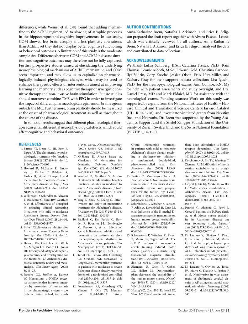
CORRELATIONS (AD AND HC)Alzheimer disease assessment scale-cognitive subscale scoreswere significantly correlated with LICI (τ= 0.352, p= 0.010)(Figure 4A) and with mean atrophy over seven brain regions(τ= 0.304, p= 0.021), specifically with atrophy of the left IPL(τ= 0.277, p= 0.036). Brain-scalp distance in MC was cor-related with rMT (τ= 0.287, p= 0.029) and aMT (τ= 0.319,p= 0.015) and mean brain-scalp distance was correlated with rMT(τ= 0.297, p= 0.024). RMT was correlated with aMT (τ= 0.688,
FIGURE 2 |TMS-driven measures of corticomotor reactivity andplasticity in Alzheimer’s patients treated with acetylcholinesteraseinhibitors (AChEI) or a combination of AChEI and memantine (COM),and a healthy age-matched control group (HC). (A) Resting motor
threshold measured from the first dorsal interosseus muscle (mean±SE).(B) Brain plasticity measures (mean±SE) over a time-period of 90 min postiTBS. (C) Paired-pulse TMS-measures of long-interval intracortical inhibition(LICI).
FIGURE 3 | Morphometric, neuropsychological, and behavioral measures of AChEI, COM, and HC. (A) Brain atrophy as indicated by brain-scalp distancesobtained from seven different brain areas (mean±SE). (B) ADAS-Cog scores (mean±SE). (C) ADCS-ADL scores (mean±SE).
Frontiers in Psychiatry | Neurodegeneration October 2013 | Volume 4 | Article 124 | 4
Brem et al. Pharmacological effects in AD
FIGURE 4 | (A) Correlation between long-interval intracortical inhibition (LICI)and ADAS-Cog scores of all study participants. Higher ADAS-Cog scoresindicate more dysfunction. (B) Correlation between average brain plasticity
within 90 min after intermittent theta burst stimulation (iTBS) and time sincediagnosis. (C) Correlation between average brain plasticity and time sincemedication onset.
p < 0.001), SICI (τ=−0.476, p= 0.001), and ICF (τ=−0.316,p= 0.022). Interestingly, disease duration was positively cor-related with average plasticity measures (τ= 0.456, p= 0.019),specifically at T20 (τ= 0.441, p= 0.019). Time since medica-tion intake was also positively correlated with average plastic-ity measures (τ= 0.402, p= 0.037). After removal of one out-lier correlations remained significant for both disease duration(τ= 0.437, p= 0.025) (Figure 4B) and time since medicationonset (τ= 0.388, p= 0.047) (Figure 4C).
DISCUSSIONWe investigated the impact of pharmacological monotherapy(AChEI) and combined interventions (AChEI and memantine)on neurophysiological indices of brain reactivity and plasticityin patients with AD and age-matched HCs. Neurophysiologicalfindings were related to cognitive function, disease duration, andmeasures of brain atrophy.
Patient groups showed differences in rMT and brain atrophy inMC, with COM showing lower motor thresholds but less atrophythan AChEI. Since the current induced by TMS decreases steeplywith distance, a greater atrophy would be expected to result inhigher MTs. Yet, most studies to date report lower MTs indicat-ing higher motor cortical excitability in AD patients (13), but alsoopposite effects have been found (30). Differences in MT betweenCOM and AChEI might therefore simply reflect differences in thedegree of brain atrophy. Nonetheless, this is somewhat surpris-ing, since greater atrophy would be expected to indicate moreprogressed disease stage, which might lead to the combinationtherapy, rather than be found in patients on monotherapy. Dis-ease duration in COM and AChEI was however not significantlydifferent. The addition of memantine to a regime of AChEI maytherefore possibly slow down progression of atrophy; a researchquestion that warrants further studies.
Measures of plasticity showed differences between COM andAChEI patients. COM showed similar plasticity effects as theHC group, while plasticity was reduced in AChEI. This is inline with a previous study (31) showing impaired brain plastic-ity in AD patients. We found no indication that the reductionin plasticity could be related to the greater atrophy in the left
MC measured in AChEI. Future studies need to carefully controlfor atrophy and adjust stimulation intensity accordingly. How-ever, our results suggest that atrophy does not account for theobserved changes in plasticity or the implied medication effects.Disease duration, as expected, was correlated with degree of atro-phy. Nonetheless, unexpectedly, we found that plasticity measurespositively correlated with duration of disease and time since med-ication intake. Our data do not allow us to disentangle whetherthis unexpected finding is related to the duration of the diseaseor the medication treatment itself. The possibility that medica-tion treatment might reverse an age- and disease-related trend forprogressive loss of efficacy of plasticity mechanisms is intrigu-ing. Longitudinal studies would be important to address thisquestion.
Long-interval intracortical inhibition was the only measurethat was impaired in both patient groups when compared toHC. To our knowledge this is the first study investigating LICIin AD patients. LICI and SAI have inhibitory interactions (25),which may explain our findings. SAI has been singled out as ameasure reliably reduced in AD [e.g., Ref. (32)] and is a predic-tor of memory dysfunctions (33). Nonetheless, the mechanismsremain unclear. Evidence shows that LICI is mediated throughlong-lasting GABAB-dependent inhibitory postsynaptic poten-tials (IPSPs) and activation of pre-synaptic GABAB receptorson inhibitory interneurons (34). Interactions between choliner-gic and GABA-ergic pathways may account for our finding (35).Cognitive functions were not only positively correlated with LICImeasures but also with brain atrophy, specifically in the left IPL,an area which has been previously shown to be affected early andprominently in AD patients (36). In future studies, the use of TMScombined with electroencephalography (EEG) would offer a valu-able means to investigate cortical reactivity and plasticity directlyin IPL and relate neurophysiological and anatomical findings moredirectly.
Cognitive impairments in AD have been linked to cholinergicdysfunctions (1), on the other hand, glutamate receptor hyperac-tivity leads to neurotoxic effects and contributes to brain atrophy(37). Studies investigating effects of memantine on brain atrophyshow contradictory results. Wilkinson et al. (3) did not report any
www.frontiersin.org October 2013 | Volume 4 | Article 124 | 5
Brem et al. Pharmacological effects in AD
differences, while Weiner et al. (38) found that adding meman-tine to the AChEI regimen led to slowing of atrophic processesin the hippocampus and cognitive improvements. In our study,COM showed less brain atrophy and less plasticity aberrationsthan AChEI, yet they did not display better cognitive functioningor behavioral outcomes. A limitation of this study is the moderatesample size. Differences between COM and AChEI in disease dura-tion and cognitive outcomes may therefore not be fully captured.Further prospective studies aimed at elucidating the underlyingneurophysiological mechanisms of AChEI, memantine, and COMseem important, and may allow us to capitalize on pharmaco-logically induced physiological changes, which may be used toenhance therapeutic effects of interventions aimed at improvinglearning and memory, such as cognitive therapy or synergistic cog-nitive therapy and non-invasive brain stimulation. Future studiesshould moreover combine TMS with EEG in order to investigatethe impact of different pharmacological regimens on brain regionsoutside the MC. Furthermore, brain plasticity should be measuredat the onset of pharmacological treatment as well as throughoutthe course of the disease.
In sum, our results suggest that different pharmacological ther-apies can entail differential neurophysiological effects, which couldaffect cognitive and behavioral outcomes.
AUTHOR CONTRIBUTIONSAnna-Katharine Brem, Natasha J. Atkinson, and Erica E. Selig-son prepared the draft report together with Alvaro Pascual-Leone,which was critically reviewed by all authors. Anna-KatharineBrem, Natasha J. Atkinson, and Erica E. Seligson analyzed the dataand contributed to data collection.
ACKNOWLEDGMENTSWe thank Lukas Schilberg, B.Sc., Catarina Freitas, Ph.D., RainThomas, Léonie Asboth, M.Sc., Edward Gold, Christina Carbone,Ilya Vidrin, Cory Kosche, Jessica Olson, Fritz Ifert-Miller, andZachary Gray for their support in data collection; Lisa Iguchi,Ph.D. for the neuropsychological exams; Ann Connor, RN, MSfor help with patient assessments and study oversight, and Drs.Daniel Press, MD and Mark Eldaief, MD for assistance with theneurological exams. Funding sources: Work on this study wassupported by a grant from the National Institutes of Health – Har-vard Clinical and Translational Science Center/Harvard Catalyst(UL1 RR025758), and investigator-initiated grants from NexstimInc., and Neuronix. Dr. Brem was supported by the Young Aca-demics Support and the Stiefel-Zangger Foundation of the Uni-versity of Zurich, Switzerland, and the Swiss National Foundation(PBZHP1_147196).
REFERENCES1. Bartus RT, Dean RL III, Beer B,
Lippa AS. The cholinergic hypothe-sis of geriatric memory dysfunction.Science (1982) 217:408–14. doi:10.1126/science.7046051
2. Howard R, McShane R, Linde-say J, Ritchie C, Baldwin A,Barber R, et al. Donepezil andmemantine for moderate-to-severeAlzheimer’s disease. N Engl J Med(2012) 366:893–903. doi:10.1056/NEJMoa1106668
3. Wilkinson D, Schindler R, SchwamE, Waldemar G, Jones RW, GauthierS, et al. Effectiveness of donepezilin reducing clinical worseningin patients with mild-to-moderateAlzheimer’s disease. Dement Geri-atr Cogn Disord (2009) 28:244–51.doi:10.1159/000241877
4. Birks J. Cholinesterase inhibitors forAlzheimer’s disease. Cochrane Data-base Syst Rev (2006) (1). doi:10.1002/14651858.CD005593
5. Hansen RA, Gartlehner G, WebbAP, Morgan LC, Moore CG, JonasDE. Efficacy and safety of donepezil,galantamine, and rivastigmine forthe treatment of Alzheimer’s dis-ease: a systematic review and meta-analysis. Clin Interv Aging (2008)3:211–25.
6. Parsons CG, Stöffler A, DanyszW. Memantine: a NMDA recep-tor antagonist that improves mem-ory by restoration of homeostasisin the glutamatergic system – toolittle activation is bad, too much
is even worse. Neuropharmacology(2007) 53:699–723. doi:10.1016/j.neuropharm.2007.07.013
7. McShane R, Areosa Sastre A,Minakaran N. Memantine fordementia. Cochrane DatabaseSyst Rev (2006) (2). doi:10.1002/14651858.CD003154.pub5
8. Winblad B, Gauthier S, AströmD, Stender K. Memantine benefitsfunctional abilities in moderate tosevere Alzheimer’s disease. J NutrHealth Aging (2010) 14:770–4. doi:10.1007/s12603-010-0122-x
9. Yang Z, Zhou X, Zhang Q. Effec-tiveness and safety of memantinetreatment for Alzheimer’s disease.J Alzheimers Dis (2013) 36:445–58.doi:10.3233/JAD-130395
10. Babiloni C, Del Percio C, Bor-det R, Bourriez J-L, BentivoglioM, Payoux P, et al. Effects ofacetylcholinesterase inhibitors andmemantine on resting-state elec-troencephalographic rhythms inAlzheimer’s disease patients. ClinNeurophysiol (2013) 124:837–50.doi:10.1016/j.clinph.2012.09.017
11. Tariot PN, Farlow MR, GrossbergGT, Graham SM, McDonald S,Gergel I, et al. Memantine treatmentin patients with moderate to severeAlzheimer disease already receivingdonepezil: a randomized controlledtrial. JAMA (2004) 291:317–24. doi:10.1001/jama.291.3.317
12. Porsteinsson AP, Grossberg GT,Mintzer J, Olin JT; Meman-tine MEM-MD-12 Study
Group. Memantine treatmentin patients with mild to moderateAlzheimer’s disease already receiv-ing a cholinesterase inhibitor:a randomized, double-blind,placebo-controlled trial. CurrAlzheimer Res (2008) 5:83–9.doi:10.2174/156720508783884576
13. Freitas C, Mondragón-Llorca H,Pascual-Leone A. Noninvasive brainstimulation in Alzheimer’s disease:systematic review and perspec-tives for the future. Exp Geron-tol (2011) 46:611–27. doi:10.1016/j.exger.2011.04.001
14. Schwenkreis P, Witscher K, JanssenF, Addo A, Dertwinkel R, Zenz M,et al. Influence of the N-methyl-D-aspartate antagonist memantine onhuman motor cortex excitability.Neurosci Lett (1999) 270:137–40.doi:10.1016/S0304-3940(99)00492-9
15. Schwenkreis P, Witscher K, PlegerB, Malin J-P, Tegenthoff M. TheNMDA antagonist memantineaffects training induced motorcortex plasticity – a study usingtranscranial magnetic stimula-tion. BMC Neurosci (2005) 6:35.doi:10.1186/1471-2202-6-35
16. Ziemann U, Chen R, CohenLG, Hallett M. Dextromethor-phan decreases the excitability ofthe human motor cortex. Neurol-ogy (1998) 51:1320–4. doi:10.1212/WNL.51.5.1320
17. Huang Y-Z, Chen R-S, Rothwell JC,Wen H-Y. The after-effect of human
theta burst stimulation is NMDAreceptor dependent. Clin Neuro-physiol (2007) 118:1028–32. doi:10.1016/j.clinph.2007.01.021
18. Korchounov A, Ilic TV, Schwinge T,Ziemann U. Modification of motorcortical excitability by an acetyl-cholinesterase inhibitor. Exp BrainRes (2005) 164:399–405. doi:10.1007/s00221-005-2326-6
19. Liepert J, Bär KJ, Meske U, WeillerC. Motor cortex disinhibition inAlzheimer’s disease. Clin Neu-rophysiol (2001) 112:1436–41.doi:10.1016/S1388-2457(01)00554-5
20. Pennisi G, Alagona G, Ferri R,Greco S, Santonocito D, PappalardoA, et al. Motor cortex excitabil-ity in Alzheimer disease: oneyear follow-up study. NeurosciLett (2002) 329:293–6. doi:10.1016/S0304-3940(02)00701-2
21. Di Lazzaro V, Oliviero A, PilatoF, Saturno E, Dileone M, MarraC, et al. Neurophysiological pre-dictors of long term response toAChE inhibitors in AD patients. JNeurol Neurosurg Psychiatry (2005)76:1064–9. doi:10.1136/jnnp.2004.051334
22. Di Lazzaro V, Oliviero A, TonaliPA, Marra C, Daniele A, Profice P,et al. Noninvasive in vivo assess-ment of cholinergic cortical cir-cuits in AD using transcranial mag-netic stimulation. Neurology (2002)59:392–7. doi:10.1212/WNL.59.3.392
Frontiers in Psychiatry | Neurodegeneration October 2013 | Volume 4 | Article 124 | 6
Brem et al. Pharmacological effects in AD
23. Nardone R,Golaszewski S,LadurnerG, Tezzon F, Trinka E. A reviewof transcranial magnetic stimula-tion in the in vivo functional evalua-tion of central cholinergic circuits indementia. Dement Geriatr Cogn Dis-ord (2011) 32:18–25. doi:10.1159/000330016
24. Di Lazzaro V, Oliviero A, Profice P,Pennisi MA, Di Giovanni S, ZitoG, et al. Muscarinic receptor block-ade has differential effects on theexcitability of intracortical circuitsin the human motor cortex. ExpBrain Res (2000) 135:455–61. doi:10.1007/s002210000543
25. Udupa K, Ni Z, Gunraj C, Chen RInteractions between short latencyafferent inhibition and long inter-val intracortical inhibition. ExpBrain Res (2009) 199:177–83. doi:10.1007/s00221-009-1997-9
26. Rossi S, Hallett M, Rossini PM,Pascual-Leone A. Safety, ethical con-siderations, and application guide-lines for the use of transcranial mag-netic stimulation in clinical prac-tice and research. Clin Neurophysiol(2009) 120:2008–39. doi:10.1016/j.clinph.2009.08.016
27. Price CJ, Winterburn D, GiraudAL, Moore CJ, Noppeney U.Cortical localisation of the visualand auditory word form areas: areconsideration of the evidence.Brain Lang (2003) 86:272–86.doi:10.1016/S0093-934X(02)00544-8
28. Rusjan PM, Barr MS, Farzan F,Arenovich T, Maller JJ, Fitzgerald
PB, et al. Optimal transcranialmagnetic stimulation coil place-ment for targeting the dorsolat-eral prefrontal cortex using novelmagnetic resonance image-guidedneuronavigation. Hum Brain Mapp(2010) 31:1643–52. doi:10.1002/hbm.20964
29. Sajonz B, Kahnt T, Margulies DS,Park SQ, Wittmann A, Stoy M, et al.Delineating self-referential process-ing from episodic memory retrieval:common and dissociable networks.Neuroimage (2010) 50:1606–17.doi:10.1016/j.neuroimage.2010.01.087
30. Perretti A, Grossi D, Fragassi N,Lanzillo B, Nolano M, Pisacreta AI,et al. Evaluation of the motor cortexby magnetic stimulation in patientswith Alzheimer disease. J Neurol Sci(1996) 135:31–7. doi:10.1016/0022-510X(95)00244-V
31. Koch G, Di Lorenzo F, Bonní S,Ponzo V, Caltagirone C, MartoranaA. Impaired LTP- but not LTD-like cortical plasticity in Alzheimer’sdisease patients. J Alzheimers Dis(2012) 31:593–9.
32. Di Lazzaro V, Pilato F, DileoneM, Saturno E, Oliviero A, MarraC, et al. In vivo cholinergiccircuit evaluation in frontotem-poral and Alzheimer dementias.Neurology (2006) 66:1111–3.doi:10.1212/01.wnl.0000204183.26231.23
33. Young-Bernier M, Kamil Y, Trem-blay F, Davidson PSR. Associa-tions between a neurophysiological
marker of central cholinergic activ-ity and cognitive functions in youngand older adults. Behav BrainFunct (2012) 8:17. doi:10.1186/1744-9081-8-17
34. McDonnell MN, Orekhov Y, Zie-mann U. The role of GABA(B)receptors in intracortical inhibitionin the human motor cortex. ExpBrain Res (2006) 173:86–93. doi:10.1007/s00221-006-0365-2
35. Anderson JJ, Kuo S, Chase TN, Eng-ber TM. GABAA and GABAB recep-tors differentially regulate striatalacetylcholine release in vivo. Neu-rosci Lett (1993) 160:126–30. doi:10.1016/0304-3940(93)90395-2
36. Im K, Lee J-M, Seo SW, YoonU, Kim ST, Kim Y-H, et al. Vari-ations in cortical thickness withdementia severity in Alzheimer’sdisease. Neurosci Lett (2008) 436:227–31. doi:10.1016/j.neulet.2008.03.032
37. Meier-Ruge WA, Bertoni-FreddariC. Pathogenesis of decreasedglucose turnover and oxidativephosphorylation in ischemic andtrauma-induced dementia of theAlzheimer type. Ann N Y Acad Sci(1997) 826:229–41. doi:10.1111/j.1749-6632.1997.tb48474.x
38. Weiner MW, Sadowsky C, Sax-ton J, Hofbauer RK, Graham SM,Yu SY, et al. Magnetic resonanceimaging and neuropsychologicalresults from a trial of memantinein Alzheimer’s disease. AlzheimersDement (2011) 7:425–35. doi:10.1016/j.jalz.2010.09.003
Conflict of Interest Statement: Theauthors declare that the research wasconducted in the absence of anycommercial or financial relationshipsthat could be construed as a poten-tial conflict of interest. Alvaro Pascual-Leone serves on the scientific advisoryboards for Nexstim, Neuronix, StarlabNeuroscience, Neuroelectrics, and Neo-Sync; and is listed as an inventor onseveral issued and pending patents onthe real-time integration of transcranialmagnetic stimulation (TMS) with elec-troencephalography (EEG) and mag-netic resonance imaging (MRI).
Received: 31 July 2013; accepted: 21 Sep-tember 2013; published online: 07 Octo-ber 2013.Citation: Brem A-K, Atkinson NJ, Selig-son EE and Pascual-Leone A (2013) Dif-ferential pharmacological effects on brainreactivity and plasticity in Alzheimer’sdisease. Front. Psychiatry 4:124. doi:10.3389/fpsyt.2013.00124This article was submitted to Neurode-generation, a section of the journal Fron-tiers in Psychiatry.Copyright © 2013 Brem, Atkinson, Selig-son and Pascual-Leone. This is an open-access article distributed under the termsof the Creative Commons AttributionLicense (CC BY). The use, distribution orreproduction in other forums is permitted,provided the original author(s) or licensorare credited and that the original publica-tion in this journal is cited, in accordancewith accepted academic practice. No use,distribution or reproduction is permittedwhich does not comply with these terms.
www.frontiersin.org October 2013 | Volume 4 | Article 124 | 7
Related Documents