RESEARCH ARTICLE Differential mRNA Expression and Glucocorticoid-Mediated Regulation of TRPM6 and TRPM7 in the Heart and Kidney throughout Murine Pregnancy and Development James S. M. Cuffe, Sarah Steane, Karen M. Moritz, Tamara M. Paravicini* School of Biomedical Sciences, The University of Queensland, Brisbane, Australia * [email protected] Abstract The transient receptor potential (TRP) channels TRPM6 and TRPM7 are critically involved in maintaining whole body and cellular Mg 2+ homeostasis and ensuring the normal function of organs such as the heart and kidney. However, we do not know how the expression of TRPM6 and TPRM7 in these organs changes throughout fetal development and adult life, and whether this expression can be hormonally regulated. This study determined the ontog- eny of TRPM6 and TRPM7 mRNA expression from mid-gestation through to adulthood in the mouse. In a second series of experiments, we examined how maternal administration of the glucocorticoids corticosterone and dexamethasone between embryonic days 12.5–15 affected TRPM6 and TRPM7 channel mRNA expression in the mother and fetus. Whilst renal TRPM7 expression was relatively constant throughout development, renal TRPM6 ex- pression was markedly upregulated after birth. In contrast, cardiac TRPM7 expression was 2–4 fold higher in the fetus than in the adult. Surprisingly, TRPM6 expression was detected in the fetal heart (qPCR and in situ hybridization). Glucocorticoid administration during ges- tation increased fetal cardiac expression of both channels without affecting renal expres- sion. In contrast, in the dam renal TRPM6 and TRPM7 expression was increased by glucocorticoids with no change in the cardiac channel expression. These data suggest that TRPM6 and TRPM7 channels are important in organogenesis, and that elevated maternal glucocorticoid levels can alter the expression of these channels. This suggests that pertur- bations in hormonal regulatory systems during pregnancy may adversely impact upon nor- mal fetal development, at least in part by altering expression of TRPM channels. PLOS ONE | DOI:10.1371/journal.pone.0117978 February 18, 2015 1 / 17 OPEN ACCESS Citation: Cuffe JSM, Steane S, Moritz KM, Paravicini TM (2015) Differential mRNA Expression and Glucocorticoid-Mediated Regulation of TRPM6 and TRPM7 in the Heart and Kidney throughout Murine Pregnancy and Development. PLoS ONE 10(2): e0117978. doi:10.1371/journal.pone.0117978 Academic Editor: Jodi Pawluski, University of Rennes-1, FRANCE Received: April 4, 2014 Accepted: January 6, 2015 Published: February 18, 2015 Copyright: © 2015 Cuffe et al. This is an open access article distributed under the terms of the Creative Commons Attribution License, which permits unrestricted use, distribution, and reproduction in any medium, provided the original author and source are credited. Data Availability Statement: All relevant data are within the paper and its Supporting Information files. Funding: The authors have no support or funding to report. Competing Interests: The authors have declared that no competing interests exist.

Welcome message from author
This document is posted to help you gain knowledge. Please leave a comment to let me know what you think about it! Share it to your friends and learn new things together.
Transcript

RESEARCH ARTICLE
Differential mRNA Expression andGlucocorticoid-Mediated Regulation ofTRPM6 and TRPM7 in the Heart and Kidneythroughout Murine Pregnancy andDevelopmentJames S. M. Cuffe, Sarah Steane, Karen M. Moritz, Tamara M. Paravicini*
School of Biomedical Sciences, The University of Queensland, Brisbane, Australia
AbstractThe transient receptor potential (TRP) channels TRPM6 and TRPM7 are critically involved
in maintaining whole body and cellular Mg2+ homeostasis and ensuring the normal function
of organs such as the heart and kidney. However, we do not know how the expression of
TRPM6 and TPRM7 in these organs changes throughout fetal development and adult life,
and whether this expression can be hormonally regulated. This study determined the ontog-
eny of TRPM6 and TRPM7 mRNA expression from mid-gestation through to adulthood in
the mouse. In a second series of experiments, we examined how maternal administration of
the glucocorticoids corticosterone and dexamethasone between embryonic days 12.5–15
affected TRPM6 and TRPM7 channel mRNA expression in the mother and fetus. Whilst
renal TRPM7 expression was relatively constant throughout development, renal TRPM6 ex-
pression was markedly upregulated after birth. In contrast, cardiac TRPM7 expression was
2–4 fold higher in the fetus than in the adult. Surprisingly, TRPM6 expression was detected
in the fetal heart (qPCR and in situ hybridization). Glucocorticoid administration during ges-
tation increased fetal cardiac expression of both channels without affecting renal expres-
sion. In contrast, in the dam renal TRPM6 and TRPM7 expression was increased by
glucocorticoids with no change in the cardiac channel expression. These data suggest that
TRPM6 and TRPM7 channels are important in organogenesis, and that elevated maternal
glucocorticoid levels can alter the expression of these channels. This suggests that pertur-
bations in hormonal regulatory systems during pregnancy may adversely impact upon nor-
mal fetal development, at least in part by altering expression of TRPM channels.
PLOS ONE | DOI:10.1371/journal.pone.0117978 February 18, 2015 1 / 17
OPEN ACCESS
Citation: Cuffe JSM, Steane S, Moritz KM, ParaviciniTM (2015) Differential mRNA Expression andGlucocorticoid-Mediated Regulation of TRPM6 andTRPM7 in the Heart and Kidney throughout MurinePregnancy and Development. PLoS ONE 10(2):e0117978. doi:10.1371/journal.pone.0117978
Academic Editor: Jodi Pawluski, University ofRennes-1, FRANCE
Received: April 4, 2014
Accepted: January 6, 2015
Published: February 18, 2015
Copyright: © 2015 Cuffe et al. This is an openaccess article distributed under the terms of theCreative Commons Attribution License, which permitsunrestricted use, distribution, and reproduction in anymedium, provided the original author and source arecredited.
Data Availability Statement: All relevant data arewithin the paper and its Supporting Information files.
Funding: The authors have no support or funding toreport.
Competing Interests: The authors have declaredthat no competing interests exist.

IntroductionMagnesium is an abundant intracellular cation that is crucial for many fundamental processesinvolved in normal cell function, including protein synthesis, DNA replication and energy me-tabolism [1]. In light of this, serum magnesium levels are kept tightly controlled, with overallbody magnesium homeostasis reflecting the balance between intestinal absorption and renalexcretion [2]. Intracellular magnesium concentrations are also maintained within a narrowrange, however until recently the molecular mechanisms regulating magnesium transport atthe cellular level were poorly understood.
Two members of the transient receptor potential (TRP) channel superfamily, TRPM6 andTRPM7, have now been identified as critical regulators of magnesium homeostasis. Geneticanalyses of patients with autosomal-recessive hypomagnesemia and secondary hypocalcemia(HSH) indicate that TRPM6 is an essential gene for magnesium homeostasis. Studies of HSHpatients by two independent research groups identified multiple mutations in TRPM6 thatcaused abnormal renal magnesium handling and hypomagnesemia [3–5]. TRPM6 is predomi-nantly expressed in the lung, cecum, colon and renal tubules [6]. Whilst overall magnesium ho-meostasis is influenced by intestinal absorption and dynamic exchange with bone, the majorsite of regulation is the kidney, which controls magnesium excretion to balance intestinal ab-sorption [3]. The amount of magnesium lost in the urine is ultimately determined by howmuch magnesium is actively reabsorbed in the distal convoluted tubule, a process thought tobe mediated (at least in part) by TRPM6 [7]. Thus, we can consider TRPM6 to be a regulator ofwhole body magnesium homeostasis.
In contrast, the TRPM7 channel is ubiquitously expressed [6,8], with high levels of expres-sion seen in the heart and kidney [9]. A magnesium- and calcium-permeable ion channel withhomology to TRPM6, the primary physiological role of TRPM7 appears to be the maintenanceof cellular magnesium homeostasis [8,10]. TRPM7 is also critical for cell growth, as genetic de-letion of TRPM7 in cultured cells prevents proliferation, an effect that can be reversed with ei-ther external magnesium supplementation or overexpression of other cellular magnesiumtransporters [10–13]. Like TRPM6, TRPM7 has also been shown to be critical for systemicmagnesium homeostasis [14].
Both TRPM6 and TRPM7 appear to play critical roles in embryonic development. Global dele-tion of TRPM7 in transgenic mice is lethal, causing embryonic loss before day 7.5 of embryogene-sis [15]. Mice lacking TRPM6 also show high rates of embryonic loss (often before embryonic day12.5), and those that survived to this point exhibited neural tube defects [16]. However, despitethese observations, the regulation of TRPM6 and TRPM7 throughout development, and the po-tential roles of these channels in fetal organ development, are yet to be fully defined.
Multiple factors can regulate TRPM6 and TRPM7 expression in adulthood; these include al-tered dietary magnesium intake [6,17,18] and steroid hormones such as aldosterone and gluco-corticoids [17,19]. The latter may be of particular relevance during development, as pregnantwomen are often exposed to either natural or synthetic glucocorticoids during gestation [20]for numerous reasons. Whilst the prevalence of glucocorticoid exposure during pregnancy isdifficult to quantify, the overall incidence is likely to be high. Importantly, prenatal exposure toelevated glucocorticoid levels has been shown to disturb normal fetal growth and organ devel-opment [20]. The developing heart and kidney have been shown to be particularly vulnerableto maternal glucocorticoid exposure, which can alter development of these organs and ‘pro-gram’ disease in adult life [21,22]. Given that TRPM6 and TRPM7 are critical for normal fetaldevelopment and that glucocorticoids may affect the expression of these channels, it is possiblethat glucocorticoid-induced changes in fetal magnesium channel expression during gestationmay alter magnesium homeostasis and thereby affect fetal development.
Magnesium Channels and Glucocorticoids in Development
PLOS ONE | DOI:10.1371/journal.pone.0117978 February 18, 2015 2 / 17
This leads to the two main aims of the present study. First, we aimed to characterize howthe expression of TRPM6 and TRPM7 changes throughout the development of the heart andkidney, from the fetus into adulthood. The second aim was to determine whether maternal ad-ministration of glucocorticoids during mid-gestation affects the maternal and fetal expressionof TRPM6 and 7 in the heart and kidney.
Materials and Methods
Animal studiesAll animal procedures were approved by The University of Queensland Anatomical Biosci-ences Animal Ethics Committee (AEC approval number SBMS/355/09) and conducted in ac-cordance with the Australian Code of Practice for the Care and Use of Animals for ScientificPurposes. Female C57Bl6/J mice were time-mated overnight at 8–10 weeks of age. Pregnancywas confirmed the following morning by the presence of a seminal plug, and this time point de-fined as embryonic day (E) 0.5. For ontogeny studies, dams and pups were euthanized at eitherE14.5 or E17.5, or allowed to deliver naturally. For animals that delivered, the offspring wereeuthanized at post-natal day 30 (PN30) or as adults. Following euthanasia the hearts and kid-neys were collected and snap frozen in liquid nitrogen (for quantitative PCR) or fixed in 4%paraformaldehyde followed by paraffin embedding (for in situ hybridization).
Glucocorticoid treatmentTo determine the effects of glucocorticoids on TRPM6 and TRPM7 expression during develop-ment, pregnant females were treated with glucocorticoids for 60 hours from E12.5 as describedpreviously [23–25]. Dams were anaesthetized using isoflurane (2%) and an osmotic minipump(model #1003D, Alzet, CA) inserted subcutaneously in the subscapular region. The osmoticpumps contained the synthetic glucocorticoid dexamethasone (Dex, dexamethasone sodiumphosphate, Intervet, Australia; 1 μg/kg/hr), the endogenous rodent glucocorticoid corticoste-rone (Cort, Sigma-Aldrich Australia; 33 μg/kg/hr) or 0.9% saline as a control. All drugs weredelivered at a rate of 1 μL/hr. Mice were then euthanized either during the infusion period atE14.5 (after* 48 hours of glucocorticoid treatment) or at E17.5 (* 60 h after the cessation ofglucocorticoid treatment) and tissues collected from the pups and dams as detailed above.These doses of glucocorticoids (delivered during this period of gestation in the mouse) areknown to adversely affect fetal and placental growth, and cause alterations in the hearts andkidneys of adult offspring [23–25].
Plasma electrolytesWhole blood was collected by cardiac puncture from the dams at E14.5 following euthanasia.Plasma was separated by centrifugation and stored at −70°C until assay. Plasma electrolyteswere measured using an automated analyzer (Cobas Integra 400 Plus, Roche Diagnostics).
Gene expressionGene expression of TRPM6 and TRPM7 was measured using quantitative reverse transcriptasePCR (qPCR). Total RNA was extracted from frozen heart and kidney samples using a commer-cially available extraction kit (RNeasy, Qiagen) according to the manufacturer’s instructions.All samples were treated with DNase to remove contaminating genomic DNA and quantifiedspectrophotometrically before being reverse transcribed using random primers (Invitrogen Su-perscript III). Taqman qPCR was performed using exon-spanning gene expression assays (Ap-plied Biosystems) for Trpm6 (Mm00463112_m1, assay location 1523 spanning exons 13–14)
Magnesium Channels and Glucocorticoids in Development
PLOS ONE | DOI:10.1371/journal.pone.0117978 February 18, 2015 3 / 17
and Trpm7 (Mm00457998_m1, assay location 1785 spanning exons 13–14). Ribosomal 18Swas amplified in the same reaction tube using a VIC-labelled probe and primers (Applied Bio-systems endogenous control reagents). Expression levels were analyzed using the comparative2-ddCt method [26]. Expression of the sex-specific gene Xist (Mm01232884_m1) was used toconfirm fetal sex [24]. As there were no sex-specific expression differences in any tissue fromE14.5, E17.5 or PN30 mice, the data from both sexes has been combined at these time points.
In situ hybridizationTo localize the distribution of Trpm6 and Trpm7 RNA, 10 μm sections were taken from paraf-fin embedded hearts and kidneys from fetal (E17.5) and adult mice. Antisense probes (679 and613 nucleotides for TRPM6 and TRPM7 respectively) and matching sense negative controlprobes were designed using NCBI sequence data (Trpm6 NM_153417; Trpm7 NM_021450)and generated from pooled adult kidney and heart cDNA using the following primers: TRPM6Forward primer TAA TAC GAC TCA CTA TAG GGG CCT GTC AAA GAA GAA GAGGAA; TRPM6 reverse primer AAT TAA CCC TCA CTA AAG GGG GGG AGA AAA GACTTC ACA ATG; TRPM7 forward primer TAA TAC GAC TCA CTA TAG GGG TGG GAGAAA ACT TGA CTG ACC; TRPM7 reverse primer AAT TAA CCC TCA CTA AAG GGCTTA GCT GAA TGG CTG TGA CTG. These primers included a leading promoter sequencefor either the T7 RNA polymerase (forward primers) or T3 RNA polymerase (reverse primers).200 ng of each PCR product was used to generate both the sense and antisense DIG labelledRNA probes using either the T7 or T3 RNA polymerase. In situ hybridization was performedas previously described [27]. Briefly, slides were post-fixed with 4% paraformaldehyde, treatedwith proteinase K and acetylated before hybridization with antisense probes at 70°C overnight.Additional slides were treated in an identical manner and were hybridized with the senseprobes to act as negative controls. Slides were then stained with NBT/BCIP (blue) and counter-stained using nuclear fast red. Sections were scanned using an Aperio scanscope XT slide scan-ner (Aperio, Vista, CA).
StatisticsData is presented as mean ± SEM and analyzed using one-way ANOVA with either Tukey's(ontogeny) or Dunnett's (glucocorticoid treatment) post-hoc tests for multiple comparisons.All data was analyzed using GraphPad Prism 5, and P<0.05 was consideredstatistically significant.
Results
Renal TRPM6/7 expressionTRPM6 and TRPM7 mRNA transcripts were detected in the kidney at all developmental timepoints examined (S1 Table), with both channels showing specific differences in expressionthroughout development. In whole kidney homogenates, TRPM6 expression was* 15–30fold less in fetuses (E14.5 and E17.5) compared to adult males (Fig. 1A, P<0.05). In adult mice,TRPM6 showed sex-specific regulation in the kidney, with non-pregnant females having twicethe expression of males. This sex-specific increase in renal TRPM6 expression was blunted inpregnant mice (Fig. 1A). In contrast, renal TRPM7 expression remained constant throughoutfetal development and early life. In adult mice, renal TRPM7 expression was also increased(*1.8 fold, P<0.05 vs males) in non-pregnant females compared to males, with this increasebeing absent in pregnant animals (Fig. 1B).
Magnesium Channels and Glucocorticoids in Development
PLOS ONE | DOI:10.1371/journal.pone.0117978 February 18, 2015 4 / 17

Fig 1. Expression of TRPM6 and TRPM7mRNA in the mouse kidney during development. Expression of TRPM6 (A) and TRPM7 (B) mRNA (relative to18S rRNA) in the kidneys of mice at embryonic days 14.5 and 17.5 (E14.5 and E17.5), postnatal day 30 (PN30), and adult male, female (Fem) and pregnantfemale (Fem+) animals. Data is expressed as mean ± SEM relative to the adult male, n = 5 (E14.5), 5 (E17.5), 9 (PN30), 5 (adult male), 5 (adult female), 5(pregnant female). * P<0.05 vs E14.5 and E17.5; ** P<0.05 vs adult male.
doi:10.1371/journal.pone.0117978.g001
Magnesium Channels and Glucocorticoids in Development
PLOS ONE | DOI:10.1371/journal.pone.0117978 February 18, 2015 5 / 17

Cardiac TRPM6/7 expressionSurprisingly, TRPM6 expression (as determined by qPCR) was detected in the heart at all de-velopmental time points examined, albeit at much lower levels than in the kidney (S1 Table).Expression in whole heart homogenates increased between E14.5 and E17.5 before decreasingat PN30 (Fig. 2A). TRPM7 was also expressed in the heart throughout development, with theexpression levels* 2–4 fold higher in fetal hearts than in adult male hearts (Fig. 2B, P<0.05).
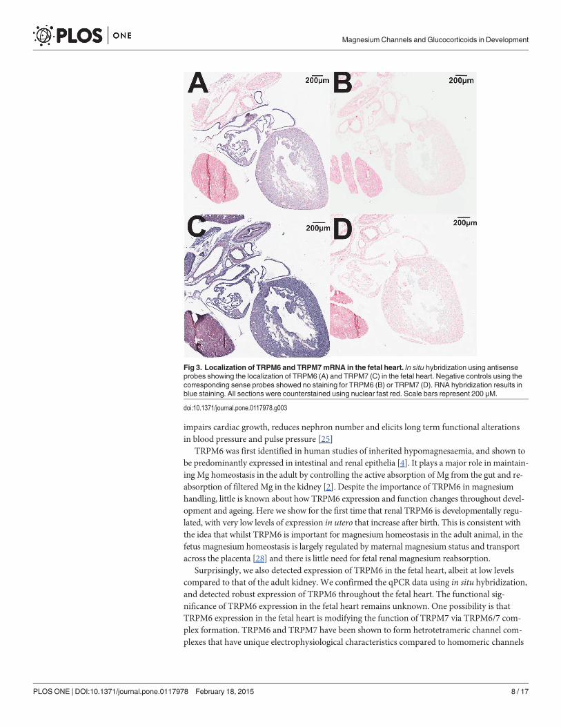
Localization of TRPM6/7 channel expressionIn situ hybridization studies supported the qPCR results demonstrating that TRPM6 mRNA isexpressed in the fetal heart at E17.5 (Fig. 3A). The negative control sense probe showed nostraining in the adjacent section (Fig. 3B), indicating the specificity of staining. TRPM7 wasubiquitously expressed throughout the fetal heart (Fig. 3C) with the sense probe demonstratingno staining (Fig. 3D). In situ hybridization of TRPM6 in adult kidneys demonstrated the ex-pected tubular localization in the adult kidney (Fig. 4A), whilst the sense probe showed nostaining in an adjacent section (Fig. 4B). TRPM7 also demonstrated a tubular localization inthe adult kidney (Fig. 4C), and staining specificity was again confirmed by using a sense probeas a negative control (Fig. 4D).
Regulation of TRPM6/7 channels by glucocorticoidsThe expression of both TRPM6 and TPRM7 mRNA was regulated by maternal glucocorticoidadministration in a time- and tissue-dependent manner. In the fetal heart, TRPM6 expressionwas increased*1.7 fold at E14.5 by the administration of either Cort or Dex to the dam(Fig. 5A). A similar trend was seen for TRPM7 expression, although this was only statisticallysignificant following Cort exposure (Fig. 5C). By E17.5 (* 60 h after the cessation of glucocor-ticoid treatment) fetal cardiac TRPM6 and TRPM7 expression had returned to the level of thesaline control (Fig. 5 B & D). In contrast to the effects on the fetal heart, administration of glu-cocorticoids did not alter TRPM6 and TRPM7 mRNA levels in the maternal heart (Fig. 6).
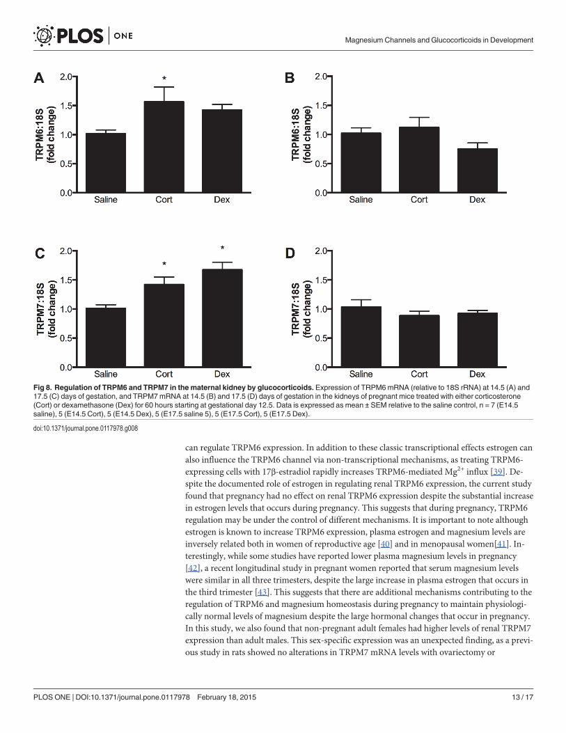
Maternal glucocorticoid administration had no effect on the expression of TRPM6 orTRPM7 in the fetal kidney at E14.5 (Fig. 7), but increased TRPM6 and TRPM7 expression inthe maternal kidney at this time (Fig. 8A & C). Maternal and fetal renal expression of TRPM6and TRPM7 were similar in all groups at E17.5,* 60 h after the end of glucocorticoid treat-ment (Fig. 8B & D). Despite these changes in magnesium channel expression, glucocorticoidtreatment caused no significant changes in plasma electrolytes in the dam (Mg2+ plasma con-centrations in mmol/L: saline 0.69±0.06; corticosterone 0.72±0.08; dexamethasone 0.71±0.05).
DiscussionThis study is the first to demonstrate that TRPM6 and TRPM7 are differentially expressed inthe heart and kidney throughout gestation and into adulthood. We have uniquely identifiedmRNA for TRPM6 (traditionally considered to be an epithelial channel) in the fetal mouseheart, with expression levels being higher at E17.5 than in PN30 offspring. Importantly, weshowed that administration of glucocorticoids during pregnancy can regulate TRPM6 andTRPM7 expression in a time- and tissue-dependent fashion in both the dam and fetus. Whenconsidered in context of recent findings suggesting a critical role for TRPM channels in organ-ogenesis, these data suggest a novel mechanism by which maternal glucocorticoid exposuremay influence the developing fetus. This is of particular interest as we have shown previouslythat maternal exposure to either corticosterone or dexamethasone impairs placental develop-ment and fetal growth [23,24], and that our model of maternal dexamethasone exposure
Magnesium Channels and Glucocorticoids in Development
PLOS ONE | DOI:10.1371/journal.pone.0117978 February 18, 2015 6 / 17

Fig 2. Expression of TRPM6 and TRPM7mRNA in the mouse heart during development. Cardiacexpression of TRPM6 (A) and TRPM7 (B) mRNA (relative to 18S rRNA) at embryonic days 14.5 and 17.5(E14.5 and E17.5), postnatal day 30 (PN30), as well as in adult male, female (Fem) and pregnant female(Fem+). Data is expressed as mean ± SEM relative to the adult male, n = 5 (E14.5), 6 (E17.5), 9 (PN30), 6(adult male), 5 (adult female), 4 (pregnant female). * P<0.05 vs E17.5.
doi:10.1371/journal.pone.0117978.g002
Magnesium Channels and Glucocorticoids in Development
PLOS ONE | DOI:10.1371/journal.pone.0117978 February 18, 2015 7 / 17
impairs cardiac growth, reduces nephron number and elicits long term functional alterationsin blood pressure and pulse pressure [25]
TRPM6 was first identified in human studies of inherited hypomagnesaemia, and shown tobe predominantly expressed in intestinal and renal epithelia [4]. It plays a major role in maintain-ing Mg homeostasis in the adult by controlling the active absorption of Mg from the gut and re-absorption of filtered Mg in the kidney [2]. Despite the importance of TRPM6 in magnesiumhandling, little is known about how TRPM6 expression and function changes throughout devel-opment and ageing. Here we show for the first time that renal TRPM6 is developmentally regu-lated, with very low levels of expression in utero that increase after birth. This is consistent withthe idea that whilst TRPM6 is important for magnesium homeostasis in the adult animal, in thefetus magnesium homeostasis is largely regulated by maternal magnesium status and transportacross the placenta [28] and there is little need for fetal renal magnesium reabsorption.
Surprisingly, we also detected expression of TRPM6 in the fetal heart, albeit at low levelscompared to that of the adult kidney. We confirmed the qPCR data using in situ hybridization,and detected robust expression of TRPM6 throughout the fetal heart. The functional sig-nificance of TRPM6 expression in the fetal heart remains unknown. One possibility is thatTRPM6 expression in the fetal heart is modifying the function of TRPM7 via TRPM6/7 com-plex formation. TRPM6 and TRPM7 have been shown to form hetrotetrameric channel com-plexes that have unique electrophysiological characteristics compared to homomeric channels
Fig 3. Localization of TRPM6 and TRPM7mRNA in the fetal heart. In situ hybridization using antisenseprobes showing the localization of TRPM6 (A) and TRPM7 (C) in the fetal heart. Negative controls using thecorresponding sense probes showed no staining for TRPM6 (B) or TRPM7 (D). RNA hybridization results inblue staining. All sections were counterstained using nuclear fast red. Scale bars represent 200 μM.
doi:10.1371/journal.pone.0117978.g003
Magnesium Channels and Glucocorticoids in Development
PLOS ONE | DOI:10.1371/journal.pone.0117978 February 18, 2015 8 / 17

[29,30]. Chubanov et al. demonstrated that TRPM6/TRPM7 heterooligomerization was essen-tial for forming a functional TRPM6-containing channel complex in the membrane of epitheli-al cells [31], suggesting that interactions between TRPM6 and TRPM7 have an importantbiological role. TRPM7 is critical for early cardiogenesis [32], and it is possible that early myo-cardial development may be influenced by both homotetrameric TRPM7 channels and hetero-meric TRPM6/7 channel complexes. Alternatively, TRPM6 may be important in developmentfor reasons unrelated to TRPM7. Studies in transgenic animals show that TRPM6 deficiency is
Fig 4. Localization of TRPM6 and TRPM7mRNA in the adult kidney. In situ hybridization using antisense probes showing the localization of TRPM6 (A)and TRPM7 (C) in the adult kidney. Negative controls using the corresponding sense probes showed no staining for TRPM6 (B) or TRPM7 (D) in the adultkidney. RNA hybridization results in blue staining. All sections were counterstained using nuclear fast red. Scale bars represent 100 μM.
doi:10.1371/journal.pone.0117978.g004
Magnesium Channels and Glucocorticoids in Development
PLOS ONE | DOI:10.1371/journal.pone.0117978 February 18, 2015 9 / 17

embryonically lethal, with most pups dying before E12.5 [16,33]. Animals that survived toterm often displayed neural tube defects, a surprising finding given that studies in the adultsupport a primarily intestinal and renal role of TRPM6 [16]. Dietary supplementation of mag-nesium to the dams did not rescue the TRPM6-/- mice [16,33], suggesting that the importanceof TRPM6 in development may extend beyond magnesium homeostasis. As these studies haveinvolved global deletion of TRPM6, determining the functional significance of TRPM6 in thedevelopment of individual organs will require the use of transgenic animals with tissue-specificdeletion of this channel. It is important to note that there are differences between the pheno-type of TRPM6-/- mice and patients with loss-of-function TRPM6 mutations. In humans, mu-tations in the TRPM6 gene lead to hypomagnesaemia with secondary hypocalcaemia (HSH),an autosomal recessive condition characterized by excessive renal magnesium wasting [4,5].The electrolyte abnormalities lead to neurological symptoms (e.g. seizures, neuromuscular ab-normalities) that manifest in infancy and can be controlled with prompt initiation of magne-sium therapy [34]. The contrast between the clinical presentation of TRPM6 mutation inhumans and the lethal phenotype of TRPM6-/- mice suggests that the physiological and devel-opmental roles of TRPM6 are yet to be fully elucidated.
Fig 5. Regulation of TRPM6 and TRPM7 in the fetal heart by glucocorticoids. Expression of TRPM6 (top panels) and TRPM7 (bottom panels) mRNA(relative to 18S rRNA) in the fetal hearts of mice exposed to either corticosterone (Cort) or dexamethasone (Dex) for 60 hours during gestation. In panels Aand C, expression was measured at embryonic day 14.5, immediately after glucocorticoid exposure. In panels B and D, expression was measured atembryonic day 17.5,* 60 hours after the cessation of glucocorticoid treatment. Data is expressed as mean ± SEM relative to the saline control, n = 12(E14.5 saline), 10 (E14.5 Cort), 12 (E14.5), 10 (E17.5 saline), 11 (E17.5 Cort), 4 (E17.5 Dex). * P<0.05 vs saline control.
doi:10.1371/journal.pone.0117978.g005
Magnesium Channels and Glucocorticoids in Development
PLOS ONE | DOI:10.1371/journal.pone.0117978 February 18, 2015 10 / 17

The pattern of TRPM7 expression in the heart throughout development mirrored that ofTRPM6: both were more highly expressed in the fetus than in the adult. However, the differ-ence in expression levels was much more pronounced for TRPM6 than for TRPM7 (15–30 foldvs 2–4 fold higher in the fetal heart compared to the adult). High levels of TRPM7 expressionin the fetal heart have been previously reported by Jin et al, who showed that at E9.5 expressionof TRPM7 in the mouse was predominantly localized to the heart before becoming more ubiq-uitously expressed throughout the fetus from E11.5 − 14.5 [15]. We have used a quantitativemethod to show that expression of TRPM7 in the mouse heart remains high until late gesta-tion, before declining after birth and remaining relatively constant in adulthood. A number ofrecent studies have identified a critical but complex role for TRPM7 in cardiac developmentand function. Cardiac-specific deletion of TRPM7 in early cardiogenesis (prior to E9) is embry-onic lethal, whilst embryos with cardiac deletion of TRPM7 at a later time point (after E12.5)survived to adulthood with no overt alterations in basal cardiac function [32]. In the adult,TRPM7 in the sinoatrial node is required to maintain automaticity [35], and its importance incardiac electrophysiology is further highlighted by work showing upregulation of TRPM7 in fi-broblasts from patients with atrial fibrillation, an effect that may contribute to fibrogenesis[36]. Thus, TRPM7 appears to be a critically important protein for cardiac function, making it
Fig 6. Regulation of TRPM6 and TRPM7 in the maternal heart by glucocorticoids. Expression of TRPM6mRNA (relative to 18S rRNA) at 14.5 (A) and17.5 (C) days of gestation, and TRPM7mRNA at 14.5 (B) and 17.5 (D) days of gestation in the hearts of pregnant mice treated with either corticosterone(Cort) or dexamethasone (Dex) for 60 hours starting at gestational day 12.5. Data is expressed as mean ± SEM relative to the saline control, n = 4 (E14.5saline), 5 (E14.5 Cort), 5 (E14.5 Dex), 5 (E17.5 saline), 5 (E17.5 Cort), 5 (E17.5 Dex).
doi:10.1371/journal.pone.0117978.g006
Magnesium Channels and Glucocorticoids in Development
PLOS ONE | DOI:10.1371/journal.pone.0117978 February 18, 2015 11 / 17

important to understand the potential factors, such as sex and age, which may regulate and/ordisrupt expression of this protein.
In contrast to the marked upregulation of renal TRPM6 that occurs after birth, expressionof TRPM7 in the kidney remains relatively constant throughout fetal development and adult-hood. This suggests that TRPM7 may play a more prominent role during renal developmentthan TRPM6, contributing to developmental processes such as cell proliferation. Indeed, tissuespecific knockouts have shown that TRPM7 is important in nephrogenesis, as deletion ofTRPM7 in the metanephric mesenchyme reduced the number of glomeruli formed in utero[37]. Whilst few studies have directly examined the functional importance of TRPM7 in theadult kidney, it is known that factors such as chronic aldosterone administration [17] and is-chemia-reperfusion injury [38] can increase renal TRPM7 mRNA levels, suggesting that thisprotein may also be important for renal health in the adult.
Interestingly, in the adult kidney both TRPM6 and TRPM7 exhibited sex-specific expres-sion differences. In the adult kidney TRPM6 expression was* 2-fold higher in females than inmales. This is consistent with previous work demonstrating that ovariectomy reduces renalTRPM6 expression by* 50% [6]. This ovariectomy-induced reduction in renal TRPM6 wasnormalized by administration of exogenous 17β-estradiol, thus demonstrating that estrogen
Fig 7. Regulation of TRPM6 and TRPM7 in the fetal kidney by glucocorticoids. Expression of TRPM6 (top panels) and TRPM7 (bottom panels) mRNA(relative to 18S rRNA) in the fetal kidneys of mice exposed to either corticosterone (Cort) or dexamethasone (Dex) for 60 hours during gestation. In panels Aand C, expression was measured at embryonic day14.5, immediately after glucocorticoid exposure. In panels B and D, expression was measured atembryonic day 17.5,* 60 hours after the cessation of glucocorticoid treatment. Data is expressed as mean ± SEM relative to the saline control, n = 11(E14.5 saline), 8 (E14.5 Cort), 6 (E14.5 Dex), 6 (E17.5 saline), 6 (E17.5 Cort), 6 (E17.5 Dex).
doi:10.1371/journal.pone.0117978.g007
Magnesium Channels and Glucocorticoids in Development
PLOS ONE | DOI:10.1371/journal.pone.0117978 February 18, 2015 12 / 17
can regulate TRPM6 expression. In addition to these classic transcriptional effects estrogen canalso influence the TRPM6 channel via non-transcriptional mechanisms, as treating TRPM6-expressing cells with 17β-estradiol rapidly increases TRPM6-mediated Mg2+ influx [39]. De-spite the documented role of estrogen in regulating renal TRPM6 expression, the current studyfound that pregnancy had no effect on renal TRPM6 expression despite the substantial increasein estrogen levels that occurs during pregnancy. This suggests that during pregnancy, TRPM6regulation may be under the control of different mechanisms. It is important to note althoughestrogen is known to increase TRPM6 expression, plasma estrogen and magnesium levels areinversely related both in women of reproductive age [40] and in menopausal women[41]. In-terestingly, while some studies have reported lower plasma magnesium levels in pregnancy[42], a recent longitudinal study in pregnant women reported that serum magnesium levelswere similar in all three trimesters, despite the large increase in plasma estrogen that occurs inthe third trimester [43]. This suggests that there are additional mechanisms contributing to theregulation of TRPM6 and magnesium homeostasis during pregnancy to maintain physiologi-cally normal levels of magnesium despite the large hormonal changes that occur in pregnancy.In this study, we also found that non-pregnant adult females had higher levels of renal TRPM7expression than adult males. This sex-specific expression was an unexpected finding, as a previ-ous study in rats showed no alterations in TRPM7 mRNA levels with ovariectomy or
Fig 8. Regulation of TRPM6 and TRPM7 in the maternal kidney by glucocorticoids. Expression of TRPM6mRNA (relative to 18S rRNA) at 14.5 (A) and17.5 (C) days of gestation, and TRPM7mRNA at 14.5 (B) and 17.5 (D) days of gestation in the kidneys of pregnant mice treated with either corticosterone(Cort) or dexamethasone (Dex) for 60 hours starting at gestational day 12.5. Data is expressed as mean ± SEM relative to the saline control, n = 7 (E14.5saline), 5 (E14.5 Cort), 5 (E14.5 Dex), 5 (E17.5 saline 5), 5 (E17.5 Cort), 5 (E17.5 Dex).
doi:10.1371/journal.pone.0117978.g008
Magnesium Channels and Glucocorticoids in Development
PLOS ONE | DOI:10.1371/journal.pone.0117978 February 18, 2015 13 / 17

exogenous 17β-estradiol [6]. This further suggests that the sexual disparity in renal TRPM7 ex-pression in mice seen in the present study is not directly related to estrogen levels.
After establishing the ontogeny of TRPM6 and TRPM7 expression in the heart and kidneys,we then examined how maternal administration of glucocorticoids affected these channels dur-ing pregnancy. Maternal exposure to both natural and synthetic glucocorticoids is common inhuman pregnancy [20], and is associated with adverse long-term outcomes [44]. Glucocorti-coids have been shown to affect the expression of TRPM channels in non-pregnant adults butthe effects of administration during pregnancy are unknown. In the maternal kidney, mRNAlevels of TRPM7 were increased by both corticosterone and dexamethasone, whilst corticoste-rone also increased TRPM6 mRNA. These changes were temporary, as channel expression hadreturned to control levels by 60 h after the end of glucocorticoid treatment. The glucocorticoid-induced changes in channel expression in the dam were organ-specific, as expression ofTPRM6 and TRPM7 in the heart were unaltered by either dexamethasone or corticosterone.Renal-specific alterations in TRPM6 expression following dexamethasone treatment have beenreported previously in male rats [19]. At present, it is not known whether organ-specific alter-ations in channel expression are due to the direct versus indirect effects of glucocorticoids, ortissue-specific differences in glucocorticoid metabolism and receptor signaling. The presentstudy suggests that regulation of renal expression of TRPM6 and TRPM7 by glucocorticoidsalso occurs in pregnant females.
We have shown that short term maternal glucocorticoid exposure alters fetal renal and car-diac development during pregnancy and predisposes offspring to disease in adulthood[25,45,46] In fact, in the same model of dexamethasone exposure as the current study wefound impaired fetal heart growth, reduced nephron endowment and increased pulse pressurein male offspring [25]. In the current study TRPM channels in the fetal kidney were unaffectedby glucocorticoids. The ontogeny data showed TRPM6 levels to be low in the fetal kidneywhen fetal magnesium homeostasis is largely regulated by maternal magnesium. Thus, it is per-haps unsurprising that whilst glucocorticoids influence renal magnesium reabsorption in theadult, they do not affect TRPM6 expression in the fetal kidney. TRPM7 has been shown to bean important mediator of nephron formation [37], however we found that TRPM7 expressionwas not affected by glucocorticoid exposure. Thus, in the current study, factors unrelated toTRPM channel expression are likely to be responsible for the glucocorticoid induced nephrondeficits previously reported. In contrast, glucocorticoids increased TRPM6 and TRPM7 mRNAlevels in the fetal heart at E14.5. These glucocorticoid induced changes in TRPM channel ex-pression may be important considering that both TRPM6 and TRPM7 are highly expressedduring fetal cardiac development. Although the consequences of glucocorticoid inducedalterations in cardiac TRPM channel expression during development are unknown, they mayinfluence the expression of cardiac growth factors. We have previously reported that dexa-methasone increases cardiac expression of IGF-1[25], a hormone which itself is involved incellular magnesium metabolism [47]. Thus, it is tempting to speculate that the previously re-ported changes in heart formation may be linked to the altered expression of these channels.
One limitation of the present study is the absence of data showing changes in the protein ex-pression of TRPM6 and TRPM7. Despite numerous attempts we have been unable to use com-mercially available antibodies to quantify changes in TRPM6/7 protein by Western blotting, asnone of the antibodies we tested showed specificity for the target protein. The ability of somecommercial antibodies to demonstrate nonspecific binding, particularly in tissue homogenates,is a cause of concern that is limiting the progress of a number of fields. Here, whilst we assumethat protein expression of TRPM6 and TRPM7 will parallel our qPCR and in situ hybridizationdata, the lack of reliable tools means that we cannot provide direct evidence for this.
Magnesium Channels and Glucocorticoids in Development
PLOS ONE | DOI:10.1371/journal.pone.0117978 February 18, 2015 14 / 17
The present study describes an mRNA expression profile for TRPM6 and TRPM7 that isboth developmentally regulated and affected by maternal glucocorticoid administration. Thesespatiotemporal alterations in expression profiles support previous work showing that thesechannels are important for normal fetal organogenesis, particularly of the heart. Furthermore,we show that maternal exposure to elevated glucocorticoid levels can directly alter expressionof these channels in the developing fetal heart. Consequently, maternal perturbations that dis-rupt the normal hormonal systems associated with pregnancy may influence fetal magnesiumexposure and the development of the growing fetus.
Supporting InformationS1 Table. Expression levels of TRPM6 and TRPM7 mRNA in the mouse heart and kidneythroughout development.(DOCX)
Author ContributionsConceived and designed the experiments: JC TP KM. Performed the experiments: JC SS TP.Analyzed the data: JC SS TP. Wrote the paper: JC KM TP.
References1. Wolf FI, Trapani V (2008) Cell (patho)physiology of magnesium. Clinical science 114: 27–35. PMID:
18047467
2. Konrad M, Schlingmann KP, Gudermann T (2004) Insights into the molecular nature of magnesium ho-meostasis. American journal of physiology Renal physiology 286: F599–F605. PMID: 15001450
3. Hoenderop JGJ, Bindels RJM (2008) Calciotropic and magnesiotropic TRP channels. Physiology 23:32–40. doi: 10.1152/physiol.00039.2007 PMID: 18268363
4. Schlingmann K, Weber S, Peters M, Niemann Nejsum L, Vitzthum H, et al. (2002) Hypomagnesemiawith secondary hypocalcemia is caused by mutations in TRPM6, a newmember of the TRPM genefamily. Nature genetics 31: 166–170. PMID: 12032568
5. Walder RY, Landau D, Meyer P, Shalev H, Tsolia M, et al. (2002) Mutation of TRPM6 causes familialhypomagnesemia with secondary hypocalcemia. Nature genetics 31: 171–174. PMID: 12032570
6. GroenestegeWMT, Hoenderop JG, van den Heuvel L, Knoers N, Bindels RJ (2006) The epithelialMg2+ channel transient receptor potential melastatin 6 is regulated by dietary Mg2+ content andestrogens. J Am Soc Nephrol 17: 1035–1043. PMID: 16524949
7. Voets T, Nilius B, Hoefs S, van der Kemp A, Droogmans G et al. (2003) TRPM6 Forms the Mg2+ InfluxChannel Involved in Intestinal and Renal Mg2+ Absorption. Journal of Biological Chemistry 279: 19–25.PMID: 14576148
8. Nadler MJ, Hermosura MC, Inabe K, Perraud AL, Zhu Q, et al. (2001) LTRPC7 is a Mg.ATP-regulateddivalent cation channel required for cell viability. Nature 411: 590–595. PMID: 11385574
9. Runnels LW, Yue L, ClaphamDE (2001) TRP-PLIK, a bifunctional protein with kinase and ion channelactivities. Science 291: 1043–1047. PMID: 11161216
10. Schmitz C, Perraud A-L, Johnson CO, Inabe K, Smith MK, et al. (2003) Regulation of vertebrate cellularMg2+ homeostasis by TRPM7. Cell 114: 191–200. PMID: 12887921
11. Li M, Du J, Jiang J, RatzanW, Su LT, et al. (2007) Molecular Determinants of Mg2+ and Ca2+ Perme-ability and pH Sensitivity in TRPM6 and TRPM7. Journal of Biological Chemistry 282: 25817–25830.PMID: 17599911
12. Deason-Towne F, Perraud A-L, Schmitz C (2011) The Mg2+ transporter MagT1 partially rescues cellgrowth and Mg2+ uptake in cells lacking the channel-kinase TRPM7. FEBS Lett 585: 2275–2278. doi:10.1016/j.febslet.2011.05.052 PMID: 21627970
13. Sahni J, Nelson B, Scharenberg AM (2007) SLC41A2 encodes a plasma-membrane Mg2+ transporter.Biochem J 401: 505–513. PMID: 16984228
14. Ryazanova LV, Rondón LJ, Zierler S, Hu Z, Galli J, et al. (2010) TRPM7 is essential for Mg2+ homeosta-sis in mammals. Nat Commun 1: 109. PMID: 21045827
Magnesium Channels and Glucocorticoids in Development
PLOS ONE | DOI:10.1371/journal.pone.0117978 February 18, 2015 15 / 17
15. Jin J, Desai BN, Navarro B, Donovan A, Andrews NC, et al. (2008) Deletion of Trpm7 disrupts embryon-ic development and thymopoiesis without altering Mg2+ homeostasis. Science 322: 756–760. doi: 10.1126/science.1163493 PMID: 18974357
16. Walder RY, Yang B, Stokes JB, Kirby PA, Cao X, et al. (2009) Mice defective in Trpm6 show embryonicmortality and neural tube defects. HumMol Genet 18: 4367– doi: 10.1093/hmg/ddp392 PMID:19692351
17. Sontia B, Montezano ACI, Paravicini T, Tabet F, Touyz RM (2008) Downregulation of renal TRPM7 andincreased inflammation and fibrosis in aldosterone-infused mice: effects of magnesium. Hypertension51: 915–921. doi: 10.1161/HYPERTENSIONAHA.107.100339 PMID: 18268139
18. Rondón LJ, Groenestege WMT, Rayssiguier Y, Mazur A (2008) Relationship between low magnesiumstatus and TRPM6 expression in the kidney and large intestine. Am J Physiol Regul Integr Comp Phy-siol 294: R2001–R2007. doi: 10.1152/ajpregu.00153.2007 PMID: 18385471
19. Nijenhuis T, Hoenderop JGJ, Bindels RJM (2004) Downregulation of Ca2+ and Mg2+ transport proteinsin the kidney explains tacrolimus (FK506)-induced hypercalciuria and hypomagnesemia. J Am SocNephrol 15: 549–557. PMID: 14978156
20. Singh RR, Cuffe JSM, Moritz KM (2012) Short- and long-term effects of exposure to natural and syn-thetic glucocorticoids during development. Clin Exp Pharmacol Physiol 39: 979–989. doi: 10.1111/1440-1681.12009 PMID: 22971052
21. Seckl JR, Holmes MC (2007) Mechanisms of disease: glucocorticoids, their placental metabolism andfetal “programming” of adult pathophysiology. Nat Clin Pract Endocrinol Metab 3: 479–488. PMID:17515892
22. Harris A, Seckl J (2011) Glucocorticoids, prenatal stress and the programming of disease. Horm Behav59: 279–289. doi: 10.1016/j.yhbeh.2010.06.007 PMID: 20591431
23. Cuffe JSM, Dickinson H, Simmons DG, Moritz KM (2011) Sex specific changes in placental growth andMAPK following short term maternal dexamethasone exposure in the mouse. Placenta 32: 981–989.doi: 10.1016/j.placenta.2011.09.009 PMID: 21974799
24. Cuffe JSM, O'Sullivan L, Simmons DG, Anderson ST, Moritz KM (2012) Maternal corticosterone expo-sure in the mouse has sex-specific effects on placental growth and mRNA expression. Endocrinology153: 5500–5511. doi: 10.1210/en.2012-1479 PMID: 22919064
25. O'Sullivan L, Cuffe JSM, Paravicini TM, Campbell S, Dickinson H, et al. (2013) Prenatal exposure todexamethasone in the mouse alters cardiac growth patterns and increases pulse pressure in agedmale offspring. PLoS ONE 8: e69149. doi: 10.1371/journal.pone.0069149 PMID: 23935943
26. Schmittgen TD, Livak KJ (2008) Analyzing real-time PCR data by the comparative CT method. Nat Pro-toc 3: 1101–1108. PMID: 18546601
27. Simmons DG, Rawn S, Davies A, Hughes M, Cross JC (2008) Spatial and temporal expression of the23 murine Prolactin/Placental Lactogen-related genes is not associated with their position in the locus.BMCGenomics 9: 352. doi: 10.1186/1471-2164-9-352 PMID: 18662396
28. Barri M, Abbas SK, Pickard DW, Hammonds RG,WoodWI, et al. (1990) Fetal magnesium homeosta-sis in the sheep. Exp Physiol 75: 681–688. PMID: 1700914
29. Li M (2006) Functional Characterization of Homo- and Heteromeric Channel Kinases TRPM6 andTRPM7. J Gen Physiol 127: 525–537. PMID: 16636202
30. Zhang Z, Yu H, Huang J, Faouzi M, Schmitz C, et al. (2014) The TRPM6 Kinase Domain Determinesthe Mg�ATP Sensitivity of TRPM7/M6 Heteromeric Ion Channels. Journal of Biological Chemistry 289:5217–5227. doi: 10.1074/jbc.M113.512285 PMID: 24385424
31. Chubanov V, Waldegger S, Mederos y Schnitzler M, Vitzthum H, Sassen MC, et al. (2004) Disruption ofTRPM6/TRPM7 complex formation by a mutation in the TRPM6 gene causes hypomagnesemia withsecondary hypocalcemia. Proc Natl Acad Sci USA 101: 2894–2899. PMID: 14976260
32. Sah R, Mesirca P, Mason X, GibsonW, Bates-Withers C, et al. (2013) Timing of myocardial trpm7 dele-tion during cardiogenesis variably disrupts adult ventricular function, conduction, and repolarization.Circulation 128: 101–114. doi: 10.1161/CIRCULATIONAHA.112.000768 PMID: 23734001
33. Woudenberg-Vrenken TE, Sukinta A, van der Kemp AW, Bindels RJM, Hoenderop JGJ (2011) Tran-sient receptor potential melastatin 6 knockout mice are lethal whereas heterozygous deletion results inmild hypomagnesemia. Nephron Physiol 117: p11–p19. doi: 10.1159/000320580 PMID: 20814221
34. Schlingmann KP, Sassen MC, Weber S, Pechmann U, Kusch K, et al. (2005) Novel TRPM6mutationsin 21 families with primary hypomagnesemia and secondary hypocalcemia. J Am Soc Nephrol 16:3061–3069. PMID: 16107578
35. Sah R, Mesirca P, Van den Boogert M, Rosen J, Mably J, et al. (2013) Ion channel-kinase TRPM7 is re-quired for maintaining cardiac automaticity. Proc Natl Acad Sci USA 110: E3037–E3046. doi: 10.1073/pnas.1311865110 PMID: 23878236
Magnesium Channels and Glucocorticoids in Development
PLOS ONE | DOI:10.1371/journal.pone.0117978 February 18, 2015 16 / 17
36. Du J, Xie J, Zhang Z, Tsujikawa H, Fusco D, et al. (2010) TRPM7-mediated Ca2+ signals confer fibro-genesis in human atrial fibrillation. 106: 992–1003. doi: 10.1161/CIRCRESAHA.109.206771 PMID:20075334
37. Jin J, Wu L-J, Jun J, Cheng X, Xu H, et al. (2012) The channel kinase, TRPM7, is required for early em-bryonic development. Proc Natl Acad Sci USA 109: E225–E233. doi: 10.1073/pnas.1120033109PMID: 22203997
38. Meng Z, Wang X, Yang Z, Xiang F (2012) Expression of Transient Receptor Potential Melastatin 7 Up-regulated in the Early Stage of Renal Ischemia-Reperfusion. TPS 44: 1206–1210.
39. Cao G, van der Wijst J, van der Kemp A, van Zeeland F, Bindels RJ, et al. (2009) Regulation of the epi-thelial Mg2+ channel TRPM6 by estrogen and the associated repressor protein of estrogen receptor ac-tivity (REA). J Biol Chem 284: 14788–14795. doi: 10.1074/jbc.M808752200 PMID: 19329436
40. Muneyvirci-Delale O, Nacharaju VL, Altura BM, Altura BT (1998) Sex steroid hormones modulateserum ionized magnesium and calcium levels throughout the menstrual cycle in women. Fertil Steril69: 958–962. PMID: 9591508
41. Muneyyirci-Delale O, Nacharaju VL, Dalloul M, Altura BM, Altura BT (1999) Serum ionized magnesiumand calcium in women after menopause: inverse relation of estrogen with ionized magnesium. FertilSteril 71: 869–872. PMID: 10231048
42. Kisters K, Körner J, Louwen F, Witteler R, Jackisch C, et al. (1998) Plasma and membrane Ca2+ andMg2+ concentrations in normal pregnancy and in preeclampsia. Gynecol Obstet Invest 46: 158–163.PMID: 9736795
43. Tabrizi FM, Pakdel FG (2014) Serum Level of SomeMinerals during Three Trimesters of Pregnancy inIranianWomen and Their Newborns: A Longitudinal Study. Indian J Clin Biochem 29: 174–180. doi:10.1007/s12291-013-0336-x PMID: 24757299
44. Doyle LW, Ford GW, Davis NM, Callanan C (2000) Antenatal corticosteroid therapy and blood pressureat 14 years of age in preterm children. Clinical science 98: 137–142. PMID: 10657267
45. Moritz KM, De Matteo R, Dodic M, Jefferies AJ, Arena D, et al. (2011) Prenatal glucocorticoid exposurein the sheep alters renal development in utero: implications for adult renal function and blood pressurecontrol. AJP: Regulatory, Integrative and Comparative Physiology 301: R500–R509.
46. Singh RR, Moritz KM, Bertram JF, Cullen-McEwen LA (2007) Effects of dexamethasone exposure onrat metanephric development: in vitro and in vivo studies. American journal of physiology Renal physiol-ogy 293: F548–F554. PMID: 17537984
47. Dominguez LJ, Barbagallo M, Sowers JR, Resnick LM (1998) Magnesium responsiveness to insulinand insulin-like growth factor I in erythrocytes from normotensive and hypertensive subjects. J ClinEndocrinol Metab 83: 4402–4407. PMID: 9851785
Magnesium Channels and Glucocorticoids in Development
PLOS ONE | DOI:10.1371/journal.pone.0117978 February 18, 2015 17 / 17
Related Documents