ORIGINAL PAPER Differential morphofunctional characteristics and gene expression in fast and slow muscle of rats with monocrotaline-induced heart failure Raquel Santilone Bertaglia • Joyce Reissler • Francis Silva Lopes • Walter Luiz Garrido Cavalcante • Fernanda Regina Carani • Carlos Roberto Padovani • Sergio Augusto Rodrigues • Anto ˆnio Carlos Cigogna • Robson Francisco Carvalho • Ana Ange ´lica Henrique Fernandes • Marcia Gallacci • Maeli Dal Pai Silva Received: 23 February 2011 / Accepted: 28 March 2011 / Published online: 21 April 2011 Ó Springer Science+Business Media B.V. 2011 Abstract Heart failure (HF) is characterized by limited exercise tolerance, skeletal muscle atrophy, a shift toward fast muscle fiber, and myogenic regulatory factor (MRF) changes. Reactive oxygen species (ROS) also contribute to target organ damage in this syndrome. In this study, we investigated and compared morphofunctional characteris- tics and gene expression in Soleus (SOL—oxidative and slow twitching muscle) and in Extensor Digitorum Longus (EDL—glycolytic and fast twitching muscle) during HF. Two groups of rats were used: control (CT) and heart failure (HF), induced by a single injection of monocrota- line. MyoD and myogenin gene expression were determined by RT-qPCR, and MHC isoforms by SDS– PAGE; muscle fiber type frequency and cross sectional area (CSA) were analyzed by mATPase. A biochemical study was performed to determine lipid hydroperoxide (LH), glutathione peroxidase (GSH-Px), and superoxide dismutase (SOD); myography was used to determine amplitude, rise time, fall time, and fatigue resistance in both muscles. HF showed SOL and EDL muscle atrophy in all muscle fiber types; fiber frequency decreased in type IIC and muscle contraction fall time increased only in SOL muscle. Myogenin mRNA expression was lower in SOL and myoD decreased in HF EDL muscle. LH increased, and SOD and GSH-Px activity decreased only in HF SOL muscle. HF EDL muscle did not present changes in MHC distribution, contractile properties, HL concentration, and antioxidant enzyme activity. In conclusion, our results indicate that monocrotaline induced HF promoted more prominent biochemical, morphological and functional changes in SOL (oxidative and slow twitching muscle). Although further experiments are required to better deter- mine the mechanisms involved in HF pathophysiology, our results contribute to understanding the muscle-specific changes that occur in this syndrome. Keywords Skeletal muscle Oxidative stress Fiber types Myogenic regulatory factors Monocrotaline Introduction Heart failure (HF) is a clinical syndrome characterized by limited exercise tolerance with the early appearance of dyspnea and fatigue (Coats et al. 1994) and by high mortality. It has been shown that the increased levels of circulating inflammatory cytokines together with R. S. Bertaglia J. Reissler F. R. Carani R. F. Carvalho M. D. P. Silva (&) Department of Morphology, Institute of Biosciences, UNESP, Sa ˜o Paulo State University, 18618-000 Botucatu, SP, Brazil e-mail: [email protected] F. S. Lopes Department of Physiotherapy, UNOESTE, 19050-920 Presidente Prudente, SP, Brazil W. L. G. Cavalcante M. Gallacci Department of Phamacology, Institute of Biosciences, UNESP, Sa ˜o Paulo State University, 18618-000 Botucatu, SP, Brazil C. R. Padovani S. A. Rodrigues Department of Bioestatistics, Institute of Biosciences, UNESP, Sa ˜o Paulo State University, 18618-000 Botucatu, SP, Brazil A. C. Cigogna Department of Internal Medicine, School of Medicine, UNESP, Sa ˜o Paulo State University, 18618-000 Botucatu, SP, Brazil A. A. H. Fernandes Department of Chemistry and Biochemistry, Institute of Biosciences, UNESP, Sa ˜o Paulo State University, 18618-000 Botucatu, SP, Brazil 123 J Mol Hist (2011) 42:205–215 DOI 10.1007/s10735-011-9325-7

Welcome message from author
This document is posted to help you gain knowledge. Please leave a comment to let me know what you think about it! Share it to your friends and learn new things together.
Transcript
ORIGINAL PAPER
Differential morphofunctional characteristics and gene expressionin fast and slow muscle of rats with monocrotaline-induced heartfailure
Raquel Santilone Bertaglia • Joyce Reissler • Francis Silva Lopes •
Walter Luiz Garrido Cavalcante • Fernanda Regina Carani • Carlos Roberto Padovani •
Sergio Augusto Rodrigues • Antonio Carlos Cigogna • Robson Francisco Carvalho •
Ana Angelica Henrique Fernandes • Marcia Gallacci • Maeli Dal Pai Silva
Received: 23 February 2011 / Accepted: 28 March 2011 / Published online: 21 April 2011
� Springer Science+Business Media B.V. 2011
Abstract Heart failure (HF) is characterized by limited
exercise tolerance, skeletal muscle atrophy, a shift toward
fast muscle fiber, and myogenic regulatory factor (MRF)
changes. Reactive oxygen species (ROS) also contribute to
target organ damage in this syndrome. In this study, we
investigated and compared morphofunctional characteris-
tics and gene expression in Soleus (SOL—oxidative and
slow twitching muscle) and in Extensor Digitorum Longus
(EDL—glycolytic and fast twitching muscle) during HF.
Two groups of rats were used: control (CT) and heart
failure (HF), induced by a single injection of monocrota-
line. MyoD and myogenin gene expression were
determined by RT-qPCR, and MHC isoforms by SDS–
PAGE; muscle fiber type frequency and cross sectional
area (CSA) were analyzed by mATPase. A biochemical
study was performed to determine lipid hydroperoxide
(LH), glutathione peroxidase (GSH-Px), and superoxide
dismutase (SOD); myography was used to determine
amplitude, rise time, fall time, and fatigue resistance in
both muscles. HF showed SOL and EDL muscle atrophy in
all muscle fiber types; fiber frequency decreased in type IIC
and muscle contraction fall time increased only in SOL
muscle. Myogenin mRNA expression was lower in SOL
and myoD decreased in HF EDL muscle. LH increased,
and SOD and GSH-Px activity decreased only in HF SOL
muscle. HF EDL muscle did not present changes in MHC
distribution, contractile properties, HL concentration, and
antioxidant enzyme activity. In conclusion, our results
indicate that monocrotaline induced HF promoted more
prominent biochemical, morphological and functional
changes in SOL (oxidative and slow twitching muscle).
Although further experiments are required to better deter-
mine the mechanisms involved in HF pathophysiology, our
results contribute to understanding the muscle-specific
changes that occur in this syndrome.
Keywords Skeletal muscle � Oxidative stress �Fiber types � Myogenic regulatory factors � Monocrotaline
Introduction
Heart failure (HF) is a clinical syndrome characterized by
limited exercise tolerance with the early appearance of
dyspnea and fatigue (Coats et al. 1994) and by high
mortality. It has been shown that the increased levels
of circulating inflammatory cytokines together with
R. S. Bertaglia � J. Reissler � F. R. Carani �R. F. Carvalho � M. D. P. Silva (&)
Department of Morphology, Institute of Biosciences, UNESP,
Sao Paulo State University, 18618-000 Botucatu, SP, Brazil
e-mail: [email protected]
F. S. Lopes
Department of Physiotherapy, UNOESTE, 19050-920
Presidente Prudente, SP, Brazil
W. L. G. Cavalcante � M. Gallacci
Department of Phamacology, Institute of Biosciences, UNESP,
Sao Paulo State University, 18618-000 Botucatu, SP, Brazil
C. R. Padovani � S. A. Rodrigues
Department of Bioestatistics, Institute of Biosciences, UNESP,
Sao Paulo State University, 18618-000 Botucatu, SP, Brazil
A. C. Cigogna
Department of Internal Medicine, School of Medicine, UNESP,
Sao Paulo State University, 18618-000 Botucatu, SP, Brazil
A. A. H. Fernandes
Department of Chemistry and Biochemistry, Institute
of Biosciences, UNESP, Sao Paulo State University,
18618-000 Botucatu, SP, Brazil
123
J Mol Hist (2011) 42:205–215
DOI 10.1007/s10735-011-9325-7
neuroendocrine activation and catabolic/anabolic imbal-
ance produces skeletal muscle myopathy characterized by
muscle wastage, reduced oxidative capacity, a shift from
slow fatigue resistant Type I to fast less fatigue resistant
Type II fibers, and atrophy (Dalla Libera et al. 2004;
De Sousa et al. 2000; Lipkin et al. 1988; Mancini et al.
1992; Vescovo et al. 1998). These changes further depress
exercise capacity.
In HF, different pathways regulate phenotypic changes
in skeletal muscle (Allen et al. 2001; Carvalho et al. 2006;
Filippatos et al. 2005; Spangenburg et al. 2002), including
the myogenic regulatory factors (MRFs), a family of
transcriptional factors that control the expression of several
skeletal muscle specific genes (Hughes et al. 1993; Hughes
et al. 1999). The family has four members: MyoD,
myogenin, Myf5, and MRF4. MRFs form dimers with
ubiquitous E proteins (e.g. E12 or E47) resulting in hete-
rodimeric complexes that bind to the E-box consensus
DNA sequence (50-CANNTG-30) found in the regulatory
region of many muscle-specific genes (Murre et al. 1989).
During embryogenesis, MRFs are critical for establishing
myogenic lineage and controlling terminal differentiation
of myoblasts (Parker et al. 2003). Several studies have
suggested that the MyoD transcript is prevalent in fast
glycolytic muscle, whereas the myogenin transcript is
mainly found in slow-oxidative muscle (Hughes et al.
1993). Studies have shown that myogenin is more involved
with oxidative gene expression and metabolic enzyme
activity than contractile characteristics (Ekmark et al.
2003; Hughes et al. 1999; Siu et al. 2004).
Reactive oxygen species (ROS) also contribute to target
organ damage in heart failure syndrome (Lapu-Bula 2007).
The use of oxygen in the oxidative metabolism results in
ROS production (Feuers 1998). Several factors may be
involved in this process. Firstly, impaired oxygen or sub-
strate delivery to the muscle could lead to hypoxia and
reoxygenation resulting in ROS generation. However,
impaired oxygen delivery cannot be the sole cause of these
changes because metabolic abnormalities are detected even
in the presence of adequate blood flow. Secondly, various
neurohumoral factors including catecholamines, angioten-
sin II, and cytokines can also activate ROS generation
(Drexler 1992).
Skeletal muscle contains an enzymatic antioxidative
system encompassing superoxide dismutase (SOD), gluta-
thione peroxidase (GPX), and catalase (Cat), which protect
the cells from attacks by ROS (Powers et al. 1999). Oxi-
dative stress is an imbalance between oxidant and antiox-
idant systems, favoring the former (Nishiyama et al. 1998).
Recently, the role of oxidative stress in skeletal muscle has
been explored as a mechanism of HF progression (Dalla
Libera et al. 2005; Kinugawa et al. 2000; Tsutsui et al.
2001; Tsutsui et al. 2008; Vescovo et al. 2008) and has
been linked to exercise intolerance in patients with HF
(Nishiyama et al. 1998).
Tsutsui et al. (2001) showed in the soleus and gastroc-
nemius muscles of rats with myocardial infarction induced
heart failure, that mitochondrial activity decreased and
increased ROS production. However, antioxidant enzyme
activities, including superoxide dismutase, catalase, and
glutathione peroxidase, were similar between groups.
According to Dalla Libera et al. (2005), in the mono-
crotaline induced heart failure rat model, decreased muscle
function and exercise capacity were due to the oxidation of
proteins actin, myosin and tropomyosin. During HF, oxi-
dative damage can occur in the myosin heavy chain iso-
form which may in part contribute to skeletal muscle
dysfunction which occurs in this syndrome (Coirault et al.
2007). These results support the hypothesis that oxidative
stress may cause (at least in part) skeletal muscle dys-
function in heart failure. However, little is known about
oxidative stress damage on the morphofunctional charac-
teristics in oxidative and glycolytic muscles during HF. In
this study, we investigated and compared morphofunc-
tional characteristics in SOL (oxidative and slow twitching
muscle), and in EDL (glycolytic and fast twitching muscle)
in monocrotaline induced heart failure.
Materials and methods
Experimental model
Twenty weaned male Wistar rats (3–4 weeks old;
80–100 g) were obtained from the Central Animal House at
Sao Paulo State University. Heart failure (HF) was experi-
mentally induced in ten rats (HF group) by a single intra-
peritoneal (i.p., 30 mg/kg) injection of monocrotaline
(MCT—SIGMA, C-2401), a widely accepted model of
heart failure (Dalla Libera et al. 2001; Leineweber et al.
2002; van Albada et al. 2010; Vescovo et al. 1998). MCT is
a pyrrolizidine alkaloid that induces pulmonary vascular
disease with severe right ventricle hypertrophy and failure
(Reindel et al. 1990; Vescovo et al. 1989) without itself
producing changes in skeletal muscle (Vescovo et al. 1998).
Preliminary experiments conducted in our laboratory
revealed that 30 mg/kg i.p. was the appropriate MCT dose
for animals with regard to survival and HF induction
(Carvalho et al. 2006; Carvalho et al. 2010). MCT-treated
rats were allowed to eat freely from a supply of standard rat
cubes. Ten control rats (CT group) were injected with saline
and given the same quantity of food consumed the previous
day by the treated rats. HF and CT rats were studied 22 days
after monocrotaline administration when HF had developed
overt heart failure. At the end of the experimental period,
animals were sacrificed by decapitation and body weight
206 J Mol Hist (2011) 42:205–215
123
(BW) and SOL and EDL muscle weight were evaluated.
The EDL/BW and SOL/BW ratios were used as indexes of
muscle atrophy. Left ventricle weight (LVW), right ven-
tricle weight (RVW), and atrium weight (ATW) normalized
by body weight (LVW/BW, RVW/BW, and ATW/BW
respectively) were used as indexes of heart hypertrophy.
This experiment was approved by the Institute of Biosci-
ences Ethics Committee, UNESP, Botucatu, SP, Brazil
(Protocol. Number 103/2009-CEEA).
Histochemical and morphometric procedures
SOL and EDL muscle were removed and the middle portion
frozen in liquid nitrogen at -156�C. Samples were kept at
-80�C until use. Histological sections (12 lm thick) were
obtained in a cryostat (JUNG CM1800, Leica Germany) at
-24�C to determine muscle fiber-type frequency and cross
sectional area (CSA), using myofibrillar adenosine triphos-
phatase (mATPase) histochemistry after preincubation at
pH 4.2 and 4.5 (Brooke and Kaiser 1970; Guth and Samaha
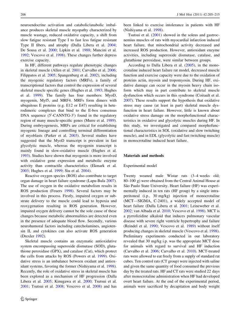
1969). Muscle fiber types were classified as Types I, IC, IIC,
and IIA in SOL and I, IIC, IIA, IIAD and IID/B in EDL
muscles (Staron et al. 1999) (Fig. 1). Fiber cross-sectional
area for each fiber type, and fiber-type frequencies were
determined using Image Analysis System Software (Leica,
Germany). These parameters were calculated in two random
fields per animal using a 209 objective.
Electrophoretic separation of myosin heavy chain
(MHC)
MHC isoform analysis was performed by sodium dodecyl
sulphate polyacrylamide gel electrophoresis (SDS–PAGE)
in duplicate (maximum 5% variation). Ten serial cross
sections (12 lm thick) were collected from each muscle
sample and placed in a solution (0.5 ml) containing glycerol
10% (w/v), 2-mercaptoethanol 5% (v/v) and sodium dode-
cylsulfate (SDS) 2.3% (w/v) in a Tris/HCl buffer 0.9% (pH
6.8) (w/v). The final solution was shaken for 1 min and
heated for 10 min at 60�C (Campos et al. 2002). Small
quantities (30 ll) of the extracts were submitted to electro-
phoresis reaction (SDS–PAGE 7–10%) using a 4% stacking
gel for 26 h at 180 V; the maximum current was limited to
13 mA. The gels were stained with Coomassie Blue (Barr
and Pette 1988) and used to identify isoforms according to
molecular weight. EDL muscle showed bands at the MHCI,
MHCIIa, MHCIId and MHCIIb levels and SOL muscle
showed bands at the MHCI and MHCIIa levels (Fig. 2). The
gels were photographed, images captured by VDS Software
(Pharmacia Biotech), and their relative percentages quanti-
fied by densitometry using Image Master VDS Software
(version 3.0). Identification of MHC isoforms was accom-
plished by comigration of plantaris muscle samples using a
control animal as reference.
RNA isolation and analysis
Total RNA was extracted from SOL and EDL muscles with
TRIzol Reagent (Invitrogen, USA). Frozen muscles were
mechanically homogenized on ice in 1 ml ice-cold TRIzol
reagent. Total RNA was solubilized in nuclease-free H2O,
incubated in DNase I (Invitrogen, USA) to remove any
DNA present in the sample, and quantified by measuring
the optical density (OD) at 260 nm. RNA purity was
ensured by obtaining a 260/280 nm OD ratio of *2.0.
Degradation of RNA samples was monitored by the
observation of appropriate 28S–18S ribosomal RNA ratios
as determined by GelRed staining of the agarose gels.
Fig. 1 Cross sections of SOL (a) and EDL (b) muscles showing
fiber-type distribution using myofibrillar adenosine triphosphatase
(mATPase) reaction after preincubation at pH 4.5. SOL (I, type I; IC,
type IC; IIC, type IIC; A, type IIA); EDL (I, type I; IIC, type IIC; A,
type IIA; AD, type IIAD; DB, type IID ? type IIB) muscle fibers. Bar
40 lm
J Mol Hist (2011) 42:205–215 207
123
Reverse transcription
For each sample, cDNA was synthesized from 2 lg of total
RNA by using components from the High Capacity cDNA
Reverse Transcription Kit (Applied Biosystems, USA). The
reaction contained 10 ll 109 Reverse Transcription Buffer,
4 ll 259 dNTPs, 10 ll 109 random primers, 100 units of
RNase inhibitor (Applied Biosystems, USA), 250 units of
MultiScribeTM Reverse Transcriptase, and the final volume
was adjusted to 100 ll with nuclease-free H2O. The primers
were allowed to anneal for 10 min at 25�C before the
reaction proceeded for 2 h at 37�C. Control ‘‘No RT’’
reactions were performed by omitting the RT enzyme. These
reactions were then PCR amplified to ensure that DNA did
not contaminate the RNA. The resulting cDNA samples
were aliquoted and stored at -20�C.
Real-time qPCR
Two microliters of cDNA, corresponding to 20 ng of total
RNA, from the Reverse transcription reaction were used as a
template in the real-time qPCR, performed in a 7300 Real-
Time PCR System (Applied Biosystems, USA). Cycling
conditions were as follows: 95�C for 10 min followed by 40
cycles of 95�C for 15 s and 60�C for 1 min. The reactions
were run in duplicate using 0.4 lM of each primer and 29
Power SYBR Green PCR master mix (Applied Biosystems,
USA) in a final volume of 25 ll. Primer sequences were
designed using the Primer Express v3.0 software (Applied
Biosystems, USA) and are listed in the Table 1. Melting
dissociation curves and agarose gel electrophoresis were
performed to confirm that only a single product was
amplified. Control reactions were run lacking cDNA tem-
plate to check for reagent contamination. Relative gene
expression was calculated using the Comparative CT
Method (Livak and Schmittgen 2001). The gene expression
and the most stable reference genes were obtained using
geNorm (version 3.5, written by Vandesompele et al. 2002).
Biochemical study
Ten animals from each group were used in the biochemical
study. Samples (200 mg) of SOL and EDL muscle were
weighed and homogenized in 5 ml cold phosphate buffer
(0.1 M, pH 7.4) containing 1 mM ethylenediaminete-
traacetic acid (EDTA). Tissue homogenates were prepared
in a motor-driven Teflon glass Potter–Elvehjem tissue
homogenizer (1 min 100 rpm) immersed in ice water. The
homogenate was centrifuged at 10,000 rpm for 15 min and
supernatant was used to determine total protein (TP), lipid
hydroperoxide (LH), glutathione peroxidase (GSH-Px),
superoxide dismutase (SOD). LH was measured through
hydroperoxide-mediated Fe2? oxidation under acid condi-
tions (Jiang et al. 1991). Samples were added to reaction
mixtures containing 100 lM xylenol orange, 250 lM
FeSO4, 25 lM H2SO4, and 4 mM butylated hydroxytolu-
ene (BHT) in 90% (v/v) methanol. The mixtures were
incubated for 30 min at room temperature prior to mea-
surement at 560 nm. GSH-Px was assayed using 0.15 M
pH 7.0 phosphate buffer containing 5 mM EDTA, 0.1 ml
0.0084 M NADPH, 0.005 ml GSSG-reductase (Sigma),
0.01 ml 1.125 M NaN4 (sodium azide), and 0.1 ml 0.15 M
GSH (Hopkins and Tudhope 1973). Superoxide dismutase
activity was determined by the ability of the enzyme to
inhibit reduction of nitro blue tetrazolium (NBT; Sigma).
NBT reduction rate in the absence of tissue was used as a
reference. One unit of SOD was defined as the amount of
protein required to decrease the reference rate to 50%
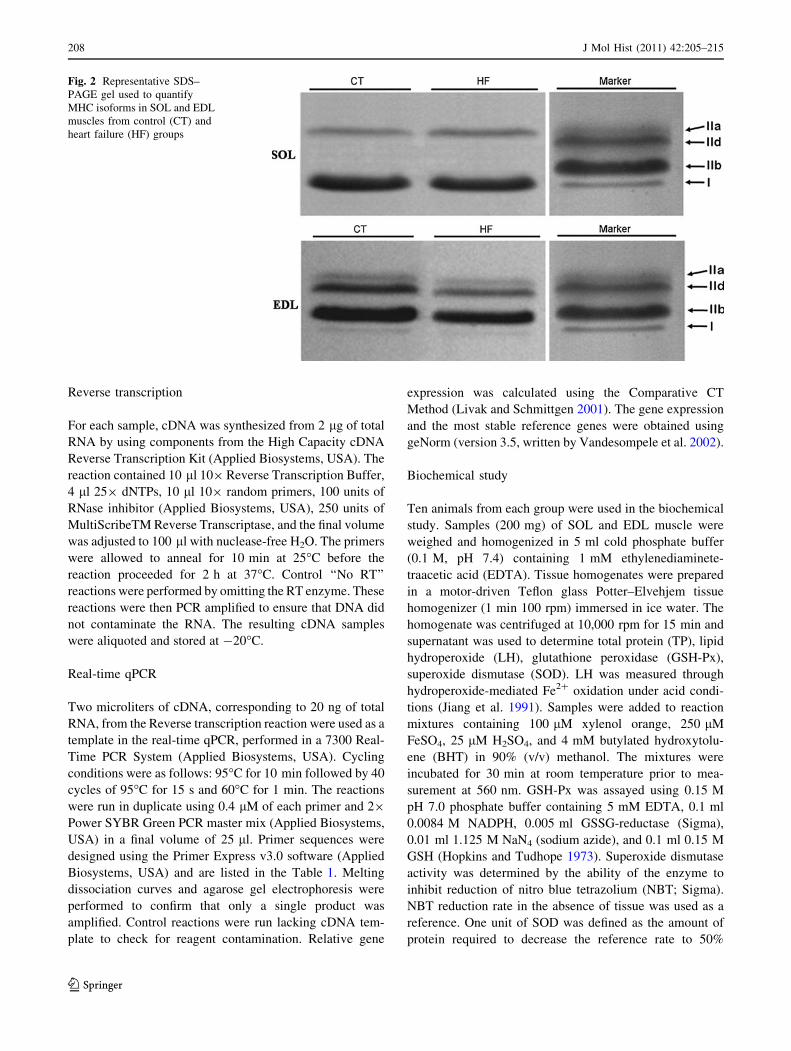
Fig. 2 Representative SDS–
PAGE gel used to quantify
MHC isoforms in SOL and EDL
muscles from control (CT) and
heart failure (HF) groups
208 J Mol Hist (2011) 42:205–215
123
maximum inhibition. All data were expressed in SOD units
per mg protein (Ewing and Janero 1995). Enzyme activity
was determined using a microplate reader (Bio-tech
Instruments INC USA). Spectrophotometric determinations
were performed in a Pharmacia Biotech spectrophotometer
(974213, Cambridge, England). All reagents were from
Sigma (Sigma. St. Louis, MO, USA). The extinction
coefficient for NADH at 340 nm was 6.22 lM-1 cm-1 and
for DTNB at 412 nm was 13.6 mM-1 cm-1.
Fatigue resistance and contractile properties
Samples of EDL or SOL muscle preparations were
removed and mounted for myographic recording in vitro,
according to Gallacci and Oliveira (1994). The preparation
was mounted vertically in a conventional isolated organ-
bath chamber containing 25 ml of physiological solution
composed of (mmol/l): NaCl, 135; KCl, 5; MgCl2, 1;
CaCl2, 2; NaHCO3, 15; NaH2PO4, 1; glucose, 11. This
solution was bubbled with carbogen (95% O2 and 5%
CO2), to maintain pH between 7.4 and 7.5, and temperature
at 27�C. The preparations were attached to isometric force
transducers (Grass, FT03) for recording twitch tension.
Transducer signal outputs were amplified and recorded on
computer via a transducer signal conditioner (Gould,
13-6615-50) with an AcquireLab Data Acquisition System
(Gould). Once mounted in the test apparatus, muscles were
allowed 10 min to equilibrate to the experimental tem-
perature. A series of brief tetanic contractions were used to
establish the muscle length that produces maximum tetanic
force. The isometric contractile properties of the muscles
and their resistance to fatigue were assessed in sequence.
Direct contractions were evoked by supramaximal pulses
(0.2 Hz, 4 ms, and 30 V), delivered from an electrical
stimulator (Grass, S88), through a bipolar electrode posi-
tioned on opposite sides of the muscle. To avoid interfer-
ence from indirect contractions, these experiments were
performed in the presence of pancuronium bromide
(2 9 10-6 M). Fatigue resistance protocol consisted of 30
tetanic contractions. For EDL muscle the frequency was
200 Hz and for SOL muscle 120 Hz. Tetani duration was
0.9 s for EDL and 1.5 s for SOL muscle. These combina-
tions of pulse frequency and tetanus duration resulted in
both muscles receiving the same number of stimulus pulses
in each tetanus (Barclay 1992). Fatigue resistance was
defined as the force developed in the last contraction of a
series relative to the force in the first contraction and was
expressed as a percentage. The contractile properties
studied were amplitude, and rise time and fall time of the
first tetanic contraction. The rise time of contraction was
determined between 10 and 90% of maximum amplitude
development and the fall time was determined between 90
and 10% after maximum amplitude. Following recordings,
muscles were dried and weighed. Force was normalized for
muscle cross-sectional area.
Statistical analysis
Anatomical data, muscle fiber cross-sectional area, bio-
chemical data, and contractile property values were
expressed as means ± standard deviation (SD). Compari-
sons between groups were made using the Student’s
unpaired t test. Muscle fiber frequency, MHC isoform
content values, gene expression, and fatigue resistance
values were expressed as median, maximum and minimum
values. Comparisons between groups were by Mann–
Whitney test (Zar 1999). Differences were considered to be
significant when P \ 0.05.
Results
Presence of heart failure in the monocrotaline-treated
rats
After 22 days, all monocrotaline treated rats showed heart
failure at post-mortem, confirmed by atrium and right
ventricular hypertrophies (RVW/BW [ 0.80), pleural and
Table 1 Oligonucleotide primers used for Real-Time PCR amplification of reverse transcribed RNA
Product Accession No Sequence (50–30) PCR Length (bp)
MyoD NM_176079.1 TTTTTCATGCGACTCACAGC 137
GAAGGCAGGGCTTAAGTGTG
Myogenin M24393.1 T GCCACAAGCCAGACTACCCACC 246
CGGGGCACTCACTGTCTCTCAA
Acidic ribosomal phosphoprotein NM_022402 CCTGCACACTCGCTTCCTAGAG 74
CAACAGTCGGGTAGCCAATCTG
Hypoxanthine guanine phosphoribosyl NM_012583 CTCATGGACTGATTATGGACAGGAC 123
GCAGGTCAGCAAAGAACTTATAGCC
Accession No, GenBank accession number, bp base pairs
J Mol Hist (2011) 42:205–215 209
123

pericardial effusions, and congested liver. No alterations
were found in control rats.
There was no significant difference in BW between HF
and CT. Heart weight was increased in HF compared to
CT, as demonstrated by LVW, RVW, and ATW and by the
RVW/BW and ATW/BW heart hypertrophy indexes
(Table 2).
Muscle fiber type frequencies and atrophy
The SOL and EDL atrophy indexes (SOL/BW; EDL/BW)
were significantly decreased in HF compared to CT
(Table 2). Using the myofibrillar adenosine triphosphatase
(mATPase) histochemical reaction after preincubation at
pH 4.2 and 4.5 in both muscles, all muscle fiber types
showed CSA decreased in HF compared to CT (P \ 0.05)
(Figs. 3 and 4). Muscle fiber frequency only decreased in
HF Type IIC SOL muscle compared to CT.
The representative SDS–PAGE gel used to quantify
MHC isoforms is shown in Fig. 2. SOL muscle revealed
MHCI and MHCIIa isoforms; EDL muscle presented
MHCI, MHCIIa, MHCIId, and MHCIIb isoforms. The
percentage distribution of MHCs was not different between
groups (P [ 0.05).
Gene expression analysis by RT-qPCR
The measurement of gene expression by RT-qPCR
revealed that SOL muscle mRNA expression of Myogenin
was significantly lower in HF than CT. EDL muscle
mRNA expression of MyoD was significantly decreased in
HF compared to CT (Figs. 5, 6).
Biochemical analysis
In HF SOL muscle, lipid hydroperoxide increased and
superoxide-dismutase and glutathione peroxidase activity
reduced compared to CT. In EDL muscle, lipid hydroper-
oxide, superoxide-dismutase and glutathione peroxidase
activity were similar between groups (Table 3).
Contractile properties and fatigue resistance tests
Fall time was prolonged in HF SOL muscle compared to
CT (Table 4). However, there were no significant changes
in amplitude, rise time, and fatigue resistance of both EDL
and SOL muscles in HF compared to CT.
Fig. 3 Cross-sectional area (CSA; lm2) of SOL muscle fiber types I,
IC, IIC and IIA in control (CT, n = 10) and heart failure (HF,
n = 10) groups. Values are expressed as mean ± SD. *P \ 0.05:
Statistical significance versus control group
Table 2 Anatomical data of CT and HF
Experimental groups
CT (n = 10) HF (n = 13)
BW (g) 166.83 ± 11.94 173.79 ± 15.66
RVW (mg) 133 (116; 153) 382 (346; 460)**
RVW/BW (mg) 0.79 (0.69; 0.80) 2.21(1.87; 2.64)**
LVW (mg) 395.00 (365; 462) 489.00 (417; 559)*
LVW/BW (mg) 2.38 (2.25; 2.71) 2.85 (2.29; 3.30)*
ATW (mg) 59.00 (46.00; 74.00) 129.00 (71; 169)**
ATW/BW (mg) 0.35 (0.26; 0.46) 0.85 (0.39; 0.99)**
SOL (mg) 91.00 ± 17.00 81.00 ± 11.00
SOL/BW (mg) 0.54 ± 0.08 0.46 ± 0.05*
EDL (mg) 89.00 ± 8.00 76.00 ± 11.00*
EDL/BW (mg) 0.54 ± 0.04 0.44 ± 0.04**
Values are means ± SD (BW, SOL, SOL/BW, EDL, EDL/BW) and median (maximum—minimum value) (RVW, RVW/BW, LVW, LVW/BW,
ATW, ATW/BW); n number of animals, CT control group, HF heart failure group, BW body weight, LVW left ventricle weight, RVW right
ventricle weight, ATW atrium weight, EDL extensor digitorum longus weight, SOL soleus weight
* P \ 0.05 and ** P \ 0.001; Statistical significance versus control group
210 J Mol Hist (2011) 42:205–215
123

Discussion
The aim of this study was to investigate and compare the
morphofunctional characteristics in SOL and EDL muscles
in a monocrotaline-induced model of heart failure. The
major finding is that monocrotaline-induced heart failure
promoted distinct alterations in skeletal muscles with dis-
tinct morphofunctional characteristics. These alterations
were higher in SOL (oxidative and slow twitching muscle)
than in EDL (glycolytic and fast twitching muscle). We
showed atrophy in both SOL and EDL muscle; oxidative
stress, down-regulation in myogenin mRNA expression,
and prolonged tetanic contraction fall time only in SOL
muscle; and down-regulation in MyoD mRNA expression
only in EDL muscle.
Skeletal muscle atrophy in HF has been commonly
described in clinical (Anker et al. 1999; Mancini et al.
Fig. 4 Cross-sectional area (CSA; lm2) of EDL muscle fiber types I,
IIC, IIA, IIA/D, and IIDB in control (CT, n = 10) and heart failure
(HF, n = 10) groups. Values are expressed as mean ± SD.
*P \ 0.05: Statistical significance versus control group
Fig. 5 mRNA abundance estimated by RT-qPCR of MyoD and
Myogenin (Myog) in SOL muscle of the control (CT, n = 10) and
heart failure (HF, n = 10) groups. Data were expressed as minimum,
1st quartile, median, 3rd quartile and maximum values. Gene
expression was normalized to the reference gene Hypoxanthine–
guanine phosphoribosyltransferase (HPRT) and acidic ribosomal
phosphoprotein (ARBP) from the same RT product. Normalized data
are expressed in arbitrary units (AU). *P \ 0.05: statistical signifi-
cance versus control group
Fig. 6 mRNA abundance estimated by RT-qPCR of MyoD and
Myogenin (Myog) in EDL muscle of the control (CT, n = 10) and
heart failure (HF, n = 10) groups. Data were expressed as minimum,
1st quartile, median, 3rd quartile and maximum values. Gene
expression was normalized to the reference gene Hypoxanthine–
guanine phosphoribosyltransferase (HPRT) and acidic ribosomal
phosphoprotein (ARBP) from the same RT product. Normalized data
are expressed in arbitrary units (AU). *P \ 0.05: statistical signifi-
cance versus control group
J Mol Hist (2011) 42:205–215 211
123
1992; Sullivan et al. 1990) and experimental studies
(Carvalho et al. 2003; Dalla Libera et al. 2004; Delp et al.
1997; Schulze et al. 2003). However, skeletal muscle
atrophy is dependent on HF model used and type of muscle
examined. In experiments using monocrotaline to induce
HF in rats, atrophy was not seen in the diaphragm (Lopes
et al. 2008), but EDL muscle did show atrophy (Carvalho
et al. 2006; Carvalho et al. 2010). Martinez et al. (2010)
observed atrophy in the SOL muscle of rats with myocar-
dial infarction induced HF. Carvalho et al. (2003) showed
muscle atrophy in rats with aortic stenosis (AS) induced
HF. In our study, HF promoted SOL and EDL muscle
atrophy 22 days after monocrotaline treatment.
Some factors seem to be linked to muscle atrophy in HF;
these include disuse conditions (Gundersen and Merlie
1994), aging (Alway et al. 2002), reduced physical and
neuromuscular activity, systemic activation of neurohor-
mones, and inflammatory cytokines (Dalla Libera et al.
2001; Filippatos et al. 2005; Toth et al. 2006). Preliminary
experiments conducted in our lab showed that skeletal
muscle atrophy in HF may be involved with changes in
MRF gene expression (Carvalho et al. 2006; Martinez et al.
2010), and changes in MAFbx/atrogin-1 and Murf1 atr-
ogenes (Carvalho et al. 2010).
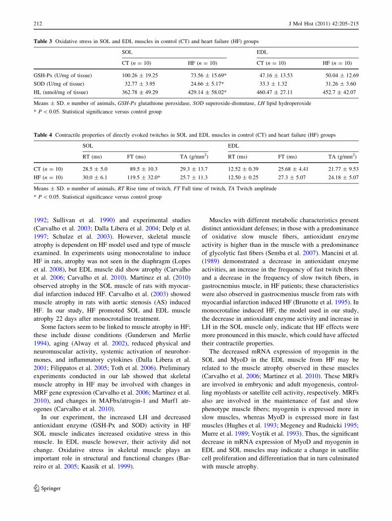
In our experiment, the increased LH and decreased
antioxidant enzyme (GSH-Px and SOD) activity in HF
SOL muscle indicates increased oxidative stress in this
muscle. In EDL muscle however, their activity did not
change. Oxidative stress in skeletal muscle plays an
important role in structural and functional changes (Bar-
reiro et al. 2005; Kaasik et al. 1999).
Muscles with different metabolic characteristics present
distinct antioxidant defenses; in those with a predominance
of oxidative slow muscle fibers, antioxidant enzyme
activity is higher than in the muscle with a predominance
of glycolytic fast fibers (Semba et al. 2007). Mancini et al.
(1989) demonstrated a decrease in antioxidant enzyme
activities, an increase in the frequency of fast twitch fibers
and a decrease in the frequency of slow twitch fibers, in
gastrocnemius muscle, in HF patients; these characteristics
were also observed in gastrocmenius muscle from rats with
myocardial infarction induced HF (Brunotte et al. 1995). In
monocrotaline induced HF, the model used in our study,
the decrease in antioxidant enzyme activity and increase in
LH in the SOL muscle only, indicate that HF effects were
more pronounced in this muscle, which could have affected
their contractile properties.
The decreased mRNA expression of myogenin in the
SOL and MyoD in the EDL muscle from HF may be
related to the muscle atrophy observed in these muscles
(Carvalho et al. 2006; Martinez et al. 2010). These MRFs
are involved in embryonic and adult myogenesis, control-
ling myoblasts or satellite cell activity, respectively. MRFs
also are involved in the maintenance of fast and slow
phenotype muscle fibers; myogenin is expressed more in
slow muscles, whereas MyoD is expressed more in fast
muscles (Hughes et al. 1993; Megeney and Rudnicki 1995;
Murre et al. 1989; Voytik et al. 1993). Thus, the significant
decrease in mRNA expression of MyoD and myogenin in
EDL and SOL muscles may indicate a change in satellite
cell proliferation and differentiation that in turn culminated
with muscle atrophy.
Table 3 Oxidative stress in SOL and EDL muscles in control (CT) and heart failure (HF) groups
SOL EDL
CT (n = 10) HF (n = 10) CT (n = 10) HF (n = 10)
GSH-Px (U/mg of tissue) 100.26 ± 19.25 73.56 ± 15.69* 47.16 ± 13.53 50.04 ± 12.69
SOD (U/mg of tissue) 32.77 ± 3.95 24.66 ± 5.17* 33.3 ± 1.32 31.26 ± 3.60
HL (nmol/mg of tissue) 362.78 ± 49.29 429.14 ± 58.02* 460.47 ± 27.11 452.7 ± 42.07
Means ± SD. n number of animals, GSH-Px glutathione peroxidase, SOD superoxide-dismutase, LH lipid hydroperoxide
* P \ 0.05. Statistical significance versus control group
Table 4 Contractile properties of directly evoked twitches in SOL and EDL muscles in control (CT) and heart failure (HF) groups
SOL EDL
RT (ms) FT (ms) TA (g/mm2) RT (ms) FT (ms) TA (g/mm2)
CT (n = 10) 28.5 ± 5.0 89.5 ± 10.3 29.3 ± 13.7 12.52 ± 0.39 25.68 ± 4.41 21.77 ± 9.53
HF (n = 10) 30.0 ± 6.1 119.5 ± 32.0* 25.7 ± 11.3 12.50 ± 0.25 27.3 ± 5.07 24.18 ± 5.07
Means ± SD. n number of animals, RT Rise time of twitch, FT Fall time of twitch, TA Twitch amplitude
* P \ 0.05. Statistical significance versus control group
212 J Mol Hist (2011) 42:205–215
123
In HF, changes in skeletal muscle phenotype charac-
teristics are frequently seen (Dalla Libera et al. 2010;
Vescovo et al. 1998) with a decrease in slow and an
increase in fast type fibers, making the muscle faster. In our
study, there was a significant decrease in the frequency of
hybrid-type IIC fibers in the SOL muscle. This was not
accompanied by a significant increase in the frequency of
fiber Type IIA. Electrophoresis analysis showed a tendency
to a decrease in the relative percentage of MHC I and an
increase in the relative percentage of MHC IIA. Therefore,
we believe that in the acute HF model used in our study,
there was a tendency for the SOL muscle to acquire the fast
phenotype. This statement is in accordance with the gene
expression results.
In relation to contractile properties, there were no
changes in fatigue resistance and muscular maximum
amplitude in the muscles studied in HF. De Sousa et al.
(2001) showed no changes in the contractile properties of
SOL muscle from rats with AS induced HF. However,
contraction fall time in HF SOL muscle was higher than in
the control group. This finding is in accordance with to
Ertunc et al. (2009) who showed that HF leads to prolon-
gation of twitch fall time only in SOL muscle. Kuno et al.
(1988) reported that muscles with higher proportions of
type II fibers (fast twitch and glycolytic metabolism) have
longer fall times than muscles with a predominance of type
I fibers (slow twitch and oxidative metabolism). Thus, our
findings regarding the SOL muscle in monocrotaline
induced HF confirm the trend towards the faster phenotype
in this muscle.
In conclusion, our results indicate that HF induced by
monocrotaline promoted biochemical, morphological, and
functional changes, more prominent in SOL (oxidative and
slow twitching muscle). Although further experiments are
required to better determine the mechanisms involved in
the pathophysiology of Heart Failure, our results contrib-
utes to the understanding of the muscle-specific changes
that occur in this syndrome.
Acknowledgments This study was supported by FAPESP (Fun-
dacao de Amparo to Pesquisa do Estado de Sao Paulo) Process no
2009/51060-8 and CAPES. This work is part of the M.Sc. Thesis
presented by R.S.B. to Sao Paulo State University (UNESP) in 2011.
References
Allen DL, Sartorius CA, Sycuro LK, Leinwands LA (2001) Different
pathway regulate of the skeletal myosin heavy chain. J Biol
Chem 274:43524–43533
Alway SE, Degens H, Lowe DA, Krishnamurthy G (2002) Increased
myogenic repressor Id mRNA and protein levels in hind limb
muscles of aged rats. Am J Physiol Regul Integr Comp Physiol
282:R411–R422
Anker SD, Ponikowski PP, Clark AL, Leyva F, Rauchhaus M, Kemp
M, Teixeira MM, Hellewell PG, Hooper J, Poole-Wilson PA,
Coats AJ (1999) Cytokines and neurohormones relating to body
composition alterations in the wasting syndrome of chronic heart
failure. Eur Heart J 20:683–693
Barclay CJ (1992) Effects of fatigue on rate of isometric force
development in mouse fast- and slow- twitch muscle. Am J
Physiol 263:C1065–C1072 (Cell Physiol 32)
Barr A, Pette D (1988) Three fast myosin heavy chain in adult rat
skeletal muscle. FEBS Lett 233:153–155
Barreiro E, de la Puente B, Minguella J, Corominas JM, Serrano S,
Hussain SNA, Gea J (2005) Oxidative stress and respiratory
muscle dysfunction in severe chronic obstructive pulmonary
disease. Am J Respir Crit Care Med 171:1116–1124
Brooke MH, Kaiser KK (1970) Three ‘‘myosin adenosine triphos-
phatase’’ systems: the nature of their pH lability and sulfhydryl
dependence. J Histochem Cytochem 18:670–672
Brunotte F, Thompson CH, Adamopoulos S, Coats A, Unitt J,
Lindsay D, Kaklamanis L, Radda GK, Rajagopalan B (1995) Rat
skeletal muscle metabolism in experimental heart failure: effects
of physical training. Acta Physiol Scand 154:439–447
Campos GE, Luecke TJ, Wendeln HK, Toma K, Hagerman FC,
Murray TF, Ragg KE, Ratamess NA, Kraemer WJ, Staron RS
(2002) Muscular adaptations in response to three different
resistance-training regimens: specificity of repetition maximum
training zones. Eur J Appl Physiol 88:50–60
Carvalho RF, Cicogna AC, Campos GE, De Assis JM, Padovani CR,
Okoshi MP, Dal Pai-Silva M (2003) Myosin heavy chain
expression and atrophy in rat skeletal muscle during transition
from cardiac hypertrophy to heart failure. Int J Exp Pathol
84:201–206
Carvalho RF, Cicogna AC, Campos GE, Lopes FS, Sugizaki MM,
Nogueira CR, Dal Pai-Silva M (2006) Heart failure alters MyoD
and MRF4 expression in rat skeletal muscle. Int J Exp Pathol
87:219–225
Carvalho RF, Castan EP, Coelho CA, Lopes FS, Almeida FL,
Michelin A, de Souza RW, Araujo JP Jr, Cicogna AC, Dal Pai-
Silva M (2010) Heart failure increases atrogin-1 and MuRF1
gene expression in skeletal muscle with fiber type-specific
atrophy. J Mol Histol 41:81–87
Coats AJS, Clark AL, Piepoli M, Volterrani M, Poole-Wilson PA
(1994) Symptoms and quality of life in heart failure. The muscle
hypothesis. Br Heart J 72:36–39
Coirault C, Guellich A, Barbry T, Samuel JL, Riou B, Lecar-pentier Y
(2007) Oxidative stress of myosin contributes to skeletal muscle
dysfunction in rats with chronic heart failure. Am J Physiol Heart
Circ Physiol 292:H1009–H1017
Dalla Libera L, Sabbadini R, Renken C, Ravara B, Sandri M, Betto R,
Angelini A, Vescovo G (2001) Apoptosis in the skeletal muscle
of rats with heart failure is associated with increased serum
levels of TNF-alpha and sphingosine. J Mol Cell Cardiol
33:1871–1878
Dalla Libera L, Ravara B, Volterrani M, Gobbo V, Della Barbera M,
Angelini A (2004) Beneficial effects of GH/IGF-1 on skeletal
muscle atrophy and function in experimental heart failure. Am J
Physiol Cell Physiol 286:C138–C144
Dalla Libera L, Ravara B, Gobbo V, Betto DD, Germinario E,
Angelini A, Vescovo G (2005) Skeletal muscle myofibrillar
protein oxidation in heart failure and the protective of Carve-
dilol. J Mol Cell Cardiol 38:803–807
Dalla Libera L, Ravara B, Gobbo V, Betto DD, Germinario E,
Angelini A, Evangelista S, Vescovo G (2010) Skeletal muscle
protein oxidation in chronic right heart failure in rat: can
different beta-blockers prevent it to the same degree? Int J
Cardiol 143:192–199
J Mol Hist (2011) 42:205–215 213
123
De Sousa E, Veksler V, Bigard X, Mateo P, Ventura-Clapier R (2000)
Heart failure affects mitochondrial but not intrinsic properties of
skeletal muscle. Circulation 102:1847–1853
De Sousa E, Veksler V, Bigard X, Mateo P, Serrurier B, Ventura-
Clapier R (2001) Dual influence of disease and increased load on
diaphragm muscle in heart failure. J Mol Cell Cardiol
33:699–710
Delp M, Duan C, Mattson JP, Musch TI (1997) Changes in skeletal
muscle biochemistry and histology relative to fiber type in rats
with heart failure. J Appl Physiol 83:1291–1299
Drexler H (1992) Skeletal muscle failure in heart failure. Circulation
85:1621–1623
Ekmark M, Gronevik E, Schjerling P, Gundersen K (2003) Myogenin
induces higher oxidative capacity in pre-existing mouse muscle
fibres after somatic DNA transfer. J Physiol 548(Pt1):259–269
Ertunc M, Sara Y, Onur R (2009) Differential Contractile Impairment
of fast-and slow-twitch skeletal muscle in a rat model of
doxorubicin-induced congestive heart failure. Pharnacology
84:240–248
Ewing JF, Janero DR (1995) Microplate superoxide dismutase assay
employing a nonenzymatic superoxide generation. Annal Bio-
chem 232:243–248
Feuers RJ (1998) The effects of dietary restriction on mitochondrial
dysfunction in aging. Annal NY Acad Sci 125:192–201
Filippatos GS, Anker SD, Kremastinos DT (2005) Pathophysiology of
peripheral muscle wasting in cardiac cachexia. Curr Opin Clin
Nutr Metab Care 8(3):249–254
Gallacci M, Oliveira AC (1994) Pre- and postsynaptic mechanisms
involved in tetanic fade induced by pancuronium in the isolated
rat muscle. Pharmacology 49:265–270
Gundersen K, Merlie JP (1994) Id-1 as a possible transcriptional
mediator of muscle disuse atrophy. Proc Natl Acad Sci
91:3647–3651
Guth L, Samaha FJ (1969) Qualitative differences between acto
myosin ATPase of slow and fast mammalian muscle. Exp Neurol
25:138–152
Hopkins J, Tudhope GR (1973) Glutathione peroxidase in human red
cells in health and disease. Br J Haematol 25:563–575
Hughes SM, Taylor JM, Tapscott SJ, Gurley CM, Carter WJ, Peterson
CA (1993) Selective accumulation of MyoD and Miogenin
mRNAs in fast and slow muscle is controlled by innervation and
hormones. Development 118:1137–1147
Hughes SM, Chi MM, Lowry OH, Gundersen K (1999) Myogenin
induces a shift of enzyme activity from glycolytic to oxidative
metabolism in muscles of transgenic mice. J Cell Biol
145:633–642
Jiang ZY, Woollard ACS, Wolf SP (1991) Lipid hydroperoxide
measurement by oxidation of Fe2? in the presence of xylenol
orange. Comparison with the TBA assay and an iodometric
method. Lipids 26:853–856
Kaasik A, Minajeva A, De Sousa E, Ventura-Clapier R, Veksler V
(1999) Nitric oxide inhibits cardiac energy production via
inhibition of mitochondrial creatine kinase. FEBS Lett
444:75–77
Kinugawa S, Tsutsui H, Hayashidani S, Ide T, Suematsu N, Satoh S,
Utsumi H, Takeshita A (2000) Treatment with dimethylthiourea
prevents left ventricular remodeling and failure after experi-
mental myocardial infarction in mice: role of oxidative stress.
Circ Res 87:392–398
Kuno S, Katsuta S, Anno I, Matsumoto K, Akisada M (1988)
Relationship between MR relaxation time and muscle fiber
composition. Radiology 169:567–568
Lapu-Bula OfiliE (2007) From hypertension to heart failure: role of
nitric oxide-mediated endothelial dysfunction and emerging
insights from myocardial contrast echocardiography. Am J
Cardiol 26:7–14
Leineweber K, Brandt K, Wludyka B, Beilfuss A, Ponicke K,
Heinroth-Hoffmann I, Brodde OE (2002) Ventricular hypertro-
phy plus neurohumoral activation is necessary to alter the
cardiac beta-adrenoceptor system in experimental heart failure.
Circ Res 91:1056–1062
Lipkin DP, Jones DA, Round JM, Poole-Wilson PA (1988) Abnor-
malities of skeletal muscle in patients with chronic heart failure.
Int J Cardiol 18:187–195
Livak KJ, Schmittgen TD (2001) Analysis of relative gene expression
data using real-time quantitative PCR and the 2(-Delta Delta
C(T)) method. Methods 25:402–408
Lopes FS, Carvalho RF, Campos GER, Sugizaki MM, Padovani CR,
Nogueira CR, Cicogna AC, Dal Pai-Silva M (2008) Down-
regulation of MyoD gene expression in rat diaphragm muscle
with heart failure. Int J Exp Path 89:216–222
Mancini DM, Coyle E, Coggan A, Beltz J, Ferraro N, Montain S,
Wilson JR (1989) Contribution of intrinsic skeletal muscle
changes to 31P NMR skeletal muscle metabolic abnormalities in
patients with chronic heart failure. Circulation 80:1338–1346
Mancini DM, Walter G, Reichek N, Lenkinski R, McCully KK,
Mullen JL, Wilson JR (1992) Contribution of skeletal muscle
atrophy to exercise intolerance and altered muscle metabolism in
heart failure. Circulation 85:1364–1373
Martinez PF, Okoshi K, Zornoff LA, Carvalho RF, Oliveira Junior
SA, Lima AR, Campos DH, Damatto RL, Padovani CR,
Nogueira CR, Dal Pai-Silva M, Okoshi MP (2010) Chronic
heart failure-induced skeletal muscle atrophy, necrosis, and
changes in myogenic regulatory factors. Med Sci Monit
16:BR374–BR383
Megeney LA, Rudnicki MA (1995) Determination versus differen-
tiation and the MyoD family of transcription factors. Biochem
Cell Biol 73:723–732
Murre C, Mccaw PS, Vaessin H, Caudy M, Jan LY, Yan JN, Cabrera
CV, Buskin JN, Hauschka SD, Lassar AB, Weintraub H,
Baltimore D (1989) Interactions between heterologous helix-
loop-helix proteins generate complexes that bind specifically to a
common DNA sequence. Cell 58:537–544
Nishiyama Y, Ikeda H, Haramaki N, Yoshida N, Imaizumi T (1998)
Oxidative stress is related to exercise intolerance in patients with
heart failure. Am Heart J 135:115–120
Parker MH, Seale P, Rudnicki MA (2003) Looking back to the
embryo: defining transcriptional networks in adult myogenesis.
Nat Rev Genet 4:497–507
Powers SK, Ji LL, Leeuwenburgh C (1999) Exercise training-induced
alterations in skeletal muscle antioxidant capacity: a brief
review. Med Sci Sports Exerc 31:987–997
Reindel JF, Ganey JG, Wagner Rf, Slocombe RF, Roth AR (1990)
Development of morphologic, hemodynamic, and biochemical
changes in lung of rats given monocrotaline pirrole. Toxicol
Appl Pharmacol 106:179–200
Schulze PC, Gielen S, Adams V, Linke A, Mobius-Winkler S, Erbs S,
Kratzsch J, Hambrecht R, Schuler G (2003) Muscular levels of
proinflammatory cytokines correlate with a reduced expression
of insulin-like growth factor-1 in chronic heart failure. Basic Res
Cardiol 98:267–274
Semba RD, Lauretani F, Ferrucci L (2007) Carotenoids as protection
against sarcopenia in older adults. Arch Biochem Biophys
458:141–145
Siu PM, Donley DA, Bryner RW, Alway SE (2004) Myogenin and
oxidative enzyme gene expression levels are elevated in rat
soleus muscles after endurance training. J Appl Physiol 97:
277–285
Spangenburg EE, Talmadge RJ, Musch TI, Pfeifer PC, McAllister
RM, Williams JH (2002) Changes in skeletal muscle myosin
heavy chain isoform content during congestive heart failure. Eur
J Appl Physiol 87:182–186
214 J Mol Hist (2011) 42:205–215
123
Staron RS, Kraemer WJ, Hikida RS, Fry AC, Murray JD, Campos GE
(1999) Fiber type composition of four hind limb muscles of adult
Fisher 344 rats. Histochem Cell Biol 111:117–123
Sullivan MJ, Green HJ, Cobb FR (1990) Skeletal muscle biochem-
istry and histology in ambulatory patients with long-term heart
failure. Circulation 81:518–527
Toth MJ, Palmer BM, Lewinter MM (2006) Effect of heart failure on
skeletal muscle myofibrillar protein content, isoform expression
and calcium sensitivity. Int J Cardiol 107:211–219
Tsutsui H, Ide T, Hayashidani S, Suematsu N, Shiomi T, Wen J,
Nakamura K, Ichikawa K, Utsumi H, Takeshita A (2001)
Enhanced generation of reactive oxygen species in the limb
skeletal muscles from a murine infarct model of heart failure.
Circulation 104:134–136
Tsutsui H, Kinugawa S, Matsushima S (2008) Oxidative stress and
mitochondrial DNA damage in heart failure (Suppl A):A31–A37
Van Albada ME, Bartelds B, Wijnberg H, Mohaupt S, Dickinson MG,
Schoemaker RG, Kooi KA, Gerbens F, Berger RM (2010) Gene
expression profile in flow-associated pulmonary arterial hyper-
tension with neointimal lesions. Am J Physiol Lung Cell Mol
Physiol 298:L483–L491
Vandesompele J, De Preter K, Pattyn F, Poppe B, Van Roy N, De
Paepe A, Speleman F (2002) Accurate normalization of Real-
Time quantitative RT–PCR by geometric averaging of multiple
internal control genes. Genome Biol 3(7):34.1–34.11
Vescovo G, Harding SE, Jones M, Dalla Libera L, Pessina AC, Poole-
Wilson PA (1989) Contractile abnormalities of single right
ventricular myocytes isolated from rats with right ventricular
hypertrophy. J Mol Cell Cardiol 21(Suppl 5):103–111
Vescovo G, Ceconi C, Bernocchi P, Ferrari R, Carraro U, Ambrosio
GB, Libera LD (1998) Skeletal muscle myosin heavy chain
expression in rats with monocrotaline-induced cardiac hypertro-
phy and failure. Relation to blood flow and degree of muscle
atrophy. Cardiovasc Res 39:233–241
Vescovo G, Ravara B, Libera LD (2008) Skeletal muscle myofibrillar
protein oxidation and exercise capacity in heart failure. Basic
Res Cardiol 103:285–290
Voytik SL, Przyborski M, Badylak SF, Konieczny SF (1993)
Differential expression of muscle regulatory factor genes in
normal and denervated adult rat hind limb muscle. Dev Dynam
198:214–224
Zar JH (1999) Biostatistical analysis. Prentice-Hall, New Jersey
J Mol Hist (2011) 42:205–215 215
123
Related Documents