Differential immune activation following encapsulation of immunostimulatory CpG oligodeoxynucleotide in nanoliposomes Erdem Erikçi a, 1 , Mayda Gursel b , _ Ihsan Gürsel a, * a Bilkent University, Department of Molecular Biology and Genetics, Biotherapeutic ODN Lab, Bilkent, 06800 Ankara, Turkey b Bilkent University, University Campus Housing, Block # 17/2, Bilkent, 06800 Ankara, Turkey article info Article history: Received 12 September 2010 Accepted 27 October 2010 Available online xxx Keywords: Liposomes Vaccine Immune response CpG DNA TLR Nanotechnology abstract The immunogenicity of a vaccine formulation is closely related to the effective internalization by the innate immune cells that provide prolonged and simultaneous delivery of antigen and adjuvant to relevant antigen presenting cells. Endosome associated TLR9 recognizes microbial unmethylated CpG DNA. Clinical applications of TLR9 ligands are significantly hampered due to their pre-mature in vivo digestion and rapid clearance. Liposome encapsulation is a powerful tool to increase in vivo stability as well as enhancing internalization of its cargo to relevant immune cells. The present study established that encapsulating CpG motifs in different liposomes having different physicochemical properties altered not only encapsulation efficiency, but also the release and delivery rates that ultimately impacted in vitro and ex-vivo cytokine production rates and types. Moreover, different liposomes encapsulating CpG ODN significantly increased Th1-biased cytokines and chemokines gene transcripts Additional studies demonstrated that co-stimulatory and surface marker molecules significantly upregulated upon lipo- some/CpG injection. Finally, co-encapsulating model antigen ovalbumin with CpG ODN adjuvant in nanoliposomes profoundly augmented Th1 and cell mediated anti-Ova specific immune response. Collectively, this work established an unappreciated immunoregulatory property of nanoliposomes mediating immunity against protein antigen and could be harnessed to design more effective therapeutic vaccines or stand alone immunoprotective agents targeting infectious diseases, as well as cancer or allergy. Ó 2010 Elsevier Ltd. All rights reserved. 1. Introduction Innate immune cells respond to diverse components of microbial pathogens. These molecular patterns collectively are known as the pathogen associated molecular patterns (PAMPs). Host cell expressed pattern recognition receptors (PRRs) [1] recognizes PAMPs and generally initiates a signaling cascade leading to orchestrated innate immune activation [2]. The most studied PRR family is the Toll-like receptors (TLRs) [3,4], which are expressed by various cells of tissues such as spleen, lung, muscle, intestines and blood cells [5,6]. Recognition of diverse array of microbial compo- nents by different TLRs separates self antigen from non-self antigens [7] and it is the link between innate and adaptive immunity [8,9]. Microbial nucleic acids are recognized by endosomal Toll-like receptors on immune system cells of the vertebrates. Bacterial DNA containing CpG motifs and dsRNA, are recognized by TLR9 [10,11] and TLR3 [12] respectively in both murine and human immune cells whereas ssRNA are recognized by murine TLR7 and human TLR8 [13]. Certain TLRs induce a Th1 dominant immune response, yet others may drive a Th2-biased immune activation through MyD88 or TRIF pathways [14]. CpG ODNs with different sequences can also yield differential immune response [15,16]. CpG-B (or K-ODN) triggers monocytes and B cells to proliferate and secrete IL6 and IgM [15]. Moreover it induces plasmacytoid dendritic cells (pDC) to mature and also secrete TNFa by proceeding signaling from late endosome through MyD88 and IRF5. Whereas, CpG-A (or D- ODNs) induces pDCs to secrete IFNa [17] by signaling from early endosome through MyD88 and IRF7 [18] and it induces NK cells to produce IFNg [15]. Nucleic acid based TLR ligands are promising vaccine adjuvants, anti-allergens, immunoprotective and anticancer agents [19]. However their clinical applications are significantly hampered due to their pre-mature in vivo digestion by endonucleases and rapid clearance via serum protein adsorption leading to limited in vivo * Corresponding author. Tel.: þ90 312 290 24 08; fax: þ90 312 266 50 97. E-mail address: [email protected] ( _ I. Gürsel). 1 Present Address: Department of Molecular Cell Biology, Max Planck Institute of Biophysical Chemistry, 37077 Göttingen, Germany. Contents lists available at ScienceDirect Biomaterials journal homepage: www.elsevier.com/locate/biomaterials 0142-9612/$ e see front matter Ó 2010 Elsevier Ltd. All rights reserved. doi:10.1016/j.biomaterials.2010.10.054 Biomaterials xxx (2010) 1e9 Please cite this article in press as: Erikçi E, et al., Differential immune activation following encapsulation of immunostimulatory CpG oligo- deoxynucleotide in nanoliposomes, Biomaterials (2010), doi:10.1016/j.biomaterials.2010.10.054

Welcome message from author
This document is posted to help you gain knowledge. Please leave a comment to let me know what you think about it! Share it to your friends and learn new things together.
Transcript

lable at ScienceDirect
Biomaterials xxx (2010) 1e9
Contents lists avai
Biomaterials
journal homepage: www.elsevier .com/locate/biomater ia ls
Differential immune activation following encapsulation of immunostimulatoryCpG oligodeoxynucleotide in nanoliposomes
Erdem Erikçi a,1, Mayda Gursel b, _Ihsan Gürsel a,*aBilkent University, Department of Molecular Biology and Genetics, Biotherapeutic ODN Lab, Bilkent, 06800 Ankara, TurkeybBilkent University, University Campus Housing, Block # 17/2, Bilkent, 06800 Ankara, Turkey
a r t i c l e i n f o
Article history:Received 12 September 2010Accepted 27 October 2010Available online xxx
Keywords:LiposomesVaccineImmune responseCpG DNATLRNanotechnology
* Corresponding author. Tel.: þ90 312 290 24 08; fE-mail address: [email protected] (_I. Gür
1 Present Address: Department of Molecular Cell BiBiophysical Chemistry, 37077 Göttingen, Germany.
0142-9612/$ e see front matter � 2010 Elsevier Ltd.doi:10.1016/j.biomaterials.2010.10.054
Please cite this article in press as: Erikçi E,deoxynucleotide in nanoliposomes, Biomate
a b s t r a c t
The immunogenicity of a vaccine formulation is closely related to the effective internalization by theinnate immune cells that provide prolonged and simultaneous delivery of antigen and adjuvant torelevant antigen presenting cells. Endosome associated TLR9 recognizes microbial unmethylated CpGDNA. Clinical applications of TLR9 ligands are significantly hampered due to their pre-mature in vivodigestion and rapid clearance. Liposome encapsulation is a powerful tool to increase in vivo stability aswell as enhancing internalization of its cargo to relevant immune cells. The present study establishedthat encapsulating CpG motifs in different liposomes having different physicochemical properties alterednot only encapsulation efficiency, but also the release and delivery rates that ultimately impacted in vitroand ex-vivo cytokine production rates and types. Moreover, different liposomes encapsulating CpG ODNsignificantly increased Th1-biased cytokines and chemokines gene transcripts Additional studiesdemonstrated that co-stimulatory and surface marker molecules significantly upregulated upon lipo-some/CpG injection. Finally, co-encapsulating model antigen ovalbumin with CpG ODN adjuvant innanoliposomes profoundly augmented Th1 and cell mediated anti-Ova specific immune response.Collectively, this work established an unappreciated immunoregulatory property of nanoliposomesmediating immunity against protein antigen and could be harnessed to design more effective therapeuticvaccines or stand alone immunoprotective agents targeting infectious diseases, as well as cancer orallergy.
� 2010 Elsevier Ltd. All rights reserved.
1. Introduction
Innate immune cells respond todiverse components ofmicrobialpathogens. These molecular patterns collectively are known as thepathogen associated molecular patterns (PAMPs). Host cellexpressed pattern recognition receptors (PRRs) [1] recognizesPAMPs and generally initiates a signaling cascade leading toorchestrated innate immune activation [2]. The most studied PRRfamily is the Toll-like receptors (TLRs) [3,4], which are expressed byvarious cells of tissues such as spleen, lung, muscle, intestines andblood cells [5,6]. Recognition of diverse array of microbial compo-nents bydifferent TLRs separates self antigen fromnon-self antigens[7] and it is the link between innate and adaptive immunity [8,9].Microbial nucleic acids are recognized by endosomal Toll-like
ax: þ90 312 266 50 97.sel).ology, Max Planck Institute of
All rights reserved.
et al., Differential immune arials (2010), doi:10.1016/j.bio
receptors on immune system cells of the vertebrates. Bacterial DNAcontaining CpG motifs and dsRNA, are recognized by TLR9 [10,11]and TLR3 [12] respectively in both murine and human immunecells whereas ssRNA are recognized by murine TLR7 and humanTLR8 [13]. Certain TLRs induce a Th1 dominant immune response,yet others may drive a Th2-biased immune activation throughMyD88 or TRIF pathways [14]. CpG ODNs with different sequencescan also yield differential immune response [15,16]. CpG-B (orK-ODN) triggers monocytes and B cells to proliferate and secreteIL6 and IgM [15]. Moreover it induces plasmacytoid dendritic cells(pDC) tomature and also secrete TNFa by proceeding signaling fromlate endosome through MyD88 and IRF5. Whereas, CpG-A (or D-ODNs) induces pDCs to secrete IFNa [17] by signaling from earlyendosome through MyD88 and IRF7 [18] and it induces NK cells toproduce IFNg [15].
Nucleic acid based TLR ligands are promising vaccine adjuvants,anti-allergens, immunoprotective and anticancer agents [19].However their clinical applications are significantly hampered dueto their pre-mature in vivo digestion by endonucleases and rapidclearance via serum protein adsorption leading to limited in vivo
ctivation following encapsulation of immunostimulatory CpG oligo-materials.2010.10.054

Table 2
E. Erikçi et al. / Biomaterials xxx (2010) 1e92
stability and activity [20]. This problem can be overcome byencapsulating nucleic acid based TLR ligands into a depot carrier,whereas provide an increased in vivo stability as well as morepronounced targeting/internalization to relevant innate immunecells [21e23]. Liposomal encapsulation is an efficient tool that canimprove stability, reduce pre-mature clearance and increase ther-apeutic efficiency of its cargo by prolonging the duration in circu-lation and thus improve bioavailability to relevant cells.
Liposomes are synthetically made phospholipids vesicle bila-yers. By mixing various types of phospholipids with differentmolar ratios, it is possible to generate different liposomes pos-sessing different physicochemical characteristics ranging fromlamellarity to size or net surface charge [23] as well as PEGmediated hydrated surface. In this study, two different classes ofTLR9 ligands (K and D-type CpG motifs) were encapsulated withinfive different liposomes possessing different surface charge andmodification. The characteristic of liposome-ODN mediated differ-ential innate immune activationwas evaluated. Moreover, potentialof these liposomes co-encapsulating antigen and adjuvant asvaccine carriers were also assessed.
2. Materials and methods
2.1. Reagents
All cell culture media components were from Hyclone (USA). Cytokine ELISAreagents such as recombinant cytokines, monoclonal unlabeled and biotinylatedantibodies against IL6 and IFNg, streptavidin-alkaline phosphatase (SAeAKP) andp-nitrophenyl phosphate disodium salt substrate (PNPP) were purchased fromThermo Scientific or Endogen Pierce (USA). Immunoglobulin ELISA reagents; goatanti-mouse IgG, IgG1, IgG2a, IgG2b monoclonal antibodies conjugated with alkalinephosphatase (AP) were from Southern Biotech (USA). Injectable endotoxin-free OVAwas obtained from Pierce (USA). DNase/RNase free water was obtained fromHyclone (USA). TRI Reagent (Trizol) for RNA isolation was from Invitrogen (USA).DyNAmo� cDNA Synthesis kit, DyNAzyme� II PCRMasterMix for PCRwas obtainedfrom Finnzymes (Finland). 10e150 bp DNA ladder was from Fermentas, and100e1000 bp DNA ladder was from Jena Bioscience.
L-a-Phosphatidylcholine (PC) was purchased from Sigma Aldrich (USA).Cholesterol (Chol), 3b-[N-(N0 ,N0-Dimethylaminoethane)-carbamoyl]CholesterolHydrochloride (DC-Chol), 1,2-Dioleoyl-sn-Glycero-3-Phosphoethanolamine-N-[Methoxy(Polyethylene glycol)-2000] (Ammonium Salt) (PEG-PE), 1,2-Dioleoyl-sn-Glycero-3-Phosphoethanolamine (DOPE) were all from Avanti Polar Lipids (USA).Heidolph Laborota Collegiate Rotary Evaporator (Germany), Vibra Cell cup typesonicator (Sonics and Materials Co., USA) and Maxi Dry Lyo, Heto-Holten (Denmark)freeze dryer were used during liposome preparation.
2.2. TLR ligands
TLR ligands for stimulation assays were as follows and supplied from severalvendors: peptidoglycan (PGN) isolated from B.subtilis; (Fluka, Switzerland), lipo-polysaccharide (LPS) (isolated from E.coli; Sigma, USA), and different classes of CpGmotif expressing ODN and control GpC ODNs (please see Table 1 for sequence andsize details) were synthesized by Alpha DNA (Montreal, Canada), and was kindlyprovided by Dr. Dennis M. Klinman (NCI/NIH, USA). All ODNs were free of endotoxinand protein. Bases shown in capital letter have phosphorothioate linkage and thosein lower case have phosphodiester linkages. CpG or flip (GpC) motifs are underlined.
2.3. Maintenance of animals
Adult male or female BALB/c or C57/BL6 mice (6e10 weeks old) were used for invivo experiments as well as generating primary spleen cells for in vitro assays. The
Table 1Names, sizes and sequences of the CpG ODNs used in stimulation assays.
ODN Name and Size Sequence
1555 (15mer) K-type GCTAGACGTTAGCGTK23 (12mer) K-type TCGAGCGTTCTCD35 (20mer) D-type GGtgcatcgatgcaggggGGD3CG (20mer) D-type GGtcgatcgatcgaggggGG1612 (15mer) Control ODN K-type GCTAGAGCTTAGGCTI-127 (20mer) Control ODN D-type GGtgcatgcatgcaggggGG
Please cite this article in press as: Erikçi E, et al., Differential immune adeoxynucleotide in nanoliposomes, Biomaterials (2010), doi:10.1016/j.bio
animals were kept in the animal holding facility of the Department of MolecularBiology and Genetics at Bilkent University under controlled ambient conditions(22 � 2 �C) regulated with 12 h light and 12 h dark cycles. They were provided withunlimited access of food and water. All experimental procedures have beenapproved by the animal ethical committee of Bilkent University (Bil-AEC, Protocol# 07/0029).
2.4. Liposome preparation
Cholesterol and various phospholipids (Avanti Polar Lipids, Alabaster, AL) werecombined in different ratios as shown in Table 2. Lipids were prepared in chloroformas stock solutions of 10 mg/ml and were stored at �40 �C until use. The liposomepreparation method was reported earlier [23].
Briefly, different ratios of phospholipid mixtures in chloroformwere evaporatedin a round bottom flask using a rotary evaporator at 37 �C for 45e60min. The solventfree lipid film was purged with argon or nitrogen to eliminate residual chloroformand replace oxygen, thereby preventing lipid peroxidation. To generate emptymultilamellar vesicles (MLVs), sterile glass beads and 1 ml of PBS was added to each20 mmol total dry lipid film. The MLV solution was then taken into sterile glass vialand sonicated 5e8 times for 30 s intervals at 4 �C using a cup sonicator. Thegenerated small unilamellar vesicles (SUVs) were then mixed with 1 mg/ml ODNsolution, and promptly frozen in liquid nitrogen and then freeze dried overnight. Atthis stage, the lipid/ODN powder is formed.
ODN encapsulation within the liposomes was achieved during controlled-rehydration step20. DNase/RNase free dH2O (1:10 ratio of original solution volumebefore freezing) was added onto dehydrated ODN/liposome powder and vigorouslyvortexed for 15 s every 5 min for 30 min at room temperature. At the end of 30 min,PBS (1:10 original SUV/ODN solution volume) was added and gently mixed for10 min. The liposome solution was completed to one ml by adding PBS and a finalliposome concentration of 20 mM lipid/mg DNA is adjusted. The size of the finalliposome preparation was reduced to <150 nm by extrusion through polycarbonatefilters. Liposome formulations were stored at 4 �C until use.
2.5. Stimulation assays
BALB/c mice (3 months old) were sacrificed and their spleens were extracted.Single-cell suspensions from spleens were prepared in RPMI 1640 supplementedwith 5% FCS, 50 mg/ml penicillin/streptomycin, 2 mM L-glutamine, 10 mM HEPES,0.11 mg/ml sodium pyruvate and 0.5 mM 2-ME. 4 � 105 cells were layered in eachwell of 96 well microtiter plates. Cells were stimulated with defined concentrationsof ODNs and liposome formulations in triplicate. Final cell concentration at eachwellwas adjusted to 2 � 106 cells/ml. The cells were cultured at 37 �C in a 5% CO2
incubator for 36 h unless otherwise stated. After incubation supernatant werecollected and stored at �20 �C for further use.
2.6. ELISA
The 96-well microtiter plates (Nunc, Denmark) were coated with 10 mg/ml ofAbs that are specific for mouse IFNg and IL6. The plates were blocked with PBS-5%BSA. Supernatants from cultured cells were added. Following the steps of incubatingwith biotin-labeled anti-cytokine Ab and phosphate-conjugated avidin and PnPPsubstrate, the cytokine content were quantitated according to standard curvesgenerated by using known amount of recombinant mouse cytokines.
2.7. Semi-quantitative RT-PCR
Total RNA from 1 �107 of stimulated single-cell suspension of splenocytes wereextracted by Trizol extractionmethod. Synthesis of single-stranded DNA frommRNAwas performed by cDNA synthesis kit (Finnzymes cDNA synthesis kit, Finland)according to themanufacturer’s protocol. PCRwas performed using 12.5 ml of mastermix (Finnzymes PCR Master Mix, Finland) 1 mg cDNA, 10 pmol/ml sense primer,10 pmol/ml antisense primer and 9.5 ml of ddH2O. PCR conditions for b-actin, CD40and IL18; 94 �C for 30s, 55 �C for 30s, 72 �C for 30s, 34 cycles and final extension at
Lipid composition and molar ratios used to generate different liposome types.
Liposome Type Liposome Composition (molar ratio)
Neutral PC:Chol (1:1)Anionic PC:DOPE:PS (1:0.5:0.25)Cationic DC-Chol:PC:DOPE (4:6:0.06)Stealth Chol:DOPE:PEG-PE (4:6:0.06)Sterically Stabilized Cationic
Liposomes (SSCL)DC-Chol:DOPE:PEG-PE (4:6:0.06)
PC, phosphatidylcholine; Chol, cholesterol; DOPE, dioleylphosphatidylethanol-amine; PS, phosphatidylserine; DC-Chol, dimethylaminoethanecarbamol-choles-terol; PEG-PE, polyethylene glycol phosphatididyl ethanolamine.
ctivation following encapsulation of immunostimulatory CpG oligo-materials.2010.10.054
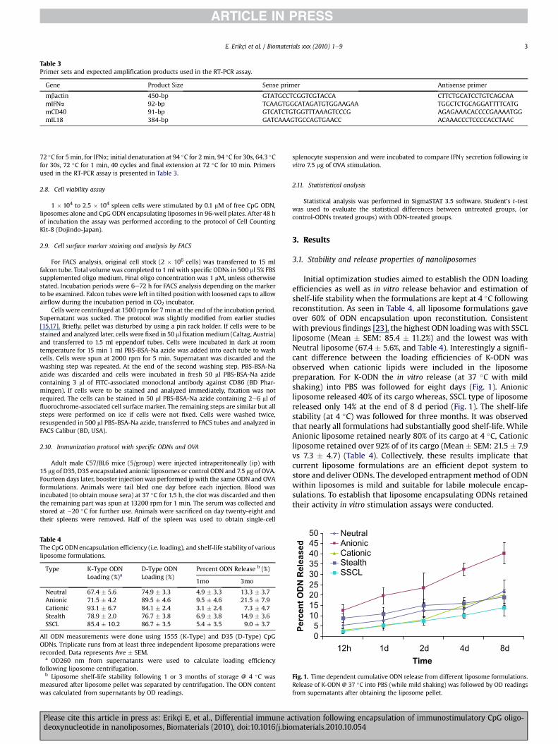
Table 3Primer sets and expected amplification products used in the RT-PCR assay.
Gene Product Size Sense primer Antisense primer
mbactin 450-bp GTATGCCTCGGTCGTACCA CTTCTGCATCCTGTCAGCAAmIFNa 92-bp TCAAGTGGCATAGATGTGGAAGAA TGGCTCTGCAGGATTTTCATGmCD40 91-bp GTCATCTGTGGTTTAAAGTCCCG AGAGAAACACCCCGAAAATGGmIL18 384-bp GATCAAAGTGCCAGTGAACC ACAAACCCTCCCCACCTAAC
E. Erikçi et al. / Biomaterials xxx (2010) 1e9 3
72 �C for 5min, for IFNa; initial denaturation at 94 �C for 2min, 94 �C for 30s, 64.3 �Cfor 30s, 72 �C for 1 min, 40 cycles and final extension at 72 �C for 10 min. Primersused in the RT-PCR assay is presented in Table 3.
2.8. Cell viability assay
1 � 104 to 2.5 � 104 spleen cells were stimulated by 0.1 mM of free CpG ODN,liposomes alone and CpG ODN encapsulating liposomes in 96-well plates. After 48 hof incubation the assay was performed according to the protocol of Cell CountingKit-8 (Dojindo-Japan).
2.9. Cell surface marker staining and analysis by FACS
For FACS analysis, original cell stock (2 � 106 cells) was transferred to 15 mlfalcon tube. Total volumewas completed to 1 ml with specific ODNs in 500 ml 5% FBSsupplemented oligo medium. Final oligo concentration was 1 mM, unless otherwisestated. Incubation periods were 6e72 h for FACS analysis depending on the markerto be examined. Falcon tubes were left in tilted position with loosened caps to allowairflow during the incubation period in CO2 incubator.
Cells were centrifuged at 1500 rpm for 7min at the end of the incubation period.Supernatant was sucked. The protocol was slightly modified from earlier studies[15,17]. Briefly, pellet was disturbed by using a pin rack holder. If cells were to bestained and analyzed later, cells were fixed in 50 ml fixationmedium (Caltag, Austria)and transferred to 1.5 ml eppendorf tubes. Cells were incubated in dark at roomtemperature for 15 min 1 ml PBS-BSA-Na azide was added into each tube to washcells. Cells were spun at 2000 rpm for 5 min. Supernatant was discarded and thewashing step was repeated. At the end of the second washing step, PBS-BSA-Naazide was discarded and cells were incubated in fresh 50 ml PBS-BSA-Na azidecontaining 3 ml of FITC-associated monoclonal antibody against CD86 (BD Phar-mingen). If cells were to be stained and analyzed immediately, fixation was notrequired. The cells can be stained in 50 ml PBS-BSA-Na azide containing 2e6 ml offluorochrome-associated cell surface marker. The remaining steps are similar but allsteps were performed on ice if cells were not fixed. Cells were washed twice,resuspended in 500 ml PBS-BSA-Na azide, transferred to FACS tubes and analyzed inFACS Calibur (BD, USA).
2.10. Immunization protocol with specific ODNs and OVA
Adult male C57/BL6 mice (5/group) were injected intraperitoneally (ip) with15 mg of D35, D35 encapsulated anionic liposomes or control ODN and 7.5 mg of OVA.Fourteen days later, booster injectionwas performed ip with the same ODN and OVAformulations. Animals were tail bled one day before each injection. Blood wasincubated (to obtain mouse sera) at 37 �C for 1.5 h, the clot was discarded and thenthe remaining part was spun at 13200 rpm for 1 min. The serum was collected andstored at �20 �C for further use. Animals were sacrificed on day twenty-eight andtheir spleens were removed. Half of the spleen was used to obtain single-cell
Table 4The CpG ODN encapsulation efficiency (i.e. loading), and shelf-life stability of variousliposome formulations.
Type K-Type ODNLoading (%)a
D-Type ODNLoading (%)
Percent ODN Release b (%)
1mo 3mo
Neutral 67.4 � 5.6 74.9 � 3.3 4.9 � 3.3 13.3 � 3.7Anionic 71.5 � 4.2 89.5 � 4.6 9.5 � 4.6 21.5 � 7.9Cationic 93.1 � 6.7 84.1 � 2.4 3.1 � 2.4 7.3 � 4.7Stealth 78.9 � 2.0 76.7 � 3.8 6.9 � 3.8 14.9 � 3.6SSCL 85.4 � 10.2 86.7 � 3.5 5.4 � 3.5 9.0 � 3.7
All ODN measurements were done using 1555 (K-Type) and D35 (D-Type) CpGODNs. Triplicate runs from at least three independent liposome preparations wererecorded. Data represents Ave � SEM.
a OD260 nm from supernatants were used to calculate loading efficiencyfollowing liposome centrifugation.
b Liposome shelf-life stability following 1 or 3 months of storage @ 4 �C wasmeasured after liposome pellet was separated by centrifugation. The ODN contentwas calculated from supernatants by OD readings.
Please cite this article in press as: Erikçi E, et al., Differential immune adeoxynucleotide in nanoliposomes, Biomaterials (2010), doi:10.1016/j.bio
splenocyte suspension and were incubated to compare IFNg secretion following invitro 7.5 mg of OVA stimulation.
2.11. Statististical analysis
Statistical analysis was performed in SigmaSTAT 3.5 software. Student’s t-testwas used to evaluate the statistical differences between untreated groups, (orcontrol-ODNs treated groups) with ODN-treated groups.
3. Results
3.1. Stability and release properties of nanoliposomes
Initial optimization studies aimed to establish the ODN loadingefficiencies as well as in vitro release behavior and estimation ofshelf-life stability when the formulations are kept at 4 �C followingreconstitution. As seen in Table 4, all liposome formulations gaveover 60% of ODN encapsulation upon reconstitution. Consistentwith previous findings [23], the highest ODN loadingwas with SSCLliposome (Mean � SEM: 85.4 � 11.2%) and the lowest was withNeutral liposome (67.4 � 5.6%, and Table 4). Interestingly a signifi-cant difference between the loading efficiencies of K-ODN wasobserved when cationic lipids were included in the liposomepreparation. For K-ODN the in vitro release (at 37 �C with mildshaking) into PBS was followed for eight days (Fig. 1). Anionicliposome released 40% of its cargo whereas, SSCL type of liposomereleased only 14% at the end of 8 d period (Fig. 1). The shelf-lifestability (at 4 �C) was followed for three months. It was observedthat nearly all formulations had substantially good shelf-life. WhileAnionic liposome retained nearly 80% of its cargo at 4 �C, Cationicliposome retained over 92% of of its cargo (Mean � SEM: 21.5 � 7.9vs 7.3 � 4.7) (Table 4). Collectively, these results implicate thatcurrent liposome formulations are an efficient depot system tostore and deliver ODNs. The developed entrapment method of ODNwithin liposomes is mild and suitable for labile molecule encap-sulations. To establish that liposome encapsulating ODNs retainedtheir activity in vitro stimulation assays were conducted.
05
101520253035404550
12h 1d 2d 4d 8dTime
Percen
t O
DN
R
eleased
NeutralAnionicCationicStealthSSCL
Fig. 1. Time dependent cumulative ODN release from different liposome formulations.Release of K-ODN @ 37 �C into PBS (while mild shaking) was followed by OD readingsfrom supernatants after obtaining the liposome pellet.
ctivation following encapsulation of immunostimulatory CpG oligo-materials.2010.10.054

E. Erikçi et al. / Biomaterials xxx (2010) 1e94
3.2. In vitro activities of liposomal formulations
To compare the activity of K-ODN (1555, or K23) and D-ODN(D35, and D35-3CG) alone or when they are encapsulated into fivedifferent liposome types, spleen cells were stimulated with variousdoses of free or liposome encapsulating CpG ODNs. IFNg and IL6production were checked from cell supernatants by ELISA. Whilefree 1555 ODN is active at 0.3 mM concentration (it yielded35.6 � 11.3 ng/ml, IL6 and 628 � 18 pg/ml, IFNg) when used withcationic and SSCL liposomes, it induced significantly high IFNg thanthe free form (approximately 35 � 5 and 25 � 4 fold more
Fig. 2. IFNg and IL6 production by splenocytes following stimulation with CpG ODN 1555.anionic, cationic, stealth or SSCL liposomes. Fold change of IFNg and IL6 levels in cell supernindex was based on 1555 mediated induction levels) for IFNg, 628 � 18 pg/ml, and for IL6, 3
and 3 mM of K23 (b) and D35 (c) either free (No Lipo) or within neutral, anionic, ca(PGN) (5 mg/ml) and various liposomes without CpG ODNs were used as positive and negatwere detected by ELISA. Data are representative of five independent experiments run in tr
Please cite this article in press as: Erikçi E, et al., Differential immune adeoxynucleotide in nanoliposomes, Biomaterials (2010), doi:10.1016/j.bio
respectively, Fig. 2a, p > 0.001). This augmented activity is solelydependent on the encapsulated CpG ODN, since neither the freeliposome itself nor the liposomes encapsulating the control ODNswere active (Fig. 2d). IL6 response is also high for 1555 encapsu-lating SSCL liposome (3 fold more IL6 compared to free ODN,p > 0.01). However, in neutral, stealth or anionic liposomes 1555CpG ODN activity was lost (Fig. 2a).
Alternative design of K-ODN, K23, was also used for stimulation.Of note themost distinguishable features of K23 from 1555 are, K23is shorter (12mer vs 15mer) and it contains two CpG motifs in itssequence, whereas 1555 is 15-mer and has only one CpG motif. To
Splenocytes was stimulated with 0.3 mM of 1555 either in solution or within neutral,atants was calculated according to stimulation with No Lipo (i.e. only 1555; stimulation5.6 � 11.3 ng/ml. IFNg and IL6), (a). Splenocytes were stimulated with 0.3 mM , 1 mMtionic, stealth or SSCL liposomes. (d) Lipopolysacharide (LPS) (5 mg/ml), peptidoglycanive controls. Cell supernatants were collected at 36 h of incubation IFNg and IL6 levelsiplicates. * not determined, **; not detectable.
ctivation following encapsulation of immunostimulatory CpG oligo-materials.2010.10.054

E. Erikçi et al. / Biomaterials xxx (2010) 1e9 5
our surprise, following stimulation with K23-liposome formula-tions, the cytokine production levels (IFNg and IL6) from spleencells, resembled more to D-Type ODN-liposome stimulations ratherthan 1555-liposome formulation activities. At the lowest dose ofK23 (0.3 mM) neutral and anionic liposomes gave 3 fold more IL6(Fig. 2b). Although free K23 could not stimulate any detectable IFNgsecretion at that concentration, K23 ODN in neutral or anionicliposomes induced approximately 5 and 10 fold more IFNg secre-tion (p > 0.01), respectively. Considering the lowest stimulationdose, while 1555 induces the strongest IFNg and IL6 secretionwhenthey are encapsulated into SSCL liposome, K23 (Fig. 2b) and D35(Fig. 2c) yielded robust cytokine production either with anionic orneutral liposomes but not with SSCL. As the dose of ODNsincreased, the IFNg secretion increased correspondingly (Fig. 2band c). Stimulation with D-ODN encapsulating various liposomesyielded cytokine responses unparallel to ODN1555 type. IFNg andIL6 production from mouse splenocytes were significantlyincreased when D-ODN was encapsulated in neutral, anionic orstealth liposomes but not in cationic or SSCL liposomes (Fig. 2c).These results strongly support the view of differential immuneactivation by different liposomes. To determine whether loss ofIFNg is due to a shift in the cytokine milieu towards a Th2-biasedimmune response, IL4 levels in cell supernatants were alsochecked, (IL4 suppresses IFNg production [24]). IL4 was notdetected in any of liposome/ODN-treated cell supernatants (datanot shown).
3.3. Cytokine expression by liposomal formulations
We have speculated that positively charged liposomes couldalter subcellular location of D-ODN and thus prevent signaling toinitiate from early endosome through MyD88 and IRF7. If this isthe case, therefore, IFNa transcript levels should be lower uponstimulation with cationic and SSCL liposomes compared toneutral, anionic or stealth liposomes (liposome types lacking
Fig. 3. IFNa, CD40 and IL18 transcript levels of spleen cells upon K-ODN and D-ODN stimulaD-type ODN alone (No Lipo) or with various combinations of liposomes (neutral, anionic,isolated and RT-PCR was performed.
Please cite this article in press as: Erikçi E, et al., Differential immune adeoxynucleotide in nanoliposomes, Biomaterials (2010), doi:10.1016/j.bio
positively charged lipid). Total RNA of spleen cells were purified1h or 8h after stimulations, and transcript levels of IFNa, inaddition to IL18 and CD40 was checked by RT-PCR. As shown inFig. 3, positively charged liposomes (Cationic and SSCL lipo-somes) encapsulating D35 (or D-3CG ODN) gave either similar orlower levels of mRNA transcripts for the tested cytokines andsurface marker molecule CD40 to that of free D35. The transcriptlevels of spleen cells stimulated with neutral, anionic and stealthliposomes encapsulating D35 surpassed the performance of freeor control D-ODN encapsulating liposomes (Fig. 3). As expected,CpG ODN 1555 (or K23 ODN) did not induce production of IFNamessage. These results strongly suggested that when positivelycharged liposomes are internalized by immune cells theypossibly either interfere or modify the fate of D-ODN subcellularlocalization and alter IFNa secretion effect. Neutral or negativelycharged liposome formulations improved TLR9/D-ODN interactionand induced more pronounced IFNa transcipt. Of note, no signifi-cant differencewas detected either in CD40or in IL18 genemessagelevels following free or liposomal CpG ODN stimulations (i.e. 1 or8 h).
3.4. Liposomal CpG ODN mediated proliferation
Cell viability and proliferation assays were performed to revealthe cytotoxic or proliferative effect of stimulation with free CpGODN, and their liposome encapsulated forms on spleen cells. Therewas no adverse effect on cell viability when the liposome encap-sulating ODN ligands were used to treat the spleen cells (data notshown). Table 5 summarizes proliferative index upon treatmentwith different liposome formulations. As expected all ODNsequences in their free forms induced very strong cell proliferation.Neutral and stealth liposomes were neither significantly contrib-uting nor inhibiting the proliferative potential of the ligands.Cationic, anionic and SSCL liposome types, however, reduced thedegree of ligand dependent cell division.
tion. 10 � 106 spleen cells were stimulated with 1 mM control ODN (1612), K-type andcationic, stealth, SSCL). After 1 h and 8 h of incubation, total RNA of splenocytes was
ctivation following encapsulation of immunostimulatory CpG oligo-materials.2010.10.054

Table 5Cell proliferation following treatment either with free or liposome encapsulatingTLR ligands.
Types No Ligand Control ODN 1555 D-35
No Lipo 1.00 � 0.09 1.38 � 0.12 8.38 � 1.47** 5.77 � 0.65**Neutral Lipo 1.55 � 0.42 2.35 � 0.92 5.55 � 1.39** 4.30 � 0.74*Anionic Lipo 0.92 � 0.34 1.56 � 0.22 2.16 � 0.82 3.81 � 0.53*Cationic Lipo 1.12 � 0.45 1.65 � 0.50 1.15 � 0.10 1.89 � 0.52Stealth Lipo 1.34 � 0.23 2.08 � 0.54 3.88 � 1.14* 2.48 � 0.64*SSCL Lipo 0.89 � 0.44 1.29 � 0.24 1.69 � 0.44 1.21 � 0.31
Stimulation index (SI) values are given. SI is calculated as the fold proliferation overuntreated cells. Triplicate wells (Ave � SEM) of two independent experiments arereported, * 0.05 < p, ** 0.01 < p compared to untreated (no ligand) group.
E. Erikçi et al. / Biomaterials xxx (2010) 1e96
3.5. APC activation by liposomal D-ODN
A naive T cell requires a second signal to proliferate and differ-entiate into effector T cells following engagement with an antigen.This second signal is known as co-stimulation. Co-stimulation ismediated by co-stimulatory molecules, such as B7-1 (CD80) andB7-2 (CD86) expressed on mouse antigen presenting cells (APCs)including DCs, macrophages and B cells. The CD86 is expressedconstitutively at low levels on APCs and its upregulation is initiatedupon recognition either by a PAMP (i.e. through TLR triggering) orexposure to inflammatory cytokines such as IFNg, IL12 or IL6. Ofnote, the CD80 upregulation requires longer exposure andmay takedays [25,26]. We investigated the activation ability (by anti-CD86staining) of APCs by liposomes encapsulating only D-Type ODNligand. This is due to the fact that, so far our findings indicated thatthe most promising vaccine adjuvant candidate is D-ODN encap-sulated in negatively charged liposome. For this reason, these
02468
10121416
Naïve No Lipo Neutral An
CD
86+
cells (%
)C
D86+
cells (%
)
0
5
10
15
20
25
30
35
Naïve No Lipo Neutral An
A
B
Fig. 4. FACS analyses of CD86 positive DC populations following (a) in vitro stimulation andencapsulating or free 1 mM D35 and the percentage of CD86 expressing cells were determencapsulated D35. 4 h later mice were sacrificed and their spleens were extracted. 2 � 106 sdetermined by FACS.
Please cite this article in press as: Erikçi E, et al., Differential immune adeoxynucleotide in nanoliposomes, Biomaterials (2010), doi:10.1016/j.bio
studies were only conducted with D-ODN formulations. Upon invitro stimulation with five different liposome formulations, CD86staining by FACS revealed that previous IL6 and IFNg findings are inaccordance with the co-stimulatory molecule upregulation (Fig. 4Aand B). Similar to previous observations, the most potent formu-lation was anionic liposome encapsulating D-ODN (12.7 � 1.2%CD86 positive DC population). The least effective treatment wasagain cationic and SSCL liposome types. Compared to free D-ODN,more than 4 fold CD86 upregulation was obtained with anionicliposomal D-ODN treatment.
The utmost aim of this study was to design an effective vaccinecarrier co-encapsulating adjuvant and antigen of interest in a singledepot package. As presented earlier, negatively charged liposomesencapsulating D-ODN’s breadth of immune activity is morepronounced than other studied ODNs. Although in vitro studies areimportant, it does not warrant reproducible in vivo performance.
To reveal the in vivo activity of various liposome-D-ODNformulations, mice were ip injected and 4h post-injection, spleencells were removed and ex-vivo cultured for an additional of 24h(no further external stimulation at this stage) and then stainedwithanti-CD86-FITC antibody to detect CD86 þ cells. Knowing that freeoligo is less stable than liposomal counterpart and also the inter-nalization efficiencies of the free vs liposomal formulations vary atgreat extend [23,27,28], we intentionally injected 2.5 fold more ofthe free oligo (50 mg) during ip injections as opposed to 20 mgliposomal D-ODN. As presented in Fig. 4, compared to untreatedanimals, all tested free or liposomal D-ODN formulations inducedsignificantly higher CD86 upregulation fromDC. Among D35 group,similar what was seen during in vitro assays, anionic-D35 gave themost profound upregulation, (>3 fold more induction compared tofree D-ODN group) CD86 upregulation was also stronger either
ionic Cationic Stealth SSCL
ionic Cationic Stealth SSCL
(b) in vivo stimulation. (A) 1 � 107 splenocytes were stimulated with various liposomeined by FACS. (B) Mice were immunized with 50 mg of free D35 or various liposomeplenocytes were incubated for 24 h and then percentage of CD86 expressing cells were
ctivation following encapsulation of immunostimulatory CpG oligo-materials.2010.10.054

E. Erikçi et al. / Biomaterials xxx (2010) 1e9 7
with neutral or stealth D-ODN encapsulating liposomes (19.1 � 1.5and 18.5 � 2.0 respectively).
3.6. In vivo immunization studies
Following in-vitro and ex-vivo analyses, we next tested theseD-ODN-liposome formulations in immunization studies. For this,a model protein antigen (Ovalbumin) was either mixed with freeD-ODN or co-encapsulated form in anionic liposomes wereprepared. Initial experiments revealed that encapsulating CpG andmixing with free Ag, or encapsulating Ova in liposomes and mixingwith free D-ODN prior to injections had no significant contributionto anti-Ova specific Ab titers (data not shown). Furthermore, twodifferent populations of liposomes (i.e. liposomal D-ODN and Ovaloaded liposomes) and mixing just before immunization also didnot significantly contribute anti-Ova Ab titers (data not shown).
As presented in Table 6, naive mice were divided into 5 differenttreatment groups. Before primary injection background un-immunized mouse sera was obtained. The next day (at d ¼ 0) micewere i.p immunized with the indicated groups. On d ¼ 13, allanimals were tail bled and sera was saved to study primary anti-OVA Ig responses by ELISA. The next day, booster injections wereperformed. On d ¼ 28 mice were first bled and then sacrificed, andspleens were frozen down for PCR study. The collected mouseprimary and secondary sera were subjected to total IgG, anti-OVAIgG1, IgG2a and IgG2b ELISA following serial titrations (Table 6). It isclearly demonstrated that present vaccine formulation (Gr#5), thatis, ovalbumin and D-ODN as the adjuvant, co-encapsulated ina single liposome vesicle (anionic liposomes) induced >150 foldmore IgG over free Ag þ CpG mixed group (i.e. Gr# 3), and 14 foldmore fromAgþ Control ODN (Gr#4). In vaccine trials, in addition tototal IgG which is an indication of the generated humoral immu-nity, it is very important to mount cell mediated immune response,in which one can predict by analyzing the level of IgG2a titers. Asseen in (Table 6a), over 30 fold more IgG2a from anionic liposomesco-encapsulating Ova and D-ODN (i.e. Gr#5) was detectedcompared to Gr#3 (428 � 50 vs 14 � 7, respectively).
Booster injection is the cardinal phase of any vaccinationprotocol, since it induces both effector T andmemory B cells againstinjected antigen. As seen in Table 6b, a very potent anti-Ovahumoral immunity is initiated at the end of 4 weeks (Gr#5, Table6b). A very strong improvement in total IgG (that correspondedto>22 fold more IgG) was achieved when liposomal formulation isused instead of its free formulation (Gr#5 and Gr#3 titers are 28670
Table 6Primary and secondary anti-Ova IgG subclass responses of mice immunized with free or
A. Primary IgG LevelsGr.# Treatment Groups IgG1 Untreated 7 � 42 Control ODN + OVA 7 � 43 D35 + OVA 28 � 144 Anionic Lipo(Control ODN + OVA) 300 � 1505 Anionic Lipo(D35 + OVA) 4250 � 1000*
B. Secondary IgG Levels.Gr.# Treatment Groups IgG1 Untreated 7 � 12 Control ODN + OVA 7 � 23 D35 + OVA 1300 � 4504 Anionic Lipo(Control ODN + OVA) 5168 � 2590*5 Anionic Lipo(D35 + OVA) 28672 � 5050a**
Female Balb/c mice (6e8 wks old, 5/group) was immunized with OVA (5 mg/ml) plus conton d¼ 13 before booster injection. Twoweeks post-booster injectionmice were first tail breported as 1/T for primary and secondary antibody response. Results (mean titer� SD) arsignificance of Gr#3 vs Gr#4 or Gr#5 paired t-test analysis comparison), ** 0.0001 < p (
a 0.001 < p (Gr#4 vs Gr#5 paired t-test analysis comparison).
Please cite this article in press as: Erikçi E, et al., Differential immune adeoxynucleotide in nanoliposomes, Biomaterials (2010), doi:10.1016/j.bio
are 1300, respectively). Moreover, IgG1 anti-OVA titers were alsosignificantly higher with liposomal formulation (more than 26 foldrose Gr#5 vs Gr#3). Of note, IgG2a level with Gr#5 was >13 foldmore compared to Gr#3 animals. To further investigate the abilityof the liposome formulation to mount cell mediated immuneresponse, IFNg production of the immunized animals wereanalyzed from the recovered spleen cell supernatants following invitro Ova stimulation in culture for 36 h (Fig. 5). Data revealed thatover 7 fold more IFNg secretionwas detected from Gr#5 animals tothat of Gr#3 immunized mice. These findings collectively demon-strated that delivery of D-ODN and Ova antigen encapsulated inanionic liposome induced very strong humoral and cell mediatedimmunity than free D-ODN plus Ova formulation (Table 6 andFig. 5).
4. Discussion
This work investigated the immune activity of liposomesharboring different classes of CpG ODNs, an important immuneadjuvant intended for clinical use. In the present study, the shelf lifeof the generated liposomes showed quite promising stability andretention profiles up to three months (Table 4), duration muchlonger than any conventional vaccination scheme. Moreover, wedemonstrated that K- and D-type ODNmediated cytokine secretionis differentially controlled by the liposome surface charge (Fig. 2).This data is in agreement with previously reported finding [23].Interestingly, alternative design of K-ODN, K23, behaved differentlyin liposomes optimal for 1555 sequence (Fig. 2b). The vesiclelamellarity or the vesicle fluidity of the liposome formulations doesnot seem to influence the resultant immune activation induced byCpG ODNs, since the generated vesicles are kept at fixedmolar ratiofor all preparations and following liposome reconstitution theirsizes were reduced to less than 150 nm via extrusion.
We have revealed that neutral, stealth and anionic liposomesencapsulating D-type ODNs boost IFNg secretion compared to freeform (Fig. 2c). Therefore we can conclude that neutral or anionicliposomes encapsulating D-ODN can promote substantial amountof cytokine secretion even at very low doses. Depot effect andprevention of pre-mature clearance in addition to sparing ODNfrom nuclease attack are important features provided by liposomeencapsulation.
We have suspected that positively charged liposomes preventODN to be sequestered in early endosome where initiation ofD-ODN mediated signaling cascade is required [15e18] and is
liposome encapsulating Ag and CpG ODN.
IgG1 IgG2a IgG2b7 � 4 7 � 4 7 � 47 � 4 7 � 2 7 � 1
14 � 7 14 � 7 7 � 235 � 10 7 � 2 7 � 1
112 � 12* 428 � 50 7 � 1
IgG1 IgG2a IgG2b7 � 2 7 � 2 7 � 47 � 4 7 � 2 7 � 1
828 � 245 112 � 12 112 � 124355 � 1430* 448 � 20* 112 � 20
21672 � 7150a** 1500 � 250a* 28 � 4
rol or CpG ODN (15 mg/ml) (free or liposome encapsulated forms) at day 1 and bleedled and then sacrificed. Sera was titrated and assayed for anti-OVA IgG subclasses ande combination of two independent experiments, *0.01< p (represent the statistiacalGr#3 vs Gr#4 or Gr#5 paired t-test analysis comparison).
ctivation following encapsulation of immunostimulatory CpG oligo-materials.2010.10.054

0
40
80
120
160
200
240
Unt
reat
ed
Con
trol O
DN
+O
VA
D35
+OVA
Anio
nic
Lipo
(Con
rol
OD
N+O
VA)
Anio
nic
Lipo
(D35
+OVA
)
IF
Ng
(p
g/m
l)
Fig. 5. Female Balb/c mice (6e8 wks old, 5/group) was immunized with OVA (5 mg/ml) plus control or CpG ODN (15 mg/ml) (free or liposome encapsulated forms), two weeks post-booster injection mice were sacrificed, spleen cells (5 � 106/ml) from each animal were recovered and cultured in the presence of 7.5 mg OVA for 36h. Supernatants were collectedand IFNg ELISA was studied to detect recall antigen response. Results are reported as IFNg (pg/ml) (mean � SD) for each group. Paired t-test analysis comparison between Gr#3 andGr#5 is P > 0.001.
E. Erikçi et al. / Biomaterials xxx (2010) 1e98
necessary for the production of IFNa transcript. We observed thatD35 encapsulating cationic and SSCL liposomes failed to induceIFNa message as much as free D35 (following 8 h of treatment).However, neutral, anionic and stealth liposomes induced signifi-cantly high IFNa transcript (Fig. 3). Therefore, this data implied thatpositively charged liposomes somehow interfere with D-ODN/TLR9interaction in the early endosome.
CD40 is a receptor expressed on macrophages and important inactivation of infected macrophages with intracellular pathogens byCD4 T cells expressing CD40 ligand. We demonstrated that free D35also induces CD40 expression (Fig. 3). Therefore we can say that K-ODN and D-ODN encapsulation into any of the liposomes do notaugment its expression compared to free form, however, positivelycharged liposomes decreased the amount of CD40 transcript. IL18 isconsidered as IFNg-inducing factor. Together with IL12, IL18induces high amount of IFNg expression [29]. It is an importantcytokine in innate [30,31] and adaptive [32] immune regulation.Except the free form, liposomal forms of D35 do not induce IL18expression. We can conclude that IFNg induction by either lipo-somal D-ODN or K-ODN is not maintained by increased levels ofIL18 production.
We observed that upon control ODN encapsulating liposometreatment considerable levels of IFNa, CD40 and IL18 was inducedby immune cells. These stimulations are TLR independent since thecontrol ODN does not contain any active CpGmotif. However, it is ofgreat importance to evaluate the activity of control ODN that isinternalized into phagocytic cells within liposomes and whether itinitiates a signaling pathways through recognition by cytosolic DNAsensors such as DAI [33] or AIM2 [34] and leads to these transcriptupregulations.
In vaccine induced immunity an important step is to stronglyactivate T cells and for this co-stimulatory molecule upregulationon APCs is critical [35e37]. In this study, unexpectedly, anionicliposome encapsulating D-ODN consistently upregulated CD86both upon in vitro and in vivo stimulation (Fig. 4). This effect wasvery encouraging since raising cell mediated immunity is asimportant as mounting a humoral response for the success of anyimmunization.
Immunization with liposome encapsulating antigen and CpGODN compared to their free forms gave significantly high amount ofIgG titers in mice. This heightened anti-ova response is provided bysimultaneous signals delivered by the formulation leading to
Please cite this article in press as: Erikçi E, et al., Differential immune adeoxynucleotide in nanoliposomes, Biomaterials (2010), doi:10.1016/j.bio
efficient naive T cell activation. In a depot vesicle formulation boththe antigen and CpG adjuvant are together and liposomes indeedmimics a natural course of a pathogenic insult.
In the literature, Joseph et al. demonstrated that when micewere vaccinated once with liposomes (DMPC:DMPG mole ratio of9:1) co-encapsulating 25 mg CpG ODN and and 0.5 mg influenzavirus subunit hemagglutinin and neuraminidase (HN) antigen, nodetectable IgG1 titer and 550 IgG2a titer was observed at 21 dayspost-injection [38]. We have injected mice with anionic liposomeco-encapsulating 7.5 mg ova and 15 mg D-ODN. Only 13 days later wehave obtained 122 IgG1 titer and 428 IgG2a titer. At the end of4 weeks these titers were much higher than reported amounts byJoseph et al. Although amount and type of CpG ODN as well asantigen types and amounts are different, one can speculate that theimmune response initiated by D-ODN encapsulating anionic lipo-some formulation is more immunogenic yielding stronger Th1-biased immunity against tested ovalbumin.
We previously demonstrated that when mice immunized withSSCL co-encapsulating 10 mg 1555 and 2 mg OVA. Following boosterinjection, total IgG titer reached 19000, the present study showedthat a 1.5 fold more IgG (a titer of 28672) with a different type ofCpG ODN and liposome type could be attained. Collectively, thepresent in vivo experiments demonstrated that contrary to theestablished dogma that D-type ODN is not that immunogenic thanK-type, our findings suggests that when encapsulated withina proper liposome, D-ODN can induce antigen specific immunity.
5. Conclusion
This study established that different liposomes induced differ-ential innate immune activation otherwise not possible to obtainwith two different classes of TLR9 ligands (i.e. K- and D-types). Theinformation from this work demonstrated an unappreciatedfeature of liposome-mediated immune modulation and could beharnessed to design more effective therapeutic vaccines targetingseveral infectious diseases, cancer and asthma or allergy.
Acknowledgements
This work was partially supported by the Scientific and Techno-logical Research Council of Turkey (TUBITAK) grants: SBAG106S102,and SBAG108S316 and EU/FP6/Marie Curie Reintegration Grant
ctivation following encapsulation of immunostimulatory CpG oligo-materials.2010.10.054

E. Erikçi et al. / Biomaterials xxx (2010) 1e9 9
(# 036615) to IG. E.E. received post-graduate program financialsupport from TUBITAK throughout his MSc thesis. We thank toGizem Tincer and C. Fuat Yagci for their excellent technical supportand Burcu C. Insal for her assistance during animal procedures.Authors declare that they do not have any conflicting interest.
Appendix
Figures with essential color discrimination. Fig. 1 in this articleare difficult to interpret in black and white. The full color imagescan be found in the on-line version, at doi:10.1016/j.biomaterials.2010.10.054.
References
[1] Medzhitov R, Janeway Jr CA. Innate immune recognition and control ofadaptive immune responses. Semin Immunol 1998;10(5):351e3.
[2] Takeshita F, Gursel I, Ishii KJ, Suzuki K, Gursel M, Klinman DM. Signal trans-duction pathways mediated by the interaction of CpG DNA with Toll-likereceptor 9. Semin Immunol 2004;16(1):17e22.
[3] TakedaK,KaishoT,AkiraS.Toll-like receptors.AnnRevImmunol2003;21:335e76.[4] Akira S, Takeda K. Toll-like receptor signalling. Nat Rev Immunol 2004;4
(7):499e511.[5] Zarember KA, Godowski PJ. Tissue expression of human Toll-like receptors
and differential regulation of Toll-like receptor mRNAs in leukocytes inresponse to microbes, their products, and cytokines. J Immunol 2002;168(2):554e61.
[6] Kabelitz D. Expression and function of Toll-like receptors in T lymphocytes.Curr Opin Immunol 2007;19(1):39e45.
[7] Blander JM, Medzhitov R. Toll-dependent selection of microbial antigens forpresentation by dendritic cells. Nature 2006;440(7085):808e12.
[8] Palm NW, Medzhitov R. Pattern recognition receptors and control of adaptiveimmunity. Immunol Rev 2009;227(1):221e33.
[9] Pasare C, Medzhitov R. Toll-like receptors: linking innate and adaptiveimmunity. Microbe Infect 2004;6(15):1382e7.
[10] Hemmi H, Takeuchi O, Kawai T, Kaisho T, Sato S, Sanjo H, et al. A Toll-likereceptor recognizes bacterial DNA. Nature 2000;408(6813):740e5.
[11] Takeshita F, Leifer CA, Gursel I, Ishii KJ, Takeshita S, Gursel M, et al. Cuttingedge: role of Toll-like receptor 9 in CpG DNA-induced activation of humancells. J Immunol 2001;167(7):3555e8.
[12] Alexopoulou L, Holt AC, Medzhitov R, Flavell RA. Recognition of double-stranded RNA and activation of NF-kappaB by Toll-like receptor 3. Nature2001;413(6857):732e8.
[13] Heil F, Hemmi H, Hochrein H, Ampenberger F, Kirschning C, Akira S, et al.Species-specific recognition of single-stranded RNA via toll-like receptor7 and 8. Science 2004;303(5663):1526e9.
[14] Iwasaki A, Medzhitov R. Toll-like receptor control of the adaptive immuneresponses. Nat Immunol 2004;5(10):987e95.
[15] Gursel M, Verthelyi D, Gursel I, Ishii KJ, Klinman DM. Differential andcompetitive activation of human immune cells by distinct classes of CpGoligodeoxynucleotide. J Leukoc Biol 2002;71(5):813e20.
[16] Kerkmann M, Rothenfusser S, Hornung V, Towarowski A, Wagner M, Sarris A,et al. Activation with CpG-A and CpG-B oligonucleotides reveals two distinctregulatory pathways of type I IFN synthesis in human plasmacytoid dendriticcells. J Immunol 2003;170(9):4465e74.
Please cite this article in press as: Erikçi E, et al., Differential immune adeoxynucleotide in nanoliposomes, Biomaterials (2010), doi:10.1016/j.bio
[17] Gursel M, Gursel I, Mostowski HS, Klinman DM. CXCL16 influences the natureand specificity of CpG-induced immune activation. J Immunol 2006;177(3):1575e80.
[18] Honda K, Ohba Y, Yanai H, Negishi H, Mizutani T, Takaoka A, et al. Spatio-temporal regulation of MyD88-IRF-7 signalling for robust type-I interferoninduction. Nature 2005;434(7036):1035e40.
[19] Klinman DM, Currie D, Gursel I, Verthelyi D. Use of CpG oligodeoxynucleotidesas immune adjuvants. Immunol Rev 2004;199:201e16.
[20] Barry ME, Pinto-Gonzalez D, Orson FM, McKenzie GJ, Petry GR, Barry MA. Roleof endogenous endonucleases and tissue site in transfection and CpG-medi-ated immune activation after naked DNA injection. Hum Gene Ther 1999;10(15):2461e80.
[21] Torchilin VP. Recent advances with liposomes as pharmaceutical carriers. NatRev Drug Discov 2005;4(2):145e60.
[22] PapahadjopoulosD, Allen TM,GabizonA,MayhewE,MatthayK,Huang SK, et al.Sterically stabilized liposomes: improvements in pharmacokinetics and anti-tumor therapeutic efficacy. Proc Natl Acad Sci (USA) 1991;88(24):11460e4.
[23] Gursel I, Gursel M, Ishii KJ, Klinman DM. Sterically stabilized cationic lipo-somes improve the uptake and immunostimulatory activity of CpG oligonu-cleotides. J Immunol 2001;167(6):3324e8.
[24] Tanaka T, Hu-Li J, Seder RA, Fazekas de St Groth B, Paul WE. Interleukin4 suppresses interleukin 2 and interferon gamma production by naive T cellsstimulated by accessory cell-dependent receptor engagement. Proc Natl AcadSci (USA) 1993;90(13):5914e8.
[25] Greenwald RJ, Freeman GJ, Sharpe AH. The B7 family revisited. Ann RevImmunol 2005;23:515e48.
[26] Abbas AK, Lichtman AH, Pillai S. Cellular and molecular Immunology. 6th ed.Philadelphia: Saunders Elsevier; 2007.
[27] Fenske DB, MacLachlan I, Cullis PR. Long-circulating vectors for the systemicdelivery of genes. Curr Opin Mol Ther 2001;3(2):153e8.
[28] Mutwiri GK, Nichani AK, Babiuk S, Babiuk LA. Strategies for enhancing theimmunostimulatory effects of CpG oligodeoxynucleotides. J Contr Rel 2004;97(1):1e17.
[29] Dinarello CA, Fantuzzi G. Interleukin-18 and host defense against infection.J Infect Dis 2003;187(Suppl. 2):S370e84.
[30] Srinivasan A, Salazar-Gonzalez RM, Jarcho M, Sandau MM, Lefrancois L,McSorley SJ. Innate immune activation of CD4 T cells in salmonella-infectedmice is dependent on IL-18. J Immunol 2007;178(10):6342e9.
[31] French AR, Holroyd EB, Yang L, Kim S, Yokoyama WM. IL-18 acts synergisti-cally with IL-15 in stimulating natural killer cell proliferation. Cytokine2006;35(5e6):229e34.
[32] Iwai Y, Hemmi H, Mizenina O, Kuroda S, Suda K, Steinman RM. An IFN-gamma-IL-18 signaling loop accelerates memory CD8þ T cell proliferation.PLOS One 2008;3(6). e2404.
[33] Takaoka A, Wang Z, Choi MK, Yanai H, Negishi H, Ban T, et al. DAI (DLM-1/ZBP1) is a cytosolic DNA sensor and an activator of innate immune response.Nature 2007;448(7152):501e5.
[34] Hornung V, Ablasser A, Charrel-Dennis M, Bauernfeind F, Horvath G,Caffrey DR, et al. AIM2 recognizes cytosolic dsDNA and forms a caspase-1-activating inflammasome with ASC. Nature 2009;458(7237):514e8.
[35] Janeway Jr CA, Medzhitov R. Introduction: the role of innate immunity in theadaptive immune response. Semin Immunol 1998;10(5):349e50.
[36] O’Hagan DT, Valiante NM. Recent advances in the discovery and delivery ofvaccine adjuvants. Nat Rev Drug Discov 2003;2(9):727e35.
[37] Pashine A, Valiante NM, Ulmer JB. Targeting the innate immune response withimproved vaccine adjuvants. Nat Med 2005;11(4 Suppl):S63e8.
[38] Joseph A, Louria-Hayon I, Plis-Finarov A, Zeira E, Zakay-Rones Z, Raz E, et al.Liposomal immunostimulatory DNA sequence (ISS-ODN): an efficient paren-teral and mucosal adjuvant for influenza and hepatitis B vaccines. Vaccine2002;20(27e28):3342e54.
ctivation following encapsulation of immunostimulatory CpG oligo-materials.2010.10.054
Related Documents


