Differential Expression of miRNAs in Brassica napus Root following Infection with Plasmodiophora brassicae Shiv S. Verma 1 , Muhammad H. Rahman 1 , Michael K. Deyholos 2 , Urmila Basu 1 , Nat N. V. Kav 1 * 1 Department of Agricultural, Food and Nutritional Science, University of Alberta, Edmonton, Alberta, Canada, 2 Department of Biological Sciences, University of Alberta, Edmonton, Alberta, Canada Abstract Canola (oilseed rape, Brassica napus L.) is susceptible to infection by the biotrophic protist Plasmodiophora brassicae, the causal agent of clubroot. To understand the roles of microRNAs (miRNAs) during the post-transcriptional regulation of disease initiation and progression, we have characterized the changes in miRNA expression profiles in canola roots during clubroot disease development and have compared these to uninfected roots. Two different stages of clubroot development were targeted in this miRNA profiling study: an early time of 10-dpi for disease initiation and a later 20-dpi, by which time the pathogen had colonized the roots (as evident by visible gall formation and histological observations). P. brassicae responsive miRNAs were identified and validated by qRT-PCR of miRNAs and the subsequent validation of the target mRNAs through starBase degradome analysis, and through 59 RLM-RACE. This study identifies putative miRNA-regulated genes with roles during clubroot disease initiation and development. Putative target genes identified in this study included: transcription factors (TFs), hormone-related genes, as well as genes associated with plant stress response regulation such as cytokinin, auxin/ethylene response elements. The results of our study may assist in elucidating the role of miRNAs in post- transcriptional regulation of target genes during disease development and may contribute to the development of strategies to engineer durable resistance to this important phytopathogen. Citation: Verma SS, Rahman MH, Deyholos MK, Basu U, Kav NNV (2014) Differential Expression of miRNAs in Brassica napus Root following Infection with Plasmodiophora brassicae. PLoS ONE 9(1): e86648. doi:10.1371/journal.pone.0086648 Editor: Tianzhen Zhang, Nanjing Agricultural University, China Received August 15, 2013; Accepted December 17, 2013; Published January 31, 2014 Copyright: ß 2014 Verma et al. This is an open-access article distributed under the terms of the Creative Commons Attribution License, which permits unrestricted use, distribution, and reproduction in any medium, provided the original author and source are credited. Funding: Funding from the Natural Sciences and Engineering Research Council (NSERC) of Canada, Agriculture Funding Consortium, Alberta Canola Producers Commission, and Alberta Crop Industry Development Fund is gratefully acknowledged. The funders had no role in study design, data collection and analysis, decision to publish, or preparation of the manuscript. Competing Interests: The authors have declared that no competing interests exist. * E-mail: [email protected] Introduction Plant pathogens are devastating biological factors that adversely affect plant growth and development [1] Various plant pathogen infections can cause up to 30% yield losses in many crops [2]. Infection of the Brassicaceae family with the obligate biotrophic pathogen Plasmodiophora brassicae Woronin, a cercozoan protist belonging to the class phytomyxea, results in the development of root galls (clubroots) and consequent stunting of plants [3,4]. Clubroot disease has been reported in more than 60 countries resulting in overall reduction in the yield of canola by about 10– 15% [5]. In Alberta, Canada, approximately 94% of plants were observed to be affected in most infected fields, resulting in an estimated yield loss of about 30% [6]. Several potential management strategies can be used to control P. brassicae infestation on canola and other cruciferous crops. For example, biocontrol agents (Bacillus subtilis and Gliocladium catenulatum) and fungicides (Fluazinam and Cyazofamid) have been used to lower disease severity [7]. However, a longer-term solution would be the development of durable resistance to this pathogen through classical breeding or by genetic modification, which necessitates the identification of targets for genetic manipulation. In previous studies, genotypes of the Brassica species with resistance to broad-spectrum pathotypes of P. brassicae were identified [8], and these were classified as pathotype-dependent resistance or race-specific [9]. Recently, [10] ten P. brassicae genes were identified that are expressed during the infection of Chinese cabbage (B. rapa subsp. pekinensis). These genes were identical to those previously observed to be modulated during infection of Arabidopsis plants with P. brassicae [11]. Also, a group of scientists from Japan recently identified clubroot resistance genes (Crr1a, CRa and CRb) through map-based cloning, which confer resistance against P. brassicae (pathotype group 3) in B. rapa [12,13,14]. Moreover, transcriptomic [15] and proteomic analyses [16,17] have previously indicated the involvement of hormone regulation during clubroot infection. Furthermore, the plant hormones auxin and cytokinin have also been implicated in development of root galls in cruciferous crops [18,19,20,21]. Despite these reports, information on regulatory mechanisms involving TFs and changes in post-transcriptional regulation at miRNA level during P. brassicae infection or club formation is lacking. MicroRNAs are a highly conserved class of small noncoding RNAs that regulate gene expression by post-transcriptional repression [22,23]. Emerging evidence indicates that hosts’ endogenous small RNAs represent an important mechanism of control in plant immune responses [24] and hormone signaling at the time of stress [25]. For example, ath-miR160 and ath-miR167 are involved in pathogenesis and target the auxin-response-factor (ARF) [26,27]. Another microRNA, ath-miR164, has been implicated in auxin homeostasis and lateral root development [28], which may have a bearing on clubroot development. Therefore, it is conceivable that miRNAs may be involved in PLOS ONE | www.plosone.org 1 January 2014 | Volume 9 | Issue 1 | e86648

Welcome message from author
This document is posted to help you gain knowledge. Please leave a comment to let me know what you think about it! Share it to your friends and learn new things together.
Transcript
Differential Expression of miRNAs in Brassica napus Rootfollowing Infection with Plasmodiophora brassicaeShiv S. Verma1, Muhammad H. Rahman1, Michael K. Deyholos2, Urmila Basu1, Nat N. V. Kav1*
1 Department of Agricultural, Food and Nutritional Science, University of Alberta, Edmonton, Alberta, Canada, 2 Department of Biological Sciences, University of Alberta,
Edmonton, Alberta, Canada
Abstract
Canola (oilseed rape, Brassica napus L.) is susceptible to infection by the biotrophic protist Plasmodiophora brassicae, thecausal agent of clubroot. To understand the roles of microRNAs (miRNAs) during the post-transcriptional regulation ofdisease initiation and progression, we have characterized the changes in miRNA expression profiles in canola roots duringclubroot disease development and have compared these to uninfected roots. Two different stages of clubroot developmentwere targeted in this miRNA profiling study: an early time of 10-dpi for disease initiation and a later 20-dpi, by which timethe pathogen had colonized the roots (as evident by visible gall formation and histological observations). P. brassicaeresponsive miRNAs were identified and validated by qRT-PCR of miRNAs and the subsequent validation of the target mRNAsthrough starBase degradome analysis, and through 59 RLM-RACE. This study identifies putative miRNA-regulated genes withroles during clubroot disease initiation and development. Putative target genes identified in this study included:transcription factors (TFs), hormone-related genes, as well as genes associated with plant stress response regulation such ascytokinin, auxin/ethylene response elements. The results of our study may assist in elucidating the role of miRNAs in post-transcriptional regulation of target genes during disease development and may contribute to the development of strategiesto engineer durable resistance to this important phytopathogen.
Citation: Verma SS, Rahman MH, Deyholos MK, Basu U, Kav NNV (2014) Differential Expression of miRNAs in Brassica napus Root following Infection withPlasmodiophora brassicae. PLoS ONE 9(1): e86648. doi:10.1371/journal.pone.0086648
Editor: Tianzhen Zhang, Nanjing Agricultural University, China
Received August 15, 2013; Accepted December 17, 2013; Published January 31, 2014
Copyright: � 2014 Verma et al. This is an open-access article distributed under the terms of the Creative Commons Attribution License, which permitsunrestricted use, distribution, and reproduction in any medium, provided the original author and source are credited.
Funding: Funding from the Natural Sciences and Engineering Research Council (NSERC) of Canada, Agriculture Funding Consortium, Alberta Canola ProducersCommission, and Alberta Crop Industry Development Fund is gratefully acknowledged. The funders had no role in study design, data collection and analysis,decision to publish, or preparation of the manuscript.
Competing Interests: The authors have declared that no competing interests exist.
* E-mail: [email protected]
Introduction
Plant pathogens are devastating biological factors that adversely
affect plant growth and development [1] Various plant pathogen
infections can cause up to 30% yield losses in many crops [2].
Infection of the Brassicaceae family with the obligate biotrophic
pathogen Plasmodiophora brassicae Woronin, a cercozoan protist
belonging to the class phytomyxea, results in the development of
root galls (clubroots) and consequent stunting of plants [3,4].
Clubroot disease has been reported in more than 60 countries
resulting in overall reduction in the yield of canola by about 10–
15% [5]. In Alberta, Canada, approximately 94% of plants were
observed to be affected in most infected fields, resulting in an
estimated yield loss of about 30% [6]. Several potential
management strategies can be used to control P. brassicae
infestation on canola and other cruciferous crops. For example,
biocontrol agents (Bacillus subtilis and Gliocladium catenulatum) and
fungicides (Fluazinam and Cyazofamid) have been used to lower
disease severity [7]. However, a longer-term solution would be the
development of durable resistance to this pathogen through
classical breeding or by genetic modification, which necessitates
the identification of targets for genetic manipulation.
In previous studies, genotypes of the Brassica species with
resistance to broad-spectrum pathotypes of P. brassicae were
identified [8], and these were classified as pathotype-dependent
resistance or race-specific [9]. Recently, [10] ten P. brassicae genes
were identified that are expressed during the infection of Chinese
cabbage (B. rapa subsp. pekinensis). These genes were identical to
those previously observed to be modulated during infection of
Arabidopsis plants with P. brassicae [11]. Also, a group of scientists
from Japan recently identified clubroot resistance genes (Crr1a,
CRa and CRb) through map-based cloning, which confer resistance
against P. brassicae (pathotype group 3) in B. rapa [12,13,14].
Moreover, transcriptomic [15] and proteomic analyses [16,17]
have previously indicated the involvement of hormone regulation
during clubroot infection. Furthermore, the plant hormones auxin
and cytokinin have also been implicated in development of root
galls in cruciferous crops [18,19,20,21]. Despite these reports,
information on regulatory mechanisms involving TFs and changes
in post-transcriptional regulation at miRNA level during P.
brassicae infection or club formation is lacking.
MicroRNAs are a highly conserved class of small noncoding
RNAs that regulate gene expression by post-transcriptional
repression [22,23]. Emerging evidence indicates that hosts’
endogenous small RNAs represent an important mechanism of
control in plant immune responses [24] and hormone signaling at
the time of stress [25]. For example, ath-miR160 and ath-miR167
are involved in pathogenesis and target the auxin-response-factor
(ARF) [26,27]. Another microRNA, ath-miR164, has been
implicated in auxin homeostasis and lateral root development
[28], which may have a bearing on clubroot development.
Therefore, it is conceivable that miRNAs may be involved in
PLOS ONE | www.plosone.org 1 January 2014 | Volume 9 | Issue 1 | e86648
mediating important plant processes following infection with P.
brassicae, which results in the development of root galls, leading to
stunting and decreased productivity.
Most methods for identification of biotic responsive miRNAs
are not sensitive enough to identify miRNAs expressed at low
levels. Recently, microarrays of miRNAs have become available
for high-throughput global analysis of miRNAs expression patterns
in response to pathogens [29]. This valuable tool has been further
exploited in a number of plants to study the miRNA: mRNA
interactions in tomato, rice and Arabidopsis [30,31,32] which may
lead to the identification of miRNAs that are involved in plant
responses to specific stresses, including P. brassicae challenge.
In this study, we report the miRNA expression profiles in roots
of B. napus plants in response to challenge with clubroot pathogen
infection. These results are discussed in context with hormone
homeostasis, and the regulation of TFs during disease develop-
ment and progression.
Results and Discussion
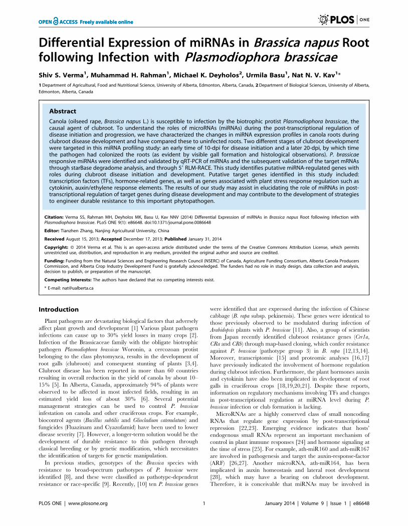
Phenotypic Changes in Roots with Response to P.brassicae
At the earlier time point of 10 day post inoculation (dpi),
compared to the uninoculated controls (Figure 1A), no obvious
swelling of the pathogen treated roots was observed (Figure 1B).
However, at 20 dpi, the inoculated roots exhibited swelling
(Figure 1D, arrows), which is a typical characteristic of the
clubroot infection. The uninfected roots of the corresponding
stage, on the other hand, exhibited typical long and fibrous root
system (Figure 1C). Although there was no visible symptom
development at 10 dpi, microscopic investigation showed the
colonization of the roots by pathogen (Figure 1F). While
uninoculated roots were elongated and highly vacuolated
(Figure 1E), the inoculated roots exhibited the presence of
evacuated zoosporangia (Figure 1F, open arrow) similar to the
observations with turnip at day 12 (Asano et al. 1999), as well as
primary plasmodium with zoospores (Figure 1F, closed arrows).
Therefore, despite the absence of visible morphological symptoms
of clubroot infection at 10 dpi, histological observations indicated
that the Plasmodiophora infection and colonization has indeed begun
at this time point. At 20 dpi, the cortical cells showed the evidence
of hypertrophy, along with the presence of secondary plasmodium
(Figure 1H, arrowheads) compared to elongated, vacuolated cells
of the uninfected control (Figure 1G). The increased hypertrophy
in clubroot infected Arabidopsis root cells has been reported to be
responsible for gall formation [33].
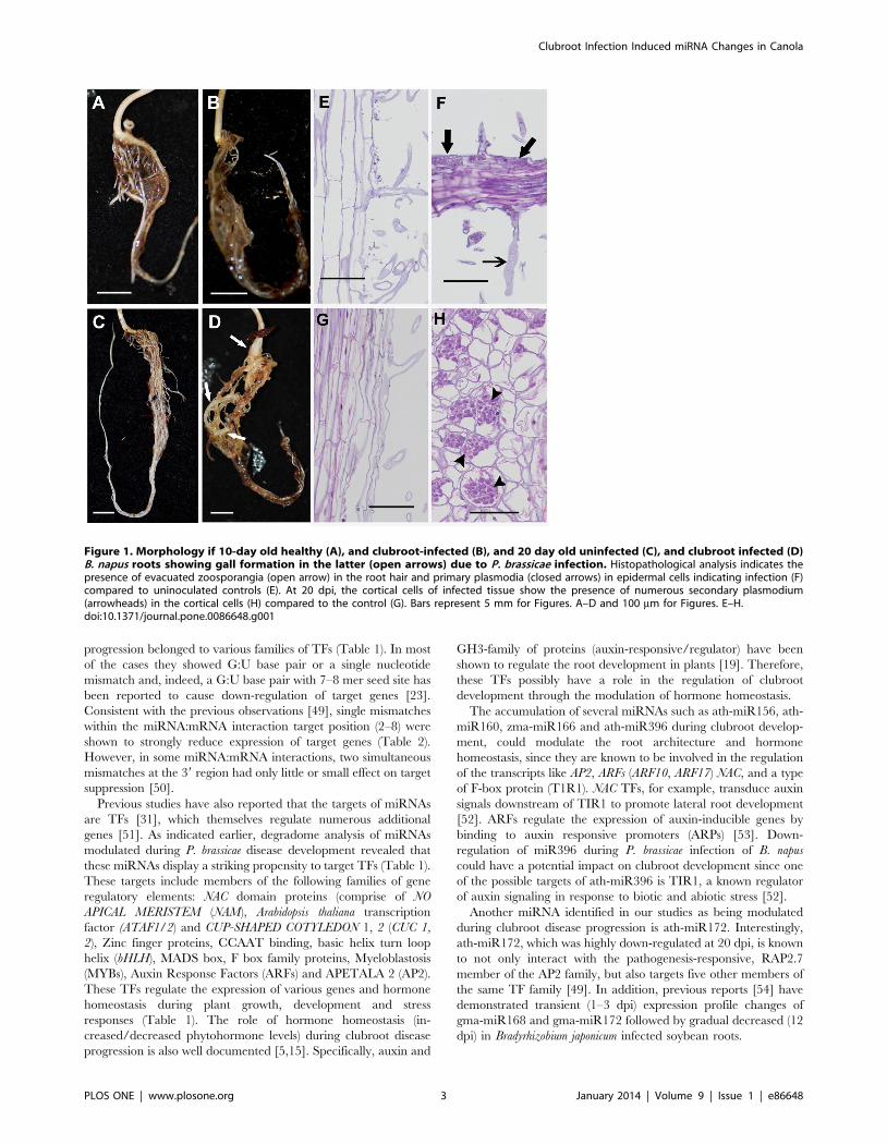
P. brassicae Responsive miRNAThe differential expression of B. napus miRNAs following
inoculation with the biotrophic protist P. brassicae was compared to
a mock sample (untreated) at two time points (10 and 20 dpi) of
clubroot disease development. In the miRNA based microarray,
we observed that ten miRNAs were differentially expressed at the
earlier time point (10 dpi; Figure 2A) and 34 miRNA showed
differential expression at the later time point (20 dpi; Figure 2B, C)
in response to P. brassicae infection. Among those miRNAs that
were modulated at 10 dpi, most of them (bdi-miR156, ath-
miR156h, ath-miR824 and peu-miR2916) showed an increase in
abundance, while the level of mtr-miR169f decreased (Figure 2A).
On the other hand, at 20 dpi, 21 miRNAs were increased in
abundance, whereas 13 miRNAs exhibited decreased abundance
(Figure 2B, C). Interestingly, the miRNAs ahy-miR156b-3p and
zma-miR166n increased at 10 dpi, but decreased in abundance at
20 dpi; whereas levels of ath-miR854a and cre-miR909.1
decreased at 10 dpi and increased at 20 dpi (Figure 2, S1, S2).
Among the differentially expressed P. brassicae miRNAs, ath-
miR156 increased during the early time point (10 dpi) and
decreased at the later stage (20 dpi). This miRNA has been
previously reported to be involved in hormone homeostasis
(abscisic acid signaling, gibberellin response), in mediating
responses to abiotic stresses [34,35] and is also induced by Turnip
Mosaic Virus (TuMV) infection in transgenic Arabidopsis plants
[36,37]. The zma-miR166, which is also increased in abundance
at 10 dpi (Figure 2A), has previously been shown to be involved in
the regulation of class-III homeodomain leucine zipper (HD-ZIP
III) genes [38]. The HD-ZIP III TFs have also been implicated in
lateral root development of A. thaliana [39]. The expression of zma-
miR166n increased at 10 dpi and decreased at 20 dpi.
Interestingly, the over-expression of mtr-miR166 has been
previously shown to reduce the number of symbiotic nodules
and lateral roots in Medicago truncatula [40]. Our results suggest that
the miRNAs, which have been implicated in root development
and/or in mediating plant responses to pathogens, are modulated
during clubroot development.
Interestingly, we identified eight miR169s, all of which showed
increased expression due to infection of B. napus with P. brassicae
(Fig. 2b, c) at 20 dpi. However, alignment of these mature miRNA
sequences exhibited at least 18 of the 21 bases being conserved,
with only between one and three bases at the termini being
dissimilar (data not shown), indicating their widespread and highly
conserved nature among plant species. The conservation of
miRNA sequences between different plant species, with variations
of one or a few base pairs, has previously been reported [25]. The
expression of ath-miR169 was found to be increase at 20 dpi. A
similar increase in abundance of the ath-miR169 has been
reported following pathogen attack in the Arabidopsis [24] as well as
in response to osmotic stress in rice [31,41]. However, the
abundance of ath-miR172 decreased at 20 dpi following P.
brassicae infection and was not detectable at 10 dpi. It has been
previously reported that the abundance of ath-miR172 was
decreased due to UV-B radiation in Arabidopsis [42], and in
response to viral infection in tomato [30]. The levels of aqc, osa,
ppt-miR160 increased at 20 dpi and were not detectable at 10 dpi.
These miRNAs have been shown to be increased in pine stems
with response to the rust fungus [43] but increased in Arabidopsis
due to bacterial infection [44]. Increased expression of ath-
miR160 in response to hypoxia in Arabidopsis [45] and maize [46]
possibly indicates the involvement of the miRNAs in both abiotic
and biotic stress response.
Our observations also revealed that ath-miR396 was differen-
tially expressed during the disease progression and decreased at 20
dpi (Figure 2C). The expression of tae-miR396 has been reported
to be decreased in wheat (Triticum aestivum) following powdery
mildew infection [47]. Interestingly, the expression of cre-miR909
decreased at 10 dpi, while at the later stage of disease progression
(20 dpi), it was increased. The role of cre-miR909 in relation to its
involvement in stress response has not been reported yet. In the
current study, miRNA microarray analysis led to the identification
of several pathogen (P. brassicae) responsive miRNAs in B. napus.
Interestingly, most of these miRNAs were decreased in abun-
dance, which suggests that expression of their target genes could
be enhanced in response to P. brassicae infection.
miRNA-mRNA Target PredictionPotential miRNA targets were identified using degradome
analysis [48]. In our study, many of the predicted target genes of
miRNAs that were differentially expressed during clubroot disease
Clubroot Infection Induced miRNA Changes in Canola
PLOS ONE | www.plosone.org 2 January 2014 | Volume 9 | Issue 1 | e86648
progression belonged to various families of TFs (Table 1). In most
of the cases they showed G:U base pair or a single nucleotide
mismatch and, indeed, a G:U base pair with 7–8 mer seed site has
been reported to cause down-regulation of target genes [23].
Consistent with the previous observations [49], single mismatches
within the miRNA:mRNA interaction target position (2–8) were
shown to strongly reduce expression of target genes (Table 2).
However, in some miRNA:mRNA interactions, two simultaneous
mismatches at the 39 region had only little or small effect on target
suppression [50].
Previous studies have also reported that the targets of miRNAs
are TFs [31], which themselves regulate numerous additional
genes [51]. As indicated earlier, degradome analysis of miRNAs
modulated during P. brassicae disease development revealed that
these miRNAs display a striking propensity to target TFs (Table 1).
These targets include members of the following families of gene
regulatory elements: NAC domain proteins (comprise of NO
APICAL MERISTEM (NAM), Arabidopsis thaliana transcription
factor (ATAF1/2) and CUP-SHAPED COTYLEDON 1, 2 (CUC 1,
2), Zinc finger proteins, CCAAT binding, basic helix turn loop
helix (bHLH), MADS box, F box family proteins, Myeloblastosis
(MYBs), Auxin Response Factors (ARFs) and APETALA 2 (AP2).
These TFs regulate the expression of various genes and hormone
homeostasis during plant growth, development and stress
responses (Table 1). The role of hormone homeostasis (in-
creased/decreased phytohormone levels) during clubroot disease
progression is also well documented [5,15]. Specifically, auxin and
GH3-family of proteins (auxin-responsive/regulator) have been
shown to regulate the root development in plants [19]. Therefore,
these TFs possibly have a role in the regulation of clubroot
development through the modulation of hormone homeostasis.
The accumulation of several miRNAs such as ath-miR156, ath-
miR160, zma-miR166 and ath-miR396 during clubroot develop-
ment, could modulate the root architecture and hormone
homeostasis, since they are known to be involved in the regulation
of the transcripts like AP2, ARFs (ARF10, ARF17) NAC, and a type
of F-box protein (T1R1). NAC TFs, for example, transduce auxin
signals downstream of TIR1 to promote lateral root development
[52]. ARFs regulate the expression of auxin-inducible genes by
binding to auxin responsive promoters (ARPs) [53]. Down-
regulation of miR396 during P. brassicae infection of B. napus
could have a potential impact on clubroot development since one
of the possible targets of ath-miR396 is TIR1, a known regulator
of auxin signaling in response to biotic and abiotic stress [52].
Another miRNA identified in our studies as being modulated
during clubroot disease progression is ath-miR172. Interestingly,
ath-miR172, which was highly down-regulated at 20 dpi, is known
to not only interact with the pathogenesis-responsive, RAP2.7
member of the AP2 family, but also targets five other members of
the same TF family [49]. In addition, previous reports [54] have
demonstrated transient (1–3 dpi) expression profile changes of
gma-miR168 and gma-miR172 followed by gradual decreased (12
dpi) in Bradyrhizobium japonicum infected soybean roots.
Figure 1. Morphology if 10-day old healthy (A), and clubroot-infected (B), and 20 day old uninfected (C), and clubroot infected (D)B. napus roots showing gall formation in the latter (open arrows) due to P. brassicae infection. Histopathological analysis indicates thepresence of evacuated zoosporangia (open arrow) in the root hair and primary plasmodia (closed arrows) in epidermal cells indicating infection (F)compared to uninoculated controls (E). At 20 dpi, the cortical cells of infected tissue show the presence of numerous secondary plasmodium(arrowheads) in the cortical cells (H) compared to the control (G). Bars represent 5 mm for Figures. A–D and 100 mm for Figures. E–H.doi:10.1371/journal.pone.0086648.g001
Clubroot Infection Induced miRNA Changes in Canola
PLOS ONE | www.plosone.org 3 January 2014 | Volume 9 | Issue 1 | e86648
Furthermore, the downregulation of ath-miR169 a/b/c was
reported [55] in wild-type Arabidopsis due to drought stress, while
transgenic Arabidopsis plants overexpressing ath-miR169a showed
enhanced leaf water loss and susceptibility to drought. In contrast,
the miR169 family was increased in rice (Oryza sativa) during
drought stress [56], and the overexpression of miR169 (sly-
miR169c) in transgenic tomato led to decreased transpiration rate
and enhanced drought tolerance [57]. ath-miR169 is known to
target mRNAs of genes that encode members of CCAAT binding
TF, as well as allowing the expression of Nuclear Factor Y (NFY)
[58], which has important implications in stress responses [55].
The abundance of zma-miR166 and aqc-miR160 was observed
to increase at 20 dpi (Table 2b, c). ath-miR160 is known to post-
transcriptionally regulate TFs involved in lateral root development
in Arabidopsis [59]. Our results are further supported by the small
RNA-expression profiling of Arabidopsis leaves collected at 1 and 3
(dpi) with Pseudomonas syringae [44], which identified ath-miR160 as
being highly induced. In addition to its role in lateral root
development, ath-miR160 is also involved in the regulation of the
TFs involved in auxin response signaling and targets ARFs:
ARF10, ARF16, and ARF17, which are involved in root
development in A. thaliana [60]. It is known that ARF8 and
ARF17 regulate the transcription level of GH3-like genes, which in
turn, regulates auxin level in plants [61,62]. Free auxins or their
conjugates have been shown to play crucial roles during clubroot
disease development in B. napus [19]. Furthermore, it has been
demonstrated that A. thaliana ARFs, ARF10 and ARF16, targeted
by ath-miR160, control root cap cell formation [53]. ARF mutant
lines and ath-mir160-overexpressing lines showed the root tip
defect with uncontrolled cell division and blocked cell differenti-
ation in the root distal region. This resulted in a tumor-like root
apex and loss of gravity sensing [53], and resembles clubroot
disease phenotype of canola. Similarly, ath-miR166, which post-
transcriptionally regulates HD-ZIP III genes, is also involved in
lateral root development in Arabidopsis [39]. In-situ expression
analysis has revealed that these target genes are spatially co-
expressed with mtr-miR166 in vascular bundle and in the apical
region of roots [40]. The over expression of mtr-miR166 has been
Figure 2. miRNA-microarray expression of P. brassicae responsive miRNAs exhibiting differential expression at 10- (A) and 20- dpi(B, C) following pathogen infection.doi:10.1371/journal.pone.0086648.g002
Clubroot Infection Induced miRNA Changes in Canola
PLOS ONE | www.plosone.org 4 January 2014 | Volume 9 | Issue 1 | e86648
shown to reduce the number of symbiotic nodules and lateral root
development in Medicago truncatula [40]. In addition, ath-miR164
targets the TF NAC domain containing proteins (NAC/ATAF/
CUC1), which regulates auxin signaling during lateral root
development, and is also up-regulated during the P. brassicae
disease progression and development in A. thaliana [28]. These
results provide additional evidence for the important roles played
by ath-miR160 and mtr-miR166 during root development and
clubroot disease progression.
Moreover, the expression of many TFs was observed to be
modulated in response to P. brassicae inoculation. These TFs
belong to several major families, including ARF, AP2, MYB,
Basic-Helix-Loop-Helix, homeobox, and zinc-finger family pro-
teins. Also, it has been reported earlier that some of these TFs are
involved in hormone homeostasis. For example, the modulation of
cytokinin [15] and auxin [62] during clubroot development has
previously been reported. As well, the involvement of auxins in the
development of clubroot disease is also well documented [20,21].
Furthermore, consistent with our microarray results carried out on
Brassica napus infected with clubroot disease, transgenic Arabidopsis
plants with lower cytokinin levels were found to be more tolerant
to clubroot [15]. All of these TFs, therefore, may conceivably have
a bearing on the development of clubroot disease in canola.
Additional work to confirm these hypotheses through silencing of
the target genes and determining their effect on the clubroot
initiation and progression to establish what role they may play in
pathogenesis in B. napus is currently underway in our laboratory.
Relative Quantification of miRNA using qRT-PCRStem-loop qRT-PCR is a reliable and established method of
detection and measurement of expression level of miRNAs. The
stem-loop primer increases the sensitivity of reaction such that this
method can significantly distinguish between two miRNAs with
only a single nucleotide change [63,64]. We used stem–loop RT
followed by TaqMan (Applied Biosystems, USA) PCR analysis to
validate and measure the expression of a selected sub-set of ten
differentially expressed miRNAs from the ones identified in the
miRNA microarray analysis as indicated previously. Endogenous
controls, snoR66 was used as references at 10 dpi and 20 dpi,
along with non-template controls in each set of experiments.
qRT-PCR results indicated that at 10 dpi, the expression of four
out of ten miRNAs (ahy-miR156b-3p, aqc-miR159, mtr-miR169f
and ppt-miR896) increased (Figure 3A), while the expression of
aqc-miR160, ath-miR160a and osa-miR160e decreased
(Figure 3A). At 20 dpi, two miRNAs (ahy-miR156b-3p and mtr-
miR169f) exhibited increased expression (Figure 3B), while five
miRNAs (aqc-miR160a, ath-miR160a, ath-miR172, osa-miR160e
and ppt-miR896) showed decreased expression (Figure 3C).
Although some of the miRNAs showed similar expression
patterns in both microarray and qRT-PCR experiments, this was
not true in all cases. Similar non-correlation between microarray
and qRT-PCR has previously been reported [65]. Indeed, a
systematic analysis of different platforms of miRNA expression
concluded that although the intra-platform correlation was very
high, the same did not apply for inter-platform comparisons [66].
Genome Wide Mapping of mRNA Target Cleavage SitemiRNA:mRNA interaction was analyzed through 59-RLM
RACE of ten selected sets to elucidate putative targets. 59-RLM
RACE generated products between 200 to 1000 bp and the major
PCR products of predicted size resulting from miRNA-guided
cleavage event were determined through mapping (Figure 4).
These products were cloned, and subsequently sequenced. The
results of 59RLM-RACE revealed that out of 10 miRNA:mRNA
interactions, six showed G:U base pairs or mismatches with no
consistency in the position of the seed site. It has been previously
reported that single G:U base-pair mismatch diminish the
expression of the target genes [50]. The second significant feature
here is that most of the mismatches observed in our case involved a
single base and at a position between 2–8, and any mismatches
between positions 2–8 has previously been reported to strongly
reduce the expression of target genes [50]. Mostly the 59 ends of
target genes terminated at a position corresponding to the middle
of the region of complementarity with respective to their miRNA
and directed the cleavage (Figure 4A). This is not surprising given
the fact that the aforementioned feature is the characteristic of
RISC-catalyzed cleavage events [67]. The sequence analyses of
the 59-RLM RACE products thus indicate the potential mRNA
targets and the miRNA-mRNA interaction sites. The results of 59
RLM-RACE (Figure 4B) were also in agreement with the findings
of degradome analysis, where most of the tested miRNAs
indicated TFs as being the major targets. For example, ath-
miR157 binds to the mRNA of Squamosa binding like protein
(SPL15) regulating plant growth and development along with
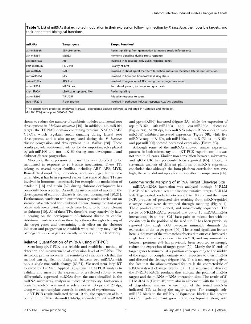
Table 1. List of miRNAs that exhibited modulation in their expression following infection by P. brassicae, their possible targets, andtheir annotated biological functions.
miRNAs Target gene Target Function*
ath-miR156h SBP-Like genes Auxin signalling; from germination to mature seeds; inflorescence
ath-miR159 MYB33 hormone signalling during stress response
aqc-miR160a ARF Involved in regulating early auxin response genes
zma-miR166n HD-ZIPIII Polarity of Leaf
osa-miR164c NAC Involved in shoot apical meristem formation and auxin-mediated lateral root formation.
mtr-miR169d NFY Involved in hormone homeostasis during stress
vvi-miR172a AP2 like Involved in regulation of TFs during the pathogen response
ath-miR824 MADS box Root development, trichome and guard cells
cre-miR909 LEA/Auxin repressed like Auxin signalling
ath-miR396 TIR1/GRF Adaptive response to stress
peu-miR2916 F-box protein Involved in pathogen induced response; Aux/IAA signalling
*The targets were predicted employing starBase - degradome analysis software as indicated in ‘‘Materials and Methods’’.doi:10.1371/journal.pone.0086648.t001
Clubroot Infection Induced miRNA Changes in Canola
PLOS ONE | www.plosone.org 5 January 2014 | Volume 9 | Issue 1 | e86648
abiotic and biotic stress signaling [68]. Similarly, ath-miR824 is
involved in the regulation of mRNA of MADS-box protein
(AGL16-II) while ath-miR172 targets the mRNA of TOE2, which
belongs to pathogenesis related AP2 like ethylene responsive
factors. Similarly, ath-miR159 binds to the mRNA of MYB101
and ath-miR160 post-transcriptionally regulate the expression of
ARF17. ARF17 is known to be involved in adventitious root
development [62] and auxin mediated signaling pathways in
plants [26]. On the other hand, ath-miR162 targets the A. thaliana
endoribonuclease such as Dicer Like Protein 1 (DCL1) [69], which
is a RNA helicase involved in the miRNA processing, having
important implications in plant growth and development [70].
The current results of miRNA mapping and cleavage site
determination through 59RLM RACE suggest that DCL1, MYB,
AP2, MADS-box, ARF and TOE are major target gene for the
miRNAs (miR162, miR160, miR157, miR824, miR396, miR854
and miR172.) involved in the disease modulation and progression
of clubroot.
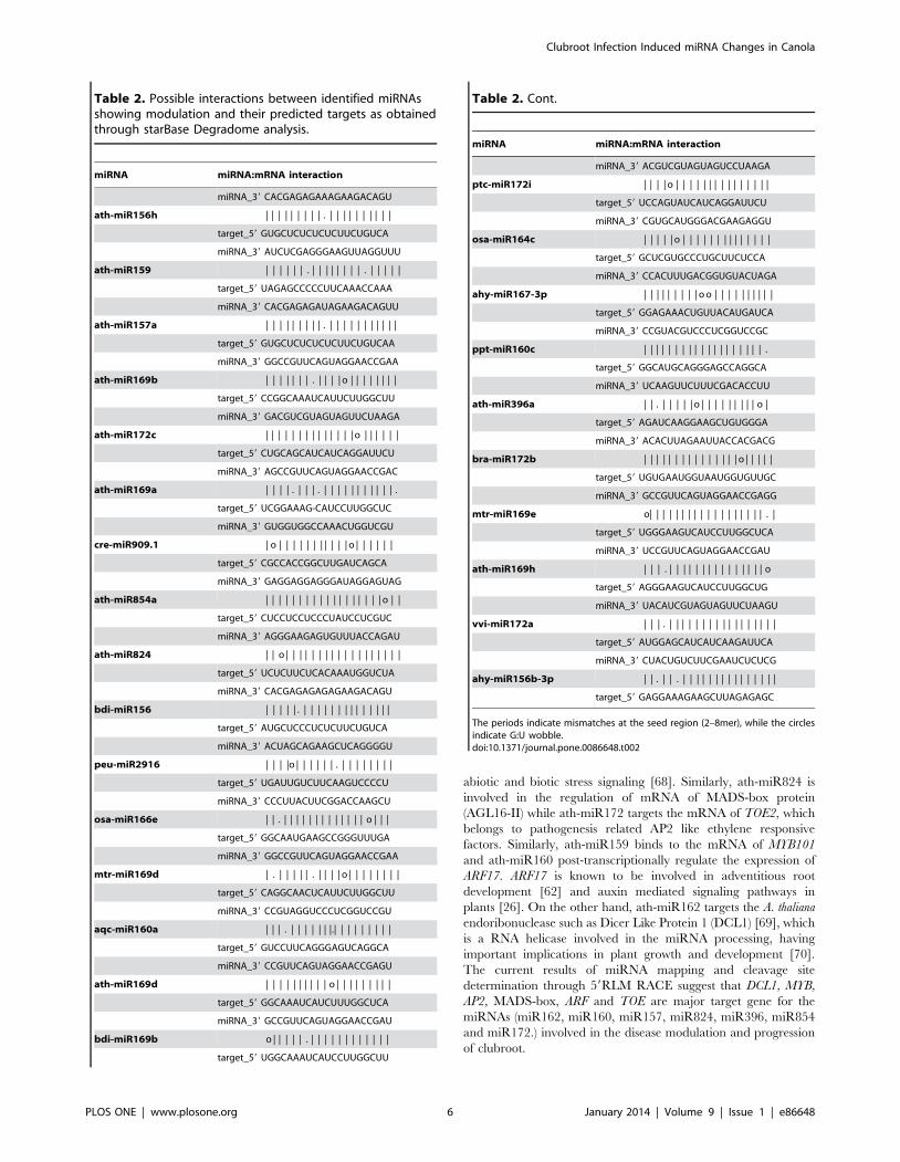
Table 2. Possible interactions between identified miRNAsshowing modulation and their predicted targets as obtainedthrough starBase Degradome analysis.
miRNA miRNA:mRNA interaction
miRNA_39 CACGAGAGAAAGAAGACAGU
ath-miR156h | | | | | | | | | . | | | | | | | | | |
target_59 GUGCUCUCUCUCUUCUGUCA
miRNA_39 AUCUCGAGGGAAGUUAGGUUU
ath-miR159 | | | | | | . | | | | | | | | . | | | | |
target_59 UAGAGCCCCCUUCAAACCAAA
miRNA_39 CACGAGAGAUAGAAGACAGUU
ath-miR157a | | | | | | | | | . | | | | | | | | | | |
target_59 GUGCUCUCUCUCUUCUGUCAA
miRNA_39 GGCCGUUCAGUAGGAACCGAA
ath-miR169b | | | | | | | . | | | | o | | | | | | | |
target_59 CCGGCAAAUCAUUCUUGGCUU
miRNA_39 GACGUCGUAGUAGUUCUAAGA
ath-miR172c | | | | | | | | | | | | | | o | | | | | |
target_59 CUGCAGCAUCAUCAGGAUUCU
miRNA_39 AGCCGUUCAGUAGGAACCGAC
ath-miR169a | | | | . | | | . | | | | | | | | | | | .
target_59 UCGGAAAG-CAUCCUUGGCUC
miRNA_39 GUGGUGGCCAAACUGGUCGU
cre-miR909.1 | o | | | | | | | | | | | o | | | | | |
target_59 CGCCACCGGCUUGAUCAGCA
miRNA_39 GAGGAGGAGGGAUAGGAGUAG
ath-miR854a | | | | | | | | | | | | | | | | | | o | |
target_59 CUCCUCCUCCCUAUCCUCGUC
miRNA_39 AGGGAAGAGUGUUUACCAGAU
ath-miR824 | | o | | | | | | | | | | | | | | | | | |
target_59 UCUCUUCUCACAAAUGGUCUA
miRNA_39 CACGAGAGAGAGAAGACAGU
bdi-miR156 | | | | | . | | | | | | | | | | | | | |
target_59 AUGCUCCCUCUCUUCUGUCA
miRNA_39 ACUAGCAGAAGCUCAGGGGU
peu-miR2916 | | | |o | | | | | | . | | | | | | | |
target_59 UGAUUGUCUUCAAGUCCCCU
miRNA_39 CCCUUACUUCGGACCAAGCU
osa-miR166e | | . | | | | | | | | | | | | | o | | |
target_59 GGCAAUGAAGCCGGGUUUGA
miRNA_39 GGCCGUUCAGUAGGAACCGAA
mtr-miR169d | . | | | | | . | | | | o | | | | | | | |
target_59 CAGGCAACUCAUUCUUGGCUU
miRNA_39 CCGUAGGUCCCUCGGUCCGU
aqc-miR160a | | | . | | | | | | |.| | | | | | | | |
target_59 GUCCUUCAGGGAGUCAGGCA
miRNA_39 CCGUUCAGUAGGAACCGAGU
ath-miR169d | | | | | | | | | | o | | | | | | | | |
target_59 GGCAAAUCAUCUUUGGCUCA
miRNA_39 GCCGUUCAGUAGGAACCGAU
bdi-miR169b o | | | | | . | | | | | | | | | | | |
target_59 UGGCAAAUCAUCCUUGGCUU
Table 2. Cont.
miRNA miRNA:mRNA interaction
miRNA_39 ACGUCGUAGUAGUCCUAAGA
ptc-miR172i | | | | o | | | | | | | | | | | | | | |
target_59 UCCAGUAUCAUCAGGAUUCU
miRNA_39 CGUGCAUGGGACGAAGAGGU
osa-miR164c | | | | | o | | | | | | | | | | | | | |
target_59 GCUCGUGCCCUGCUUCUCCA
miRNA_39 CCACUUUGACGGUGUACUAGA
ahy-miR167-3p | | | | | | | | | o o | | | | | | | | | |
target_59 GGAGAAACUGUUACAUGAUCA
miRNA_39 CCGUACGUCCCUCGGUCCGC
ppt-miR160c | | | | | | | | | | | | | | | | | | | .
target_59 GGCAUGCAGGGAGCCAGGCA
miRNA_39 UCAAGUUCUUUCGACACCUU
ath-miR396a | | . | | | | | o | | | | | | | | | o |
target_59 AGAUCAAGGAAGCUGUGGGA
miRNA_39 ACACUUAGAAUUACCACGACG
bra-miR172b | | | | | | | | | | | | | | | o | | | | |
target_59 UGUGAAUGGUAAUGGUGUUGC
miRNA_39 GCCGUUCAGUAGGAACCGAGG
mtr-miR169e o| | | | | | | | | | | | | | | | | | . |
target_59 UGGGAAGUCAUCCUUGGCUCA
miRNA_39 UCCGUUCAGUAGGAACCGAU
ath-miR169h | | | . | | | | | | | | | | | | | | | o
target_59 AGGGAAGUCAUCCUUGGCUG
miRNA_39 UACAUCGUAGUAGUUCUAAGU
vvi-miR172a | | | . | | | | | | | | | | | | | | | | |
target_59 AUGGAGCAUCAUCAAGAUUCA
miRNA_39 CUACUGUCUUCGAAUCUCUCG
ahy-miR156b-3p | | . | | . | | | | | | | | | | | | | | |
target_59 GAGGAAAGAAGCUUAGAGAGC
The periods indicate mismatches at the seed region (2–8mer), while the circlesindicate G:U wobble.doi:10.1371/journal.pone.0086648.t002
Clubroot Infection Induced miRNA Changes in Canola
PLOS ONE | www.plosone.org 6 January 2014 | Volume 9 | Issue 1 | e86648
Conclusion
Clubroot disease of Brassicaceae family has potential to substan-
tially limit canola production. A miRNA microarray-based
approach was used to identify miRNA expression and regulation
during the disease initiation and progression. Results from this
study provide a useful basis towards understanding the dynamics
of miRNAs related to P. brassicae infection of canola. The
differential expression analysis of miRNA during pathogenesis of
P. brassicae was carried out at two developmentally distinct time
points. The expression of various miRNAs was observed to be
modulated during the process of clubroot initiation and develop-
ment. Many of these miRNAs were involved in the regulation of
gene activity by targeting, among others, TFs, ARFs, MYB, MADS
box, AP2 and ERFs. Based on available literature, many of these
are known to be involved in stress-responses and developmental
events related to pathogenesis.
The results documented here lead us to conclude that a diverse
array of miRNAs and their target genes are modulated during the
pathogenesis of canola by P. brassicae. The current study enhances
our understanding related to the biological processes involved in P.
brassicae-canola interactions and may lead to unique ways to
generate disease resistant phenotypes.
Materials and Methods
Plant Growth and PathogenSeeds of Brassica napus were germinated on moist filter paper in
petri dishes and placed under a light/dark (16 h/8 h) regime for 7
days at 2261uC. Clubroot gall pathotype SACAN03-1 (St. Albert,
Canada type 03-1), was isolated from infected tissues stored at
220uC (collected from Southern Alberta region), [71]. Resting
spores were extracted by homogenizing mature clubroot galls of
Chinese cabbage, followed by filtration through six layers of
cheesecloth and two subsequent centrifugation steps (2,5006g) for
10 min [72]. The resting spores of P. brassicae were resuspended in
autoclaved, deionized water and the number of spores in the
suspension was counted using a haemocytometer. One week-old
seedlings were individually dipped into the spore suspensions
(16107 spores/mL) for 1–2 seconds and planted in flats (363 cm,
one seed per insert) containing LA4 Aggregate Plus (Sunshine
Professional Peat-Lite Mix; Sungro Horticulture, Vancouver, BC,
Canada). Plants were placed in a growth chamber with an 18 h
photoperiod (light intensity of 130 mmol m22 s21) with a day
(2161uC)/night (1861uC) temperature cycle for 1 week, with a
water tray underneath.
Figure 3. Relative abundance of miRNA in B. napus plant infected with P. brassicae. (a) Relative accumulation of miRNA showing thequantitative expression at 10 dpi and (b) Relative accumulation of miRNA showing the quantitative expression at 20 dpi. The expression of sonR66was used as internal control in the experiment. The error bars show the standard deviation.doi:10.1371/journal.pone.0086648.g003
Clubroot Infection Induced miRNA Changes in Canola
PLOS ONE | www.plosone.org 7 January 2014 | Volume 9 | Issue 1 | e86648

Figure 4. miRNA mapping and cleavage site determination through 59 RLM RACE. Agarose gel image of 59 RACE products (A) and thetarget mRNA cleavage sites (B). The targeted mRNA section and miRNA sequences, along with mismatch (es), if any, are shown as the expandedregion. The 59ends of the cleaved product determined by sequencing is indicated by the vertical arrowheads, along with the numbers of clonesanalyzed. The horizontal arrowheads indicated the gene-specific primer sites used for 59RLM-RACE.doi:10.1371/journal.pone.0086648.g004
Clubroot Infection Induced miRNA Changes in Canola
PLOS ONE | www.plosone.org 8 January 2014 | Volume 9 | Issue 1 | e86648
Sampling and RNA IsolationOne-week old seedlings of B. napus cv. Westar were divided into
two groups; one was inoculated with P. brassicae while the other,
uninoculated group, served as controls in the experiment. Tissues
were harvested at 10 and 20-days-post inoculation (dpi) for RNA
isolation. Plant roots from these groups were pooled separately and
total RNA was isolated using the TRI-Reagent (Ambion, USA)
and used in microarray experiments.
Microscopy of B. napus Infected with P. brassicae at TwoTime Points
Pathogen-inoculated roots of B. napus cv Westar, as well as
uninoculated controls, were cut into small (10–15 mm) segments
and fixed in FAA (formalin, acetic acid and ethyl alcohol, [73]
under vacuum at room temperature overnight. Following fixation,
the root segments were dehydrated in a series of graded ethanol/
water solutions, changed to toluene and later infiltrated with
ParaplastH using a Leica TP1020 Tissue Processor. Longitudinal
sections (6 mm thickness) were prepared using an AO Rotary
microtome, affixed to the glass slides, de-paraffinated with toluene,
rehydrated through a graded ethanol series and stained with
Harris Hematoxylin and counterstained with Eosin Y [74]. Slides
were subsequently dehydrated in ethanol followed by toluene and
mounted with DPXH mounting medium (Sigma Aldrich, USA).
The sections were viewed with a Zeiss Axioskop, analyzed using
AxioVisionTM software and photographed with Zeiss Axiocam.
mParafloTM miRNA MicroarrayThe miRNA microarray experiment was performed at LC
Sciences, Houston, Texas (http://www.lcsciences.com). Chip
hybridization experiments were carried out in duplicate using
total RNA samples (2 to 5 mg) which were size fractionated using a
YM-100 Microcon centrifugal filter (Millipore, USA). The small
RNAs were polyadenylated (39) using poly (A) polymerase (Applied
Biosystems, USA). An oligonucleotide tag (Cy3 or Cy5, for
individual treatments) was then ligated to the polyadenylated small
RNA for fluorescent dye staining. Hybridization was performed
overnight on mParaflo microfluidic chips (MRA-1038B) using a
micro-circulation pump (Atactic Technologies, USA) where each
detection probe consisted of a chemically modified nucleotide-
coding segment complementary to known target plant microRNA
from PMRD (Plant microRNA Database; (www.bioinformatics.
cau.edu.cn/PMRD) and miRdata base 15 was used. This array
includes over 5600 probes from experimentally as well as
computationally identified plant microRNAs mainly from Arabi-
dopsis thaliana, Brassica oleracea, B. rapa and B. napus, Oryza sativa,
Populus trichocarpa, Physcomitrella patens, Glycine max, Zea mays, Vitis
vinifera and Triticum aestivum. Blank cells and other non-homologous
nucleic acid probes served as negative controls. Hybridization was
performed overnight on a single-channel mParaFlo microfluidic
chip at 34uC as described by [32]. Data were analyzed by
subtracting the background and normalizing the signals using a
LOWESS filter (locally weighted regression). The ratio of the two
sets of detected signals (log2) and P-values were calculated through
t-test. Differentially detected signals are reported for those with P-
values ,0.05. A transcript was considered as detectable when the
signal intensity was higher than 36the background standard
deviation and the spot CV was ,0.5, where CV was calculated as
standard deviation/signal intensity. The miRNA-microarray data
have been submitted to public repository, Gene Expression
Omnibus/NCBI database (accession number-GSE51590).
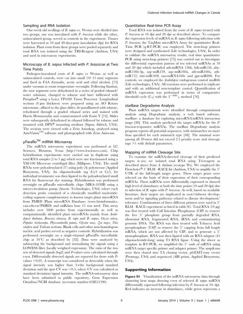
Quantitative Real-time PCR AssayTotal RNA was isolated from the roots of B. napus treated with
P. brassicae at 10 dpi and 20 dpi as described above. To compare
the expression levels of miRNA in B. napus following infection with
P. brassicae, the TaqMan microRNA Assay for quantitative Real-
Time PCR (q-RT-PCR) was employed. The stem-loop primers
were designed and synthesized (Life technologies, USA). In order
to validate the miRNA microarray results, real time quantitative
PCR using stem-loop primers [75] was carried out to investigate
the differential expression pattern of ten selected miRNAs at 10
and 20 dpi which included ath-miRNA 156h, bdi-miR156, ahy-
miR156b-3p, aqc-miR159, aqc-miR160a, ath-miR160a, ath-
miR172, mtr-miR169f, osa-miRNA160e and ppt-miR896. For
controls, we employed the Arabidopsis endogenous control snoR66
(Life technologies, USA). All reactions were performed in triplicate
and with an additional non-template control. Quantification of
miRNA expression was performed in terms of comparative
threshold cycle (CT) with the 22DDCT method [76].
starBase Degradome AnalysisPlant miRNA targets were identified through computational
analysis using Degradome analysis, a web based software,
starBase: a database for exploring microRNA:mRNA interaction
maps [48]. This analysis predicted the target mRNAs for the P.
brassicae-responsive miRNAs. The starBase Degradome analysis
program reports all potential sequences, with mismatches no more
than specified for each mismatch type [48]. The minimal score
among all 20-mers did not exceed 2.5 penalty score and cleavage
tags .1 with default parameters.
Mapping of mRNA Cleavage SiteTo examine the miRNA-directed cleavage of their predicted
targets in vivo, we isolated total RNA using Tri-reagent as
described above from A. thaliana (ecotype Ws). We employed the
FirstchoiceH 59 RLM –RACE kit (Ambion, USA) to amplify the 59
UTR of the full-length target genes. These target genes were
selected on the basis of their expression of their corresponding
miRNAs. These miRNAs were differentially expressed or showed
high level of abundance at both the time points (10 and 20 dpi) due
to infection of B. napus with P. brassicae. As well, based on available
literature, their targets are implicated in root/clubroot develop-
ment and/or signaling pathways related to disease development/
tolerance. Combinations of three different primers were sued in 59
RLM –RACE experiment as listed in table-S1. Total RNA (10 mg)
was first treated with Calf Intestine Phosphatase (CIP) to remove
the free 59 phosphate group from partially degraded RNA,
ribosomal RNA, fragmented RNA, tRNA and contaminating
genomic DNA. The RNA was then treated with Tobacco Acid
pyrophosphate (TAP) to remove the 59 capping from full length
mRNA, which are not affected by CIP, and to generate a 59
monophosphate. RNA was then ligated with an RNA adaptor (45
oligonucleotide-long) using T4 RNA ligase. Using the above as
template in RT-PCR, we amplified the 59 ends of mRNA using
miRNA target specific primer and adapter primer. The ampilcons
were then cloned into TA cloning vector, pGEMT-easy vector
(Promega, USA) and sequenced (ABI prism, Applied Biosystems,
USA).
Supporting Information
Figure S1 Visualization of the miRNA microarray data through
clustering heat maps showing t-test of selected B. napus miRNA
differentially expressed following infection by P. brassicae at 10- dpi.
Red indicates an increase in abundance, while green represents a
Clubroot Infection Induced miRNA Changes in Canola
PLOS ONE | www.plosone.org 9 January 2014 | Volume 9 | Issue 1 | e86648
decrease in abundance of miRNAs at a P value of less than 0.01
(P,0.05).
(TIF)
Figure S2 Visualization of the miRNA microarray data through
clustering heat maps showing t-test of selected B. napus miRNA
differentially expressed following infection by P. brassicae at 20- dpi.
Red indicates an increase in abundance, while green represents a
decrease in abundance of miRNAs at a P value of less than 0.01
(P,0.05).
(TIF)
Table S1 List of primers used in 59 RLM-RACE.
(DOCX)
Acknowledgments
We would like to thank Dr. Stephen Strelkov and Mr. Victor Manoli, Plant
Pathology Lab, Department of Agricultural, Food and Nutritional Science,
University of Alberta, for providing the clubroot pathogen used in this
study, and for help with the inoculations, respectively.
Author Contributions
Conceived and designed the experiments: NNVK SSV. Performed the
experiments: SSV MHR. Analyzed the data: SSV MHR NNVK MKD
UB. Wrote the paper: SSV MHR NNVK.
References
1. Brown JK, Hovmoller MS (2002) Aerial dispersal of pathogens on the global and
continental scales and its impact on plant disease. Science 297: 537–541.
2. Pande S, Siddique KHM, Kishore GK, Bayaa B, Gaur PM, et al. (2005)Ascochyta blight of chickpea(Cicer arietinum L.): a review of biology, pathogenic-
ity, and disease management. Aust J Agri Res 56: 317–332.
3. Strelkov SE, Manolii VP, Cao T, Xue S, Hwang SF (2007) Pathotype
classification of Plasmodiophora brassicae and its occurrence in Brassica napus in
Alberta, Canada. J Phytopathol 155: 706–712.
4. Hwang SF, Cao T, Xiao Q, Ahmed HU, Manolii VP, et al. (2012) Effects of
fungicide, seeding date and seedling age on clubroot severity, seedlingemergence and yield of canola. Can J of Plant Sci 92: 1175–1186.
5. Dixon G (2009) The Occurrence and Economic Impact of Plasmodiophora
brassicae and Clubroot Disease. J of Plant Growth Regul 28: 194–202.
6. Tewari JP, Strelkov SE, Orchard D, Hartman M, Lange RM, et al. (2005)
Identification of clubroot of crucifers on canola (Brassica napus) in Alberta.
Can J Plant Pathol 27: 143–144.
7. Peng SE, McGregor GL, Lahlali R, Gossen BD, Hwang SF, et al. (2011)
Potential biological control of clubroot on canola and crucifer vegetable crops.
Plant Pathol 60: 566–574.
8. Hirai M, Harada T, Kubo N, Tsukada M, Suwabe K, et al. (2004) A novel locus
for clubroot resistance in Brassica rapa and its linkage markers. Theor Appl
Genet 108: 639–643.
9. Diederichsen E, Frauen M, Linders EA, Hatakeyama K, Hirai M (2009) Status
and Perspectives of Clubroot Resistance Breeding in Crucifer Crops. J of Plant
Growth Regul 28: 265–281.
10. Sundelin T, Jensen DF, Lubeck M (2011) Identification of expressed genes
during infection of Chinese cabbage (Brassica rapa subsp. pekinensis) by
Plasmodiophora brassicae. J Eukaryot Microbiol 58: 310–314.
11. Bulman S, Ridgway HJ, Eady C, Conner AJ (2007) Intron-rich gene structure in
the intracellular plant parasite Plasmodiophora brassicae. Protist 158: 423–433.
12. Ueno H, Matsumoto E, Aruga D, Kitagawa S, Matsumura H, et al. (2012)
Molecular characterization of the CRa gene conferring clubroot resistance in
Brassica rapa. Plant Mol Biol 80: 621–629.
13. Kato T, Hatakeyama K, Fukino N, Matsumoto S (2013) Fine mapping of the
clubroot resistance gene CRb and development of a useful selectable marker in
Brassica rapa. Breed Sci 63: 116–124.
14. Hatakeyama K, Suwabe K, Tomita RN, Kato T, Nunome T, et al. (2013)
Identification and characterization of Crr1a, a gene for resistance to clubroot
disease (Plasmodiophora brassicae Woronin) in Brassica rapa L. PLoS ONE 8:
e54745.
15. Siemens J, Keller I, Sarx J, Kunz S, Schuller A, et al. (2006) Transcriptome
analysis of Arabidopsis clubroots indicate a key role for cytokinins in disease
development. Mol Plant Microbe Interact 19: 480–494.
16. Cao T, Srivastava S, Rahman M, Kav N, Hotte N, et al. (2008) Proteome-level
changes in the roots of Brassica napus as a result of Plasmodiophora brassicae
infection. Plant Sci 174: 97–115.
17. Devos S, Laukens K, Deckers P, Van Der Straeten D, Beeckman T, et al. (2006)
A hormone and proteome approach to picturing the initial metabolic events
during Plasmodiophora brassicae infection on Arabidopsis. Mol Plant Microbe
Interact 19: 1431–1443.
18. Siemens J, Graf H, Bulman S, In O, Ludwig-Muller J (2009) Monitoring
expression of selected Plasmodiophora brassicae genes during clubroot development
in Arabidopsis thaliana. Plant Pathol 58: 130–136.
19. Ludwig-Muller J, Julke S, Bierfreund NM, Decker EL, Reski R (2009) Moss
(Physcomitrella patens) GH3 proteins act in auxin homeostasis. New Phytol 181:
323–338.
20. Agarwal A, Kaul V, Faggian R, Rookes JE, Ludwig-Muller J, et al. (2011)
Analysis of global host gene expression during the primary phase of the
Arabidopsis thaliana Plasmodiophora brassicae interaction. Funct Plant Biol 38: 462–
478.
21. Gravot A, Deleu C, Wagner G, Lariagon C, Lugan R, et al. (2012) Arginase
induction represses gall development during clubroot infection in Arabidopsis.
Plant Cell Physiol 53: 901–911.
22. Carrington JC, Ambros V (2003) Role of microRNAs in plant and animaldevelopment. Science 301: 336–338.
23. Bartel DP (2004) MicroRNAs: genomics, biogenesis, mechanism, and function.Cell 116: 281–297.
24. Li Y, Zhang Q, Zhang J, Wu L, Qi Y, et al. (2010) Identification of microRNAsinvolved in pathogen-associated molecular pattern-triggered plant innate
immunity. Plant Physiol 152: 2222–2231.
25. Axtell MJ, Bowman JL (2008) Evolution of plant microRNAs and their targets.
Trends Plant Sci 13: 343–349.
26. Mallory AC, Bartel DP, Bartel B (2005) MicroRNA-directed regulation of
Arabidopsis AUXIN RESPONSE FACTOR17 is essential for proper develop-ment and modulates expression of early auxin response genes. Plant Cell 17:
1360–1375.
27. Bartel B, Bartel DP (2003) MicroRNAs: at the root of plant development? Plant
Physiol 132: 709–717.
28. Guo HS, Xie Q, Fei JF, Chua NH (2005) MicroRNA directs mRNA cleavage of
the transcription factor NAC1 to downregulate auxin signals for Arabidopsis
lateral root development. Plant Cell 17: 1376–1386.
29. Lu C, Souret F (2010) High-throughput approaches for miRNA expressionanalysis. Methods Mol Biol 592: 107–125.
30. Naqvi AR, Haq QM, Mukherjee SK (2010) MicroRNA profiling of tomato leafcurl New Delhi virus (tolcndv) infected tomato leaves indicates that deregulation
of mir159/319 and mir172 might be linked with leaf curl disease. Virol J 7: 281.
31. Liu HH, Tian X, Li YJ, Wu CA, Zheng CC (2008) Microarray-based analysis of
stress-regulated microRNAs in Arabidopsis thaliana. RNA 14: 836–843.
32. Ding Y, Chen Z, Zhu C (2011) Microarray-based analysis of cadmium-
responsive microRNAs in rice (Oryza sativa). J Exp Bot 62: 3563–3573.
33. Malinowski R, Smith JA, Fleming AJ, Scholes JD, Rolfe SA (2012) Gall
formation in clubroot-infected Arabidopsis results from an increase in existingmeristematic activities of the host but is not essential for the completion of the
pathogen life cycle. Plant J 71: 226–238.
34. Sunkar R, Zhu JK (2004) Novel and stress-regulated microRNAs and other
small RNAs from Arabidopsis. Plant Cell 16: 2001–2019.
35. Achard P, Herr A, Baulcombe DC, Harberd NP (2004) Modulation of floral
development by a gibberellin-regulated microRNA. Development 131: 3357–3365.
36. Kasschau KD, Xie Z, Allen E, Llave C, Chapman EJ, et al. (2003) P1/HC-Pro,a viral suppressor of RNA silencing, interferes with Arabidopsis development and
miRNA unction. Dev Cell 4: 205–217.
37. Navarro L, Jay F, Nomura K, He SY, Voinnet O (2008) Suppression of the
microRNA pathway by bacterial effector proteins. Science 321: 964–967.
38. Floyd SK, Bowman JL (2004) Gene regulation: ancient microRNA target
sequences in plants. Nature 428: 485–486.
39. Hawker NP, Bowman JL (2004) Roles for Class III HD-Zip and KANADI genes
in Arabidopsis root development. Plant Physiol 135: 2261–2270.
40. Boualem A, Laporte P, Jovanovic M, Laffont C, Plet J, et al. (2008)
MicroRNA166 controls root and nodule development in Medicago truncatula.Plant J 54: 876–887.
41. Sunkar R, Zhou X, Zheng Y, Zhang W, Zhu JK (2008) Identification of noveland candidate miRNAs in rice by high throughput sequencing. BMC Plant Biol
8: 1471–2229.
42. Zhou X, Wang G, Zhang W (2007) UV-B responsive microRNA genes in
Arabidopsis thaliana: Mol Syst Biol 3: 103.
43. Lu S, Sun YH, Amerson H, Chiang VL (2007) MicroRNAs in loblolly pine
(Pinus taeda L.) and their association with fusiform rust gall development. Plant J51: 1077–1098.
44. Fahlgren N, Howell MD, Kasschau KD, Chapman EJ, Sullivan CM, et al.(2007) High-throughput sequencing of Arabidopsis microRNAs: evidence for
frequent birth and death of MIRNA genes. PLoS ONE 2: e219.
45. Moldovan D, Spriggs A, Yang J, Pogson BJ, Dennis ES, et al. (2009) Hypoxia-
responsive microRNAs and trans-acting small interfering RNAs in Arabidopsis.J Exp Bot 61: 165–177.
46. Zhang L, Chia JM, Kumari S, Stein JC, Liu Z, et al. (2009) A genome-widecharacterization of microRNA genes in maize. PLoS Genet 5: e1000716.
Clubroot Infection Induced miRNA Changes in Canola
PLOS ONE | www.plosone.org 10 January 2014 | Volume 9 | Issue 1 | e86648
47. Xin M, Wang Y, Yao Y, Xie C, Peng H, et al. (2010) Diverse set of microRNAs
are responsive to powdery mildew infection and heat stress in wheat (Triticum
aestivum L.). BMC Plant Biol 10: 1471–2229.
48. Yang JH, Li JH, Shao P, Zhou H, Chen YQ, et al. (2011) starBase: a database
for exploring microRNA-mRNA interaction maps from Argonaute CLIP-Seqand Degradome-Seq data. Nucleic Acids Res 39: 30.
49. Schwab R, Palatnik JF, Riester M, Schommer C, Schmid M, et al. (2005)Specific effects of microRNAs on the plant transcriptome. Dev Cell 8: 517–527.
50. Brennecke J, Stark A, Russell RB, Cohen SM (2005) Principles of microRNA-
target recognition. PLoS Biol 3: e85.51. Zhang H, Forde BG (1998) An Arabidopsis MADS box gene that controls
nutrient-induced changes in root architecture. Science 279: 407–409.52. Xie Q, Frugis G, Colgan D, Chua NH (2000) Arabidopsis NAC1 transduces auxin
signal downstream of TIR1 to promote lateral root development. Genes Dev 14:3024–3036.
53. Wang JW, Wang LJ, Mao YB, Cai WJ, Xue HW, et al. (2005) Control of root
cap formation by MicroRNA-targeted auxin response factors in Arabidopsis. PlantCell 17: 2204–2216.
54. Subramanian S, Fu Y, Sunkar R, Barbazuk WB, Zhu JK, et al. (2008) Novel andnodulation-regulated microRNAs in soybean roots. BMC Genomics 9: 1471–
2164.
55. Li WX, Oono Y, Zhu J, He XJ, Wu JM, et al. (2008) The Arabidopsis NFYA5transcription factor is regulated transcriptionally and posttranscriptionally to
promote drought resistance. Plant Cell 20: 2238–2251.56. Zhao B, Liang R, Ge L, Li W, Xiao H, et al. (2007) Identification of drought-
induced microRNAs in rice. Biochem Biophys Res Commun 354: 585–590.57. Zhang X, Zou Z, Gong P, Zhang J, Ziaf K, et al. (2011) Over-expression of
microRNA169 confers enhanced drought tolerance to tomato. Biotechnol Lett
33: 403–409.58. Testa A, Donati G, Yan P, Romani F, Huang TH, et al. (2005) Chromatin
immunoprecipitation (ChIP) on chip experiments uncover a widespreaddistribution of NF-Y binding CCAAT sites outside of core promoters. J Biol
Chem 280: 13606–13615.
59. Dastidar MG, Jouannet V, Maizel A (2012) Root branching: mechanisms,robustness, and plasticity. Wiley Interdiscip Rev Dev Biol 1: 329–343.
60. Gutierrez L, Bussell JD, Pacurar DI, Schwambach J, Pacurar M, et al. (2009)Phenotypic plasticity of adventitious rooting in Arabidopsis is controlled by
complex regulation of AUXIN RESPONSE FACTOR transcripts andmicroRNA abundance. Plant Cell 21: 3119–3132.
61. Tian C-E, Muto H, Higuchi K, Matamura T, Tatematsu K, et al. (2004)
Disruption and overexpression of auxin response factor 8 gene of Arabidopsis
affect hypocotyl elongation and root growth habit, indicating its possible
involvement in auxin homeostasis in light condition. The Plant J 40: 333–343.
62. Sorin C, Bussell JD, Camus I, Ljung K, Kowalczyk M, et al. (2005) Auxin and
light control of adventitious rooting in Arabidopsis require ARGONAUTE1.
Plant Cell 17: 1343–1359.
63. Mestdagh P, Feys T, Bernard N, Guenther S, Chen C, et al. (2008) High-
throughput stem-loop RT-qPCR miRNA expression profiling using minute
amounts of input RNA. Nucleic Acids Res 36: e143.
64. Kramer MF (2011) Stem-loop RT-qPCR for miRNAs. Curr Protoc Mol Biol
2011.
65. Git A, Dvinge H, Salmon-Divon M, Osborne M, Kutter C, et al. (2010)
Systematic comparison of microarray profiling, real-time PCR, and next-
generation sequencing technologies for measuring differential microRNA
expression. RNA 16: 991–1006.
66. Wang B, Howel P, Bruheim S, Ju J, Owen LB, et al. (2011) Systematic
Evaluation of Three microRNA Profiling Platforms: Microarray, Beads Array,
and Quantitative Real-Time PCR Array. PLoS ONE 6: e17167.
67. Axtell MJ, Bartel DP (2005) Antiquity of microRNAs and their targets in land
plants. Plant Cell 17: 1658–1673.
68. Chen X, Zhang Z, Liu D, Zhang K, Li A, et al. (2010) SQUAMOSA promoter-
binding protein-like transcription factors: star players for plant growth and
development. J Integr Plant Biol 52: 946–951.
69. Chen X (2009) Small RNAs and their roles in plant development. Annu Rev
Cell Dev Biol 25: 21–44.
70. Linder P, Owttrim GW (2009) Plant RNA helicases: linking aberrant and
silencing RNA. Trends Plant Sci 14: 344–352.
71. Strelkov SE, Tewari JP, Smith-Dagenhardt E (2006) Characterization of
Plasmodiophora brassicae populations from Alberta. Can J Plant Pathol 28: 467–
474.
72. Sacristan M, Hoffmann F (1979) Direct infection of embryogenic tissue cultures
of haploid Brassica napus with resting spores of Plasmodiophora brassicae. Theor Appl
Genet 54: 129–132.
73. Yeung EC, Saxena PK (2005) Histological techniques. In: Somatic Embryo-
genesis in Woody Plants, Jain SM, Gupta PK (eds.), Springer, Dordrecht, 517–
537.
74. Dougherty WJ (1981) Preparation of semi-thin sections of tissues embedded in
water-soluble methacrylate for light microscopy. In: Staining Procedures, 4th
Edition, Clark G (ed.), Williams and Wilkins, Baltimore, MD, USA, 27–38.
75. Czimmerer Z, Hulvely J, Simandi Z, Varallyay E, Havelda Z, et al. (2013) A
versatile method to design stem-loop primer-based quantitative PCR assays for
detecting small regulatory RNA molecules. PLoS ONE 8: e55168.
76. Livak KJ, Schmittgen TD (2001) Analysis of relative gene expression data using
real-time quantitative PCR and the 22DDCT method. Methods 25: 402–408.
Clubroot Infection Induced miRNA Changes in Canola
PLOS ONE | www.plosone.org 11 January 2014 | Volume 9 | Issue 1 | e86648
Related Documents