Differential Distribution of Major Brain Gangliosides in the Adult Mouse Central Nervous System Katarina Vajn 1 , Barbara Viljetic ´ 2 , Ivan Vec ˇ eslav Degmec ˇic ´ 3 , Ronald L. Schnaar 4 , Marija Heffer 1 * 1 Department of Medical Biology, University of Osijek School of Medicine, Osijek, Croatia, 2 Department of Chemistry, Biochemistry and Clinical Chemistry, University of Osijek School of Medicine, Osijek, Croatia, 3 Animal Facility, University of Osijek School of Medicine, Osijek, Croatia, 4 Departments of Pharmacology and Neuroscience, The Johns Hopkins School of Medicine, Baltimore, Maryland, United States of America Abstract Gangliosides - sialic acid-bearing glycolipids - are major cell surface determinants on neurons and axons. The same four closely related structures, GM1, GD1a, GD1b and GT1b, comprise the majority of total brain gangliosides in mammals and birds. Gangliosides regulate the activities of proteins in the membranes in which they reside, and also act as cell-cell recognition receptors. Understanding the functions of major brain gangliosides requires knowledge of their tissue distribution, which has been accomplished in the past using biochemical and immunohistochemical methods. Armed with new knowledge about the stability and accessibility of gangliosides in tissues and new IgG-class specific monoclonal antibodies, we investigated the detailed tissue distribution of gangliosides in the adult mouse brain. Gangliosides GD1b and GT1b are widely expressed in gray and white matter. In contrast, GM1 is predominately found in white matter and GD1a is specifically expressed in certain brain nuclei/tracts. These findings are considered in relationship to the hypothesis that gangliosides GD1a and GT1b act as receptors for an important axon-myelin recognition protein, myelin-associated glycoprotein (MAG). Mediating axon-myelin interactions is but one potential function of the major brain gangliosides, and more detailed knowledge of their distribution may help direct future functional studies. Citation: Vajn K, Viljetic ´ B, Degmec ˇic ´ IV, Schnaar RL, Heffer M (2013) Differential Distribution of Major Brain Gangliosides in the Adult Mouse Central Nervous System. PLoS ONE 8(9): e75720. doi:10.1371/journal.pone.0075720 Editor: David J. Schulz, University of Missouri, United States of America Received June 14, 2013; Accepted August 16, 2013; Published September 30, 2013 Copyright: ß 2013 Vajn et al. This is an open-access article distributed under the terms of the Creative Commons Attribution License, which permits unrestricted use, distribution, and reproduction in any medium, provided the original author and source are credited. Funding: This study was funded by Ministry of Science, Education and Sports of the Republic of Croatia grant #219-0061194-2157 ("Role of lipid rafts and glycoconjugates in development and regeneration of nervous system") to M.H. and NIH Grant NS037096 to R.L.S. The funders had no role in study design, data collection and analysis, decision to publish, or preparation of the manuscript. Competing Interests: Some anti-ganglioside antibodies used in this study are licensed for commercial distribution by the Johns Hopkins University with Dr. Ronald L. Schnaar entitled to a share of royalty received. This does not alter the authors’ adherence to all the PLOS ONE policies on sharing data and materials. * E-mail: [email protected] Introduction Gangliosides, sialic acid-containing glycosphingolipids, are expressed widely in vertebrate tissues but at particularly high abundance in the brain, where they are major cell surface determinants on nerve cells. Four ganglioside structures, GM1, GD1a, GD1b and GT1b (Fig. 1) constitute 97% of all gangliosides in normal human brain, and the same four gangliosides similarly dominate brain ganglioside expression in mammals and birds [1]. Gangliosides are found primarily in the outer leaflet of plasma membranes where they are anchored via their ceramide lipid moiety, with their glycan structures extending into the extracel- lular space. They engage molecules laterally - in their own membranes - to regulate cell signaling, and they engage molecules on apposing cells to regulate cell-cell interactions. In combination, ganglioside recognition leads to altered cell signaling and changes in cell function and physiology [2]. Mice genetically engineered to lack major brain gangliosides appear to develop normally, but demonstrate progressive nervous system deficits, especially in axon-myelin interactions [3]. A rare human genetic disorder resulting in congenital loss of complex gangliosides is more severe, resulting in neuromuscular and cognitive developmental stagna- tion, blindness, and seizures [4]. Knowledge of the distribution of major gangliosides in the brain informs theories about their functions. The distributions of major brain gangliosides in rodent and human CNS have been studied using chemical and immunohistochemical methods [5–13]. More recently, imaging mass spectrometry (IMS) has revealed remark- ably subtle molecular distributions of ganglioside sub-species [14– 17]. However, the results sometimes conflict, possibly due to limitations in anti-ganglioside antibody specificities [6,9,10], tissue fixation methods that disrupt gangliosides [6,8–10], ganglioside degradation during analysis [14] or detergent-mediated redistri- bution in tissues [9,12]. To reassess ganglioside distribution in the adult mouse CNS, we used highly specific IgG-class monoclonal antibodies (mAb) raised against each of the major brain gangliosides. Since mice fail to raise a robust IgG response to self-gangliosides, we successfully raised these mAb’s in mice genetically engineered to lack complex gangliosides (B4galnt1-null mice) [11,18]. We used mild fixation of tissues (4% paraformaldehyde) under conditions that preserve gangliosides when compared to unfixed tissues [7,19]. Finally, to avoid artifactual tissue redistribution we did not use detergents [20,21]. The resulting differential distributions of the four major brain gangliosides have implications for understanding their functions. PLOS ONE | www.plosone.org 1 September 2013 | Volume 8 | Issue 9 | e75720

Welcome message from author
This document is posted to help you gain knowledge. Please leave a comment to let me know what you think about it! Share it to your friends and learn new things together.
Transcript
Differential Distribution of Major Brain Gangliosides inthe Adult Mouse Central Nervous SystemKatarina Vajn1, Barbara Viljetic2, Ivan Veceslav Degmecic3, Ronald L. Schnaar4, Marija Heffer1*
1 Department of Medical Biology, University of Osijek School of Medicine, Osijek, Croatia, 2 Department of Chemistry, Biochemistry and Clinical Chemistry, University of
Osijek School of Medicine, Osijek, Croatia, 3 Animal Facility, University of Osijek School of Medicine, Osijek, Croatia, 4 Departments of Pharmacology and Neuroscience,
The Johns Hopkins School of Medicine, Baltimore, Maryland, United States of America
Abstract
Gangliosides - sialic acid-bearing glycolipids - are major cell surface determinants on neurons and axons. The same fourclosely related structures, GM1, GD1a, GD1b and GT1b, comprise the majority of total brain gangliosides in mammals andbirds. Gangliosides regulate the activities of proteins in the membranes in which they reside, and also act as cell-cellrecognition receptors. Understanding the functions of major brain gangliosides requires knowledge of their tissuedistribution, which has been accomplished in the past using biochemical and immunohistochemical methods. Armed withnew knowledge about the stability and accessibility of gangliosides in tissues and new IgG-class specific monoclonalantibodies, we investigated the detailed tissue distribution of gangliosides in the adult mouse brain. Gangliosides GD1b andGT1b are widely expressed in gray and white matter. In contrast, GM1 is predominately found in white matter and GD1a isspecifically expressed in certain brain nuclei/tracts. These findings are considered in relationship to the hypothesis thatgangliosides GD1a and GT1b act as receptors for an important axon-myelin recognition protein, myelin-associatedglycoprotein (MAG). Mediating axon-myelin interactions is but one potential function of the major brain gangliosides, andmore detailed knowledge of their distribution may help direct future functional studies.
Citation: Vajn K, Viljetic B, Degmecic IV, Schnaar RL, Heffer M (2013) Differential Distribution of Major Brain Gangliosides in the Adult Mouse Central NervousSystem. PLoS ONE 8(9): e75720. doi:10.1371/journal.pone.0075720
Editor: David J. Schulz, University of Missouri, United States of America
Received June 14, 2013; Accepted August 16, 2013; Published September 30, 2013
Copyright: � 2013 Vajn et al. This is an open-access article distributed under the terms of the Creative Commons Attribution License, which permits unrestricteduse, distribution, and reproduction in any medium, provided the original author and source are credited.
Funding: This study was funded by Ministry of Science, Education and Sports of the Republic of Croatia grant #219-0061194-2157 ("Role of lipid rafts andglycoconjugates in development and regeneration of nervous system") to M.H. and NIH Grant NS037096 to R.L.S. The funders had no role in study design, datacollection and analysis, decision to publish, or preparation of the manuscript.
Competing Interests: Some anti-ganglioside antibodies used in this study are licensed for commercial distribution by the Johns Hopkins University with Dr.Ronald L. Schnaar entitled to a share of royalty received. This does not alter the authors’ adherence to all the PLOS ONE policies on sharing data and materials.
* E-mail: [email protected]
Introduction
Gangliosides, sialic acid-containing glycosphingolipids, are
expressed widely in vertebrate tissues but at particularly high
abundance in the brain, where they are major cell surface
determinants on nerve cells. Four ganglioside structures, GM1,
GD1a, GD1b and GT1b (Fig. 1) constitute 97% of all gangliosides
in normal human brain, and the same four gangliosides similarly
dominate brain ganglioside expression in mammals and birds [1].
Gangliosides are found primarily in the outer leaflet of plasma
membranes where they are anchored via their ceramide lipid
moiety, with their glycan structures extending into the extracel-
lular space. They engage molecules laterally - in their own
membranes - to regulate cell signaling, and they engage molecules
on apposing cells to regulate cell-cell interactions. In combination,
ganglioside recognition leads to altered cell signaling and changes
in cell function and physiology [2]. Mice genetically engineered to
lack major brain gangliosides appear to develop normally, but
demonstrate progressive nervous system deficits, especially in
axon-myelin interactions [3]. A rare human genetic disorder
resulting in congenital loss of complex gangliosides is more severe,
resulting in neuromuscular and cognitive developmental stagna-
tion, blindness, and seizures [4].
Knowledge of the distribution of major gangliosides in the brain
informs theories about their functions. The distributions of major
brain gangliosides in rodent and human CNS have been studied
using chemical and immunohistochemical methods [5–13]. More
recently, imaging mass spectrometry (IMS) has revealed remark-
ably subtle molecular distributions of ganglioside sub-species [14–
17]. However, the results sometimes conflict, possibly due to
limitations in anti-ganglioside antibody specificities [6,9,10], tissue
fixation methods that disrupt gangliosides [6,8–10], ganglioside
degradation during analysis [14] or detergent-mediated redistri-
bution in tissues [9,12].
To reassess ganglioside distribution in the adult mouse CNS, we
used highly specific IgG-class monoclonal antibodies (mAb) raised
against each of the major brain gangliosides. Since mice fail to
raise a robust IgG response to self-gangliosides, we successfully
raised these mAb’s in mice genetically engineered to lack complex
gangliosides (B4galnt1-null mice) [11,18]. We used mild fixation of
tissues (4% paraformaldehyde) under conditions that preserve
gangliosides when compared to unfixed tissues [7,19]. Finally, to
avoid artifactual tissue redistribution we did not use detergents
[20,21]. The resulting differential distributions of the four major
brain gangliosides have implications for understanding their
functions.
PLOS ONE | www.plosone.org 1 September 2013 | Volume 8 | Issue 9 | e75720
Materials and Methods
Ethics, Animals and Tissue CollectionFourteen brains and spinal cords were obtained from 3-month-
old wild type (C57Bl/6 strain) female mice. The protocol was
approved by Ethical Committee of University of Osijek School of
Medicine and Johns Hopkins University Animal Care and Use
Committee. Johns Hopkins is accredited by the Association for the
Assessment and Accreditation of Laboratory Animal Care
(AAALAC) International. The protocol also complied with
Directive 2010/63/EU of the European Parliament and of the
Council on the protection of animals used for scientific purposes.
Mice were deeply anesthetized with isoflurane and consequently
transcardially perfused with phosphate buffered saline (PBS) and
4% paraformaldehyde (PFA) in PBS. The brains and spinal
columns were dissected, additionally fixed in 4% PFA for 24 hours
and cryoprotected in gradients of sucrose (10–30% w/v). After
cryoprotection, spinal cords were dissected from the spinal column
and the specimens (brains and spinal cords) snap frozen by
immersion in cold 2-methylbutane.Tissues were cut on a cryostat
and immunohistochemistry was performed using 20–35 mm thick
free-floating sections.
Primary Antibody CharacterizationThe primary antibodies used in this study are listed in Table S1.
Highly specific IgG-class monoclonal antibodies against the four
major CNS gangliosides were raised in B4galnt1-null mice that lack
complex gangliosides and thus exhibit a robust IgG response to
administered gangliosides [11,18]. Each batch of antibody
produced was tested with ELISA for the antibody specificity. No
staining was present when anti-ganglioside antibodies were used
on brain sections of B4galnt1-null mice that lack complex
gangliosides.
Antibodies to myelin-associated glycoprotein (anti-MAG, Che-
micon, Temecula, CA; MAB1567, clone 513) and myelin basic
protein (anti-MBP, QED Bioscience, San Diego, CA) were used as
markers of myelinated fibers. Anti - MAG antibody detects MAG
in immunohistochemistry on frozen sections, immunocytochemis-
try and Western blot and is routinely evaluated by the
manufacturer by staining of myelinated fibers of rat hippocampus
(manufacturer’s technical sheet). In our hands, anti - MAG
antibody stained all white matter tracts in mouse CNS at the
concentration of 1.2 mg/ml. No staining was present when anti-
MAG antibody was used to stain Mag-null mice.
Anti - MBP antibody reacts with residues 130–136 in human
myelin basic protein (21.5 kD and 18.5 kD molecular forms) and it
also recognizes primate, rabbit, sheep, goat, rat and mouse myelin
basic protein (manufacturer’s technical sheet). In our hands, anti-
MBP antibody was shown to stain all white matter tracts in mouse
CNS at the concentration of 1.5 mg/ml.
For detection of brainstem catecholaminergic neurons we used
an antibody against tyrosine hydroxylase (TH) (anti- tyrosine
hydroxylase, AB152, Millipore, Billerica, MA) at a dilution of 1:
1000. Each batch of anti-TH antibody is routinely evaluated by
the manufacturer by Western Blot on PC12 lysates where it labels
a single band at approximately 62 kDa (reduced) corresponding to
tyrosine hydroxylase (manufacturer’s technical sheet). In our
hands, anti–TH antibody stained catecholaminergic neurons of
medial and lateral parabrachial nuclei, locus coeruleus, caudal
ventrolateral medulla and solitary nucleus.
ImmunohistochemistryAll steps of ganglioside immunohistochemistry were performed
at 4uC and without the use of any detergent. The tissues were
blocked in 1% bovine serum albumin (Sigma-Aldrich, St. Louis,
MO), 5% goat serum (Invitrogen, Carlsbad, CA) in PBS.
Immunohistochemistry of myelin-associated glycoprotein (MAG)
and myelin basic protein (MBP) was performed using 1% Triton
X-100 in the blocking solution.
After washing the free-floating tissues, primary antibody binding
was detected with biotin-SP-AffiniPure goat anti-mouse IgG (H+L)(Jackson Immunoresearch Labs., West Grove, PA) at 1 mg/ml
final concentration followed by Vector Elite peroxidase kit (Vector
Laboratories, Burlingame, CA) and developed with SIGMA-
FASTTM DAB with Metal Enhancer (Sigma-Aldrich, St. Louis,
MO). Stained sections were slide-mounted, air dried and scanned
with Nikon Super CoolScan 9000 ED scanner prior to cover-
slipping with Vectamount (Vector Laboratories, Burlingame, CA).
Additional images were captured using a Zeiss Axioskop 2 MOT
microscope fitted with an Olympus D70 camera. Images were
assembled in CorelDraw 12 software and, after assembly, were
adjusted for contrast, intensity and brightness.
Co-localizationCo-localization experiments were performed using affinity-
purified rabbit anti- tyrosine hydroxylase (TH) antibody and
monoclonal anti-ganglioside antibodies as previously described.
Primary TH antibody binding was detected using biotinylated
goat anti-rabbit IgG (H+L) (Jackson), and streptavidin Alexa
FluorH 488 conjugate (Invitrogen, Carlsbad, CA). Anti-ganglioside
antibodies were detected using Alexa FluorH 546 goat anti-mouse
IgG (H+L) (Invitrogen).
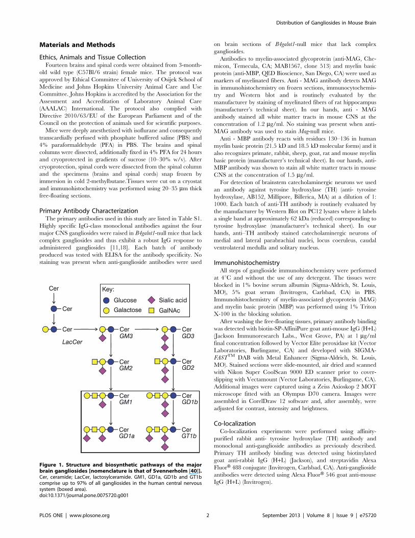
Figure 1. Structure and biosynthetic pathways of the majorbrain gangliosides [nomenclature is that of Svennerholm [40]].Cer, ceramide; LacCer, lactosylceramide. GM1, GD1a, GD1b and GT1bcomprise up to 97% of all gangliosides in the human central nervoussystem (boxed area).doi:10.1371/journal.pone.0075720.g001
Distribution of Gangliosides in Mouse Brain
PLOS ONE | www.plosone.org 2 September 2013 | Volume 8 | Issue 9 | e75720
Anterograde Axon LabelingTo determine ganglioside expression in white matter tracts, five
mice were subjected to anterograde labeling. Craniotomy was
performed on anesthetized mice and sensorimotor cortex was
injected with one injection of 0.2 ml 10% (w/v) 10,000 MW
biotinylated dextran amine (BDA, Invitrogen). After 2 weeks mice
were perfusion-fixed, brains and spinal cords were recovered as
described above, cryosections prepared, and gangliosides detected
using immunohistochemistry. BDA-labeled axons were detected
with streptavidin Alexa FluorH 488 conjugate, whereas anti-
ganglioside antibodies were detected using Alexa FluorH 546 goat
anti-mouse IgG.
Fluorescent MicroscopyFluorescent immunohistochemical images were obtained using
a Zeiss LSM 510 inverted confocal microscope (The Johns
Hopkins School of Medicine Microscope Facility), assembled in
CorelDraw 12 software and assembled images adjusted for
contrast, intensity and brightness.
Qualitative Analysis of Immunohistochemical Reactivityto Gangliosides GD1a and GT1bQualitative analysis of immunohistochemical reactivity to
gangliosides GD1a and GT1b was performed on images taken
with the same exposure times by two independent observers. The
relative expression (+++, strong signal; ++, moderate signal; +,weak signal, 2, no signal) of gangliosides GD1a and GT1b was
compared to negative control on each mouse brain region listed in
Table S2. Images of different brain regions were taken at different
exposure times, so the comparisons were made between different
antibodies in one brain region, but not between different brain
regions.
Results
General Expression Patterns of Major Brain GangliosidesThe expression patterns of gangliosides GM1, GD1a, GD1b
and GT1b were studied using immunohistochemistry on adult
C57Bl/6 mouse brains and spinal cords cut in three planes
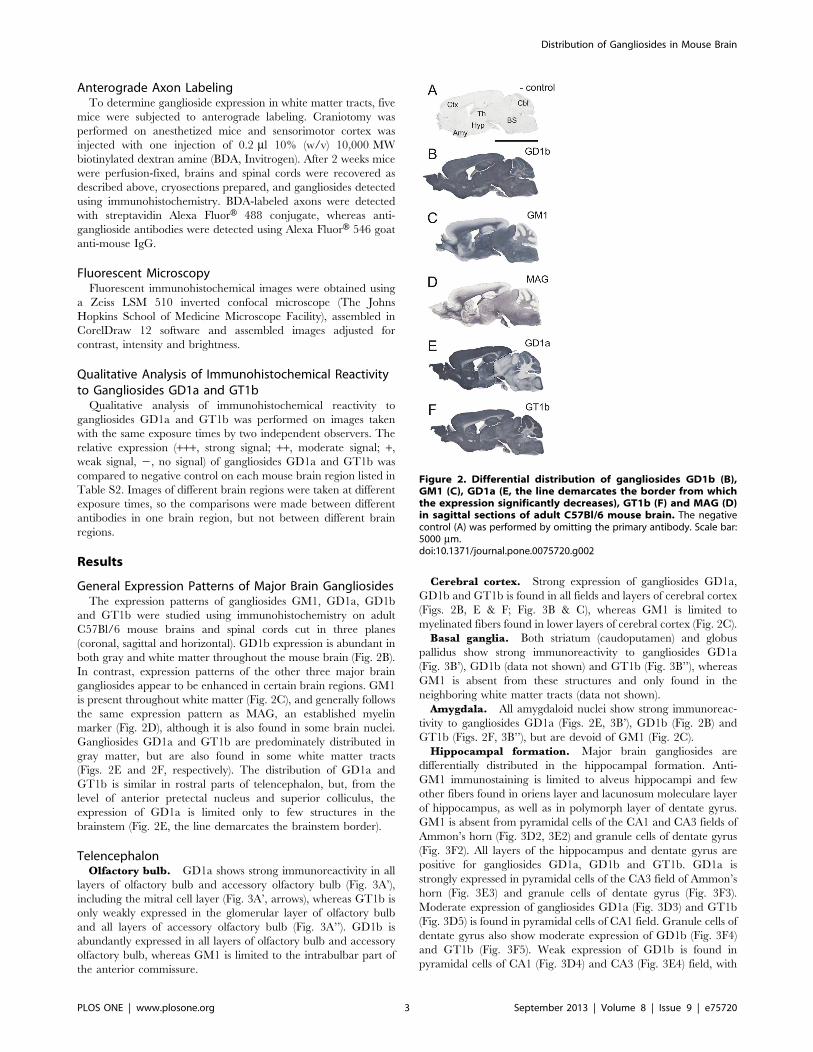
(coronal, sagittal and horizontal). GD1b expression is abundant in
both gray and white matter throughout the mouse brain (Fig. 2B).
In contrast, expression patterns of the other three major brain
gangliosides appear to be enhanced in certain brain regions. GM1
is present throughout white matter (Fig. 2C), and generally follows
the same expression pattern as MAG, an established myelin
marker (Fig. 2D), although it is also found in some brain nuclei.
Gangliosides GD1a and GT1b are predominately distributed in
gray matter, but are also found in some white matter tracts
(Figs. 2E and 2F, respectively). The distribution of GD1a and
GT1b is similar in rostral parts of telencephalon, but, from the
level of anterior pretectal nucleus and superior colliculus, the
expression of GD1a is limited only to few structures in the
brainstem (Fig. 2E, the line demarcates the brainstem border).
TelencephalonOlfactory bulb. GD1a shows strong immunoreactivity in all
layers of olfactory bulb and accessory olfactory bulb (Fig. 3A’),
including the mitral cell layer (Fig. 3A’, arrows), whereas GT1b is
only weakly expressed in the glomerular layer of olfactory bulb
and all layers of accessory olfactory bulb (Fig. 3A’’). GD1b is
abundantly expressed in all layers of olfactory bulb and accessory
olfactory bulb, whereas GM1 is limited to the intrabulbar part of
the anterior commissure.
Cerebral cortex. Strong expression of gangliosides GD1a,
GD1b and GT1b is found in all fields and layers of cerebral cortex
(Figs. 2B, E & F; Fig. 3B & C), whereas GM1 is limited to
myelinated fibers found in lower layers of cerebral cortex (Fig. 2C).
Basal ganglia. Both striatum (caudoputamen) and globus
pallidus show strong immunoreactivity to gangliosides GD1a
(Fig. 3B’), GD1b (data not shown) and GT1b (Fig. 3B’’), whereas
GM1 is absent from these structures and only found in the
neighboring white matter tracts (data not shown).
Amygdala. All amygdaloid nuclei show strong immunoreac-
tivity to gangliosides GD1a (Figs. 2E, 3B’), GD1b (Fig. 2B) and
GT1b (Figs. 2F, 3B’’), but are devoid of GM1 (Fig. 2C).
Hippocampal formation. Major brain gangliosides are
differentially distributed in the hippocampal formation. Anti-
GM1 immunostaining is limited to alveus hippocampi and few
other fibers found in oriens layer and lacunosum moleculare layer
of hippocampus, as well as in polymorph layer of dentate gyrus.
GM1 is absent from pyramidal cells of the CA1 and CA3 fields of
Ammon’s horn (Fig. 3D2, 3E2) and granule cells of dentate gyrus
(Fig. 3F2). All layers of the hippocampus and dentate gyrus are
positive for gangliosides GD1a, GD1b and GT1b. GD1a is
strongly expressed in pyramidal cells of the CA3 field of Ammon’s
horn (Fig. 3E3) and granule cells of dentate gyrus (Fig. 3F3).
Moderate expression of gangliosides GD1a (Fig. 3D3) and GT1b
(Fig. 3D5) is found in pyramidal cells of CA1 field. Granule cells of
dentate gyrus also show moderate expression of GD1b (Fig. 3F4)
and GT1b (Fig. 3F5). Weak expression of GD1b is found in
pyramidal cells of CA1 (Fig. 3D4) and CA3 (Fig. 3E4) field, with
Figure 2. Differential distribution of gangliosides GD1b (B),GM1 (C), GD1a (E, the line demarcates the border from whichthe expression significantly decreases), GT1b (F) and MAG (D)in sagittal sections of adult C57Bl/6 mouse brain. The negativecontrol (A) was performed by omitting the primary antibody. Scale bar:5000 mm.doi:10.1371/journal.pone.0075720.g002
Distribution of Gangliosides in Mouse Brain
PLOS ONE | www.plosone.org 3 September 2013 | Volume 8 | Issue 9 | e75720
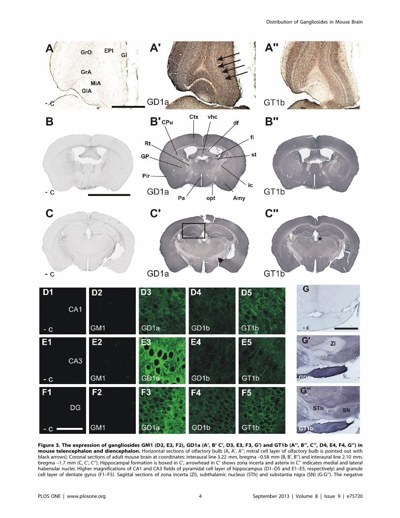
Figure 3. The expression of gangliosides GM1 (D2, E2, F2), GD1a (A’, B’ C’, D3, E3, F3, G’) and GT1b (A’’, B’’, C’’, D4, E4, F4, G’’) inmouse telencephalon and diencephalon. Horizontal sections of olfactory bulb (A, A’, A’’; mitral cell layer of olfactory bulb is pointed out withblack arrows). Coronal sections of adult mouse brain at coordinates: interaural line 3.22 mm, bregma –0.58 mm (B, B’, B’’) and interaural line 2.10 mm,bregma –1.7 mm (C, C’, C’’). Hippocampal formation is boxed in C’, arrowhead in C’ shows zona incerta and asterix in C’’ indicates medial and lateralhabenular nuclei. Higher magnifications of CA1 and CA3 fields of pyramidal cell layer of hippocampus (D1–D5 and E1–E5, respectively) and granulecell layer of dentate gyrus (F1–F5). Sagittal sections of zona incerta (ZI), subthalamic nucleus (STh) and substantia nigra (SN) (G-G’’). The negative
Distribution of Gangliosides in Mouse Brain
PLOS ONE | www.plosone.org 4 September 2013 | Volume 8 | Issue 9 | e75720
CA3 field being also weakly positive for GT1b (Fig. 3E5). The
pattern of expression of gangliosides GD1a, GD1b and GT1b is
consistent with expression primarily on neuronal plasma mem-
branes.
White matter tracts of the telencephalon. Gangliosides
GD1a and GT1b are expressed in some white matter tracts of the
telencephalon, but not others. Consistent with their expression on
myelinated axons, gangliosides GD1a and GT1b immunodetec-
tion was found to be satisfactory only if tracts were cut
longitudinally instead of perpendicularly. For that reason, brains
cut in three planes (coronal, sagittal and horizontal) were analyzed.
A subset of fibers in the corpus callosum show high immunore-
activity for GD1a (Figs. 4A, C) and GT1b (Figs. 4B, D). Based on
anterograde BDA tracing (from the sensorimotor cortex of right
hemisphere) GD1a- and GT1b-labeled fibers were identified as
myelinated commissural fibers, as shown on consecutive sections
double-stained with Alexa Fluor 488 conjugated streptavidin (for
detection of BDA) and anti-MAG (Fig. 4H) or anti-MBP (Fig. 4I)
antibodies. In contrast, gangliosides GM1 (Fig. 4F) and GD1b
(Fig. 4G) are expressed throughout corpus callosum.
DiencephalonThalamus. Most thalamic nuclei abundantly express gangli-
osides GM1 (Fig. 2C), GD1a (Figs. 2E, 3B’, 3C’), GD1b (Fig. 2B)
and GT1b (Figs. 2F, 3B’’, 3C’’). However, GD1a is only weakly
expressed in reticular nucleus of thalamus (Fig. 3B’).
Epithalamus. Medial and lateral habenular nuclei show
strong expression of GD1b (data not shown) and GT1b (Fig. 3C’’,
asterix). While there is a weak expression of GM1 (data not
shown), GD1a is completely absent from these structures (Fig. 3C’).
Hypothalamus. All hypothalamic nuclei, including paraven-
tricular and medial preoptic nuclei and anterior hypothalamic
area, abundantly express gangliosides GM1 (Fig. 2C), GD1a
(Figs. 2E, 3B’, 3C’), GD1b (Fig. 2B) and GT1b (Figs. 2F, 3B’’,
3C’’).
Subthalamus. While GT1b is expressed both in subthalamic
nucleus and zona incerta (Fig. 3G’’), GD1a is completely absent
from these structures (Fig. 3G’).
CerebellumIn the mouse cerebellum, GM1 is detected only in white matter
(Fig. 5B, asterix), while gangliosides GD1a (Figs. 5C, H), GD1b
(Fig. 5D) and GT1b (Fig. 5E, I) are found in all three layers of
cerebellar cortex (granule cell layer, Purkinje cell layer and
molecular layer). It is not clear, however, if gangliosides GD1a and
GT1b are expressed in cellular membranes of Purkinje cells or in
the synapses and fibers surrounding them. The white matter of
cerebellum and cerebellar nuclei show strong immunoreactivity
for GM1 (Fig. 2C) and GD1b (Fig. 2B) and a slight immunore-
activity for GT1b (Fig. 2F), while GD1a is completely absent from
these structures (Fig. 2E).
BrainstemGangliosides GM1, GD1b and GT1b are present in most nuclei
of the brainstem and in white matter tracts. In midbrain, moderate
expression of GT1b is found both in reticular and compact part of
substantia nigra (Fig. 3G’’) and red nucleus (Fig. 5M). While GD1a
is strongly expressed in both parts of substantia nigra (Fig 3G’), no
immunoreactivity is found in the red nucleus (Fig. 5L).
The raphe magnus and pallidus nuclei show strong expression
of GT1b (Fig. 5Q) and moderate expression of GD1a (Fig. 5P),
whereas the raphe obscurus nucleus shows only moderate
expression of GT1b (Fig. 5U) and no GD1a (Fig. 5T).
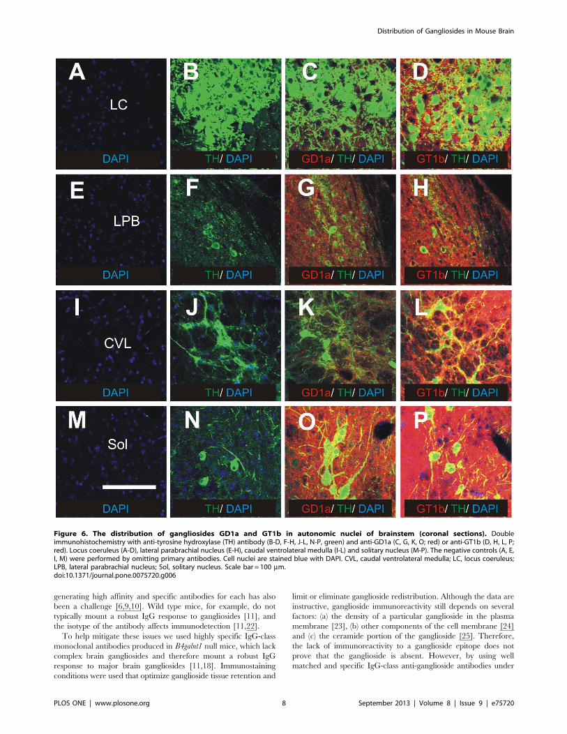
Among autonomic nuclei of the brainstem, strong GD1a
immunoreactivity is detected in periaqueductal gray matter, locus
coeruleus (Fig. 6C), subcoeruleus nucleus, medial parabrachial
nucleus, lateral parabrachial nucleus (Fig. 6G), Kolliker-Fuse
nucleus, rostral ventrolateral medulla, caudal ventrolateral me-
dulla (Fig. 6K) and solitary nucleus (Fig. 6O). Strong expression of
GT1b is found in locus coeruleus (Fig. 6D), lateral parabrachial
nucleus (Fig. 6H), caudal ventrolateral medulla (Fig. 6L), solitary
nucleus (Fig. 6P) and other brainstem nuclei that are also positive
for GD1a. GM1 and GD1b are moderately expressed in all of
these autonomic nuclei, except solitary nucleus which is devoid of
GM1 (Fig. 7B).
Moderate expression of GD1a and GT1b is also found in
mesencephalic, principal and spinal trigeminal nuclei, as well as in
pontine, medullary, parvicellular and intermediate reticular nuclei
(Fig. 7C, F), all of which project their axons to the spinal cord. The
gigantocellular reticular nucleus, involved in control of blood
pressure and axial musculature was also found to express GT1b
(Fig. 7F) and GD1a, although GD1a expression is relatively weak
(Fig. 7C). Interestingly, GD1a is not expressed in either of the
vestibular nuclei (Fig. 7C).
GD1b is strongly expressed in inferior olivary, vestibular nuclei
and solitary nucleus and to some extent in gigantocellular reticular
nucleus, parvicellular reticular nucleus and intermediate reticular
nucleus (Fig. 7E). The pattern of expression of GT1b is similar to
that of GM1, the difference being only in the solitary nucleus that
is positive for GT1b. It is also worth noticing that certain white
matter tracts are positive for GT1b such as: spinal trigeminal tract,
inferior cerebellar peduncule and pyramidal tract, the latter being
also positive for GD1a (Figs. 7F and 7C, respectively).
Spinal CordWhereas GD1b is expressed in both white and gray matter of
the spinal cord (Figs. 8D, D’, D’’), other gangliosides are limited to
more specific areas. GM1 is strongly expressed in corticospinal
tract (Figs. 8B, B’, B’’; white arrow) and moderately expressed in
other white matter, mainly in propriospinal tracts surrounding the
gray matter (Figs. 8B, B’, B’’; black arrowheads). GM1 is also
weakly expressed in gray matter. The expression of GD1a is strong
in Rexed laminae 1 and 2 of dorsal horn, moderate around central
canal and weak in other Rexed laminae of gray matter (Figs. 8C,
C’,C’’). GT1b is moderately expressed in all Rexed laminae of
gray matter (Fig. 8E, E’, E’’). Since corticospinal tract is the main
pathway involved in skilled voluntary movements, such as food
pellet reaching, the expression of gangliosides in horizontal
sections of spinal cord has been studied in detail, namely by
tracing the corticospinal tract with BDA tracer. Co-localization
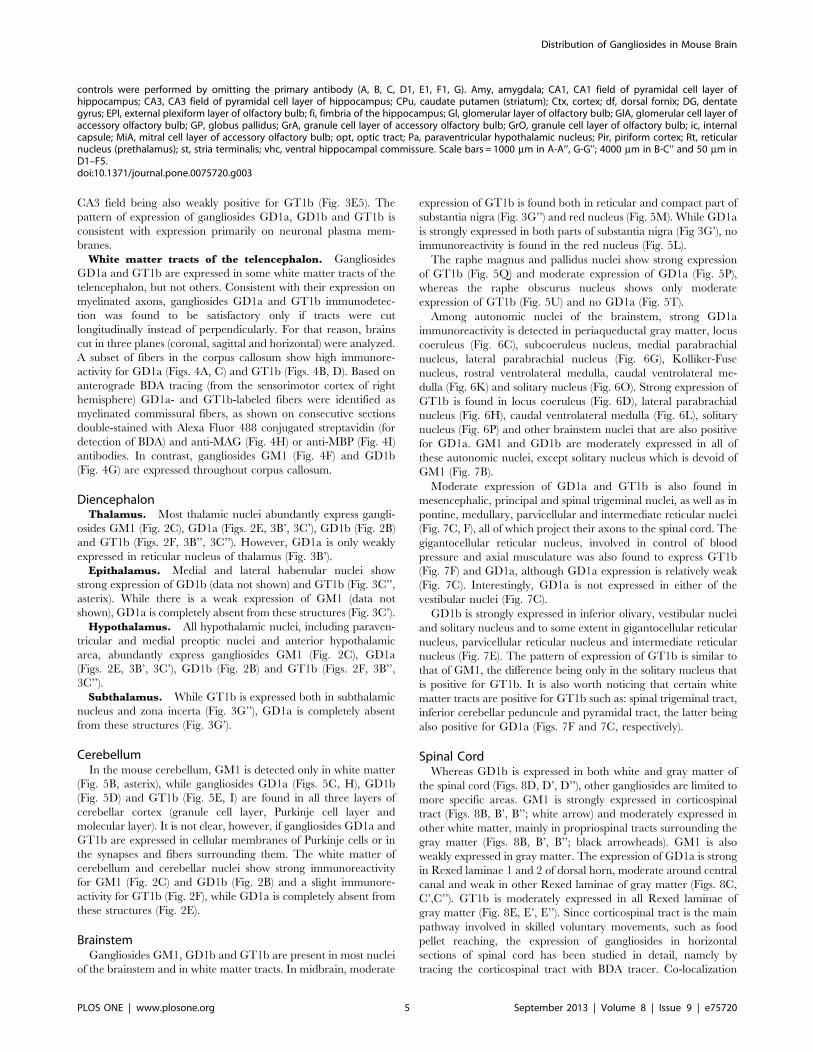
controls were performed by omitting the primary antibody (A, B, C, D1, E1, F1, G). Amy, amygdala; CA1, CA1 field of pyramidal cell layer ofhippocampus; CA3, CA3 field of pyramidal cell layer of hippocampus; CPu, caudate putamen (striatum); Ctx, cortex; df, dorsal fornix; DG, dentategyrus; EPl, external plexiform layer of olfactory bulb; fi, fimbria of the hippocampus; Gl, glomerular layer of olfactory bulb; GlA, glomerular cell layer ofaccessory olfactory bulb; GP, globus pallidus; GrA, granule cell layer of accessory olfactory bulb; GrO, granule cell layer of olfactory bulb; ic, internalcapsule; MiA, mitral cell layer of accessory olfactory bulb; opt, optic tract; Pa, paraventricular hypothalamic nucleus; Pir, piriform cortex; Rt, reticularnucleus (prethalamus); st, stria terminalis; vhc, ventral hippocampal commissure. Scale bars = 1000 mm in A-A’’, G-G’’; 4000 mm in B-C’’ and 50 mm inD1–F5.doi:10.1371/journal.pone.0075720.g003
Distribution of Gangliosides in Mouse Brain
PLOS ONE | www.plosone.org 5 September 2013 | Volume 8 | Issue 9 | e75720
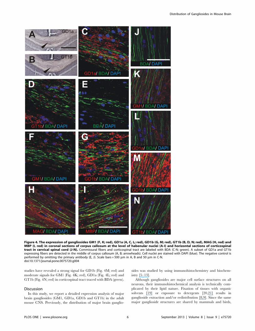
studies have revealed a strong signal for GD1b (Fig. 4M; red) and
moderate signals for GM1 (Fig. 4K; red), GD1a (Fig. 4L; red) and
GT1b (Fig. 4N; red) in corticospinal tract traced with BDA (green).
DiscussionIn this study, we report a detailed expression analysis of major
brain gangliosides (GM1, GD1a, GD1b and GT1b) in the adult
mouse CNS. Previously, the distribution of major brain ganglio-
sides was studied by using immunohistochemistry and biochem-
istry [5–13].
Although gangliosides are major cell surface structures on all
neurons, their immunohistochemical analysis is technically com-
plicated by their lipid nature. Fixation of tissues with organic
solvents [19] or exposure to detergents [20,21] results in
ganglioside extraction and/or redistribution [8,9]. Since the same
major ganglioside structures are shared by mammals and birds,
Figure 4. The expression of gangliosides GM1 (F, K; red), GD1a (A, C, L; red), GD1b (G, M; red), GT1b (B, D, N; red), MAG (H, red) andMBP (I, red) in coronal sections of corpus callosum at the level of habenular nuclei (A-I) and horizontal sections of corticospinaltract in cervical spinal cord (J-N). Commissural fibers and corticospinal tract are labeled with BDA (C-N; green). A subset of GD1a and GT1bexpressing fibers are detected in the middle of corpus callosum (A, B; arrowheads). Cell nuclei are stained with DAPI (blue). The negative control isperformed by omitting the primary antibody (E, J). Scale bars = 500 mm in A, B and 50 mm in C-N.doi:10.1371/journal.pone.0075720.g004
Distribution of Gangliosides in Mouse Brain
PLOS ONE | www.plosone.org 6 September 2013 | Volume 8 | Issue 9 | e75720
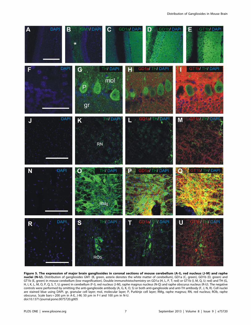
Figure 5. The expression of major brain gangliosides in coronal sections of mouse cerebellum (A-I), red nucleus (J-M) and raphenuclei (N-U). Distribution of gangliosides GM1 (B, green, asterix denotes the white matter of cerebellum), GD1a (C, green), GD1b (D, green) andGT1b (E, green) in mouse cerebellum (low magnification). Double immunohistochemistry on GD1a (H, L, P, T; red) or GT1b (I, M, Q, U; red) and TH (G,H, I, K, L, M, O, P, Q, S, T, U; green) in cerebellum (F-I), red nucleus (J-M), raphe magnus nucleus (N-Q) and raphe obscurus nucleus (R-U). The negativecontrols were performed by omitting the anti-ganglioside antibody (A, G, K, O, S) or both anti-ganglioside and anti-TH antibody (F, J, N, R). Cell nucleiare stained blue using DAPI. gr, granular cell layer; mol, molecular layer; P, Purkinje cell layer; RMg, raphe magnus; RN, red nucleus; ROb, rapheobscurus. Scale bars = 200 mm in A-E, J-M; 50 mm in F-I and 100 mm in N-U.doi:10.1371/journal.pone.0075720.g005
Distribution of Gangliosides in Mouse Brain
PLOS ONE | www.plosone.org 7 September 2013 | Volume 8 | Issue 9 | e75720
generating high affinity and specific antibodies for each has also
been a challenge [6,9,10]. Wild type mice, for example, do not
typically mount a robust IgG response to gangliosides [11], and
the isotype of the antibody affects immunodetection [11,22].
To help mitigate these issues we used highly specific IgG-class
monoclonal antibodies produced in B4galnt1 null mice, which lack
complex brain gangliosides and therefore mount a robust IgG
response to major brain gangliosides [11,18]. Immunostaining
conditions were used that optimize ganglioside tissue retention and
limit or eliminate ganglioside redistribution. Although the data are
instructive, ganglioside immunoreactivity still depends on several
factors: (a) the density of a particular ganglioside in the plasma
membrane [23], (b) other components of the cell membrane [24]
and (c) the ceramide portion of the ganglioside [25]. Therefore,
the lack of immunoreactivity to a ganglioside epitope does not
prove that the ganglioside is absent. However, by using well
matched and specific IgG-class anti-ganglioside antibodies under
Figure 6. The distribution of gangliosides GD1a and GT1b in autonomic nuclei of brainstem (coronal sections). Doubleimmunohistochemistry with anti-tyrosine hydroxylase (TH) antibody (B-D, F-H, J-L, N-P, green) and anti-GD1a (C, G, K, O; red) or anti-GT1b (D, H, L, P;red). Locus coeruleus (A-D), lateral parabrachial nucleus (E-H), caudal ventrolateral medulla (I-L) and solitary nucleus (M-P). The negative controls (A, E,I, M) were performed by omitting primary antibodies. Cell nuclei are stained blue with DAPI. CVL, caudal ventrolateral medulla; LC, locus coeruleus;LPB, lateral parabrachial nucleus; Sol, solitary nucleus. Scale bar = 100 mm.doi:10.1371/journal.pone.0075720.g006
Distribution of Gangliosides in Mouse Brain
PLOS ONE | www.plosone.org 8 September 2013 | Volume 8 | Issue 9 | e75720
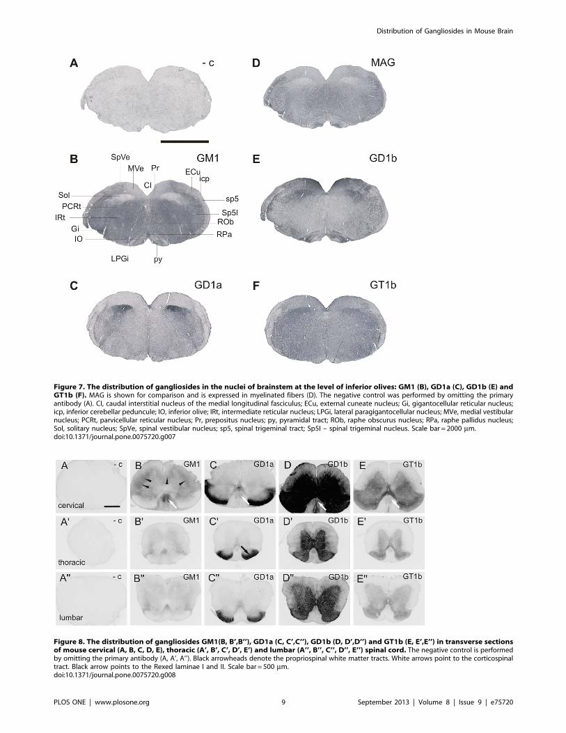
Figure 7. The distribution of gangliosides in the nuclei of brainstem at the level of inferior olives: GM1 (B), GD1a (C), GD1b (E) andGT1b (F). MAG is shown for comparison and is expressed in myelinated fibers (D). The negative control was performed by omitting the primaryantibody (A). CI, caudal interstitial nucleus of the medial longitudinal fasciculus; ECu, external cuneate nucleus; Gi, gigantocellular reticular nucleus;icp, inferior cerebellar peduncule; IO, inferior olive; IRt, intermediate reticular nucleus; LPGi, lateral paragigantocellular nucleus; MVe, medial vestibularnucleus; PCRt, parvicellular reticular nucleus; Pr, prepositus nucleus; py, pyramidal tract; ROb, raphe obscurus nucleus; RPa, raphe pallidus nucleus;Sol, solitary nucleus; SpVe, spinal vestibular nucleus; sp5, spinal trigeminal tract; Sp5I – spinal trigeminal nucleus. Scale bar = 2000 mm.doi:10.1371/journal.pone.0075720.g007
Figure 8. The distribution of gangliosides GM1(B, B’,B’’), GD1a (C, C’,C’’), GD1b (D, D’,D’’) and GT1b (E, E’,E’’) in transverse sectionsof mouse cervical (A, B, C, D, E), thoracic (A’, B’, C’, D’, E’) and lumbar (A’’, B’’, C’’, D’’, E’’) spinal cord. The negative control is performedby omitting the primary antibody (A, A’, A’’). Black arrowheads denote the propriospinal white matter tracts. White arrows point to the corticospinaltract. Black arrow points to the Rexed laminae I and II. Scale bar = 500 mm.doi:10.1371/journal.pone.0075720.g008
Distribution of Gangliosides in Mouse Brain
PLOS ONE | www.plosone.org 9 September 2013 | Volume 8 | Issue 9 | e75720
well characterized conditions, reasonable comparisons can be
inferred with respect to antibody-accessible structures.
A complementary method for analysis of ganglioside distribu-
tion in CNS tissue is imaging mass spectrometry (IMS). IMS gives
detailed information on the ceramide core of the ganglioside and is
able to assess multiple ganglioside species at a time in a single
tissue slice. However, it has the well documented potential
problem of partial loss of sialic acid during ionization and
differentiation between ganglioside isomers (such as GD1a and
GD1b) requires more detailed (MSn) analyses [15–17].
Our results show that GD1b and GT1b are widely expressed
throughout the mouse CNS, whereas the immunoreactivity to
GM1 is predominately in white matter tracts and immunoreac-
tivity to GD1a significantly decreases caudal to the superior
colliculus. Particularly strong expression of GD1a, GD1b and
GT1b is found in olfactory bulbs, neocortex, basal ganglia,
amygdaloid nuclear complex, septal regions, thalamic nuclei,
hypothalamic nuclei and hippocampal formation.
Strong expression of GD1b and GT1b is found in all three
layers of cerebellum (molecular layer, Purkinje cell layer and
granular cell layer) and cerebellar nuclei, whereas GD1a is
moderately expressed in cerebellar cortex, but completely absent
from cerebellar nuclei. Our results also show that GM1 is limited
to white matter in the cerebellum.
Among nuclei of brainstem, strong immunoreactivity to GD1a
is detected in periaqueductal gray matter, locus coeruleus,
subcoeruleus nucleus, medial and lateral parabrachial nucleus,
Kolliker-Fuse nucleus, rostral and caudal ventrolateral medulla
and solitary nucleus. Interestingly, those are all autonomic nuclei
that are also positive for tyrosine hydroxylase. It is important to
note that these nuclei send their projections to the spinal cord and
that they are involved in cardiovascular reflexes and respiratory
control but also receive sensory, nociceptive and visceral input
from periphery. In concordance with these results, previous
biochemical studies have also shown the expression of GD1a and
GT1b in locus coeruleus, as well as in dorsal raphe nucleus,
laterodorsal tegmentum and pedunculopontine tegmentum [5].
On the other hand, immunoreactivity to GD1a is weak or
absent in the reticular nucleus of thalamus, habenular nuclei, zona
incerta, subthalamic nucleus, red nucleus, superior and inferior
colliculus, cochlear nuclei, vestibular nuclei, dorsal tegmental
nucleus, motor nuclei of the trigeminal nerve, facial nucleus,
hypoglossal nucleus, gracile nucleus, cuneate nucleus, external
cuneate nucleus and ventral horn of spinal cord (for detailed
comparison of GD1a and GT1b expression see Table S2).
Another interesting finding is that certain white matter tracts
such as corpus callosum, the main comissural pathway of
telencephalon, and corticospinal tract express all four major
gangliosides, while others, e.g. white matter of cerebellum, do not.
It is not clear from our data if certain gangliosides are on
axolemma or on myelin membrane. However, biochemical studies
have shown that GM1 is part of myelin membrane, while CNS
axolemma contains all four major brain gangliosides [26].
A goal in documenting the distribution of major brain
gangliosides is to inform theories about their functions. Based on
studies of the B4galnt1-null mouse, in which all complex
gangliosides are replaced by the simpler structures GM3 and
GD3, major brain gangliosides have functional roles in long-term
axon-myelin stabilization [27]. Other studies implicate GD1a and
GT1b as complementary receptors for myelin-associated glyco-
protein (MAG), a ganglioside-binding protein expressed selectively
on myelin membranes apposed to the axon surface. Mice
engineered to lack MAG, complex gangliosides, or double null
mice have similar long-term dysmyelination phenotypes [28].
Together, these data implicate GD1a and/or GT1b as receptors
in axon-myelin interactions.
MAG is also known to inhibit axon outgrowth in vitro, and has
been proposed as one of the axon regeneration inhibitors that
limits recovery after CNS injury. MAG has several receptors on
nerve cells that mediate axon outgrowth inhibition, including
gangliosides [29], Nogo receptors (NgR’s) [30], b-integrin [31] and
PirB [32]. Comparing the distribution of MAG receptors GD1a/
GT1b to other MAG receptors may contribute to our under-
standing of selective MAG functions and mechanisms in different
neuronal pathways. In this light, it is interesting to note the
distribution of NgR’s in the CNS in comparison to gangliosides
GD1a and GT1b. Most of the studies on NgR distribution have
used in situ hybridization, although immunohistochemistry on
NgR protein has been reported by several groups [33,34]. All
studies agree that NgR’s are expressed in neocortical neurons,
pyramidal cells of hippocampus, granule cells of dentate gyrus,
most of thalamic nuclei and granule cells of cerebellum.
Two recent in vitro studies revealed that different neurons
respond to MAG inhibition of neurite outgrowth using different
signaling pathways [35,36]. While, cerebellar granule neurons
(CGNs) respond to MAG inhibition by a ganglioside – dependent
pathway, most of MAG-mediated neurite outgrowth inhibition in
DRGs is mainly through NgR-dependent pathway. In addition,
postnatal hippocampal neurons and retinal ganglion cells use both
pathways for MAG-mediated inhibition. In the light of expression
of NgR1-3 and gangliosides GD1a and GT1b in these cells in the
adult mouse CNS, it is expected that hippocampal neurons, which
strongly express both NgR1-3 [33,34] and gangliosides GD1a and
GT1b, respond to MAG inhibition using both receptors.
Interestingly, while CGNs express both gangliosides GD1a and
GT1b and NgRs, they respond to MAG inhibition primarily
through ganglioside- dependent pathway. The explanation for this
could be in the lack of NgR co-receptors or other downstream
signaling molecules in CGNs.
The studies of the distribution of major brain gangliosides in
DRGs have shown that most neurons express GT1b, while GD1a
is expressed mainly in small neurons (15–30 mm diameter),
involved in nociception, and only in 10–20% of larger neurons
[7]. This may explain why most of the MAG-mediated inhibition
in DRG cultures is NgR-dependent.
It is important to note that gangliosides GD1a and GT1b are
strongly expressed in cerebral cortex, corticospinal tract and
certain brainstem nuclei that comprise ascending and descending
tracts of spinal cord. However GD1a and GT1b expression is
weak in gracile and cuneate nuclei that comprise dorsal column
pathway. This knowledge may be helpful in designing treatments
that address MAG inhibition of axonal regeneration through
manipulation of these particular gangliosides. Along the same line,
pathway-specific treatments may be designed based on the specific
MAG receptor expression profiles. This notion is supported by the
findings that treatments that target a single molecule often fail to
evoke robust axonal regeneration after spinal cord injury [37–39].
Supporting Information
Table S1 Primary antibodies used in the study.
(DOCX)
Table S2 Qualitative analysis of immunohistochemical reactivity
to gangliosides GD1a and GT1b. +++, strong signal; ++, moderate
signal; +, weak signal, 2, no signal.
(DOCX)
Distribution of Gangliosides in Mouse Brain
PLOS ONE | www.plosone.org 10 September 2013 | Volume 8 | Issue 9 | e75720
Author Contributions
Conceived and designed the experiments: KV MH. Performed the
experiments: KV MH BV IVD. Analyzed the data: KV MH RLS.
Contributed reagents/materials/analysis tools: IVD RLS. Wrote the
paper: KV RLS MH.
References
1. Tettamanti G, Bonali F, Marchesini S, Zambotti V (1973) A new procedure for
the extraction, purification and fractionation of brain gangliosides. Biochim
Biophys Acta 296: 160–170.
2. Lopez PH, Schnaar RL (2009) Gangliosides in cell recognition and membrane
protein regulation. Curr Opin Struct Biol 19: 549–557.
3. Allende ML, Proia RL (2002) Lubricating cell signaling pathways with
gangliosides. Curr Opin Struct Biol 12: 587–592.
4. Simpson MA, Cross H, Proukakis C, Priestman DA, Neville DC, et al. (2004)
Infantile-onset symptomatic epilepsy syndrome caused by a homozygous loss-of-
function mutation of GM3 synthase. Nat Genet 36: 1225–1229.
5. Byers DM, Irwin LN, Cabeza R (2002) Ganglioside patterns mature at different
rates in functionally related subregions of the rat pons. Dev Neurosci 24: 478–
484.
6. De Baecque C, Johnson AB, Naiki M, Schwarting G, Marcus DM (1976)
Ganglioside localization in cerebellar cortex: an immunoperoxidase study with
antibody to GM1 ganglioside. Brain Res 114: 117–122.
7. Gong Y, Tagawa Y, Lunn MP, Laroy W, Heffer-Lauc M, et al. (2002)
Localization of major gangliosides in the PNS: implications for immune
neuropathies. Brain 125: 2491–2506.
8. Kotani M, Kawashima I, Ozawa H, Terashima T, Tai T (1993) Differential
distribution of major gangliosides in rat central nervous system detected by
specific monoclonal antibodies. Glycobiology 3: 137–146.
9. Laev H, Mahadik SP (1989) Topography of monosialoganglioside (GM1) in rat
brain using monoclonal antibodies. Neurosci Lett 102: 7–14.
10. Laev H, Rapport MM, Mahadik SP, Silverman AJ (1978) Immunohistological
localization of ganglioside in rat cerebellum. Brain Res 157: 136–141.
11. Lunn MP, Johnson LA, Fromholt SE, Itonori S, Huang J, et al. (2000) High-
affinity anti-ganglioside IgG antibodies raised in complex ganglioside knockout
mice: reexamination of GD1a immunolocalization. J Neurochem 75: 404–412.
12. Molander M, Berthold CH, Persson H, Fredman P (2000) Immunostaining of
ganglioside GD1b, GD3 and GM1 in rat cerebellum: cellular layer and cell type
specific associations. J Neurosci Res 60: 531–542.
13. Schwarz A, Futerman AH (1996) The localization of gangliosides in neurons of
the central nervous system: the use of anti-ganglioside antibodies. Biochim
Biophys Acta 1286: 247–267.
14. Sugiura Y, Shimma S, Konishi Y, Yamada MK, Setou M (2008) Imaging mass
spectrometry technology and application on ganglioside study; visualization of
age-dependent accumulation of C20-ganglioside molecular species in the mouse
hippocampus. PLoS One 3: e3232.
15. Goto-Inoue N, Hayasaka T, Zaima N, Kashiwagi Y, Yamamoto M, et al. (2010)
The detection of glycosphingolipids in brain tissue sections by imaging mass
spectrometry using gold nanoparticles. J Am Soc Mass Spectrom 21: 1940–1943.
16. Colsch B, Jackson SN, Dutta S, Woods AS (2011) Molecular Microscopy of
Brain Gangliosides: Illustrating their Distribution in Hippocampal Cell Layers.
ACS Chem Neurosci 2: 213–222.
17. Colsch B, Woods AS (2010) Localization and imaging of sialylated glycosphin-
golipids in brain tissue sections by MALDI mass spectrometry. Glycobiology 20:
661–667.
18. Schnaar RL, Fromholt SE, Gong Y, Vyas AA, Laroy W, et al. (2002)
Immunoglobulin G-class mouse monoclonal antibodies to major brain
gangliosides. Anal Biochem 302: 276–284.
19. Schwarz A, Futerman AH (1997) Determination of the localization of
gangliosides using anti-ganglioside antibodies: comparison of fixation methods.
J Histochem Cytochem 45: 611–618.
20. Heffer-Lauc M, Lauc G, Nimrichter L, Fromholt SE, Schnaar RL (2005)
Membrane redistribution of gangliosides and glycosylphosphatidylinositol-
anchored proteins in brain tissue sections under conditions of lipid raft isolation.
Biochim Biophys Acta 1686: 200–208.
21. Heffer-Lauc M, Viljetic B, Vajn K, Schnaar RL, Lauc G (2007) Effects of
detergents on the redistribution of gangliosides and GPI-anchored proteins inbrain tissue sections. J Histochem Cytochem 55: 805–812.
22. Schwarz A, Futerman AH (2000) Immunolocalization of gangliosides by lightmicroscopy using anti-ganglioside antibodies. Methods Enzymol 312: 179–187.
23. Nores GA, Dohi T, Taniguchi M, Hakomori S (1987) Density-dependent
recognition of cell surface GM3 by a certain anti-melanoma antibody, and GM3lactone as a possible immunogen: requirements for tumor-associated antigen and
immunogen. J Immunol 139: 3171–3176.24. Lloyd KO, Gordon CM, Thampoe IJ, DiBenedetto C (1992) Cell surface
accessibility of individual gangliosides in malignant melanoma cells to antibodies
is influenced by the total ganglioside composition of the cells. Cancer Res 52:4948–4953.
25. Tagawa Y, Laroy W, Nimrichter L, Fromholt SE, Moser AB, et al. (2002) Anti-ganglioside antibodies bind with enhanced affinity to gangliosides containing
very long chain fatty acids. Neurochem Res 27: 847–855.26. De Vries GH, Zmachinski CJ (1980) The lipid composition of rat CNS
axolemma-enriched fractions. J Neurochem 34: 424–430.
27. Sheikh KA, Sun J, Liu Y, Kawai H, Crawford TO, et al. (1999) Mice lackingcomplex gangliosides develop Wallerian degeneration and myelination defects.
Proceedings of the National Academy of Sciences of the United States ofAmerica 96: 7532–7537.
28. Pan B, Fromholt SE, Hess EJ, Crawford TO, Griffin JW, et al. (2005) Myelin-
associated glycoprotein and complementary axonal ligands, gangliosides,mediate axon stability in the CNS and PNS: neuropathology and behavioral
deficits in single- and double-null mice. Exp Neurol 195: 208–217.29. Vyas AA, Patel HV, Fromholt SE, Heffer-Lauc M, Vyas KA, et al. (2002)
Gangliosides are functional nerve cell ligands for myelin-associated glycoprotein
(MAG), an inhibitor of nerve regeneration. Proc Natl Acad Sci U S A 99: 8412–8417.
30. Liu BP, Fournier A, GrandPre T, Strittmatter SM (2002) Myelin-associatedglycoprotein as a functional ligand for the Nogo-66 receptor. Science 297: 1190–
1193.31. Atwal JK, Pinkston-Gosse J, Syken J, Stawicki S, Wu Y, et al. (2008) PirB is a
functional receptor for myelin inhibitors of axonal regeneration. Science 322:
967–970.32. Goh EL, Young JK, Kuwako K, Tessier-Lavigne M, He Z, et al. (2008) beta1-
integrin mediates myelin-associated glycoprotein signaling in neuronal growthcones. Mol Brain 1: 10.
33. Pignot V, Hein AE, Barske C, Wiessner C, Walmsley AR, et al. (2003)
Characterization of two novel proteins, NgRH1 and NgRH2, structurally andbiochemically homologous to the Nogo-66 receptor. J Neurochem 85: 717–728.
34. Venkatesh K, Chivatakarn O, Lee H, Joshi PS, Kantor DB, et al. (2005) TheNogo-66 receptor homolog NgR2 is a sialic acid-dependent receptor selective for
myelin-associated glycoprotein. J Neurosci 25: 808–822.35. Mehta NR, Lopez PH, Vyas AA, Schnaar RL (2007) Gangliosides and Nogo
receptors independently mediate myelin-associated glycoprotein inhibition of
neurite outgrowth in different nerve cells. J Biol Chem 282: 27875–27886.36. Venkatesh K, Chivatakarn O, Sheu SS, Giger RJ (2007) Molecular dissection of
the myelin-associated glycoprotein receptor complex reveals cell type-specificmechanisms for neurite outgrowth inhibition. J Cell Biol 177: 393–399.
37. Giger RJ, Venkatesh K, Chivatakarn O, Raiker SJ, Robak L, et al. (2008)
Mechanisms of CNS myelin inhibition: evidence for distinct and neuronal celltype specific receptor systems. Restor Neurol Neurosci 26: 97–115.
38. Lee JK, Zheng B (2012) Role of myelin-associated inhibitors in axonal repairafter spinal cord injury. Exp Neurol 235: 33–42.
39. Schnaar RL, Lopez PH (2009) Myelin-associated glycoprotein and its axonalreceptors. J Neurosci Res 87: 3267–3276.
40. Svennerholm L (1994) Designation and schematic structure of gangliosides and
allied glycosphingolipids. Prog Brain Res 101: XI–XIV.
Distribution of Gangliosides in Mouse Brain
PLOS ONE | www.plosone.org 11 September 2013 | Volume 8 | Issue 9 | e75720
Related Documents