BioMed Central Page 1 of 11 (page number not for citation purposes) BMC Neuroscience Open Access Research article Differential cognitive impairment for diverse forms of multiple sclerosis Javier J Gonzalez-Rosa* 1,2 , Manuel Vazquez-Marrufo 2 , Encarnacion Vaquero 2 , Pablo Duque 1 , Monica Borges 1 , Miguel A Gamero 1 , Carlos M Gomez 2 and Guillermo Izquierdo 1 Address: 1 Multiple Sclerosis Unit, Virgen Macarena Hospital, Avda Dr Fedriani s/n, 41009 Seville, Spain and 2 Laboratory of Psychophysiology, Departament of Experimental Psychology, University of Seville, Camilo Jose Cela s/n, 41018 Seville, Spain Email: Javier J Gonzalez-Rosa* - [email protected]; Manuel Vazquez-Marrufo - [email protected]; Encarnacion Vaquero - [email protected]; Pablo Duque - [email protected]; Monica Borges - [email protected]; Miguel A Gamero - [email protected]; Carlos M Gomez - [email protected]; Guillermo Izquierdo - [email protected] * Corresponding author Abstract Background: Cognitive impairment is a common feature in multiple sclerosis (MS) patients and occurs in 60% of all cases. Unfortunately, neurological examination does not always agree with the neuropsychological evaluation in determining the cognitive profile of the patient. On the other hand, psychophysiological techniques such as event-related potentials (ERPs) can help in evaluating cognitive impairment in different pathologies. Behavioural responses and EEG signals were recorded during the experiment in three experimental groups: 1) a relapsing-remitting group (RRMS), 2) a benign multiple sclerosis group (BMS) and 3) a Control group. The paradigm employed was a spatial attention task with central cues (Posner experiment). The main aim was to observe the differences in the performance (behavioural variables) and in the latency and amplitude of the ERP components among these groups. Results: Our data indicate that both MS groups showed poorer task performance (longer reaction times and lower percentage of correct responses), a latency delay for the N1 and P300 component, and a different amplitude for the frontal N1. Moreover, the deficit in the BMS group, indexed by behavioural and pyschophysiological variables, was more pronounced compared to the RRMS group. Conclusion: The present results suggest a cognitive impairment in the information processing in all of these patients. Comparing both pathological groups, cognitive impairment was more accentuated in the BMS group compared to the RMSS group. This suggests a silent deterioration of cognitive skills for the BMS that is not usually treated with pharmacological or neuropsychological therapy. Background Multiple sclerosis (MS) is a neurodegenerative disease of the Central Nervous System (CNS) in which early symp- toms can appear in young adults between 20 and 40 years old. This condition damages the myelin shield of neurons in the brain, spinal cord and optical nerves. As a conse- Published: 19 May 2006 BMC Neuroscience 2006, 7:39 doi:10.1186/1471-2202-7-39 Received: 07 December 2005 Accepted: 19 May 2006 This article is available from: http://www.biomedcentral.com/1471-2202/7/39 © 2006 Gonzalez-Rosa et al; licensee BioMed Central Ltd. This is an Open Access article distributed under the terms of the Creative Commons Attribution License (http://creativecommons.org/licenses/by/2.0 ), which permits unrestricted use, distribution, and reproduction in any medium, provided the original work is properly cited.

Welcome message from author
This document is posted to help you gain knowledge. Please leave a comment to let me know what you think about it! Share it to your friends and learn new things together.
Transcript

BioMed CentralBMC Neuroscience
ss
Open AcceResearch articleDifferential cognitive impairment for diverse forms of multiple sclerosisJavier J Gonzalez-Rosa*1,2, Manuel Vazquez-Marrufo2, Encarnacion Vaquero2, Pablo Duque1, Monica Borges1, Miguel A Gamero1, Carlos M Gomez2 and Guillermo Izquierdo1Address: 1Multiple Sclerosis Unit, Virgen Macarena Hospital, Avda Dr Fedriani s/n, 41009 Seville, Spain and 2Laboratory of Psychophysiology, Departament of Experimental Psychology, University of Seville, Camilo Jose Cela s/n, 41018 Seville, Spain
Email: Javier J Gonzalez-Rosa* - [email protected]; Manuel Vazquez-Marrufo - [email protected]; Encarnacion Vaquero - [email protected]; Pablo Duque - [email protected]; Monica Borges - [email protected]; Miguel A Gamero - [email protected]; Carlos M Gomez - [email protected]; Guillermo Izquierdo - [email protected]
* Corresponding author
AbstractBackground: Cognitive impairment is a common feature in multiple sclerosis (MS) patients andoccurs in 60% of all cases. Unfortunately, neurological examination does not always agree with theneuropsychological evaluation in determining the cognitive profile of the patient. On the otherhand, psychophysiological techniques such as event-related potentials (ERPs) can help in evaluatingcognitive impairment in different pathologies.
Behavioural responses and EEG signals were recorded during the experiment in three experimentalgroups: 1) a relapsing-remitting group (RRMS), 2) a benign multiple sclerosis group (BMS) and 3) aControl group. The paradigm employed was a spatial attention task with central cues (Posnerexperiment). The main aim was to observe the differences in the performance (behaviouralvariables) and in the latency and amplitude of the ERP components among these groups.
Results: Our data indicate that both MS groups showed poorer task performance (longer reactiontimes and lower percentage of correct responses), a latency delay for the N1 and P300 component,and a different amplitude for the frontal N1. Moreover, the deficit in the BMS group, indexed bybehavioural and pyschophysiological variables, was more pronounced compared to the RRMS group.
Conclusion: The present results suggest a cognitive impairment in the information processing inall of these patients. Comparing both pathological groups, cognitive impairment was moreaccentuated in the BMS group compared to the RMSS group. This suggests a silent deteriorationof cognitive skills for the BMS that is not usually treated with pharmacological orneuropsychological therapy.
BackgroundMultiple sclerosis (MS) is a neurodegenerative disease ofthe Central Nervous System (CNS) in which early symp-
toms can appear in young adults between 20 and 40 yearsold. This condition damages the myelin shield of neuronsin the brain, spinal cord and optical nerves. As a conse-
Published: 19 May 2006
BMC Neuroscience 2006, 7:39 doi:10.1186/1471-2202-7-39
Received: 07 December 2005Accepted: 19 May 2006
This article is available from: http://www.biomedcentral.com/1471-2202/7/39
© 2006 Gonzalez-Rosa et al; licensee BioMed Central Ltd.This is an Open Access article distributed under the terms of the Creative Commons Attribution License (http://creativecommons.org/licenses/by/2.0), which permits unrestricted use, distribution, and reproduction in any medium, provided the original work is properly cited.
Page 1 of 11(page number not for citation purposes)

BMC Neuroscience 2006, 7:39 http://www.biomedcentral.com/1471-2202/7/39
quence of that, it produces sensory, motor and cognitiveimpairments that produce physical disabilities [1,2].
Most MS patients present a clinical course in which exac-erbations and remissions occur (85–90%). If this patternis established, MS is classified as MS relapsing-remittingform (RRMS). After 10 years suffering this form of the dis-ease, 50% of patients enter in a progressive phase of theillness defined as Secondary Progressive. The Primary Pro-gressive form is characterized by a continuous progressionfrom the very beginning, without signs of remission. Areduced number of patients present a progressive clinicalcourse and some of them can present bursts (Progressive-Relapsing form). Finally, a 20% of patients show a benigncourse (BMS) of the clinical condition (see [1-6] forreviews).
The cognitive deterioration is highly prevalent as one ofthe symptoms accompanying the general deterioration ofpatients [7-9]. The more common functions usuallyimpaired are sustained attention, speed processing,abstract reasoning, verbal fluency and visuo-spatial per-ception among others. The cognitive deterioration patternmust be somehow associated with the anatomopathol-ogy, number and location of lesions, producing then ahigh individual variability. However, it is difficult todirectly associate the neurological and neuropsychologi-cal results [10,11], and the lack of correspondencebetween Magnetic Resonance Imaging (MRI) character-ised lesions and cognitive deterioration occurs very fre-quently.
This discrepancy between cognitive-behavioural function-ing and MRI signs has promoted the use of other tech-niques from neuroscience disciplines to objectivelyexplore the relation between brain dysfunctions and neu-ropsychological deterioration. One of these techniques isthe Event-Related-Potentials (ERPs). The endogenouscomponents of ERPs are particularly linked to the cogni-tive associated symptoms of the MS condition [12,13].
The reliability of ERPs results is in the same order to thoseobtained in laboratory tests [14]. Moreover, the temporalresolution of this technique is much better than othermetabolic techniques as positron emission tomographyor functional MRI. The P300 component is being usedregularly in clinical practice [14]. Latency and amplitudeof P300 are sensitive to neural degenerative conditions asseveral types of dementias and psychiatric diseases [15-19]. The current analytical tools applied to the electroen-cephalogram (EEG) allow to temporally and spatiallylocalize certain phases of cognitive processing [20]. EEGtechniques as ERPs, quantitative spectral EEG and coher-ence analysis have shown that the EEG is modified in MSwith respect to controls [21-25]. The combination of EEG
recording with an experimental design that allows meas-uring cognitive function is a powerful method to evaluatecognitive function in controls and patients. In order tostudy attentional mechanisms, the so-called Posner para-digm with central cues has been broadly used [26,27].
In this test, the subject must indicate the location of a tar-get stimulus. The location of the target stimuli is previ-ously cued (validly or invalidly) by a central arrow. Thebehavioural measures are response times, and percentageof correct responses, or response errors. There is a benefitin the RTs if the target stimulus appears in the cued loca-tion. On the contrary, there is a cost in the RTs if the targetappears in the uncued location.
The results in this paradigm and others have allowed topropose a theoretical framework about different atten-tional operations: 1) "disengagement" from the last atten-tional focus 2) "moving" the attention to the new location3) "engagement" to the new location [28]. The robustnessof the model has also been tested by different studiesincluding neuropsychological testing in injured patients,animal models, neuroimaging and behavioural variables.All these studies have suggested an attentional neural net-work including fronto-parietal areas, superior colliculusand pulvinar nucleus [28]. Moreover, the central cue isalso able to prepare the specific sensory and motor circuitsthat are going to be needed to complete the task upon theavailable information provided by the central cue[29,30].
The aim of present study was to understand if attentionalmechanism, as measured in the central cue Posner's para-digm (spatial attention and stimuli discrimination), isimpaired in MS patients. Our hypothesis is that given thedistributed nature of the attentional network, the demy-elinization process would affect its function in the differ-ent brain areas of the RRMS patients. Furthermore, thepossibility of testing a group of patients presenting BMSform would allow to establish the possible cognitive dete-rioration of these patients, which is not always detected byneurological and neuropsychological examination, as ithas been suggested by other authors [31]. The analysis ofERPs component in both groups of MS patients wouldhelp in the definition of the possible cognitive deteriora-tion of BMS patients.
ResultsThe RTs of the valid cue condition were statistically signif-icantly shorter compared to the invalid ones, showing afacilitated visuo-motor processing when attention isdirected to the validly cued position (F [1, 42] = 37.148, p< 0.001). This facilitation was present for the Control andthe RRMS group (F [2, 42] = 4.450, p < 0.018), althoughthe Control group shows a higher difference betweenthose conditions (34 ms) than the RRMS group (18 ms).
Page 2 of 11(page number not for citation purposes)
BMC Neuroscience 2006, 7:39 http://www.biomedcentral.com/1471-2202/7/39
Page 3 of 11(page number not for citation purposes)
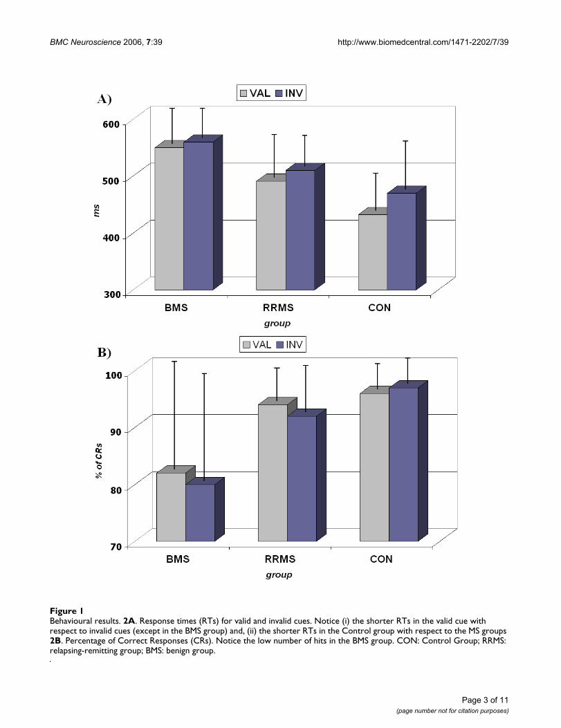
Behavioural resultsFigure 1Behavioural results. 2A. Response times (RTs) for valid and invalid cues. Notice (i) the shorter RTs in the valid cue with respect to invalid cues (except in the BMS group) and, (ii) the shorter RTs in the Control group with respect to the MS groups 2B. Percentage of Correct Responses (CRs). Notice the low number of hits in the BMS group. CON: Control Group; RRMS: relapsing-remitting group; BMS: benign group.

BMC Neuroscience 2006, 7:39 http://www.biomedcentral.com/1471-2202/7/39
The BMS group, however, did not show statistically signif-icant differences between valid and invalid reaction times(see Figure 1 and Table 1).
The intersubject comparison showed that there was aneffect of the group (F [2, 42] = 14.947, p < 0.001). Thepost-hoc analysis showed that the Control groupresponded faster than the RRMS group (p < 0.013) andthe BMS group (p < 0.001). Regarding the comparisonbetween RRMS and BMS groups, RRMS patients were sta-tistically significantly faster in responding to targets thanthe BMS group (p < 0.021) (see Figure 1 and Table 1).
In the inter-subject comparison, the analysis of the per-centage of correct responses showed that there was aneffect of the group (F [2, 42] = 6.790, p < 0.003). The post-hoc analysis revealed that this difference was statisticallysignificant in the comparison between the Control groupand the BMS group (p < 0.002) and between the RRMSand BMS groups (p < 0.022), and was not statistically sig-nificant between the RRMS group and the Control (seeFigure 1 and Table 1).
With regard to the analysis of errors, the ANOVA showeda statistically significant interaction between the effects ofthe following factors: cue, type of error and group (F [2,42] = 4.136; p < 0.020). The interpretation of this resultcomes from different evidences. First of all, there weremore errors in the valid cue than in the invalid condition,and the most frequent type of error was the one termed"missed". Moreover, this difference was exclusivelypresent for the BMS group compared to RRMS (p < 0.033)and Control groups (p < 0.003).
In the analysis of the amplitude of the P1 component,there was not statistical difference between the differentgroups (F [2, 42] = 1.786; p < 0.180). However, theobserved P1 amplitude in the Control and RRMS groups
showed higher amplitude for valid trials compared toinvalid trials, which was not observed in the BMS group.
There was no statistical difference for the P1 latencybetween the different groups. With respect to the latencyof the N1 component, the ANOVA showed a statisticallysignificant effect of the electrode factor (F [3, 42] =30.226, p < 0.001). The post-hoc analysis showed that thedifferences were due to anterior-posterior differences inlatencies (Fz-Oc = p < 0.001; Fz-Pz = p < 0.004; Oc-Cz = p< 0.001). These differences appeared in both: valid andinvalid. The effect of the group factor was statistically sig-nificant for the latency of the N1 component (F [2, 42] =4.259, p < 0.021). The post-hoc analysis revealed that thisdifference was due to a statistically significant delayedlatency of the N1 component of the BMS group withrespect to the Control group (p < 0.017) (see Figure 2 andTable 1).
For the latency of the P300 component, the ANOVA anal-ysis revealed a statistically significant effect of the groupfactor (F [2, 42] = 24.879, p < 0.001). The post-hoc analy-sis showed that the latency for the Control group (332 ±23 ms) was faster (p < 0.001) than the latency for both MSgroups (RRMS: 369 ± 28 ms; BMS: 388 ± 38 ms). The post-hoc analysis also confirmed the statistically signficant dif-ferences between MS groups (p < 0.042).
The amplitude of the early frontal N1 was clearly differentbetween the Control group and the MS groups. In fact, theMS groups presented, at that latency, a positive voltage inthe Fz electrode instead of the negativity observed in con-trols (Figure 2). The amplitude of the early N1 componentat Fz electrode showed a statistical difference for the effectof the group factor [F (1, 42) = 6.585; p < 0.003]. The post-hoc analysis showed a statistical difference between Con-trol and RRMS groups (p < 0.036) and between Controlcompared to BMS group (p < 0.005). There were not sta-
Table 1: Descriptive and Statistical results of the behavioural and ERPs data. CON: Control Group; RRMS: relapsing-remitting group; BMS: benign group.
Reaction Times Correct Response % Latency N1 Latency P300 Amplitude N1 frontal
Descriptive Valid Invalid Total Valid Invalid Total Valid Invalid Total Valid Invalid Total Valid Invalid TotalBMS 549 ± 63 560 ± 45 555 ± 51 81 ± 23 79 ± 21 80 ± 23 179 ± 14 179 ± 10 179 ± 12 383 ± 22 386 ± 21 385 ± 21 .97 ± 1.2 .97 ± 2.1 .97 ± 1.6
RRMS 492 ± 47 510 ± 43 501 ± 43 94 ± 05 92 ± 07 93 ± 06 172 ± 13 170 ± 15 171 ± 14 365 ± 19 372 ± 24 368 ± 21 .47 ± .1.2 .33 ± 1.5 .40 ± 1.3CON 433 ± 50 467 ± 56 450 ± 53 96 ± 04 97 ± 05 96 ± 05 164 ± 12 167 ± 11 165 ± 12 341 ± 15 342 ± 12 341 ± 14 -.85 ± 2.4 -.63 ± .87 -.74 ± 1.6
Anova F p F p F p F p F pCue 37.148 <0.001 0.785 0.381 0.001 0.992 1.376 0.258 0.009 0.925Elec 30.226 <0.001 3.731 0.017
Cue*Group 4.450 0.018 0.575 0.567 0.975 0.386 0.398 0.674 0.151 0.861Group 14.947 <0.001 6.790 0.003 4.259 0.021 24.879 <0.001 6.685 0.003
Post Hoc RRMS BMS CON RRMS BMS CON RRMS BMS CON RRMS BMS CON RRMS BMS CONRRMS 0.021 0.013 0.022 0.980 0.287 0.517 0.042 <0.001 0.827 0.036BMS <0.001 0.002 0.017 <0.001 0.005
Page 4 of 11(page number not for citation purposes)

BMC Neuroscience 2006, 7:39 http://www.biomedcentral.com/1471-2202/7/39
tistically significant differences between RRMS and BMSgroups.
DiscussionThe performance in the attentional task was poorer forBMS group compared to the RRMS group and for both MSgroups with respect to the Control group. This differencein behaviour was statistically significant and was consist-
ent across all behavioural variables (RTs, errors and per-centage of correct responses). This difference cannot beexplained by a speed-accuracy trade-off because MSgroups were also less accurate than Control group, partic-ularly in the case of BMS group. The increase in RTs is acommon result obtained in the MS literature [24,32] andsuggests that BMS patients also show a deterioration invisuo-motor processing as other subtypes of MS.
ERPs to the standard stimuli in the Fz, Cz, Pz and Oc electrodes for the Control, RRMS and BMS groupsFigure 2ERPs to the standard stimuli in the Fz, Cz, Pz and Oc electrodes for the Control, RRMS and BMS groups. Notice the delayed latency of the P300 (inverted triangle) and posterior N1 (asterisk). Also notice the lack of the early frontal N1 in the BMS and RRMS group (the horizontal line indicates the time window used for statistical analysis). CON: Control group; RRMS: relaps-ing-remitting group; BMS: benign group
Page 5 of 11(page number not for citation purposes)
BMC Neuroscience 2006, 7:39 http://www.biomedcentral.com/1471-2202/7/39
The behavioural results, using the central cue paradigm,replicate the studies where an improvement of the RTs isobserved for the valid cues compared to the invalid ones[26,33]. This is explained by the need of a reorientingmechanism in the invalid condition that it is not neces-sary for the valid condition. The differences obtained inthe RTs during the Posner paradigm with central cuesindicate that the performance in the spatial attentionprocessing in the BMS group is impaired, given that therewas not a statistically significant difference between RTsfor valid and invalid cues in the BMS group and the laten-cies were delayed compared to the Control group. Thisimpairment could involve both orienting (indicated bythe longer RTs in both conditions for BMS) and reorient-ing mechanisms (indicated by the lack of statistical signif-icance between valid and invalid conditions).
The present study reveals that validity effects wereobtained in Control and RRMS groups but were notobtained in the BMS group. It is possible that the lack ofthe validity effect in the BMS group is caused by a pro-longed duration of the moving and engaging operations,which would produce a divided attention situationinstead of an oriented spatial attention condition, as itoccurs in the Control and RRMS groups.
To check if these patients were orienting or not to the cue,we checked if the P1 component showed a change ofamplitude to the stimuli when they were presented in thevalidly field with respect to when the cue invalidly cuedthe position of the stimulus [34]. The result of this analy-sis was that although there were not statistically signifi-cant differences, a higher amplitude was observed for thevalidly cued stimulus with respect to the invalidly cuedstimulus in the Control and RRMS groups. However, forthe BMS group, the amplitude of P1 was practically equalfor both conditions, suggesting that it is probable thatthese patients carried out a divided attention setting dur-ing the course of the task.
The analysis of errors yielded a similar conclusion thanthe RTs analysis, an increase in the number of errors in theBMS group with respect to RRMS and controls. The overallconclusion is that both MS groups showed a behaviouraldeterioration compared to controls, but this deteriorationwas higher in the BMS group than in the RRMS group.
The latency of the P1 component did not reflect statisti-cally significant differences among the experimentalgroups. These results suggest that the differences found inthe behavioural variables among the diverse groups arenot due to a different processing in the speed of sensory-visual pathways. One interesting phenomenon appearingin the ERPs at the same latency than P1 was the inversionof polarity that occurs in MS patients of the early N1 fron-
tal component. The early N1 frontal component thatappeared in controls has been previously described [35].They have suggested that this initial volley of informationarrives to the frontal cortex through the dorsal stream andcan be used for an alerting function in the frontal cortexwhile more elaborated information arrives from compu-tation in higher order visual cortex. The patient's inver-sion of polarity obtained in the N1 frontal componentcould be due to a lack of this communication because ofcentral demyelinization. If the frontal N1 component isnot formed, the posterior P1 could have a much morefrontal extension and produce the inversion of polarityobserved in MS patients in frontal electrodes. A functionalconsequence of that would be the lack of the fronto-occip-ital attentional control that would occur after the frontalN1 component [35].
The latency of the posterior N1 was increased in the BMSgroup with respect to controls. Given the implication ofthe N1 in the object discrimination process and its locali-zation in the occipito-ventral areas [20,29], the increase ofN1 latency in BMS group would implicate a delay in visualdiscrimination processes with respect to controls.
On the other hand, we can observe the significant delay inthe latency of the P300 component for MS groups, beingthe BMS group the one that exhibited the longest latencieswhen compared to the Control group. Indeed, the BMSgroup showed statistical differences even with the RRMSgroup. Part of this delay is partially due to the N1 latencydelay, at least for the BMS group. This latency delay of theP3 component has been observed in previous studies withdifferent pathologies. In the particular case of the multiplesclerosis, Polich [23] suggested a cognitive deficit associ-ated to the anomalies in the latency and amplitude of theP300 component. Ellger [25] presented a significant delayof the latency of the P300 in different groups of MS,although the BMS was not studied. Aminof [36] indicatedthat the changes in the components of the ERPs in the MSare due to a central disorder that correlates with thepatient's cognitive state. Dujardin [37] suggested, usingbehavioural variables, that the attentional deficit in MSdoes not depend on the attentional gain mechanisms.Instead, the deficit would be caused by the lack of centralresources in the processing of visual information. Ourstudy reinforces these previous empirical results and sup-ports the hypothesis that the deficit exhibited is related tothe cognitive processing and not only dependent on sen-sory processing deterioration.
It is necessary to highlight that in our study, the measuresof P300 peak latency were carried out with standard stim-uli (that does not require motor response). Therefore, it ispossible to discard any contribution from motor poten-tials. Moreover, the impaired performance in behaviour
Page 6 of 11(page number not for citation purposes)
BMC Neuroscience 2006, 7:39 http://www.biomedcentral.com/1471-2202/7/39
Page 7 of 11(page number not for citation purposes)
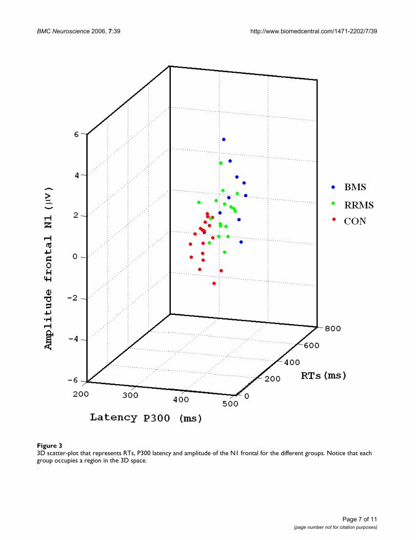
3D scatter-plot that represents RTs, P300 latency and amplitude of the N1 frontal for the different groupsFigure 33D scatter-plot that represents RTs, P300 latency and amplitude of the N1 frontal for the different groups. Notice that each group occupies a region in the 3D space.
BMC Neuroscience 2006, 7:39 http://www.biomedcentral.com/1471-2202/7/39
for the MS patients observed in this study cannot beexplained exclusively in terms of motor dysfunction. Thedelay in the P300 component has to be related to a cogni-tive impairment in the MS patients. It is important tohighlight that the capacity of attentional orientation inthe visual world is a function of enormous importance. Itis not only related to the visuo-attentional process itself,but it is involved in many processes (e.g.: learning, mem-ory, etc).
As mentioned above, one of the most surprising results isthe worse execution in the task for what is called benigncourse of disease (BMS group). In the clinical practice,these patients show a slow evolution of the disease with-out the presence of evident clinical signs for many yearsand, therefore, with no pharmacological therapy applied[1-4,38-40]. However, our test has allowed us to checkthat these patients show a poorer behavioural executionin terms of RTs and errors compared with the RRMSgroup. Moreover, the BMS group presented the mostimpaired ERPs in both the latencies of the posterior N1and P300, and in the absence of the frontal N1.
According to those results, it is possible to ask if thesetechniques could be useful for diagnostic purposes. Thepresent results suggest indeed that differences take placeamong the different studied groups. However, given thenumber of cases studied in our study, it seems daring todefine these measures as diagnostic tools. We believe thatmore studies are necessary to confirm the validity of theseparameters in the diagnosis of multiple sclerosis and beincluded in future diagnostic criteria.
However, the evidences found in the study suggest thatthe behavioural and psychophysiological measures can begood estimators of the degree of attentional impairmentin multiple sclerosis and help professionals to apply theappropriate therapeutic measures (see Figure 3).
On the other hand, these results prove that the neurolog-ical and cognitive evolution of a MS patient does not cor-relate. In fact, in subjects with a low level of neurologicalincapacity (EDSS < 3 or 4), the cognitive profile isaffected. To conclude, these data suggest that the cognitiveimpairment in MS could develop in a subtle way duringthe course of the disease, producing a deterioration simi-lar to that experienced by RRMS patients. This fact pointsto the necessity of increasing the research in clinical andlaboratory parameters in the prediction and more properdefinition of benign form of multiple sclerosis [31]. Theseresults confirm that the use of psychophysiological tech-niques would be recommended to explore the cognitiveprofile in multiple sclerosis patients as a complementarytest for the neurological and neuropsychological examina-tion.
ConclusionCognitive deterioration is a highly prevalent symptomaccompanying the general deterioration of MS patients. Inthe present experiment, these patients showed an atten-tional impairment indexed by ERPs and behaviouralresponses. The main result was that BMS group presenteda higher deterioration than RRMS group. The latter resultsuggests that it could be necessary to follow up the cogni-tive evolution of the patients with BMS and to considertherapeutic alternatives to those offered today. Psycho-physiological techniques combined with specific tasks(e.g. Posner paradigm) can be helpful in evaluating cogni-tive impairment in MS.
MethodsSubjects and procedureThree different groups of subjects participated in thestudy: The first group included 17 patients (women 12;age 38.88 ± 9.04 years old) diagnosed with RRMS withoutclear signs of motor impairment, and with a disease dura-tion of 4.67 ± 4.13 years. The score in Multiple SclerosisExpanded Disability Status Scale (EDSS) was 1.6 ± 0.9.The second group included 10 patients (women 6; age42.30 ± 7.21 years old) diagnosed with BMS. Thesepatients had a disease duration of 12.09 ± 4.77 years andat least 8 years of disease evolution scoring under 3.5 inthe EDSS scale. Diagnosis was made following the criteriaof Poser [1]. The exclusion criteria for the MS groups were:the presence of a relapse, medication in the last month,and/or the presence of clear signs of depression. Allpatients were clinically stable at baseline (i.e., had noexacerbations within several months before of studyentry). There were no patients treated with any medica-tion at the time of evaluation, and only a few presentedhistory of corticoids treatment in past relapses. Patientspreviously assessed at the Multiple Sclerosis Unit of theNeurology service of the Virgen Macarena Hospital(Seville, Spain) participated voluntarily in the Psycho-physiological testing.
The data from both clinical groups were compared with agroup of 18 healthy subjects (women 15; age 36.54 ± 8.73years old) similar in age, gender proportion and educa-tional level. The experimental protocol was approved bythe ethical committee of the Hospital. After a full explana-tion of the nature and general objectives of the experi-ments, a written consent statement was obtained fromsubjects: MS patients and controls.
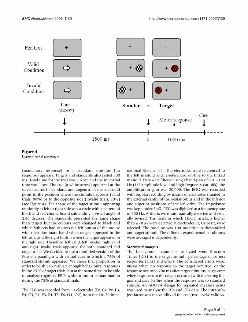
Experimental protocolBehavioural responses were recorded during the Posnerparadigm. Five blocks with 200 trials each were presented.The subjects had 2–3 minutes of rest between blocks. Atrial consisted in a central cue (lasting 300 ms) pointingto the left or to the right side of the screen, where a target
Page 8 of 11(page number not for citation purposes)
BMC Neuroscience 2006, 7:39 http://www.biomedcentral.com/1471-2202/7/39
(mandatory response) or a standard stimulus (noresponse) appears. Targets and standards also lasted 300ms. Total time for the trial was 1.5 sec and the inter-trialtime was 1 sec. The cue (a white arrow) appeared at thescreen centre. In standards and targets trials the cue couldpoint to the position where the stimulus appears (validtrials, 80%) or to the opposite side (invalid trials, 20%)(see Figure 4). The shape of the target stimuli appearingrandomly at left or right side was a circle with a pattern ofblack and red checkerboard subtending a visual angle of2.46 degrees. The standards presented the same shapethan targets but the colours were changed to black andwhite. Subjects had to press the left button of the mousewith their dominant hand when targets appeared in theleft side, and the right button when the target appeared inthe right side. Therefore, left valid, left invalid, right validand right invalid trials appeared for both: standard andtarget trials. We decided to use a modified version of thePosner's paradigm with central cues in which a 75% ofstandard stimuli appeared. We chose that proportion inorder to be able to evaluate enough behavioural responsesin the 25 % of target trials, but at the same time, to be ableto analyse cognitive ERPs without motor contaminationduring the 75% of standard trials.
The EEG was recorded from 13 electrodes (Fz, Cz, Pz, F3,F4, C3, F4, P3, F4, T5, F6, O1, O2) from the 10–20 Inter-
national System [41]. The electrodes were referenced tothe left mastoid and re-referenced off-line to the linkedmastoid. Data were filtered using a band-pass of 0.01–100Hz (1/2 amplitude low- and high-frequency cut-offs); theamplification gain was 20,000. The EOG was recordedwith bipolar recording by means of electrodes situated inthe external canthi of the ocular orbits and in the inferiorand superior positions of the left orbit. The impedancewas kept under 5 kΩ. EEG was digitised at a frequency rateof 500 Hz. Artifacts were automatically detected and visu-ally revised. The trials in which HEOG artefacts higherthan ± 70 µV were detected at electrodes Fz, Cz or Pz, wererejected. The baseline was 100 ms prior to thestandardand target stimuli. The different experimental conditionswere averaged independently.
Statistical analysisThe behavioural parameters analysed were ReactionTimes (RTs) to the target stimuli, percentage of correctresponses (CRs) and errors. The considered errors were:missed when no response to the target occurred, or theresponse occurred 700 ms after target stimulus; target errorwhen responses to the targets occurred with the wrong fin-ger; and false positive when the response was to standardstimuli. An ANOVA design for repeated measurementswas used to analyse the RTs and CRs data. The intra-sub-ject factor was the validity of the cue (two levels: valid vs.
Experimental paradigmFigure 4Experimental paradigm.
Page 9 of 11(page number not for citation purposes)
BMC Neuroscience 2006, 7:39 http://www.biomedcentral.com/1471-2202/7/39
invalid) and the intersubject factor was the subject's group(three levels: RRMS, BMS and Controls). The Bonferronicorrection was used for post-hoc analysis. In the case oferrors, a mixed factorial ANOVA designed for repeatedmeasurements was used. The intra-subject factors were:the validity of the cue (two levels: valid vs. Invalid) andthe classification of errors (misses, target error, and falsepositive); the intersubject factor was the subject's group(three levels: RRMS, BMS and Controls).
Latency was measured for P1, N1 and P300 components.The P1 latency was obtained as the highest positive deflec-tion in the time window 90–160 ms for the electrode Oc(electrode Oc was computed off-line as the mean voltageof O1 and O2); the N1 component as the highest negativedeflection in the time window 130–210 ms for the elec-trodes Fz, Cz, Pz and Oc, and the P300 as the highest pos-itive deflection in the time window 280–400 ms for theelectrodes Fz, Cz and Pz.
The statistical significance of latency differences in the P1component was tested independently by means of one-factor ANOVA (three levels: RRMS, BMS and Controls).To analyse the latency of the N1 and P300 component, amixed factorial designed for repeated measurements wasapplied to the latency data. For N1 and P300 analysis, theintra-subject factors were: a) validity of the cue (two lev-els: Valid and invalid) and b) electrode. The inter-subjectfactor was the subject's group (Three levels: RRMS, BMSand Controls).
Our initial objectives were related to the latency analysisof ERPs. However, after visual inspection of ERPs in themidline, an inversion of polarity in the frontal N1 compo-nent was obvious for the MS groups (see Figure 2). Thishighly unexpected result motivated the amplitude analy-sis of the frontal N1 in the Fz electrode in which a positivedeflection appeared for the MS groups. The mean voltagebetween 110 and 130 ms in the electrode Fz was com-puted in the three subject groups. An ANOVA design forrepeated measurements was used to analyse the ampli-tude of the frontal N1. The intra-subject factor was thevalidity of the cue (two levels: valid vs. Invalid), and theintersubject factor was the subject's group (three levels:RRMS, BMS and Controls).
In order to verify the effect of validity in the early phase ofattentional processing, the differences of amplitude forthe P1 component were analysed. The P1 amplitude wascalculated based in a positive deflection which appearedin the posterior electrodes in all groups. The mean voltageoccurring between 120 and 150 ms in the electrode P3and P4 was computed in all stimular conditions (leftvalid, left invalid, right valid and right invalid trials). AnANOVA design for repeated measurements was used to
analyse the amplitude of the P1. The intra-subject factorswere: a) validity of the cue (two levels: valid vs. invalid),b) side of visual field stimulation (two levels: left vs. right)and c) electrode (two levels: p3 and p4). The intersubjectfactor was the subject's group (three levels: RRMS, BMSand Controls).
AbbreviationsANOVA analyses of variance
BMS benign multiple sclerosis
CRs correct responses
EDSS expanded disability status scale
EEG electroencephalography
ERPs event-related potentials
MRI Magnetic Resonance Imaging
MS multiple sclerosis
RRMS relapsing-remitting multiple sclerosis
RTs reaction times
Authors' contributionsMV and JJGR, with CG and PD participated in the plan-ning of the study. JJGR and MV participated in the acqui-sition and execution of the study and performed the dataanalysis. MV, CG, GI, EV, PD and JJGR contributed to theinterpretation of results. The patients were selected by PD,MB, MAG and GI. JJGR drafted the manuscript. The man-uscript was subsequently revised by MV, CG, EV and PDand GI, and all authors gave final approval.
AcknowledgementsThis study has been supported by Virgen Macarena Hospital Association from Seville, by Ministerio de Educación y Ciencia through to Ramon y Cajal fellowship to M. Vazquez, and by Neuroinvest.
References1. Poser Poser CM, Paty DW, Scheinberg L, McDonald WI, Davis FA,
Ebers GC, Johnson KP, Sibley WA, Silberberg DH, Tourtellotte WW:New diagnostic criteria for multiple sclerosis: guidelinesresearch protocols. Ann Neurol 1983, 13:227-31.
2. Keegan BM, Noseworthy JH: Multiple sclerosis. Annu Rev Med2002, 53:285-302.
3. Kujala P, Portin R, Ruutianinen J: The progress of cognitivedecline in Multiple Sclerosis: A controled 3 year follow-up.Brain 1997, 120:289-297.
4. Scolding N: The differential diagnosis of multiple sclerosis. JNeurol Neurosurg Psychiatry 2001, 71(suppl 2):9-15.
5. McDonald WI, Compston A, Edan G, Goodkin D, Hartung HP, LublinFD, McFarland HF, Paty DW, Polman CH, Reingold SC, Sandberg-Wollheim M, Sibley W, Thompson A, van den Noort S, WeinshenkerBY, Wolinsky JS: Recommended diagnostic criteria for multi-
Page 10 of 11(page number not for citation purposes)
http://www.ncbi.nlm.nih.gov/entrez/query.fcgi?cmd=Retrieve&db=PubMed&dopt=Abstract&list_uids=6847134
http://www.ncbi.nlm.nih.gov/entrez/query.fcgi?cmd=Retrieve&db=PubMed&dopt=Abstract&list_uids=6847134
http://www.ncbi.nlm.nih.gov/entrez/query.fcgi?cmd=Retrieve&db=PubMed&dopt=Abstract&list_uids=6847134
http://www.ncbi.nlm.nih.gov/entrez/query.fcgi?cmd=Retrieve&db=PubMed&dopt=Abstract&list_uids=9117376

BMC Neuroscience 2006, 7:39 http://www.biomedcentral.com/1471-2202/7/39
Publish with BioMed Central and every scientist can read your work free of charge
"BioMed Central will be the most significant development for disseminating the results of biomedical research in our lifetime."
Sir Paul Nurse, Cancer Research UK
Your research papers will be:
available free of charge to the entire biomedical community
peer reviewed and published immediately upon acceptance
cited in PubMed and archived on PubMed Central
yours — you keep the copyright
Submit your manuscript here:http://www.biomedcentral.com/info/publishing_adv.asp
BioMedcentral
ple sclerosis; guidelines from the international panel on thediagnosis of multiple sclerosis. Ann Neurol 2001, 50:121-127.
6. Ebers GC: Natural history of multiple sclerosis. J Neurol Neuro-surg Psychiatry 2001, 71(suppl 2):16-19.
7. Foong J, Rozewicz L, Quaghebeur G, Thompson AJ, Miller DH, RonMA: Neuropsychological deficits in multiple sclerosis afteracute relapse. J Neurol Neurosurg Psychiatry 1998, 64(4):529-32.
8. Rao SM: Neuropsychology of multiple sclerosis. Curr Opin Neu-rol 1995, 8(3):216-20.
9. Brassington JC, Marsh NV: Neuropsychological aspects of mul-tiple sclerosis. Neuropsychol Rev 1998, 8:43-77.
10. Gadea M, Martinez-Bisbal MC, Marti-Bonmati L, Espert R, CasanovaB, Coret F, Celda B: Spectroscopic axonal damage of the rightlocus coeruleus relates to selective attention impairment inearly stage relapsing-remitting multiple sclerosis. Brain 2004,127(pt 1):89-98.
11. Rao SM: Cognitive function in patients with multiple sclerosis:Impairment and treatment. Int J of MS Care 2004, 1:9-22.
12. O'Connor P, Marchetti P, Lee L, Perera M: Evoked potentialabnormality scores are a useful measure of disease burden inrelapsing-remitting multiple sclerosis. Ann Neurol 1998,44(3):404-7.
13. Comi G, Leocani L, Locatelli T, Medaglini S, Martinelli V: Electro-physiological investigations in multiple sclerosis dementia.Electroencephalogr Clin Neurophysiol Suppl 1999, 50:480-5.
14. Polich J, Herbst KL: P300 as a clinical assay: rationale, evalua-tion, and findings. Int J Psychophysiol 2000, 38(1):3-19.
15. Polich J: P300 clinical utility and control of variability. J ClinNeurophy 1998, 15(1):14-33.
16. Fuhr P, Kappos L: Evoked potentials for evaluation of multiplesclerosis. Clin Neurophysio 2001, 112(12):2185-2189.
17. Swanwick GR, Rowan M, Coen RF, O'Mahony D, Lee H, Lawlor BA,Walsh JB, Coakley D: Clinical application of electrophysiologi-cal markers in the differential diagnosis of depression andvery mild Alzheimer's disease. J Neurol Neurosurg Psychiatry 1996,60(1):82-6.
18. Wang H, Wang Y, Wang D, Cui L, Tian S, Zhang Y: Cognitiveimpairment in Parkinson's disease revealed by event-relatedpotential N270. J Neurol Sci 2002, 194(1):49-53.
19. Frodl T, Meisenzahl EM, Muller D, Holder J, Juckel G, Moller HJ,Hegerl U: P300 subcomponents and clinical symptoms inschizophrenia. Int J Psychophysiol 2002, 43(3):237-46.
20. Hillyard SA, Anllo-Vento L: Event-related brain potentials in thestudy of visual selective attention. Proc Natl Acad Sci USA 1998,95(3):781-7.
21. Sfagos C, Papageorgiou CC, Kosma KK, Kodopadelis E, Uzunoglu NK,Vassilopoulos D, Rabavilas AD: Working memory deficits inmultiple sclerosis: a controlled study with auditory P600 cor-relates. J Neurol Neurosurg Psychiatry 2003, 74(9):1231-5.
22. Leocani L, Locatelli T, Martinelli V, Rovaris M, falautano M, Fillipi M,magnani G, Comi G: Electroencephalographic coherence anal-ysis in multiple sclerosis: correlation with clinical neuropsy-chological and MRI findings. J Neurol Neurosurg Psychiatry 2000,69:192-198.
23. Polich J, Romine JS, Sipe JC, Aung M, Dalessio DJ: P300 in multiplesclerosis: a preliminary report. Int J Psychophysiol 1992,12(2):155-63.
24. Gerschlager W, Beisteiner R, Deecke L, Dirnberger G, Endl W, Kol-legger H, Lindinger G, Vass K, Lang W: Electrophysiological, neu-ropsychological and clinical findings in multiple sclerosispatients receiving interferon β-1b: a 1-year follow-up. EurNeurol 2000, 44:205-209.
25. Ellger T, Bethke F, Frase A, Luettman RJ, Buchheister A, RingelsteinEB, Evers S: Event-related potentiels in different subtypes ofmultiple sclerosis: a cross-sectional study. J Neurol Scien 2002,205:35-40.
26. Posner MI, Walker JA, Friendrich FJ, Rafal RD: Effects of parietalinjury on covert orienting of attention. J Neurosci 1984,4:1863-74.
27. Posner MI, Early TS, Reiman E, Pardo PJ: Asymmetries in hemi-spherical control of attention in schizophrenia. Arch Gen Psy-chiatry 1988, 45:814-821.
28. Posner MI, Petersen SE: The attention system of the humanbrain. Annu Rev Neurosci 1990, 13:25-42.
29. Gomez C, Clark V, Hillyard S: Localization of neural generatorsduring visual selective attention. Brain Topogr 1994, 7:41-51.
30. Gomez CM, Fernandez A, Maestu F, Amo C, Gonzalez-Rosa JJ,Vaquero E, Ortiz T: Task-specific sensoryand motor prepara-tory activation revealed by contingent magnetic variation.Brain Res Cogn Brain Res 2004, 21(1):59-68.
31. Ramsaransing G, Mauritis N, Zwanikken C, De Keyser J: Early pre-diction of a benign course of multiple sclerosis on clinicalgrounds: a systematic review. Mult Scler 2001, 7(5):345-347.
32. Vitkovitch M, Bishop S, Dancey C, Richards A: Stroop interferenceand negative priming in patients with multiple sclerosis. Neu-ropsychologia 2002, 40(9):1570-6.
33. Posner MI: Orienting and attention. Q J Exp Psychol 1980, 32:3-25.34. Mangun GR, Hillyard SA: Allocation of visual attention to spatial
locations: tradeoff functions for event-related brain poten-tials and detection performance. Percept Psychophys 1990,47(6):532-50.
35. Foxe JJ, Simpson GV: Flow activation from V1 to frontal cortexin humans. A framework for defining "early" visual process-ing. Exp Brain Res 2002, 142(1):139-50.
36. Aminoff JC, Goodin DS: Long-latency cerebral event-relatedpotentials in multiple sclerosis. J Clin Neurophysiol 2001,18(4):372-7.
37. Dujardin K, Donce AC, Hautecoeur P: Attention impairment nrecently diagnosed multiple sclerosis. Eur J Neurol 1998,5:61-66.
38. Horsfield MA, Lai M, Webb SL, Barker GJ, Tofts PS, Turner R, RudgeP, Miller DH: Apparent diffusion coefficients in benign and sec-ondary progressive multiple sclerosis by nuclear magneticresonance. Magn Reson Med 1996, 36(3):393-400.
39. Kidd D, Thompson AJ, Kendal BE, Miller DH, McDonald WI: Benignform of multiple sclerosis: MRI evidence for less frequent andless inflammatory disease activity. J Neurol Neurosurg Psychiatry1994, 57(9):1070-1072.
40. Falini A, Calabrese G, Fillipi M, Origi D, Lipari S, Colombo B, Comi Gand Scotti G: Benign versus secundary-progresive multiplesclerosis: the potencial role of proton MR spectrcopy indefining the nature of disability. Am J Neuroradiol 1998,19:223-229.
41. Jasper HH: The ten-twenty electrode system. Electroencephalog-raphy and Clinical Neurophysiology 1958, 10:371-375.
Page 11 of 11(page number not for citation purposes)
http://www.ncbi.nlm.nih.gov/entrez/query.fcgi?cmd=Retrieve&db=PubMed&dopt=Abstract&list_uids=9576548
http://www.ncbi.nlm.nih.gov/entrez/query.fcgi?cmd=Retrieve&db=PubMed&dopt=Abstract&list_uids=9576548
http://www.ncbi.nlm.nih.gov/entrez/query.fcgi?cmd=Retrieve&db=PubMed&dopt=Abstract&list_uids=7551121
http://www.ncbi.nlm.nih.gov/entrez/query.fcgi?cmd=Retrieve&db=PubMed&dopt=Abstract&list_uids=9658410
http://www.ncbi.nlm.nih.gov/entrez/query.fcgi?cmd=Retrieve&db=PubMed&dopt=Abstract&list_uids=9658410
http://www.ncbi.nlm.nih.gov/entrez/query.fcgi?cmd=Retrieve&db=PubMed&dopt=Abstract&list_uids=9749611
http://www.ncbi.nlm.nih.gov/entrez/query.fcgi?cmd=Retrieve&db=PubMed&dopt=Abstract&list_uids=9749611
http://www.ncbi.nlm.nih.gov/entrez/query.fcgi?cmd=Retrieve&db=PubMed&dopt=Abstract&list_uids=9749611
http://www.ncbi.nlm.nih.gov/entrez/query.fcgi?cmd=Retrieve&db=PubMed&dopt=Abstract&list_uids=8558158
http://www.ncbi.nlm.nih.gov/entrez/query.fcgi?cmd=Retrieve&db=PubMed&dopt=Abstract&list_uids=8558158
http://www.ncbi.nlm.nih.gov/entrez/query.fcgi?cmd=Retrieve&db=PubMed&dopt=Abstract&list_uids=8558158
http://www.ncbi.nlm.nih.gov/entrez/query.fcgi?cmd=Retrieve&db=PubMed&dopt=Abstract&list_uids=9448241
http://www.ncbi.nlm.nih.gov/entrez/query.fcgi?cmd=Retrieve&db=PubMed&dopt=Abstract&list_uids=9448241
http://www.ncbi.nlm.nih.gov/entrez/query.fcgi?cmd=Retrieve&db=PubMed&dopt=Abstract&list_uids=1592668
http://www.ncbi.nlm.nih.gov/entrez/query.fcgi?cmd=Retrieve&db=PubMed&dopt=Abstract&list_uids=1592668
http://www.ncbi.nlm.nih.gov/entrez/query.fcgi?cmd=Retrieve&db=PubMed&dopt=Abstract&list_uids=6737043
http://www.ncbi.nlm.nih.gov/entrez/query.fcgi?cmd=Retrieve&db=PubMed&dopt=Abstract&list_uids=6737043
http://www.ncbi.nlm.nih.gov/entrez/query.fcgi?cmd=Retrieve&db=PubMed&dopt=Abstract&list_uids=3415424
http://www.ncbi.nlm.nih.gov/entrez/query.fcgi?cmd=Retrieve&db=PubMed&dopt=Abstract&list_uids=3415424
http://www.ncbi.nlm.nih.gov/entrez/query.fcgi?cmd=Retrieve&db=PubMed&dopt=Abstract&list_uids=2183676
http://www.ncbi.nlm.nih.gov/entrez/query.fcgi?cmd=Retrieve&db=PubMed&dopt=Abstract&list_uids=2183676
http://www.ncbi.nlm.nih.gov/entrez/query.fcgi?cmd=Retrieve&db=PubMed&dopt=Abstract&list_uids=7803199
http://www.ncbi.nlm.nih.gov/entrez/query.fcgi?cmd=Retrieve&db=PubMed&dopt=Abstract&list_uids=7803199
http://www.ncbi.nlm.nih.gov/entrez/query.fcgi?cmd=Retrieve&db=PubMed&dopt=Abstract&list_uids=7367577
http://www.ncbi.nlm.nih.gov/entrez/query.fcgi?cmd=Retrieve&db=PubMed&dopt=Abstract&list_uids=2367174
http://www.ncbi.nlm.nih.gov/entrez/query.fcgi?cmd=Retrieve&db=PubMed&dopt=Abstract&list_uids=2367174
http://www.ncbi.nlm.nih.gov/entrez/query.fcgi?cmd=Retrieve&db=PubMed&dopt=Abstract&list_uids=2367174
http://www.ncbi.nlm.nih.gov/entrez/query.fcgi?cmd=Retrieve&db=PubMed&dopt=Abstract&list_uids=8875409
http://www.ncbi.nlm.nih.gov/entrez/query.fcgi?cmd=Retrieve&db=PubMed&dopt=Abstract&list_uids=8875409
http://www.ncbi.nlm.nih.gov/entrez/query.fcgi?cmd=Retrieve&db=PubMed&dopt=Abstract&list_uids=8875409
http://www.ncbi.nlm.nih.gov/entrez/query.fcgi?cmd=Retrieve&db=PubMed&dopt=Abstract&list_uids=8089671
http://www.ncbi.nlm.nih.gov/entrez/query.fcgi?cmd=Retrieve&db=PubMed&dopt=Abstract&list_uids=8089671
http://www.ncbi.nlm.nih.gov/entrez/query.fcgi?cmd=Retrieve&db=PubMed&dopt=Abstract&list_uids=8089671
http://www.ncbi.nlm.nih.gov/entrez/query.fcgi?cmd=Retrieve&db=PubMed&dopt=Abstract&list_uids=9504469
http://www.ncbi.nlm.nih.gov/entrez/query.fcgi?cmd=Retrieve&db=PubMed&dopt=Abstract&list_uids=9504469
Related Documents