JOURNAL OF VIROLOGY, Mar. 2008, p. 2975–2988 Vol. 82, No. 6 0022-538X/08/$08.000 doi:10.1128/JVI.02216-07 Copyright © 2008, American Society for Microbiology. All Rights Reserved. Differential CD4 versus CD8 T-Cell Responses Elicited by Different Poxvirus-Based Human Immunodeficiency Virus Type 1 Vaccine Candidates Provide Comparable Efficacies in Primates † Petra Mooij, 1 * Sunita S. Balla-Jhagjhoorsingh, 1 ‡ Gerrit Koopman, 1 Niels Beenhakker, 1 Patricia van Haaften, 1 § Ilona Baak, 1 ¶ Ivonne G. Nieuwenhuis, 1 Ivanela Kondova, 1 Ralf Wagner, 2 Hans Wolf, 2 Carmen E. Go ´mez, 3 Jose ´ L. Na ´jera, 3 Victoria Jime ´nez, 3 Mariano Esteban, 3 and Jonathan L. Heeney 1,4 Department of Virology, Biomedical Primate Research Center, 2288 GJ Rijswijk, The Netherlands 1 ; Institut fu ¨r Medizinische Mikrobiologie und Hygiene der Universita ¨t Regensburg 39053, Germany 2 ; Department of Cell and Molecular Biology, Centro Nacional de Biotecnologia, CSIC, Madrid 28049, Spain 3 ; and the University of Cambridge, Department of Veterinary Medicine, Cambridge CB3 0ES, United Kingdom 4 Received 11 October 2007/Accepted 13 December 2007 Poxvirus vectors have proven to be highly effective for boosting immune responses in diverse vaccine settings. Recent reports reveal marked differences in the gene expression of human dendritic cells infected with two leading poxvirus-based human immunodeficiency virus (HIV) vaccine candidates, New York vaccinia virus (NYVAC) and modified vaccinia virus Ankara (MVA). To understand how complex genomic changes in these two vaccine vectors translate into antigen-specific systemic immune responses, we undertook a head-to-head vaccine immunogenicity and efficacy study in the pathogenic HIV type 1 (HIV-1) model of AIDS in Indian rhesus macaques. Differences in the immune responses in outbred animals were not distinguished by enzyme- linked immunospot assays, but differences were distinguished by multiparameter fluorescence-activated cell sorter analysis, revealing a difference between the number of animals with both CD4 and CD8 T-cell responses to vaccine inserts (MVA) and those that elicit a dominant CD4 T-cell response (NYVAC). Re- markably, vector-induced differences in CD4 /CD8 T-cell immune responses persisted for more than a year after challenge and even accompanied antigenic modulation throughout the control of chronic infection. Importantly, strong preexposure HIV-1/simian immunodeficiency virus-specific CD4 T-cell responses did not prove deleterious with respect to accelerated disease progression. In contrast, in this setting, animals with strong vaccine-induced polyfunctional CD4 T-cell responses showed efficacies similar to those with stronger CD8 T-cell responses. The global spread of human immunodeficiency virus (HIV) has reached pandemic proportions (http://www.unaids.org). Despite more than two decades of research since the discovery of HIV as the etiologic agent of AIDS, the development of an effective HIV type 1 (HIV-1) vaccine remains an unfulfilled priority. While it is generally accepted that ultimately a pro- phylactic HIV-1 vaccine should induce both humoral and cell- mediated immune responses to a number of different HIV antigens (40, 63), envelope-based immunogens capable of in- ducing broadly neutralizing responses currently are not avail- able (13, 79, 98). Recent approaches have focused on vaccines capable of inducing potent CD8 T-cell responses to control the virus load, to reduce transmission, and to slow disease development (26, 53). Evidence from both humans and non- human primates for the role of T-cell responses in the control of HIV includes the correlation between HIV-specific CD8 T cells and the control of plasma viremia (51, 52, 99); the asso- ciation of certain restricting major histocompatibility complex (MHC) class I alleles, conserved T-cell epitopes, and slow disease progression (14, 27, 28, 48, 55, 59, 61, 64, 70, 72, 90, 100); and the rapid increase in viral load after experimental CD8 lymphocyte depletion in simian immunodeficiency virus (SIV)- or simian-human immunodeficiency virus (SHIV)-in- fected rhesus macaques (2, 54, 86), providing a strong rationale for the development of T-cell-based vaccines. Recently the quality of the HIV-specific CD8 T cells associated with the control of HIV-1 virus loads in human long-term nonproges- sors has been described, revealing characteristics of a polyfunc- tional profile simultaneously capable of degranulation and of producing gamma interferon (IFN-), interleukin-2 (IL-2), tu- mor necrosis factor alpha (TNF-), and macrophage inflam- matory protein 1- (9). In contrast, while anti-HIV CD8 and CD4 T-cell responses have been demonstrated to have a positive effect on controlling virus loads, HIV-1-specific CD4 T-cell responses also have been implicated as being deleteri- ous. Indeed, the finding that HIV-1 preferentially infects HIV- specific CD4 T cells has suggested a possible contraindication * Corresponding author. Mailing address: Department of Virology, Biomedical Primate Research Centre (BPRC), P.O. Box 3306, 2280 GH Rijswijk, The Netherlands. Phone: 31 152842615. Fax: 31 152842601. E-mail: [email protected]. † Supplemental material for this article may be found at http://jvi .asm.org/. ‡ Present address: Institute of Tropical Medicine, B2000 Antwerp, Belgium. § Present address: LUMC, 2300 RC Leiden, The Netherlands. ¶ Present address: NFI, 2497 BG The Hague, The Netherlands. Published ahead of print on 9 January 2008. 2975

Welcome message from author
This document is posted to help you gain knowledge. Please leave a comment to let me know what you think about it! Share it to your friends and learn new things together.
Transcript

JOURNAL OF VIROLOGY, Mar. 2008, p. 2975–2988 Vol. 82, No. 60022-538X/08/$08.00�0 doi:10.1128/JVI.02216-07Copyright © 2008, American Society for Microbiology. All Rights Reserved.
Differential CD4� versus CD8� T-Cell Responses Elicited by DifferentPoxvirus-Based Human Immunodeficiency Virus Type 1 Vaccine
Candidates Provide Comparable Efficacies in Primates�†Petra Mooij,1* Sunita S. Balla-Jhagjhoorsingh,1‡ Gerrit Koopman,1 Niels Beenhakker,1
Patricia van Haaften,1§ Ilona Baak,1¶ Ivonne G. Nieuwenhuis,1 Ivanela Kondova,1Ralf Wagner,2 Hans Wolf,2 Carmen E. Gomez,3 Jose L. Najera,3
Victoria Jimenez,3 Mariano Esteban,3 and Jonathan L. Heeney1,4
Department of Virology, Biomedical Primate Research Center, 2288 GJ Rijswijk, The Netherlands1;Institut fur Medizinische Mikrobiologie und Hygiene der Universitat Regensburg 39053, Germany2;
Department of Cell and Molecular Biology, Centro Nacional de Biotecnologia, CSIC,Madrid 28049, Spain3; and the University of Cambridge, Department of
Veterinary Medicine, Cambridge CB3 0ES, United Kingdom4
Received 11 October 2007/Accepted 13 December 2007
Poxvirus vectors have proven to be highly effective for boosting immune responses in diverse vaccine settings.Recent reports reveal marked differences in the gene expression of human dendritic cells infected with twoleading poxvirus-based human immunodeficiency virus (HIV) vaccine candidates, New York vaccinia virus(NYVAC) and modified vaccinia virus Ankara (MVA). To understand how complex genomic changes in thesetwo vaccine vectors translate into antigen-specific systemic immune responses, we undertook a head-to-headvaccine immunogenicity and efficacy study in the pathogenic HIV type 1 (HIV-1) model of AIDS in Indianrhesus macaques. Differences in the immune responses in outbred animals were not distinguished by enzyme-linked immunospot assays, but differences were distinguished by multiparameter fluorescence-activated cellsorter analysis, revealing a difference between the number of animals with both CD4� and CD8� T-cellresponses to vaccine inserts (MVA) and those that elicit a dominant CD4� T-cell response (NYVAC). Re-markably, vector-induced differences in CD4�/CD8� T-cell immune responses persisted for more than a yearafter challenge and even accompanied antigenic modulation throughout the control of chronic infection.Importantly, strong preexposure HIV-1/simian immunodeficiency virus-specific CD4� T-cell responses did notprove deleterious with respect to accelerated disease progression. In contrast, in this setting, animals withstrong vaccine-induced polyfunctional CD4� T-cell responses showed efficacies similar to those with strongerCD8� T-cell responses.
The global spread of human immunodeficiency virus (HIV)has reached pandemic proportions (http://www.unaids.org).Despite more than two decades of research since the discoveryof HIV as the etiologic agent of AIDS, the development of aneffective HIV type 1 (HIV-1) vaccine remains an unfulfilledpriority. While it is generally accepted that ultimately a pro-phylactic HIV-1 vaccine should induce both humoral and cell-mediated immune responses to a number of different HIVantigens (40, 63), envelope-based immunogens capable of in-ducing broadly neutralizing responses currently are not avail-able (13, 79, 98). Recent approaches have focused on vaccinescapable of inducing potent CD8� T-cell responses to controlthe virus load, to reduce transmission, and to slow disease
development (26, 53). Evidence from both humans and non-human primates for the role of T-cell responses in the controlof HIV includes the correlation between HIV-specific CD8� Tcells and the control of plasma viremia (51, 52, 99); the asso-ciation of certain restricting major histocompatibility complex(MHC) class I alleles, conserved T-cell epitopes, and slowdisease progression (14, 27, 28, 48, 55, 59, 61, 64, 70, 72, 90,100); and the rapid increase in viral load after experimentalCD8� lymphocyte depletion in simian immunodeficiency virus(SIV)- or simian-human immunodeficiency virus (SHIV)-in-fected rhesus macaques (2, 54, 86), providing a strong rationalefor the development of T-cell-based vaccines. Recently thequality of the HIV-specific CD8� T cells associated with thecontrol of HIV-1 virus loads in human long-term nonproges-sors has been described, revealing characteristics of a polyfunc-tional profile simultaneously capable of degranulation and ofproducing gamma interferon (IFN-�), interleukin-2 (IL-2), tu-mor necrosis factor alpha (TNF-�), and macrophage inflam-matory protein 1-� (9). In contrast, while anti-HIV CD8� andCD4� T-cell responses have been demonstrated to have apositive effect on controlling virus loads, HIV-1-specific CD4�
T-cell responses also have been implicated as being deleteri-ous. Indeed, the finding that HIV-1 preferentially infects HIV-specific CD4� T cells has suggested a possible contraindication
* Corresponding author. Mailing address: Department of Virology,Biomedical Primate Research Centre (BPRC), P.O. Box 3306, 2280 GHRijswijk, The Netherlands. Phone: 31 152842615. Fax: 31 152842601.E-mail: [email protected].
† Supplemental material for this article may be found at http://jvi.asm.org/.
‡ Present address: Institute of Tropical Medicine, B2000 Antwerp,Belgium.
§ Present address: LUMC, 2300 RC Leiden, The Netherlands.¶ Present address: NFI, 2497 BG The Hague, The Netherlands.� Published ahead of print on 9 January 2008.
2975

for the prophylactic induction of strong HIV-1-specific CD4�
T-cell responses (12, 23).Two of the leading poxvirus-based vaccine vector candidates
for the delivery of HIV antigens for the induction of T-cell-mediated immune responses include modified vaccinia virusAnkara (MVA) and New York vaccinia virus (NYVAC) vec-tors. Following the successful global eradication of smallpox inthe 1970s, attenuated vaccinia vectors now have the advantageof the relative absence of preexisting immunity to poxvirus inthe large young human population at risk for HIV-1 infection.In animal models, MVA as a vaccine vector has been found toinduce very immunogenic responses to its inserts when admin-istered by systemic and mucosal routes as well as providingprotection against various infectious agents, including immu-nodeficiency viruses (for reviews, see references 18, 30, and92). The NYVAC vector is derived from the vaccinia virusstrain Copenhagen (94), is able to express multiple antigensfrom a wide range of species (93), and has been evaluated inseveral preclinical and clinical trials (7, 29, 45, 66, 87). Thecomplements of genes that have been altered, modified, or lostare very different between these two vectors, as has recentlybeen revealed by gene profiling (36). In studies on humanmonocyte-derived dendritic cells (MDDC) infected with eitherMVA or NYVAC vector (37), type 1 IFN, IL-6, and toll-likereceptor pathways were selectively induced by MVA at themRNA level (37). Although IL-12�, IFN-�, and TNF-� wereupregulated by both vectors, they were increased to higherlevels by MVA than by NYVAC. In mice, a comparison of theimmune responses revealed a greater magnitude of T-cell re-sponses to HIV-1 inserts expressed by MVA than those ex-pressed by NYVAC (33). However, differences in polyfunc-tional T-cell subsets have not been explored in either human ornonhuman primates.
Here, we have undertaken a detailed head-to-head compar-ison between MVA and NYVAC vectors expressing identicalSIV/HIV-1 gene inserts. The study design utilized the DNAprime/poxvirus boost strategy (4, 15, 22, 34, 38, 41–43, 49, 58,65, 68, 80). The immunization protocol was based on the sameEuroVacc clinical trial design as that recently completed withhuman volunteers (39). Vaccine constructs contained identicalSIV/HIV-1 antigen inserts to allow a proper immunologiccomparison of MVA- and NYVAC-based vectors. As HIV-1Env was one of the dominant immunogenic components, effi-cacy subsequently was evaluated in the SHIV model of AIDSin rhesus macaques (Macaca mulatta of Indian origin).
MATERIALS AND METHODS
Vaccines. (i) DNA immunogens HIV-1 Env and SIV Gag-Pol-Nef. SIVmac239
pcDNA-gag-pol-nef and SHIV89.6P pcDNA-env (KB9 molecular clone; 47) con-structs were made and provided by R. Wagner (University of Regensburg,Germany). The techniques used for the construction of synthetic polygenes weresimilar to those that were used for preparing the pcDNA-gag-pol-nef of HIV-1IIIB (20) and pcDNA-env of Chinese clade C HIV-1 strain CN54 (88), asdescribed elsewhere (33).
RNA- and codon-optimized gag-pol-nef and env gene vector inserts weredesigned by Geneart AG using the GeneOptimizer software package. This pro-cedure allows several parameters to be optimized in parallel, including theadaptation of the coding sequence to human codon usage (96) and the elimina-tion of cryptic splice sites, AU-rich sequence clusters, palindromic sequences,direct repeats, and unfavorable restriction sites, and the introduction of favor-able sequence elements, e.g., the 5� Kozak sequence. SIVmac239 gag-pol-nef andSHIV89.6P env (gp120) sequences were used. The codon-optimized SHIV89.6P
env construct comprises 1,500 nucleotides (nt) encoding an artificial signal pep-tide (MDRAKLLLLLLLLLLPQAQ) followed by gp120 SHIV89.6P. The 5� partof the 4,254-nt SIVmac239 gag-pol-nef polygene construct encodes the group-specific antigen with a G2A modification that renders this polyprotein myristy-lation deficient. The gag sequence is followed by a 952-bp fragment containingthe 5� part of pol, including a D577N mutation leading to the inactivation of theviral protease. A 618-bp fragment containing a scrambled nef variant (the 5� endof nt 8170 to 8469 linked to the 3� end of nt 8470 to 8787) was fused to the 3� endof pol coding sequence, replacing the active site of the reverse transcriptase. The3� pol reading frame (nt 2527 to 3591) lacking the integrase gene was extendedby the 3� end of the scrambled nef gene. The sequence stretch (nt 2407 to 2514)encoding the active site of the reverse transcriptase (amino acids [aa] 1382 to1417 in Gag-Pol-Nef) was translocated to the 3� end of the polygene construct,resulting in an open reading frame encoding the budding-defective �160-kDanonglycosylated artificial Gag-Pol-Nef polyprotein. A schematic representationof the Gag-Pol-Nef polyprotein is given in Fig. 1. The synthetic SIVmac239
gag-pol-nef gene was cloned into the unique KpnI restriction site of the pCR-Script Amp(�) cloning vector (SrfI to KpnI in the 3� region of the synthetic gene;Stratagene, La Jolla, CA), resulting in the vector pCR-Script-C-syn-gag-pol-nef.The synthetic SHIV89.6P env-gp120 gene was cloned into the SrfI and XhoIrestriction sites of the pCR-Script Amp(�) cloning vector, resulting in the vectorpCR-Script-C-syn-env-gp120. The SrfI restriction site thereby was destroyed. ThepCR-Script-based vectors were used to generate the DNA vaccine candidateplasmids. Both plasmids lack any antibiotic resistance gene and instead includeseveral copies of a lacI repressor binding site. Upscaling was performed by CobraBiomanufacturing Plc., Keele, Staffordshire, United Kingdom.
(ii) Generation of vaccine vectors. Both the NYVAC (94) and MVA vectorsexpressing SIVmac239 gag-pol-nef and SHIV89.6P env were constructed as de-scribed elsewhere (32, 33) by using the DNA plasmids described above that wereprovided by R. Wagner and the pLZAW1 cloning vector that was provided bySanofi-Pasteur. The resulting plasmids direct the insertion of the foreign genesinto the thymidine kinase locus of the MVA and NYVAC genomes, allowing theconstruction of recombinant viruses lacking a selectable marker. The resultingMVA-89.6P-SIVGPN and NYVAC-89.6P-SIVGPN vaccine vectors contain twoearly/late promoters in a back-to-back orientation individually driving SHIV89.6P
env and SIVmac239 gag-pol-nef genes. The vaccine vectors were purified throughtwo 45% (wt/vol) sucrose cushions and titrated by being immunostained inchicken embryo fibroblasts. The genetic homogeneity and purity of the MVA-89.6P-SIVGPN and NYVAC-89.6P-SIVGPN vectors were confirmed by PCRanalyses. The expression of 89.6P-gp120 and SIVGPN proteins by the vaccinevectors was confirmed by Western blotting.
Study design. The study comprised three groups of seven animals each plustwo naıve, infection control animals (outbred rhesus macaques [Macaca mulatta]of Indian origin). At weeks 0 and 4, two groups of seven animals were immunized(primed) with DNA. Prior to administration, the DNA constructs pcDNA-gag-pol-nef-SIVmac239 and pcDNA-env-SHIV89.6P were mixed together. A total of 4mg of DNA was administered intramuscularly (i.m.) in the upper leg. One groupof seven control animals received empty DNA vectors. At weeks 20 and 24, onegroup of seven animals was immunized (boosted) with MVA-89.6P-SIVGPN(5 � 108 PFU/500 �l i.m. in the right upper arm), while another group of sevenanimals was boosted with NYVAC-89.6P-SIVGPN (5 � 108 PFU/500 �l i.m).Seven control animals were boosted with empty NYVAC vectors (5 � 108
PFU/500 �l i.m. in the right upper arm).Eight weeks after the final immunization, all animals were challenged with 50
to 100 50% monkey infectious doses (MID50) of the pathogenic virus strainSHIV89.6P (17, 47, 77, 78), generously provided by N. Letvin (Beth Israel Dea-coness Medical Center, Boston, MA), by the intravenous route (1 ml of a 1:1,000dilution of the virus stock, containing 3.2 � 104 50% tissue culture infectiousdoses/ml [1 � 108 RNA equivalents/ml] and approximately 5 � 104 MID50/ml).The study protocol and experimental procedures were approved by the institute’sanimal ethical care and use committee and were performed in accordance withDutch law and international ethical and scientific standards and guidelines.
Enumeration of peptide-specific T-cell responses by IFN-�, IL-2, or IL-4ELISpot assays. The quantification of antigen-specific cytokine-secreting cellswas performed on freshly isolated peripheral blood mononuclear cells (PBMC)by IFN-�, IL-2, and IL-4 enzyme-linked immunospot (ELISpot) assays accordingto the manufacturer’s instructions (U-Cytech, Utrecht, The Netherlands). Thepositive control was 1 �g/ml staphylococcal enterotoxin B (SEB), while thenegative control was medium alone. Antigen-specific responses were measuredagainst the following peptide pools (5 �g/ml each): 15-mers with an 11-aa overlapspanning the entire Gag-Pol-Nef polyprotein (provided by EuroVacc consor-tium; Synpep Corporation, Dublin, CA), and 20-mers overlapping by 10 aa andspanning the Env gp120 of SHIV89.6P (NIH AIDS Research and Reference
2976 MOOIJ ET AL. J. VIROL.
Reagent Program, Rockville, MD). Peptides were allocated into pools as follows:Gag1, 54 peptides (SIVg1 to SIVg213); Gag2, 54 peptides (SIVg217 toSIVgp429); Pol1, 60 peptides (SIVgp433 to SIVp669); Pol2, 60 peptides(SIVp673 to SIVpn689 and SIVnp941 to SIVp1157); Pol3, 59 peptides(SIVp1161 to SIVp1393); Nef, 62 peptides (SIVpn693 to SIVnp937); and Env, 49peptides (4702 to 4749). Results are expressed as the mean number of spot-forming cells (SFC) per 106 PBMC from triplicate assays minus backgroundvalues (mean numbers of SFC plus twice the standard deviations of triplicateassays with medium alone). Mean SFC for each antigen (responses to both Gagpools were combined, as were those from the three Pol pools) were reported forindividual macaques. Responses were considered positive if more than 50 SFC/106 PBMC (after background values were subtracted) were detected.
Phenotyping of T-cell responses by intracytoplasmic staining of cytokines.The phenotype of responding T cells was analyzed by intracytoplasmic staining ofcytokines and fluorescence-activated cell sorter (FACS) analysis as describedelsewhere (6), with minor modifications. PBMC were incubated overnight in thepresence of the costimulatory molecules CD28 and CD49d (2 �g/ml each), withmedium alone (negative control), with 1 �g/ml SEB (positive control), or with 1�g/ml of the same peptide pools as those used in the ELISpot assay describedabove. Cells were stained with directly conjugated antibodies (Becton Dickinson[BD] Biosciences) to CD3 (peridinin-chlorophyll protein [PerCP] labeled), CD4(phycoerythrin-CY7 [PE-CY7] labeled), CD8 (allophycocyanin-CY7 [APC-CY7] labeled), and CD14 and CD20 (energy-coupled dye [ECD] labeled; Beck-man Coulter, Marseille, France) to identify monocytes and B cells. The pheno-typing of memory cells was possible by staining them with antibodies to CCR7(fluorescein isothiocyanate [FITC] labeled; R&D Systems, Minneapolis, MN)and CD45RA (custom biotin labeled [BD Biosciences], followed by streptavidinlabeling [Pacific Orange; Molecular Probes, Leiden, The Netherlands]). The cellswere fixed and permeabilized with Cytofix/Cytoperm buffer (BD Biosciences)and stained intracellularly with antibodies to IFN-� (APC labeled) and IL-2 (PElabeled) in perm/wash solution (BD Biosciences). A total of 100,000 to 200,000events were acquired on the FACS Aria (BD Biosciences), and FACS Divasoftware was used for analysis. The two-proportion z test (two tailed) with a
significance level of 0.05 was used to calculate whether antigen-specific responseswere significantly greater than negative control responses. If yes, then the num-ber of antigen-specific cytokine-producing T cells per 106 lymphocytes was cal-culated and is shown in the figures.
Antigen-specific proliferation. The proliferative capacity upon antigen stimu-lation of PBMC was investigated using the 5-(and 6-)carboxyfluorescein diac-etate, succinimidyl ester (CFSE) proliferation assay as described elsewhere (57,75). Cells were stimulated with medium alone, the Gag peptide pool or the Envpeptide pool (both at 4 �g/peptide/ml), or concanavalin A (5 �g/ml). Cells wereincubated for 6 days at 37°C and 5% CO2. Cells were stained for viability with theLive/Dead fixable violet dead cell dye (L34955; Molecular Probes). Subse-quently, cells were exposed to antibodies from BD Biosciences (CD3APC [custommade], CD4PerCPCy5.5, CD8APCCy7) CD14ECD, and from Beckman Coulter(CD20ECD). Cells were fixed overnight in 2% paraformaldehyde at 4°C, and datawere acquired on a FACS Aria (BD Biosciences). Responses that were greaterthan twice the background values were considered to be positive.
Gene array analysis. Human MDDC were infected with MVA or NYVACand incubated for 6 h. Cells were collected and processed for RNA extraction.Chips carrying oligonucleotides from 19,256 genes were used to profileMDDC gene expression and hybridized cDNA samples from infected anduninfected (mock-infected) cells at 6 h postinfection as well as NYVAC-infected samples versus MVA-infected samples at 6 h postinfection. In eachanalysis, genes with an interreplicative mean signal of less than 100 or aninterreplicative standard deviation of greater than 1 were filtered out. Themicroarray labeling, slide treatment, and gene expression analysis were de-scribed previously (37). The values in Table 1 are changes and were obtainedfrom expression ratios. Ratio values of 1 were transformed into theirnegative reciprocals, i.e., the more negative a value is, the greater the down-regulation it represents (a change of 2-fold is equivalent to a ratio of 0.5).The data for MVA versus NYVAC come from an independent hybridization,so variability with respect to the theoretical values calculated from the othertwo hybridizations is expected.
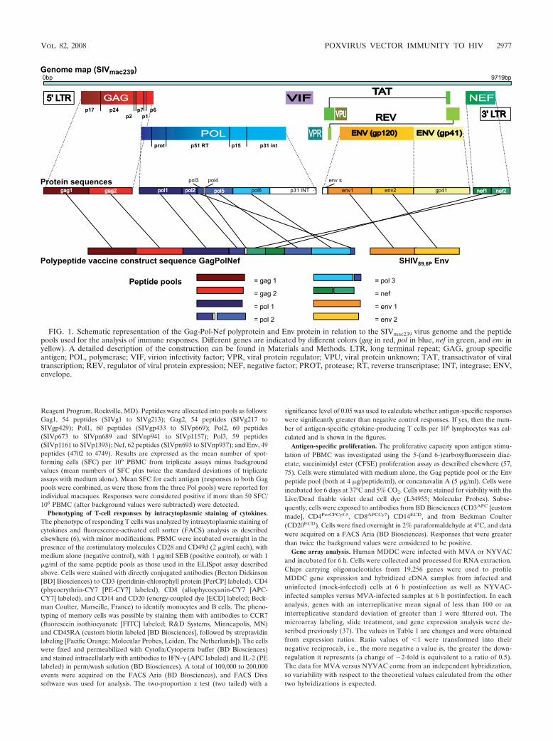
FIG. 1. Schematic representation of the Gag-Pol-Nef polyprotein and Env protein in relation to the SIVmac239 virus genome and the peptidepools used for the analysis of immune responses. Different genes are indicated by different colors (gag in red, pol in blue, nef in green, and env inyellow). A detailed description of the construction can be found in Materials and Methods. LTR, long terminal repeat; GAG, group specificantigen; POL, polymerase; VIF, virion infectivity factor; VPR, viral protein regulator; VPU, viral protein unknown; TAT, transactivator of viraltranscription; REV, regulator of viral protein expression; NEF, negative factor; PROT, protease; RT, reverse transcriptase; INT, integrase; ENV,envelope.
VOL. 82, 2008 POXVIRUS VECTOR IMMUNITY TO HIV 2977

Virus neutralization assay. The neutralizing activity of the sera from immu-nized and infected rhesus macaques was assayed against the challenge virusstrain SHIV89.6P. Virus stocks were prepared on human PBMC. For the neu-tralization assay, 50 tissue culture infectious doses/ml of SHIV89.6P were incu-bated with serum (final dilution, 1/15) from each animal in duplicate for 1 h at37°C in a total volume of 150 �l Dulbecco’s modified Eagle’s medium with 10%fetal calf serum in 96-well flat-bottomed culture plates. Freshly trypsinized
TZM-bl cells (10,000 cells in 100 �l medium containing 37.5 �g/ml DEAE-dextran) were added to each well. Control wells received cells plus virus (viruscontrol) or cells only (background control) (97). After 48 h of culture, superna-tant was removed and 150 �l lysis buffer (phosphate-buffered saline, 1% Triton,1 mM CaCl2, and 1 mM MgCl2) was added and incubated for 45 min at 4°C. Onehundred microliters of cell lysate was transferred to 96-well black/white solidplates for the measurement of luminescence using a Victor 3 light luminometer.
TABLE 1. Differences in mRNA levels of immunomodulators possibly involved in CD4� and CD8� T-cellactivation after MVA and NYVAC infection of human MDDCa
Description GenBankaccession no.
Genedesignation
Fold change in transcription level for:
MVA vs mockinfection
NYVAC vs mockinfection
MVA vsNYVAC
IFN-induced protein with tetratricopeptide repeats 4 NM_001549 IFIT4 52.45 3.37 11.44TNF (TNF superfamily, member 2) NM_000594 TNF 52.21 6.79 10.38IFN-induced protein with tetratricopeptide repeats 1 NM_001548 IFIT1 47.8 4.65 7.38Mitogen-activated protein kinase kinase 5 NM_002757 MAP2K5 15.59 2.07 6.54RNA helicase NM_014314 RIG-I 21.81 5.72Small inducible cytokine A4 NM_002984 SCYA4 3.74 1.14 5.61IFN-induced, hepatitis C-associated microtubular aggregate
protein (44 kDa)NM_006417 MTAP44 20.95 2.63 5.52
IL-12B (natural killer cell stimulatory factor 2, cytotoxiclymphocyte maturation factor 2)
NM_002187 IL12B 9.76 5.44
IFN-�1, fibroblast NM_002176 IFNB1 137.82 5.06Melanoma differentiation-associated protein-5 NM_022168 MDA5 7.77 1.67 5.05Cyclin B1 NM_031966 CCNB1 7.87 1.06 3.64Small inducible cytokine A5 (RANTES) NM_002985 SCYA5 4.17 1.17 3.06IL-6 signal transducer (gp130, oncostatin M receptor) NM_002184 IL6ST 2.15 1.19 2.91V-fos FBJ murine osteosarcoma viral oncogene homolog NM_005252 FOS 3.38 1.00 2.86GRO2 oncogene (SCYB2) NM_002089 GRO2 6.58 2.22 2.78TNF (ligand) superfamily, member 13b NM_006573 TNFSF13B 1.84 1.21 2.67Small inducible cytokine subfamily B (Cys-X-Cys), member 10 NM_001565 SCYB10 8.33 2.35IRF-7 NM_004031 IRF7 2.52 1.3 2.31Menage a trois 1 (CAK assembly factor) NM_002431 MNAT1 5.85 1.72 2.27Small inducible cytokine A3 NM_002983 SCYA3 4.22 1.12Colony-stimulating factor 2 (granulocyte-macrophage) NM_000758 CSF2 3.13 1.08 1.94Growth arrest and DNA damage-inducible, � type NM_015675 GADD45B 2.22 1.56 1.31Nuclear factor of activated T cells 5, tonicity responsive AF346509 NFAT5 2.81 1.24IL-10 receptor, � NM_001558 IL10RA 1.39 �2.12 1.21Major histocompatibility complex, class II, DQ�1 NM_002123 HLA-DQB1 2.18 2.49 1.02Proteasome (prosome, macropain) subunit, � type, 10 NM_002801 PSMB10 �3.04 1.20 1.03IFN consensus sequence binding protein 1, IRF-8 NM_002163 ICSBP1 1.19 3.32 1.07TNF (ligand) superfamily, member 9 (4-1BB-L) NM_003811 TNFSF9 1.59 �2.10 1.07Inhibitor of DNA binding 2B, dominant-negative helix-loop-
helix proteinM96843 ID2B �2.16 1.78 1.07
CD86 antigen (CD28 antigen ligand 2, B7-2 antigen) NM_006889 CD86 1.44 1.12 1.13Transforming growth factor �1 NM_000660 TGFB1 �2.43 �2.09 1.16CD80 antigen (CD28 antigen ligand 1, B7-1 antigen) NM_005191 CD80 1.53 1.14 1.20V-jun sarcoma virus 17 oncogene homolog (avian) NM_002228 JUN 1.91 2.93 1.34IL-10 receptor, � Z17227 IL10RB �2.59 1.21 1.38MHC class II, DR�5 NM_002125 HLA-DRB5 �2.92 �5.59 1.65IL-8 NM_000584 IL-8 �2.90 1.39 1.89Ras homolog gene family, member B NM_004040 ARHB 1.50 2.08 1.97IFN-� receptor 2 (IFN-� transducer 1) NM_005534 IFNGR2 �2.52 1.18 �2.05Secreted phosphoprotein 1 (osteopontin, bone sialoprotein I,
early T-lymphocyte activation 1)NM_000582 SPP1 1.60 �2.64 �2.13
Jun D proto-oncogene NM_005354 JUND 1.41 2.78 �2.42IL-1� NM_000575 IL1A 1.43 2.01 �2.42Mal, T-cell differentiation protein 2 NM_052886 MAL2 �2.49 1.09 �2.54IRF-5 NM_002200 IRF5 �2.31 1.06 �2.65IL-1� NM_000576 IL1B �2.25 1.39 �3.14Prostate differentiation factor NM_004864 PLAB 1.77 3.32 �3.94FOS-like antigen 2 AK055579 FOSL2 1.36 4.13 �4.09Deoxythymidylate kinase (thymidylate kinase) NM_012145 DTYMK 1.54 12.02 �8.18Cyclin-dependent kinase inhibitor 2C (p18, inhibits CDK4) NM_001262 CDKN2C �3.10 1.18
a The description, accession number, gene designation, and change in transcription levels are indicated for each gene in three different hybridizations. Change valueswere obtained from the ratio of intensities. The change is shown in boldface for genes that are significantly upregulated (�2-fold) or downregulated (2-fold) inMVA-infected versus mock-infected MDDC, NYVAC-infected versus mock-infected MDDC, or MVA-infected versus NYVAC-infected MDDC. Upregulation ordownregulation of 2-fold is considered not significant. FBJ, Finkel-Biskis-Jinkins; GRO2, growth-related gene; MAL, myelin and lymphocyte protein.
2978 MOOIJ ET AL. J. VIROL.

One hundred microliters of Britelite reagent (Perkin Elmer) was added. Theluminescence from samples before immunization (groups 1 and 2) or beforechallenge (group 3) was taken as equivalent to 100% of infected cells or alter-natively as 0% neutralization.
Determination of plasma virus loads. The plasma virus load was determinedwith quantitative competitive RNA reverse transcription-PCR using plasma fromEDTA-treated blood samples (95). The lower detection limit was 40 RNAequivalents/ml.
T-lymphocyte subset analysis. Quantitative changes in PBMC subsets afterchallenge were monitored by FACS analysis as previously described (62). Thefollowing monoclonal antibody combinations were used: (i) CD20FITC, CD16PE,HLA-DRPerCP, CD3APC, CD4PECY7, and CD8APC-CY7 and (ii) CCR7FITC,CD62LPE, and CD45RABiotin followed by streptavidinPerCP, CD3APC, CD4PE-CY7,and CD8APC-CY7. Monoclonal antibodies were obtained from BD Biosciences(CD20FITC, clone L27; CD16PE, clone B7.3.1; HLA-DRPerCP, clone L243; CD3APC,clone SP34; CD4PE-CY7, clone SK3; CD8APC-CY7, clone SK1; and CD45RABiotin,clone 5H9), R&D Systems (CCR7FITC, clone FAB197F), or AbD Serotec (Dussel-dorf, Germany) (CD62LPE, clone FMC46). Polystyrene fluorospheres (BeckmanCoulter) were used to calculate the absolute lymphocyte counts. Flow cytometry wasperformed on a FACS Aria (BD Biosciences). A total of 20,000 events in thelymphocyte gate were analyzed per monoclonal antibody mix.
Statistical analysis. Statistical analysis was performed by the Wilcoxon’s ranksum test or unpaired t test (with Welch correction) depending on the normaldistribution of the data. A P value 0.05 was considered significant.
Nucleotide sequence accession number. The DNA sequence of MVA-89.6p-SIVGPN in the thymidine kinase viral locus has been deposited in GenBankunder accession number EU359568 (see Table S1 in the supplemental material).
RESULTS
Antigen-specific IFN-�, IL-2, and IL-4 ELISpot responses.To investigate possible differences in the modulation of anti-gen-specific cytokine responses by the genetically distinctMVA and NYVAC vectors, we performed IFN-�, IL-2, and
IL-4 ELISpot assays to detect peptide-specific responses toeach of the four vaccine-encoded antigens Env, Gag, Pol, andNef. Priming with DNA induced low-level Env-specific cyto-kine responses (Fig. 2, top). As anticipated, antigen-specificresponses were further increased after boosts with either typeof poxvirus vector, increasing IFN-�, IL-2, and IL-4 responsesto all vaccine antigens (Fig. 2). The second poxvirus immuni-zation resulted in an additional increase in the Env-specificIFN-� responses (for MVA, from 560 to 1,171 mean SFC/106
PBMC [P 0.035]; for NYVAC, from 446 to 1,074 meanSFC/106 PBMC [P 0.004]) and Gag-specific IFN-� responses(from 101 to 331 mean SFC/106 PBMC [P 0.045] for MVA;Wilcoxon rank sum test). The highest observed response was toEnv, which consistently scored positive in ELISpot assays dur-ing follow-up. This observation was entirely consistent withfindings of the human clinical trial (39).
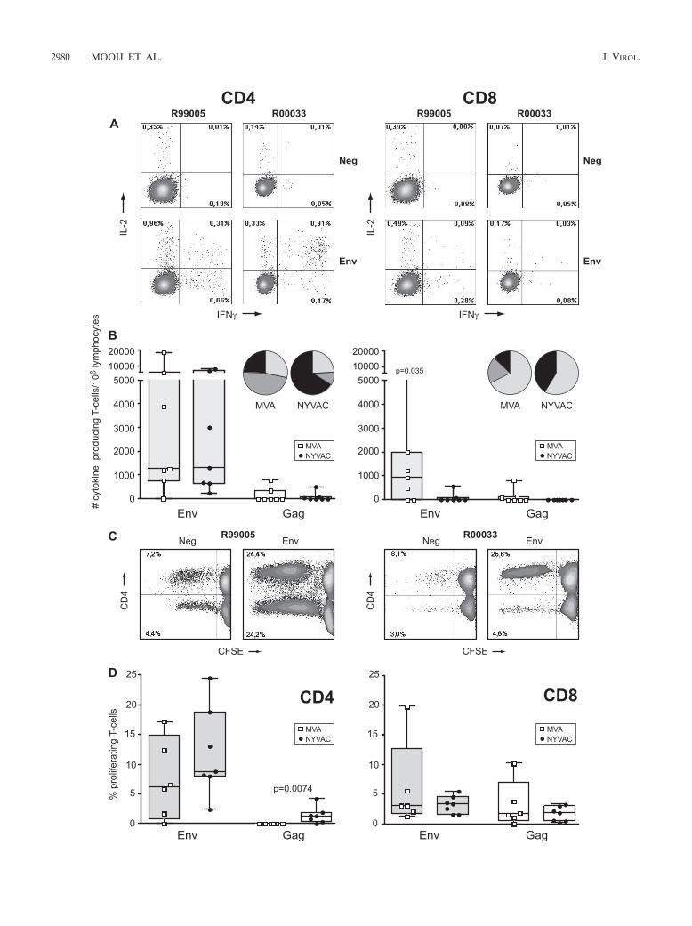
Qualitative differences between poxvirus vector vaccine can-didates reveal preferential CD8� versus CD4� T-cell re-sponses. Multiparameter intracellular FACS analysis was em-ployed to determine the phenotypic characteristics of thecytokine-producing T-cell populations (Fig. 3A). Followingpoxvirus vector immunizations, Env-specific cytokine-produc-ing T cells were dominant in animals of both MVA andNYVAC groups (Fig. 3A and B). This confirmed the ELISpotdata but revealed that responses were mainly from CD4� Tcells (Fig. 3A and B) of the central memory (CD45RA/CCR7�) and effector memory (CD45RA/CCR7) pheno-types (data not shown) in both groups. Additionally, five out ofseven MVA-boosted animals also developed Env-specific cy-
FIG. 2. T-cell ELISpot responses elicited over time. Shown are IFN-� (left panels), IL-2 (middle panels), and IL-4 (right panels) ELISpots fromindividual animals immunized with SHIV immunogens delivered by DNA/MVA or DNA/NYVAC to HIV-189.6P Env and SIVmac239 Gag, Pol, andNef peptides. Background responses (mean numbers of SFC plus twice the standard deviations of triplicate assays with medium alone) weresubtracted. Responses are presented as the number of SFC per 106 PBMC. Arrows indicate immunization time points at weeks 0, 4 (DNA), 20,and 24 (poxvirus).
VOL. 82, 2008 POXVIRUS VECTOR IMMUNITY TO HIV 2979
2980 MOOIJ ET AL. J. VIROL.

tokine responses mediated by CD8� T cells (P � 0.035) (Fig.3B, right) and had an effector phenotype (CD8�/CD45RA�/CCR7) (data not shown). Further analysis of the antigen-specific T cells revealed that the CD4� and CD8� T cells of themajority of the immunized animals produced either IFN-�alone or IFN-� plus IL-2 (Fig. 3B). The frequency of IFN-�-producing CD8� T cells was higher in MVA-boosted animalsthan in NYVAC-boosted animals (P � 0.0187).
To further corroborate polyfunctionality by using FACS-based assays, the proliferative capacity of both CD4� andCD8� T-cell populations was studied in each animal from bothimmunization groups (Fig. 3C and D). Proliferation wasmainly Env specific, consistent with ELISpot and intracellularcytokine staining (ICS) data. In MVA-boosted animals, Env-specific proliferation was mediated by both CD4� and CD8� Tcells, while in NYVAC-boosted animals Env-specific prolifer-ation was preferentially mediated by CD4� T cells (Fig. 3C andD). Furthermore, Gag-specific proliferating CD4� T cells weredetected in NYVAC-immunized animals (P � 0.0074), whilein MVA-boosted animals the Gag-specific proliferating T cellswere CD8� (Fig. 3D). The calculation of the relative contri-bution of CD4� and CD8� T cells to the total response re-vealed a significantly larger proportion of CD8� T-cell-medi-ated antigen-specific responses induced by MVA compared tothose induced by NYVAC immunization (25% versus 7.3% oftotal cytokine responses [P � 0.042] and 59.8% versus 31.4%of total proliferative responses [P � 0.0227]).
Gene array analysis supports differential poxvirus vector-induced CD4�/CD8� T-cell responses. The reanalysis of geneprofiling data from human dendritic cells (DC) activated byMVA or NYVAC infection (Table 1) revealed a highly markedupregulation in MVA-infected cells at the mRNA level ofIL-12, IFN-�, and IFN-� as well as IFN regulatory factor 7(IRF-7), which is implicated in the type I IFN production(MDA5, RIG). Consequently, many IFN-stimulated genes(ISGs) also were differentially upregulated in MVA-infectedDC (i.e., IFIT1 [ISG56], IFIT4 [ISG60], and SCYB10 [aCXCR3 ligand chemokine chiefly active on effector Th1 cells]genes). Moreover, the differentiation program initiated byIL-12 and IFN-�/� regulates numerous genes involved in sev-eral functions. Of these, genes relevant for effector cell regu-lation of gene expression, such as the GADD45B (56) and thetranscription factor NFAT5 (91) genes, genes involved in sig-
nal transduction (MAP2K5 gene) and cell cycle regulation(cyclin B1 gene) (60), and members of the TNF family (5) areconsistently upregulated in MVA infection. Interestingly, genesencoding proinflammatory cytokines such as TNF and IL-6 andCC chemokines such as SCYA3, SCYA4, and SCYA5(RANTES), which are involved in the modulation of the immuneresponse, are differentially expressed by MVA and NYVAC vec-tors. These profiling data support the preferential stimulation ofCD8� T cells by MVA. In the case of NYVAC, it is significantthat the expression of all of the above-described CD8� T-cell-stimulatory genes is markedly reduced after virus infection of DC,further supporting a differential CD8 versus CD4 behavior be-tween the two vaccine vector strains in vivo.
Vaccine efficacy against pathogenic challenge. After chal-lenge, all animals became infected (Fig. 4). Peak virus loads at2 weeks after challenge ranged from 4.1 � 103 to 2.5 � 106
RNA copies/ml and were similar in all three groups (Fig. 4,top). Subsequently, virus loads in five control animals re-mained high and persisted in four of these five animals atabove 105 RNA copies/ml, followed by ensuing AIDS-like dis-ease. Upon necropsy, histology confirmed the diagnosis ofAIDS, hallmarked by lymphoid depletion in the peripheral andmesenteric lymph nodes, spleen, gut-associated lymphoid tis-sue, and tonsils. In contrast to controls, six out of seven im-munized animals from each group were able to reduce thevirus load to below 104 RNA copies/ml (Fig. 4, top left). His-tological examination of lymphoid tissues upon necropsy re-vealed mild lymphoid depletion of gut-associated lymphoidtissue in only one animal of the MVA group (98051). Allimmunized animals remained healthy during the study periodand showed prolonged survival compared to that of the controlanimals (Fig. 5).
Absolute CD4� T-cell loss inversely correlated with virusload after challenge (Fig. 4, middle row). In four of five controlanimals with high virus loads, a dramatic progressive loss ofCD4� T cells occurred, while in another control animal a10-fold reduction of absolute CD4� T-cell counts was ob-served. In both immunized groups (n � 14), only the twoanimals that were unable to control virus load below 104 RNAcopies/ml developed evidence of CD4� T-cell loss (Fig. 4).
A more sensitive indicator of changes within the CD4� T-cell population is the loss of the CD4� central memory T-cellsubset (Tcm; CD4�/CCR7�/CD45RA) (71). In control ani-
FIG. 3. Vaccine-induced antigen-specific CD4� and CD8� T-cell responses 2 weeks prior to challenge. (A) Representative flow cytometryprofiles of vaccine-induced CD4� (left) and CD8� (right) T-cell responses directed against Env in monkeys R99005 and R00033 immunized withSHIV immunogens delivered by DNA/MVA or DNA/NYVAC, respectively. CD4 and CD8 T-cell responses were defined using polychromatic flowcytometry. Blood mononuclear cells were stimulated with the relevant peptide pools and stained with CD4, CD8, IFN-�, and IL-2 antibodies.(B) Cytokine production by CD4� (left) and CD8� (right) T cells of all individual animals immunized with SHIV immunogens delivered byDNA/MVA (open squares) or DNA/NYVAC (black dots) to HIV-189.6p Env (gray boxes) and SIVmac239 Gag peptides (clear boxes) as measuredby ICS assays 2 weeks before challenge. Box-whisker plots indicate the interquartile ranges and the medians (horizontal lines) of the groups.Responses are presented as the numbers of cytokine-producing CD3� T cells per 106 lymphocytes. Pie charts represent the average response forall animals of each group of the CD4� and CD8� HIV-189.6p Env- and SIVmac239 Gag-specific T-cell responses grouped by function (expressingeither one cytokine, IFN-� [light gray] or IL-2 [dark gray], or the two simultaneously [black]) relative to the total antigen-specific response.(C) Representative flow cytometry profiles of CFSE-labeled PBMC of monkeys R99005 and R00033 immunized with SHIV immunogens deliveredby DNA/MVA or DNA/NYVAC, respectively, unstimulated (Neg) or stimulated with Env peptides, cultured for 6 days, and stained with CD3,CD4, and CD8 antibodies. (D) Percentage of antigen-specific proliferating CD4� and CD8� T cells of all individual animals (dots) immunized withSHIV immunogens delivered by DNA/MVA (open squares) or DNA/NYVAC (black dots) to HIV-189.6p Env (gray boxes) and SIVmac239 Gagpeptides (clear boxes) 2 weeks before challenge. Responses are presented as percentages of proliferating (CFSE low) CD3�/CD4� andCD3�/CD8� T cells. Background responses (medium alone) were subtracted.
VOL. 82, 2008 POXVIRUS VECTOR IMMUNITY TO HIV 2981

mals with high virus loads, a decline of the CD4� Tcm wasobserved after challenge. In the immunized animals from bothgroups, the CD4� Tcm population remained relatively stableuntil 30 weeks after infection. Subsequently, a slow decline of
the CD4� Tcm population also could be observed in theseanimals (Fig. 4, bottom).
Changes in antigen-specific T-cell responses following infec-tion. To further elucidate immune responses associated withprotection from disease in the immunized groups, antigen-specific cellular immune responses were studied after chal-lenge. Surprisingly, anamnestic T-cell responses were not ob-served immediately following challenge. This may have beendue to an initial decline of antigen-specific IFN-�, IL-2, andIL-4 responses in both vaccine groups (data not shown). Incontrast to the control animals, HIV-1 Env-, SIV Gag-, andSIV Nef-specific IFN-� responses in vaccinated animals at 22weeks after challenge were similar to those observed beforechallenge (Fig. 6, left). These responses subsequently persisteduntil 41 weeks after challenge (Fig. 6, right).
At the time of euthanasia (66 weeks after challenge forimmunized animals and earlier for symptomatic control ani-mals), antigen-specific cytokine induction was monitored intra-cellularly by polychromatic FACS analysis (ICS). In contrast tothe prechallenge ICS data, postinfection responses were di-rected primarily against SIV Gag and, to a lesser extent,
FIG. 4. Vaccine efficacy. The viral RNA load (top), absolute number of circulating CD4� T cells (middle), and percentage of CD4� Tcm cells(bottom) after the challenge of each individual animal are presented. The left column represents results from animals immunized with SHIVimmunogens delivered by DNA/MVA, the middle column shows results from animals immunized with SHIV immunogens delivered by DNA/NYVAC, and the right column presents results from control animals.
FIG. 5. Survival. The percentages of animals that remained diseasefree after SHIV89.6P challenge are shown. The animals were immu-nized with SHIV immunogens delivered by DNA/MVA (solid line) orDNA/NYVAC (dashed line) or were control animals (declining line).
2982 MOOIJ ET AL. J. VIROL.

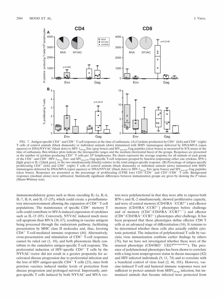
against HIV-1 Env (Fig. 7A). The Gag-specific CD4� T cellswere of the central memory (CD45RA/CCR7�) and effectormemory (CD45RA/CCR7) phenotypes, while the CD8� Tcells were of the effector phenotype (CD45RA�/CCR7)(data not shown). Similarly to the situation prior to challenge,CD8-mediated cytokine responses were higher in the MVA-boosted animals (P � 0.0148 for SIV Gag) than in theNYVAC-boosted animals, and a relatively higher proportionof the total cytokine response was mediated by CD8� T cells inthis group (74% for MVA versus 20% for NYVAC; P �0.0072). The predominant cytokine produced was IFN-�, with
a small contribution from IFN-� and IL-2 double-positive cells(Fig. 7A). In contrast to controls, detectable levels of Env- andGag-specific proliferating CD4� and CD8� T cells were ob-served in immunized animals at the time of euthanasia (Fig.7B). Although low, the frequency of Gag-specific CD4� pro-liferating T cells was significantly higher in NYVAC-boostedanimals than in MVA-boosted animals (P � 0.0416).
Neutralizing antibodies. As expected with this T-cell-basedvaccine approach, prior to challenge there were insufficientneutralizing antibody responses to protect animals from infec-tion (Fig. 8). After challenge, neutralizing antibodies devel-oped quickly in immunized animals (at similar rates in MVA-and NYVAC-immunized animals) and were significantlyhigher at 6 weeks after challenge compared to antibody titersin controls (P � 0.0012 for MVA versus controls and P �0.0023 for NYVAC versus controls).
DISCUSSION
This head-to-head study evaluated the immunogenicity andefficacy of two DNA prime/poxvirus boost HIV-1 vaccine can-didates in the Indian rhesus macaque model of AIDS. Ourresults demonstrated that both poxvirus boosts were highlyimmunogenic, significantly boosting DNA-primed cellular im-mune responses to the HIV-1 and SIV inserts. This observa-tion was consistent with earlier findings in which a broadeningof the DNA-primed immune response to other (subdominant)epitopes by poxvirus boosting was reported (42, 83). The Env-specific vaccine responses prior to challenge were dominantand mediated mainly by CD4� T cells. This may be attribut-able to the vaccine construct, which was designed such that theEnv protein would be secreted, likely favoring CD4� T-cellresponses (84). The cause of the relative dominance of Envremains speculative and may be related to the level of antigenexpression at the cellular level. However, Western blot analysisof MVA- and NYVAC-infected chicken embryo fibroblastsshowed similar levels of expression of both Env protein and thepolyprotein Gag-Pol-Nef. When the immunogenicity of thesevaccine vectors was evaluated in HLA-A2 class I transgenicmice, a similar Env immunodominant response was observed(33). Factors such as MHC binding affinity, the efficiency ofepitope processing, and competition between T cells for accessto antigen-presenting cells could contribute to the observedimmunodominant response (32, 73, 81).
Phenotypic analyses by ICS and CFSE proliferation assaysrevealed that the MVA vaccine vector induced CD8� T-cellresponses in addition to CD4 responses, while the NYVACvector boosted CD4� T-cell-mediated responses to a higherlevel than MVA. These differences were likely influenced bythe different immune-modulatory effects that these poxvirusvectors have on their host cells. Moreover, in a recent study ofmice that evaluated the biodistribution of MVA and NYVACrecombinant viruses, a more sustained infection was inducedby NYVAC than by MVA (31), which possibly influences theT-cell response. In contrast to CD8� T cells, for which arelatively short antigen pulse seems sufficient for antigen-pre-senting cells to drive clonal expansion and differentiation, an-tigen persistence is required for CD4� T cells throughout theirexpansion phase (69). A human gene profiling analysis ofMVA-infected HeLa cells has revealed an upregulation of
FIG. 6. T-cell ELISpot responses after challenge. IFN-� produc-tion by PBMC of individual control animals (black diamonds) or ani-mals immunized with SHIV immunogens delivered by DNA/MVA(open squares) or DNA/NYVAC (black dots) to HIV-189.6P Env andSIVmac239 Gag, Pol, and Nef peptides at 22 weeks after challenge (pc)(left panels) and at 41 weeks after challenge (right panels). Box-whisker plots indicate the interquartile ranges and the medians (hor-izontal lines) of the groups. Background responses (mean numbers ofSFC plus twice the standard deviations of triplicate assays with me-dium alone) were subtracted. Responses are presented as the numberof SFC per 106 PBMC.
VOL. 82, 2008 POXVIRUS VECTOR IMMUNITY TO HIV 2983
immunomodulatory genes such as those encoding IL-1a, IL-6,IL-7, IL-8, and IL-15 (35), which could create a proinflamma-tory microenvironment allowing the expansion of CD8� T-cellpopulations. The maintenance of specific CD8� memory Tcells could contribute to MVA-induced expression of cytokinessuch as IL-15 (85). Conversely, NYVAC induced much morecell apoptosis than MVA (36, 67), resulting in vaccine antigensbeing processed through the endocytosis pathway, facilitatingpresentation by MHC class II molecules and, thus, favoringCD4� T-cell-mediated immune responses (44). Alternatively,cross-presentation and stimulation of CD8� T-cell responsescannot be ruled out (1, 10), and both phenomena likely con-tribute to the cumulative antigen-specific T-cell response. Thepreferential induction of HIV-specific CD4� T cells by theNYVAC vector did not result in a greater propensity for ac-celerated disease progression due to preferential infection andthe loss of HIV antigen-specific CD4� T cells (23), since bothpoxvirus vaccines induced similar levels of protection fromdisease progression and prolonged survival. Importantly, anti-gen-specific T cells induced by both NYVAC and MVA vec-
tors were polyfunctional in that they were able to express bothIFN-� and IL-2 simultaneously, showed proliferative capacity,and were of central memory (CD45RA/CCR7�) and effectormemory (CD45RA/CCR7) phenotypes before challengeand of memory (CD4�/CD45RA/CCR7�/) and effector(CD8�/CD45RA�/CCR7) phenotypes after challenge. It hasbeen proposed that these phenotypes define effector CD8 Tcells at an advanced stage of differentiation (16). It remains tobe determined whether these cells also actually exhibit cyto-toxic potential. The induction of polyfunctional T cells by vac-cinia virus immunization confirms earlier findings by others(76), but we have not investigated whether these were of theunusual phenotype (CD45RO CD27intermediate). The pres-ence of polyfunctional phenotypes has been shown to correlatewith a long-term nonprogressor status in rhesus macaques (82)and HIV-infected individuals (9, 11, 74) and to correlate witha beneficial control of virus load (2, 46, 101). However, vac-cine-induced T-cell and humoral immune responses were notsufficient to protect animals from SHIV89.6p infection, but im-munized animals that became infected were protected from
FIG. 7. Antigen-specific CD4� and CD8� T-cell responses at the time of euthanasia. (A) Cytokine production by CD4� (left) and CD8� (right)T cells of control animals (black diamonds) or individual animals (dots) immunized with SHIV immunogens delivered by DNA/MVA (opensquares) or DNA/NYVAC (black dots) to HIV-189.6p Env (gray boxes) and SIVmac239 Gag peptides (clear boxes) as measured by ICS assays at thetime of euthanasia. Box-whisker plots indicate the interquartile ranges and the medians (horizontal lines) of the groups. Responses are presentedas the number of cytokine-producing CD3� T cells per 106 lymphocytes. Pie charts represent the average response for all animals of each groupof the CD4� and CD8� HIV-189.6p Env- and SIVmac239 Gag-specific T-cell responses grouped by function (expressing either one cytokine, IFN-�[light gray] or IL-2 [dark gray], or the two simultaneously [black]) relative to the total antigen-specific response. (B) Percentage of antigen-specificproliferating CD4� (left) and CD8� (right) T cells of control animals (black diamonds) or individual animals (dots) immunized with SHIVimmunogens delivered by DNA/MVA (open squares) or DNA/NYVAC (black dots) to HIV-189.6p Env (gray boxes) and SIVmac239 Gag peptides(clear boxes). Responses are presented as the percentage of proliferating (CFSE low) CD3�/CD4� and CD3�/CD8� T cells. Backgroundresponses (medium alone) were subtracted. Statistically significant differences between immunization groups are given by showing the P values(Mann-Whitney test).
2984 MOOIJ ET AL. J. VIROL.

disease progression for more than a year (66 weeks), while themajority of control animals developed evidence of AIDS-likedisease. Others also have previously shown vaccine-inducedcontrol of the SHIV89.6P load and protection from diseaseprogression (3, 8, 19, 21, 24). It is possible that protection isrelatively easy to induce in the challenge model used (25). Itremains to be determined whether the observed equivalentprotection mediated by differential CD4 and CD8 responses isrestricted to the SHIV89.6P challenge model or if similar resultswould have been obtained following challenge with the morepathogenic SIVmac239.
The majority of immunized animals in our study controlledvirus loads and maintained CD4� T-cell numbers, including
the CD4� Tcm population, at preinfection levels. The poten-tial contributions of the various antigen-specific CD4� orCD8� T-cell-mediated mechanisms to the improved survival inthe immunized animals need further investigation. The pres-ervation of CD4� T cells in immunized and in control animalswithout disease allowed the development of neutralizing anti-bodies by B cells after challenge, which also may ultimatelyhave facilitated long-term control.
The Env-dominant response observed prior to infectiongradually shifted toward a Gag-dominant response followinginfection. The vaccine-induced Env-specific responses weremediated mainly by CD4� T cells, which are the targets forSHIV infection. Since all vaccinated animals became infected,the function and the number of these Env-specific CD4� Tcells might have been transiently affected by the virus infection(50, 89), even though no clear CD4� T-cell decline was ob-served. The postchallenge induction of Gag as well as Nefresponses represents an anamnestic response preferentiallyobserved in immunized animals. This underscores the conclu-sion that, despite the relative immunodominance of Env,immunization against the other vaccine antigens has been ben-eficial.
Although immunization did not protect against SHIV infec-tion, viral loads were reduced after the acute phase, CD4�
Tcm cells were maintained, and prolonged survival was in-duced. However, the relative loss of Env-specific responses,reduced CD4 responses, reduced IL-2 production (accordingto ELISpot and ICS assays), and reduced proliferative capacityafter challenge would indicate an insidious loss of T-cell func-tion, which may eventually have an effect on long-term survival.While both vaccine candidates induced similar levels of effi-cacy, this efficacy apparently was exerted through differentmechanisms, which may have implications for the long-termmanagement of the infection and future refinements in HIVvaccine design.
ACKNOWLEDGMENTS
We are indebted to T. de Koning and H. van Westbroek for docu-mentation. We thank D. Mortier, S. Hofman, Z. Fagrouch, and W.Koornstra for technical support. We are grateful to Jose ManuelGonzalez for bioinformatic assistance in gene array analysis.
This study was supported by funding from the EuroVacc (EuropeanVaccine Effort Against HIV/AIDS) project QLK2-CT-1999-01321.
REFERENCES
1. Albert, M. L. 2004. Death-defying immunity: do apoptotic cells influenceantigen processing and presentation? Nat. Rev. Immunol. 4:223–231.
2. Amara, R. R., C. Ibegbu, F. Villinger, D. C. Montefiori, S. Sharma, P.Nigam, Y. Xu, H. M. McClure, and H. L. Robinson. 2005. Studies using aviral challenge and CD8 T cell depletions on the roles of cellular andhumoral immunity in the control of an SHIV-89.6P challenge in DNA/MVA-vaccinated macaques. Virology 343:246–255.
3. Amara, R. R., K. Patel, G. Niedziela, P. Nigam, S. Sharma, S. I. Staprans,D. C. Montefiori, L. Chenareddi, J. G. Herndon, H. L. Robinson, H. M.McClure, and F. J. Novembre. 2005. A combination DNA and attenuatedsimian immunodeficiency virus vaccine strategy provides enhanced protec-tion from simian/human immunodeficiency virus-induced disease. J. Virol.79:15356–15367.
4. Amara, R. R., F. Villinger, J. D. Altman, S. L. Lydy, S. P. O’Neil, S. I.Staprans, D. C. Montefiori, Y. Xu, J. G. Herndon, L. S. Wyatt, M. A.Candido, N. L. Kozyr, P. L. Earl, J. M. Smith, H. L. Ma, B. D. Grimm, M. L.Hulsey, J. Miller, H. M. McClure, J. M. McNicholl, B. Moss, and H. L.Robinson. 2001. Control of a mucosal challenge and prevention of AIDS bya multiprotein DNA/MVA vaccine. Science 292:69–74.
5. Anel, A., A. Bosque, J. Naval, A. Pineiro, L. Larrad, M. A. Alava, and M. J.Martinez-Lorenzo. 2007. Apo2L/TRAIL and immune regulation. Front.Biosci. 12:2074–2084.
FIG. 8. Neutralizing antibody titers. Relative inhibition of virusinfection of indicator cells with the challenge virus SHIV89.6p is shown.The infection of indicator cells with virus incubated with individualsera from before immunization or before challenge (controls) was setat 0% inhibition.
VOL. 82, 2008 POXVIRUS VECTOR IMMUNITY TO HIV 2985
6. Balla-Jhagjhoorsingh, S. S., G. Koopman, P. Mooij, W. Koornstra, S.McCormack, J. Weber, G. Pantaleo, and J. L. Heeney. 2004. Long-termpersistence of HIV-1 vaccine-induced CD4�CD45RACD62LCCR7
memory T-helper cells. AIDS 18:837–848.7. Benson, J., C. Chougnet, M. Robert-Guroff, D. Montefiori, P. Markham, G.
Shearer, R. C. Gallo, M. Cranage, E. Paoletti, K. Limbach, D. Venzon, J.Tartaglia, and G. Franchini. 1998. Recombinant vaccine-induced protec-tion against the highly pathogenic simian immunodeficiency virusSIVmac251: dependence on route of challenge exposure. J. Virol. 72:4170–4182.
8. Bertley, F. M., P. A. Kozlowski, S. W. Wang, J. Chappelle, J. Patel, O.Sonuyi, G. Mazzara, D. Montefiori, A. Carville, K. G. Mansfield, and A.Aldovini. 2004. Control of simian/human immunodeficiency virus viremiaand disease progression after IL-2-augmented DNA-modified vaccinia virusAnkara nasal vaccination in nonhuman primates. J. Immunol. 172:3745–3757.
9. Betts, M. R., M. C. Nason, S. M. West, S. C. De Rosa, S. A. Migueles, J.Abraham, M. M. Lederman, J. M. Benito, P. A. Goepfert, M. Connors, M.Roederer, and R. A. Koup. 2006. HIV nonprogressors preferentially main-tain highly functional HIV-specific CD8� T cells. Blood 107:4781–4789.
10. Blachere, N. E., R. B. Darnell, and M. L. Albert. 2005. Apoptotic cellsdeliver processed antigen to dendritic cells for cross-presentation. PLoSBiol. 3:e185.
11. Boaz, M. J., A. Waters, S. Murad, P. J. Easterbrook, and A. Vyakarnam.2002. Presence of HIV-1 Gag-specific IFN-gamma� IL-2� and CD28�
IL-2� CD4 T cell responses is associated with nonprogression in HIV-1infection. J. Immunol. 169:6376–6385.
12. Brenchley, J. M., L. E. Ruff, J. P. Casazza, R. A. Koup, D. A. Price, andD. C. Douek. 2006. Preferential infection shortens the life span of humanimmunodeficiency virus-specific CD4� T cells in vivo. J. Virol. 80:6801–6809.
13. Burton, D. R., R. C. Desrosiers, R. W. Doms, W. C. Koff, P. D. Kwong, J. P.Moore, G. J. Nabel, J. Sodroski, I. A. Wilson, and R. T. Wyatt. 2004. HIVvaccine design and the neutralizing antibody problem. Nat. Immunol.5:233–236.
14. Carrington, M., and S. J. O’Brien. 2003. The influence of HLA genotype onAIDS. Annu. Rev. Med. 54:535–551.
15. Cebere, I., L. Dorrell, H. McShane, A. Simmons, S. McCormack, C.Schmidt, C. Smith, M. Brooks, J. E. Roberts, S. C. Darwin, P. E. Fast, C.Conlon, S. Rowland-Jones, A. J. McMichael, and T. Hanke. 2006. Phase Iclinical trial safety of DNA- and modified virus Ankara-vectored humanimmunodeficiency virus type 1 (HIV-1) vaccines administered alone and ina prime-boost regime to healthy HIV-1-uninfected volunteers. Vaccine24:417–425.
16. Champagne, P., G. S. Ogg, A. S. King, C. Knabenhans, K. Ellefsen, M. Nobile,V. Appay, G. P. Rizzardi, S. Fleury, M. Lipp, R. Forster, S. Rowland-Jones,R. P. Sekaly, A. J. McMichael, and G. Pantaleo. 2001. Skewed maturation ofmemory HIV-specific CD8 T lymphocytes. Nature 410:106–111.
17. Collman, R., J. W. Balliet, S. A. Gregory, H. Friedman, D. L. Kolson, N.Nathanson, and A. Srinivasan. 1992. An infectious molecular clone of anunusual macrophage-tropic and highly cytopathic strain of human immu-nodeficiency virus type 1. J. Virol. 66:7517–7521.
18. Corbett, M., W. M. Bogers, J. L. Heeney, S. Gerber, C. Genin, A. Didierlaurent, H.Oostermeijer, R. Dubbes, G. Braskamp, S. Lerondel, C. E. Gomez, M. Esteban, I.Kondova, P. Mooij, S. S. Balla-Jhagjhoorsingh, N. Beenhakker, G. Koopman, S.Van der Burg, J. P. Kraehenbuhl, and A. Le Pape. Aerosol immunization withNYVAC and MVA vectored vaccines: simple, safe and immunogenic. Proc. Natl.Acad. Sci. USA, in press.
19. Demberg, T., R. H. Florese, M. J. Heath, K. Larsen, I. Kalisz, V. S. Kaly-anaraman, E. M. Lee, R. Pal, D. Venzon, R. Grant, L. J. Patterson, B.Korioth-Schmitz, A. Buzby, D. Dombagoda, D. C. Montefiori, N. L. Letvin,A. Cafaro, B. Ensoli, and M. Robert-Guroff. 2007. A replication-competentadenovirus-human immunodeficiency virus (Ad-HIV) tat and Ad-HIV envpriming/Tat and envelope protein boosting regimen elicits enhanced pro-tective efficacy against simian/human immunodeficiency virus SHIV89.6Pchallenge in rhesus macaques. J. Virol. 81:3414–3427.
20. Didierlaurent, A., J. C. Ramirez, M. Gherardi, S. C. Zimmerli, M. Graf,H. A. Orbea, G. Pantaleo, R. Wagner, M. Esteban, J. P. Kraehenbuhl, andJ. C. Sirard. 2004. Attenuated poxviruses expressing a synthetic HIV pro-tein stimulate HLA-A2-restricted cytotoxic T-cell responses. Vaccine 22:3395–3403.
21. Doria-Rose, N. A., C. Ohlen, P. Polacino, C. C. Pierce, M. T. Hensel, L.Kuller, T. Mulvania, D. Anderson, P. D. Greenberg, S. L. Hu, and N. L.Haigwood. 2003. Multigene DNA priming-boosting vaccines protect ma-caques from acute CD4� T-cell depletion after simian-human immunode-ficiency virus SHIV89.6P mucosal challenge. J. Virol. 77:11563–11577.
22. Dorrell, L., H. Yang, B. Ondondo, T. Dong, K. di Gleria, A. Suttill, C.Conlon, D. Brown, P. Williams, P. Bowness, N. Goonetilleke, T. Rostron, S.Rowland-Jones, T. Hanke, and A. McMichael. 2006. Expansion and diver-sification of virus-specific T cells following immunization of human immu-nodeficiency virus type 1 (HIV-1)-infected individuals with a recombinantmodified vaccinia virus Ankara/HIV-1 Gag vaccine. J. Virol. 80:4705–4716.
23. Douek, D. C., J. M. Brenchley, M. R. Betts, D. R. Ambrozak, B. J. Hill, Y.Okamoto, J. P. Casazza, J. Kuruppu, K. Kunstman, S. Wolinsky, Z. Gross-man, M. Dybul, A. Oxenius, D. A. Price, M. Connors, and R. A. Koup. 2002.HIV preferentially infects HIV-specific CD4� T cells. Nature 417:95–98.
24. Egan, M. A., S. Y. Chong, S. Megati, D. C. Montefiori, N. F. Rose, J. D.Boyer, M. K. Sidhu, J. Quiroz, M. Rosati, E. B. Schadeck, G. N. Pavlakis,D. B. Weiner, J. K. Rose, Z. R. Israel, S. A. Udem, and J. H. Eldridge. 2005.Priming with plasmid DNAs expressing interleukin-12 and simian immu-nodeficiency virus gag enhances the immunogenicity and efficacy of anexperimental AIDS vaccine based on recombinant vesicular stomatitis vi-rus. AIDS Res. Hum. Retrovir. 21:629–643.
25. Feinberg, M. B., and J. P. Moore. 2002. AIDS vaccine models: challengingchallenge viruses. Nat. Med. 8:207–210.
26. Fischer, W., S. Perkins, J. Theiler, T. Bhattacharya, K. Yusim, R. Funkhouser, C.Kuiken, B. Haynes, N. L. Letvin, B. D. Walker, B. H. Hahn, and B. T. Korber. 2007.Polyvalent vaccines for optimal coverage of potential T-cell epitopes in globalHIV-1 variants. Nat. Med. 13:100–106.
27. Frahm, N., S. Adams, P. Kiepiela, C. H. Linde, H. S. Hewitt, M. Lichterfeld,K. Sango, N. V. Brown, E. Pae, A. G. Wurcel, M. Altfeld, M. E. Feeney,T. M. Allen, T. Roach, M. A. St. John, E. S. Daar, E. Rosenberg, B. Korber,F. Marincola, B. D. Walker, P. J. Goulder, and C. Brander. 2005. HLA-B63presents HLA-B57/B58-restricted cytotoxic T-lymphocyte epitopes and isassociated with low human immunodeficiency virus load. J. Virol. 79:10218–10225.
28. Frahm, N., P. Kiepiela, S. Adams, C. H. Linde, H. S. Hewitt, K. Sango,M. E. Feeney, M. M. Addo, M. Lichterfeld, M. P. Lahaie, E. Pae, A. G.Wurcel, T. Roach, M. A. St. John, M. Altfeld, F. M. Marincola, C. Moore,S. Mallal, M. Carrington, D. Heckerman, T. M. Allen, J. I. Mullins, B. T.Korber, P. J. Goulder, B. D. Walker, and C. Brander. 2006. Control ofhuman immunodeficiency virus replication by cytotoxic T lymphocytes tar-geting subdominant epitopes. Nat. Immunol. 7:173–178.
29. Franchini, G., S. Gurunathan, L. Baglyos, S. Plotkin, and J. Tartaglia.2004. Poxvirus-based vaccine candidates for HIV: two decades of experi-ence with special emphasis on canarypox vectors. Expert Rev. Vaccines3:S75–S88.
30. Gherardi, M. M., and M. Esteban. 2005. Recombinant poxviruses as mu-cosal vaccine vectors. J. Gen. Virol. 86:2925–2936.
31. Gomez, C. E., J. L. Najera, E. Domingo-Gil, L. Ochoa-Callejero, G. Gonzalez-Aseguinolaza, and M. Esteban. 2007. Virus distribution of the attenuated MVAand NYVAC poxvirus strains in mice. J. Gen. Virol. 88:2473–2478.
32. Gomez, C. E., J. L. Najera, E. P. Jimenez, V. Jimenez, R. Wagner, M. Graf,M. J. Frachette, P. Liljestrom, G. Pantaleo, and M. Esteban. 2007. Head-to-head comparison on the immunogenicity of two HIV/AIDS vaccinecandidates based on the attenuated poxvirus strains MVA and NYVACcoexpressing in a single locus the HIV-1(BX08) gp120 and HIV-1(IIIB)Gag-Pol-Nef proteins of clade B. Vaccine 25:2863–2885.
33. Gomez, C. E., J. L. Najera, V. Jimenez, K. Bieler, J. Wild, L. Kostic, S.Heidari, M. Chen, M. J. Frachette, G. Pantaleo, H. Wolf, P. Liljestrom, R.Wagner, and M. Esteban. 2007. Generation and immunogenicity of novelHIV/AIDS vaccine candidates targeting HIV-1 Env/Gag-Pol-Nef antigensof clade C. Vaccine 25:1969–1992.
34. Goonetilleke, N., S. Moore, L. Dally, N. Winstone, I. Cebere, A. Mahmoud,S. Pinheiro, G. Gillespie, D. Brown, V. Loach, J. Roberts, A. Guimaraes-Walker, P. Hayes, K. Loughran, C. Smith, J. De Bont, C. Verlinde, D.Vooijs, C. Schmidt, M. Boaz, J. Gilmour, P. Fast, L. Dorrell, T. Hanke, andA. J. McMichael. 2006. Induction of multifunctional human immunodefi-ciency virus type 1 (HIV-1)-specific T cells capable of proliferation inhealthy subjects by using a prime-boost regimen of DNA- and modifiedvaccinia virus Ankara-vectored vaccines expressing HIV-1 Gag coupled toCD8� T-cell epitopes. J. Virol. 80:4717–4728.
35. Guerra, S., L. A. Lopez-Fernandez, R. Conde, A. Pascual-Montano, K.Harshman, and M. Esteban. 2004. Microarray analysis reveals character-istic changes of host cell gene expression in response to attenuated modifiedvaccinia virus Ankara infection of human HeLa cells. J. Virol. 78:5820–5834.
36. Guerra, S., L. A. Lopez-Fernandez, A. Pascual-Montano, J. L. Najera, A.Zaballos, and M. Esteban. 2006. Host response to the attenuated poxvirusvector NYVAC: upregulation of apoptotic genes and NF- B-responsivegenes in infected HeLa cells. J. Virol. 80:985–998.
37. Guerra, S., J. L. Najera, J. M. Gonzalez, L. Lopez, N. Climent, J. M. Gatell,T. Gallart, and M. Esteban. 2007. Distinct gene expression profiling afterinfection of immature human monocyte-derived dendritic cells by the at-tenuated poxvirus vectors MVA and NYVAC. J. Virol. 81:8707–8721.
38. Hanke, T., R. V. Samuel, T. J. Blanchard, V. C. Neumann, T. M. Allen, J. E.Boyson, S. A. Sharpe, N. Cook, G. L. Smith, D. I. Watkins, M. P. Cranage,and A. J. McMichael. 1999. Effective induction of simian immunodeficiencyvirus-specific cytotoxic T lymphocytes in macaques by using a multiepitopegene and DNA prime-modified vaccinia virus Ankara boost vaccinationregimen. J. Virol. 73:7524–7532.
39. Harari, A., P. A. Bart, W. Stohr, G. Tapia, M. Garcia, E. Medjitna-Rais, S.Burnet, O. Erlwein, T. Barber, C. Moog, P. Liljestrom, R. Wagner, H. Wolf,J. P. Kraehenbuhl, M. Esteban, J. L. Heeney, M. J. Frachette, J. Tartaglia,
2986 MOOIJ ET AL. J. VIROL.

S. McCormack, A. Babiker, J. Weber, and G. Pantaleo. 2008. An HIV-1clade C DNA prime, NYVAC boost vaccine regimen induces vigorous,broad, polyfunctional and long-lasting T-cell responses. J. Exp. Med. 205:63–67.
40. Heeney, J. L., and S. A. Plotkin. 2006. Immunological correlates of protec-tion from HIV infection and disease. Nat. Immunol. 7:1281–1284.
41. Hel, Z., J. Nacsa, E. Tryniszewska, W. P. Tsai, R. W. Parks, D. C. Montefiori, B. K.Felber, J. Tartaglia, G. N. Pavlakis, and G. Franchini. 2002. Containment ofsimian immunodeficiency virus infection in vaccinated macaques: correlation withthe magnitude of virus-specific pre- and postchallenge CD4� and CD8� T cellresponses. J. Immunol. 169:4778–4787.
42. Hel, Z., W. P. Tsai, A. Thornton, J. Nacsa, L. Giuliani, E. Tryniszewska, M.Poudyal, D. Venzon, X. Wang, J. Altman, D. I. Watkins, W. Lu, A. vonGegerfelt, B. K. Felber, J. Tartaglia, G. N. Pavlakis, and G. Franchini.2001. Potentiation of simian immunodeficiency virus (SIV)-specificCD4(�) and CD8(�) T cell responses by a DNA-SIV and NYVAC-SIVprime/boost regimen. J. Immunol. 167:7180–7191.
43. Im, E. J., J. P. Nkolola, K. di Gleria, A. J. McMichael, and T. Hanke. 2006.Induction of long-lasting multi-specific CD8� T cells by a four-componentDNA-MVA/HIVA-RENTA candidate HIV-1 vaccine in rhesus macaques.Eur. J. Immunol. 36:2574–2584.
44. Inaba, K., S. Turley, F. Yamaide, T. Iyoda, K. Mahnke, M. Inaba, M. Pack,M. Subklewe, B. Sauter, D. Sheff, M. Albert, N. Bhardwaj, I. Mellman, andR. M. Steinman. 1998. Efficient presentation of phagocytosed cellular frag-ments on the major histocompatibility complex class II products of den-dritic cells. J. Exp. Med. 188:2163–2173.
45. Kanesa-Thasan, N., J. J. Smucny, C. H. Hoke, D. H. Marks, E. Konishi, I.Kurane, D. B. Tang, D. W. Vaughn, P. W. Mason, and R. E. Shope. 2000.Safety and immunogenicity of NYVAC-JEV and ALVAC-JEV attenuatedrecombinant Japanese encephalitis virus-poxvirus vaccines in vaccinia-non-immune and vaccinia-immune humans. Vaccine 19:483–491.
46. Kannanganat, S., B. G. Kapogiannis, C. Ibegbu, L. Chennareddi, P.Goepfert, H. L. Robinson, J. Lennox, and R. R. Amara. 2007. Humanimmunodeficiency virus type 1 controllers but not noncontrollers maintainCD4 T cells coexpressing three cytokines. J. Virol. 81:12071–12076.
47. Karlsson, G. B., M. Halloran, J. Li, I. W. Park, R. Gomila, K. A. Reimann,M. K. Axthelm, S. A. Iliff, N. L. Letvin, and J. Sodroski. 1997. Character-ization of molecularly cloned simian-human immunodeficiency virusescausing rapid CD4� lymphocyte depletion in rhesus monkeys. J. Virol.71:4218–4225.
48. Kaslow, R. A., M. Carrington, R. Apple, L. Park, A. Munoz, A. J. Saah, J. J.Goedert, C. Winkler, S. J. O’Brien, C. Rinaldo, R. Detels, W. Blattner, J.Phair, H. Erlich, and D. L. Mann. 1996. Influence of combinations ofhuman major histocompatibility complex genes on the course of HIV-1infection. Nat. Med. 2:405–411.
49. Kent, S. J., A. Zhao, S. J. Best, J. D. Chandler, D. B. Boyle, and I. A.Ramshaw. 1998. Enhanced T-cell immunogenicity and protective efficacy ofa human immunodeficiency virus type 1 vaccine regimen consisting ofconsecutive priming with DNA and boosting with recombinant fowlpoxvirus. J. Virol. 72:10180–10188.
50. Koopman, G., H. Niphuis, W. Newman, T. K. Kishimoto, V. C. Maino, andJ. L. Heeney. 2001. Decreased expression of IL-2 in central and effectorCD4 memory cells during progression to AIDS in rhesus macaques. AIDS15:2359–2369.
51. Koup, R. A., J. T. Safrit, Y. Cao, C. A. Andrews, G. McLeod, W. Borkowsky,C. Farthing, and D. D. Ho. 1994. Temporal association of cellular immuneresponses with the initial control of viremia in primary human immunode-ficiency virus type 1 syndrome. J. Virol. 68:4650–4655.
52. Kuroda, M. J., J. E. Schmitz, W. A. Charini, C. E. Nickerson, M. A. Lifton,C. I. Lord, M. A. Forman, and N. L. Letvin. 1999. Emergence of CTLcoincides with clearance of virus during primary simian immunodeficiencyvirus infection in rhesus monkeys. J. Immunol. 162:5127–5133.
53. Letvin, N. L. 2006. Progress and obstacles in the development of an AIDSvaccine. Nat. Rev. Immunol. 6:930–939.
54. Lifson, J. D., J. L. Rossio, M. Piatak, Jr., T. Parks, L. Li, R. Kiser, V.Coalter, B. Fisher, B. M. Flynn, S. Czajak, V. M. Hirsch, K. A. Reimann,J. E. Schmitz, J. Ghrayeb, N. Bischofberger, M. A. Nowak, R. C. Desrosiers,and D. Wodarz. 2001. Role of CD8� lymphocytes in control of simianimmunodeficiency virus infection and resistance to rechallenge after tran-sient early antiretroviral treatment. J. Virol. 75:10187–10199.
55. Loffredo, J. T., J. Maxwell, Y. Qi, C. E. Glidden, G. J. Borchardt, T. Soma,A. T. Bean, D. R. Beal, N. A. Wilson, W. M. Rehrauer, J. D. Lifson, M.Carrington, and D. I. Watkins. 2007. Mamu-B*08-positive macaques con-trol simian immunodeficiency virus replication. J. Virol. 81:8827–8832.
56. Lu, B. 2006. The molecular mechanisms that control function and death ofeffector CD4� T cells. Immunol. Res. 36:275–282.
57. Lyons, A. B., and C. R. Parish. 1994. Determination of lymphocyte divisionby flow cytometry. J. Immunol. Methods 171:131–137.
58. Makitalo, B., P. Lundholm, J. Hinkula, C. Nilsson, K. Karlen, A. Morner, G.Sutter, V. Erfle, J. L. Heeney, B. Wahren, G. Biberfeld, and R. Thorstensson.2004. Enhanced cellular immunity and systemic control of SHIV infection by
combined parenteral and mucosal administration of a DNA prime MVA boostvaccine regimen. J. Gen. Virol. 85:2407–2419.
59. McNeil, A. J., P. L. Yap, S. M. Gore, R. P. Brettle, M. McColl, R. Wyld, S.Davidson, R. Weightman, A. M. Richardson, and J. R. Robertson. 1996.Association of HLA types A1-B8-DR3 and B27 with rapid and slow pro-gression of HIV disease. QJM 89:177–185.
60. Mescher, M. F., J. M. Curtsinger, P. Agarwal, K. A. Casey, M. Gerner, C. D.Hammerbeck, F. Popescu, and Z. Xiao. 2006. Signals required for program-ming effector and memory development by CD8� T cells. Immunol. Rev.211:81–92.
61. Migueles, S. A., M. S. Sabbaghian, W. L. Shupert, M. P. Bettinotti, F. M.Marincola, L. Martino, C. W. Hallahan, S. M. Selig, D. Schwartz, J. Sul-livan, and M. Connors. 2000. HLA B*5701 is highly associated with restric-tion of virus replication in a subgroup of HIV-infected long term nonpro-gressors. Proc. Natl. Acad. Sci. USA 97:2709–2714.
62. Mooij, P., W. M. Bogers, H. Oostermeijer, W. Koornstra, P. J. Ten Haaft,B. E. Verstrepen, G. Van Der Auwera, and J. L. Heeney. 2000. Evidence forviral virulence as a predominant factor limiting human immunodeficiencyvirus vaccine efficacy. J. Virol. 74:4017–4027.
63. Mooij, P., and J. L. Heeney. 2001. Rational development of prophylacticHIV vaccines based on structural and regulatory proteins. Vaccine 20:304–321.
64. Mothe, B. R., J. Weinfurter, C. Wang, W. Rehrauer, N. Wilson, T. M. Allen,D. B. Allison, and D. I. Watkins. 2003. Expression of the major histocom-patibility complex class I molecule Mamu-A*01 is associated with control ofsimian immunodeficiency virus SIVmac239 replication. J. Virol. 77:2736–2740.
65. Mwau, M., I. Cebere, J. Sutton, P. Chikoti, N. Winstone, E. G. Wee, T.Beattie, Y. H. Chen, L. Dorrell, H. McShane, C. Schmidt, M. Brooks, S.Patel, J. Roberts, C. Conlon, S. L. Rowland-Jones, J. J. Bwayo, A. J.McMichael, and T. Hanke. 2004. A human immunodeficiency virus 1(HIV-1) clade A vaccine in clinical trials: stimulation of HIV-specific T-cellresponses by DNA and recombinant modified vaccinia virus Ankara(MVA) vaccines in humans. J. Gen. Virol. 85:911–919.
66. Myagkikh, M., S. Alipanah, P. D. Markham, J. Tartaglia, E. Paoletti, R. C.Gallo, G. Franchini, and M. Robert-Guroff. 1996. Multiple immunizationswith attenuated poxvirus HIV type 2 recombinants and subunit boostsrequired for protection of rhesus macaques. AIDS Res. Hum. Retrovir.12:985–992.
67. Najera, J. L., C. E. Gomez, E. Domingo-Gil, M. M. Gherardi, and M.Esteban. 2006. Cellular and biochemical differences between two attenu-ated poxvirus vaccine candidates (MVA and NYVAC) and role of the C7Lgene. J. Virol. 80:6033–6047.
68. Nkolola, J. P., E. G. Wee, E. J. Im, C. P. Jewell, N. Chen, X. N. Xu, A. J.McMichael, and T. Hanke. 2004. Engineering RENTA, a DNA prime-MVA boost HIV vaccine tailored for eastern and central Africa. GeneTher. 11:1068–1080.
69. Obst, R., H. M. van Santen, D. Mathis, and C. Benoist. 2005. Antigenpersistence is required throughout the expansion phase of a CD4(�) T cellresponse. J. Exp. Med. 201:1555–1565.
70. O’Connor, D. H., B. R. Mothe, J. T. Weinfurter, S. Fuenger, W. M. Rehrauer,P. Jing, R. R. Rudersdorf, M. E. Liebl, K. Krebs, J. Vasquez, E. Dodds, J.Loffredo, S. Martin, A. B. McDermott, T. M. Allen, C. Wang, G. G. Doxiadis,D. C. Montefiori, A. Hughes, D. R. Burton, D. B. Allison, S. M. Wolinsky, R.Bontrop, L. J. Picker, and D. I. Watkins. 2003. Major histocompatibility com-plex class I alleles associated with slow simian immunodeficiency virus diseaseprogression bind epitopes recognized by dominant acute-phase cytotoxic-T-lymphocyte responses. J. Virol. 77:9029–9040.
71. Okoye, A., M. Meier-Schellersheim, J. M. Brenchley, S. I. Hagen, J. M.Walker, M. Rohankhedkar, R. Lum, J. B. Edgar, S. L. Planer, A. Legasse,A. W. Sylwester, M. Piatak, Jr., J. D. Lifson, V. C. Maino, D. L. Sodora,D. C. Douek, M. K. Axthelm, Z. Grossman, and L. J. Picker. 2007. Pro-gressive CD4� central memory T cell decline results in CD4� effectormemory insufficiency and overt disease in chronic SIV infection. J. Exp.Med. 204:2171–2185.
72. Pal, R., D. Venzon, N. L. Letvin, S. Santra, D. C. Montefiori, N. R. Miller,E. Tryniszewska, M. G. Lewis, T. C. VanCott, V. Hirsch, R. Woodward, A.Gibson, M. Grace, E. Dobratz, P. D. Markham, Z. Hel, J. Nacsa, M. Klein,J. Tartaglia, and G. Franchini. 2002. ALVAC-SIV-gag-pol-env-based vac-cination and macaque major histocompatibility complex class I (A*01)delay simian immunodeficiency virus SIVmac-induced immunodeficiency.J. Virol. 76:292–302.
73. Palmowski, M. J., E. M. Choi, I. F. Hermans, S. C. Gilbert, J. L. Chen, U.Gileadi, M. Salio, A. Van Pel, S. Man, E. Bonin, P. Liljestrom, P. R.Dunbar, and V. Cerundolo. 2002. Competition between CTL narrows theimmune response induced by prime-boost vaccination protocols. J. Immu-nol. 168:4391–4398.
74. Pantaleo, G., and A. Harari. 2006. Functional signatures in antiviral T-cellimmunity for monitoring virus-associated diseases. Nat. Rev. Immunol.6:417–423.
75. Parish, C. R. 1999. Fluorescent dyes for lymphocyte migration and prolif-eration studies. Immunol. Cell Biol. 77:499–508.
VOL. 82, 2008 POXVIRUS VECTOR IMMUNITY TO HIV 2987
76. Precopio, M. L., M. R. Betts, J. Parrino, D. A. Price, E. Gostick, D. R.Ambrozak, T. E. Asher, D. C. Douek, A. Harari, G. Pantaleo, R. Bailer, B. S.Graham, M. Roederer, and R. A. Koup. 2007. Immunization with vacciniavirus induces polyfunctional and phenotypically distinctive CD8� T cellresponses. J. Exp. Med. 204:1405–1416.
77. Reimann, K. A., J. T. Li, R. Veazey, M. Halloran, I. W. Park, G. B.Karlsson, J. Sodroski, and N. L. Letvin. 1996. A chimeric simian/humanimmunodeficiency virus expressing a primary patient human immunodefi-ciency virus type 1 isolate env causes an AIDS-like disease after in vivopassage in rhesus monkeys. J. Virol. 70:6922–6928.
78. Reimann, K. A., J. T. Li, G. Voss, C. Lekutis, K. Tenner-Racz, P. Racz, W.Lin, D. C. Montefiori, D. E. Lee-Parritz, Y. Lu, R. G. Collman, J. Sodroski,and N. L. Letvin. 1996. An env gene derived from a primary human immu-nodeficiency virus type 1 isolate confers high in vivo replicative capacity toa chimeric simian/human immunodeficiency virus in rhesus monkeys. J. Vi-rol. 70:3198–3206.
79. Richman, D. D., T. Wrin, S. J. Little, and C. J. Petropoulos. 2003. Rapidevolution of the neutralizing antibody response to HIV type 1 infection.Proc. Natl. Acad. Sci. USA 100:4144–4149.
80. Robinson, H. L., D. C. Montefiori, R. P. Johnson, K. H. Manson, M. L.Kalish, J. D. Lifson, T. A. Rizvi, S. Lu, S. L. Hu, G. P. Mazzara, D. L.Panicali, J. G. Herndon, R. Glickman, M. A. Candido, S. L. Lydy, M. S.Wyand, and H. M. McClure. 1999. Neutralizing antibody-independent con-tainment of immunodeficiency virus challenges by DNA priming andrecombinant pox virus booster immunizations. Nat. Med. 5:526–534.
81. Rodriguez, F., S. Harkins, M. K. Slifka, and J. L. Whitton. 2002. Immu-nodominance in virus-induced CD8� T-cell responses is dramatically mod-ified by DNA immunization and is regulated by gamma interferon. J. Virol.76:4251–4259.
82. Sadagopal, S., R. R. Amara, D. C. Montefiori, L. S. Wyatt, S. I. Staprans,N. L. Kozyr, H. M. McClure, B. Moss, and H. L. Robinson. 2005. Signaturefor long-term vaccine-mediated control of a simian and human immuno-deficiency virus 89.6P challenge: stable low-breadth and low-frequency T-cell response capable of coproducing gamma interferon and interleukin-2.J. Virol. 79:3243–3253.
83. Santra, S., D. H. Barouch, B. Korioth-Schmitz, C. I. Lord, G. R. Krivulka,F. Yu, M. H. Beddall, D. A. Gorgone, M. A. Lifton, A. Miura, V. Philippon,K. Manson, P. D. Markham, J. Parrish, M. J. Kuroda, J. E. Schmitz, R. S.Gelman, J. W. Shiver, D. C. Montefiori, D. Panicali, and N. L. Letvin. 2004.Recombinant poxvirus boosting of DNA-primed rhesus monkeys augmentspeak but not memory T lymphocyte responses. Proc. Natl. Acad. Sci. USA101:11088–11093.
84. Santra, S., Y. Sun, J. G. Parvani, V. Philippon, M. S. Wyand, K. Manson,A. Gomez-Yafal, G. Mazzara, D. Panicali, P. D. Markham, D. C. Monte-fiori, and N. L. Letvin. 2007. Heterologous prime/boost immunization ofrhesus monkeys by using diverse poxvirus vectors. J. Virol. 81:8563–8570.
85. Schluns, K. S., K. Williams, A. Ma, X. X. Zheng, and L. Lefrancois. 2002.Cutting edge: requirement for IL-15 in the generation of primary andmemory antigen-specific CD8 T cells. J. Immunol. 168:4827–4831.
86. Schmitz, J. E., M. J. Kuroda, S. Santra, V. G. Sasseville, M. A. Simon, M. A.Lifton, P. Racz, K. Tenner-Racz, M. Dalesandro, B. J. Scallon, J. Ghrayeb,M. A. Forman, D. C. Montefiori, E. P. Rieber, N. L. Letvin, and K. A.Reimann. 1999. Control of viremia in simian immunodeficiency virus infec-tion by CD8� lymphocytes. Science 283:857–860.
87. Siemens, D. R., S. Crist, J. C. Austin, J. Tartaglia, and T. L. Ratliff. 2003.Comparison of viral vectors: gene transfer efficiency and tissue specificity ina bladder cancer model. J. Urol. 170:979–984.
88. Su, L., M. Graf, Y. Zhang, H. von Briesen, H. Xing, J. Kostler, H. Melzl, H.Wolf, Y. Shao, and R. Wagner. 2000. Characterization of a virtually full-length human immunodeficiency virus type 1 genome of a prevalent inter-subtype (C/B�) recombinant strain in China. J. Virol. 74:11367–11376.
89. Sun, Y., S. R. Permar, A. P. Buzby, and N. L. Letvin. 2007. Memory CD4�
T-lymphocyte loss and dysfunction during primary simian immunodefi-ciency virus infection. J. Virol. 81:8009–8015.
90. Sun, Y., J. E. Schmitz, A. P. Buzby, B. R. Barker, S. S. Rao, L. Xu, Z. Y.Yang, J. R. Mascola, G. J. Nabel, and N. L. Letvin. 2006. Virus-specificcellular immune correlates of survival in vaccinated monkeys after simianimmunodeficiency virus challenge. J. Virol. 80:10950–10956.
91. Sundrud, M. S., and A. Rao. 2007. New twists of T cell fate: control of T cellactivation and tolerance by TGF-beta and NFAT. Curr. Opin. Immunol.19:287–293.
92. Sutter, G., and C. Staib. 2003. Vaccinia vectors as candidate vaccines: thedevelopment of modified vaccinia virus Ankara for antigen delivery. Curr.Drug Targets Infect. Disord. 3:263–271.
93. Tartaglia, J., W. I. Cox, S. Pincus, and E. Paoletti. 1994. Safety and immu-nogenicity of recombinants based on the genetically-engineered vacciniastrain, NYVAC. Dev. Biol. Stand. 82:125–129.
94. Tartaglia, J., M. E. Perkus, J. Taylor, E. K. Norton, J. C. Audonnet, W. I.Cox, S. W. Davis, J. van der Hoeven, B. Meignier, M. Riviere, et al. 1992.NYVAC: a highly attenuated strain of vaccinia virus. Virology 188:217–232.
95. Ten Haaft, P., B. Verstrepen, K. Uberla, B. Rosenwirth, and J. Heeney.1998. A pathogenic threshold of virus load defined in simian immunodefi-ciency virus- or simian-human immunodeficiency virus-infected macaques.J. Virol. 72:10281–10285.
96. Wada, K., S. Aota, R. Tsuchiya, F. Ishibashi, T. Gojobori, and T. Ikemura.1990. Codon usage tabulated from the GenBank genetic sequence data.Nucleic Acids Res. 18(Suppl.):2367–2411.
97. Wei, X., J. M. Decker, H. Liu, Z. Zhang, R. B. Arani, J. M. Kilby, M. S.Saag, X. Wu, G. M. Shaw, and J. C. Kappes. 2002. Emergence of resistanthuman immunodeficiency virus type 1 in patients receiving fusion inhibitor(T-20) monotherapy. Antimicrob. Agents Chemother. 46:1896–1905.
98. Wei, X., J. M. Decker, S. Wang, H. Hui, J. C. Kappes, X. Wu, J. F.Salazar-Gonzalez, M. G. Salazar, J. M. Kilby, M. S. Saag, N. L. Komarova,M. A. Nowak, B. H. Hahn, P. D. Kwong, and G. M. Shaw. 2003. Antibodyneutralization and escape by HIV-1. Nature 422:307–312.
99. Wilson, N. A., J. Reed, G. S. Napoe, S. Piaskowski, A. Szymanski, J. Furlott,E. J. Gonzalez, L. J. Yant, N. J. Maness, G. E. May, T. Soma, M. R.Reynolds, E. Rakasz, R. Rudersdorf, A. B. McDermott, D. H. O’Connor,T. C. Friedrich, D. B. Allison, A. Patki, L. J. Picker, D. R. Burton, J. Lin,L. Huang, D. Patel, G. Heindecker, J. Fan, M. Citron, M. Horton, F. Wang,X. Liang, J. W. Shiver, D. R. Casimiro, and D. I. Watkins. 2006. Vaccine-induced cellular immune responses reduce plasma viral concentrations af-ter repeated low-dose challenge with pathogenic simian immunodeficiencyvirus SIVmac239. J. Virol. 80:5875–5885.
100. Yant, L. J., T. C. Friedrich, R. C. Johnson, G. E. May, N. J. Maness, A. M.Enz, J. D. Lifson, D. H. O’Connor, M. Carrington, and D. I. Watkins. 2006.The high-frequency major histocompatibility complex class I allele Mamu-B*17 is associated with control of simian immunodeficiency virus SIVmac239replication. J. Virol. 80:5074–5077.
101. Zimmerli, S. C., A. Harari, C. Cellerai, F. Vallelian, P. A. Bart, and G.Pantaleo. 2005. HIV-1-specific IFN-gamma/IL-2-secreting CD8 T cells sup-port CD4-independent proliferation of HIV-1-specific CD8 T cells. Proc.Natl. Acad. Sci. USA 102:7239–7244.
2988 MOOIJ ET AL. J. VIROL.
Related Documents