Differential brain activation associated with laser-evoked burning and pricking pain: An event-related fMRI study Dieuwke S. Veldhuijzen a,c,g, * , Michael I. Nemenov d,e , Michael Keaser a , Jiachen Zhuo f , Rao P. Gullapalli f , Joel D. Greenspan a,b,c a Department of Biomedical Sciences, Dental School, University of Maryland, Baltimore, MD, USA b Program in Neuroscience, University of Maryland, Baltimore, MD, USA c Research Center for Neuroendocrine Influences on Pain, University of Maryland, Baltimore, MD, USA d Department of Anesthesia, Stanford University, Palo Alto, CA, USA e Lasmed LLC, Mountain View, CA, USA f Department of Diagnostic Radiology, University of Maryland School of Medicine, Baltimore, MD, USA g Division of Perioperative Care and Emergency Medicine, University Medical Center Utrecht, Rudolf Magnus Institute of Neuroscience, Heidelberglaan 100, 3584 CX Utrecht, The Netherlands article info Article history: Received 5 September 2008 Received in revised form 23 October 2008 Accepted 28 October 2008 Keywords: Burning pain Event-related fMRI Diode laser Pricking pain abstract An important question remains as to how the brain differentially processes first (pricking) pain mediated by Ad-nociceptors versus second (burning) pain mediated by C-nociceptors. In the present cross-over randomized, within]-subjects controlled study, brain activity patterns were examined with event-related fMRI while pricking and burning pain were selectively evoked using a diode laser. Stimuli evoking equiv- alent pain intensities were delivered to the dorsum of the left foot. Different laser parameters were used to elicit pricking (60 ms pulse duration) and burning (2.0 s pulse duration) pain. Whole brain group anal- ysis showed that several brain areas were commonly activated by pricking and burning pain, including bilateral thalamus, bilateral anterior insula, bilateral posterior parietal lobule, contralateral dorsolateral prefrontal cortex, ipsilateral cerebellum, and mid anterior cingulate cortex. These findings show that pricking and burning pain were associated with activity in many of the same nociceptive processing brain regions. This may be expected given that Ad-and C-nociceptive signals converge to a great extent at the level of the dorsal horn. Other brain regions showed differential processing. Stronger activation in the pricking pain condition was found in the ipsilateral hippocampus, bilateral parahippocampal gyrus, bilat- eral fusiform gyrus, contralateral cerebellum and contralateral cuneus/parieto-occipital sulcus. Stronger activation in the burning pain condition was found in the ipsilateral dorsolateral prefrontal cortex. These differential activation patterns suggest preferential importance of Ad-fiber signals versus C-fiber signals for these specific brain regions. Ó 2008 International Association for the Study of Pain. Published by Elsevier B.V. All rights reserved. 1. Introduction Different types of nociceptive afferents evoke qualitatively dif- ferent sensations. First pain is described as ‘sharp’ or ‘pricking’, and is mediated by myelinated Ad-fibers. Second pain is described as ‘dull’ or ‘burning’ and is mediated by the slower unmyelinated C-fibers. It remains largely unclear how the signals conveyed by these fibers are differentially processed in the human brain. Knowledge about the cortical representation of signals originating from C- and Ad-fiber nociceptors may provide new insights into the biological significance of these distinct pain pathways. Recently, reports emerged on differential involvement of nociceptive fibers in pain syndromes [27,44], suggesting that insights on differential nociceptor processing may be helpful in establishing better diag- noses. The ability to reliably differentiate between pain-mediating fibers may also provide unique utility in predicting the efficacy of treatments. Few studies modeled brain responses associated with first and second pain. In a laser-evoked potential (LEP) study, brain activa- tion in response to second pain was found bilaterally in the sec- ondary somatosensory cortex (SII) and the anterior cingulate cortex (ACC) [43], while a magnetoencephalography (MEG) study found second pain-related activation in the contralateral primary somatosensory cortex (SI) and bilateral SII [33]. However, both studies did not compare findings to the cortical processing of first pain. Studies directly comparing brain activation patterns of first www.elsevier.com/locate/pain PAIN 141 (2009) 104–113 0304-3959/$34.00 Ó 2008 International Association for the Study of Pain. Published by Elsevier B.V. All rights reserved. doi:10.1016/j.pain.2008.10.027 * Corresponding author. Address: Division of Perioperative Care and Emergency Medicine, University Medical Center Utrecht, Rudolf Magnus Institute of Neurosci- ence, Heidelberglaan 100, 3584 CX Utrecht, The Netherlands. Tel.: +31 88 75 56163; fax: +31 88 75 55511. E-mail address: [email protected] (D.S. Veldhuijzen).

Welcome message from author
This document is posted to help you gain knowledge. Please leave a comment to let me know what you think about it! Share it to your friends and learn new things together.
Transcript

w w w . e l s e v i e r . c o m / l o c a t e / p a i n
PAIN 141 (2009) 104–113
Differential brain activation associated with laser-evoked burningand pricking pain: An event-related fMRI study
Dieuwke S. Veldhuijzen a,c,g,*, Michael I. Nemenov d,e, Michael Keaser a,Jiachen Zhuo f, Rao P. Gullapalli f, Joel D. Greenspan a,b,c
a Department of Biomedical Sciences, Dental School, University of Maryland, Baltimore, MD, USAb Program in Neuroscience, University of Maryland, Baltimore, MD, USAc Research Center for Neuroendocrine Influences on Pain, University of Maryland, Baltimore, MD, USAd Department of Anesthesia, Stanford University, Palo Alto, CA, USAe Lasmed LLC, Mountain View, CA, USAf Department of Diagnostic Radiology, University of Maryland School of Medicine, Baltimore, MD, USAg Division of Perioperative Care and Emergency Medicine, University Medical Center Utrecht, Rudolf Magnus Institute of Neuroscience,Heidelberglaan 100, 3584 CX Utrecht, The Netherlands
a r t i c l e i n f o a b s t r a c t
Article history:Received 5 September 2008Received in revised form 23 October 2008Accepted 28 October 2008
Keywords:Burning painEvent-related fMRIDiode laserPricking pain
0304-3959/$34.00 � 2008 International Associationdoi:10.1016/j.pain.2008.10.027
* Corresponding author. Address: Division of PerioMedicine, University Medical Center Utrecht, Rudolf Mence, Heidelberglaan 100, 3584 CX Utrecht, The Nethefax: +31 88 75 55511.
E-mail address: [email protected] (D
An important question remains as to how the brain differentially processes first (pricking) pain mediatedby Ad-nociceptors versus second (burning) pain mediated by C-nociceptors. In the present cross-overrandomized, within]-subjects controlled study, brain activity patterns were examined with event-relatedfMRI while pricking and burning pain were selectively evoked using a diode laser. Stimuli evoking equiv-alent pain intensities were delivered to the dorsum of the left foot. Different laser parameters were usedto elicit pricking (60 ms pulse duration) and burning (2.0 s pulse duration) pain. Whole brain group anal-ysis showed that several brain areas were commonly activated by pricking and burning pain, includingbilateral thalamus, bilateral anterior insula, bilateral posterior parietal lobule, contralateral dorsolateralprefrontal cortex, ipsilateral cerebellum, and mid anterior cingulate cortex. These findings show thatpricking and burning pain were associated with activity in many of the same nociceptive processing brainregions. This may be expected given that Ad-and C-nociceptive signals converge to a great extent at thelevel of the dorsal horn. Other brain regions showed differential processing. Stronger activation in thepricking pain condition was found in the ipsilateral hippocampus, bilateral parahippocampal gyrus, bilat-eral fusiform gyrus, contralateral cerebellum and contralateral cuneus/parieto-occipital sulcus. Strongeractivation in the burning pain condition was found in the ipsilateral dorsolateral prefrontal cortex. Thesedifferential activation patterns suggest preferential importance of Ad-fiber signals versus C-fiber signalsfor these specific brain regions.
� 2008 International Association for the Study of Pain. Published by Elsevier B.V. All rights reserved.
1. Introduction
Different types of nociceptive afferents evoke qualitatively dif-ferent sensations. First pain is described as ‘sharp’ or ‘pricking’,and is mediated by myelinated Ad-fibers. Second pain is describedas ‘dull’ or ‘burning’ and is mediated by the slower unmyelinatedC-fibers. It remains largely unclear how the signals conveyed bythese fibers are differentially processed in the human brain.Knowledge about the cortical representation of signals originatingfrom C- and Ad-fiber nociceptors may provide new insights into the
for the Study of Pain. Published by
perative Care and Emergencyagnus Institute of Neurosci-
rlands. Tel.: +31 88 75 56163;
.S. Veldhuijzen).
biological significance of these distinct pain pathways. Recently,reports emerged on differential involvement of nociceptive fibersin pain syndromes [27,44], suggesting that insights on differentialnociceptor processing may be helpful in establishing better diag-noses. The ability to reliably differentiate between pain-mediatingfibers may also provide unique utility in predicting the efficacy oftreatments.
Few studies modeled brain responses associated with first andsecond pain. In a laser-evoked potential (LEP) study, brain activa-tion in response to second pain was found bilaterally in the sec-ondary somatosensory cortex (SII) and the anterior cingulatecortex (ACC) [43], while a magnetoencephalography (MEG) studyfound second pain-related activation in the contralateral primarysomatosensory cortex (SI) and bilateral SII [33]. However, bothstudies did not compare findings to the cortical processing of firstpain. Studies directly comparing brain activation patterns of first
Elsevier B.V. All rights reserved.

D.S. Veldhuijzen et al. / PAIN 141 (2009) 104–113 105
and second pain consistently found largely overlapping activationpatterns for both fibers, but differential processing was also found.In a MEG study, common activation was found in SII, while differ-ential activation for first pain was found in SI and for second painin ACC [51]. In another MEG study, common activation was foundin bilateral SII, contralateral posterior parietal cortex, and in a fewsubjects in cingulate, insula, prefrontal, and inferior temporoparie-tal regions, but no differential activation was found [24]. In a re-cent functional magnetic resonance imaging (fMRI) study,common activation was found in bilateral thalamus, SII, posteriorACC, and ipsilateral middle insula, while significantly stronger acti-vation for second pain was found in ACC, pre-SMA, and bilateralanterior insula [54]. Although these studies yielded valuable infor-mation, some issues arise. C-fiber stimulation can also reflectinnocuous warmth processing, and pain intensity scores were fre-quently not reported. Further, studies did not equalize perceivedpain intensity between first and second pain, which could con-found the perceptual quality and intensity of pain.
In this study, event-related fMRI was used to selectively evaluatebrain activity associated with pricking and burning pain induced bya diode laser. Perceived pain intensity was equalized between firstand second pain. We hypothesized that the spatial representationof nociceptive signals conducted by the two fiber types would besimilar to a large extent. However, differences in spatial processingwere also expected, because of distinct qualitative differences inpain sensations. In addition, we explored the possibility that inputfrom the fibers could be differentially processed in brain regions notnecessarily related to the perception of pain.
2. Materials and methods
2.1. Subjects
Ten healthy volunteers (5 females and 5 males; 1 left-handed)completed this study. Of these ten participants, one subject re-ported a significantly higher pain intensity rating (VAS rating of76 on a 0–100 scale) during the burning pain condition in the fMRIsession than the other subjects. Statistical analysis identified thepain rating of this subject as a significant outlier; therefore, thissubject was excluded from further analysis. Four additional sub-jects were screened for this study, but were excluded from full par-ticipation based on the following criteria: dual sensation ofpricking and burning pain upon stimulation (n = 1), inconsistentpain ratings (n = 1), or insensitivity to laser stimuli within thesafety margins (n = 2). Mean age of the remaining nine subjectswho completed the study was 29.7 ± 8.7 SD (standard deviation),ranging from 22 to 49 years. Participants who completed the studyhad at least 4 years of college education, none of them sufferedfrom a serious medical of psychiatric illness, they were good sleep-ers (mean 7.1 ± 1.1 SD hours a night), all except one subject did notsmoke, they used small amounts of caffeinated drinks (mean1.3 ± 0.8 SD per day), used alcohol moderately (mean 2.1 ± 2.3 SDglasses a week), and exercised light to moderately. One femalewas in her early follicular phase of her menstrual cycle, two fe-males were in their mid-luteal phases, and two women were intheir premenstrual phases when tested. The University of Mary-land Institutional Review Board for the Protection of Human Sub-jects approved the procedures and protocol for this study and theparticipants’ informed consent was obtained in accordance withthe Declaration of Helsinki.
2.2. Stimulation paradigms
A diode laser (LASS-10 M, Lasmed LLC, CA, USA; patented in2004) with a power of 20 W, wavelength of 980 nm, and an
adjustable spot diameter between 1 and 5 mm diameters wasused to apply the painful stimuli to the skin of the dorsum ofthe left foot. Selective activation of Ad-fibers was accomplishedby using a diode laser with a high rate heating, high energy, briefpulses, and small spot sizes. Selective activation of C-fibers wasaccomplished by low rate heating with long pulses, low energyand larger spot sizes [62,64,66,68]. The diode laser has been pre-viously used to selectively activate transient receptor potentialvanilloid (TRPV1 and TRPV2) positive cells [32], to selectivelyactivate C- and Ad- fibers in rats [62], to elicit a heat-activated in-ward current Iheat in dorsal root ganglion neurons of rats in vitro,and to elicit laser-evoked potentials (LEPs) associated with activa-tion of Ad- and C-nociceptors in humans [28,67]. The diode laserdistributes energy principally at the level of cutaneous nocicep-tive endings (20–570 lm) [59], due to less absorption(�0.5 cm�1 for 970 nm) thereby avoiding superficial skin lesions[36,45]. Accordingly, it allows for simultaneous and homogeneousheating at a skin depth of 20–570 lm [40,67]. Other commonlyused lasers, such as the CO2 or Thulium-YAG laser, show higherabsorption (�63 cm�1 for 2010 nm for Thulium and �300 cm�1
for 10.2 lm for CO2 lasers) and more superficial distribution ofenergy [57]. As they require conduction through tissue to reachnociceptive endings, risk of burns is higher than with the diodelaser. Further, the delay time between superficial skin heatingand heating at 450 lm skin depth could be up to several seconds[60]. Different parameters of laser stimulation were used to elicitpricking and burning pain sensations. For pricking pain, shortpulses of 60 ms with a spot size of 1 mm in diameter were ap-plied and the laser beam was positioned 5 cm from the skin sur-face. The mean intensity used to evoke pricking pain was 715 mJ/mm2, ranging from 390 to 868 mJ/mm2. For burning pain, longpulses of 2 s duration with a spot size of 5 mm in diameter wereapplied with the laser beam positioned 15 cm from the skin sur-face. The mean intensity used to evoke burning pain was 468 mJ/mm2, ranging from 428 to 499 mJ/mm2. An aiming beam of670 nm wavelength was used to identify the stimulated area.The differential activation of the burning and pricking pain wasconfirmed by the subjects’ verbal reports. For this purpose, sub-jects were asked to consider several mechanical- and tempera-ture-related descriptors, partly adapted from the McGill painquestionnaire, and to identify which descriptors applied to theirsensation. Reports of ‘‘pricking” and ‘‘sharp” were interpreted asassociated with Ad-fiber stimulation, while reports of ‘‘warm/hot”and ‘‘burning” were considered to be associated with C-fiberstimulation.
Intensities for pricking and burning pain were chosen based onsubjects’ perceived pain threshold and pain perception. Before MRIscanning, in a separate screening session, sequences of laser stim-uli were given which started with low, often undetectable levels,and intensities were increased stepwise until pain of around 20on a visual analogue scale (VAS) ranging between 0 (no pain) and100 (maximum tolerable pain) was reported. This relatively lowpain intensity was chosen to avoid burns that could potentially oc-cur with more intense stimulation, particularly for less pain sensi-tive individuals. Then, this stimulus level was repeated andadjusted if necessary until a response near 20 was given reliablyfor 10 consecutive stimuli of similar intensity. Mean pain intensityrating for the pricking pain stimuli was 22.4 (±SD = 6.7) and meanpain intensity for the burning pain stimuli was 25.4 (±SD = 6.6) inthe screening session. To avoid sensitization or fatigue of the noci-ceptors, the stimulation site on the dorsum of the foot was slightlydifferent for the pricking and burning pain stimulation. Also, thetemperature of the skin surface can affect the sensitivity to the la-ser pulse; with lower skin temperatures, higher energy levels needto be used to reach the same level of pain perception. Therefore,the temperature of the foot surface was monitored carefully at

106 D.S. Veldhuijzen et al. / PAIN 141 (2009) 104–113
the start and end of each functional run. The foot was kept warmby means of a reheatable bean sock between sessions.
2.3. Experimental design
Subjects participated in two sessions: a screening session and aMRI scanning session one or two days later. In the screening ses-sion, subjects were tested for their sensitivity to the experimentalprocedure. During the MRI scans, laser stimuli were presented inseparate blocks of burning or pricking pain stimuli, with 30 stimuliin each block. The order of presentation of pricking and burningpain stimuli in this MRI session was counter-balanced across sub-jects. An event-related design was used, and the inter-stimulusinterval was varied between 14 and 18 s in a random sequence trialby trail to reduce anticipatory effects and artifacts occurring at fixedfrequencies. A stimulus-free baseline period of 30 s was given be-fore and after the presentation of each block of stimuli. After thislast baseline period, subjects were asked to rate the average painintensity across all stimuli in the block on a computerized VAS(DAPSYS; Brian Turnquist, Johns Hopkins University, http://www.dapsys.net) using an MR-compatible trackball (Fellowes,www.fellowes.com), which controls a cursor moving along a verti-cal VAS. The extremes of the VAS were labeled ‘‘no sensation” and‘‘most intense pain imaginable”, with a marker for ‘‘just painful” lo-cated at the lower quarter of the scale. Subjects were instructed touse the range from ‘‘no sensation” to ‘‘just painful” to rate non-pain-ful sensations and to use the range from ‘‘just painful” to ‘‘most in-tense pain imaginable” to rate painful sensations. Ratings werestored as numbers from 0 to 100 with 25 corresponding to ‘‘justpainful”. This scale was chosen to give the subject the opportunityto rate intensities below pain thresholds in case the average stimu-lus intensity was not painful. Subjects practiced with this comput-erized VAS during the screening session. Scores on this 25–100 painintensity scale were converted to a 0–100 pain intensity scale to al-low comparison with rating data collected during the screening ses-sion. Subjects were also asked to rate average pain unpleasantnessusing the same trackball on a 0 (not unpleasant at all) to 100 (mostunpleasant imaginable) scale. Lastly, they were asked to choosedescriptors representing their laser-evoked sensations from a list,partly adapted from the McGill pain questionnaire.
For stimulus presentation, the laser beam was mounted in aMRI compatible micromanipulator stand which could be adjustedin vertical and horizontal directions. The stand was fixed to a mo-vable table to allow easy repositioning after application of eachstimulus. One investigator stayed in the scanner room during theMRI sessions to change the position of the laser beam after eachstimulus. Before each stimulus was applied, the distance of the la-ser collimator to the skin was confirmed by measurements with aruler without touching the skin.
2.4. Image acquisition
Functional MRI scans were acquired using echo planar imaging(EPI) on a 3.0 T Tim Trio scanner (Siemens Medical Solutions, Mal-vern, PA) with an 8-channel head coil with parallel imaging capa-bility. A gradient echo single shot EPI sequence was used to provide3.594 � 3.594mm acquired resolution over a 23-cm field of view(FOV). T2* weighting from this sequence was accomplished witha gradient echo time (TE) of 30 ms and flip angle 90�. The repetitiontime (TR) was 2000 ms, which allowed the whole brain to be cov-ered using 24 slices, with a slice thickness of 6 mm and no gaps be-tween slices. An interleaved slice sequence was used. A 3D T1 MP-RAGE volumetric scan was acquired for anatomical reference of thefunctional scans with parameters: 3.44 ms TE, 2250 ms TR, 900 msTI, flip angle 9�, 96 slices, slice thickness of 1.5 mm and a0.9 � 0.9 mm in-plane resolution over a 23 cm FOV.
2.5. Image processing and analysis
Analysis of Functional NeuroImages (AFNI; http://afni.nimh.-nih.gov) was used for image processing and analysis. The first fourvolumes were removed from the functional scan series to allowfor signal equilibration. Remaining volumes were corrected for slicetiming differences, and spatially aligned to the first volume for mo-tion correction. Spiking artifacts were reduced (Despike routine inAFNI) and the time series were temporally smoothed using a movingthree-point weighted average. After co-registration to the T1-weig-thed structural volume, the functional volumes were normalized instereotactic space (Talairach template) and spatially smoothedusing a 5-mm full-width, half-maximum Gaussian blur. Linear, sec-ond-order, and third-order trends within the time series were re-moved, and voxelwise normalization was achieved by dividing thesignal intensity at each time point by the voxels mean intensity.
A general linear model (GLM) was used to model temporally dis-crete responses to pricking or burning pain stimuli. Responses weremodeled with a voxelwise regression of fMRI signal time coursesassuming a standard gamma-response function. Based on the tim-ing of the stimulus delivery it was predicted that the peak of burn-ing pain condition would be delayed compared to the pricking paincondition. This was indeed confirmed when GLM regressions wereperformed at multiple lag times of event-related average wave-forms from significant clusters. The peak amplitude for the burningpain condition occurred approximately 2 s later than the peak forthe pricking pain condition. Further, the waveform for the prickingpain condition was best matched with a 2-s delay on the gammafunction. Based on this observation, a lag of 2 s was used for dataderived from the pricking pain condition and a lag of 4 s was usedfor data derived from the burning pain condition.
Individual activation maps were created to determine individ-ual differences in activation, based on a region of interest (ROI) ap-proach. ROIs included the thalamus, the foot representation of theSI, SII, the posterior insula, the anterior insula, rostral ACC, mid-ACC, the supplementary motor area (SMA), and the dorsolateralprefrontal cortex (DLPFC). Anatomic criteria for these a priori ROIshave been given elsewhere [38]. Significant voxel clusters wereidentified on group activation maps for pricking and burning painseparately.
Additionally, group analyses were performed on whole brainactivation. A group overlap map of voxel clusters that were com-monly activated in both conditions was created. Further, groupcontrast maps were calculated of activation that differed signifi-cantly between the pricking and burning pain conditions. Func-tionally activated regions were identified based on anatomicallocations with the underlying structural images. The data werecorrected for multiple comparisons by using minimum cluster sizethresholds as determined by the AFNI routine AlphaSim. The min-imum cluster size threshold for the ROI activation analyses was 4voxels in original space, which corresponds to �310 mm3 withp < 0.01. The minimum cluster size threshold in the group overlapanalysis was 4 voxels in original space, which corresponds to�310 mm3 at an overall p < 0.01, with a minimum individual voxelthreshold of p < 0.01 in each region. In the group contrast analysis,the minimum cluster size was 6 voxels in original space, whichcorresponds to �465 mm3 at an overall p < 0.05, with a minimumindividual voxel threshold of p < 0.05 in each region.
3. Results
3.1. Behavioral data
Stimulus intensities for the pricking and burning pain condi-tions were determined on an individual basis in a screening session
D.S. Veldhuijzen et al. / PAIN 141 (2009) 104–113 107
preceding the MRI session. Subjects were also asked to rate theiroverall pain intensity and pain unpleasantness after each func-tional scan in the MRI session. No significant differences in painintensity or unpleasantness ratings were found between thescreening and the MRI sessions, or between the burning and prick-ing pain conditions within each session (Fig. 1). Therefore, thestimuli felt equally painful in both sessions, and importantly, painintensities were perceptually equalized among subjects for the dif-ferent pain conditions, despite some variations in foot skin temper-atures. The foot dorsum was found to be significantly cooler in thepricking pain condition than in the burning pain condition(mean = 31.3 �C, ±SD = 0.90 for pricking pain and mean = 32.3 �C,±SD = 0.52 for burning pain; p < 0.036), even though these condi-tions were presented in a counter-balanced order across subjects.No significant effect of order of stimulation was found, i.e., the footwas not warmer when the pricking pain condition followed theburning pain condition. Further, as can be expected, the tempera-ture of the foot cooled down significantly over the time course ofeach functional scan (p < 0.045), but no significant interactionwas found for temperature change between the start and end ofeach functional run and pain conditions.
Subjects further chose among a set of descriptors to character-ize the sensory qualities they felt after each functional run in theMRI session. In general, subjects used the same descriptors in thefMRI session as the training session. For the pricking pain conditionall subjects used the descriptors ‘sharp’ and ‘stinging’, while for theburning pain condition, all subjects used the descriptors ‘burning’
Fig. 1. Pain intensity and pain unpleasantness ratings. The box plots presented on thestimulation separately. Additionally, individual scores for each subject are presented as oin the screening session for the burning and pricking conditions separately. (B) Averagepricking conditions separately. No significant differences in pain intensity ratings were foMRI session. The box plots presented on the right side of the figure represent pain unpleunpleasantness rating over 10 trials in the screening session. (B) Average unpleasantnesspain unpleasantness ratings were found between conditions of burning and pricking pa
and/or ‘hot’. However, three subjects also used the descriptors‘burning’ and/or ‘hot’ in the pricking pain conditions, while three(partly different) subjects also used ‘stinging’ and ‘sharp’ in theburning pain condition. Upon query, these sensations were de-scribed as distinctly different from those evoked by the other stim-ulus condition. The ‘burning’ and/or ‘hot’ sensation in the prickingpain condition was described as a ‘sharp hot prick’, which was notassociated with a dull burning pain as described in the burningpain condition. The ‘stinging’ or ‘sharp’ sensation in the burningpain condition was described as a sensation which was clearlyassociated with the concomitant burning sensation, and differentfrom the pinprick sensation evoked in the other stimulus condi-tion. In the training session, it was also verified that subjects didnot experience ‘‘double sensations” with these stimuli, indicatingthat perceptions were not based on volleys of A- and C-fiber inputsdriven together with the same stimulus.
3.3. Imaging results
3.3.1. Single subject analysisPricking and burning painful stimuli commonly activated brain
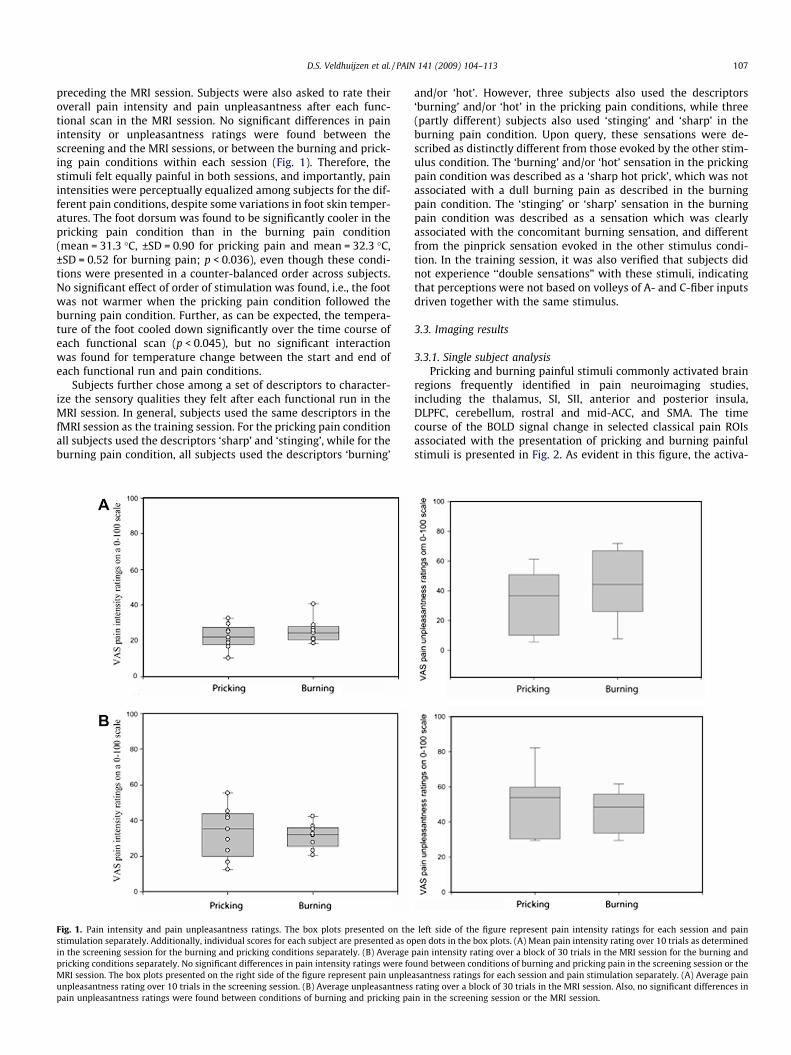
regions frequently identified in pain neuroimaging studies,including the thalamus, SI, SII, anterior and posterior insula,DLPFC, cerebellum, rostral and mid-ACC, and SMA. The timecourse of the BOLD signal change in selected classical pain ROIsassociated with the presentation of pricking and burning painfulstimuli is presented in Fig. 2. As evident in this figure, the activa-
left side of the figure represent pain intensity ratings for each session and painpen dots in the box plots. (A) Mean pain intensity rating over 10 trials as determinedpain intensity rating over a block of 30 trials in the MRI session for the burning andund between conditions of burning and pricking pain in the screening session or theasantness ratings for each session and pain stimulation separately. (A) Average painrating over a block of 30 trials in the MRI session. Also, no significant differences in
in in the screening session or the MRI session.

Pricking pain
Seconds
2 4 6 8 10 12 14
% B
OLD
Sig
nal C
hange
-0.10
-0.05
0.00
0.05
0.10
0.15
0.20
0.25
0.30
0.35
0.40
Contralateral DLPFC
Contralateral thalamus
Contralateral anterior INS
Contralateral posterior INS
mid ACC
anterior ACC
Burning pain
Seconds
2 4 6 8 10 12 14
% B
OLD
Sig
nal C
hange
-0.10
-0.05
0.00
0.05
0.10
0.15
0.20
0.25
0.30
0.35
0.40
Contralateral DLPFC
Contralateral thalamus
Contralateral anterior INS
Contralateral posterior INS
mid ACC
anterior ACC
A
B
Fig. 2. Time course of brain activation following pricking and burning pain. Time course of BOLD signal change in selected ROIs following laser stimulation in the (A) prickingpain and (B) burning pain conditions. Stimulation onset was at 0 s. The right side of the brain is contralateral to stimulation. Error bars represent standard errors.
108 D.S. Veldhuijzen et al. / PAIN 141 (2009) 104–113
tion associated with the burning pain stimuli peaked about 2 s la-ter than the activation associated with the pricking pain stimuli.This pattern was consistent for all the brain regions responding
to both types of stimuli. Several mechanisms could account forthis finding. First, this temporal difference could be explainedby the fact that the stimulus duration for the burning pain condi-

Table 1Individual subject activation in pain-related regions for each condition.
Region Side Pricking pain (n = 9) Burning pain (n = 9)
Thalamus Ipsilateral 7 5Contralateral 9 4
SI Ipsilateral 3 3Contralateral 7 3
SII Ipsilateral 9 8Contralateral 8 6
Anterior-INS Ipsilateral 8 7Contralateral 8 7
Posterior-INS Ipsilateral 7 6Contralateral 8 6
DLPFC Ipsilateral 7 7Contralateral 8 8
Rostral-ACC Midline 7 6Mid-ACC Midline 7 6SMA Midline 9 8
Abbreviations: ACC, anterior cingulate cortex; DLPFC, dorsolateral prefrontal cortex;INS, insula; SMA, supplementary motor cortex.
D.S. Veldhuijzen et al. / PAIN 141 (2009) 104–113 109
tion was nearly 2 s longer than for the pricking pain condition,and that afferent activation in the burning pain condition wouldbe maximal near the end of that stimulation period. Activationtime of the nociceptors was different due to the stimulationparameters necessary to selectively activate the fibers. Second,the difference in conduction velocities between Ad- and C-fibersis also likely to contribute to the temporal difference betweenthe pricking pain and the burning pain-related activation. Third,differences in central processing of information conducted bythe afferents, such as spinothalamic pathway versus a more pro-priospinal pathway, could potentially result in the differences inarrival time at the cortical level. Different lags for the prickingand burning pain conditions were used for further analysis, such
Fig. 3. Activation and overlap maps. Activation maps for the pricking (column 1) andbetween these conditions (column 3; superimposition of the two activation maps). The leto stimulation. Functional activation maps are overlaid on a normalized anatomical MRI iare presented in the images. The color bar represents the extent of activation in z-scores oin the group overlap analysis was 4 voxels in original space at an overall p < 0.01.
that an additional 2-s delay was used for the burning pain com-pared to the pricking pain condition.
Table 1 represents the individual variation in activation of sev-eral regions previously associated with pain processing and se-lected for the ROI analysis. Many areas show consistentactivation within and between subjects, however, some areas, suchas SI and the thalamus in the burning pain condition, only showactivation in a limited number of subjects. The thalamus onlyshowed activation in about half of the subjects for the burningpain condition, yet in almost all subjects for the pricking paincondition.
3.3.2. Group analysisFollowing individual subject analysis, the activation common to
the burning and pricking pain conditions was identified by a groupoverlap map (Fig. 3; overall p < 0.01, corrected for cluster size). Sig-nificant overlap in activation was found for pricking and burningpain in several brain areas, including the bilateral thalamus, bilat-eral anterior insula, bilateral posterior parietal lobule, mid-ACC,contralateral dorsolateral prefrontal cortex, and ipsilateral cerebel-lum (Table 2 and Fig. 3).
Pricking and burning pain also evoked differential responses inselective brain regions. Group contrast maps were used to identifydifferential activation between pricking and burning pain condi-tions. Several areas showed stronger activation following prickingpain compared to burning pain (overall p < 0.05, corrected forcluster size), including the ipsilateral hippocampus, bilateral para-hippocampal gyrus, bilateral fusiform gyrus, contralateral cerebel-lum, and contralateral cuneus/parieto-occipital sulcus. Only onearea showed stronger activation with burning pain compared topricking pain: a region in the ipsilateral middle frontal gyrus, com-prising part of the DLPFC (Table 3, Fig. 4).
burning (column 2) pain conditions separately, and the overlap map of activationft side of each image corresponds to the right side of the brain, which is contralateraln Talairach space. The coordinates of slice cuts through the axial and sagittal planesf the pricking and burning pain activation maps. The minimum cluster size threshold

Table 2Brain regions that show overlap in activation in pricking and burning pain conditions.
Region Side Pricking pain Burning pain
TAL coordinates Z-score TAL coordinates Z-score
x y z x y z
Thalamus Ipsilateral �8 �1 9 4.05 �5 �17 10 3.95Contralateral 6 �2 7 3.62 11 �3 13 3.80
Anterior insula Ipsilateral �37 14 8 4.35 �39 11 3 3.43Ipsilateral �37 �8 16 3.34 �30 �5 19 3.15Contralateral 39 5 19 4.15 43 17 20 3.88
Anterior cingulate cortex Midline �2 17 30 3.35 �1 19 37 3.38Dorsolateral prefrontal cortex Contralateral 36 3 24 4.47 42 12 31 5.11Posterior parietal lobe Ipsilateral �57 �33 25 4.22 �56 �39 26 4.07
Contralateral 50 �30 15 3.51 47 �37 20 3.14Cerebellum Ipsilateral �36 �50 �29 3.43 �38 �53 �37 3.46
Abbreviations: TAL, Talairach coordinates (x,y,z). Note that activation maps for each condition separately show activation in more regions than shown here. Data presented inthis table represent overlap in activation between conditions.
Table 3Brain regions showing a significant contrast for pricking pain versus burning pain.
Region Side TAL coordinates Volume (mm3) Z-score
x y z
Pricking > burningHippocampus Ipsilateral �29 �16 �10 805 3.43Parahippocampal gyrus Ipsilateral �11 �34 �4 465 2.52
Contralateral 20 �34 �1 476 3.14Fusiform gyrus Ipsilateral �36 �43 �9 901 3.62
Contralateral 30 �40 �12 585 3.56Cerebellum Contralateral 8 �66 �25 1133 3.23Cuneus/parieto-occipital sulcus Contralateral 7 �80 17 1047 2.72
Burning > prickingDorsolateral prefrontal cortex Ipsilateral �34 27 34 511 �2.61
Fig. 4. Contrast maps. Brain regions differentially activated by the burning and pricking pain stimulation. The left side of each image corresponds to the right side of the brain,which is contralateral to stimulation. Functional run activation is overlaid on a normalized anatomical MRI in Talairach space. The coordinates of slice cuts through the axialand coronal planes are presented in the images. Orange regions represent stronger activation in the pricking condition versus burning condition, while blue areas representstronger activation in the burning condition versus pricking condition. In the group contrast analysis, the minimum cluster size was 6 voxels in original space at an overallp < 0.05.
110 D.S. Veldhuijzen et al. / PAIN 141 (2009) 104–113

D.S. Veldhuijzen et al. / PAIN 141 (2009) 104–113 111
4. Discussion
Event-related fMRI was used to evaluate brain activation pat-terns associated with pricking and burning pain induced by a diodelaser. Only few studies directly compared these pain conditions,and the present study is innovative regarding perceptually equaliz-ing pain perception between conditions and assuring that the stim-ulation of C-fibers was painful. Several brain regions were found tobe commonly activated, but differential activation was observedfor select brain regions.
4.1. Individual variation in brain activation
Pain-evoked activation in the brain was found bilaterally in allpredefined ROIs. SI activation upon painful laser-evoked stimula-tion has been reported previously [4,5], although it was least con-sistently activated among the subjects in the present study. SIactivity is often absent in pain imaging studies [14]. It is hypothe-sized that in the present study, it was hard to identify significantclusters of activation in SI because the foot representation is rela-tively small. Both the ipsilateral and contralateral thalamus wereonly activated in about half of the subjects in the burning pain con-dition, but in most or all subjects in the pricking pain condition.Along with tissue volume considerations described above, differ-ences in activation in the thalamus could result from time-dis-persed arrival of input from the fibers evoked by the laser. In theburning pain condition, the stimulus was of longer duration andsignals are likely to arrive at the thalamus more dispersed thanin the pricking pain condition.
4.2. Shared neural representation
Conjunction analysis indicated overlap in activation of prickingand burning pain in several brain areas including thalamus, insula,mid-ACC, cerebellum, contralateral DLPFC, and the posterior parie-tal lobe. Activation in these brain areas is typically found in painimaging studies [2,29,46,53,55,61]. The findings of the presentstudy demonstrate that these brain areas are associated with painprocessing of both types of nociceptive inputs. Common activationto pricking and burning pain is expected since Ad- and C-nocicep-tive signals converge to a great extent at the level of the dorsalhorn (e.g., [31,52]). Overlap in the cortical representation of thesepain conditions may possibly reflect a role of these regions in rep-resenting pain salience [23]. Nevertheless, Ad- and C-fiber-medi-ated nociceptive signals are neurobiologically distinct, whichimplies some level of differential CNS processing.
4.3. Differences in brain activation
Group contrast analyses identified brain areas that were differ-entially activated in the burning pain condition versus prickingpain condition, including the hippocampal formation, cerebellum,fusiform gyrus, cuneus/parieto-occipital sulcus, and DLPFC.
While not typically considered a nociceptive processing region,activation in the hippocampal formation has been previously re-ported in response to painful heat [21,50] and laser stimulation[3]. The hippocampal formation has been traditionally associatedwith recent memory consolidation [1], spatial memory [16,30],and fear-initiated avoidance behavior [35]. Studies using specificconditioning paradigms demonstrated that the hippocampus isalso involved in learning associations between pain and predictivecues [49,50]. It is perhaps in its role in learned associations that thedifferential activation between pain pathways in the hippocampushas relevance. Given the need for precise timing of information forappropriately learned associations, it would be reasonable to pro-
pose that nociceptive signals from the Ad-fibers provide the mostuseful input. A separate cluster of activation was identified in thefusiform gyrus adjacent to the activated region of the parahippo-campal gyrus. Given that there is no prior evidence of nociceptiveinput to the fusiform gyrus, the most conservative interpretationwould be that it represents an extension of parahippocampal gyrusactivation.
Activity in the cerebellum is frequently found in pain neuroim-aging studies. Cerebellar activation is generally considered to beprimarily associated with motor responses [12]. Some studiespoint to a more direct role of the cerebellum in pain processingby showing the release of endogenous opioids upon electrical stim-ulation of the cerebellum in animals [22]. The need for temporallyprecise information may also be relevant for brain areas involvedin initiating, propagating, and executing defensive motor re-sponses to first pain [12,25,47,56,58].
Differential activation was also found for a multi-sensory inte-gration region: the parieto-occipital sulcus. This area is recognizedas a site of integration of innocuous somatosensory signals and vi-sual information [15]. A network of motor, pain, and sensory areasincluding the posterior parietal cortex was previously found to beactivated in response to a prickle sensation evoked by noxious coldstimuli [19]. Because the parieto-occipital sulcus is not routinelyfound activated in pain neuroimaging studies, it remains to bedetermined whether preferential Ad nociceptive activation is aconsistent property of this region.
Although the contralateral (right) DLPFC was commonly acti-vated in both the pricking and burning pain conditions, the ipsilat-eral (left) DLPFC was found to be significantly more activated in theburning pain condition. Transcranial magnetic stimulation studiesdemonstrate that prefrontal regions can modulate pain perception.Stimulation of the DLPFC increased pain tolerance upon handimmersion in cold water, however, only when stimulated on theright hemisphere [26]. Significant effects of left prefrontal stimula-tion were found on decreased patient-controlled analgesia use [6].It has been postulated that pain modulation in these regions occursby means of attentional control of pain. Lesion studies, neuronalrecordings, and neuroimaging findings demonstrate that a networkof frontal and parietal structures is involved in attentional control[13,17,34]. In a previous study, the perceptual illusion of ‘paradox-ical heat’, isolated from the perception of pain, was associated withactivity in the right anterior-mid insula [20]. The current studyspecifically focuses on the perception of pain and the findings sug-gest that the C-fiber-mediated nociceptive signals engage the leftDLPFC to a greater extent than the Ad-mediated signals.
It should be noted that differential information processing mayoccur in principle nociceptive processing regions of the cerebrumin a manner that is not easily resolved with fMRI. For instance,the relatively low temporal resolution of fMRI limits its ability todetect differences in the timing of neural processing associatedwith different nociceptive inputs. Additionally, fMRI is not likelyto be able to discern the situation in which small discrete neuralmodules are differentially active within a given ROI, which mayvery well be the case for the early nociceptive processing regions[18].
4.4. Selective activation of nociceptive fibers
Laser stimulation selectively activates nociceptors while avoid-ing concomitant activation of mechanoreceptors [48]. Imagingstudies show that laser stimulation commonly activates corticalareas, including SI, SII, insula, amygdala, ACC, and multiple pre-frontal regions (e.g., [5,7]). However, previous studies frequentlyactivated nociceptors simultaneously rather than selectively.Selective activation of Ad- or C-nociceptors can be achieved bymaking use of differences in resistance to ischemic pressure be-

112 D.S. Veldhuijzen et al. / PAIN 141 (2009) 104–113
tween fibers [9–11], differences in heat thresholds between fibers[37], differences in distribution density of the fibers [8,41,42], anddifferential responses of fibers to the heating rate of the skin and topharmacological treatment [40,62–66,68]. The present study wasdesigned to separate C- and Ad-fiber activation by exploiting differ-ences in thermal thresholds, rate of thermal activation sensitivity,and spatial properties of the nociceptor types. Previous studiesusing contact heat stimulation found that low heating rates (inthe range of 1�2 �C per second) evoked nociceptive responses thatwere primarily mediated by C-fibers, while high heating rates (inthe range of higher than 2 �C per second) produced nociceptive re-sponses primarily mediated by Ad-fibers [63,64,66,68]. With laserstimulation, these same principles apply although heating ratesare faster [39].
4.5. Conclusion
The principle finding from the present study is that many brainareas are commonly engaged in processing of pricking and burningpain mediated by Ad- and C-fiber nociceptors, respectively. Yet,there are some regions that show preferential activation relatedto Ad-fiber input versus C-fiber input. Stronger activation in thepricking pain condition was found in the hippocampal formation,cerebellum, fusiform gyrus, and cuneus/parieto-occipital sulcus.Stronger activation in the burning pain condition was found inthe dorsolateral prefrontal cortex. It remains to be determinedwhether such differences are related to perception, motor reac-tions, or other aspects of nociceptive processing.
Acknowledgments
All authors declare that there are no financial interests to dis-close. This work was supported by P50-AR49555 to J.D.G. andR44DA016840-03 to M.I.N.
References
[1] Alvarez P, Squire LR. Memory consolidation and the medial temporal lobe: asimple network model. Proc Natl Acad Sci 1994;91:7041–5.
[2] Apkarian AV, Bushnell MC, Treede R-D, Zubieta J-K. Human brain mechanismsof pain perception and regulation in health and disease. Eur J Pain2005;9:463–84.
[3] Bingel U, Quante M, Knab R, Bromm B, Weiller C, Büchel C. Subcorticalstructures involved in pain processing: evidence from single-trial fMRI. Pain2002;99:313–21.
[4] Bingel U, Quante M, Knab R, Bromm B, Weiller C, Büchel C. Single trial fMRIreveals significant contralateral bias in responses to laser pain within thalamusand somatosensory cortices. NeuroImage 2003;18:740–8.
[5] Bingel U, Lorenz J, Glauche V, Knab R, Gläscher J, Weiller C, et al. Somatotopicorganization of human somatosensory cortices for pain: a single trial fMRIstudy. NeuroImage 2004;23:224–32.
[6] Borckardt JJ, Weinstein M, Reeves S, Kozel A, Nahas Z, Smith AR, et al.Postoperative left prefrontal repetitive transcranial magnetic stimulationreduces patient-controlled analgesia use. Anesthesiology 2006;105:557–62.
[7] Bornhövd K, Quante M, Glauche V, Bromm B, Weiller C, B}uchel C. Painfulstimuli evoke different stimulus–response functions in the amygdala,prefrontal, insula and somatosensory cortex: a single-trial fMRI study. Brain2002;125:1326–36.
[8] Bragard D, Chen AC, Plaghki L. Direct isolation of ultra-late (C-fibre) evokedbrain potentials by CO2 laser stimulation of tiny cutaneous surface areas inman. Neurosci Lett 1996;209:81–4.
[9] Bromm B, Neitzel H, Tecklenburg A, Treede RD. Evoked cerebral potentialcorrelates of C-fibre activity in man. Neurosci Lett 1983;43:109–14.
[10] Bromm B, Treede RD. Nerve fibre discharges, cerebral potentials andsensations induced by CO2 laser stimulation. Hum Neurobiol 1984;3:33–40.
[11] Bromm B, Treede RD. Human cerebral potentials evoked by CO2 laser stimulicausing pain. Exp Brain Res 1987;67:153–62.
[12] Büchel C, Bornhövd K, Quante M, Glauche V, Bromm B, Weiller C.Dissociable neural responses related to pain intensity, stimulus intensity,and stimulus awareness within the anterior cingulate cortex: a parametricsingle-trial laser functional magnetic resonance imaging study. J Neurosci2002;22:970–6.
[13] Buschman TJ, Miller EK. Top-down versus bottom-up control of attention inthe prefrontal and posterior parietal cortices. Science 2007;315:1860–2.
[14] Bushnell MC, Duncan GH, Hofbauer RK, Ha B, Chen J-I, Carrier B. Painperception: is there a role for primary somatosensory cortex? Proc Natl AcadSci 1999;96:7705–9.
[15] Calvert GA. Crossmodal processing in the human brain: insights fromfunctional neuroimaging studies. Cereb Cortex 2001;11:1110–23.
[16] Clark RE, Broadbent NJ, Squire LR. The hippocampus and spatial memory:findings with a novel modification of the water maze. J Neurosci2007;27:6647–54.
[17] Corbetta M, Shulman GL. Control of goal-directed and stimulus-drivenattention in the brain. Nat Rev Neurosci 2002;3:201–15.
[18] Davis KD. Neurophysiological and anatomical considerations in functionalimaging of pain. Pain 2003;105:1–3.
[19] Davis KD, Pope GE, Crawley AP, Mikulis DJ. Neural correlates of pricklesensation: a percept-related fMRI study. Nat Neurosci 2002;5:1121–2.
[20] Davis KD, Pope GE, Crawley AP, Mikulis DJ. The perceptual illusion of‘‘paradoxical heat” engages the insular cortex. J Neurophysiol2004;92:1248–51.
[21] Derbyshire SWG, Jones AKP, Gyulai F, Clark S, Townsend D, Firestone LL. Painprocessing during three levels of noxious stimulation produces differentialpatterns of central activity. Pain 1997;73:431–45.
[22] Dey PK, Ray AK. Anterior cerebellum as a site for morphine analgesia and post-stimulation analgesia. Indian J Physiol Pharmacol 1982;26:3–12.
[23] Downar J, Mikulis DJ, Davis KD. Neural correlates of the prolonged salience ofpainful stimulation. NeuroImage 2003;20:1540–51.
[24] Forss N, Raij TT, Seppä M, Hari R. Common cortical network for first and secondpain. NeuroImage 2005;24:132–42.
[25] Gracely RH, Geisser ME, Giesecke T, Grant MAB, Petzke F, Williams DA, et al.Pain catastrophizing and neural responses to pain among persons withfibromyalgia. Brain 2004;127:835–43.
[26] Graff-Guerrero A, González-Olvera J, Fresán A, Gómez-Martín D, Méndez-Núnez JC, Pellicer F. Repetitive transcranial magnetic stimulation ofdorsolateral prefrontal cortex increases tolerance to human experimentalpain. Cog Brain Res 2005;25:153–60.
[27] Granot M, Buskila D, Granovsky Y, Sprecher E, Neumann L, Yarnitsky D.Simultaneous recording of late and ultra-late pain evoked potentials infibromyalgia. Clin Neurophysiol 2001;112:1881–7.
[28] Greffrath W, Nemenov MI, Schwarz S, Baumgärtner U, Vogel H, Arendt-NielsenL, et al. Inward currents in primary nociceptive neurons of rat and painsensation in humans elicited by infrared diode laser pulses. Pain2002;99:145–55.
[29] Hofbauer RK, Rainville P, Duncan GH, Bushnell MC. Cortical representation ofthe sensory dimension of pain. J Neurophysiol 2001;86:402–11.
[30] Hollup SA, Kjelstrup KG, Hoff J, Moser M-B, Moser EI. Impaired recognition ofthe global location during spatial navigation in rats with hippocampal lesions.J Neurosci 2001;21:4505–13.
[31] Jänig W. Neuronal mechanisms of pain with special emphasis on visceral anddeep somatic pain. Acta Neurochir Suppl 1987;38:16–32.
[32] Jiang N, Cooper BY, Nemenov MI. Non-invasive diode laser activation oftransient receptor potential proteins in nociceptors. In: Hamblin MR, WaynantRW, Anders J, editors. Mechanisms for low-light therapy II. Proceedings of theSPIE 2007.
[33] Kakigi R, Tran TD, Qiu Y, Wang X, Nguyen TB, Inui K, et al. Cerebral responsesfollowing stimulation of unmyelinated C-fibers in humans: electro- andmagneto-encephalographic study. Neurosci Res 2003;45:255–75.
[34] Kastner S, Ungerleider L. Mechanisms of visual attention in the human cortex.Annu Rev Neurosci 2000;23:315–41.
[35] Kjelstrup KG, Tuvnes FA, Steffenach H-A, Murison R, Moser EI, Moser M-B.Reduced fear expression after lesions of the ventral hippocampus. Proc NatlAcad Sci 2002;99:10825–30.
[36] Kou L, Labrie D, Chylek P. Refractive indices of water and ice in the 0.65- to 2.5-lm spectral range. Appl Opt 1993;32:3531–40.
[37] Magerl W, Ali Z, Ellrich J, Meyer RA, Treede RD. C- and A delta-fibercomponents of heat-evoked cerebral potentials in healthy human subjects.Pain 1999;82:127–37.
[38] Moulton EA, Keaser ML, Gullapalli RP, Maitra R, Greenspan JD. Sex differencesin the cerebral BOLD signal response to painful heat stimuli. Am J PhysiolRegul Integr Comp Physiol 2006;291:R257–67.
[39] Nemenov MI, Zaitsev VG, Mikkelsen J. Limitations of laser application in painresearch. In: International association for the study of pain, Ninth WorldCongress on Pain, 1999, Vienna, Austria [abstract].
[40] Nemenov MI. Portable laser and process for producing controlled pain USAPatent #7402167, issued 2008, priority date 2002.
[41] Ochoa J, Mair WGP. The normal sural nerve in man. Ultrastructure andnumbers of fibers and cells I. Acta Neuropath 1969;13:197–216.
[42] Opsommer E, Masquelier E, Plaghki L. Determination of nerve conductionvelocity of C-fibres in humans from thermal thresholds to contact heat(thermode) and from evoked brain potentials to radiant heat (CO2 laser).Neurophysiol Clin 1999;29:411–22.
[43] Opsommer E, Weiss T, Plaghki L, Miltner WHR. Dipole analysis of ultralate (C-fibres) evoked potentials after laser stimulation of tiny cutaneous surface areasin humans. Neurosci Lett 2001;298:41–4.
[44] Orstavik K, Weidner C, Schmidt R, Schmelz M, Hilliges M, Jorum E, et al.Pathological C-fibres in patients with a chronic painful condition. Brain2003;126:567–78.
[45] Palmer KF, Williams D. Optical properties of water in the near infrared. J OptSoc Am 1974;64:1107–10.

D.S. Veldhuijzen et al. / PAIN 141 (2009) 104–113 113
[46] Peyron R, Laurent B, García-Larrea L. Functional imaging of brain responses topain. A review and meta-analysis. Neurophysiol Clin 2000;30:263–88.
[47] Peyron R, Kupers R, Jehl JL, Garcia-Larrea L, Convers P, Barral FG, et al. Centralrepresentation of the RIII flexion reflex associated with overt motor reaction:an fMRI study. Clin Neurophysiol 2007;37:249–59.
[48] Plaghki L, Mouraux A. How do we selectively activate skin nociceptors with ahigh power infrared laser? Physiology and biophysics of laser stimulation. ClinNeurophysiol 2003;33:269–77.
[49] Ploghaus A, Tracey I, Clare S, Gati JS, Rawlins NP, Matthews PM. Learning aboutpain: the neural substrate of the prediction error for aversive events. Proc NatlAcad Sci 2000;97:9281–6.
[50] Ploghaus A, Narain C, Beckmann CF, Clare S, Bantick S, Wise R, et al.Exacerbation of pain by anxiety is associated with activity in a hippocampalnetwork. J Neurosci 2001;21:9896–903.
[51] Ploner M, Gross J, Timmermann L, Schnitzler A. Cortical representation of firstand second pain sensation in humans. Proc Natl Acad Sci 2002;99:12444–8.
[52] Price DD, Wagman IH. Relationships between pre- and postsynaptic effects ofA and C fiber inputs to dorsal horn of M. mulatta. Exp Neurol 1973;40:90–103.
[53] Price DD. Psychological and neural mechanisms of the affective dimension ofpain. Science 2000;288:1769–72.
[54] Qiu Y, Noguchi Y, Honda M, Nakata H, Tamura Y, Tanaka S, et al. Brainprocessing of signals ascending through unmyelinated C fibers in humans: anevent-related functional magnetic resonance study. Cereb Cortex2006;16:1289–95.
[55] Rainville P, Duncan GH, Price DD, Carrier B, Bushnell MC. Pain affect encodedin human anterior cingulate but not somatosensory cortex. Science1997;277:968–71.
[56] Saab CY, Kawasaki M, Al-Chaer ED, Willis WD. Cerebellar cortical stimulationincreases spinal visceral nociceptive responses. J Neurophysiol2001;85:2359–63.
[57] Spiegel J, Hansen C, Treede R-D. Clinical evaluation for the assessment ofimpaired pain sensitivity by thulium-laser evoked potentials. ClinNeurophysiol 2000;111:725–35.
[58] Sullivan MJL, Thon B, Haythornthwaite JA, Keefe F, Martin M, Bradley LA, et al.Theoretical perspectives on the relationship between catastrophizing andpain. Clin J Pain 2001;17:52–64.
[59] Tilman DB, Treede RD, Meyer RA, Campbell JN. Response of C fiber nociceptorsin the anesthetized monkey to heat stimuli: estimates of receptor depth andthreshold. J Physiol 1995;485:753–65.
[60] Treede RD, Meyer RA, Raja SN, Campbell JN. Evidence for two different heattransduction mechanisms in nociceptive primary afferents innervatingmonkey skin. J Physiol 1995;483:747–58.
[61] Treede R-D, Kenshalo DR, Gracely RH, Jones AKP. The cortical representation ofpain. Pain 1999;79:105–11.
[62] Tzabazis A, Klyukinov M, Manering N, Nemenov MI, Schafer SL, Yeomans DC.Differential activation of trigeminal C or Ad nociceptors by infrared diode laserin rats: behavioral evidence. Brain Res 2005;1037:148–56.
[63] Yarnitsky D, Ochoa JL. Studies of heat pain sensation in man: perceptionthresholds, rate of stimulus rise and reaction time. Pain 1990;40:85–91.
[64] Yarnitsky D, Simone DA, Dotson RM, Cline MA, Ochoa JL. Single C nociceptorresponses and psychophysical parameters of evoked pain: effect of rate of riseof heat stimuli in humans. J Physiol 1992;450:581–92.
[65] Yeomans DC, Cooper BY, Vierck CJ. Effects of systemic morphine on responsesof primates to first or second pain sensations. Pain 1996;66:253–63.
[66] Yeomans DC, Pirec V, Proudfit HK. Nociceptive responses to high and low ratesof noxious cutaneous heating are mediated by different nociceptors in the rat:behavioral evidence. Pain 1996;68:133–40.
[67] Yeomans DC, Plaghki L, Herbette S, Klyukinov M, Nemenov MI. Cortical evokedpotentials and psychophysical evaluation of pain evoked by infrared diodelaser stimulation of skin in humans. In: International association for the studyof pain, Tenth world congress on pain, 2008, Glasgow, UK [abstract].
[68] Zachariou V, Goldstein BD, Yeomans DC. Low but not high rate noxious radiantskin heating evokes a capsaicin-sensitive increase in spinal cord dorsal hornrelease of substance P. Brain Res 1997;752:143–50.
Related Documents