Immunity 25, 309–318, August 2006 ª2006 Elsevier Inc. DOI 10.1016/j.immuni.2006.05.017 Differential Activity of IL-12 and IL-23 in Mucosal and Systemic Innate Immune Pathology Holm H. Uhlig, 1,4,5 Brent S. McKenzie, 2,4 Sophie Hue, 1 Claire Thompson, 1 Barbara Joyce-Shaikh, 2 Renata Stepankova, 3 Nicolas Robinson, 1 Sofia Buonocore, 1 Helena Tlaskalova-Hogenova, 3 Daniel J. Cua, 2, * and Fiona Powrie 1, * 1 Sir William Dunn School of Pathology University of Oxford South Parks Road OX1 3RE Oxford United Kingdom 2 Schering-Plough Biopharma (formerly DNAX research) 901 California Avenue Palo Alto, California 94304 3 Institute of Microbiology and Gnotobiology of the Czech Academy of Sciences 54922 Praque and Novy Hradek Czech Republic Summary The CD40-CD154 pathway is important in the patho- genesis of inflammatory bowel disease. Here we show that injection of an agonistic CD40 mAb to T and B cell- deficient mice was sufficient to induce a pathogenic systemic and intestinal innate inflammatory response that was functionally dependent on tumor necrosis factor-a and interferon-g as well as interleukin-12 p40 and interleukin-23 p40 secretion. CD40-induced colitis, but not wasting disease or serum proinflammatory cytokine production, depended on interleukin-23 p19 secretion, whereas interleukin-12 p35 secretion con- trolled wasting disease and serum cytokine produc- tion but not mucosal immunopathology. Intestinal inflammation was associated with IL-23 (p19) mRNA- producing intestinal dendritic cells and IL-17A mRNA within the intestine. Our experiments identified IL-23 as an effector cytokine within the innate intestinal im- mune system. The differential role of IL-23 in local but not systemic inflammation suggests that it may make a more specific target for the treatment of IBD. Introduction The inflammatory bowel diseases (IBD) encompassing Crohn’s disease (CD) and ulcerative colitis are chronic inflammatory disorders of the gastrointestinal tract that affect approximately 0.1% of Western populations (Bouma and Strober, 2003; Shanahan, 2002). Available evidence suggests that IBD involves an aberrant inflam- matory response to intestinal bacteria in genetically susceptible individuals. The immune pathogenesis of CD is associated with increased inflammatory cytokines including tumor necrosis factor-a (TNF-a), interferon-g (IFN-g), and interleukin-12 and interleukin-23 p40 (IL- 12p40, IL-23p40). Accordingly, anti-TNF-a therapy has been shown to be beneficial in CD patients (Sandborn and Faubion, 2004). However, a limitation of this ap- proach is that TNF-a also plays a pivotal role in protec- tion from infection. Indeed, sustained blockade of TNF-a has been reported to increase susceptibility to infection such as reactivation of Mycobacterium tuber- culosis (Sandborn and Faubion, 2004). These findings highlight the need to develop more specific strategies that discriminate the local pathogenic inflammatory response from systemic host protective immunity. Interactions between CD40 and CD154 (CD40L) are important in the initiation and maintenance of T cell- mediated intestinal inflammation. In the inflamed intesti- nal tissue of human IBD patients, as well as in mouse models of IBD, CD40 + antigen-presenting cells (APC) are found in association with CD40L + T cells (Liu et al., 1999, 2000; Polese et al., 2002). Importantly, blockade of CD40-CD40L interactions inhibits the development of colitis and can ameliorate established disease in mouse models (Cong et al., 2000; De Jong et al., 2000; Kelsall et al., 1996; Liu et al., 2000). These results suggest that after antigen encounter, activated CD4 + CD40L + T cells stimulate CD40 + APC via the CD40L-CD40 path- way. This in turn leads to further activation of APC and T cells, establishing a positive feedback loop of immune activation (Diehl et al., 2000; van Kooten and Bancher- eau, 2000). In this type of inflammatory cascade, both CD4 + T cells and APC can contribute to immunopathol- ogy with effector mechanisms including cytokine pro- duction (Strober et al., 2002). CD40 stimulation of myeloid cells can lead to IL-12 production (Stuber et al., 1996). This cytokine plays a key role in the inflammatory response primarily due to its ability to induce IFN-g production by T cells and NK cells (Trinchieri et al., 2003). In intestinal inflamma- tion, administration of neutralizing IL-12p40 mAbs ame- liorates colitis in several different models (Neurath et al., 1996; Simpson et al., 1998). This therapeutic effect is linked to reductions in IL-12-driven IFN-g secretion as well as the induction of Fas-mediated apoptosis of Th1 cells (Fuss et al., 1999). In a recent study, anti-IL-12p40 treatment in CD led to an amelioration of inflammation in some patients (Mannon et al., 2004). Clinical improve- ment was associated with a reduction in the production of IL-12 and IFN-g by mononuclear cells from the intes- tine. However, it is now known that IL-12p40 forms heter- odimers not only with IL-12p35 (IL-12p35p40; IL-12p70) but also with IL-23p19 (IL-23p19p40). These results raise the possibility that activities previously ascribed to IL-12 may in fact be attributable to IL-23. Indeed, the immune pathological response in experimental autoimmune encephalomyelitis (Cua et al., 2003) and collagen- induced arthritis (Murphy et al., 2003) has been shown to be functionally dependent on IL-23 and not IL-12. To assess the role of CD40-mediated effector func- tion in the innate response and to further probe the *Correspondence: [email protected] (D.J.C.); fiona.powrie@ path.ox.ac.uk (F.P.) 4 These authors contributed equally to this work. 5 Present address: Children’s Hospital, Department of Pediatric Gastroenterology, University Children’s Hospital, Oststr. 21-25, 04317 Leipzig, Germany.

Welcome message from author
This document is posted to help you gain knowledge. Please leave a comment to let me know what you think about it! Share it to your friends and learn new things together.
Transcript

Immunity 25, 309–318, August 2006 ª2006 Elsevier Inc. DOI 10.1016/j.immuni.2006.05.017
Differential Activity of IL-12 and IL-23in Mucosal and Systemic Innate Immune Pathology
Holm H. Uhlig,1,4,5 Brent S. McKenzie,2,4 Sophie Hue,1
Claire Thompson,1 Barbara Joyce-Shaikh,2
Renata Stepankova,3 Nicolas Robinson,1
Sofia Buonocore,1 Helena Tlaskalova-Hogenova,3
Daniel J. Cua,2,* and Fiona Powrie1,*1Sir William Dunn School of PathologyUniversity of OxfordSouth Parks RoadOX1 3RE OxfordUnited Kingdom2Schering-Plough Biopharma (formerly DNAX
research)901 California AvenuePalo Alto, California 943043 Institute of Microbiology and Gnotobiology
of the Czech Academy of Sciences54922 Praque and Novy HradekCzech Republic
Summary
The CD40-CD154 pathway is important in the patho-
genesis of inflammatory bowel disease. Here we showthat injection of an agonistic CD40 mAb to T and B cell-
deficient mice was sufficient to induce a pathogenicsystemic and intestinal innate inflammatory response
that was functionally dependent on tumor necrosisfactor-a and interferon-g as well as interleukin-12 p40
and interleukin-23 p40 secretion. CD40-induced colitis,but not wasting disease or serum proinflammatory
cytokine production, depended on interleukin-23 p19secretion, whereas interleukin-12 p35 secretion con-
trolled wasting disease and serum cytokine produc-tion but not mucosal immunopathology. Intestinal
inflammation was associated with IL-23 (p19) mRNA-producing intestinal dendritic cells and IL-17A mRNA
within the intestine. Our experiments identified IL-23as an effector cytokine within the innate intestinal im-
mune system. The differential role of IL-23 in local butnot systemic inflammation suggests that it may make
a more specific target for the treatment of IBD.
Introduction
The inflammatory bowel diseases (IBD) encompassingCrohn’s disease (CD) and ulcerative colitis are chronicinflammatory disorders of the gastrointestinal tractthat affect approximately 0.1% of Western populations(Bouma and Strober, 2003; Shanahan, 2002). Availableevidence suggests that IBD involves an aberrant inflam-matory response to intestinal bacteria in geneticallysusceptible individuals. The immune pathogenesis of
*Correspondence: [email protected] (D.J.C.); fiona.powrie@
path.ox.ac.uk (F.P.)4 These authors contributed equally to this work.5 Present address: Children’s Hospital, Department of Pediatric
Gastroenterology, University Children’s Hospital, Oststr. 21-25,
04317 Leipzig, Germany.
CD is associated with increased inflammatory cytokinesincluding tumor necrosis factor-a (TNF-a), interferon-g
(IFN-g), and interleukin-12 and interleukin-23 p40 (IL-12p40, IL-23p40). Accordingly, anti-TNF-a therapy hasbeen shown to be beneficial in CD patients (Sandbornand Faubion, 2004). However, a limitation of this ap-proach is that TNF-a also plays a pivotal role in protec-tion from infection. Indeed, sustained blockade ofTNF-a has been reported to increase susceptibility toinfection such as reactivation of Mycobacterium tuber-culosis (Sandborn and Faubion, 2004). These findingshighlight the need to develop more specific strategiesthat discriminate the local pathogenic inflammatoryresponse from systemic host protective immunity.
Interactions between CD40 and CD154 (CD40L) areimportant in the initiation and maintenance of T cell-mediated intestinal inflammation. In the inflamed intesti-nal tissue of human IBD patients, as well as in mousemodels of IBD, CD40+ antigen-presenting cells (APC)are found in association with CD40L+ T cells (Liu et al.,1999, 2000; Polese et al., 2002). Importantly, blockadeof CD40-CD40L interactions inhibits the developmentof colitis and can ameliorate established disease inmouse models (Cong et al., 2000; De Jong et al., 2000;Kelsall et al., 1996; Liu et al., 2000). These results suggestthat after antigen encounter, activated CD4+CD40L+
T cells stimulate CD40+ APC via the CD40L-CD40 path-way. This in turn leads to further activation of APC andT cells, establishing a positive feedback loop of immuneactivation (Diehl et al., 2000; van Kooten and Bancher-eau, 2000). In this type of inflammatory cascade, bothCD4+ T cells and APC can contribute to immunopathol-ogy with effector mechanisms including cytokine pro-duction (Strober et al., 2002).
CD40 stimulation of myeloid cells can lead to IL-12production (Stuber et al., 1996). This cytokine playsa key role in the inflammatory response primarily dueto its ability to induce IFN-g production by T cells andNK cells (Trinchieri et al., 2003). In intestinal inflamma-tion, administration of neutralizing IL-12p40 mAbs ame-liorates colitis in several different models (Neurath et al.,1996; Simpson et al., 1998). This therapeutic effect islinked to reductions in IL-12-driven IFN-g secretion aswell as the induction of Fas-mediated apoptosis of Th1cells (Fuss et al., 1999). In a recent study, anti-IL-12p40treatment in CD led to an amelioration of inflammationin some patients (Mannon et al., 2004). Clinical improve-ment was associated with a reduction in the productionof IL-12 and IFN-g by mononuclear cells from the intes-tine. However, it is now known that IL-12p40 forms heter-odimers not only with IL-12p35 (IL-12p35p40; IL-12p70)but also with IL-23p19 (IL-23p19p40). These results raisethe possibility that activities previously ascribed to IL-12may in fact be attributable to IL-23. Indeed, the immunepathological response in experimental autoimmuneencephalomyelitis (Cua et al., 2003) and collagen-induced arthritis (Murphy et al., 2003) has been shownto be functionally dependent on IL-23 and not IL-12.
To assess the role of CD40-mediated effector func-tion in the innate response and to further probe the

Immunity310
role of IL-12 and IL-23 in this pathway, we injected T andB cell-deficient mice with an agonistic CD40 mAb. Anti-CD40 stimulation led to a systemic and local inflamma-tory disease characterised by wasting disease, spleno-megaly, increases in serum inflammatory cytokines,and colitis. Functional analysis showed that the sys-temic inflammatory response was driven by IL-12 andnot IL-23, whereas local intestinal inflammation requiredthe presence of IL-23 and was independent of IL-12.These results newly identify IL-23 as a key effector cyto-kine within the innate intestinal immune system andpoint to divergent roles for IL-12 and IL-23 in local andsystemic inflammation.
Results
Anti-CD40 Induces Wasting Disease and Colitisin T Cell-Deficient Mice
To assess the effects of CD40 stimulation on the activa-tion of the innate immune system, we injected immuno-deficient recombinase-activating gene 1-deficient (Rag1KO) mice with 200 mg CD40 mAb. Clinically, the mice de-veloped wasting disease as well as gastrointestinalsymptoms including diarrhea and anal inflammation.CD40 mAb-treated Rag1 KO mice developed rapidweight loss, up to 20% within the first 4 days (Figure 1A).7 days after antibody challenge, the weight was still sub-stantially decreased compared to isotype- or PBS-treated control mice. At this time, all CD40 mAb-treatedmice developed splenomegaly. Other pathologicalchanges in these mice included hepatopathy, lympha-denomegaly of the mesenteric lymph node (MLN), andcolon pathology, as indicated by swelling of the colonwall and presence of edema. 10 days after CD40 stimu-lation, the mice recovered clinically but still showedmacroscopical signs of colon pathology, whereas 3weeks after the initiation of the immune response, nopathology was observed (data not shown). None ofthese changes were seen when two different isotypecontrol antibodies or PBS injections were used. C.B-17SCID mice developed similar pathology after anti-CD40 stimulation (data not shown).
Consistent with macroscopic observations, 7 daysafter CD40 mAb challenge, mice had histological signsof colitis with pronounced epithelial hyperplasia andmarked leukocyte infiltration within the lamina propria,goblet cell depletion, and epithelial cell destruction(Figures 1B and 1C and Figures S1A–S1C in the Supple-mental Data available with this article online). 10 daysafter CD40 activation, there was still marked leucocyticinfiltration, but by 3 weeks after CD40 stimulation, allhistological changes had resolved (data not shown).
In summary, our results indicate that CD40 mAbtreatment in immunodeficient mice induces weightloss and intestinal pathology.
Innate Immune Activation after Anti-CD40
Stimulation In VivoIn line with the pathological and histopathologicalobservations, marked increases in total leukocyteswere found in the spleen and locally in the colon inanti-CD40-treated mice (Figure S1C). CD40 is abun-dantly expressed on CD11chi dendritic cells (DCs) ofSCID or Rag1 KO mice (Figure 2A), and these cells
became activated with increases in MHC-II, CD80, andCD86 expression at early time points after anti-CD40stimulation (Figure 2A). There was also a marked in-crease in the density of CD11chi DC in leucocytic clus-ters in the colon (Figure 2B).
Changes in serum cytokines and chemokines wereanalyzed to determine whether these correlate with thedevelopment of anti-CD40-induced disease. Serumwas assayed at day 3, when the anti-CD40-stimulatedmice had their maximum weight loss and the first signsof colon pathology, as well as at day 7, when intestinalpathology peaked. Increased amounts of TNF-a, IL-6,IFN-g, MCP-1, and IL-12p70 were found in CD40 mAb-treated mice. There was no significant change in IL-10(Figure 2C).
Anti-CD40 treatment induced comparable local andsystemic immune pathology in germ-free and specificpathogen-free (SPF) housed animals, suggesting thatthe CD40-mediated activation pathway is not depen-dent on the preactivation of the innate immune systemby replicating resident bacteria (Supplemental Dataand Figure S2).
Anti-CD40-Induced Immune Pathology Depends
on Cytokine SecretionTo test whether proinflammatory cytokines playeda functional role in the development of the anti-CD40-mediated immunopathology, Rag1 KO mice were coin-jected with CD40 mAb together with anti-TNF-a, anti-IL-12 and IL-23 p40 (anti-p40), or anti-IFN-g. Anti-p40had the most marked effect, leading to inhibition of wast-ing disease, increases in serum cytokines, and colonimmune pathology (Figures 3 and 4). Anti-TNF-a andanti-IFN-g also significantly inhibited wasting, althoughneither afforded complete protection from increases inserum cytokine levels or colitis (Figure 3). Together thedata indicate that proinflammatory cytokines play akey role in anti-CD40-induced systemic and intestinalimmune pathology.
Differential Roles for IL-12 and IL-23 in Systemic
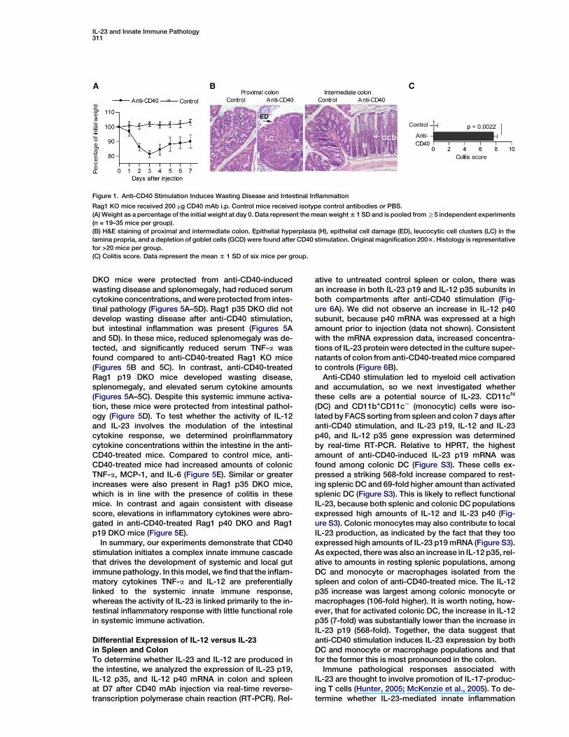
and Gut ImmunopathologyAnti-IL-12p40 neutralizes the activity not only of IL-12(p35p40) but also IL-23, which is composed of a hetero-dimer of p40 with p19 (Trinchieri et al., 2003). To unravelthe role of IL-23 in this system, immunodeficient micewere coinjected with anti-CD40 together with neutraliz-ing anti-IL-12 and -23p40 (anti-p40), anti-IL-23p19(anti-p19), or an isotype control antibody. Anti-p40inhibited systemic immune activation as well as localcolonic pathology (Figure 4). In contrast, anti-CD40and anti-p19 coinjected mice developed wasting dis-ease (Figure 4A) and showed serum amounts of theinflammatory mediators MCP-1, IL-6, and TNF-a thatwere similar to anti-CD40-treated controls (Figure 4B).Strikingly, anti-IL-23p19-treated mice did not developsigns of intestinal inflammation (Figure 4C). These ex-periments suggest differential roles for IL-12 and IL-23in systemic and mucosal innate immunopathology.
To further investigate this, we crossed mice that aredeficient for IL-12p35 or IL-23p19 as well as IL-12 andIL-23p40 onto a Rag1-deficient background (Rag1 p35DKO, Rag1 p19 DKO, Rag1 p40 DKO). These doubleknockout mice were injected with anti-CD40. Rag1 p40
IL-23 and Innate Immune Pathology311
Figure 1. Anti-CD40 Stimulation Induces Wasting Disease and Intestinal Inflammation
Rag1 KO mice received 200 mg CD40 mAb i.p. Control mice received isotype control antibodies or PBS.
(A) Weight as a percentage of the initial weight at day 0. Data represent the mean weight 6 1 SD and is pooled from R5 independent experiments
(n = 19–35 mice per group).
(B) H&E staining of proximal and intermediate colon. Epithelial hyperplasia (H), epithelial cell damage (ED), leucocytic cell clusters (LC) in the
lamina propria, and a depletion of goblet cells (GCD) were found after CD40 stimulation. Original magnification 2003. Histology is representative
for >20 mice per group.
(C) Colitis score. Data represent the mean 6 1 SD of six mice per group.
DKO mice were protected from anti-CD40-inducedwasting disease and splenomegaly, had reduced serumcytokine concentrations, and were protected from intes-tinal pathology (Figures 5A–5D). Rag1 p35 DKO did notdevelop wasting disease after anti-CD40 stimulation,but intestinal inflammation was present (Figures 5Aand 5D). In these mice, reduced splenomegaly was de-tected, and significantly reduced serum TNF-a wasfound compared to anti-CD40-treated Rag1 KO mice(Figures 5B and 5C). In contrast, anti-CD40-treatedRag1 p19 DKO mice developed wasting disease,splenomegaly, and elevated serum cytokine amounts(Figures 5A–5C). Despite this systemic immune activa-tion, these mice were protected from intestinal pathol-ogy (Figure 5D). To test whether the activity of IL-12and IL-23 involves the modulation of the intestinalcytokine response, we determined proinflammatorycytokine concentrations within the intestine in the anti-CD40-treated mice. Compared to control mice, anti-CD40-treated mice had increased amounts of colonicTNF-a, MCP-1, and IL-6 (Figure 5E). Similar or greaterincreases were also present in Rag1 p35 DKO mice,which is in line with the presence of colitis in thesemice. In contrast and again consistent with diseasescore, elevations in inflammatory cytokines were abro-gated in anti-CD40-treated Rag1 p40 DKO and Rag1p19 DKO mice (Figure 5E).
In summary, our experiments demonstrate that CD40stimulation initiates a complex innate immune cascadethat drives the development of systemic and local gutimmune pathology. In this model, we find that the inflam-matory cytokines TNF-a and IL-12 are preferentiallylinked to the systemic innate immune response,whereas the activity of IL-23 is linked primarily to the in-testinal inflammatory response with little functional rolein systemic immune activation.
Differential Expression of IL-12 versus IL-23in Spleen and Colon
To determine whether IL-23 and IL-12 are produced inthe intestine, we analyzed the expression of IL-23 p19,IL-12 p35, and IL-12 p40 mRNA in colon and spleenat D7 after CD40 mAb injection via real-time reverse-transcription polymerase chain reaction (RT-PCR). Rel-
ative to untreated control spleen or colon, there wasan increase in both IL-23 p19 and IL-12 p35 subunits inboth compartments after anti-CD40 stimulation (Fig-ure 6A). We did not observe an increase in IL-12 p40subunit, because p40 mRNA was expressed at a highamount prior to injection (data not shown). Consistentwith the mRNA expression data, increased concentra-tions of IL-23 protein were detected in the culture super-natants of colon from anti-CD40-treated mice comparedto controls (Figure 6B).
Anti-CD40 stimulation led to myeloid cell activationand accumulation, so we next investigated whetherthese cells are a potential source of IL-23. CD11chi
(DC) and CD11b+CD11c2 (monocytic) cells were iso-lated by FACS sorting from spleen and colon 7 days afteranti-CD40 stimulation, and IL-23 p19, IL-12 and IL-23p40, and IL-12 p35 gene expression was determinedby real-time RT-PCR. Relative to HPRT, the highestamount of anti-CD40-induced IL-23 p19 mRNA wasfound among colonic DC (Figure S3). These cells ex-pressed a striking 568-fold increase compared to rest-ing splenic DC and 69-fold higher amount than activatedsplenic DC (Figure S3). This is likely to reflect functionalIL-23, because both splenic and colonic DC populationsexpressed high amounts of IL-12 and IL-23 p40 (Fig-ure S3). Colonic monocytes may also contribute to localIL-23 production, as indicated by the fact that they tooexpressed high amounts of IL-23 p19 mRNA (Figure S3).As expected, there was also an increase in IL-12 p35, rel-ative to amounts in resting splenic populations, amongDC and monocyte or macrophages isolated from thespleen and colon of anti-CD40-treated mice. The IL-12p35 increase was largest among colonic monocyte ormacrophages (106-fold higher). It is worth noting, how-ever, that for activated colonic DC, the increase in IL-12p35 (7-fold) was substantially lower than the increase inIL-23 p19 (568-fold). Together, the data suggest thatanti-CD40 stimulation induces IL-23 expression by bothDC and monocyte or macrophage populations and thatfor the former this is most pronounced in the colon.
Immune pathological responses associated withIL-23 are thought to involve promotion of IL-17-produc-ing T cells (Hunter, 2005; McKenzie et al., 2005). To de-termine whether IL-23-mediated innate inflammation

Immunity312
Figure 2. Immune Activation after Anti-CD40 Stimulation
Rag1 KO mice received 200 mg CD40 mAb i.p. or isotype antibody.
(A) Left: Dot plot of FACS staining for CD11c and isotype as well as CD11c and CD40 of mesenteric lymph node cells (MLN) of untreated SCID
mice. Right: Rag1 KO MLN cells were isolated at day 3 after CD40 mAb and isotype treatment, pooled from six mice per group, and analyzed for
CD40, CD80, CD86, and MHC-II on CD11c+ gated cells. Histogram plots show activation markers of Rag1 KO mice after CD40 mAb (bold line) or
isotype treatment (dotted line). Isotype control for the respective activation marker is shown for anti-CD40-treated mice (filled area). Isotype
control-treated mice expressed a similar isotype staining as the CD40 mAb-treated mice (not shown). Numbers indicate the percentage of cells
that are positive for the respective activation marker (Isotype; Anti-CD40). Similar results were obtained when spleen cells were analyzed.
(B) CD40 mAb treatment results in the accumulation of CD11c+ cells in the colonic lamina propria of Rag1 KO mice. Large clusters of CD11c+ cells
are present in the proximal colon of anti-CD40-treated mice. The scattered distribution of CD11c+ cells present in isotype control-treated mice is
shown for comparison. Histological pictures are representative of n R 4 mice per group. Original magnification 2003. Isotype control gave
no staining.
(C) CD40 mAb treatment leads to increased serum amounts of IL-6, MCP-1, IFN-g, IL-12p70, and TNF-a. Serum cytokines concentrations were
analyzed 3 and 7 days after injection. Data represent mean 6 SD. Data points lower than standard curve data were set as 1 pg/ml. Significance
was tested by the Mann and Whitney U test. Data from two independent experiments were pooled (n = 3–7 mice per group). A further independent
experiment gave a similar result.
is also linked to increased IL-17 production, we exam-ined the expression of IL-17 mRNA (now termedIL-17A) in colon and spleen of Rag1 KO mice afterCD40 mAb injection. As shown in Figure 6C, comparedto colons from unstimulated mice, there was a 65-foldincrease in IL-17 transcripts after anti-CD40 stimulation.However, no such increase was observed in the spleen.As with other inflammatory cytokines, increases incolonic IL-17 mRNA were dependent on IL-23 and not
IL-12 (Figure 6D). These results indicate that anti-CD40stimulation induces IL-17 mRNA production by non-Tcells and that this response is localized to the colon.
Discussion
The IBDs are of complex multifactorial pathogene-sis and involve the activation of the innate and adap-tive immune system. Animal models have suggested

IL-23 and Innate Immune Pathology313
Figure 3. Anti-CD40-Induced Immunopathology Depends Functionally on TNF-a, IL-12 and IL-23p40 as well as IFN-g
Rag1 KO mice recieved 200 mg CD40 mAb i.p. or isotype antibody. In some groups, anti-IL-12/23p40 (500 mg), anti-TNF-a (1 mg), or anti-IFN-g
(1–2 3 2 mg) were coadministered together with anti-CD40 and analyzed for weight, serum cytokine concentration, and development of colitis.
Data represent mean 6 SD. Significance was tested by the Mann and Whitney U test.
(A) Weight as a percentage of the initial weight. Data from R2 experiments were pooled.
(B) TNF-a, IL-6, and MCP-1 serum concentration at day 7 (n = 3 per group).
(C) Colitis score (n = 7–15 mice per group).
T cell-dependent and -independent mechanisms of in-testinal inflammation (Strober et al., 2002). In IBD, thereis evidence that alterations in the innate immune re-sponse contribute to disease development becausemutations in the NOD2 gene, which lead to abnormalfunction of this innate pathogen recognition receptor,confer susceptibility to CD in some patients (Eckmannand Karin, 2005). In IBD patients as well as in many modelsituations, the CD40L-CD40 pathway plays an importantrole in the crosstalk between the innate and adaptiveimmune response. We therefore developed a modelwhere CD40L-expressing activated T cells were replacedwith an agonist CD40 mAb and demonstrate that CD40-mediated effector function, in the absence of furtherT cell help, is sufficient to induce a pathogenic systemicand local inflammatory response. Anti-CD40-induceddisease is accompanied by accumulation of myeloid cellsincluding activated cytokine-producing DC in spleen andcolon. Production of the inflammatory cytokine IL-12 andIL-23p40 subunit plays a pivotal role in the pathogenesisof systemic and mucosal disease. To date, the functionsof IL-23 have only been described in models that involveT cells (Cua et al., 2003; Murphy et al., 2003). However,our data reveal distinct roles for IL-12 and IL-23 withinthe innate immune system. Thus, IL-12 is a key moleculefor systemic immune activationwhereas IL-23 drives localintestinal inflammation.
With our model of innate immune activation, we showthat a number of proinflammatory cytokines are associ-
ated with weight loss and colon pathology. Inhibition ofTNF-a and IFN-g had some protective effect on thesystemic immune pathology and colitis, but only block-ade of the p40 subunit of IL-12 and IL-23 completely pre-vented wasting and colitis. These results suggest thatIL-12 and IL-23p40 is a key factor within the proinflam-matory cytokine cascade. There is considerable evi-dence that early expression of IL-12 and/or IL-23 directsa variety of downstream effectors of inflammation (forreviews, see Hunter, 2005; Langrish et al., 2004;Trinchieri et al., 2003; McKenzie et al., 2005). Thus, it islikely that the inflammatory cascade induced by CD40 li-gation triggers the production of IL-12 and/or IL-23,which induces the release of other proinflammatorycytokines such as TNF-a, IFN-g, and IL-6 by DC andmacrophages as well as IFN-g by NK cells (Ma, 2001;Mason et al., 2002). Indeed, we found that myeloidcells, in particular CD40-expressing CD80+CD86+MHC-II+CD11c+ DC become activated after anti-CD40 stimu-lation and that IL-12 and IL-23 mRNA-producing mono-cytic cells and DCs accumulate within the colon andspleen. The presence of IFN-g acts synergistically withCD40 stimulation (Cua et al., 2003; Isler et al., 1999),making it likely that, after anti-CD40 stimulation, signifi-cant synergistic interactions between different cyto-kines and cell types contribute to immunopathology.
Our dissection of the role of IL-23 and IL-12 in anti-CD40-induced pathology revealed a surprising distinc-tion in the regulation of systemic and tissue-specific
Immunity314
Figure 4. CD40-Induced Intestinal Immunopathology Depends Functionally on IL-23p19
Rag1 KO mice were either left untreated or injected with 125 mg of anti-CD40. In some groups, IL-12 and IL-23p40 (anti-p40; 500 mg), IL-23p19
(anti-p19, 1 mg), or isotype control (1 mg) mAbs were coinjected together with anti-CD40 at day 0 and 3 and analyzed for weight, serum cytokine
concentration, and development of colitis. Data are representative of three independent experiments. Mean value is shown 6 SD, and signifi-
cance was tested by the Mann and Whitney U test.
(A) Weight as a percentage of the initial weight (n = 7 per group); *untreated versus isotype control p < 0.001 and isotype control versus anti-p19
p < 0.001.
(B) TNF-a, IL-6, and MCP-1 serum concentration at day 3 (n = 7 per group). ND, not determined.
(C) H&E staining of proximal colon at day 7. Anti-CD40-mediated pathology including epithelial hyperplasia, lamina propria infiltration, and
a reduction of goblet cells was completely inhibited by blocking with anti-p40 or anti-p19. Colitis score (n = 4–7 mice per group).
inflammation. Where IL-23 was essential for local innateinflammation in the colon, it played no role in systemicinflammatory changes such as weight loss, splenomeg-aly, and elevated serum cytokines. On the other hand,animals with specific IL-12 deficiency (p35 KO) still de-veloped colitis but were protected from acute systemicinflammation, including rises in serum proinflammatorycytokines, indicating that IL-12 is not required for innatemucosal pathology but is required for systemic re-sponses induced by anti-CD40 treatment. These resultssuggest that IL-12 and IL-23 define an axis of innateregulation that differentially governs systemic and localinflammatory responses.
Precisely how IL-12 controls the systemic inflamma-tory response has not been investigated here. However,it seems likely that anti-CD40-induced myeloid cellactivation is dependent on an autocrine activation loopdriven by IL-12. Thus, after CD40 ligation, myeloid cellsproduce a burst of inflammatory cytokines includingIL-12, which leads to IFN-g production by NK cells andother cells. This in turn can feed back to induce furthermyeloid cell activation, resulting in increased IL-12 aswell as proinflammatory TNF-a, MCP-1, and IL-6.
In mice with anti-CD40-induced colitis, there wasa marked increase in IL-23p19 mRNA produced byCD11chi DCs, indicating that these cells not only are in-volved in the initiation of the IL-23-mediated cascadebut also contribute to IL-23 production under pathogenicconditions. Consistent with these results, increasedIL-23 production by mouse DC has been reported afterin vitro anti-CD40 stimulation (Krajina et al., 2003; Morelliet al., 2001). Furthermore, the production of IL-23 but notIL-12 has been found in CD11c+ cells of the terminalileum as a consequence of bacterial stimulation (Beckeret al., 2003). The ability of anti-IL-23p19 to inhibit localbut not systemic immune responses suggests thatIL-23 may be an important mediator of tissue inflamma-tion where it may amplify the inflammatory response viaeffects on activated macrophages or dendritic cells(Cua et al., 2003). Indeed, not only the production ofIL-23 but also expression of the IL-23 receptor and func-tional stimulation via IL-23 have been shown in murineand human dendritic cells and macrophages (Krajinaet al., 2003; Morelli et al., 2001; Oppmann et al., 2000;Parham et al., 2002), which supports the basis of an au-tocrine mode of amplification within the innate immune

IL-23 and Innate Immune Pathology315
Figure 5. Differential Role of IL-12 and IL-23 in Systemic and Mucosal Innate Immunity
Rag1 KO mice as well as double knockout mice (Rag1 p40 DKO, Rag1 p35 DKO, and Rag1 p19 DKO) were injected with 125 mg anti-CD40. Data
are representative of three independent experiments. Mean value is shown 6 SD.
(A) Weight as a percentage of the initial weight (n = 6 per group).
(B) Spleen weight at day 7 (n = 6 per group).
(C) TNF-a, IL-6, and MCP-1 serum concentration at day 3 (n = 6 per group).
(D) H&E staining of proximal colon at day 7. Anti-CD40-mediated pathology including epithelial hyperplasia, lamina propria infiltration,
and a reduction of goblet cells was completely prevented in Rag1 p40 DKO and Rag1 p19 DKO but not Rag1 p35 DKO mice. Colitis score
(n = 4–5 per group).
(E) Cytokine concentrations in colon homogenates at day 7 were measured by CBA and normalized to total protein content for each sample.
Results represent mean cytokine levels from two experiments (n = 6–9 mice per group).
system. Thus, IL-23 production is likely to be a primarystep in the mucosal inflammatory autocrine cascadethat drives local expression of proinflammatory media-tors. In support of this, there was increased productionof IL-23 in the colon compared to the spleen after anti-CD40 stimulation, and increases in colonic inflammatorycytokines such as TNF-a and IL-6 were dependent onIL-23. However, since IL-23 production in our model isnot restricted to myeloid cells within the colon, it seemsunlikely that the tissue-specific activity of IL-23 is solelya consequence of increased IL-23 production.
Investigation of IL-23-mediated effects within differ-ent T cell-dependent models has revealed that IL-23promotes the development of autoreactive T cells thatmediate destruction via release of the proinflammatorycytokines IL-17, IL-6, and TNF-a (Langrish et al., 2005).On the basis of their unique development and function,these IL-17-producing T cells have been grouped intotheir own lineage, now termed Th17 (Harrington et al.,2005; Langrish et al., 2005; Park et al., 2005; McKenzieet al., 2005). Through neutralizing antibody and geneablation studies, we now show that IL-23 is the key driver

Immunity316
Figure 6. Induction of IL-23 and IL-17 after
Stimulation with Anti-CD40 In Vivo
Rag1 KO mice were injected with anti-CD40
i.p. At day 7, spleen and colon homogenate
mRNA were assayed via real-time RT-PCR.
Mean 6 SD are presented.
(A) Induction of IL-23 p19 mRNA expression
in the colon. IL-23 p19, IL-12 p35, and IL-12
and IL-23 p40 mRNA expression was deter-
mined in isotype or anti-CD40-treated mice
at day 7 (n = 2–6 per group). Data are normal-
ized for HPRT mRNA content and expressed
as fold change over untreated spleen or over
untreated colon.
(B) Induction of IL-23 protein (IL-23p19p40) in
colon after anti-CD40 stimulation. Spleen and
colon pieces from anti-CD40 and control mice
were cultured for 24 hr. IL-23 protein in the
supernatant was measured by ELISA (n = 4–5
per group). Significance was tested by the
Mann and Whitney U test. *not detectable.
(C) Expression of IL-17-A mRNA is induced in colon after anti-CD40 stimulation (n = 6 per group). Data are normalized for HPRT mRNA content and
plotted as fold change over untreated spleen or over untreated colon. Data represent the mean 6 SD and are pooled from two experiments.
(D) Expression of IL-17-A mRNA induced after anti-CD40 stimulation depends on IL-23.
Rag1 KO,Rag1 p40DKO, Rag1 p35DKO,and Rag1 p19 DKOmice were injected with 200 mg anti-CD40 (n= 4–6mice pergroup). Expression of IL-17-
A mRNA was assayed in colon via real-time RT-PCR. Data are normalized for HPRT mRNA content and plotted as fold change over untreated colon.
of T cell-independent mucosal inflammation. Thus, inthis setting, IL-23 promotes tissue destruction that isnot mediated via induction of IL-17-producing T cells.However, somewhat unexpectedly we found that anti-CD40 stimulation in Rag1 KO mice led to an increase inIL-17 mRNA expression, indicating T cell-independentsources of IL-17. Strikingly, IL-17 induction was evidentin the colon but not the spleen, providing a potentialmechanism for our findings that IL-23 drives intestinalbut not systemic immune pathology. Like other inflam-matory cytokines, induction of IL-17 mRNA in the colonwas dependent on IL-23 and not IL-12. Further experi-ments are required to identify the innate source ofIL-17 and to test its functional role. In addition to effectson IL-17, we have previously shown that IL-23 syner-gises with anti-CD40 to induce TNF-a, IL-1b, and IFN-gproduction by macrophages (Cua et al., 2003), suggest-ing that IL-23-mediated macrophage/myeloid cell acti-vation may also contribute to intestinal pathology.
In conclusion, the experiments presented here sup-port a role for the innate immune system as a potenteffector arm involved in intestinal tissue pathology. Weuncover a striking dichotomy in the regulation of sys-temic and local inflammation, where IL-12 promotesinnate systemic responses and IL-23 directs local in-flammation. These experiments raise the possibilitythat some of the therapeutic effects of anti-IL-12/23p40mAbs in colitis models (Neurath et al., 1995; Schmidtet al., 2002) and in CD (Mannon et al., 2004) may bemediated via effects on IL-23. Our results suggest thatIL-23 and activated IL-23-secreting DC/myeloid cellsmay be effective targets for therapeutic strategies intissue-specific inflammatory conditions such as IBD.Blocking IL-23 may inhibit not only maintenance ofpathogenic Th17 responses but also local myeloid ef-fector cell responses. Importantly, our results suggestthat targeting IL-23 may be effective for the treatmentof tissue-restricted inflammatory conditions such asIBD while maintaining the ability to mount protective im-mune responses.
Experimental Procedures
Mice
CB-17 SCID (SCID), C57BL/6 recombinase-activating gene 1-defi-
cient (Rag1 KO), and double deficient for recombinase-activating
gene 1 as well as IL-12 or IL-23 or IL-12 and IL-23 (Rag1 p35 DKO,
Rag1 p19 DKO, Rag1 p40 DKO) mice were bred under specific path-
ogen-free conditions and kept in microisolators with filtered air at
the Pathology Support Building of the Sir William Dunn School of
Pathology (Oxford, UK) and the DNAX Research Institute (Palo
Alto, CA). Sentinel animals from the SCID and Rag1 KO mouse col-
onies were tested to be free from the intestinal pathogens Helico-
bacter spp. and Citrobacter rodentium. Mice were used at 7–12
week of age. Experiments were performed according to the UK
Scientific Procedures Act 1986.
In Vivo Antibody Treatment
Mice were injected i.p. with the anti-CD40, IgG2a monoclonal anti-
body FGK45 (Rolink et al., 1996). FGK45 was purified from hybridoma
supernatant via affinity chromatography. If not stated differently, 200
mg FGK45 in PBS was used. Isotype control rat IgG2a k R35-95 (BD
Pharmingen) or IgG2a GL117 (Ferlin et al., 1996) as well as PBS injec-
tions were used in age- and sex-matched control mice. No differ-
ences were observed between isotype control and PBS-treated
animals (n > 10 for each group). After antibody injection, mice were
observed daily and—if not otherwise stated—sacrificed after 7 days
or when they were moribund or had lost up to 20% of their initial
weight.
To interfere with specific cytokine activities in vivo, mice received
1 mg TNF-a blocking mAb (clone XT22), 2–4 mg of IFN-g blocking
mAb (clone XMG1.2), 500 mg of IL-12 and IL-23 p40 mAb (anti-p40,
clone CB17.8.20), or IL-23 p19 (anti-p19, clone MB490; B.S.M. and
D.J.C., unpublished data) i.p. together with anti-CD40 (clone
FGK45). Control mice received IgG2a (clone GL117), IgG1 (clone
27F11), or PBS together with FGK45.
The amount of lipopolysacharide (LPS, endotoxin) in all FGK45
antibody preparations was less than 1 endotoxin unit (limulus ame-
bocyte lysate assay) per dose and mouse. The antibody prepara-
tions that were used in the germ-free experiment were screened
for sterility and low endotoxin content.
FACS
The following antibodies were used for flow cytometry: anti-mouse
MHC class II I-Ad/I-Ed FITC (2G9), CD11c PE (HL3), CD40 FITC
(HM40-3), CD80 FITC (16-10A1), and CD86 FITC (GL1), as well as
isotype control antibodies for activation markers (all PharMingen).

IL-23 and Innate Immune Pathology317
Stained cells were analyzed with BD FACS Calibur or FACS Sort and
CellQuest Software (Becton Dickinson, San Jose, CA).
Tissue Preparation
Cells of spleen and MLN were prepared essentially as described
previously (Malmstrom et al., 2001). Spleen and MLN were cut into
pieces and incubated for 25 min under agitation at 37�C in the pres-
ence of 1 mg/ml collagenase/dispase (Sigma, St. Louis, MO) and 100
U/ml DNase (Sigma) before 5 min of deaggregation in the presence
of 10 mM EDTA. The tissue was then passed through a 70 mm mem-
brane to generate single-cell suspensions.
Lamina propria (LP) lymphocytes were purified as described
(Powrie et al., 1994). In brief, colon tissue was cut into 0.5 cm pieces
and incubated in Ca- and Mg-free PBS containing 10% heat-inacti-
vated FCS (GIBCO-BRL) and 5 mM EDTA to release intraepithelial
lymphocytes. The remaining tissue was further digested with colla-
genase/dispase (100 U/ml; Sigma Chemical Co.), and the LP cells
were then layered on a 30%/40%/75% Percoll gradient (Amersham
Pharmacia Biotech). Cells were recovered after centrifugation
(600 3 g, 20 min) at the 40%/75% Percoll interface.
Histology
For H&E histology, tissue samples were fixed in 3.7% formalin. Par-
affin-embedded sections were cut and stained with haematoxylin
and eosin. 5 mm colon sections were analyzed. By adding the indi-
vidual parameter epithelial hyperplasia, goblet cell depletion, lamina
propria infiltrate, and epithelial cell damage (0 no pathology, 1 mild
changes, 2 intermediate, 3 severe changes), a colitis score (0–12)
was calculated. The degree of epithelial hyperplasia and the lamina
propria infiltrate were measured quantitatively.
For immunohistochemistry, tissue samples were snap frozen in
OTC medium (Sakura, Zoeterwude, The Netherlands). After section-
ing, the 6 mm slides were acetone fixed and processed including
blocking steps with 10% normal goat serum and blocking of endog-
enous peroxidase with H2O2 as well as glucose oxidase and sodium
azide. The primary monoclonal antibodies against mouse CD11c
(HL3 or N418) were detected with POD-labeled donkey anti-hamster
antibodies (Jackson ImmunoResearch, West Grove, PA) and the
tyramide amplification method with Cy5-tyramide (NEN, Boston,
MA). Cell nuclei in the tissue were counterstained with DAPI (Sigma).
Zeiss Axioplan microscopes were used including a Kodak Spot-2
digital camera system and image software (Diagnostic Instruments
Inc., MI).
Detection of Cytokines via a Cytometric Bead Array System
and ELISA
25 ml undiluted mouse serum samples were used for the parallel de-
tection of mouse IL-6, IL-10, MCP-1, IFN-g, TNF-a, and IL-12p70 in
mouse serum. The Luminex assay system (Luminex Corporation)
or the mouse inflammation bead array system (BD Biosciences,
UK) was used according to the manufacturer’s instructions and
analyzed with a FACSCalibur (Becton Dickinson).
Frozen intestinal tissue samples (pooled from 2–3 mice per group)
were homogenized in PBS containing a cocktail of protease inhibi-
tors (Roche) via a Polytron homogenizer. After centrifugation at
10,000 3 g to pellet debris, concentrations of cytokines in superna-
tants were measured with the cytometric bead assay (BD Biosci-
ences, UK). Cytokine levels were normalized to the total protein
levels present in each sample, which were measured by the Bradford
assay (Bio-Rad).
Pieces of colon or spleen were cultured in complete media for 24
hr. Supernatant was collected for IL-23p19p40 quantification by
ELISA (eBioscience, Insight Biotechnology Limited, UK). The sensi-
tivity limit of the IL-23 ELISA is 30 pg/ml.
Detection of Cytokines by Quantitative PCR
Total RNA from spleen or colon homogenates was isolated with the
RNAeasy mini kit (Quiagen). To analyze mRNA levels in myeloid cells
of mice after anti-CD40 or isotype treatment, spleen and colon were
removed immediately after sacrifice, digested, and FACS sorted for
CD11chigh and CD11clowCD11bhigh cells. RNA was prepared with
RNeasy Mini columns (Quiagen) and DNase digestion. After RNA
preparation, the cDNA was transcribed with a single reverse
transcriptase synthesis step with Superscript reagents (Invitrogen,
Paisley, UK). cDNA samples were stored at 220�C. Quantitative
PCR primers were designed with Primer Express software from
ABI (Applied Biosystems, Foster City, CA). Quantitative PCR was
performed with the Taqman Sequence detector 1.6.3 Abi software
(Applied Biosystems, UK). For all quantitative PCR amplification re-
actions, the following parameters were used. After an initial incuba-
tion for 2 min at 50�C and 10 min at 95�C, cycles of denaturation at
95�C for 15 s were followed by annealing and elongation for 2 min
at 60�C. 40 to 50 cycles were applied. As an alternative approach,
samples were detected with SYBR green incorporation into double-
stranded PCR products. The specificity of the reaction was tested by
product separation on gel electrophoresis or by melting curve anal-
ysis in case of SYBR green incorporation. The following reagents
were used for cDNA amplification: IL-23 p19 primer AGCGGGAC
ATATGAATCTACTAAGAGA, GTCCTAGTAGGGAGGTGTGAAGTTG,
and FAM/TAMRA-labeled probe CCAGTTCTGCTTGCAAAGGATCC
GC; IL-12 p35 primer TACTAGAGAGACTTCTTCCACAACAAGAG,
TCTGGTACATCTTCAAGTCCTCATAGA, and FAM/TAMRA-labeled
probe AGACGTCTTTGATGATGACCCTGTGCCT; IL-12 and 23 p40
primer GACCATCACTGTCAAAGAGTTTCTAGAT, AGGAAAGTCTT
GTTTTTGAAATTTTTTAA, and FAM/TAMRA-labeled probe CCACT
CACATCTGCTGCTCCACAAGAAG; HPRT primer GACCGGTCCC
GTCATGC, TCATAACCTGGTTCATCATCGC; VIC/TAMRA-labeled
probe ACCCGCAGTCCCAGCGTCGTC; IL17A primer GCTCCA
GAAGGCCCTCAG, CTTTCCCTCGCATTGACA; and VIC/TAMRA-
labeled probe ACCTCAACCGTTCCACGTCACCCTG.
For each individual sample, cytokine gene expression was com-
pared to expression of the housekeeping gene HPRT. Mean relative
expression of cytokine genes were calculated and differences
calculated using the 2-DC(t) method.
Statistics
The two-tailed Mann and Whitney U Test and Fisher exact test were
performed with GraphPad Prism 3.00 (GraphPad Software, San
Diego, CA). p values %0.05 were regarded as significant. Data are
presented as mean 6 1 SD.
Supplemental Data
Supplemental Data include three figures and Supplemental Experi-
mental Procedures and can be found with this article online at
http://www.immunity.com/cgi/content/full/25/2/309/DC1/.
Acknowledgments
We would like to thank U. Yrlid, O. Annacker, and K. Maloy for critical
comments, K. Nolan for help and advice with the quantitative PCR,
W. Young for antibody preparation, N. White and L. Darley for help
with histology and microscopy, as well as the PSB staff, especially
M. Coates and S. Laynes, for excellent animal care. This work was
supported by the European Union Grant QLRT-CT-1999-00050
(H.H.U., R.S., H.T.-H.), EU-IMDEMI MRTN-CT2004-05632 (S.B.), the
Academy of Sciences of the Czech Republic No. A5020205 (H.T.-H.,
R.S.), the Deutsche Forschungsgemeinschaft (H.H.U. 128-2) and the
Wellcome Trust (F.P.). B.S.M., B.J.-S., and D.J.C. are employed by
Schering-Plough Biopharma (formerly DNAX research).
Received: March 29, 2005
Revised: April 19, 2006
Accepted: May 25, 2006
Published online: August 17, 2006
References
Becker, C., Wirtz, S., Blessing, M., Pirhonen, J., Strand, D., Becht-
hold, O., Frick, J., Galle, P.R., Autenrieth, I., and Neurath, M.F.
(2003). Constitutive p40 promoter activation and IL-23 production
in the terminal ileum mediated by dendritic cells. J. Clin. Invest.
112, 693–706.
Bouma, G., and Strober, W. (2003). The immunological and genetic
basis of inflammatory bowel disease. Nat. Rev. Immunol. 3, 521–533.
Cong, Y., Weaver, C.T., Lazenby, A., and Elson, C.O. (2000). Colitis
induced by enteric bacterial antigen-specific CD4+ T cells requires

Immunity318
CD40-CD40 ligand interactions for a sustained increase in mucosal
IL-12. J. Immunol. 165, 2173–2182.
Cua, D.J., Sherlock, J., Chen, Y., Murphy, C.A., Joyce, B., Seymour,
B., Lucian, L., To, W., Kwan, S., Churakova, T., et al. (2003). Interleu-
kin-23 rather than interleukin-12 is the critical cytokine for autoim-
mune inflammation of the brain. Nature 421, 744–748.
De Jong, Y.P., Comiskey, M., Kalled, S.L., Mizoguchi, E., Flavell,
R.A., Bhan, A.K., and Terhorst, C. (2000). Chronic murine colitis is
dependent on the CD154/CD40 pathway and can be attenuated by
anti-CD154 administration. Gastroenterology 119, 715–723.
Diehl, L., Den Boer, A.T., van der Voort, E.I., Melief, C.J., Offringa, R.,
and Toes, R.E. (2000). The role of CD40 in peripheral T cell tolerance
and immunity. J. Mol. Med. 78, 363–371.
Eckmann, L., and Karin, M. (2005). NOD2 and Crohn’s disease: loss
or gain of function? Immunity 22, 661–667.
Ferlin, W.G., Severinson, E., Strom, L., Heath, A.W., Coffman, R.L.,
Ferrick, D.A., and Howard, M.C. (1996). CD40 signaling induces in-
terleukin-4-independent IgE switching in vivo. Eur. J. Immunol. 26,
2911–2915.
Fuss, I.J., Marth, T., Neurath, M.F., Pearlstein, G.R., Jain, A., and
Strober, W. (1999). Anti-interleukin 12 treatment regulates apoptosis
of Th1 T cells in experimental colitis in mice. Gastroenterology 117,
1078–1088.
Harrington, L.E., Hatton, R.D., Mangan, P.R., Turner, H., Murphy,
T.L., Murphy, K.M., and Weaver, C.T. (2005). Interleukin 17-produc-
ing CD4+ effector T cells develop via a lineage distinct from the
T helper type 1 and 2 lineages. Nat. Immunol. 6, 1123–1132.
Hunter, C.A. (2005). New IL-12-family members: IL-23 and IL-27,
cytokines with divergent functions. Nat. Rev. Immunol. 5, 521–531.
Isler, P., de Rochemonteix, B.G., Songeon, F., Boehringer, N., and
Nicod, L.P. (1999). Interleukin-12 production by human alveolar
macrophages is controlled by the autocrine production of interleu-
kin-10. Am. J. Respir. Cell Mol. Biol. 20, 270–278.
Kelsall, B.L., Stuber, E., Neurath, M., and Strober, W. (1996). Interleu-
kin-12 production by dendritic cells. The role of CD40-CD40L inter-
actions in Th1 T-cell responses. Ann. N Y Acad. Sci. 795, 116–126.
Krajina, T., Leithauser, F., Moller, P., Trobonjaca, Z., and Reimann, J.
(2003). Colonic lamina propria dendritic cells in mice with CD4+
T cell-induced colitis. Eur. J. Immunol. 33, 1073–1083.
Langrish, C.L., McKenzie, B.S., Wilson, N.J., de Waal Malefyt, R.,
Kastelein, R.A., and Cua, D.J. (2004). IL-12 and IL-23: master regula-
tors of innate and adaptive immunity. Immunol. Rev. 202, 96–105.
Langrish, C.L., Chen, Y., Blumenschein, W.M., Mattson, J., Basham,
B., Sedgwick, J.D., McClanahan, T., Kastelein, R.A., and Cua, D.J.
(2005). IL-23 drives a pathogenic T cell population that induces
autoimmune inflammation. J. Exp. Med. 201, 233–240.
Liu, Z., Colpaert, S., D’Haens, G.R., Kasran, A., de Boer, M., Rut-
geerts, P., Geboes, K., and Ceuppens, J.L. (1999). Hyperexpression
of CD40 ligand (CD154) in inflammatory bowel disease and its
contribution to pathogenic cytokine production. J. Immunol. 163,
4049–4057.
Liu, Z., Geboes, K., Colpaert, S., Overbergh, L., Mathieu, C., Here-
mans, H., de Boer, M., Boon, L., D’Haens, G., Rutgeerts, P., and
Ceuppens, J.L. (2000). Prevention of experimental colitis in SCID
mice reconstituted with CD45RBhigh CD4+ T cells by blocking the
CD40-CD154 interactions. J. Immunol. 164, 6005–6014.
Ma, X. (2001). TNF-alpha and IL-12: a balancing act in macrophage
functioning. Microbes Infect. 3, 121–129.
Malmstrom, V., Shipton, D., Singh, B., Al-Shamkhani, A., Puklavec,
M.J., Barclay, A.N., and Powrie, F. (2001). CD134L expression on
dendritic cells in the mesenteric lymph nodes drives colitis in
T cell-restored SCID mice. J. Immunol. 166, 6972–6981.
Mannon, P.J., Fuss, I.J., Mayer, L., Elson, C.O., Sandborn, W.J.,
Present, D., Dolin, B., Goodman, N., Groden, C., Hornung, R.L.,
et al. (2004). Anti-interleukin-12 antibody for active Crohn’s disease.
N. Engl. J. Med. 351, 2069–2079.
Mason, N., Aliberti, J., Caamano, J.C., Liou, H.C., and Hunter, C.A.
(2002). Cutting edge: identification of c-Rel-dependent and -inde-
pendent pathways of IL-12 production during infectious and inflam-
matory stimuli. J. Immunol. 168, 2590–2594.
McKenzie, B.S., Kastelein, R.A., and Cua, D.J. (2005). Understanding
the IL-23-IL-17 immune pathway. Trends Immunol. 27, 17–23.
Morelli, A.E., Zahorchak, A.F., Larregina, A.T., Colvin, B.L., Logar,
A.J., Takayama, T., Falo, L.D., and Thomson, A.W. (2001). Cytokine
production by mouse myeloid dendritic cells in relation to differenti-
ation and terminal maturation induced by lipopolysaccharide or
CD40 ligation. Blood 98, 1512–1523.
Murphy, C.A., Langrish, C.L., Chen, Y., Blumenschein, W., McClana-
han, T., Kastelein, R.A., Sedgwick, J.D., and Cua, D.J. (2003). Diver-
gent pro- and antiinflammatory roles for IL-23 and IL-12 in joint
autoimmune inflammation. J. Exp. Med. 198, 1951–1957.
Neurath, M.F., Fuss, I., Kelsall, B.L., Stuber, E., and Strober, W.
(1995). Antibodies to interleukin 12 abrogate established experimen-
tal colitis in mice. J. Exp. Med. 182, 1281–1290.
Neurath, M.F., Fuss, I., Kelsall, B., Meyer zum Buschenfelde, K.H.,
and Strober, W. (1996). Effect of IL-12 and antibodies to IL-12 on
established granulomatous colitis in mice. Ann. N Y Acad. Sci.
795, 368–370.
Oppmann, B., Lesley, R., Blom, B., Timans, J.C., Xu, Y., Hunte, B.,
Vega, F., Yu, N., Wang, J., Singh, K., et al. (2000). Novel p19 protein
engages IL-12p40 to form a cytokine, IL-23, with biological activities
similar as well as distinct from IL-12. Immunity 13, 715–725.
Parham, C., Chirica, M., Timans, J., Vaisberg, E., Travis, M., Cheung,
J., Pflanz, S., Zhang, R., Singh, K.P., Vega, F., et al. (2002). A receptor
for the heterodimeric cytokine IL-23 is composed of IL-12Rbeta1
and a novel cytokine receptor subunit, IL-23R. J. Immunol. 168,
5699–5708.
Park, H., Li, Z., Yang, X.O., Chang, S.H., Nurieva, R., Wang, Y.H.,
Wang, Y., Hood, L., Zhu, Z., Tian, Q., and Dong, C. (2005). A distinct
lineage of CD4 T cells regulates tissue inflammation by producing in-
terleukin 17. Nat. Immunol. 6, 1133–1141.
Polese, L., Angriman, I., Cecchetto, A., Norberto, L., Scarpa, M., Ruf-
folo, C., Barollo, M., Sommariva, A., and D’Amico, D.F. (2002). The
role of CD40 in ulcerative colitis: histochemical analysis and clinical
correlation. Eur. J. Gastroenterol. Hepatol. 14, 237–241.
Powrie, F., Leach, M.W., Mauze, S., Menon, S., Caddle, L.B., and
Coffman, R.L. (1994). Inhibition of Th1 responses prevents inflam-
matory bowel disease in scid mice reconstituted with CD45RBhi
CD4+ T cells. Immunity 1, 553–562.
Rolink, A., Melchers, F., and Andersson, J. (1996). The SCID but not
the RAG-2 gene product is required for S mu-S epsilon heavy chain
class switching. Immunity 5, 319–330.
Sandborn, W.J., and Faubion, W.A. (2004). Biologics in inflamma-
tory bowel disease: how much progress have we made? Gut 53,
1366–1373.
Schmidt, C., Marth, T., Wittig, B.M., Hombach, A., Abken, H., and
Stallmach, A. (2002). Interleukin-12 antagonists as new therapeutic
agents in inflammatory bowel disease. Pathobiology 70, 177–183.
Shanahan, F. (2002). Crohn’s disease. Lancet 359, 62–69.
Simpson, S.J., Shah, S., Comiskey, M., de Jong, Y.P., Wang, B., Miz-
oguchi, E., Bhan, A.K., and Terhorst, C. (1998). T cell-mediated
pathology in two models of experimental colitis depends predomi-
nantly on the interleukin 12/Signal transducer and activator of tran-
scription (Stat)-4 pathway, but is not conditional on interferon
gamma expression by T cells. J. Exp. Med. 187, 1225–1234.
Strober, W., Fuss, I.J., and Blumberg, R.S. (2002). The immunology
of mucosal models of inflammation. Annu. Rev. Immunol. 20, 495–
549.
Stuber, E., Strober, W., and Neurath, M. (1996). Blocking the CD40L-
CD40 interaction in vivo specifically prevents the priming of T helper
1 cells through the inhibition of interleukin 12 secretion. J. Exp. Med.
183, 693–698.
Trinchieri, G., Pflanz, S., and Kastelein, R.A. (2003). The IL-12 family
of heterodimeric cytokines: new players in the regulation of T cell
responses. Immunity 19, 641–644.
van Kooten, C., and Banchereau, J. (2000). CD40-CD40 ligand.
J. Leukoc. Biol. 67, 2–17.
Related Documents