Research paper Different modalities of NaCl osmogen in biodegradable microspheres for bone deposition of risedronate sodium by alveolar targeting Maha Nasr a,⇑ , Gehanne A.S. Awad a , Samar Mansour a , Ismail Taha b , Abdelhamid Al Shamy a , Nahed D. Mortada a a Pharmaceutics Department, Faculty of Pharmacy, Ain Shams University, Cairo, Egypt b Hot Lab. Center, Atomic Energy Authority, Cairo, Egypt article info Article history: Received 25 January 2011 Accepted in revised form 22 July 2011 Available online 29 July 2011 Keywords: Risedronate Surface engineer Microspheres Pulmonary Osmogen abstract Risedronate sodium was formulated into polylactide-co-glycolic acid microspheres for pulmonary deliv- ery using the w/o/w double emulsion technique. Sodium chloride was used as osmogen in either the internal or external aqueous phase to surface-engineer the particles to achieve favorable properties. The prepared microspheres were characterized for the surface morphology, entrapment efficiency, in vitro release behavior, particle size, surface area, aerodynamic as well as powder flow properties. Fur- thermore, the safety of the drug and the selected formula were assessed by MTT viability test performed on Calu-3 cell line as well as histopathological lung tissue examination. A novel in vivo approach based on the radiolabeling of risedronate sodium with I 125 was developed in order to assess its deposition in the bones of male albino rats. The majority of the prepared microspheres exhibited high entrapment effi- ciency, sustained release profile up to 15 days, suitable geometric and aerodynamic particle sizes as well as good flow properties. The safety of the drug and the selected formula were proven by the high cell via- bility percentage of Calu-3 cells as well as the normal lung histology after intra-tracheal administration. The in vivo study showed high bone deposition for risedronate sodium following the pulmonary route, suggesting that it could be utilized as an alternative route of administration for delivery of bisphosphonates. Ó 2011 Elsevier B.V. All rights reserved. 1. Introduction Porous or surface modified polylactide-co-glycolide (PLGA) microspheres are commonly employed as a suitable carrier for pul- monary administered drugs owing to their biocompatibility, controlled release behavior, and their well-known safety profile [1]. Various approaches have been attempted in the literature to incorporate water soluble drugs into biodegradable microspheres at high loading. However, the double (w/o/w) emulsion method re- mains the most commonly used method for achieving such pur- pose [2]. Upon using osmogens as NaCl in the preparation of these microparticles, the organic phase of a w/o/w emulsion acts as semipermeable membrane allowing the passage of water across the organic phase [3,4], which in turn leads to the production of diverse modalities of microspheres, differing in their morphology, entrapment efficiency properties, release capabilities, and aerody- namic deposition in the lung. Bisphosphonates (BPs), the gold-standard pharmacological treatment of osteoporosis [5], suffer extremely low bioavailability (less than 1%) upon their oral intake owing to their high polarity and hydrophilicity. Furthermore, they exhibit several gastric and esophageal side effects such as erosive esophagitis, gastritis, and ulcers. These adverse effects are hypothesized to be caused by the reflex of the acidic gastric contents along with the undissolved drug crystals back to the esophagus, exposing it to the free acid form of the drug, or due to the exacerbation of an existing esoph- ageal disorder [6,7]. They might also be caused by the local reac- tion of the mucosa upon contact with the concentrated form of the drug [8–10]. The local tissue damage and irritation at the sites of injection precluded the use of intra-muscular and subcutaneous routes of administration for delivery of BPs. Slow release of BPs is also critical, since it was reported that rapid injection of BPs can lead to renal failure due to the formation of complexes with cal- cium in the blood, which are held back in the kidney [10]. A model BP, risedronate sodium (RS), was used in our study, as it possessed a high anti-resorptive activity while causing less incidence of gas- tric damage [11]. Therefore, the aim of our work was to test the possibility for delivery of RS through an alternative route (the pulmonary route), taking advantage of the neutral pH of the lung environment with 0939-6411/$ - see front matter Ó 2011 Elsevier B.V. All rights reserved. doi:10.1016/j.ejpb.2011.07.010 ⇑ Corresponding author. Ain Shams University, Faculty of Pharmacy, Department of Pharmaceutics, Monazamet El Wehda El Afrikia St., El Abbassia, Cairo, Egypt. Tel.: +20 20103450279; fax: +20 24051107. E-mail address: [email protected] (M. Nasr). European Journal of Pharmaceutics and Biopharmaceutics 79 (2011) 601–611 Contents lists available at SciVerse ScienceDirect European Journal of Pharmaceutics and Biopharmaceutics journal homepage: www.elsevier.com/locate/ejpb

Welcome message from author
This document is posted to help you gain knowledge. Please leave a comment to let me know what you think about it! Share it to your friends and learn new things together.
Transcript
European Journal of Pharmaceutics and Biopharmaceutics 79 (2011) 601–611
Contents lists available at SciVerse ScienceDirect
European Journal of Pharmaceutics and Biopharmaceutics
journal homepage: www.elsevier .com/locate /e jpb
Research paper
Different modalities of NaCl osmogen in biodegradable microspheres for bonedeposition of risedronate sodium by alveolar targeting
Maha Nasr a,⇑, Gehanne A.S. Awad a, Samar Mansour a, Ismail Taha b, Abdelhamid Al Shamy a,Nahed D. Mortada a
a Pharmaceutics Department, Faculty of Pharmacy, Ain Shams University, Cairo, Egyptb Hot Lab. Center, Atomic Energy Authority, Cairo, Egypt
a r t i c l e i n f o a b s t r a c t
Article history:Received 25 January 2011Accepted in revised form 22 July 2011Available online 29 July 2011
Keywords:RisedronateSurface engineerMicrospheresPulmonaryOsmogen
0939-6411/$ - see front matter � 2011 Elsevier B.V. Adoi:10.1016/j.ejpb.2011.07.010
⇑ Corresponding author. Ain Shams University, Facuof Pharmaceutics, Monazamet El Wehda El Afrikia St.,+20 20103450279; fax: +20 24051107.
E-mail address: [email protected] (M. Nasr).
Risedronate sodium was formulated into polylactide-co-glycolic acid microspheres for pulmonary deliv-ery using the w/o/w double emulsion technique. Sodium chloride was used as osmogen in either theinternal or external aqueous phase to surface-engineer the particles to achieve favorable properties.The prepared microspheres were characterized for the surface morphology, entrapment efficiency,in vitro release behavior, particle size, surface area, aerodynamic as well as powder flow properties. Fur-thermore, the safety of the drug and the selected formula were assessed by MTT viability test performedon Calu-3 cell line as well as histopathological lung tissue examination. A novel in vivo approach based onthe radiolabeling of risedronate sodium with I125 was developed in order to assess its deposition in thebones of male albino rats. The majority of the prepared microspheres exhibited high entrapment effi-ciency, sustained release profile up to 15 days, suitable geometric and aerodynamic particle sizes as wellas good flow properties. The safety of the drug and the selected formula were proven by the high cell via-bility percentage of Calu-3 cells as well as the normal lung histology after intra-tracheal administration.The in vivo study showed high bone deposition for risedronate sodium following the pulmonary route,suggesting that it could be utilized as an alternative route of administration for delivery ofbisphosphonates.
� 2011 Elsevier B.V. All rights reserved.
1. Introduction
Porous or surface modified polylactide-co-glycolide (PLGA)microspheres are commonly employed as a suitable carrier for pul-monary administered drugs owing to their biocompatibility,controlled release behavior, and their well-known safety profile[1].
Various approaches have been attempted in the literature toincorporate water soluble drugs into biodegradable microspheresat high loading. However, the double (w/o/w) emulsion method re-mains the most commonly used method for achieving such pur-pose [2]. Upon using osmogens as NaCl in the preparation ofthese microparticles, the organic phase of a w/o/w emulsion actsas semipermeable membrane allowing the passage of water acrossthe organic phase [3,4], which in turn leads to the production ofdiverse modalities of microspheres, differing in their morphology,entrapment efficiency properties, release capabilities, and aerody-namic deposition in the lung.
ll rights reserved.
lty of Pharmacy, DepartmentEl Abbassia, Cairo, Egypt. Tel.:
Bisphosphonates (BPs), the gold-standard pharmacologicaltreatment of osteoporosis [5], suffer extremely low bioavailability(less than 1%) upon their oral intake owing to their high polarityand hydrophilicity. Furthermore, they exhibit several gastric andesophageal side effects such as erosive esophagitis, gastritis, andulcers. These adverse effects are hypothesized to be caused bythe reflex of the acidic gastric contents along with the undissolveddrug crystals back to the esophagus, exposing it to the free acidform of the drug, or due to the exacerbation of an existing esoph-ageal disorder [6,7]. They might also be caused by the local reac-tion of the mucosa upon contact with the concentrated form ofthe drug [8–10]. The local tissue damage and irritation at the sitesof injection precluded the use of intra-muscular and subcutaneousroutes of administration for delivery of BPs. Slow release of BPs isalso critical, since it was reported that rapid injection of BPs canlead to renal failure due to the formation of complexes with cal-cium in the blood, which are held back in the kidney [10]. A modelBP, risedronate sodium (RS), was used in our study, as it possesseda high anti-resorptive activity while causing less incidence of gas-tric damage [11].
Therefore, the aim of our work was to test the possibility fordelivery of RS through an alternative route (the pulmonary route),taking advantage of the neutral pH of the lung environment with
602 M. Nasr et al. / European Journal of Pharmaceutics and Biopharmaceutics 79 (2011) 601–611
the possibility of using small dose to minimize possible localmucosal irritation. Different modalities for NaCl as osmogenthrough changing its concentration in the internal and externalaqueous phases were attempted, in which the influence of the rel-ative difference in osmotic pressure on the produced microsphereswas studied through several experimental parameters. Sodiumchloride was used as the osmogen of choice based on preliminarystudy conducted in our laboratory [12]. Cellular toxicity using MTTassay on Calu-3 cells as well as histological examination of lung tis-sue was performed in order to assess the safety of the optimizedformula. In addition, instead of using the traditional chromato-graphic methods to test the bioavailability of our optimized for-mula, a novel radiolabeling technique was utilized to calculatethe percentage of RS directly deposited in the bones after pulmon-ary administration.
2. Materials and methods
2.1. Materials
Risedronate sodium was gifted by SPIC Pharma. Co., India. PLGA50:50 and 75:25 of molecular weights 150,000 and 95,000 g/mole,respectively, were gifted by PURAC Biomaterials Co., the Nether-lands. Polyvinyl alcohol (Mowiol�4-88) Molecular weight 31,000,MTT: 3-(4,5-dimethylthiazol-2-yl)-2,5-diphenyl-tetrazolium bro-mide, HEPES buffer, fetal calf serum, gentamycin, phosphate buf-fered saline tablets (PBS), dimethyl sulfoxide (DMSO), sodiumdodecyl sulfate (SLS), sodium pentobarbital, and chloramine-Twere purchased from Sigma Chemical Co., USA. Dichloromethane(DCM), sodium hydroxide, disodium hydrogen phosphate, potas-sium dihydrogen phosphate, and sodium chloride (purity P99%)were purchased from Adwic, El Nasr pharmaceutical company,Egypt. Spectra/Por dialysis membrane, 12,000–14,000 molecularweight cutoff, was purchased from Spectrum Laboratories Inc.,Canada. L-glutamine GIBCO� was purchased from Invitrogen Co.,USA. Minimum Essential Medium Eagle (MEM medium) and Tryp-sin–EDTA were purchased from LONZA Co., Belgium. K125I was pur-chased from H1121 Budapest, Konkoly-Thege Miklòs ùt 29–33, asno carrier added solution of radionuclidic purity >99%. All otherchemicals were obtained as reagent-grade products.
2.2. Preparation of RS loaded PLGA microspheres using NaCl asosmogen
RS loaded PLGA microspheres were prepared by w/o/w doubleemulsion technique based on the results of the entrapment optimi-zation study done in a previous work in our laboratory [12]. Briefly,PLGA polymer (500 mg) of either type 50:50 or 75:25 was dissolvedin 8 ml DCM. RS (50 mg) was dissolved in 1600 ll of 1% PVAsolution as the internal aqueous phase, which was added to the or-ganic polymer solution and homogenized for 30 s at 8000 rpmusing a homogenizer (Heidolph DIAX 900, Germany). The primaryemulsion was further emulsified into 150 ml of 1% PVA solutionat 8 �C and stirred at 1500 rpm using a magnetic stirrer (IKAMAG�,model C-MAG HS 7, Germany) for 4 h. Sodium chloride was addedin various concentrations in the internal or external aqueous phaseor both. Table 1 shows the composition of different microspheregroups. The polymeric particles were centrifuged at 7000 rpm for5 min (Hermle Labortechnik GmBH, model Z216MK, Germany)and washed with three portions of distilled water. The micro-spheres were vacuum-dried for 24 h at ambient conditions andthen stored in a desiccator for further investigations.
The amount of residual DCM was then determined using a gaschromatographic system equipped with flame ionization detector(Shimadzu, model GC-17A). The microspheres were dissolved in
a standard solution (10 mg microspheres in 10 ml DMF), the oventemperature was 80 �C, injector temperature was 150 �C, and thedetector temperature was 200 �C [13]. The residual solvent contentwas calculated using a standard curve constructed with knowndilutions of DCM.
2.3. Characterization of the prepared microspheres
2.3.1. Determination of RS entrapment efficiency into the preparedmicrospheres
The EE% of RS in PLGA microspheres was determined using anextraction procedure in alkaline medium [12]. A constant amountof microspheres (10 mg) was dissolved in 5 ml DCM. RS was thenextracted by 20 ml of 1 M sodium hydroxide solution on three di-vided portions and assayed spectrophotometrically in the NaOHsolution at 262 nm (UV-1601 PC, Shimadzu, Japan) according tothe following equation:
EE% ¼ Amount of drug in microspheresAmount of drug used in the formulation
� 100 ð1Þ
2.3.2. Scanning electron microscopy (SEM)The morphology and surface characteristics of microspheres
were examined by SEM (JSM 5500, Jeol, Japan). The samples weremounted onto aluminum stubs using double-sided adhesive tapeonto which the microspheres were applied and sputter coated withthin layer of gold particles for 1 min (SPI-Module, USA) prior toexamination by SEM at 10 kV.
2.3.3. Particle size analysisThe particle size was measured using laser diffraction particle
size analyzer (Mastersizer X, Malvern Instruments Ltd., UK). Theprepared microspheres were placed in a sample holding cell withstirrer so that the sample, diluted with distilled water (refractiveindex 1.33), was stirred to keep the sample in suspension whileparticle size was being measured. The polydispersity of the parti-cles was expressed in terms of the SPAN index according to the fol-lowing equation:
SPAN index ¼ D½v;90� � D½v ;10�D½v ;50� ð2Þ
where D[v, 90], D[v, 10], and D[v, 50] are the respective diameters at90%, 10%, and 50%.
2.3.4. Particle flow and aerodynamic propertiesPowder density was estimated by tapped density measure-
ments. A known weight (150 mg) of PLGA microspheres was trans-ferred to a graduated 1 ml syringe, and the initial volume wasrecorded [14,15]. The syringe was tapped up to a volume plateau[16]. Tapped density of the particles was calculated as the ratio be-tween the sample weight (g) and the final volume (ml) occupiedafter 1250 tappings [17]. The theoretical mass mean aerodynamicdiameter (MMADth) of the particles was calculated using theequation:
MMADth ¼ dðq=qoXÞ1=2 ð3Þ
where d is the geometric mean diameter obtained from particle sizeanalysis, q is the tapped density, qo is the reference density of 1 g/cm3, and X is the shape factor, which equals to 1 for a sphere[17,18].
Furthermore, to obtain information about the microparticles’compressibility and flowability properties, the compressibilityindex (Carr’s index) was estimated by calculating the relative per-cent difference between bulk and tapped density as stated by theUS Pharmacopoeia:
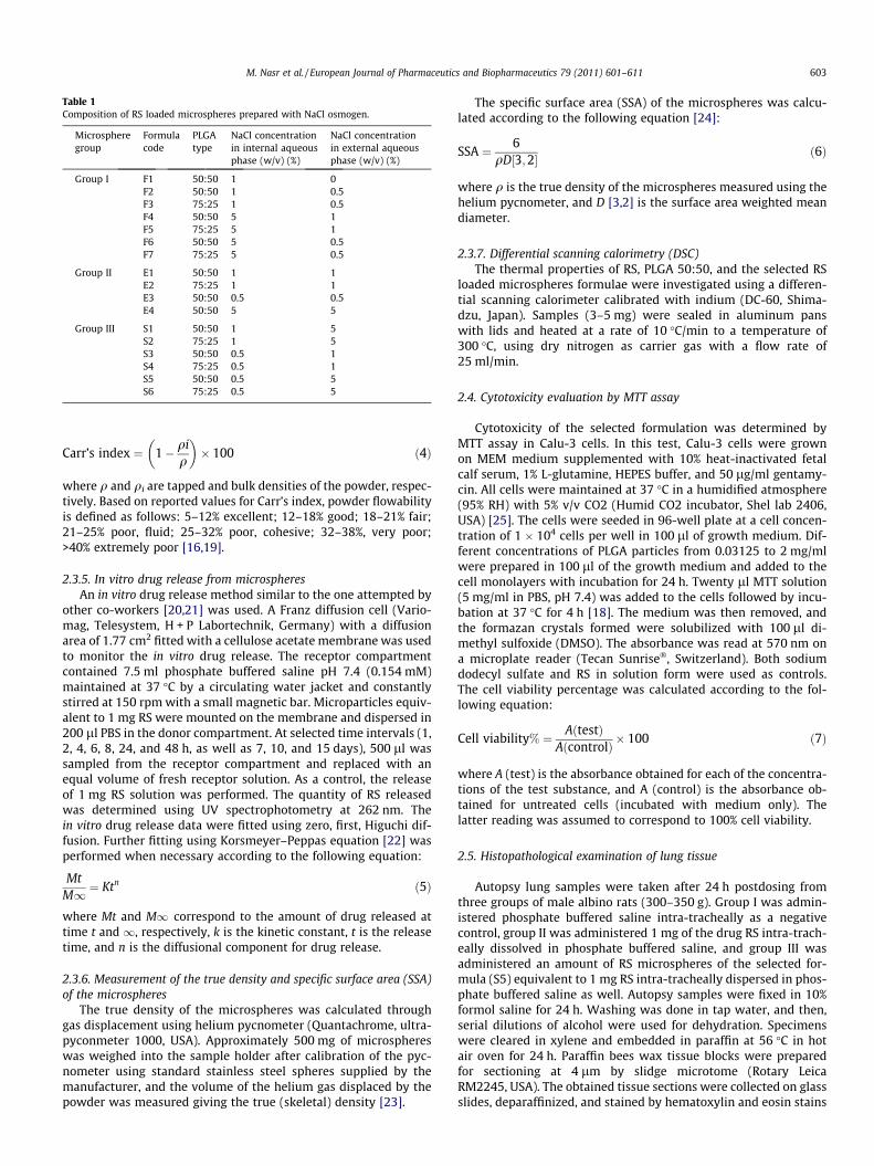
Table 1Composition of RS loaded microspheres prepared with NaCl osmogen.
Microspheregroup
Formulacode
PLGAtype
NaCl concentrationin internal aqueousphase (w/v) (%)
NaCl concentrationin external aqueousphase (w/v) (%)
Group I F1 50:50 1 0F2 50:50 1 0.5F3 75:25 1 0.5F4 50:50 5 1F5 75:25 5 1F6 50:50 5 0.5F7 75:25 5 0.5
Group II E1 50:50 1 1E2 75:25 1 1E3 50:50 0.5 0.5E4 50:50 5 5
Group III S1 50:50 1 5S2 75:25 1 5S3 50:50 0.5 1S4 75:25 0.5 1S5 50:50 0.5 5S6 75:25 0.5 5
M. Nasr et al. / European Journal of Pharmaceutics and Biopharmaceutics 79 (2011) 601–611 603
Carr’s index ¼ 1� qiq
� �� 100 ð4Þ
where q and qi are tapped and bulk densities of the powder, respec-tively. Based on reported values for Carr’s index, powder flowabilityis defined as follows: 5–12% excellent; 12–18% good; 18–21% fair;21–25% poor, fluid; 25–32% poor, cohesive; 32–38%, very poor;>40% extremely poor [16,19].
2.3.5. In vitro drug release from microspheresAn in vitro drug release method similar to the one attempted by
other co-workers [20,21] was used. A Franz diffusion cell (Vario-mag, Telesystem, H + P Labortechnik, Germany) with a diffusionarea of 1.77 cm2 fitted with a cellulose acetate membrane was usedto monitor the in vitro drug release. The receptor compartmentcontained 7.5 ml phosphate buffered saline pH 7.4 (0.154 mM)maintained at 37 �C by a circulating water jacket and constantlystirred at 150 rpm with a small magnetic bar. Microparticles equiv-alent to 1 mg RS were mounted on the membrane and dispersed in200 ll PBS in the donor compartment. At selected time intervals (1,2, 4, 6, 8, 24, and 48 h, as well as 7, 10, and 15 days), 500 ll wassampled from the receptor compartment and replaced with anequal volume of fresh receptor solution. As a control, the releaseof 1 mg RS solution was performed. The quantity of RS releasedwas determined using UV spectrophotometry at 262 nm. Thein vitro drug release data were fitted using zero, first, Higuchi dif-fusion. Further fitting using Korsmeyer–Peppas equation [22] wasperformed when necessary according to the following equation:
MtM1 ¼ Ktn ð5Þ
where Mt and M1 correspond to the amount of drug released attime t and 1, respectively, k is the kinetic constant, t is the releasetime, and n is the diffusional component for drug release.
2.3.6. Measurement of the true density and specific surface area (SSA)of the microspheres
The true density of the microspheres was calculated throughgas displacement using helium pycnometer (Quantachrome, ultra-pyconmeter 1000, USA). Approximately 500 mg of microsphereswas weighed into the sample holder after calibration of the pyc-nometer using standard stainless steel spheres supplied by themanufacturer, and the volume of the helium gas displaced by thepowder was measured giving the true (skeletal) density [23].
The specific surface area (SSA) of the microspheres was calcu-lated according to the following equation [24]:
SSA ¼ 6qD½3;2� ð6Þ
where q is the true density of the microspheres measured using thehelium pycnometer, and D [3,2] is the surface area weighted meandiameter.
2.3.7. Differential scanning calorimetry (DSC)The thermal properties of RS, PLGA 50:50, and the selected RS
loaded microspheres formulae were investigated using a differen-tial scanning calorimeter calibrated with indium (DC-60, Shima-dzu, Japan). Samples (3–5 mg) were sealed in aluminum panswith lids and heated at a rate of 10 �C/min to a temperature of300 �C, using dry nitrogen as carrier gas with a flow rate of25 ml/min.
2.4. Cytotoxicity evaluation by MTT assay
Cytotoxicity of the selected formulation was determined byMTT assay in Calu-3 cells. In this test, Calu-3 cells were grownon MEM medium supplemented with 10% heat-inactivated fetalcalf serum, 1% L-glutamine, HEPES buffer, and 50 lg/ml gentamy-cin. All cells were maintained at 37 �C in a humidified atmosphere(95% RH) with 5% v/v CO2 (Humid CO2 incubator, Shel lab 2406,USA) [25]. The cells were seeded in 96-well plate at a cell concen-tration of 1 � 104 cells per well in 100 ll of growth medium. Dif-ferent concentrations of PLGA particles from 0.03125 to 2 mg/mlwere prepared in 100 ll of the growth medium and added to thecell monolayers with incubation for 24 h. Twenty ll MTT solution(5 mg/ml in PBS, pH 7.4) was added to the cells followed by incu-bation at 37 �C for 4 h [18]. The medium was then removed, andthe formazan crystals formed were solubilized with 100 ll di-methyl sulfoxide (DMSO). The absorbance was read at 570 nm ona microplate reader (Tecan Sunrise�, Switzerland). Both sodiumdodecyl sulfate and RS in solution form were used as controls.The cell viability percentage was calculated according to the fol-lowing equation:
Cell viability% ¼ AðtestÞAðcontrolÞ � 100 ð7Þ
where A (test) is the absorbance obtained for each of the concentra-tions of the test substance, and A (control) is the absorbance ob-tained for untreated cells (incubated with medium only). Thelatter reading was assumed to correspond to 100% cell viability.
2.5. Histopathological examination of lung tissue
Autopsy lung samples were taken after 24 h postdosing fromthree groups of male albino rats (300–350 g). Group I was admin-istered phosphate buffered saline intra-tracheally as a negativecontrol, group II was administered 1 mg of the drug RS intra-trach-eally dissolved in phosphate buffered saline, and group III wasadministered an amount of RS microspheres of the selected for-mula (S5) equivalent to 1 mg RS intra-tracheally dispersed in phos-phate buffered saline as well. Autopsy samples were fixed in 10%formol saline for 24 h. Washing was done in tap water, and then,serial dilutions of alcohol were used for dehydration. Specimenswere cleared in xylene and embedded in paraffin at 56 �C in hotair oven for 24 h. Paraffin bees wax tissue blocks were preparedfor sectioning at 4 lm by slidge microtome (Rotary LeicaRM2245, USA). The obtained tissue sections were collected on glassslides, deparaffinized, and stained by hematoxylin and eosin stains
604 M. Nasr et al. / European Journal of Pharmaceutics and Biopharmaceutics 79 (2011) 601–611
for histopathological examination through the electric light micro-scope (Axiostar plus, Zeiss, USA).
2.6. In vitro aerosolization study
The selected microspheres formula (S5) was examined for itsaerosolization properties using Next Generation Impactor (NGI)(Copley M170, UK), which was considered the impactor of choicefor DPI testing [26]. Microspheres in an amount equivalent to1 mg RS were filled into size three capsule, which was then placedin Aerolizer� inhaler device (Novartis, UK). Deposition behavior ofthe microspheres was performed at a flow rate 30 L/min for 8 susing a vacuum pump (Copley HCP5, UK). The microspheres depos-ited in each stage of the NGI were collected using 1 N NaOH as acollecting solvent and analyzed for drug content using the methodpreviously described in Section 2.3.1. The microspheres depositingon the inhaler, adapter, preseparator (PS), and remaining in thecapsule were also analyzed for drug content. The emitted fraction(EF%) was determined gravimetrically [27] using the followingequation:
EF% ¼ mfull �mempty
mpowder
� �� 100 ð8Þ
where mfull and mempty refer to the weight of the capsule before andafter the aerosolization, respectively, and mpowder is the initialweight of the microspheres placed in the capsule.
The fine particle fraction (FPF%) was calculated according to thefollowing equation [27]:
FPF<4:46 lm ð%Þ ¼Amount of drug in microspheres with aerodynamic diameters < 4:46 lm
Sum of the amount f drug collected from all NGI stagesþ throatþ adapter
� �� 100 ð9Þ
The experimental mass median aerodynamic diameter (MMAD)of the aerosolized formula was calculated by analyzing the amountof drug deposited in different NGI stages.
2.7. Radiolabeling of RS using K125I
2.7.1. Radioiodination labeling techniqueRS was dissolved in distilled water at a concentration of 50 mg/
2 ml, and the pH was adjusted to nine using phosphate buffer.Freshly prepared chloramine-T solution (50 lg/5 ll) was addedto RS solution followed by the addition of 3.5 m Ci K125I at 25 �C.After 1 h, the reaction was quenched through the reduction ofremaining chloramine-T using (150 lg/5 ll) Na2S2O5. The labeledcompound was investigated using electrophoresis analysis andthin layer chromatography.
2.7.2. Calculating the reaction yield using electrophoresis and thinlayer chromatography
Electrophoresis was done using a programmable electrophore-sis power supply (E.C. 3000p-series 90, E-C Apparatus corporation,St. Petersburg, USA) using cellulose acetate strips (45 cm). Cathodeand anode poles as well as application points were indicated onthese strips and were subsequently moistened with buffer solution(0.05 M phosphate buffer pH 7). Ten microliters of the reactionmixture samples was set on the strips and introduced in theelectrophoresis chamber at a distance of 10 cm from the cathode.Current at 300 V was applied for 1½ h. Developed strips were driedand cut into 1 cm segments, then counted by a well-type NaI scin-tillation counter. The radiochemical yield was calculated as the ra-tio of the radioactivity of the labeled product to the totalradioactivity:
% Radiochemical yield of125I� RS
¼ Peakactiv ity of 125I � RSTotal activity
� 100 ð10Þ
The radiochemical yield of 125I-RS was further confirmed using thinlayer chromatography (TLC) [28]. Silica gel 60 supported on alumi-num sheets (20 � 2 cm2) was activated by heating at 110 �C for 2 h.Volumes of 10 lL were placed 2 cm from the base of TLC, thendeveloped using mobile phase chloroform/ethanol/ammonia(90:10:0.5). The strips were dried under inert N2, cut into 1 cmsegments, and then assayed for radioactivity. The retention factorwas calculated according to the following equation, and the radio-chemical yield of I125 labeled RS was determined as stated in Eq.(10):
RF ¼Distance travelled by 125I-RSDistance travelled by solvent
ð11Þ
2.8. In vivo determination of the amount of bone deposited 125I-RS
The bone deposition study was carried out on four groups ofmale albino rats of six rats each weighing 300–350 g. The animalswere fed a normal diet and water ad libitum in a constant temper-ature environment of 25 �C, and a period of 7 days was allowed foracclimatization of rats before any experimental manipulation wasundertaken. Rats were anesthetized using intra-peritonial injectionof sodium pentobarbital (32 mg/kg), followed by orotracheal instil-lation of the selected 125I-RS labeled formula dispersed in 200 llphosphate buffered saline (pH 7.4).
The selected formula (S5) was administered intra-tracheally, inan amount equivalent to 1 mg 125I-RS to groups I and II, whichwere sacrificed after 24 h and 7 days postdosing, respectively.Groups III and IV were injected an amount of 1 mg of the drug125I-RS in solution form in the tail vein and were sacrificed alsoafter 24 h and 7 days, respectively. Samples of fresh blood were ta-ken immediately before killing the rats. The bones and thyroidglands were dissected and placed in pre-weighed counting vials,and the activity was counted and expressed as a percentage ofthe inhaled or injected activity. Blood and bone were assumed tobe 7% and 12% of the total rat body weight, respectively [29]. Cor-rections were made for background radiation during experiment.The percentage of injected/inhaled dose (% I.D.) was calculatedaccording to the following equation:
%I:D: ¼ C2 �W2 � 100W1 � S
ð12Þ
where C2 is the sample count per unit time, W2 is organ mass (g), W1
is sample mass (g), and S is the activity of the injected/inhaled doseexpressed as count per unit time. All animal procedures were ap-proved by the Ethics Committee of the Faculty of Pharmacy AinShams University, in accordance with the guidelines of the Experi-ments and Advanced Pharmaceutical Research Unit (EAPRU).
2.9. Statistical analysis
All experiments were repeated at least three times. Resultswere expressed as mean ± standard deviation (S.D) or standard er-ror (S.E). One-way ANOVA followed by Tukey Kramer test wasdone using GraphPad� Instat software. A difference between
ys
M. Nasr et al. / European Journal of Pharmaceutics and Biopharmaceutics 79 (2011) 601–611 605
means was considered significant if the p-value was less than orequal to 0.05.
Car
r’s
inde
xm
ean
±S.
D.
MM
AD
th
mea
n±
S.D
.C
um
ula
tive
amou
nt
rele
ased
%af
ter
15da
mea
n±
S.D
.
35.3
0±
5.42
4.76
±0.
21N
.D.*
35.0
9±
3.51
4.78
±0.
1448
.38
±0.
9832
.86
±6.
603.
86±
0.14
43.8
1±
3.60
32.6
5±
0.92
N.D
.�50
.66
±6.
80
33.1
0±
6.14
4.72
±0.
1545
.13
±2.
4330
.40
±2.
065.
86±
0.05
58.9
7±
0.26
29.2
6±
3.39
5.68
±0.
09N
.D.*
34.4
3±
4.83
4.00
±0.
1334
.93
±2.
08
44.2
8±
3.76
3.54
±0.
0723
.14
±1.
8334
.56
±3.
853.
93±
0.12
35.1
9±
1.13
40.8
5±
5.22
3.44
±0.
1331
.69
±2.
20
17.4
0±
1.10
3.47
±0.
1567
.79
±0.
94
20.1
9±
5.44
2.80
±0.
0660
.22
±3.
5319
.80
±4.
033.
50±
0.03
63.5
9±
3.21
17.6
7±
1.77
2.80
±0.
08N
.D.*
16.0
9±
1.01
2.24
±0.
0864
.09
±3.
9117
.78
±3.
742.
72±
0.05
46.2
0±
2.56
3. Results and discussion
3.1. Preparation of RS loaded PLGA microspheres using NaCl asosmogen
Sodium chloride is commonly used as porogen owing to its os-motic effects. It is well known that water molecules may pass fromone aqueous phase to the other, based on the difference in osmoticpressure [4]. Materials such as electrolytes and drugs in eitheraqueous phases are also known to exert a similar osmotic effect[3]. Therefore, in our study, the osmotic gradient was variedthrough changing the amount of NaCl in the internal and externalaqueous phases, in order to study its effect on EE%, and accord-ingly, two groups of microspheres were prepared with differentconcentrations of NaCl in the internal/external phases (groups Iand III), and one group was prepared with equal concentration ofNaCl on both sides (group II), serving as our control group. Uponexceeding the 5% NaCl concentration in the internal aqueousphase, rapid transfer of water occurred from the outside towardthe internal aqueous phase leading to bursting of the micro-spheres; therefore, a maximum limit of 5% NaCl was used in ourpresent study.
No residual DCM was detected using gas chromatography in theprepared microspheres, which complies with the requirements ofthe USPXXVIII for volatile impurities [13].
Tabl
e2
Char
acte
riza
tion
ofRS
load
edPL
GA
mic
rosp
here
spr
epar
edus
ing
NaC
las
osm
ogen
.
Gro
up
Form
ula
code
EE%
mea
n±
S.D
.D
istr
ibu
tion
mod
alsi
ze(l
m)
mea
n±
S.D
.SP
AN
inde
xm
ean
±S.
D.
Tapp
edde
nsi
ty(g
/ml)
mea
n±
S.D
.
IF1
17.2
3±
0.16
8.39
±0.
511.
67±
0.06
0.32
3±
0.03
F224
.15
±1.
048.
39±
0.36
1.65
±0.
040.
325
±0.
02F3
21.9
8±
0.78
6.84
±0.
531.
65±
0.08
0.31
9±
0.02
F436
.81
±3.
32(4
.27 ,
10.4
7)±
0.89
2.87
±0.
390.
313
±0.
01
F534
.40
±1.
028.
41±
0.42
1.38
±0.
040.
315
±0.
02F6
21.2
1±
1.07
(6.8
,10.
48)
±0.
923.
02±
0.41
0.31
3±
0.00
F717
.39
±1.
96(7
.22,
10.2
3 )±
0.30
1.70
±0.
300.
308
±0.
01
IIE1
38.2
7±
0.64
(4.8
1,6.
83)
±0.
251.
52±
0.02
0.34
4±
0.02
E224
.42
±1.
885.
58±
0.47
1.58
±0.
050.
403
±0.
01E3
25.3
0±
1.38
6.77
±0.
361.
48±
0.04
0.33
8±
0.02
E457
.12
±1.
455.
71±
0.54
1.63
±0.
050.
363
±0.
03
III
S181
.19
±2.
86(3
.99,
5.58
)±
0.44
1.39
±0.
070.
387
±0.
03
S268
.85
±2.
904.
53±
0.68
1.35
±0.
140.
383
±0.
02S3
55.7
1±
2.81
(3.9
7,5.
59)
±0.
211.
25±
0.20
0.39
2±
0.01
S418
.19
±1.
014.
48±
0.58
1.84
±0.
250.
391
±0.
02S5
95.3
7±
2.22
3.68
±0.
691.
28±
0.03
0.37
2±
0.03
S643
.90
±2.
314.
45±
0.85
1.22
±0.
040.
373
±0.
01
Un
derl
ined
nu
mbe
rsin
dist
ribu
tion
mod
alsi
zere
pres
ent
pred
omin
ant
popu
lati
on.
�N
.D.:
Not
dete
rmin
edsi
nce
F4sh
owed
2eq
ual
lypr
edom
inan
tpo
pula
tion
s.*
N.D
.:N
otde
term
ined
sin
cefo
rmu
lae
F1,F
7,an
dS4
had
very
low
EE%
valu
es(l
ess
than
20%
).
3.2. Determination of RS entrapment efficiency into the preparedmicrospheres
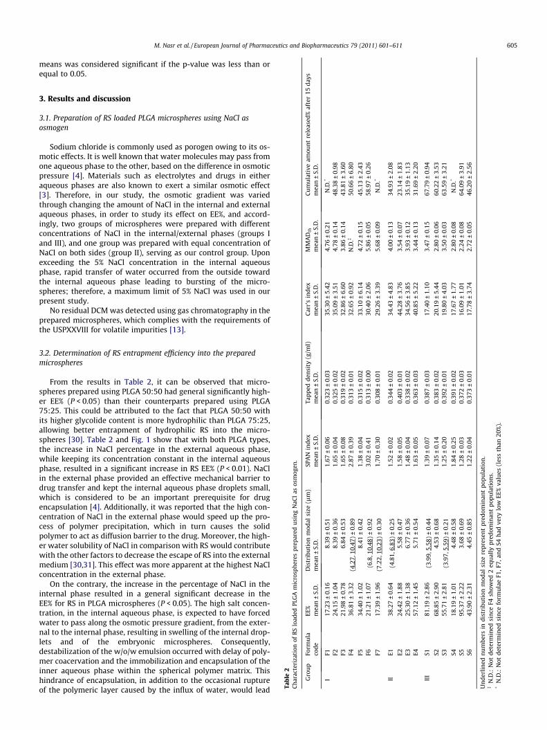
From the results in Table 2, it can be observed that micro-spheres prepared using PLGA 50:50 had general significantly high-er EE% (P < 0.05) than their counterparts prepared using PLGA75:25. This could be attributed to the fact that PLGA 50:50 withits higher glycolide content is more hydrophilic than PLGA 75:25,allowing better entrapment of hydrophilic RS into the micro-spheres [30]. Table 2 and Fig. 1 show that with both PLGA types,the increase in NaCl percentage in the external aqueous phase,while keeping its concentration constant in the internal aqueousphase, resulted in a significant increase in RS EE% (P < 0.01). NaClin the external phase provided an effective mechanical barrier todrug transfer and kept the internal aqueous phase droplets small,which is considered to be an important prerequisite for drugencapsulation [4]. Additionally, it was reported that the high con-centration of NaCl in the external phase would speed up the pro-cess of polymer precipitation, which in turn causes the solidpolymer to act as diffusion barrier to the drug. Moreover, the high-er water solubility of NaCl in comparison with RS would contributewith the other factors to decrease the escape of RS into the externalmedium [30,31]. This effect was more apparent at the highest NaClconcentration in the external phase.
On the contrary, the increase in the percentage of NaCl in theinternal phase resulted in a general significant decrease in theEE% for RS in PLGA microspheres (P < 0.05). The high salt concen-tration, in the internal aqueous phase, is expected to have forcedwater to pass along the osmotic pressure gradient, from the exter-nal to the internal phase, resulting in swelling of the internal drop-lets and of the embryonic microspheres. Consequently,destabilization of the w/o/w emulsion occurred with delay of poly-mer coacervation and the immobilization and encapsulation of theinner aqueous phase within the spherical polymer matrix. Thishindrance of encapsulation, in addition to the occasional ruptureof the polymeric layer caused by the influx of water, would lead
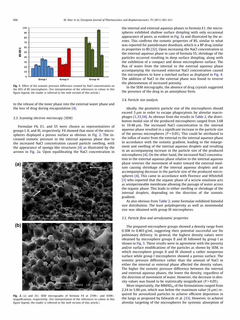
Fig. 1. Effect of the osmotic pressure difference created by NaCl concentration onthe EE% of RS microspheres. (For interpretation of the references to colour in thisfigure legend, the reader is referred to the web version of this article.)
606 M. Nasr et al. / European Journal of Pharmaceutics and Biopharmaceutics 79 (2011) 601–611
to the release of the inner phase into the external water phase andthe loss of drug during encapsulation [4].
3.3. Scanning electron microscopy (SEM)
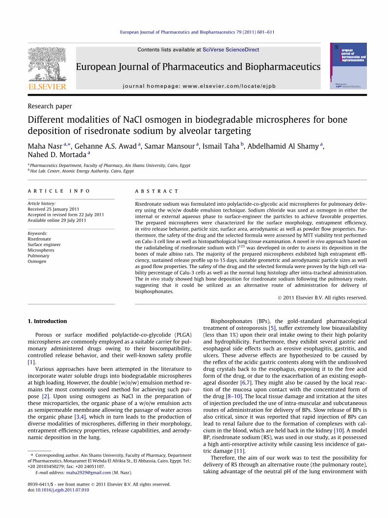
Formulae F4, E1, and S5 were chosen as representatives ofgroups I, II, and III, respectively. F4 showed that most of the micro-spheres displayed a porous surface as obvious in Fig. 2. The in-creased osmotic pressure in the internal aqueous phase due tothe increased NaCl concentration caused particle swelling, withthe appearance of spongy-like structures [4] as illustrated by thearrows in Fig. 2a. Upon equilibrating the NaCl concentration in
Fig. 2. (a) and (b): SEM micrographs of formula F4 at 2700� and 4300�magnifications, respectively. (For interpretation of the references to colour in thisfigure legend, the reader is referred to the web version of this article.)
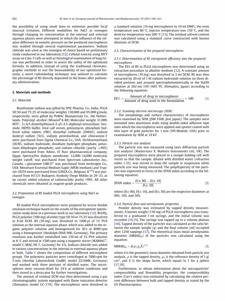
the internal and external aqueous phases in formula E1, the micro-spheres exhibited shallow surface dimpling with only occasionalappearance of pores, as evident in Fig. 3a and illustrated by the ar-rows. This confirms the osmotic properties of RS, similar to whatwas reported for pamidronate disodium, which is a BP drug similarin properties to RS [32]. Upon increasing the NaCl concentration inthe external aqueous phase in case of formula S5, shrinkage of theparticles occurred resulting in deep surface dimpling, along withthe exhibition of a compact and dense microspheres surface. Theflux of water from the internal to the external aqueous phaseaccompanying the increased enternal NaCl concentration causedthe microspheres to have a notched surface as displayed in Fig. 4.The addition of NaCl in the external phase was found to reversethe phenomenon of increased porosity.
In the SEM micrographs, the absence of drug crystals suggestedthe presence of the drug in an amorphous form.
3.4. Particle size analysis
Ideally, the geometric particle size of the microspheres shouldexceed 3 lm in order to escape phagocytosis by alveolar macro-phages [1,33,34]. As obvious from the results in Table 2, the distri-bution modal size of the produced microspheres ranged from 3.68to 10.48 lm. The increased NaCl concentration in the internalaqueous phase resulted in a significant increase in the particle sizeof the porous microspheres (P < 0.05). This could be attributed tothe influx of water from the external to the internal aqueous phasein accordance with the osmotic gradient, leading to the enlarge-ment and swelling of the internal aqueous droplets and resultingin an accompanying increase in the particle size of the producedmicrospheres [4]. On the other hand, the increased NaCl concentra-tion in the external aqueous phase relative to the internal aqueousphase reverses the movement of water toward the external med-ium causing shrinkage of the internal aqueous droplets and anaccompanying decrease in the particle size of the produced micro-spheres [4]. This came in accordance with Florence and Whitehill[3] who reported that the organic phase of a w/o/w emulsion actsas semipermeable membrane allowing the passage of water acrossthe organic phase. This leads to either swelling or shrinkage of theinternal droplets, depending on the direction of the osmoticgradient.
As also obvious from Table 2, some formulae exhibited bimodalsize distribution. The least polydispersity as well as monomodalsize was obtained with group III microspheres.
3.5. Particle flow and aerodynamic properties
The prepared microsphere groups showed a density range from0.308 to 0.403 g/ml, suggesting their potential successful use forpulmonary delivery. In general, the highest density values wereobtained by microsphere groups II and III followed by group I asshown in Fig. 5. These results were in agreement with the porosityand/or surface modifications of the particles as shown by SEM, inwhich microsphere groups II and III showed a rather nonporoussurface while group I microspheres showed a porous surface. Theosmotic pressure difference rather than the amount of NaCl ineither the internal or external phase affected the density values.The higher the osmotic pressure difference between the internaland external aqueous phases, the lower the density, regardless ofthe direction of movement of water. However, the decrease in den-sity values was found to be statistically insignificant (P > 0.05).
More importantly, the MMADth of the formulations ranged from2.24 to 5.86 lm, which was below the maximum value (6 lm) re-quired for aerosolized particles to achieve efficient deposition inthe lungs as proposed by Edwards et al. [33]. However, to achievealveolar targeting of the microspheres for systemic absorption of
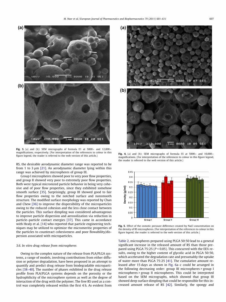
Fig. 3. (a) and (b): SEM micrographs of formula E1 at 5000� and 12,000�magnifications, respectively. (For interpretation of the references to colour in thisfigure legend, the reader is referred to the web version of this article.)
Fig. 4. (a) and (b): SEM micrographs of formula S5 at 5000� and 10,000�magnifications. (For interpretation of the references to colour in this figure legend,the reader is referred to the web version of this article.)
Fig. 5. Effect of the osmotic pressure difference created by NaCl concentration onthe density of RS microspheres. (For interpretation of the references to colour in thisfigure legend, the reader is referred to the web version of this article.)
M. Nasr et al. / European Journal of Pharmaceutics and Biopharmaceutics 79 (2011) 601–611 607
RS, the desirable aerodynamic diameter range was reported to befrom 1 to 3 lm [21]. An aerodynamic diameter lying within thisrange was achieved by microspheres of group III.
Group I microspheres showed poor to very poor flow properties,and group II showed very poor to extremely poor flow properties.Both were typical micronized particle behavior in being very cohe-sive and of poor flow properties, since they exhibited somehowsmooth surface [35]. Surprisingly, group III showed good to fairflow properties owing to the notched surface and nonsmoothstructure. The modified surface morphology was reported by Chanand Chew [36] to improve the dispersibility of the microparticlesowing to the reduced cohesion and the less close contact betweenthe particles. This surface dimpling was considered advantageousto improve particle dispersion and aerosolization via reduction inparticle–particle contact energies [37]. This came in accordancewith Healy et al. [14] who reported that particle engineering tech-niques may be utilized to optimize the micromeritic properties ofthe particles to counteract cohesiveness and poor flowability/dis-persion associated with microparticles.
3.6. In vitro drug release from microspheres
Owing to the complex nature of the release from PLA/PLGA sys-tems, a range of models, involving contributions from either diffu-sion or polymer degradation, have been proposed in an attempt toquantify and predict drug release from biodegradable microparti-cles [38–40]. The number of phases exhibited in the drug releaseprofile from PLA/PLGA systems depends on the porosity or thehydrophilicity of the microsphere system as well as the degree ofinteraction of the drug with the polymer. The free RS used as a con-trol was completely released within the first 4 h. As evident from
Table 2, microspheres prepared using PLGA 50:50 lead to a generalsignificant increase in the released amount of RS than those pre-pared using PLGA 75:25 (P < 0.05). This concurred with the EE% re-sults, owing to the higher content of glycolic acid in PLGA 50:50,which accelerated the degradation rate and presumably the uptakeof water more than PLGA 75:25 [41]. The cumulative amount re-leased after 15 days as shown in Fig. 6a–c could be arranged inthe following decreasing order: group III microspheres > group Imicrospheres > group II microspheres. This could be interpretedbased on the SEM micrographs, which showed that group IIIshowed deep surface dimpling that could be responsible for this in-creased amount release of RS [42]. Similarly, the spongy and
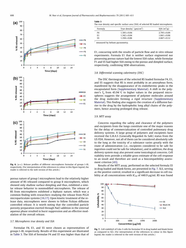
Fig. 6. (a–c): Release profiles of different microspheres formulae of groups I–III,respectively. (For interpretation of the references to colour in this figure legend, thereader is referred to the web version of this article.)
Table 3The true density and specific surface area (SSA) of selected RS loaded microspheres.
Formula True density* (gm/cm3) SSA (m2/g)
F4 1.395 ± 0.04 2.795 ± 0.09E1 1.342 ± 0.04 1.841 ± 0.06S5 1.358 ± 0.06 2.667 ± 0.12
* measured by helium pycnometry.
Fig. 7. Cell viability% of Calu-3 cells for formulae S5 in drug loaded and blank formsas compared to SLS. (For interpretation of the references to colour in this figurelegend, the reader is referred to the web version of this article.)
608 M. Nasr et al. / European Journal of Pharmaceutics and Biopharmaceutics 79 (2011) 601–611
porous nature of group I microspheres lead to the relatively higheramount of RS released compared to group II microspheres, whichshowed only shallow surface dimpling and thus, exhibited a simi-lar release behavior to nonmodified microspheres. The release ofRS from microspheres exhibited a biphasic nature, which was acommon finding with researchers studying the release from PLGAmicroparticulate systems [16,17]. Upon kinetic treatment of the re-lease data, microspheres were shown to follow Fickian diffusioncontrolled release. It is worth noting that the controlled particleporosity preparation exerted through NaCl addition in the externalaqueous phase resulted in burst suppression and an effective mod-ulation of the overall release.
3.7. Microspheres true density and SSA
Formulae F4, E1, and S5 were chosen as representatives ofgroups I–III, respectively. Results of this experiment are illustratedin Table 3. The SSA of formulae F4 and S5 was higher than that of
E1, concurring with the results of particle flow and in vitro releaseexperiments. Formula E1 that is neither surface engineered norpossessing porous nature had the lowest SSA value, while formulaeF1 and S5 had higher SSA owing to the porous and dimpled surface,respectively, confirming SEM observations.
3.8. Differential scanning calorimetry (DSC)
The DSC thermogram of the selected RS loaded formulae F4, E1,and S5 suggests that RS is most probably in an amorphous form,manifested by the disappearance of its endothermic peaks in theencapsulated form (Supplementary Material). A shift in the poly-mer’s Tg from 45.94 �C to higher values in the prepared micro-spheres suggests the arrangement of polymer molecules aroundthe drug molecules forming a rigid structure (SupplementaryMaterial). This finding also suggests the creation of a diffusion bar-rier to the drug by the hydrophobic long alkyl chains of the poly-mers, hence assuring prolonged drug release.
3.9. MTT assay
Concerns regarding the safety and clearance of the polymersand excipients from the lungs constitute one of the major reasonsfor the delay of commercialization of controlled pulmonary drugdelivery systems. A large group of polymers and excipients havereceived the G.R.A.S (Generally Regarded As Safe) status from theUS FDA. However, not all of them can be considered absolutely safeto the lung as the toxicity of a substance varies greatly with theroute of administration (i.e., excipients considered to be safe foringestion could not be safe for inhalation), and in addition, the drugdelivery system may also present some toxicological concerns. Cellviability tests provide a reliable gross estimate of the cell responseto an insult and therefore are used as a biocompatibility assess-ment criterion [43].
Results of the MTT assay, performed on the selected formula S5in drug loaded and blank forms, are presented in Fig. 7. SLS, servingas the positive control, resulted in a significant decrease in cell via-bility at all concentrations with IC50 of 440.9 lg/ml. RS was found
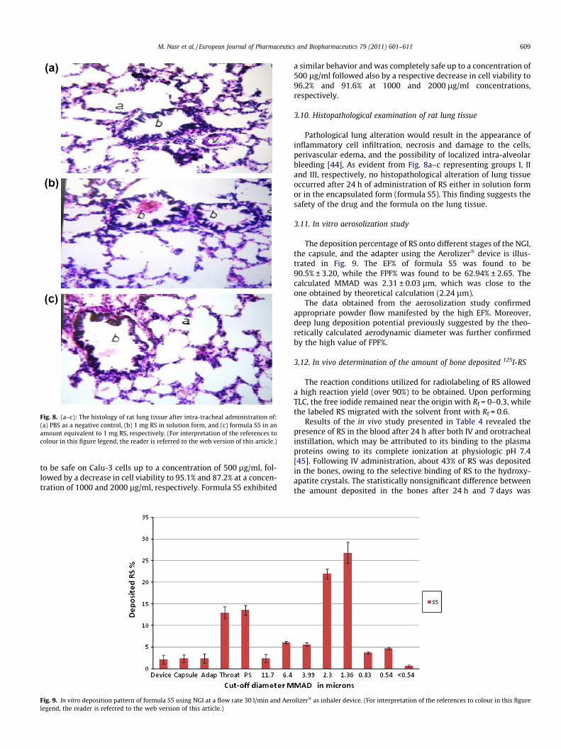
Fig. 8. (a–c): The histology of rat lung tissue after intra-tracheal administration of:(a) PBS as a negative control, (b) 1 mg RS in solution form, and (c) formula S5 in anamount equivalent to 1 mg RS, respectively. (For interpretation of the references tocolour in this figure legend, the reader is referred to the web version of this article.)
M. Nasr et al. / European Journal of Pharmaceutics and Biopharmaceutics 79 (2011) 601–611 609
to be safe on Calu-3 cells up to a concentration of 500 lg/ml, fol-lowed by a decrease in cell viability to 95.1% and 87.2% at a concen-tration of 1000 and 2000 lg/ml, respectively. Formula S5 exhibited
Fig. 9. In vitro deposition pattern of formula S5 using NGI at a flow rate 30 l/min and Aerlegend, the reader is referred to the web version of this article.)
a similar behavior and was completely safe up to a concentration of500 lg/ml followed also by a respective decrease in cell viability to96.2% and 91.6% at 1000 and 2000 lg/ml concentrations,respectively.
3.10. Histopathological examination of rat lung tissue
Pathological lung alteration would result in the appearance ofinflammatory cell infiltration, necrosis and damage to the cells,perivascular edema, and the possibility of localized intra-alveolarbleeding [44], As evident from Fig. 8a–c representing groups I, IIand III, respectively, no histopathological alteration of lung tissueoccurred after 24 h of administration of RS either in solution formor in the encapsulated form (formula S5). This finding suggests thesafety of the drug and the formula on the lung tissue.
3.11. In vitro aerosolization study
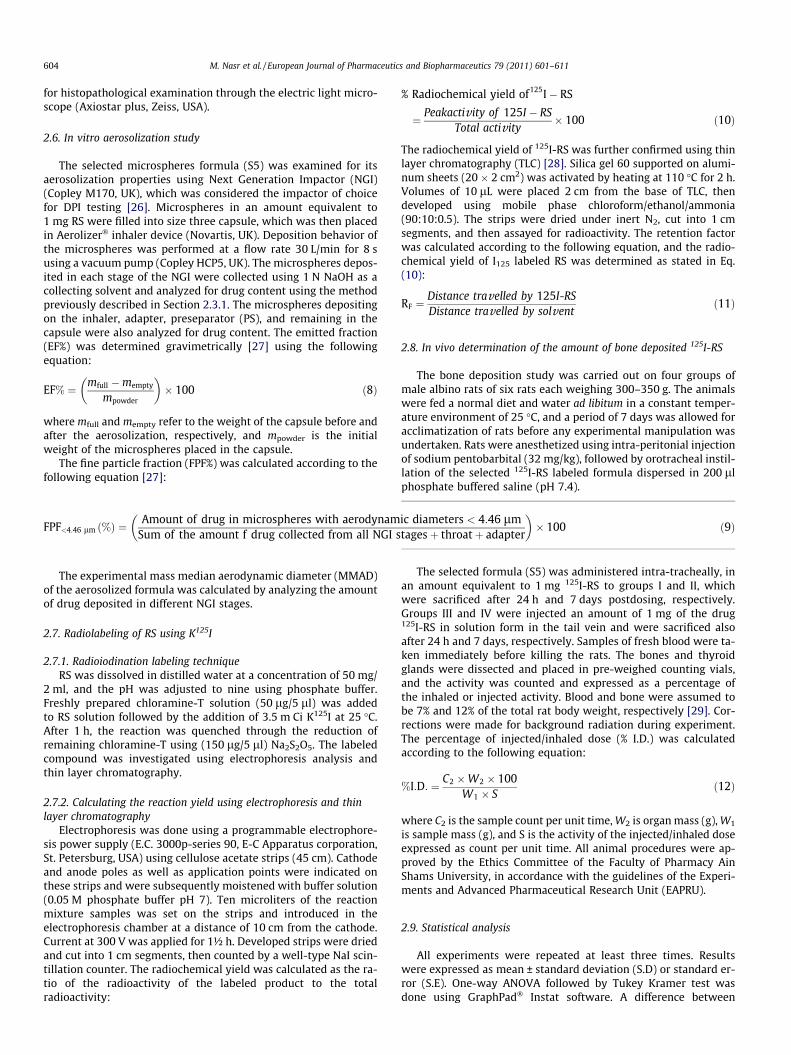
The deposition percentage of RS onto different stages of the NGI,the capsule, and the adapter using the Aerolizer� device is illus-trated in Fig. 9. The EF% of formula S5 was found to be90.5% ± 3.20, while the FPF% was found to be 62.94% ± 2.65. Thecalculated MMAD was 2.31 ± 0.03 lm, which was close to theone obtained by theoretical calculation (2.24 lm).
The data obtained from the aerosolization study confirmedappropriate powder flow manifested by the high EF%. Moreover,deep lung deposition potential previously suggested by the theo-retically calculated aerodynamic diameter was further confirmedby the high value of FPF%.
3.12. In vivo determination of the amount of bone deposited 125I-RS
The reaction conditions utilized for radiolabeling of RS alloweda high reaction yield (over 90%) to be obtained. Upon performingTLC, the free iodide remained near the origin with Rf = 0–0.3, whilethe labeled RS migrated with the solvent front with Rf = 0.6.
Results of the in vivo study presented in Table 4 revealed thepresence of RS in the blood after 24 h after both IV and orotrachealinstillation, which may be attributed to its binding to the plasmaproteins owing to its complete ionization at physiologic pH 7.4[45]. Following IV administration, about 43% of RS was depositedin the bones, owing to the selective binding of RS to the hydroxy-apatite crystals. The statistically nonsignificant difference betweenthe amount deposited in the bones after 24 h and 7 days was
olizer� as inhaler device. (For interpretation of the references to colour in this figure
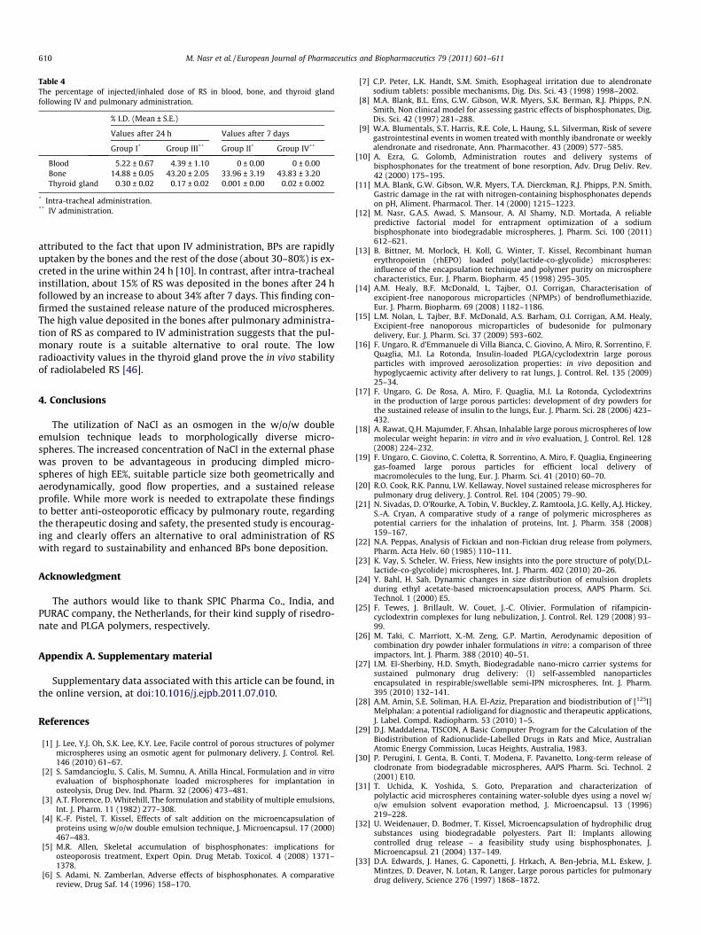
Table 4The percentage of injected/inhaled dose of RS in blood, bone, and thyroid glandfollowing IV and pulmonary administration.
% I.D. (Mean ± S.E.)
Values after 24 h Values after 7 days
Group I* Group III** Group II* Group IV**
Blood 5.22 ± 0.67 4.39 ± 1.10 0 ± 0.00 0 ± 0.00Bone 14.88 ± 0.05 43.20 ± 2.05 33.96 ± 3.19 43.83 ± 3.20Thyroid gland 0.30 ± 0.02 0.17 ± 0.02 0.001 ± 0.00 0.02 ± 0.002
* Intra-tracheal administration.** IV administration.
610 M. Nasr et al. / European Journal of Pharmaceutics and Biopharmaceutics 79 (2011) 601–611
attributed to the fact that upon IV administration, BPs are rapidlyuptaken by the bones and the rest of the dose (about 30–80%) is ex-creted in the urine within 24 h [10]. In contrast, after intra-trachealinstillation, about 15% of RS was deposited in the bones after 24 hfollowed by an increase to about 34% after 7 days. This finding con-firmed the sustained release nature of the produced microspheres.The high value deposited in the bones after pulmonary administra-tion of RS as compared to IV administration suggests that the pul-monary route is a suitable alternative to oral route. The lowradioactivity values in the thyroid gland prove the in vivo stabilityof radiolabeled RS [46].
4. Conclusions
The utilization of NaCl as an osmogen in the w/o/w doubleemulsion technique leads to morphologically diverse micro-spheres. The increased concentration of NaCl in the external phasewas proven to be advantageous in producing dimpled micro-spheres of high EE%, suitable particle size both geometrically andaerodynamically, good flow properties, and a sustained releaseprofile. While more work is needed to extrapolate these findingsto better anti-osteoporotic efficacy by pulmonary route, regardingthe therapeutic dosing and safety, the presented study is encourag-ing and clearly offers an alternative to oral administration of RSwith regard to sustainability and enhanced BPs bone deposition.
Acknowledgment
The authors would like to thank SPIC Pharma Co., India, andPURAC company, the Netherlands, for their kind supply of risedro-nate and PLGA polymers, respectively.
Appendix A. Supplementary material
Supplementary data associated with this article can be found, inthe online version, at doi:10.1016/j.ejpb.2011.07.010.
References
[1] J. Lee, Y.J. Oh, S.K. Lee, K.Y. Lee, Facile control of porous structures of polymermicrospheres using an osmotic agent for pulmonary delivery, J. Control. Rel.146 (2010) 61–67.
[2] S. Samdancioglu, S. Calis, M. Sumnu, A. Atilla Hincal, Formulation and in vitroevaluation of bisphosphonate loaded microspheres for implantation inosteolysis, Drug Dev. Ind. Pharm. 32 (2006) 473–481.
[3] A.T. Florence, D. Whitehill, The formulation and stability of multiple emulsions,Int. J. Pharm. 11 (1982) 277–308.
[4] K.-F. Pistel, T. Kissel, Effects of salt addition on the microencapsulation ofproteins using w/o/w double emulsion technique, J. Microencapsul. 17 (2000)467–483.
[5] M.R. Allen, Skeletal accumulation of bisphosphonates: implications forosteoporosis treatment, Expert Opin. Drug Metab. Toxicol. 4 (2008) 1371–1378.
[6] S. Adami, N. Zamberlan, Adverse effects of bisphosphonates. A comparativereview, Drug Saf. 14 (1996) 158–170.
[7] C.P. Peter, L.K. Handt, S.M. Smith, Esophageal irritation due to alendronatesodium tablets: possible mechanisms, Dig. Dis. Sci. 43 (1998) 1998–2002.
[8] M.A. Blank, B.L. Ems, G.W. Gibson, W.R. Myers, S.K. Berman, R.J. Phipps, P.N.Smith, Non clinical model for assessing gastric effects of bisphosphonates, Dig.Dis. Sci. 42 (1997) 281–288.
[9] W.A. Blumentals, S.T. Harris, R.E. Cole, L. Haung, S.L. Silverman, Risk of severegastrointestinal events in women treated with monthly ibandronate or weeklyalendronate and risedronate, Ann. Pharmacother. 43 (2009) 577–585.
[10] A. Ezra, G. Golomb, Administration routes and delivery systems ofbisphosphonates for the treatment of bone resorption, Adv. Drug Deliv. Rev.42 (2000) 175–195.
[11] M.A. Blank, G.W. Gibson, W.R. Myers, T.A. Dierckman, R.J. Phipps, P.N. Smith,Gastric damage in the rat with nitrogen-containing bisphosphonates dependson pH, Aliment. Pharmacol. Ther. 14 (2000) 1215–1223.
[12] M. Nasr, G.A.S. Awad, S. Mansour, A. Al Shamy, N.D. Mortada, A reliablepredictive factorial model for entrapment optimization of a sodiumbisphosphonate into biodegradable microspheres, J. Pharm. Sci. 100 (2011)612–621.
[13] B. Bittner, M. Morlock, H. Koll, G. Winter, T. Kissel, Recombinant humanerythropoietin (rhEPO) loaded poly(lactide-co-glycolide) microspheres:influence of the encapsulation technique and polymer purity on microspherecharacteristics, Eur. J. Pharm. Biopharm. 45 (1998) 295–305.
[14] A.M. Healy, B.F. McDonald, L. Tajber, O.I. Corrigan, Characterisation ofexcipient-free nanoporous microparticles (NPMPs) of bendroflumethiazide,Eur. J. Pharm. Biopharm. 69 (2008) 1182–1186.
[15] L.M. Nolan, L. Tajber, B.F. McDonald, A.S. Barham, O.I. Corrigan, A.M. Healy,Excipient-free nanoporous microparticles of budesonide for pulmonarydelivery, Eur. J. Pharm. Sci. 37 (2009) 593–602.
[16] F. Ungaro, R. d’Emmanuele di Villa Bianca, C. Giovino, A. Miro, R. Sorrentino, F.Quaglia, M.I. La Rotonda, Insulin-loaded PLGA/cyclodextrin large porousparticles with improved aerosolization properties: in vivo deposition andhypoglycaemic activity after delivery to rat lungs, J. Control. Rel. 135 (2009)25–34.
[17] F. Ungaro, G. De Rosa, A. Miro, F. Quaglia, M.I. La Rotonda, Cyclodextrinsin the production of large porous particles: development of dry powders forthe sustained release of insulin to the lungs, Eur. J. Pharm. Sci. 28 (2006) 423–432.
[18] A. Rawat, Q.H. Majumder, F. Ahsan, Inhalable large porous microspheres of lowmolecular weight heparin: in vitro and in vivo evaluation, J. Control. Rel. 128(2008) 224–232.
[19] F. Ungaro, C. Giovino, C. Coletta, R. Sorrentino, A. Miro, F. Quaglia, Engineeringgas-foamed large porous particles for efficient local delivery ofmacromolecules to the lung, Eur. J. Pharm. Sci. 41 (2010) 60–70.
[20] R.O. Cook, R.K. Pannu, I.W. Kellaway, Novel sustained release microspheres forpulmonary drug delivery, J. Control. Rel. 104 (2005) 79–90.
[21] N. Sivadas, D. O’Rourke, A. Tobin, V. Buckley, Z. Ramtoola, J.G. Kelly, A.J. Hickey,S.-A. Cryan, A comparative study of a range of polymeric microspheres aspotential carriers for the inhalation of proteins, Int. J. Pharm. 358 (2008)159–167.
[22] N.A. Peppas, Analysis of Fickian and non-Fickian drug release from polymers,Pharm. Acta Helv. 60 (1985) 110–111.
[23] K. Vay, S. Scheler, W. Friess, New insights into the pore structure of poly(D,L-lactide-co-glycolide) microspheres, Int. J. Pharm. 402 (2010) 20–26.
[24] Y. Bahl, H. Sah, Dynamic changes in size distribution of emulsion dropletsduring ethyl acetate-based microencapsulation process, AAPS Pharm. Sci.Technol. 1 (2000) E5.
[25] F. Tewes, J. Brillault, W. Couet, J.-C. Olivier, Formulation of rifampicin-cyclodextrin complexes for lung nebulization, J. Control. Rel. 129 (2008) 93–99.
[26] M. Taki, C. Marriott, X.-M. Zeng, G.P. Martin, Aerodynamic deposition ofcombination dry powder inhaler formulations in vitro: a comparison of threeimpactors, Int. J. Pharm. 388 (2010) 40–51.
[27] I.M. El-Sherbiny, H.D. Smyth, Biodegradable nano-micro carrier systems forsustained pulmonary drug delivery: (I) self-assembled nanoparticlesencapsulated in respirable/swellable semi-IPN microspheres, Int. J. Pharm.395 (2010) 132–141.
[28] A.M. Amin, S.E. Soliman, H.A. El-Aziz, Preparation and biodistribution of [125I]Melphalan: a potential radioligand for diagnostic and therapeutic applications,J. Label. Compd. Radiopharm. 53 (2010) 1–5.
[29] D.J. Maddalena, TISCON, A Basic Computer Program for the Calculation of theBiodistribution of Radionuclide-Labelled Drugs in Rats and Mice, AustralianAtomic Energy Commission, Lucas Heights, Australia, 1983.
[30] P. Perugini, I. Genta, B. Conti, T. Modena, F. Pavanetto, Long-term release ofclodronate from biodegradable microspheres, AAPS Pharm. Sci. Technol. 2(2001) E10.
[31] T. Uchida, K. Yoshida, S. Goto, Preparation and characterization ofpolylactic acid microspheres containing water-soluble dyes using a novel w/o/w emulsion solvent evaporation method, J. Microencapsul. 13 (1996)219–228.
[32] U. Weidenauer, D. Bodmer, T. Kissel, Microencapsulation of hydrophilic drugsubstances using biodegradable polyesters. Part II: Implants allowingcontrolled drug release – a feasibility study using bisphosphonates, J.Microencapsul. 21 (2004) 137–149.
[33] D.A. Edwards, J. Hanes, G. Caponetti, J. Hrkach, A. Ben-Jebria, M.L. Eskew, J.Mintzes, D. Deaver, N. Lotan, R. Langer, Large porous particles for pulmonarydrug delivery, Science 276 (1997) 1868–1872.
M. Nasr et al. / European Journal of Pharmaceutics and Biopharmaceutics 79 (2011) 601–611 611
[34] D.A. Edwards, A. Ben-Jebria, R. Langer, Recent advances in pulmonary drugdelivery using large porous inhaled particles, J. Appl. Physiol. 85 (1998) 379–385.
[35] T. Sebti, K. Amighi, Preparation and in vitro evaluation of lipidic carriers andfillers for inhalation, Eur. J. Pharm. Biopharm. 63 (2006) 51–58.
[36] H.K. Chan, N.Y. Chew, Novel alternative methods for the delivery of drugs forthe treatment of asthma, Adv. Drug Deliv. Rev. 55 (2003) 793–805.
[37] F. Mohamed, C.F. van der Walle, PLGA microcapsules with novel dimpledsurfaces for pulmonary delivery of DNA, Int. J. Pharm. 311 (2006) 97–107.
[38] J. Siepmann, A. Göpferich, Mathematical modeling of bioerodible, polymericdrug delivery systems, Adv. Drug Deliv. Rev. 48 (2001) 229–247.
[39] N. Faisant, J. Siepmann, J.P. Benoit, PLGA-based microparticles: elucidation ofmechanisms and a new, simple mathematical model quantifying drug release,Eur. J. Pharm. Sci. 15 (2002) 355–366.
[40] D.Y. Arifin, L.Y. Lee, C.H. Wang, Mathematical modeling and simulation of drugrelease from microspheres: Implications to drug delivery systems, Adv. DrugDeliv. Rev. 58 (2006) 1274–1325.
[41] F. Cui, D. Cun, A. Tao, M. Yang, K. Shi, M. Zhao, Y. Guan, Preparation andcharacterization of melittin-loaded poly (DL-lactic acid) or poly (DL-lactic-co-
glycolic acid) microspheres made by the double emulsion method, J. Control.Rel. 107 (2005) 310–319.
[42] H. Yoshizawa, S. Nishino, K. Shiomori, S. Natsugoe, T. Aiko, Y. Kitamura, Surfacemorphology control of polylactide microspheres enclosing irinotecanhydrochloride, Int. J. Pharm. 296 (2005) 112–116.
[43] C.F. Jones, D.W. Grainger, In vitro assessments of nanomaterial toxicity, Adv.Drug Deliv. Rev. 61 (2009) 438–456.
[44] H. Katsumi, M. Nakatani, J. Sano, M. Abe, K. Kusamori, M. Kurihara, R. Shiota,M. Takashima, T. Fujita, T. Sakane, T. Hibi, A. Yamamoto, Absorption and safetyof alendronate, a nitrogen-containing bisphosphonate, after intrapulmonaryadministration in rats, Int. J. Pharm. 400 (2010) 124–130.
[45] R.J. Milner, J. Farese, C.J. Henry, K. Selting, T.M. Fan, L.P. de Lorimier,Bisphosphonates and cancer, J. Vet. Intern. Med. 18 (2004) 597–604.
[46] E.A. EL-Ghany, A.M. Amine, A.S. EL-Sayed, M.T. EL-Kolaly, F. Abdel-Gelil,Radiochemical and biological characteristics of radioactive iodine labeledindomethacin for imaging of inflammation, J. Radioanal. Nucl. Chem. 266(2005) 117–124.
Related Documents