Br. J. Cancer (1990), 61, 270-275 © Macmillan Press Ltd., 1990 Different methylation of oestrogen receptor DNA in human breast carcinomas with and without oestrogen receptor R. Piva', A.P. Rimondi2, S. Hanau', I. Maestri', A. Alvisi', V.L. Kumar3 & L. del Senno' 'Istituto di Chimica Biologica e Centro di Studi Biochimici delle Patologie del Genoma Umano, Universitad di Ferrara, Italy; 2lstituto di Anatomia Patologica, Ospedale di Rovigo, Italy; and 3Laboratoire de Genetique Moleculaire des Eukaryotes du CNRS, Institut de Chimie Biologique, 67085, Strasbourg Cedex, France. Summary The methylation of the human oestrogen receptor (ER) gene was analysed by restriction enzymes in normal and neoplastic human breast tissues and cell lines. CCGG sequences in regions inside the gene, which are methylated both in normal breast and in tissues that are not the target of the oestrogen, are hypomethylated in 30% of tumours, both ER + and ER - carcinomas. Moreover, 5' sequences of the gene, which are hypomethylated in normal breast and not in tissues not the target of oestrogen, are methylated to a lower degree in ER + carcinomas, whereas they are methylated to a greater degree in ER - carcinomas. However, the same region is equally hypomethylated in both ER + and ER - cancer cell lines. Our results indicate that in breast carcinomas ER DNA methylation is deranged, and in cancer cell lines is different from that observed in primary tumours. Furthermore, the abnormal methylation in the 5' end seems to be related to abnormal expression, namely diffuse hypomethylation in carcinomas with high ER content and hypermethyla- tion in carcinomas without ER. These findings support our previous hypothesis that DNA methylation could be involved in the control of ER gene expression and demonstrate that abnormal ER gene methylation is a typical feature of breast cancers. Several experimental and clinical data have established that oestrogen plays a major role in breast development and neoplasia (Henderson et al., 1988; Dickson & Lipman, 1987), and that there is a relationship between the abnormal expres- sion of the oestrogen receptor (ER) and the growth of trans- formed mammary cells (Henderson et al., 1988; Dickson & Lipman, 1987; De Sombre et al., 1979; Perrotteau et al., 1987). Recent evidence suggests that DNA methylation, which occurs in the cytosine of CpG doublets (Razin et al., 1984), is important in multilevel mechanisms regulating gene expres- sion and differentiation in eukaryotes (Razin & Szyf, 1984; Jaenisch & Jahner, 1984). Although there are some dis- crepant reports in the literature, inverse correlation exists between methylation and expression of several normal genes (Doerfler, 1983). In addition, it is suggested that DNA methylation is deranged in cancer cells (Jones, 1986; Goelz et al., 1985), and possibly contributes to the aberrant gene expression observed in cancer (Boehm et al., 1983). As methylation of the ER gene may be one of the molecular mechanisms involved in the control of ER gene expression, changes in ER DNA methylation may be relevant in the abnormal expression of this gene and therefore for neoplastic growth and/or tumour promotion of oestrogen target cells. We have recently reported an inverse correlation between the extent of methylation and the expression of the ER gene in normal human tissues (Piva et al., 1989a). The 5' region of the gene is demethylated in normal endometrium, which contains high ER levels, and strongly methylated in white blood cells, which do not contain ER. In addition, a DNA region, internal to the gene and usually methylated in normal endometrium, is consistently hypomethylated in endometrial carcinomas, in association with a fall of ER gene expression (Piva et al., 1989b). However, it is unclear whether the decrease in expression of the ER gene is related to the abnormal hypomethylation or whether this hypomethylation is an invariable property of specific tumours. Both points could also be relevant for breast cancers. Primary breast carcinomas are known to be heterogeneous with respect to the ER protein and ER mRNA content (Henry et al., 1988), and ER + and ER - breast cancer cell lines are available. In addition, it has been suggested that changes in DNA methylation are involved in steroid-induced gene activation, thus possibly inducing progression of breast tumours from the steroid-sensitive to the steroid-insensitive state (Darbre & King, 1984). We report here the methylation and the expression of ER DNA in normal breast, and in carcinomas breast tissues and cell lines with different content of ER. Materials and methods Patients Twenty cases of primary breast cancers diagnosed as ductal carcinoma (not otherwise specified) were used in the present study. The age of the patients varied from 40 to 80 years (mean 57 years). Four patients were premenopausal and 16 post-menopausal. In addition, five cases of fibroadenomas were included, aged from 20 to 45 years. Tissue samples were frozen immediately after surgery and processed for diagnostic procedures and ER status evalu- ations. Normal breast samples from the same patients (in 11 cases) were removed from an area far from the tumour and checked in frozen sections. Only tumour samples with less than 10% of stromal tissues were analysed for DNA methylation. Tissues and their metastatic lymphonodes were stored at - 70°C until assay. Blood samples were obtained from the same patients. ER assays Sections from frozen tissues were assayed immuno- cytochemically for ER using the ER-ICA Monoclonal kit (Abbot), as previously described (Goussard et al., 1985; Di Fronzo et al., 1986; Pertschuk et al., 1985). In all cases the ER status was also determined biochemically using the dextran-coated charcol assay (DCC). Staining intensity of target cells nuclei was subjectively graded and recorded as low (+), intermediate (+ +) and high (+ + +) or negative (-), and averaged for each observed area. Cell culture Breast cancer cell lines were MCF7, T47D (both ER +) and MDA-MB-231 (ER -). Cells were grown in a-MEM medium for 4 days, in 5% CO2 humidified atmosphere, as already described (Piva et al., 1988). Correspondence: L. del Senno, Istituto di Chimica Biologica, V.L. Borsari 46, 44100 Ferrara, Italy. Received 25 July 1989. Br. J. Cancer (1990), 61, 270-275 '." Macmillan Press Ltd., 1990

Welcome message from author
This document is posted to help you gain knowledge. Please leave a comment to let me know what you think about it! Share it to your friends and learn new things together.
Transcript
Br. J. Cancer (1990), 61, 270-275 © Macmillan Press Ltd., 1990
Different methylation of oestrogen receptor DNA in human breastcarcinomas with and without oestrogen receptor
R. Piva', A.P. Rimondi2, S. Hanau', I. Maestri', A. Alvisi', V.L. Kumar3 & L. del Senno'
'Istituto di Chimica Biologica e Centro di Studi Biochimici delle Patologie del Genoma Umano, Universitad di Ferrara, Italy;2lstituto di Anatomia Patologica, Ospedale di Rovigo, Italy; and 3Laboratoire de Genetique Moleculaire des Eukaryotes du CNRS,Institut de Chimie Biologique, 67085, Strasbourg Cedex, France.
Summary The methylation of the human oestrogen receptor (ER) gene was analysed by restriction enzymesin normal and neoplastic human breast tissues and cell lines. CCGG sequences in regions inside the gene,
which are methylated both in normal breast and in tissues that are not the target of the oestrogen, are
hypomethylated in 30% of tumours, both ER + and ER - carcinomas. Moreover, 5' sequences of the gene,
which are hypomethylated in normal breast and not in tissues not the target of oestrogen, are methylated to a
lower degree in ER + carcinomas, whereas they are methylated to a greater degree in ER - carcinomas.However, the same region is equally hypomethylated in both ER + and ER - cancer cell lines. Our resultsindicate that in breast carcinomas ER DNA methylation is deranged, and in cancer cell lines is different fromthat observed in primary tumours. Furthermore, the abnormal methylation in the 5' end seems to be related toabnormal expression, namely diffuse hypomethylation in carcinomas with high ER content and hypermethyla-tion in carcinomas without ER. These findings support our previous hypothesis that DNA methylation couldbe involved in the control of ER gene expression and demonstrate that abnormal ER gene methylation is a
typical feature of breast cancers.
Several experimental and clinical data have established thatoestrogen plays a major role in breast development andneoplasia (Henderson et al., 1988; Dickson & Lipman, 1987),and that there is a relationship between the abnormal expres-sion of the oestrogen receptor (ER) and the growth of trans-formed mammary cells (Henderson et al., 1988; Dickson &Lipman, 1987; De Sombre et al., 1979; Perrotteau et al.,1987).Recent evidence suggests that DNA methylation, which
occurs in the cytosine of CpG doublets (Razin et al., 1984), isimportant in multilevel mechanisms regulating gene expres-sion and differentiation in eukaryotes (Razin & Szyf, 1984;Jaenisch & Jahner, 1984). Although there are some dis-crepant reports in the literature, inverse correlation existsbetween methylation and expression of several normal genes(Doerfler, 1983). In addition, it is suggested that DNAmethylation is deranged in cancer cells (Jones, 1986; Goelz etal., 1985), and possibly contributes to the aberrant geneexpression observed in cancer (Boehm et al., 1983). Asmethylation of the ER gene may be one of the molecularmechanisms involved in the control of ER gene expression,changes in ER DNA methylation may be relevant in theabnormal expression of this gene and therefore for neoplasticgrowth and/or tumour promotion of oestrogen target cells.We have recently reported an inverse correlation between
the extent of methylation and the expression of the ER genein normal human tissues (Piva et al., 1989a). The 5' region ofthe gene is demethylated in normal endometrium, whichcontains high ER levels, and strongly methylated in whiteblood cells, which do not contain ER. In addition, a DNAregion, internal to the gene and usually methylated in normalendometrium, is consistently hypomethylated in endometrialcarcinomas, in association with a fall of ER gene expression(Piva et al., 1989b). However, it is unclear whether thedecrease in expression of the ER gene is related to theabnormal hypomethylation or whether this hypomethylationis an invariable property of specific tumours.
Both points could also be relevant for breast cancers.Primary breast carcinomas are known to be heterogeneouswith respect to the ER protein and ER mRNA content(Henry et al., 1988), and ER + and ER - breast cancer celllines are available. In addition, it has been suggested that
changes in DNA methylation are involved in steroid-inducedgene activation, thus possibly inducing progression of breasttumours from the steroid-sensitive to the steroid-insensitivestate (Darbre & King, 1984).We report here the methylation and the expression of ER
DNA in normal breast, and in carcinomas breast tissues andcell lines with different content of ER.
Materials and methods
Patients
Twenty cases of primary breast cancers diagnosed as ductalcarcinoma (not otherwise specified) were used in the presentstudy. The age of the patients varied from 40 to 80 years
(mean 57 years). Four patients were premenopausal and 16post-menopausal. In addition, five cases of fibroadenomaswere included, aged from 20 to 45 years.
Tissue samples were frozen immediately after surgery andprocessed for diagnostic procedures and ER status evalu-ations. Normal breast samples from the same patients (in 11cases) were removed from an area far from the tumour andchecked in frozen sections. Only tumour samples with lessthan 10% of stromal tissues were analysed for DNAmethylation. Tissues and their metastatic lymphonodes were
stored at - 70°C until assay. Blood samples were obtainedfrom the same patients.
ER assays
Sections from frozen tissues were assayed immuno-cytochemically for ER using the ER-ICA Monoclonal kit(Abbot), as previously described (Goussard et al., 1985; DiFronzo et al., 1986; Pertschuk et al., 1985). In all cases theER status was also determined biochemically using thedextran-coated charcol assay (DCC). Staining intensity oftarget cells nuclei was subjectively graded and recorded aslow (+), intermediate (+ +) and high (+ + +) or negative(-), and averaged for each observed area.
Cell culture
Breast cancer cell lines were MCF7, T47D (both ER +) andMDA-MB-231 (ER -). Cells were grown in a-MEM mediumfor 4 days, in 5% CO2 humidified atmosphere, as alreadydescribed (Piva et al., 1988).
Correspondence: L. del Senno, Istituto di Chimica Biologica,V.L. Borsari 46, 44100 Ferrara, Italy.Received 25 July 1989.
Br. J. Cancer (1990), 61, 270-275 '." Macmillan Press Ltd., 1990
ER DNA METHYLATION IN BREAST CANCER 271
RNA isolation and analysis
Frozen specimens were pulverised with a microdismembrator.Powdered material was used for cytoplasmic and nucleipreparation, according to the method described by Whiteand Bancroft (1982). RNA was prepared from cytoplasm byphenol-chloroform extraction and analysed by Northern blot.Ten ILg RNA was formaldehyde denatured, electrophoresedin denaturating agarose gel (2.2 M formaldehyde) and blottedto Gene Screen Plus filters (NEN) (Piva et al., 1988).
DNA isolation and analysis
DNA was obtained from nuclei by proteinase K treatmentand phenol-chloroform extraction (Maniatis et al., 1982).Ten jig DNA was digested with an excess of HpaII and MspIrestriction enzymes respectively or with enzymes not sensitiveto methylation (5-10 units ig-' DNA) in a total reactionmixture of 250 ftl, under the conditions recommended by thesuppliers. To check that enzymatic digestion was complete, a15 ,Al aliquot was withdrawn from the reaction mixture and1 ul of bacterial phage lambda DNA (0.5 fig) was added.After incubation with the reaction mixture, the phage lambdaand human DNA samples were analysed by agarose gelelectrophoresis to ensure that digestion of lambda DNA wascomplete. After digestion and EtOH precipitation, DNAfragments were separated on 0.8% agarose gel, stained withethidium bromide and photographed through a UV trans-illuminator (Maniatis et al., 1982).
RNA and DNA hybridisation
Nucleic acids were immobilised on the filters which werehybridised in formamide at 42°C as described by the GeneScreen manual of NEN. After hybridisation, filters werewashed and treated as previously described (Piva et al.,1989b). The ER DNA probes utilised in our study were thepOR3 (Green et al., 1986) and pGHERl (Piva et al., 1989b)(see Figures 2 and 3), a cDNA and a genomic 5' endsequence respectively, which cover most of the codifyingparts of ER gene. The probes were 32P-labelled by nicktranslation or by a multiprime system (Amersham).
Results
Expression ofER gene in human breast carcinomas
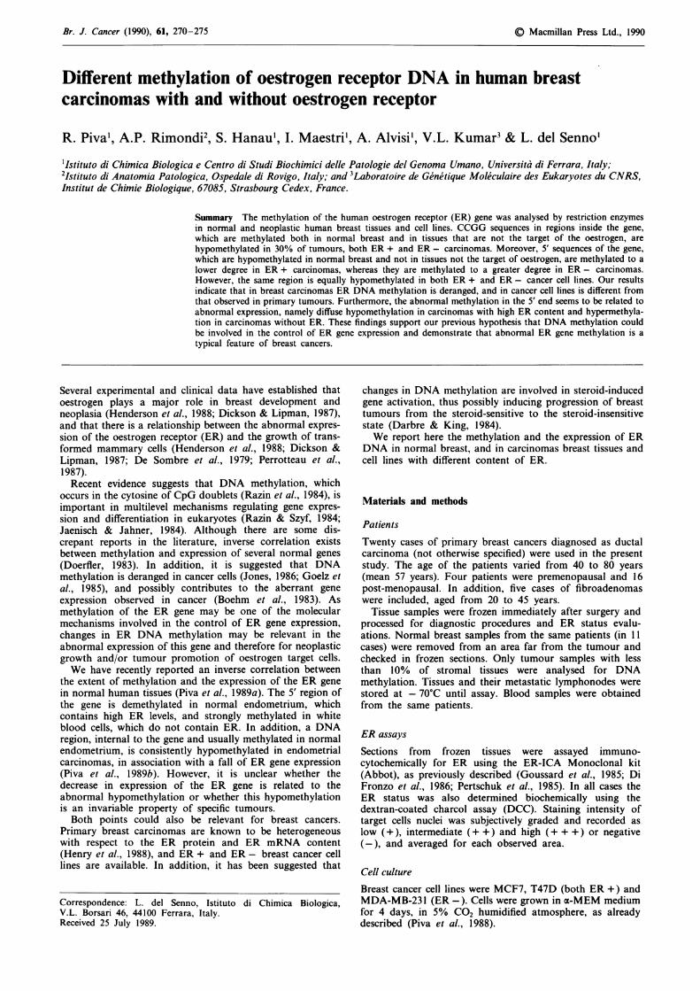
The expression of ER in the tumour tissues was studied byimmunocytochemical analysis of tissue sections and bymeasuring ER mRNA by Northern blot hybridisation, asshown in Table I and in Figure 1.According to the criteria for grading ER immunostaining,
15 cases were positive (ER +) and five negative (ER -) outof the 20 cases analysed (see Table I). Positive immunostain-ing was localised in the nucleus. Heterogeneity was commonwith respect to both the intensity of immunostaining and thepercentage of immunostained nuclei, thus reflectingheterogeneity of tumour cell populations.ER mRNA of 6.7 kb was detected in cancer specimens
with ER and in control ER + MCF7 and T47D cell lines, asshown in Figure 1. This RNA band was not detectable in theMDA-MB231 ER - cell line, and in ER - tumour samples
Table I ER content in 20 breast cancers and five fibroadenomas
Breast ER status Menopausaltumours - + ++ +++ status
Ductal carcinomas (20) 2 1 0 1 Pre3 4 6 3 Post
Fibroadenomas (5) 0 5 0 0 Pre
Cancers were all of ductal type. ER status has been evaluated byER-ICA monoclonal kit, as described in the Materials and methodssection.
- N c' v L00 0 0 0
a
-
U
mRNA ER*
28 S
Figure 1 Northern blot analysis of cytoplasmic RNA fromER- and ER + breast carcinoma tissues and cell lines. TotalRNA was denatured in formamide, electrophoresed in 1. 1%agarose gel and transferred to a filter, as described in Materialsand methods. The filter was hybridised to 32P-labelled pOR3cDNA probe. The size of ER mRNA of 6.7 kb was determinedon the basis of ribosomal RNA markers (28 and 18S). C1 andC2, ER - carcinomas; C3, C4 and C5, ER + carcinomas. MDA(ER -), T47D (ER +) and MCF7 (ER +): cancer cell lines.
(Figure 1). Abnormal ER mRNA bands were not observedin the 14 cancer samples investigated for ER RNA content,and all the 10 samples containing ER mRNA were positiveto ER antibody.
Restriction enzyme mapping ofER gene in breast carcinomas
Before analysing methylation of ER gene, we examined DNAsamples by Southern blotting technique after digestion withfour different restriction enzymes (PvuII, BamHI, Hind III,TaqI) and hybridisation to a cDNA and 5' genomic probes(shown in Figures 2 and 3). The pattern of DNA digests wasidentical in the 11 normal and 20 neoplastic breast samples(data not shown). The only differences were observed inPvuII patterns, but they were caused by a PvuII poly-morphism (RFLP) (Castagnoli et al., 1987).
Methylation inside the ER gene in human breast carcinomas
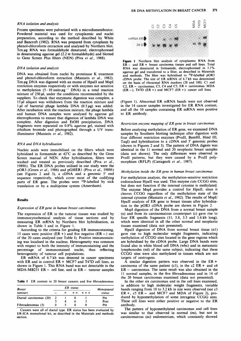
For methylation analysis, the methylation-sensitive restrictionendonuclease HpaII was used. This enzyme cuts CCGG sites,but does not function if the internal cytosine is methylated.The enzyme MspI provides a control for HpaII, since itcleaves CCGG regardless of the methylation state of theinternal cytosine (Maniatis et al., 1982). The results of MspI/HpaII analysis of ER gene in breast tissues after hybridisa-tion to the pOR3 cDNA probe are shown in Figure 2.MspI digestion of the DNA from a normal breast sample
(n) and from its carcinomatous counterpart (c) gave rise tofour ER specific fragments (11, 5.8, 3.5 and 1.6kb long),which were identical in all the other normal and neoplastictissues examined (data not presented).HpaII digestion of DNA from normal breast tissue (nl)
gave rise to high molecular weight fragments, indicatingmethylation of CCGG sites located in the gene regions whichare hybridised by the cDNA probe. Large DNA bands werefound also in white blood cell DNA (wbc) and in metastaticlymphonodes (ml) of the same subject, indicating that theseCCGG sites were also methylated in tissues which are nottargets of oestrogens.A similar digestion pattern was observed in the ER+
carcinoma of the same patient (cI), in the c2 ER + and c4ER- carcinomas. The same result was also obtained in the11 normal samples, in the five fibroadenomas and in 14 ofthe 20 breast carcinomas examined (data not presented).
In the other six carcinomas and in the cell lines examined,in addition to high molecular weight fragments, variablebands ranging from 10 to 3.2 kb in size were observed (see c3ER +, c5 ER - and MCF7 and MDA of Figure 2), pro-duced by hypomethylation of some intragenic CCGG sites.These cell lines were either positive or negative to the ERantibody.The pattern of hypomethylated carcinomas and cell lines
was similar to that observed in normal (en), but not incarcinomatous (ec) endometrium, which constantly showed

272 R. PIVA et al.
pOR3
.0 '1' N qt n a3:' C0o o o
u acUWUJ WUJ
_ 11
-a-6
- 3.5
_ 1.6
Hpall MSpl lHpall
6.3 kb
pOR3
Figure 2 Southern blot hybridisation of DNA from human normal (n) and neoplastic (c) breast tissues and cell lines (MDA,MCF7). In case 1 white blood cells (wbc) and metastatic lymphonodes (ml) are included. On the right side of the figure normal(En) and carcinomatous (Ec) endometrium are included. Samples of 10 fig of DNA were digested with either HpaII or MspI andhybridised with 32P-labelled pOR3 cDNA probe. See Materials and methods for technical details. c-1, c-2 and c-3: ER +carcinomas (+ + +, +, + + ER status respectively). c-4 and c-5: ER - carcinomas. In the lower part of the figure the ER cDNA isreported with the MspI/HpaII sites (o) and PvuIl site (P). The pOR3 probe is also shown.
the 1.6 kb fragment indicating a lower degree of methylation.Taken together the results indicate that some CCGG sites
located inside the ER gene are rarely hypomethylated inbreast carcinomas, but always in breast cell lines,independently of their ER content.The lack of the restriction map of MspI/HpaII sites in the
inner regions of the gene precludes the identification of siteswhich can be undermethylated. On the other hand, we haverecently mapped the HpaII/MspI sites in the 5' region of thegene (Piva et al., 1989a), making it easier to estimatemethylation accurately in neoplastic tissues.
Methylation of the 5' end of the ER gene
Figure 3a shows the map of the BamHI and MspI/HpaIIrestriction sites at the 5' end of the ER gene and the ER 5'genomic probe (pGHERl) with the B and A sub-fragments.
After hybridisation of BamHI digested DNA with thisprobe (Figure 3b), two bands of 2.5 and 4.7 kb appeared;they were positive to the B and A sub-fragments respectively(data not presented), and were generated by cleavage of B1,B2 and B3 sites, as shown in the map of Figure 3a and byPiva et al. (1989a).
After HpaII treatment of BamHI digested DNA fromwhite blood cells (wbc), the BamHI bands of 4.7 and 2.5 kbdisappeared, and three smaller fragments, 3.2, 2.8, 2.4 kb insize, were detected (Figure 3b). Both the 3.2 and 2.8 kbfragments originated from the 4.7 kb band by cleavage of B3and demethylated Ml and Ml13 sites respectively. The co-existence of 3.2 and 2.8 kb bands indicated that M13 was
methylated in some cells and not in others and it was refer-red to as a 'partially methylated site'. The 2.4 kb bandoriginated from the 2.5 by Bl and demethylated M2 sites.Fragments smaller than 0.6 kb originating from demethylatedHpaII/MspI sites located between M2 and MIO were notalways transferred efficiently to the filter, and were notclearly evident in the blot (Piva et al., 1989a). These siteswere found to be demethylated in all tissues, irrespective ofthe expression of the ER protein (data not shown).
In normal breast DNA (n), the 3.2 kb band was very faintand a new band of 1.6 kb was detectable, originating fromcleavage of the demethylated Ml and M2 CCGG sites. Thusthe Ml and Ml13 sites were less methylated in breast than inwhite blood cells, where the ER protein is absent, suggestingthe existence of tissue-specific methylation related to theswitch-off of the gene. The HpaII/BamHI pattern of thenormal breast samples was similar to that observed in theother normal and adenomatous breast samples examined(data not presented).
In contrast, heterogeneous methylation patterns werefound in HpaII/BamHI digests of breast carcinomas and celllines. In ER + cancers (cl and c2) the 2.8 and 2.4 kb bandswere either less marked than in normal tissue (n) or unde-tectable; the 1.6 kb band increased in intensity, and a newfragment of 0.7 kb appeared on the blot. This last fragmentoriginated from the M13 and M14 demethylated sites. Thisresult implies a loss of methylation in the 5' region of the ERgene in these two carcinomas, although they showed amethylated pattern inside the gene (cl blots of Figures 2 and3 were the same). A similar methylation pattern was observedin 13 of the 15 ER + carcinomas.
In ER - cancers (c3 and c4) the 3.2 kb band was evidentand the 2.4 was more marked than in normal tissue, whilethe 0.7 and 1.6 kb bands were fainter or absent, indicatingthe methylation of M13, M14 and Ml sites. These methyla-tion patterns were similar to those observed in metastaticlymphonodes (ml 3) and white blood cells (wbc 3 and 4),which are not known to express the ER gene. In addition,they were observed in four of the five ER - carcinomas.
In both ER - and ER + carcinoma cell lines (MDA,MCF7 and T47D of Figure 3) intense 1.6 and 0.7 kb bandswere present. A similar pattern was found in carcinomatousand normal endometrium (en and ec) which always showedthe lowest degree of methylation of the ER gene.These results indicate that Ml, M13 and M14 CCGG sites
were hypomethylated in the majority of ER + carcinomasand demethylated in cell lines, whereas they were methylatedin the majority of ER - carcinomas.
nc
kb W
1.6-M
'..i
p
ERcDNA ,3 fTER cDNA i

ER DNA METHYLATION IN BREAST CANCER 273
subfragments { BB A1wl~~~~~~ -
E-
I, moIIM M2 M10 M11 M13 M14I B2 'T(Y 1I
14.7 -I----4
i 2.5 -
2.4 -4I - 1.6 -
-- 3.2 - -1-- - ------ -2.8 -l- 0.7 -
U) U
.0
< LLOr
acu2 ; L
1.6 &
0.7
0.5 -
0.3
Bam HI B/H I B/H B/H I B/H B/H B/H MMspIE E
Figure 3 Different methylation extent of Hpall/MspI sites (M) in the 5' sequence of the ER gene in breast carcinomas and celllines. a, Map of the BamHI (B) and MspI/HpaII (M) sites in the 5' genomic region of the human ER gene. Over the map thepGHERI and subfragments A and B probes are shown. Black box is Exon 1. E = EcoRI, P = PvuII and B = BamHI. Numbersunder the map correspond to the size (in kilobases) of the restriction fragments. b, Southern blot hybridisation of DNA obtainedfrom four carcinoma tissues (cl -4), and from their normal counterparts (n). White blood cell (wbc) and metastatic lymphonodes(ml) are from the same patients. c-I and c-2: ER + carcinomas (+ + + and + in respect to the ER status). c-3 and c-4: ER -
carcinomas. MDA (ER -), MCF7 (ER +) and T47D (ER +) are cancer cell lines. On the right side of the figure endometrialnormal (En) and carcinoma (Ec) DNA are included. 10 g of DNA was digested either with BamHI or with BamHI and HpaII(B/H) and hybridised with 32P-labelled pGHERI probe.
The results of the methylation analysis are summarised inTable II.
Discussion
Human breast cancer has long been known to contain highlyvariable amounts of ER protein which can decrease fromvery high to undetectable levels (McGuire, 1978; Kodama et
al., 1985), as can be observed during the progression fromthe steroid sensitive to insensitive state (Darbre & King,1988). In order to investigate the molecular mechanismsinvolved in abnormal expression of ER gene we have studiedthe structure and the level of methylation of ER DNA inhuman breast cancers and in breast cancer cell lines with a
different content of nuclear ER.No apparent structural alterations of ER DNA were
observed in breast carcinomas, indicating that the abnormalproduction of ER is not associated with large chromosomalrearrangements or deletions. Furthermore, according to datareported by Henry et al. (1988), no abnormal ER mRNA
bands were detected in ER + cancers; however, differentlyfrom what reported by the same authors, no ER mRNA was
present in the ER - cancers investigated. On the other hand,differences in ER DNA methylation were found betweennormal, carcinomatous and cultured cancer cells in CCGGsequences located both in the core and in the 5' region of theER gene.Hypomethylation of intragenic CCGG sites (see Figure 2)
was present in 30% of breast carcinomas examined and in allthe breast cancer cells, as shown by the data of Figure 2 andTable II. Hypomethylation in the breast is different from thatobserved in endometrial carcinomas (see Figure 2 and Piva etal. (1989b)). This observation adds evidence to the fact thatboth methylating and demethylating events are stronglyrelated to the tissue type (Silva & White, 1988). Moreover,this breast ER DNA hypomethylation, although similar tothe hypomethylation typical of normal endometrium (seeFigure 2) which expresses ER to a high extent (Piva et al.,1989b), is not constantly associated with an increase of ERgene expression since the same changes were observed bothin ER + and ER - carcinomas.
a
pGHERl
E P B cap PI I I. WI,I a I I
p
B3
u
MUU
b
4.7_
2.5-
3.2-2.8 --
2.4 f
1.6 W
0.7 v-
0.3 w
-1.6
- 0.7
- 0.3
m a a s a --

274 R. PIVA et al.
Table II Quantitation of the extent of methylation in the ER DNAdisplayed by normal and neoplastic breast tissues and cell lines
Degree of ER DNA methylation
Source of S' regionDNA Inner region Ml M13 M14 ER status
Non-target tissue + + +/- +Normal breast + +/- - + +Breast ca +;+/- +/-;- - +;- +Breast ca - - - ++Breast ca - - - + + +Breast ca +;+/- +/-;+ +/-;+ +Breast ca +/- - - -
Cell linesNormal endom. +- - - - +++/++Endom. ca - - - - +
The restriction pattern of DNA from normal and cancer (ca)breast tissues and cell lines was compared with DNA from whiteblood cell (non-target tissue) and normal and neoplasticendometrium (endom.) which respectively show the highest andlowest degree of ER DNA methylation. The methylation wasquantitated as follows. Inner region (hybridised with pOR3 probe):(+) presence of only high molecular DNA fragments; (+ /-)presence of the 3.2kb band; (-) presence of the 1.6kb band. 5'region (hybridised by the pGHERl probe) = Ml: (+) presence ofthe 2.4 kb without the 1.6 kb; (+ /-) presence of the 2.4 with the1.6; (-) presence of the 1.6 only. M13: (+) presence of the 3.2without the 2.8; (+/-) presence of the 3.2 with the 2.8; (-)presence of the 2.8 only. M14: (+) absence of the 0.7; (-) presenceof the 0.7 kb band. Different methylation patterns in cancers withthe same ER status are indicated with (+; + / -; -). For examples ofthis scoring compare the autoradiographs of Figures 2 and 3. TheER status is also indicated.
These last findings demonstrate the lack of correlationbetween ER expression and methylation of CCGG sites inthe core of the ER gene. Nevertheless, we cannot exclude thepossibility that undermethylation of these sites, always pres-ent in breast cell lines, may be relevant for assessing tumourinvasiveness or progression.The CCGG sites of the 5' region of ER gene, excluding
those in the cap site region which are demethylated in alltissues (Piva et al., 1989a), are partially hypomethylated innormal breast (Figure 3b) and also in fibroadenomas (datanot presented).
In breast carcinomas a heterogeneous methylation patternis found. These sites are markedly hypomethylated in 13 ofthe 15 samples which express ER, and hypermethylated infour of the five ER- samples (Table II). This suggests that
hypomethylation in the 5' end of the gene and not in the coreof the gene is closely related to the gene expression. Thisrelationship does not exist in breast cancer cell lines becausethey are methylated to a low degree, although either positiveor negative to ER antibody. However, methylation was notas low as that found in the endometrium (Figure 3b). Theseobservations are again in agreement with the hypothesis of atissue constraint in the methylation state which may be lostin cell cultures.
Alternatively, the differences in methylation observedbetween the investigated cancer tissues could reflectheterogeneity of the cell population, with a high degree ofmethylation of ER gene where the stromal cell component isprevalent. However, the tumour specimens examined forDNA analysis contained less than 10% of stromal cells (seeMethods section), which is not enough to explain the changesin methylation observed.
In addition, the differences in ER methylation found in thebreast carcinomas should be related to the different phases ofthe menstrual cycle of the patients, as reported for the ERcontent (Smith et al., 1988). This does not appear to be thecase because the alterations in methylations were assessed inrelation to the normal counterpart of breast tissue from thesame patient (Figures 2 and 3).
It is also noteworthy that the hypomethylation describedhere appears to be specific, since it is not associated with ageneral hypomethylation because the same DNA samples arehypomethylated in 5' and not in the inner regions of thesame gene (c 1 of Figures 2 and 3).
In conclusion, the pattern of ER DNA methylation in thecore and particularly in the 5' region of the gene is differentin breast carcinomas from that observed in normal breasttissue. In breast carcinomas the heterogeneous methylationpattern of the 5' end of ER DNA is in agreement with theheterogeneity of the neoplastic cells, particularly with respectto the ER status. It would be of interest to follow the clinicalcourse of the patients with different ER methylation patternsin order to establish whether the ER DNA variations haveprognostic value. In addition to this possible clinical appli-cation, these results may shed more light on the biologicalmechanisms leading to the loss of hormone dependence inbreast cancer.This work has been supported by grants from Regione Veneto,Emilia-Romagna, CNR Progetto Finalizzato Oncologia 870126044and from MPI. We thank B. Anderson for helping us in the prepara-tion of the manuscript and 1. Perrotteau for the gift of the breastcancer cell lines.
References
BOEHM, T.L.J. & DRAHOVSKY, D. (1983). Alteration of enzymaticmethylation of DNA Cytosines by chemical carcinogens: amechanism involved in the initiation of carcinogenesis. J. NatlCancer Inst., 71, 429.
CASTAGNOLI, A., MAESTRI, I., BERNARDI, F. & DEL SENNO, L.(1987). Pvull RFLP inside the human estrogen receptor gene.Nucleic Acid Res., 15, 866.
DARBRE, P. & KING, R.J.B. (1984). Progression to steroid autonomyin SI 15 mouse mammary tumor cells: role of DNA methylation.J. Cell Biol., 99, 1410.
DARBRE, P.D. & KING, R.J.B. (1988). Role of receptor occupancy inthe transition from responsive to unresponsive states in culturedbreast tumor cells. J. Cell. Biochem., 36, 83.
DE SOMBRE, E.R., CARBONE, P.P., JENSEN, E.V. & 4 others (1979).Steroid receptors in breast cancer. N. EngI. J. Med., 301, 1011.
DICKSON, R.B. & LIPMAN, M.E. (1987). Estrogenic regulation ofgrowth and polipeptide growth factor secretion in human breastcarcinoma. Endocr. Rev., 8, 29.
Di FRONZO, G., CLEMENTE, C., CAPPELLETTI, V. & 5 others (1986).Relationship between ER-ICA and conventional steroid receptorassay in human breast cancers. Breast Cancer Res. Treat., 8, 35.
DOERFLER, W. (1983). DNA methylation and gene activity. Annu.Rev. Biochem., 52, 93.
GOELZ, S.E., VOGELSTEIN, B., HAMILTON, S.R. & FEINBERG, A.P.(1985). DNA from benign and malignant human neoplasms ishypomethylated. Science, 228, 187.
GOUSSARD, J., LECHEVREL, C., MARTIN, P.M. & ROUSSEL, G.(1985). Estrogen receptor determination with monoclonalantibodies in 160 breast tumors: comparison of the Abbot'sER-IA it with the DCC method. Bull. Cancer, 72, 168.
GREEN, S., WALTER, P., KUMAR, V. & 4 others (1986). Humanoestrogen receptor cDNA: sequence, expression and homology tov-erbA. Nature, 320, 134.
HENDERSON, B.E., ROSS, R. & BERNSTEIN, L. (1988). Estrogens as acause of human cancer: the Richard and Hinda Rosenthal AwardLecture. Cancer Res., 48, 246.
HENRY, J.A., NICHOLSON, S., FARNDON, J.R., WESTLEY, B.R. &MAY, F.E.B. (1988). Measurement of oestrogen receptor mRNAlevels in human breast tumours. Br. J. Cancer, 58, 600.
KODAMA, F., GREENE, G.L. & SALMON, S.E. (1985). Relation ofestrogen receptor expression to clonal growth and antiestrogeneffects on human breast cancer cells. Cancer Res., 45, 2720.
JAENISCH, R. & JAHNER, D. (1984). Methylation, expression andchromosomal position of genes in mammals. Biochim. Biophys.Acta, 782, 1.
JONES, P.A. (1986). DNA methylation and cancer. Cancer Res., 46,461.
McGUIRE, W.L. (1978). Steroid receptors in human breast cancer.Cancer Res., 38, 4289.
MANIATIS, T., FRITSH, E.F. & SAMBROOK, J. (1982). MolecularCloning: a Laboratory Manual. Cold Spring Harbor Laboratory:New York.

ER DNA METHYLATION IN BREAST CANCER 275
PERROTTEAU, I., SALMON, D., DE BORTOLI, M. & 5 others (1987).Immunological detection and quantification of a transforminggrowth factors in human breast carcinoma cells. Breast CancerRes. Treat., 7, 201.
PERTSCHUK, L.P., HEISEMBERG, K.B., CARTER, A.C. & SELMAN,J.G. (1985). Immunohistological localisation of estrogen receptorsin breast cancers with monoclonal antibodies. Correlation withbiochemistry and clinical endocrine response. Cancer, 55, 1513.
PIVA, R., BIANCHINI, E., KUMAR, V.L., CHAMBON, P. & DELSENNO, L. (1988). Estrogen induced increase of estrogen receptorRNA in human breast cancer cells. Biochem. Biophys. Res. Com-mun., 155, 943.
PIVA, R., KUMAR, L.V., HANAU, S. & 6 others (1989a). Expression-linked undermethylation of CpG sites at the 5' end of the humanestrogen receptor gene. Biochem. Int., 19, 267.
PIVA, R., KUMAR, L.V., HANAU, S. & 4 others (1989b). Abnormalmethylation of the estrogen receptor gene and reduced estrogenreceptor RNA levels in human endometrial carcinomas. J.Steroid Biochem., 32, 1.
RAZIN, A. & SZYF, M. (1984). DNA methylation patterns, formationand function. Biochim. Biophys. Acta, 782, 331.
SILVA, A.J. & WHITE, R. (1988). Inheritance of allelic blueprints formethylation patterns. Cell, 54, 145.
SMITH, C.M., BENN, D.E. & REEVE, T.S. (1988). Influence of themenstrual cycle on the concentration of estrogen and pro-gesterone receptors in primary breast cancer biopsies. BreastCancer Res. Treat., 11, 45.
WHITE, B.A. & BANCROFT, F.C. (1982). Cytoplasmic dot hybridiza-tion: Simple analysis of relative mRNA levels in multiple smallcell or tissue samples. J. Biol. Chem., 257, 8569.
Related Documents


![1 The role of oestrogen€¦ · The role of oestrogen •Menstrual migraine (MM) - occurs as a result of a fall in oestrogen[23, 24] •MM sufferers generally do not have hormonal](https://static.cupdf.com/doc/110x72/5edf0bffad6a402d666a66ef/1-the-role-of-oestrogen-the-role-of-oestrogen-amenstrual-migraine-mm-occurs.jpg)