Original Article Different immunization methods lead to altered gut flora and varied responses to Mycobacterium tuberculosis infection in mice Jianguo Guo 1,2 , Jun Tang 1,2 , Taisheng Kang 1,2 , Yi Xiong 3 , Zhiguang Xiang 1,2 , Chuan Qin 1,2 1 NHC Key Laboratory of Human Disease Comparative Medicine (The Institute of Laboratory Animal Sciences, CAMS and PUMC), Beijing, China 2 Key Laboratory of Human Diseases Animal Model, State Administration of Traditional Chinese Medicine, Beijing, China 3 Department of Food Science and Engineering, School of Chemistry and Chemical Engineering, Harbin Institute of Technology, Harbin, China Abstract Introduction: Vaccination is an essential means for prevention of tuberculosis infection, but the effects of various vaccines on the intestinal flora of mice and their response to Mycobacterium tuberculosis (Mtb) infection remain poorly understood. Methodology: In this study, two different vaccinations − ESAT6 and ESAT6 + TLR8 agonists - were administered to mice transgenic for human TLR8 to investigate gut microbiota characteristics following vaccination. Gut microbiota was investigated by next generation sequencing in the MiSeq Sequencing System. Adonis analysis was used to evaluate the effect of variables on gut bacterial community stucture. Chao1, Shannon index, and phylogenetic diversity index were used to explore the gut bacterial diversity. Results: The results showed that different vaccines have significant influence on mice intestinal bacteria (adonis analysis, p < 0.01), with gut bacterial diversity within the ESAT6 + TLR8 agonists group being significantly decreased compared to the ESAT6 treatment group (p < 0.01). Following infection with Mtb via tail vein injection, the bacterial community structure within the control versus vaccinated groups altered significantly (adonis analysis, p < 0.01), and the altered changed genera were markedly different between the groups. Following infection, Bifidobacteria differed between the groups, indicated that they play a vital role in the response to infection. Conclusions: Our results indicated that different vaccines might have distinct influences on intestinal flora, and their role should not be ignored. Key words: Gut microbiota; vaccine; TLR8; Mycobacterium tuberculosis. J Infect Dev Ctries 2020; 14(10):1170-1177. doi:10.3855/jidc.12697 (Received 23 March 2020 – Accepted 17 June 2020) Copyright © 2020 Guo et al. This is an open-access article distributed under the Creative Commons Attribution License, which permits unrestricted use, distribution, and reproduction in any medium, provided the original work is properly cited. Introduction Tuberculosis (TB) remains a significant threat to human health with an increasing number of cases infected with Mycobacterium tuberculosis (Mtb) worldwide. During the invasion of Mtb in the human body, many host-pathogen-recognition receptors can recognize Mtb surface molecules, such as toll-like receptors (TLRs). It has been reported that several TLRs work to recognize Mtb and activate the innate immune responses accompanied by mycobacterial infection [1]. Also, gut microbiota play an important role in the function of host immune system [2]. This microbiome is vital for the development of host immune processes responding to stimuli, which is essential for maintenance of normal homeostasis [3-7]. Thus, the roles of human microbiota within TB infection should not be overlooked. Since the characteristic of sputum microbiota during TB infection was studied by next generation sequencing in 2013 [8], dramatic changes were observed in gut microbiota of tuberculosis patients [9]. The gut microbiota of TB patients was primarily characterized by significantly decreased bacteria related by short-chain fatty acids production [9]. Much evidence indicated the close relationship between the changes of gut and lung microbiota and the Mtb infection [10]. It was also proved in mice [11]. The gut microbiota is also crucial for the response to Mtb in the lung [12]. Gut microbiota rapidly modified following Mtb infection and formed a gut bacterial community structure that was significantly different from uninfected samples [11]. Vaccination is an essential means for prevention of TB. Work is ongoing to improve the vaccine effectiveness in various ways. Evidence indicates that dysbiosis of gut microbiota could affect the function of DCs and the response of T cell against Mtb [12]. Whether different

Welcome message from author
This document is posted to help you gain knowledge. Please leave a comment to let me know what you think about it! Share it to your friends and learn new things together.
Transcript

Original Article Different immunization methods lead to altered gut flora and varied responses to Mycobacterium tuberculosis infection in mice Jianguo Guo1,2, Jun Tang1,2, Taisheng Kang1,2, Yi Xiong3, Zhiguang Xiang1,2, Chuan Qin1,2
1 NHC Key Laboratory of Human Disease Comparative Medicine (The Institute of Laboratory Animal Sciences, CAMS and PUMC), Beijing, China 2 Key Laboratory of Human Diseases Animal Model, State Administration of Traditional Chinese Medicine, Beijing, China 3 Department of Food Science and Engineering, School of Chemistry and Chemical Engineering, Harbin Institute of Technology, Harbin, China Abstract Introduction: Vaccination is an essential means for prevention of tuberculosis infection, but the effects of various vaccines on the intestinal flora of mice and their response to Mycobacterium tuberculosis (Mtb) infection remain poorly understood. Methodology: In this study, two different vaccinations − ESAT6 and ESAT6 + TLR8 agonists - were administered to mice transgenic for human TLR8 to investigate gut microbiota characteristics following vaccination. Gut microbiota was investigated by next generation sequencing in the MiSeq Sequencing System. Adonis analysis was used to evaluate the effect of variables on gut bacterial community stucture. Chao1, Shannon index, and phylogenetic diversity index were used to explore the gut bacterial diversity. Results: The results showed that different vaccines have significant influence on mice intestinal bacteria (adonis analysis, p < 0.01), with gut bacterial diversity within the ESAT6 + TLR8 agonists group being significantly decreased compared to the ESAT6 treatment group (p < 0.01). Following infection with Mtb via tail vein injection, the bacterial community structure within the control versus vaccinated groups altered significantly (adonis analysis, p < 0.01), and the altered changed genera were markedly different between the groups. Following infection, Bifidobacteria differed between the groups, indicated that they play a vital role in the response to infection. Conclusions: Our results indicated that different vaccines might have distinct influences on intestinal flora, and their role should not be ignored. Key words: Gut microbiota; vaccine; TLR8; Mycobacterium tuberculosis. J Infect Dev Ctries 2020; 14(10):1170-1177. doi:10.3855/jidc.12697 (Received 23 March 2020 – Accepted 17 June 2020) Copyright © 2020 Guo et al. This is an open-access article distributed under the Creative Commons Attribution License, which permits unrestricted use, distribution, and reproduction in any medium, provided the original work is properly cited. Introduction
Tuberculosis (TB) remains a significant threat to human health with an increasing number of cases infected with Mycobacterium tuberculosis (Mtb) worldwide. During the invasion of Mtb in the human body, many host-pathogen-recognition receptors can recognize Mtb surface molecules, such as toll-like receptors (TLRs). It has been reported that several TLRs work to recognize Mtb and activate the innate immune responses accompanied by mycobacterial infection [1]. Also, gut microbiota play an important role in the function of host immune system [2]. This microbiome is vital for the development of host immune processes responding to stimuli, which is essential for maintenance of normal homeostasis [3-7]. Thus, the roles of human microbiota within TB infection should not be overlooked. Since the characteristic of sputum microbiota during TB infection
was studied by next generation sequencing in 2013 [8], dramatic changes were observed in gut microbiota of tuberculosis patients [9]. The gut microbiota of TB patients was primarily characterized by significantly decreased bacteria related by short-chain fatty acids production [9]. Much evidence indicated the close relationship between the changes of gut and lung microbiota and the Mtb infection [10]. It was also proved in mice [11]. The gut microbiota is also crucial for the response to Mtb in the lung [12]. Gut microbiota rapidly modified following Mtb infection and formed a gut bacterial community structure that was significantly different from uninfected samples [11]. Vaccination is an essential means for prevention of TB. Work is ongoing to improve the vaccine effectiveness in various ways. Evidence indicates that dysbiosis of gut microbiota could affect the function of DCs and the response of T cell against Mtb [12]. Whether different
Guo et al. – Gut flora response to Mtb after Immune methods J Infect Dev Ctries 2020; 14(10):1170-1177.
1171
immunization methods could affect gut microbiota as well as how they affect vaccine effectiveness remains poorly understood. Our previous study evaluated the impact of ESAT6 (Abcam)-aluminum hydroxide gel (group C) and ESAT6-aluminum hydroxide gel-TL8-506 (group D) on Mtb vaccine effectiveness. It was demonstrated that group D gained better protection against Mtb infection compared to group C as supported by CFU determination in the lung, bacterial load in lung and liver, lung histology, and percentage of granuloma area within lung lobes (Figure S1) [13]. In the present study we investigated the fecal bacterial community from our previous research and compared the bacterial community structure of group B and group D within the gut microbiota responses to Mtb infection, which could lead to a better understanding of the shift of gut species within the context of infection.
Methodology Experimental process
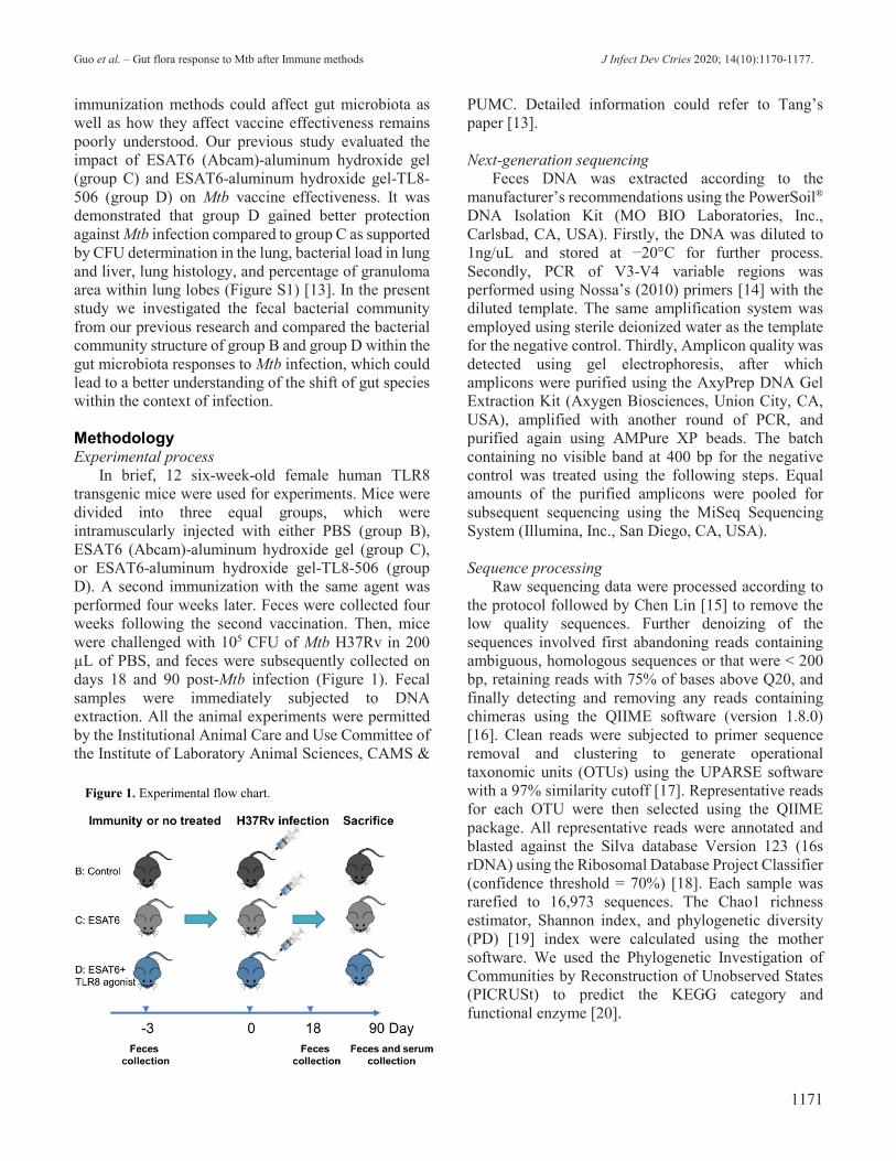
In brief, 12 six-week-old female human TLR8 transgenic mice were used for experiments. Mice were divided into three equal groups, which were intramuscularly injected with either PBS (group B), ESAT6 (Abcam)-aluminum hydroxide gel (group C), or ESAT6-aluminum hydroxide gel-TL8-506 (group D). A second immunization with the same agent was performed four weeks later. Feces were collected four weeks following the second vaccination. Then, mice were challenged with 105 CFU of Mtb H37Rv in 200 µL of PBS, and feces were subsequently collected on days 18 and 90 post-Mtb infection (Figure 1). Fecal samples were immediately subjected to DNA extraction. All the animal experiments were permitted by the Institutional Animal Care and Use Committee of the Institute of Laboratory Animal Sciences, CAMS &
PUMC. Detailed information could refer to Tang’s paper [13].
Next-generation sequencing
Feces DNA was extracted according to the manufacturer’s recommendations using the PowerSoil® DNA Isolation Kit (MO BIO Laboratories, Inc., Carlsbad, CA, USA). Firstly, the DNA was diluted to 1ng/uL and stored at −20°C for further process. Secondly, PCR of V3-V4 variable regions was performed using Nossa’s (2010) primers [14] with the diluted template. The same amplification system was employed using sterile deionized water as the template for the negative control. Thirdly, Amplicon quality was detected using gel electrophoresis, after which amplicons were purified using the AxyPrep DNA Gel Extraction Kit (Axygen Biosciences, Union City, CA, USA), amplified with another round of PCR, and purified again using AMPure XP beads. The batch containing no visible band at 400 bp for the negative control was treated using the following steps. Equal amounts of the purified amplicons were pooled for subsequent sequencing using the MiSeq Sequencing System (Illumina, Inc., San Diego, CA, USA).
Sequence processing
Raw sequencing data were processed according to the protocol followed by Chen Lin [15] to remove the low quality sequences. Further denoizing of the sequences involved first abandoning reads containing ambiguous, homologous sequences or that were < 200 bp, retaining reads with 75% of bases above Q20, and finally detecting and removing any reads containing chimeras using the QIIME software (version 1.8.0) [16]. Clean reads were subjected to primer sequence removal and clustering to generate operational taxonomic units (OTUs) using the UPARSE software with a 97% similarity cutoff [17]. Representative reads for each OTU were then selected using the QIIME package. All representative reads were annotated and blasted against the Silva database Version 123 (16s rDNA) using the Ribosomal Database Project Classifier (confidence threshold = 70%) [18]. Each sample was rarefied to 16,973 sequences. The Chao1 richness estimator, Shannon index, and phylogenetic diversity (PD) [19] index were calculated using the mother software. We used the Phylogenetic Investigation of Communities by Reconstruction of Unobserved States (PICRUSt) to predict the KEGG category and functional enzyme [20].
Figure 1. Experimental flow chart.

Guo et al. – Gut flora response to Mtb after Immune methods J Infect Dev Ctries 2020; 14(10):1170-1177.
1172
Statistical analysis The α-diversity [21] of the bacterial community was
analyzed on the basis of Chao1, Shannon–Wiener, and Faith’s phylogenetic diversity (PD) [22] index using the OTU matrix file. Phylogenetic tree was built using approximately maximum likelihood based on the evolutionary distance calculated by Jukes and Cantor model with QIIME. Phylogenetic tree was used for PD index. Principal coordinates analysis (PcoA) and a permutational multivariate analysis of variance were carried out using distance matrices (adonis) with Bray-Curtis distance in the package “vegan” in the R environment. All other statistical analyses were performed in the package “state” in the R environment. p values below 0.05 indicated statistically significant differences.
Results Effects of different immunization modes on intestinal flora within mice
PcoA based on the Bray-Curtis distance showed that the samples were clustered into three groups consistently within the method of immunization, and the gut microbiota structure differed significantly among the groups (Figure 2A). Furthermore, it was found the prevalence of classes such as Actinobacteria, Bacilli, Bacteroidia, Coriobacteriia, Erysipelotrichia were significantly different among the groups (Figure
2B). Of note, the abundances of Bacilli, Bacteroidia, and Erysipelotrichia displayed considerable changes (Figure 2B). Analysis of bacterial diversity showed that the ESAT6-aluminum hydroxide gel-TL8-506 had a highly significant effect on Chao1, Shannon diversity, and PD diversity (Figure 2C). However, the ESAT6 (Abcam)-aluminum hydroxide gel inoculation had no effect on bacterial diversity (Figure 2C).
Changes of intestinal flora within immunized mice following Mtb challenge
Bacterial community structures significantly changed following Mtb infection (Figure 3A). Taxa at the genus level with abundances greater than 0.001 were analyzed to compare the differences among groups. The communities of Lactobacillus, Oscillibacter, Ruminiclostridium, Alistipes, Bifidobacterium, and Bilophila were significantly changed (Figure 3B), with greatest variation occurring within the Lactobacillus, Alistipes, and Bifidobacterium. Lactobacillus were suppressed on day 18 following Mtb infection and recovered to original levels by day 90; Alistipes and Bifidobacterium remained unchanged on day 18 but were suppressed on day 90 (Figure 3B). Bacterial community structures within groups C and D also significantly changed following Mtb infection (Figure 3C and 3E).
(A) Principal coordinates analysis (PCoA) of samples based on Bray-Curtis distances following immunization. (B) Abundances of significantly different classes within different groups. (C) Chao1, Shannon index, and PD index in groups. Adonis and t-tests were performed in the R environment. * P < 0.05; ** P < 0.01.
Figure 2. Characteristics of gut microbiota affected by different immunization methods.

Guo et al. – Gut flora response to Mtb after Immune methods J Infect Dev Ctries 2020; 14(10):1170-1177.
1173
(A) (C) (E) PCoA of samples based on Bray-Curtis distances in group B, C, and D. (B) (D) (F) Abundances of significantly different genera following Mtb infection in groups B, C, and D. Adonis and t-tests were performed in the R environment. * P < 0.05; ** P < 0.01.
Figure 3. Response of gut microbiota within different groups to Mtb infection.

Guo et al. – Gut flora response to Mtb after Immune methods J Infect Dev Ctries 2020; 14(10):1170-1177.
1174
However, the significantly changed genera within these groups were quite different from those of group B. Parasutterella, Parabacteroides, Alistipes, Lactobacillus, Bifidobacterium were the most abundance genera present within mice in group C; Akkermansia, Klebsiella, Escherichia, Shigella, Blautia, Anaerostipes, Bilophila, Thalassospira, Lachnoclostridium, Bacteroides, Alistipes, Alloprevotella were the most abundant within group D mice (Figure 3D and 3F). Alistipes and Lactobacillus showed similar behavior to group B, while Bifidobacterium was stimulated following Mtb infection on day 18 post-infection within group C (Figure 3B and 3D). In group D, Alistipes prevalence was increased on day 18 and recovered to original levels on day 90, while the abundances of Lactobacillus and Bifidobacterium were unaffected by Mtb infection (Figure 3F). A marked increase in Bacteroides and Lachnoclostridium levels were observed following Mtb infection (Figure 3F).
Differences in intestinal flora within immunized mice following Mtb infection
The bacterial community structure was separated according to group (Figure 4A). Alphaproteobacteria, Actinobacteria, Erysipelotrichia differed among groups (Figure 4B). Bacterial diversity within infected samples was similar result to that of samples collected prior to Mtb infection (Figure 4C). On day 90 post-infection, the bacterial community still significantly differed between the groups (Figure 5A). Betaproteobacteria was the only class that differed significantly between groups B and D (Figure 5B). Chao1, Shannon and PD indexes showed no differences among groups on day 90 (Figure 5C).
Discussion
The gut microbiota affect mammalian both during homeostasis and disease states [23]. The relationship between humans and gut microbiota is vital for the maintenance of health. Vaccines provide active acquired immunity to particular pathogens, and their effectiveness is critical for disease prevention.
(A) PCoA of samples based on Bray-Curtis distances collected on day 18. (B) Abundances of significantly different classes between groups B, C, and D. (C) Chao1, Shannon, and PD indexes within different groups. Adonis and t-tests were performed in the R environment. * P < 0.05; ** P < 0.01.
Figure 4. Differences in gut microbiota between different groups on day 18 post-infection.

Guo et al. – Gut flora response to Mtb after Immune methods J Infect Dev Ctries 2020; 14(10):1170-1177.
1175
Therefore, gut microbiota composition may affect how individuals respond to vaccinations. Evidence indicates that many factors related to gut microbiota could affect the host response to vaccines. Kim et al. (2017) showed that malnutrition and poor sanitation, which affects the composition of gut microbiota, can impair the efficacy of vaccines. It was shown that antibiotic-driven intestinal dysbiosis could lead to decreased responses to adjuvant and live vaccines, and that restoration of the gutmicrobiota could recover these responses [24]. Evidence from human and mouse models both suggested that the gut microbiome could affect immune responses to vaccines [25]. However, limited studies have investigated the influence of the intestinal microbiome on vaccine responses [26]. Tuberculosis, commonly known as consumption for many years, results from infection by Mtb. The vaccine currently available for TB prevention is only BCG [27]. However, it was reported that the BCG vaccine has several limitations such as a low rate of protection [28, 29]. Therefore, the development of novel vaccines that are more effective and safer should be considered as a priority. In this study, we selected two vaccination variations: ESAT6 and ESAT6 + TLR8 agonists. The effect of immunization on intestinal flora may affect its response to Mtb infection. We found that vaccination with ESAT6 + TLR8 agonists had a great effect on gut
bacterial community and bacterial diversity compared to ESAT6 alone (Figure 2 and Figure S2). Toll-like receptor 8 is a protein in the TLR family in humans [30]. Agonists of the TLR family have been proven to enhance the protective effect of the Mtb vaccine [31-33]. Our previous study demonstrated that vaccination with ESAT6 + TLR8 agonists could strengthen the protective efficacy of Mtb immunization [13]. A diverse bacterial community could contribute to the improvement of protective efficiency. It has been shown that members of Bacteroidetes, which contain a great number of beneficial commensal microorganisms, decreased in patients experience repeated TB infections [34]. We found that the abundance of Bacteroidia increased following vaccination with ESAT6 + TLR8 agonists (Figure 2B), indicating that Bacteroidia played an important role in vaccination efficacy. Mtb infection could also affect human sputum microbiota [35]. Murine gut microbiota significantly changes following Mtb infection [11]. Our results also confirmed that Mtb infection could alter the gut microbiota following challenge with Mtb infection (Figure 3A), as Lactobacillus and Bifidobacterium were both affected by Mtb infection (Figure 3B).
Reduction of probiotics could indicate a decreased benefit of the presence of gut microbiota. Conversely, the abundance of Bifidobacterium in mice vaccinated
Figure 5. Differences between gut microbiota among different groups on day 90 post-infection.
(A) PCoA of samples based on Bray-Curtis distances collected on day 90. (B) Abundances of significantly different classes between groups B, C, and D. (C) Chao1, Shannon, and PD indexes within different groups. Adonis and t-tests were performed in the R environment. * P < 0.05; ** P < 0.01.

Guo et al. – Gut flora response to Mtb after Immune methods J Infect Dev Ctries 2020; 14(10):1170-1177.
1176
with ESAT6 increased after Mtb infection (Figure 3D) and no change occurred within the group immunized with ESAT6 + TLR8 agonists, possibly indicating no apparent negative influence of Mtb infection. However, the gut microbial community of vaccinated mice within both groups changed after Mtb infection (Figure 3C and 3E). Probiotics could benefit host by several mechanisms such as restoration of disordered microbiota [36]. It was found that probiotics benefit the immune response to oral vaccinations which could improve the vaccine efficacy [26]. Thus, application of probiotics accompanied by vaccination may increase the efficiency of Mtb infection prevention. Differences in gut microbiota between groups B, C, and D remained following Mtb inoculation on day 18 (Figure 4A), with the trend of bacterial diversity remaining unchanged (Figure 4C). Significantly different taxa at the class level decreased over time (Figure 4B, 5B, and Figure S3), and the bacterial diversity indices of the groups became indistinguishable (Figure 5C). We also found that several genera were significantly correlated with inflammation factors, including Alloprevotella, Clostridium_sensu_stricto_1, Romboutsia, Ruminococcaceae_UCG_013, Ruminococcus_1, Thalassospira, and Turicibacter (Table S1). These genera could possibly be considered as enhancers of immunity. The host-microbiota could initially protect lung from Mtb colonization [37]. Gut commensal bacteria could affect the outcome of BCG vaccination [38], and treatment of TB significantly alters the composition of the gut microbiota [39]. It was demonstrated that the addition of certain bacteria was an effective strategy for the process of prevention and treatment of tuberculosis.
Conclusions
In sum, vaccination is an effective method for prevention of tuberculosis. Our study found that different methods of vaccination distinctly influenced mice intestinal bacteria. TLR8 agonists vaccinated in combination with ESAT6 significantly affected gut microbiota and bacterial diversity. Behaviors of probiotics within the gut exhibited a significantly different response following Mtb challenge. The gut microbiota communities between the vaccinated groups also showed different patterns throughout the course of infection. All these data confirmed that vaccination strategy can affect gut microbiota structure and result in a shift of gut microbial communities following Mtb infection, and probiotics may be considered as a functional treatment to recover gut microbiota function.
These results could ultimately facilitate advances in TB treatment.
Data availability
All bacterial 16S rRNA gene sequences generated for the present study were deposited in the National Centre for Biotechnology Information Sequence Read Archive (http://www.ncbi.nlm.nih.gov/sra) under the accession number PRJNA588481. Acknowledgements This research was supported by CAMS Innovation Fund for Medical Science (CIFMS, 2018-I2M-1-001, 2017-I2M-2-005), the National Key R&D Program of China (2017YFC0702800), and Central Public-interest Scientific Institution Basal Research Fund (2016ZX310037). We are grateful for the sequencing service provided by Oebiotech Company in Shanghai. References 1. Adami AJ, Cervantes JL (2015) The microbiome at the
pulmonary alveolar niche and its role in Mycobacterium tuberculosis infection. Tuberculosis 95: 651-658.
2. Kau AL, Ahern PP, Griffin NW, Goodman AL, Gordon JI (2011) Human nutrition, the gut microbiome and the immune system. Nature 474: 327-336.
3. Chung H, Pamp SJ, Hill JA, Surana NK, Edelman SM, Troy EB, Reading NC, Villablanca EJ, Wang S, Mora JR, Umesaki Y, Mathis D, Benoist C, Relman D A, Kasper DL (2012) Gut immune maturation depends on colonization with a host-specific microbiota. Cell 149: 1578-1593.
4. Duan J, Chung H, Troy E, Kasper DL (2010) Microbial colonization drives expansion of IL-1 receptor 1-expressing and IL-17-producing gamma/delta T cells. Cell Host Microbe 7: 140-150.
5. Lathrop SK, Bloom SM, Rao SM, Nutsch K, Lio CW, Santacruz N, Peterson D A, Stappenbeck TS, Hsieh CS (2011) Peripheral education of the immune system by colonic commensal microbiota. Nature 478: 250-254.
6. Macpherson AJ, Uhr T (2004) Induction of protective IgA by intestinal dendritic cells carrying commensal bacteria. Science 303: 1662-1665.
7. Wingender G, Stepniak D, Krebs P, Lin L, McBride S, Wei B, Braun J, Mazmanian SK, Kronenberg M (2012) Intestinal microbes affect phenotypes and functions of invariant natural killer T cells in mice. Gastroenterology 143: 418-428.
8. Cheung MK, Lam WY, Fung WYW, Law PTW, Au CH, Nong WY, Kam KM, Kwan HS, Tsui SKW (2013) Sputum Microbiota in Tuberculosis as Revealed by 16S rRNA Pyrosequencing. Plos One 8.
9. Hu YF, Feng YQ, Wu JN, Liu F, Zhang ZG, Hao YN, Liang SH, Li BX, Li J, Lv N, Xu YH, Zhu BL, Sun ZG (2019) The Gut Microbiome Signatures Discriminate Healthy From Pulmonary Tuberculosis Patients. Front Cell Infect Microbiol 9.
10. Wood MR, Yu EA, Mehta S (2017) The Human Microbiome in the Fight against Tuberculosis. Am J Trop Med and Hyg 96: 1274-1284.
11. Winglee K, Eloe-Fadrosh E, Gupta S, Guo HD, Fraser C, Bishai W (2014) Aerosol Mycobacterium tuberculosis

Guo et al. – Gut flora response to Mtb after Immune methods J Infect Dev Ctries 2020; 14(10):1170-1177.
1177
Infection Causes Rapid Loss of Diversity in Gut Microbiota. Plos One 9.
12. Negi S, Pahari S, Bashir H, Agrewala JN (2019) Gut Microbiota Regulates Mincle Mediated Activation of Lung Dendritic Cells to Protect Against Mycobacterium tuberculosis. Front Immunol 10.
13. Tang J, Sun M, Shi G, Xu Y, Han Y, Li X, Dong W, Zhan L, Qin C (2017) Toll-Like Receptor 8 Agonist Strengthens the Protective Efficacy of ESAT-6 Immunization to Mycobacterium tuberculosis Infection. Front Immunol l8: 1972.
14. Nossa CW, Oberdorf WE, Yang L, Aas JA, Paster BJ, Desantis TZ, Brodie EL, Malamud D, Poles MA, Pei Z (2010) Design of 16S rRNA gene primers for 454 pyrosequencing of the human foregut microbiome. World J Gastroentero l16: 4135-4144.
15. Chen L, Fang K, Zhou J, Yang Z P, Dong X F, Dai G H, Zhang H B (2019) Enrichment of soil rare bacteria in root by an invasive plant Ageratina adenophora. Sci Total Environ 683: 202-209.
16. Caporaso JG, Kuczynski J, Stombaugh J, Bittinger K, Bushman FD, Costello EK, Fierer N, Pena AG, Goodrich JK, Gordon JI, Huttley GA, Kelley ST, Knights D, Koenig JE, Ley RE, Lozupone CA, McDonald D, Muegge BD, Pirrung M, Reeder J, Sevinsky JR, Turnbaugh PJ, Walters WA, Widmann J, Yatsunenko T, Zaneveld J, Knight R (2010) QIIME allows analysis of high-throughput community sequencing data. Nat Methods 7: 335-336.
17. Edgar RC (2013) UPARSE: highly accurate OTU sequences from microbial amplicon reads. Nat Methods 10: 996-998.
18. Wang Q, Garrity GM, Tiedje JM, Cole JR (2007) Naive Bayesian classifier for rapid assignment of rRNA sequences into the new bacterial taxonomy. Appl Environ Microbiol 73: 5261-5267.
19. Faith DP, Baker AM (2007) Phylogenetic diversity (PD) and biodiversity conservation: some bioinformatics challenges. Evol Bioinform Online 2: 121-128.
20. Langille MG, Zaneveld J, Caporaso JG, McDonald D, Knights D, Reyes JA, Clemente JC, Burkepile DE, Vega Thurber RL, Knight R, Beiko RG, Huttenhower C (2013) Predictive functional profiling of microbial communities using 16S rRNA marker gene sequences. Nat Biotechnol 31: 814-821.
21. Li B, Zhang X, Guo F, Wu W, Zhang TJWR (2013) Characterization of tetracycline resistant bacterial community in saline activated sludge using batch stress incubation with high-throughput sequencing analysis. 47: 4207-4216.
22. Faith DP (1992) Conservation evaluation and phylogenetic diversity. Biological Conservation 61: 1-10.
23. Karst SM (2016) The influence of commensal bacteria on infection with enteric viruses. Nat Rev Microbiol 14: 197-204.
24. Lynn MA, Tumes DJ, Choo JM, Sribnaia A, Blake SJ, Leong LEX, Young GP, Marshall HS, Wesselingh SL, Rogers GB, Lynn DJ (2018) Early-Life Antibiotic-Driven Dysbiosis Leads to Dysregulated Vaccine Immune Responses in Mice. Cell Host Microbe 23: 653-660.
25. Lynn DJ, Pulendran B (2018) The potential of the microbiota to influence vaccine responses. J Leukocyte Biol 103: 225-231.
26. Zimmermann P, Curtis N (2018) The influence of the intestinal microbiome on vaccine responses. Vaccine 36: 4433-4439.
27. Principi N, Esposito S (2015) The present and future of tuberculosis vaccinations. Tuberculosis 95: 6-13.
28. Mangtani P, Abubakar I, Ariti C, Beynon R, Pimpin L, Fine PE, Rodrigues LC, Smith PG, Lipman M, Whiting PF, Sterne JA (2014) Protection by BCG vaccine against tuberculosis: a
systematic review of randomized controlled trials. Clin Infect Dis 58: 470-480.
29. Fine PE (1988) BCG vaccination against tuberculosis and leprosy. Br Med Bull 44: 691-703.
30. Du X, Poltorak A, Wei Y, Beutler B (2000) Three novel mammalian toll-like receptors: gene structure, expression, and evolution. Eur Cytokine Netw 11: 362-371.
31. Mohammad O, Kaur J, Singh G, Faisal SM, Azhar A, Rauf MA, Gupta UD, Gupta P, Pal R, Zubair S (2016) TLR Agonist Augments Prophylactic Potential of Acid Inducible Antigen Rv3203 against Mycobacterium tuberculosis H37Rv in Experimental Animals. PLoS One 11: e0152240.
32. Dubois Cauwelaert N, Desbien AL, Hudson TE, Pine SO, Reed SG, Coler RN, Orr MT (2016) The TLR4 Agonist Vaccine Adjuvant, GLA-SE, Requires Canonical and Atypical Mechanisms of Action for TH1 Induction. PLoS One 11: e0146372.
33. Gillard P, Yang PC, Danilovits M, Su WJ, Cheng SL, Pehme L, Bollaerts A, Jongert E, Moris P, Ofori-Anyinam O, Demoitie MA, Castro M (2016) Safety and immunogenicity of the M72/AS01E candidate tuberculosis vaccine in adults with tuberculosis: A phase II randomised study. Tuberculosis (Edinb) 100: 118-127.
34. Luo M, Liu Y, Wu PF, Luo DX, Sun Q, Zheng H, Hu R, Pandol SJ, Li QF, Han YP, Zeng YL (2017) Alternation of Gut Microbiota in Patients with Pulmonary Tuberculosis. Frontiers in Physiology 8.
35. Cui Z, Zhou Y, Li H, Zhang Y, Zhang S, Tang S, Guo X (2012) Complex sputum microbial composition in patients with pulmonary tuberculosis. BMC Microbio l12: 276.
36. Hill C, Guarner F, Reid G, Gibson GR, Merenstein DJ, Pot B, Morelli L, Canani RB, Flint HJ, Salminen S, Calder PC, Sanders ME (2014) Expert consensus document. The International Scientific Association for Probiotics and Prebiotics consensus statement on the scope and appropriate use of the term probiotic. Nat Rev Gastroenterol Hepatol 11: 506-14.
37. Dumas A, Corral D, Colom A, Levillain F, Peixoto A, Hudrisier D, Poquet Y, Neyrolles O (2018) The Host Microbiota Contributes to Early Protection Against Lung Colonization by Mycobacterium tuberculosis. Front Immunol 9.
38. Stedman A, de Motes CM, Lesellier S, Dalley D, Chambers M, Gutierrez-Merino J (2018) Lactic acid Bacteria isolated from European badgers (Meles meles) reduce the viability and survival of Bacillus Calmette-Guerin (BCG) vaccine and influence the immune response to BCG in a human macrophage model. BMC Microbiol 18.
39. Khan N, Mendonca L, Dhariwal A, Fontes G, Menzies D, Xia JG, Divangahi M, King IL (2019) Intestinal dysbiosis compromises alveolar macrophage immunity to Mycobacterium tuberculosis. Mucosal Immunol 12: 772-783.
Corresponding authors Chuan, Qin, MD; Jianguo, Guo, PhD NHC Key Laboratory of Human Disease Comparative Medicine (The Institute of Laboratory Animal Sciences, CAMS&PUMC); Key Laboratory of Human Diseases Animal Model, State Administration of Traditional Chinese Medicine, address: Pan Jia Yuan Nan Li No. 5, Chao Yang District, 100021, Beijing, China. Phone: +86-010-87778141 Fax: +86-010 67761943 Email: [email protected]; [email protected] Conflict of interests: No conflict of interests is declared.
Guo et al. – Gut flora response to Mtb after Immune methods J Infect Dev Ctries 2020; 14(10):1170-1177.
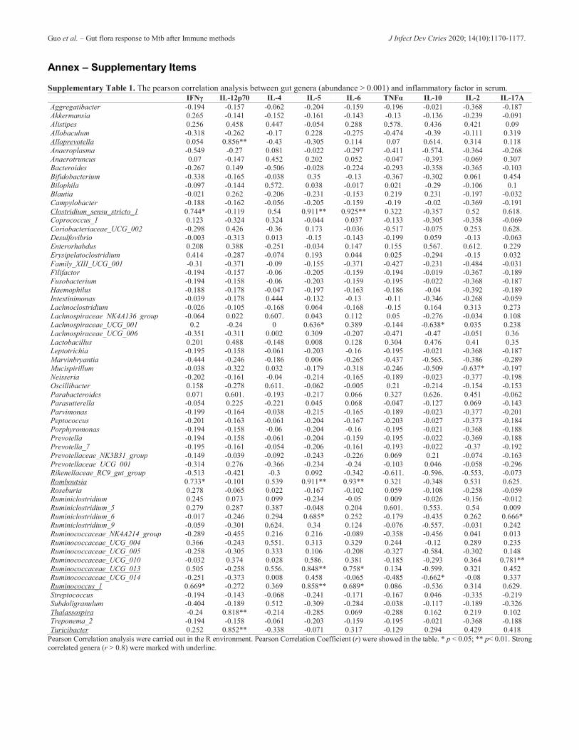
Annex – Supplementary Items Supplementary Table 1. The pearson correlation analysis between gut genera (abundance > 0.001) and inflammatory factor in serum. IFNγ IL-12p70 IL-4 IL-5 IL-6 TNFα IL-10 IL-2 IL-17A Aggregatibacter -0.194 -0.157 -0.062 -0.204 -0.159 -0.196 -0.021 -0.368 -0.187 Akkermansia 0.265 -0.141 -0.152 -0.161 -0.143 -0.13 -0.136 -0.239 -0.091 Alistipes 0.256 0.458 0.447 -0.054 0.288 0.578. 0.436 0.421 0.09 Allobaculum -0.318 -0.262 -0.17 0.228 -0.275 -0.474 -0.39 -0.111 0.319 Alloprevotella 0.054 0.856** -0.43 -0.305 0.114 0.07 0.614. 0.314 0.118 Anaeroplasma -0.549 -0.27 0.081 -0.022 -0.297 -0.411 -0.574. -0.364 -0.268 Anaerotruncus 0.07 -0.147 0.452 0.202 0.052 -0.047 -0.393 -0.069 0.307 Bacteroides -0.267 0.149 -0.506 -0.028 -0.224 -0.293 -0.358 -0.365 -0.103 Bifidobacterium -0.338 -0.165 -0.038 0.35 -0.13 -0.367 -0.302 0.061 0.454 Bilophila -0.097 -0.144 0.572. 0.038 -0.017 0.021 -0.29 -0.106 0.1 Blautia -0.021 0.262 -0.206 -0.231 -0.153 0.219 0.231 -0.197 -0.032 Campylobacter -0.188 -0.162 -0.056 -0.205 -0.159 -0.19 -0.02 -0.369 -0.191 Clostridium_sensu_stricto_1 0.744* -0.119 0.54 0.911** 0.925** 0.322 -0.357 0.52 0.618. Coprococcus_1 0.123 -0.324 0.324 -0.044 0.037 -0.133 -0.305 -0.358 -0.069 Coriobacteriaceae_UCG_002 -0.298 0.426 -0.36 0.173 -0.036 -0.517 -0.075 0.253 0.628. Desulfovibrio -0.003 -0.313 0.013 -0.15 -0.143 -0.199 0.059 -0.13 -0.063 Enterorhabdus 0.208 0.388 -0.251 -0.034 0.147 0.155 0.567. 0.612. 0.229 Erysipelatoclostridium 0.414 -0.287 -0.074 0.193 0.044 0.025 -0.294 -0.15 0.032 Family_XIII_UCG_001 -0.31 -0.371 -0.09 -0.155 -0.371 -0.427 -0.231 -0.484 -0.031 Filifactor -0.194 -0.157 -0.06 -0.205 -0.159 -0.194 -0.019 -0.367 -0.189 Fusobacterium -0.194 -0.158 -0.06 -0.203 -0.159 -0.195 -0.022 -0.368 -0.187 Haemophilus -0.188 -0.178 -0.047 -0.197 -0.163 -0.186 -0.04 -0.392 -0.189 Intestinimonas -0.039 -0.178 0.444 -0.132 -0.13 -0.11 -0.346 -0.268 -0.059 Lachnoclostridium -0.026 -0.105 -0.168 0.064 -0.168 -0.15 0.164 0.313 0.273 Lachnospiraceae_NK4A136_group -0.064 0.022 0.607. 0.043 0.112 0.05 -0.276 -0.034 0.108 Lachnospiraceae_UCG_001 0.2 -0.24 0 0.636* 0.389 -0.144 -0.638* 0.035 0.238 Lachnospiraceae_UCG_006 -0.351 -0.311 0.002 0.309 -0.207 -0.471 -0.47 -0.051 0.36 Lactobacillus 0.201 0.488 -0.148 0.008 0.128 0.304 0.476 0.41 0.35 Leptotrichia -0.195 -0.158 -0.061 -0.203 -0.16 -0.195 -0.021 -0.368 -0.187 Marvinbryantia -0.444 -0.246 -0.186 0.006 -0.265 -0.437 -0.565. -0.386 -0.289 Mucispirillum -0.038 -0.322 0.032 -0.179 -0.318 -0.246 -0.509 -0.637* -0.197 Neisseria -0.202 -0.161 -0.04 -0.214 -0.165 -0.189 -0.023 -0.377 -0.198 Oscillibacter 0.158 -0.278 0.611. -0.062 -0.005 0.21 -0.214 -0.154 -0.153 Parabacteroides 0.071 0.601. -0.193 -0.217 0.066 0.327 0.626. 0.451 -0.062 Parasutterella -0.054 0.225 -0.221 0.045 0.068 -0.047 -0.127 0.069 -0.143 Parvimonas -0.199 -0.164 -0.038 -0.215 -0.165 -0.189 -0.023 -0.377 -0.201 Peptococcus -0.201 -0.163 -0.061 -0.204 -0.167 -0.203 -0.027 -0.373 -0.184 Porphyromonas -0.194 -0.158 -0.06 -0.204 -0.16 -0.195 -0.021 -0.368 -0.188 Prevotella -0.194 -0.158 -0.061 -0.204 -0.159 -0.195 -0.022 -0.369 -0.188 Prevotella_7 -0.195 -0.161 -0.054 -0.206 -0.161 -0.193 -0.022 -0.37 -0.192 Prevotellaceae_NK3B31_group -0.149 -0.039 -0.092 -0.243 -0.226 0.069 0.21 -0.074 -0.163 Prevotellaceae_UCG_001 -0.314 0.276 -0.366 -0.234 -0.24 -0.103 0.046 -0.058 -0.296 Rikenellaceae_RC9_gut_group -0.513 -0.421 -0.3 0.092 -0.342 -0.611. -0.596. -0.553. -0.073 Romboutsia 0.733* -0.101 0.539 0.911** 0.93** 0.321 -0.348 0.531 0.625. Roseburia 0.278 -0.065 0.022 -0.167 -0.102 0.059 -0.108 -0.258 -0.059 Ruminiclostridium 0.245 0.073 0.099 -0.234 -0.05 0.009 -0.026 -0.156 -0.012 Ruminiclostridium_5 0.279 0.287 0.387 -0.048 0.204 0.601. 0.553. 0.54 0.009 Ruminiclostridium_6 -0.017 -0.246 0.294 0.685* 0.252 -0.179 -0.435 0.262 0.666* Ruminiclostridium_9 -0.059 -0.301 0.624. 0.34 0.124 -0.076 -0.557. -0.031 0.242 Ruminococcaceae_NK4A214_group -0.289 -0.455 0.216 0.216 -0.089 -0.358 -0.456 0.041 0.013 Ruminococcaceae_UCG_004 0.366 -0.243 0.551. 0.313 0.329 0.244 -0.12 0.289 0.235 Ruminococcaceae_UCG_005 -0.258 -0.305 0.333 0.106 -0.208 -0.327 -0.584. -0.302 0.148 Ruminococcaceae_UCG_010 -0.032 0.374 0.028 0.586. 0.381 -0.185 -0.293 0.364 0.781** Ruminococcaceae_UCG_013 0.505 -0.258 0.556. 0.848** 0.758* 0.134 -0.599. 0.321 0.452 Ruminococcaceae_UCG_014 -0.251 -0.373 0.008 0.458 -0.065 -0.485 -0.662* -0.08 0.337 Ruminococcus_1 0.669* -0.272 0.369 0.858** 0.689* 0.086 -0.536 0.314 0.629. Streptococcus -0.194 -0.143 -0.068 -0.241 -0.171 -0.167 0.046 -0.335 -0.219 Subdoligranulum -0.404 -0.189 0.512 -0.309 -0.284 -0.038 -0.117 -0.189 -0.326 Thalassospira -0.24 0.818** -0.214 -0.285 0.069 -0.288 0.162 0.219 0.102 Treponema_2 -0.194 -0.158 -0.061 -0.203 -0.159 -0.195 -0.021 -0.368 -0.188 Turicibacter 0.252 0.852** -0.338 -0.071 0.317 -0.129 0.294 0.429 0.418
Pearson Correlation analysis were carried out in the R environment. Pearson Correlation Coefficient (r) were showed in the table. * p < 0.05; ** p< 0.01. Strong correlated genera (r > 0.8) were marked with underline.

Guo et al. – Gut flora response to Mtb after Immune methods J Infect Dev Ctries 2020; 14(10):1170-1177.
Supplementary Figure 1. TLR8 transgenic mice immunized with TLR8 agonist carry lower bacilli load and display ameliorated
pathological lesions in tissues following a challenge. (A) Bone marrow cells were cultured and treated ex vivo as indicated for 12 h before the mRNA level of IL1β, IL6, and TNF was determined. Three mice per group. *p < 0.05, by Student’s t-test. (B) CFU (log10-transformed) of lungs in differently immunized TLR8transgenic mice 10 weeks post Mycobacterium tuberculosis challenge. (C) Time-to-detection (transformed into hours) by the MGIT 960 system in lung, spleen, and liver. (D) Lung sections stained with H&E. (E) Percentage of granuloma area in lung lobes determined by NanoZoomer S60 (Hamamatsu) and software, 3–5 sections per mice. *p < 0.05, **p < 0.01, by one-way ANOVA with a Bonferroni posttest. Data are representative of two independent experiments, each with four mice per group (Tang, et al. 2017: 1972).
Supplementary Figure 2. The significantly different genera among group B, C, and D before Mtb infection. The genera (the mean abundance > 0.001) which were significantly different among groups were showed in bar chart.
Supplementary Figure 3. The significantly different genera among groups before Mtb infection and on day 18 and day 90 challenged with Mtb. Red box indicated the abundance of the corresponding genus was significantly greater in former group; blue box indicated the abundance of the corresponding genus was significantly lower in former group; white box indicated no difference.
Related Documents


![Immunization Program Strategic Plan 2013 – 2017 · 1 2013-2017 Immunization Program [Immunization Program Strategic Plan 2013 – 2017] Maintaining and Improving Immunization Rates](https://static.cupdf.com/doc/110x72/5e18e16c0228f448f3787c8f/immunization-program-strategic-plan-2013-a-2017-1-2013-2017-immunization-program.jpg)