Differences in Tissue-Remodeling Potential of Aortic and Pulmonary Heart Valve Interstitial Cells W. DAVID MERRYMAN, M.S., 1 JUN LIAO, Ph.D., 1 ARON PAREKH, Ph.D., 1 JOSEPH E. CANDIELLO, B.S., 2 HAI LIN, Ph.D., 2 and MICHAEL S. SACKS, Ph.D. 1,3 ABSTRACT Heart valve interstitial cells (VICs) appear to have a dynamic and reversible phenotype, an attribute speculated to be necessary for valve tissue remodeling during times of development and repair. Therefore, we hypothesized that the cytoskeletal (CSK) remodeling capability of the aortic and pulmonary VICs (AVICs and PVICs, respectively), which are dominated by smooth muscle a-actin, would exhibit unique contractile behaviors when seeded on collagen gels. Using a porcine cell source, we observed that VIC pop- ulations did not contract the gels at early time points (2 and 4 hours) as dermal fibroblasts did, but formed a central cluster of cells prior to contraction. After clustering, VICs appeared to radiate out from the center of the gels, whereas fibroblasts did not migrate but contracted the gels locally. VIC gels treated with trans- forming growth factor b1 contracted the gels rapidly, revealing similar sensitivity to the cytokine. Moreover, we evaluated the initial mechanical state of the underlying CSK by comparing AVIC and PVIC stiffness with atomic force microscopy. Not only were AVICs significantly stiffer ( p < 0.001) than the PVICs, but they also contracted the gels significantly more at 24 and 48hours ( p < 0.001). Taken together, these findings suggest that the AVICs are capable of inducing greater extra cellular matrix contraction, possibly mani- festing in a more pronounced ability to remodel valvular tissues. Moreover, significant mechanobiological differences between AVICs and PVICs exist, and may have implications for understanding native valvular tissue remodeling. Elucidating these differences will also define important functional endpoints in the development of tissue engineering approaches for heart valve repair and replacement. INTRODUCTION H EART VALVE INTERSTITIAL CELLS (VICs) are important for their roles in valve physiology 1–3 and pathology, 4–8 and have come under increasing scrutiny in recent years as a design endpoint in the development of tissue-engineered heart valves (TEHVs). 9–12 They are believed to be primarily responsible for the maintenance of the valvular tissue matrix through biosynthesis and enzymatic degradation. The bio- synthetic demands in the different valves have been shown to be different and unique to each valve, thus depending on the local stress/strain environment of the VICs. 1 As is the case for most biosynthetic cells in a mechanically demand- ing environment, their mechanobiological response is re- lated to the cell’s cytoskeletal (CSK) structure and function. Hence, there is a need to examine the structural and func- tional properties of the aortic and pulmonary VIC (AVIC and PVIC, respectively) CSK for further understanding of valvular tissue remodeling. Phenotypically, the VIC is usually referred to as a myofi- broblast due to its dualistic expression of both fibroblast and smooth muscle cell markers [particularly, a-smooth muscle 1 Engineered Tissue Mechanics and Mechanobiology Laboratory, Department of Bioengineering, University of Pittsburgh, Pittsburgh, Pennsylvania. 2 Department of Bioengineering, University of Pittsburgh, Pittsburgh, Pennsylvania. 3 McGowan Institute for Regenerative Medicine, University of Pittsburgh, Pittsburgh, Pennsylvania. TISSUE ENGINEERING Volume 00, Number 00, 2007 # Mary Ann Liebert, Inc. DOI: 10.1089/ten.2006.0324 1 (page numbers are temporary)

Welcome message from author
This document is posted to help you gain knowledge. Please leave a comment to let me know what you think about it! Share it to your friends and learn new things together.
Transcript

Differences in Tissue-Remodeling Potential of Aortic
and Pulmonary Heart Valve Interstitial Cells
W. DAVID MERRYMAN, M.S.,1 JUN LIAO, Ph.D.,1 ARON PAREKH, Ph.D.,1
JOSEPH E. CANDIELLO, B.S.,2 HAI LIN, Ph.D.,2 and MICHAEL S. SACKS, Ph.D.1,3
ABSTRACT
Heart valve interstitial cells (VICs) appear to have a dynamic and reversible phenotype, an attributespeculated to be necessary for valve tissue remodeling during times of development and repair. Therefore,we hypothesized that the cytoskeletal (CSK) remodeling capability of the aortic and pulmonary VICs(AVICs and PVICs, respectively), which are dominated by smooth muscle a-actin, would exhibit uniquecontractile behaviors when seeded on collagen gels. Using a porcine cell source, we observed that VIC pop-ulations did not contract the gels at early time points (2 and 4 hours) as dermal fibroblasts did, but formed acentral cluster of cells prior to contraction. After clustering, VICs appeared to radiate out from the center ofthe gels, whereas fibroblasts did not migrate but contracted the gels locally. VIC gels treated with trans-forming growth factor b1 contracted the gels rapidly, revealing similar sensitivity to the cytokine.Moreover,we evaluated the initial mechanical state of the underlying CSK by comparing AVIC and PVIC stiffnesswith atomic force microscopy. Not only were AVICs significantly stiffer ( p<0.001) than the PVICs, but theyalso contracted the gels significantly more at 24 and 48hours ( p< 0.001). Taken together, these findingssuggest that the AVICs are capable of inducing greater extra cellular matrix contraction, possibly mani-festing in a more pronounced ability to remodel valvular tissues. Moreover, significant mechanobiologicaldifferences between AVICs and PVICs exist, and may have implications for understanding native valvulartissue remodeling. Elucidating these differences will also define important functional endpoints in thedevelopment of tissue engineering approaches for heart valve repair and replacement.
INTRODUCTION
HEART VALVE INTERSTITIAL CELLS (VICs) are important
for their roles in valve physiology1–3 and pathology,4–8
and have come under increasing scrutiny in recent years as
a design endpoint in the development of tissue-engineered
heart valves (TEHVs).9–12 They are believed to be primarily
responsible for the maintenance of the valvular tissue matrix
through biosynthesis and enzymatic degradation. The bio-
synthetic demands in the different valves have been shown
to be different and unique to each valve, thus depending on
the local stress/strain environment of the VICs.1 As is the
case for most biosynthetic cells in a mechanically demand-
ing environment, their mechanobiological response is re-
lated to the cell’s cytoskeletal (CSK) structure and function.
Hence, there is a need to examine the structural and func-
tional properties of the aortic and pulmonary VIC (AVIC
and PVIC, respectively) CSK for further understanding of
valvular tissue remodeling.
Phenotypically, the VIC is usually referred to as a myofi-
broblast due to its dualistic expression of both fibroblast and
smooth muscle cell markers [particularly, a-smooth muscle
1Engineered Tissue Mechanics and Mechanobiology Laboratory, Department of Bioengineering, University of Pittsburgh, Pittsburgh,
Pennsylvania.2Department of Bioengineering, University of Pittsburgh, Pittsburgh, Pennsylvania.3McGowan Institute for Regenerative Medicine, University of Pittsburgh, Pittsburgh, Pennsylvania.
TISSUE ENGINEERINGVolume 00, Number 00, 2007# Mary Ann Liebert, Inc.DOI: 10.1089/ten.2006.0324
1 (page numbers are temporary)

actin (SMA)].13–16 Yet, when observed histologically the pop-
ulation is typically heterogeneous with fibroblasts, smooth
muscle cells, and the aforementioned myofibroblasts. It is
not clear whether the population contains one or two distinct
phenotypes,15 but recently it has been proposed that the VIC
phenotype is both dynamic and reversible, depending on the
synthetic requirements of the tissue in times of development,
remodeling, or disease.8 In healthy adult valves, the per-
centage of smooth muscle cells and myofibroblasts is low
(*2.5%) compared to cells from valves that are in develop-
ing, diseased, or remodeling states (19–62%).8 This finding
suggests that the phenotype of the VIC population is not only
an indicator for the state of the valvular tissue, but also could
be involved in the actual etiology for some types of pathology.
The physiologic importance of phenotypically dynamic
and reversible VICs is not entirely clear, although it is gen-
erally believed that this plasticity is likely essential to effec-
tively maintain valvular tissues under varying physiological
states. Furthermore, while autologous VICs are not believed
to be an ideal cell source for TEHV due to donor site mor-
bidity, the chosen cell source’s function will likely be para-
mount since it will form the de novo ECM and be responsible
for subsequent repair and regeneration over time. Thus,
understanding the mechanism by which VICs become con-
tractile and remain in this state is undoubtedly a crucial
functional endpoint in our continued progress toward a
TEHV. Given the reported biosynthetic and mechanical
differences between the VICs of the different heart valves,1
there is a need to understand the different remodeling cap-
abilities between the semilunar VICs.
Based on the above, in the present study we hypothesized
that AVICs and PVICs will reveal differential remodeling
capabilities, as indicated by contraction of VIC-seeded free-
floating collagen gels. Further, we have previously shown
stiffness differences between the VIC populations1 with the
micropipette aspiration technique. To extend our understand-
ing of the VIC mechanical properties, we utilized a monolayer-
seeded configuration and atomic force microscopy (AFM).
Taken together, it is believed that this information will be
useful in understanding the remodeling potential of VICs in
normal and pathologic valvular tissue remodeling and during
the early stages of TEHV development.
MATERIALS AND METHODS
VIC isolation
Porcine AVICs and PVICs were isolated from the same
animal (*10 months old,*200 lbs (90 kg)). The animal was
sacrificed at a U.S. Department of Agriculture (USDA)–
approved abattoir (Thoma Meat Market, Saxonburg, PA),
and the aortic and pulmonary valves were submerged sepa-
rately in Hypothermosol FRS (BioLife Solutions, Bing-
hamton, NY) at 48C to ensure interstitial cell survival during
transport. Within 3 hours of sacrifice, the leaflets were dis-
sected, surfaces scrapped to remove all endothelial cells,
minced into 1�1 mm2 pieces, and digested with collagenase
A (Sigma, St. Louis, MO) for 30 minutes with gentle rotation
(10 rev/min). Following digestion, the cell-tissue solution
was strained with a 0.5 mm cell strainer. Both populations
were plated separately in 250 mL culture flasks in complete
media (Dulbecco’s modified Eagle’s medium (DMEM),
10% fetal bovine serum, 1% pen/strept, and 0.5% fungizone,
all from Gibco, Carlsbad, CA). Media were changed every
3 days, and the cells were passaged at 7 days. After 14 days,
the flasks were combined and each population was cryo-
preserved in DMEM with 10% dimethyl sulfoxide (DMSO)
at a density 1�106 cells/mL in a volume of 2 mL.
VIC stiffness using AFM
To perform AFM measurements of cellular stiffness, a
monolayer of seeded VICs were prepared for each cell type.
VICs were thawed and plated for 72 hours before AFM
experiments were performed. Coverslips were prepared for
collagen coating the day before seeding cells with poly-D-
lysine (5 mg/50 mL; Sigma) treatment for 5 minutes. Colla-
gen [14 mL of 5.90 mg/mL rat tail, type I collagen (BD
Biosciences, San Jose, CA)þ 2 mL of 0.02 N acetic acid]
was then placed on the coverslip surface and left overnight
before being rinsed with phosphate-buffered saline (PBS).
VICs were trypsinized, plated on the coverslips, and allowed
to attach for 3 hours.
AFM VIC stiffness measurements were made per cell
using the so-called tapping mode. The cantilever probes
used were calibrated by thermal vibrations using software
from the AFM manufacturer (MFP-3D Inverted Optical
Model; Asylum Research, Santa Barbara, CA), with canti-
lever spring constants having values between 65.23 and
72.93 pN/nm. For each VIC, approximately 70 indentions
were made over the surface of each cell using a rectilinear
grid sampling pattern (Fig. 1A). These measurements were
carried out on a total of 7 cells from each cell type (AVIC
and PVIC), for a total of 14 cells measured.
The apparent stiffness of the cell, E, was calculated based
on the Hertzian model17 of the AFM probe modeled as a
conical tip with a probe opening angle of 358 using
E ¼ F(1 � t2)
p � /(d), (1)
where f(d) is a function of the conical tip probe.18
The function f(d) is defined as
/(d) ¼ d2 2 � tan (a)
p2
� �, (2)
where a represents the probe opening angle (358) and d de-
scribing the indentation depth of the probe. The terms u and
F in Eq. 1 represent the Poisson’s ratio (set to 0.5, which
assumes an incompressible material) and the force applied to
2 MERRYMAN ET AL.
the cell by the AFM tip, respectively. With these assump-
tions, E was determined as
E ¼ F
0:594 � d2: (3)
The resulting relation between the indentation depth
and applied force (Eq. 3) was then fit with a Levenberg-
Marquardt variation of the Gauss-Newton method for fitting
parameters to a nonlinear equation (MathCAD; Mathsoft,
Needham, MA).
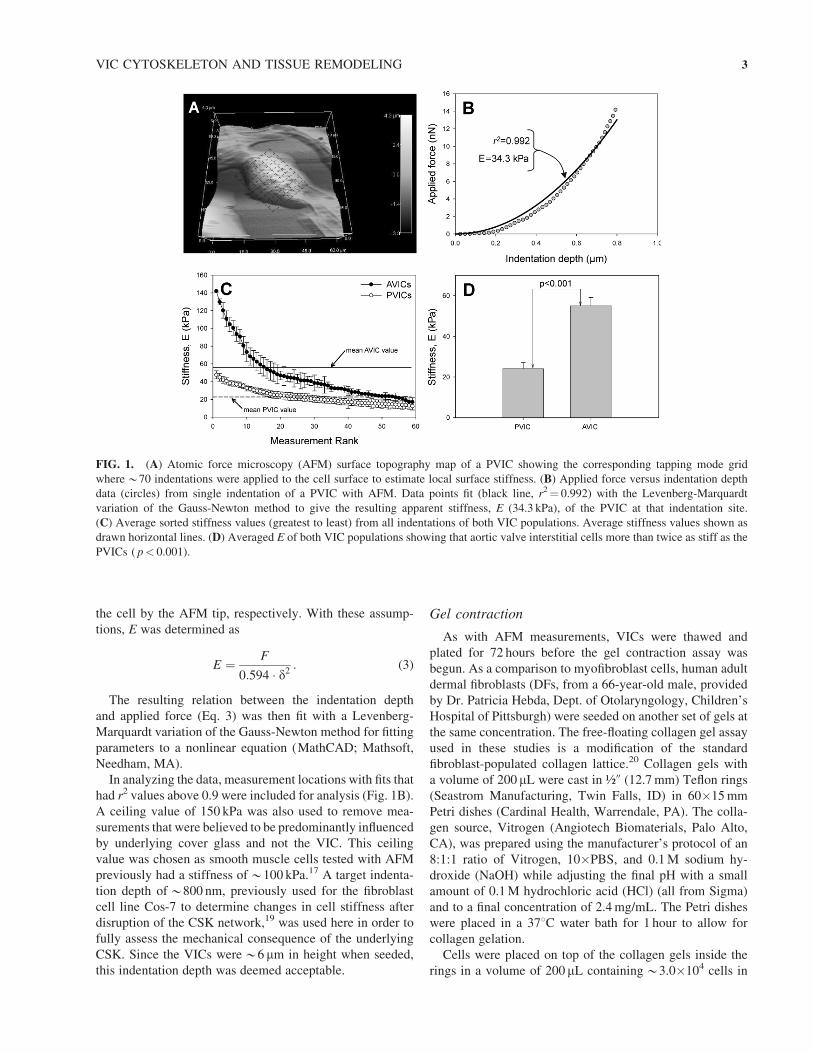
In analyzing the data, measurement locations with fits that
had r2 values above 0.9 were included for analysis (Fig. 1B).
A ceiling value of 150 kPa was also used to remove mea-
surements that were believed to be predominantly influenced
by underlying cover glass and not the VIC. This ceiling
value was chosen as smooth muscle cells tested with AFM
previously had a stiffness of*100 kPa.17 A target indenta-
tion depth of*800 nm, previously used for the fibroblast
cell line Cos-7 to determine changes in cell stiffness after
disruption of the CSK network,19 was used here in order to
fully assess the mechanical consequence of the underlying
CSK. Since the VICs were*6 mm in height when seeded,
this indentation depth was deemed acceptable.
Gel contraction
As with AFM measurements, VICs were thawed and
plated for 72 hours before the gel contraction assay was
begun. As a comparison to myofibroblast cells, human adult
dermal fibroblasts (DFs, from a 66-year-old male, provided
by Dr. Patricia Hebda, Dept. of Otolaryngology, Children’s
Hospital of Pittsburgh) were seeded on another set of gels at
the same concentration. The free-floating collagen gel assay
used in these studies is a modification of the standard
fibroblast-populated collagen lattice.20 Collagen gels with
a volume of 200 mL were cast in ½@ (12.7 mm) Teflon rings
(Seastrom Manufacturing, Twin Falls, ID) in 60�15 mm
Petri dishes (Cardinal Health, Warrendale, PA). The colla-
gen source, Vitrogen (Angiotech Biomaterials, Palo Alto,
CA), was prepared using the manufacturer’s protocol of an
8:1:1 ratio of Vitrogen, 10�PBS, and 0.1 M sodium hy-
droxide (NaOH) while adjusting the final pH with a small
amount of 0.1 M hydrochloric acid (HCl) (all from Sigma)
and to a final concentration of 2.4 mg/mL. The Petri dishes
were placed in a 378C water bath for 1 hour to allow for
collagen gelation.
Cells were placed on top of the collagen gels inside the
rings in a volume of 200 mL containing*3.0�104 cells in
FIG. 1. (A) Atomic force microscopy (AFM) surface topography map of a PVIC showing the corresponding tapping mode grid
where*70 indentations were applied to the cell surface to estimate local surface stiffness. (B) Applied force versus indentation depth
data (circles) from single indentation of a PVIC with AFM. Data points fit (black line, r2¼ 0.992) with the Levenberg-Marquardt
variation of the Gauss-Newton method to give the resulting apparent stiffness, E (34.3 kPa), of the PVIC at that indentation site.
(C) Average sorted stiffness values (greatest to least) from all indentations of both VIC populations. Average stiffness values shown as
drawn horizontal lines. (D) Averaged E of both VIC populations showing that aortic valve interstitial cells more than twice as stiff as the
PVICs ( p< 0.001).
VIC CYTOSKELETON AND TISSUE REMODELING 3

complete medium. The dishes were incubated for 30 minutes
to ensure cell adhesion to the collagen gels. Then, 7 mL of
medium was added to the dish, the Teflon rings were re-
moved, and the collagen gels were loosened from the surface
with a pipette tip. The dishes were incubated for another
30 minutes to allow for equilibration of the medium. To
examine the effects of transforming growth factor b1 (TGF-
b1) on the VICs, additional groups of PVIC (PVICsþTGF)
and AVIC (AVICsþTGF) gels were fabricated as above;
however, in the 7 mL added after cell adhesion, a single dose
of TGF-b1 (0.5 ng/mL, T5050 from Sigma)21 was added.
Collagen gels were imaged at 0, 2, 4, 24, 48, and 72 hours
following the final incubation with a digital camera (Diag-
nostic Instruments, Sterling Heights, MI) connected to a dis-
secting microscope (Olympus, Melville, NY). Changes in
collagen gel area were measured using Metamorph software
(Universal Imaging, Downingtown, PA). Percent contrac-
tion was calculated as the change in area from the initial area
at time 0. Dishes were imaged with a 1�1 cm2 visible grid
underneath to ensure uniform scaling during image analysis.
Cellular viability was assessed at each time point with a 10�light microscope to ensure that the VICs were spread and
healthy.
VIC migration and morphology
To qualitatively assess the location and migration of the
VICs at each time point, one gel for each VIC type and time
point was imaged via fluorescence microscopy. As there was
no evidence of differences between gel specimens for each
VIC population at each time point (i.e., all contracted simi-
larly), one gel was deemed sufficient. Gels were fixed in 2%
paraformaldehyde and imaged by fluorescence microscopy.
Gels were treated with 0.1% Triton X-100 for 20 minutes to
allow for infiltration of the stains. After washing three times
with 1�PBS, gels were stained with rhodamine phalloidin
for 1 hour to label the total actin of the cells. Cell nuclei were
then stained with 406-diamidino-2-phenylindole (DAPI). Gels
were put in a chambered borosilicate cover glass (Lab-Tek,
Campbell, CA) with the cell-seeded surface facing the cover
glass. The chambered cover glass was then mounted on an
inverted laser-scanning confocal microscope (Olympus Fluo-
view 1000). DAPI (excitation: 405 nm) and Cy3 (excitation:
543 nm) filter combinations were set to sequentially image
the cell nuclei and actin. For each gel, three 10� images
were taken encompassing an area from the center to the edge
of the gel. These images were concatenated to form a single
image.
Statistical analyses
Cell stiffness and percent area contraction from time 0 are
reported as mean� SE. Stiffness values were compared with
a paired Student’s t-test (SigmaStat, San Jose, CA) and
percent area contraction differences were first compared
with a two-way ANOVA and where significant differences
were found, post hoc testing was done with the unpaired
Student’s t-test (at each time point). For each gel contraction
group, a total of six gels were analyzed. Note that gels used
for fluorescence imaging were not taken from the n¼ 6 group
but were made in addition.
RESULTS
VIC stiffness with AFM
Analysis of the AFM indentation tests data resulted in at
least 60 acceptable AFM measurements per cell. As the lo-
cation of the organelles varied between cells in relation to
the AFM measurement location grid (Fig. 1A), the stiffness
values for each cell were sorted by magnitude then averaged
with the other cells from the same group. This allowed us to
more easily visualize the VIC AFM stiffness distribution
(Fig. 1C). In addition, the 60 stiffness values were averaged
together to obtain an estimated single value of E for the VIC
being tested. Resulting distributions indicated that the aver-
aged apparent stiffness (Fig. 1D) of the AVICs (55.06�4.17 kPa, n¼ 7) was approximately twice ( p< 0.001) the
FIG. 2. Light microscopy images of AVICs (left panel) and
PVICs (right panel) seeded on the free-floating collagen gels at 2,
24, and 72 hours (magnification 10�). Note the spread and attached
nature of the AVICs and PVICs at each time point.
4 MERRYMAN ET AL.

averaged apparent stiffness of the PVICs (24.11� 3.02 kPa,
n¼ 7). Since these values are averaged from multiple inden-
tations over the surface of both VIC populations (mean pop-
ulation values are shown as horizontal lines on Fig. 1C), it
indicates that there exists a significant overall stiffness dif-
ference between valve cells.
Gel contraction results
Both VIC populations appeared to be viable and well at-
tached during the duration of the contraction experiment
(Fig. 2). All gels were circular at 0 hour and underwent uni-
form contraction over the 72-hour duration of the experiment
(Fig. 3). With two-way ANOVA, both factors of time and
cell type were significant ( p< 0.001) for the gel contraction
experiment; all further statistical comparisons are with the
Student’s t-test. Gel contraction by the VIC populations
at 2 hours was negligible (AVICs: 0.22� 0.52%; PVICs:
0.78� 0.42%);however, theDFs hadsignificantly contracted
the gels (23.21� 1.79%, p< 0.001, Fig. 4A). Similarly, both
VIC populations contracted the gels insignificantly (AVICs:
0.51� 0.86%; PVICs: 0.64� 0.57%) after 4 hours; DF gels
were significantly contracted (47.54� 2.67%, p< 0.001)
compared to both VIC gels.
FIG. 3. Time course contraction of VIC-seeded collagen gels
(AVICs, left panel; PVICs, right panel) imaged at 1�; underlying
grid is 1�1 cm2 and is necessary for uniform scaling during analysis.
FIG. 4. (A) Percent area contraction by both VIC populations and
dermal fibroblasts (DFs). DFs contraction was significantly greater
(*p< 0.001) than both VIC populations at each time point. AVICs
and PVICs were not different at 2 or 4 hours; however, AVIC gels
were statistically more contracted at 24 hours ( p< 0.001) and
48 hours ( p¼ 0.009) than PVIC gels. All data points are fit with a
four-parameter sigmoid function. ‘‘�’’ symbols indicate the greatest
rate of contraction (dA/dtmax); both VIC time points occur after VIC
clustering shown in Fig. 5. No difference was observed at 72 hours as
both populations had reached a steady state and contraction was
ceased. (B) Comparison of VIC contraction results shown in (A) up
to 24 hours with VICs treated with TGF-b1 (AVICsþTGF and
PVICsþTGF). Both TGF-treated groups (circled) were statistically
more contracted ( p< 0.001) at 2 and 4 hours versus the AVIC and
PVIC groups. Additionally, at 24 hours both TGF-treated groups
were significantly more contracted ( p< 0.001) than untreated VIC
groups, and the AVICsþTGF group was statistically more con-
tracted ( p< 0.001) than the PVICsþTGF group.
VIC CYTOSKELETON AND TISSUE REMODELING 5

At 24 hours, the AVIC population (31.99� 3.44%) con-
tracted the gels significantly more ( p< 0.001, Fig. 4A) than
the PVIC population (7.57� 1.81%). DF gels were highly
contracted at 24 hours (79.46� 0.96%, p< 0.001) and no
subsequent contraction was observed. Similarly at 48 hours,
the AVIC (50.68� 2.39%) gels were significantly more
contracted ( p¼ 0.009, Fig. 4A) than the PVIC gels (34.70�4.34%). Gel contraction differences were insignificant at
72 hours (AVICs: 58.96� 1.03%; PVICs: 53.96� 2.29%).
From the shape of the contraction curves, it was deemed best
to fit all data sets with a four-parameter sigmoid func-
tion, thereby making it possible to calculate the greatest
rate of contraction (dA/dtmax) for each group. For AVIC gels,
dA/dtmax occurred at 15.18 hours and for PVIC gels at
43.59 hours (Fig. 4A). DF gel contraction was nearly linear
up to 4 hours; however, dA/dtmax occurred at 2.25 hours.
VIC-induced gel contraction in the presence of TGF-b1
was more rapid and substantial than VIC gels without cyto-
kine treatment. At 2, 4, and 24 hours, the AVICsþTGF and
PVICsþTGF gels were significantly more contracted ( p<0.001, Fig. 4B) than the normal AVIC and PVIC gels. How-
ever, there was no difference between the cytokine-treated
gels at 2 or 4 hours. At 24 hours, the AVICsþTGF gels
(81.6� 1.28%) were significantly more contracted ( p<0.001) than the PVICsþTGF gels (70.11� 1.2%). Contrac-
tion of the TGF-treated gels could not be observed past
24 hours as they became spherical with no apparent seeded
surface.
All cell populations were spatially distributed at 0 hour
(1 hour after seeding, Fig. 5). In the AVIC gels, the cells
appeared to cluster and organize around 4 hours, whereas
the PVICs lagged behind and clustered closer to 24 hours
(Fig. 5). Following central clustering, both populations gen-
erated their largest rate of contraction [shown as�-symbols
in Fig. 4A]. This was then followed, in the next time point,
with the VICs radiating out from the center of the gel (Fig. 5,
24 hours for AVICs and 48 hours for PVICs) and a decreased
rate of contraction at subsequent observations.
DISCUSSION
Observed differences in VIC stiffness
An important aspect of this study was the observation that,
as seen previously with ovine VICs measured by micropi-
pette aspiration,1 the stiffness of the AVICs was approx-
imately twofold greater than the PVICs. However, it is
important to note that the AFM values were also*100 times
greater than the micropipette values, as anticipated based on
studies with other cell types.17,22–24 The best explanation
offered to reconcile these differences concerns the assump-
tions used to model the force-deformation relation and the
actual loading technique used. With AFM, a localized force
is applied to a focal point on the cell membrane, which is
supported by the nearby structural components (microfila-
ments, nucleus, etc.). The resulting response is modeled from
this small, highly localized deformation state. Micropipette
aspiration, on the other hand, examines the macromechanics
of the cell and is primarily influenced by the cell membrane
and unanchored CSK. Our previous VIC results with micro-
pipette aspiration were modeled by assuming the cell to be
an isotropic, elastic, and incompressible material that was
homogenous (discontinuities, such as nuclei and organelles,
were ignored).
Overall, it can be argued that VIC stiffness as measured
with AFM allows for a more accurate representation of the
cellular properties and architecture of the cell than micro-
pipette aspiration. This is a result of the fact that the cells are
seeded and attached to an underlying collagen substrate so
that the CSK is extended and rigid as it is anchored at focal
adhesions. This configuration is in contrast to testing with
micropipette aspiration where the CSK is freed and not in a
physiologic state as the cells float in a solution. Ultimately,
interpretation and meaning of all cellular mechanical analysis
techniques (nonlinear viscoelastic, heterogeneous materials)
are tenuous, and direct conclusions about cell function and
biology are difficult to draw. Therefore, while these testing
techniques do not result in similar stiffness values, they do
allow for direct comparisons between PVICs and AVICs.
When considering the differences in CSK composition,1
the measured differences in E reveal that the underlying
FIG. 5. Concatenated fluorescence images [10�] of time course
migration of VIC populations seeded on free-floating collagen
gels (AVICs on left and PVICs on right) at each point of gel
contraction measurement shown in Fig. 4A. Three images were
made from the center (left) to the edge (right) of gel; central
diagram shows geometry of image capture. Note the AVIC clus-
tering near 4 hours and PVIC clustering near 24 hours. Following
this clustering, both populations contracted the gels at their greatest
rate [shown as �-symbols in Fig. 4A]. After clustering, the cells
appear to radiate outward from the center of the gel and though
contraction continued, it was less pronounced.
6 MERRYMAN ET AL.

CSK of the AVICs is a strong contributor to overall cell
stiffness. The necessity of greater CSK content in the AVIC
is believed to be required in order to effectively synthesize or
expel synthesized proteins into the ECM.1 This has been
observed previously for other types of myofibroblasts, where
human wound closure was facilitated by these dermal myo-
fibroblasts as they produced thicker collagen fibers than fi-
broblasts and contributed to 88% of closure.25 Therefore, the
mechanical properties of the AVIC imply a greater ability to
remodel the surrounding ECM compared to the PVIC.
VIC contraction of collagen gels
The primary finding from this study is the greater ability
of AVICs to contract free-floating collagen gels compared to
PVICs under identical experimental conditions. Though the
substrate of a free-floating collagen gel does not fully re-
present all the characteristics of the in vivo ECM of valvular
tissue, it does support cell survival, attachment, and con-
traction. Therefore, comparing the contractility of both VIC
populations indicates their CSK activity and resulting po-
tential for tissue remodeling.
Human adult DFs, used for comparison in the present
study, caused rapid contraction of the gels, which were
*80% contracted at 24 hours; further contraction could not
be detected as the gels became spherical. Not only did the
DFs contract the gels much faster, but also the time course of
their contraction was unlike the VIC populations (Fig. 4A).
DFs did not migrate and form a central cluster at any time
point (images not shown), but essentially attached to the
collagen gel and exerted local contraction without apprecia-
ble migration. Conversely, both VIC populations attached to
the gel and did not exert any measurable contraction at 2 or
4 hours, but appeared to migrate to form an organized cluster
in the center of the gel. Although there was an apparent lag
by the PVICs (dA/dtmax for PVIC gels was*28 hours later
than for AVIC gels), once they did form the central cluster,
they reached the level of AVIC contraction at 72 hours. After
clustering by both VIC populations, they appeared to disperse
and form radiating lines of cellular organization, which we
speculate is similar to a response that would be seen with
smooth muscle cells.
Walker et al.7 have reported AVIC gel contraction in the
presence of TGF-b1 and cytochalasin D. This study examined
AVICs only and the dose dependence of TGF-b1 on free-
floating and attached collagen gels. Our results support their
findings in that the AVICsþTGF group did contract the gels
significantly more at 24 hours than the AVIC group; however,
our results are much more pronounced than theirs for the
AVICsþTGF-b1 group (*80% vs.*20%). It is unclear why
such a large difference was observed, but we speculate that it
may be due to different-sized collagen gels, number of seeded
cells, or culture conditions. This is supported by results from
untreated AVIC gels, as their contraction was nearly 20% less
than our observed values. Moreover, Fu et al.26 investigated
ascorbic acid and growth factors [including basic fibroblast
growth factor (bFGF) and TGF] on human pediatric AVICs
for their effect on tissue development when seeded on poly-
glycolic acid coated with prolyl 4-hydroxylase. Results in-
dicated that after 7 and 28 days of culture, the bFGF and TGF
groups showed significantly higher DNA content compared to
the control. They also found denser, more organized tissue
development with pronounced matrix protein formation in the
tissue-engineered structures in the TGF group after 28 days,
while the addition of bFGF led to a markedly higher collagen
synthesis after 28 days. Our results corroborate and further
support these studies and further indicated that TGF-b1 does
serve to significantly activate the remodeling activity of both
semilunar valve cell types.
Role of CSK contraction in valve
physiology and pathology
As the VICs are the resident population of cells respon-
sible for maintaining valvular tissue homeostasis, their
mechanobiological function is clearly important. Previously,
the level of SMA found in each VIC population corre-
lated well with the molecular chaperone heat-shock protein
47 (HSP47),1 which was used as a surrogate for collagen
synthesis.27–29 Therefore, the difference observed in gel
contraction between the AVIC and PVIC populations has
two implications. First, it implies that the pulmonary val-
vular tissues remodel more slowly and have less biosynthetic
demand than aortic valvular tissues, which was suspected
from the outset.
The second and unexpected implication from the groups
treated with TGF-b1 involves valvular pathology. Our re-
sults indicate that PVICs respond similarly as AVICs to
TGF-b1, which is known to be a byproduct of infiltrating
macrophages,30 and is thought to initiate aortic valve cal-
cification.31 From this fact alone, one would suspect that
pulmonary valves may be as susceptible to pathologic con-
ditions as aortic valves, but this is not supported clinically or
epidemiologically. Therefore, one explanation for not seeing
pathologic similarities between these two valves is either a
lack of TGF-b1 near the PVICs or TGF-b1 may not be as
effective on PVICs. Current work is aimed at understanding
the roll of mechanical stimulation in the presence of TGF-b1
on the AVIC to better understand the etiologies of aortic
valve fibrosis and calcification.32
Role of CSK contraction in TEHV development
The desired endpoint of any engineered tissue is func-
tional equivalence of the replaced tissue or organ. Similarly,
the goal of a TEHV is accomplished largely at the tissue
level where the tissue biomechanical behavior is para-
mount.12 Much activity has been focused on identification
and biosynthetic activity of candidate TEHV cell sources
during in vitro tissue formation prior to implantation.33–36
The long-term ability of the chosen cell source to continually
remodel the ECM will be crucial for success of a TEHV.
VIC CYTOSKELETON AND TISSUE REMODELING 7
With this in mind and due to the phenotypic plasticity of the
native VIC population,8,11 a requirement of biosynthetic
sensitivity to various cytokines may be necessary in choosing
a suitable cell source that can be activated early during the
in vitro portion of TEHV development and later quiescent
under steady state, in vivo conditions. Regardless of the cell
source chosen for a TEHV, be it for the aortic or pulmonary
position, remodeling potential should be considered in light
of results shown here.
In summary, this work is the first to demonstrate a potential
difference in tissue-remodeling ability between the AVIC
and PVIC. Moreover, seeded cell stiffness values measured
with AFM revealed a twofold difference between AVICs
and PVICs, confirming our earlier result using micropipette
aspiration.1 These results suggest a marked difference be-
tween the two VIC populations with respect to free-floating
gel contraction, which is believed to be an indication of
tissue-remodeling potential. Overall, the PVIC appears to be
a similar, but less-robust version of the AVIC; in that, it acts
slower and in a less-pronounced fashion. Future work will be
needed to elucidate the effects of both local tissue stress and
surrounding cytokines on the VIC phenotype and biosyn-
thetic function. Additionally, the question of native VIC
phenotypic plasticity and resulting biosynthetic function will
have to be addressed for development of a successful TEHV
with whatever cell source is ultimately chosen.
ACKNOWLEDGMENTS
This work was supported by the National Heart, Lung, and
Blood Institute grant HL68818. The following individual
support from the American Heart Association Pennsylvania-
Delaware Affiliate is also gratefully acknowledged: Pre-
doctoral Fellowship (W.D. Merryman, 0515416U); Beginning
Grant-in-Aid (J. Liao, 0565346U).
REFERENCES
1. Merryman, W.D., Youn, I., Lukoff, H.D., Krueger, P.M.,
Guilak, F., Hopkins, R.A., and Sacks, M.S. Correlation be-
tween heart valve interstitial cell stiffness and transvalvular
pressure: implications for collagen biosynthesis. Am J Physiol
Heart Circ Physiol 290, H224, 2006.
2. Taylor, P.M., Batten, P., Brand, N.J., Thomas, P.S., and Ya-
coub, M.H. The cardiac valve interstitial cell. Int J Biochem
Cell Biol 35, 113, 2003.
3. Taylor, P.M., Allen, S.P., and Yacoub, M.H. Phenotypic and
functional characterization of interstitial cells from human heart
valves, pericardium and skin. J Heart Valve Dis 9, 150, 2000.
4. Schoen, F.J., and Levy, R.J. Calcification of tissue heart valve
substitutes: progress toward understanding and prevention.
Ann Thorac Surg 79, 1072, 2005.
5. Jian, B., Narula, N., Li, Q.Y., Mohler, E.R., 3rd, and Levy, R.J.
Progression of aortic valve stenosis: TGF-beta1 is present in
calcified aortic valve cusps and promotes aortic valve intersti-
tial cell calcification via apoptosis. Ann Thorac Surg 75, 457,
2003.
6. Jian, B., Xu, J., Connolly, J., Savani, R.C., Narula, N., Liang,
B., and Levy, R.J. Serotonin mechanisms in heart valve disease
I: serotonin-induced up-regulation of transforming growth
factor-beta1 via G-protein signal transduction in aortic valve
interstitial cells. Am J Pathol 161, 2111, 2002.
7. Walker, G.A., Masters, K.S., Shah, D.N., Anseth, K.S., and
Leinwand, L.A. Valvular myofibroblast activation by trans-
forming growth factor-beta: implications for pathological extra-
cellular matrix remodeling in heart valve disease. Circ Res 95,
253, 2004.
8. Rabkin-Aikawa, E., Farber, M., Aikawa, M., and Schoen, F.J.
Dynamic and reversible changes of interstitial cell phenotype
during remodeling of cardiac valves. J Heart Valve Dis 13,
841, 2004.
9. Taylor, P.M., Allen, S.P., Dreger, S.A., and Yacoub, M.H.
Human cardiac valve interstitial cells in collagen sponge: a
biological three-dimensional matrix for tissue engineering.
J Heart Valve Dis 11, 298, 2002.
10. Butcher, J.T., and Nerem, R.M. Porcine aortic valve inter-
stitial cells in three-dimensional culture: comparison of phe-
notype with aortic smooth muscle cells. J Heart Valve Dis 13,
478, 2004.
11. Rabkin, E., Hoerstrup, S.P., Aikawa, M., Mayer, J.E., Jr., and
Schoen, F.J. Evolution of cell phenotype and extracellu-
lar matrix in tissue-engineered heart valves during in-vitro
maturation and in-vivo remodeling. J Heart Valve Dis 11, 308,
2002.
12. Merryman, W.D., Engelmayr, G.C., Jr., Liao, J., and Sacks,
M.S. Defining biomechanical endpoints for tissue engineered
heart valve leaflets from native leaflet properties. Prog Pediatr
Cardiol 21, 153, 2006.
13. Messier, R.H., Jr., Bass, B.L., Aly, H.M., Jones, J.L., Dom-
kowski, P.W., Wallace, R.B., and Hopkins, R.A. Dual structural
and functional phenotypes of the porcine aortic valve inter-
stitial population: characteristics of the leaflet myofibroblast.
J Surg Res 57, 1, 1994.
14. Filip, D.A., Radu, A., and Simionescu, M. Interstitial cells
of the heart valves possess characteristics similar to smooth
muscle cells. Circ Res 59, 310, 1986.
15. Mulholland, D.L., and Gotlieb, A.I. Cell biology of valvular
interstitial cells. Can J Cardiol 12, 231, 1996.
16. Della Rocca, F., Sartore, S., Guidolin, D., Bertiplaglia, B.,
Gerosa, G., Casarotto, D., and Pauletto, P. Cell composition of
the human pulmonary valve: a comparative study with the aortic
valve—the VESALIO Project. Vitalitate Exornatum Succe-
daneum Aorticum labore Ingegnoso Obtinebitur. Ann Thorac
Surg 70, 1594, 2000.
17. Mathur, A.B., Collinsworth, A.M., Reichert, W.M., Kraus, W.E.,
and Truskey, G.A. Endothelial, cardiac muscle and skeletal mus-
cle exhibit different viscous and elastic properties as determined
by atomic force microscopy. J Biomech 34, 1545, 2001.
18. Costa, K.D., and Yin, F.C. Analysis of indentation: implica-
tions for measuring mechanical properties with atomic force
microscopy. J Biomech Eng 121, 462, 1999.
19. Kasas, S., Wang, X., Hirling, H., Marsault, R., Huni, B., Yersin,
A., Regazzi, R., Grenningloh, G., Riederer, B., Forro, L.,
8 MERRYMAN ET AL.

Dietler, G., and Catsicas, S. Superficial and deep changes of
cellular mechanical properties following cytoskeleton disas-
sembly. Cell Motil Cytoskeleton 62, 124, 2005.
20. Bell, E., Ivarsson, B., and Merrill, C. Production of a tissue-
like structure by contraction of collagen lattices by human
fibroblasts of different proliferative potential in vitro. Proc
Natl Acad Sci USA 76, 1274, 1979.
21. Cushing, M.C., Liao, J.T., and Anseth, K.S. Activation of
valvular interstitial cells is mediated by transforming growth
factor-beta1 interactions with matrix molecules. Matrix Biol
24, 428, 2005.
22. Sato, M., Theret, D.P., Wheeler, L.T., Ohshima, N., and
Nerem, R.M. Application of the micropipette technique to the
measurement of cultured porcine aortic endothelial cell visco-
elastic properties. J Biomech Eng 112, 263, 1990.
23. Guilak, F., Ting-Beall, H.P., Baer, A.E., Trickey, W.R.,
Erickson, G.R., and Setton, L.A. Viscoelastic properties of
intervertebral disc cells. Identification of two biomechanically
distinct cell populations. Spine 24, 2475, 1999.
24. Na, S., Sun, Z., Meininger, G.A., and Humphrey, J.D. On
atomic force microscopy and the constitutive behavior of
living cells. Biomech Model Mechanobiol 3, 75, 2004.
25. Berry, D.P., Harding, K.G., Stanton, M.R., Jasani, B., and
Ehrlich, H.P. Human wound contraction: collagen organization,
fibroblasts, and myofibroblasts. Plast Reconstr Surg 102, 124,
1998.
26. Fu, P., Sodian, R., Luders, C., Lemke, T., Kraemer, L., Hu-
bler, M., Weng, Y., Hoerstrup, S.P., Meyer, R., and Hetzer, R.
Effects of basic fibroblast growth factor and transforming
growth factor-beta on maturation of human pediatric aortic
cell culture for tissue engineering of cardiovascular structures.
ASAIO J 50, 9, 2004.
27. Tasab, M., Batten, M.R., and Bulleid, N.J. Hsp47: a molec-
ular chaperone that interacts with and stabilizes correctly-
folded procollagen. EMBO J 19, 2204, 2000.
28. Rocnik, E.F., van der Veer, E., Cao, H., Hegele, R.A., and
Pickering, J.G. Functional linkage between the endoplasmic
reticulum protein Hsp47 and procollagen expression in human
vascular smooth muscle cells. J Biol Chem 277, 38571, 2002.
29. Sauk, J.J., Nikitakis, N., and Siavash, H. Hsp47 a novel col-
lagen binding serpin chaperone, autoantigen and therapeutic
target. Front Biosci 10, 107, 2005.
30. Peters, T., Sindrilaru, A., Hinz, B., Hinrichs, R., Menke, A.,
Al-Azzeh, E.A., Holzwarth, K., Oreshkova, T., Wang, H.,
Kess, D., Walzog, B., Sulyok, S., Sunderkotter, C., Friedrich,
W., Wlaschek, M., Krieg, T., and Scharffetter-Kochanek, K.
Wound-healing defect of CD18(�/�) mice due to a decrease
in TGF-beta(1) and myofibroblast differentiation. EMBO J
24, 3400, 2005.
31. Edwards, J.E. Calcific aortic stenosis: pathologic features.
Mayo Clin Proc 36, 444, 1961.
32. Merryman, W.D., Lukoff, H.D., Long, R.A., Engelmayr,
G.C., Jr., Hopkins, R.A., and Sacks, M.S. Synergistic effects
of cyclic tension and transforming growth factor-b1 on the
aortic valve myofibroblast. Cardiovas Pathol (In press).
33. Sales, V.L., Engelmayr, G.C., Mettler, B.A., Johnson, J.A.,
Sacks, M.S., and Mayer, J.E., Jr. Transforming growth factor-b1
modulates extracellular matrix production, proliferation and
apoptosis of endothelial progenitor cells in tissue-engineering
scaffolds. Circulation 114 (1 Suppl), I193, 2006.
34. Perry, T.E., Kaushal, S., Nasseri, B., Sutherland, F.W.H.,
Wang, J., Guleserian, K.J., Bischoff, J., Vacanti, J.P., Sacks,
M.S., and Mayer, J.E. Peripheral blood as a cell source for
tissue engineering heart valves. Surg Forum LII, 99, 2001.
35. Perry, T.E., Kaushal, S., Sutherland, F.W., Guleserian, K.J.,
Bischoff, J., Sacks, M., and Mayer, J.E. Thoracic Surgery Di-
rectors Association Award. Bone marrow as a cell source for
tissue engineering heart valves. Ann Thorac Surg 75, 761, 2003.
36. Kadner, A., Hoerstrup, S.P., Zund, G., Eid, K., Maurus, C.,
Melnitchouk, S., Grunenfelder, J., and Turina, M.I. A new
source for cardiovascular tissue engineering: human bone
marrow stromal cells. Eur J Cardiothorac Surg 21, 1055, 2002.
Address reprint requests to:
Michael S. Sacks, Ph.D.
W.K. Whiteford Professor
Department of Bioengineering
100 Technology Drive, Room 234
University of Pittsburgh
Pittsburgh, PA 15219
E-mail: [email protected]
VIC CYTOSKELETON AND TISSUE REMODELING 9
Related Documents