Differences in the Cellular Uptake and Intracellular Itineraries of Amyloid Beta Proteins 40 and 42: Ramifications for the Alzheimer’s Drug Discovery Rajesh S. Omtri, †,‡ Michael W. Davidson, § Balasubramaniam Arumugam, ‡ Joseph F. Poduslo, ∥ and Karunya K. Kandimalla* ,†,∥ † Division of Basic Pharmaceutical Sciences, Florida A&M University College of Pharmacy and Pharmaceutical Sciences, Tallahassee, Florida, United States § National High Magnetic Field Laboratory and Department of Biological Science, The Florida State University, 1800 East Paul Dirac Drive, Tallahassee, Florida, United States ∥ Molecular Neurobiology Laboratory, Departments of Neurology, Neuroscience, and Biochemistry/Molecular Biology, Mayo Clinic College of Medicine, Rochester, Minnesota, United States ‡ NIMS Institute of Pharmacy, NIMS University, Shoba Nagar, Delhi Highway, Jaipur, Rajasthan, India ABSTRACT: Mounting evidence suggests that the pathological hallmarks of Alzheimer’s disease (AD), neurofibrillary tangles and parenchymal amyloid plaques, are downstream reflections of neurodegeneration caused by the intraneuronal accumulation of amyloid-β proteins (Aβ), particularly Aβ42 and Aβ40. While the neurotoxicity of more amyloidogenic but less abundant Aβ42 is well documented, the effect of Aβ40 on neurons has been understudied. The Aβ40 expression in the presymptomatic AD brain is ten times greater than that of Aβ42. However, the Aβ40:42 ratio decreases with AD progression and coincides with increased amyloid plaque deposition in the brain. Hence, it is thought that Aβ40 protects neurons from the deleterious effects of Aβ42. The pathophysiological pathways involved in the neuronal uptake of Aβ40 or Aβ42 have not been clearly elucidated. Lack of such critical information obscures therapeutic targets and thwarts rational drug development strategies aimed at preventing neurodegeneration in AD. The current study has shown that fluorescein labeled Aβ42 (F-Aβ42) is internalized by neurons via dynamin dependent endocytosis and is sensitive to membrane cholesterol, whereas the neuronal uptake of F-Aβ40 is energy independent and nonendocytotic. Following their uptake, both F-Aβ40 and F-Aβ42 did not accumulate in early/recycling endosomes; F-Aβ42 but not F-Aβ40 accumulated in late endosomes and in the vesicles harboring caveolin-1. Furthermore, F-Aβ42 demonstrated robust accumulation in the lysosomes and damaged their integrity, whereas F-Aβ40 showed only a sparse lysosomal accumulation. Such regulated trafficking along distinct pathways suggests that Aβ40 and Aβ42 exercise differential effects on neurons. These differences must be carefully considered in the design of a pharmacological agent intended to block the neurodegeneration triggered by Aβ proteins. KEYWORDS: Alzheimer’s disease, cellular trafficking, cholesterol, endocytosis, lysosomes, neurodegeneration ■ INTRODUCTION Deposition of Aβ proteins as parenchymal plaques is the conventional pathological hallmark of AD. However, extensive neuropathological and biochemical observations made in AD transgenic mouse models and in human AD patients suggest that intraneuronal accumulation of Aβ proteins is the pivotal event that triggers neurodegeneration in AD. 1−3 The appearance of intraneuronal Aβ leads to profound deficits in hippocampal long-term potentiation, facilitates tau hyper- phosphorylation, and disrupts proteosomal as well as mitochondrial functions. 4 Removal of intraneuronal Aβ with passive immunotherapy was shown to halt neurodegeneration and reverse behavioral deficits in AD mouse models. 2,5 However, immunotherapy with antiamyloid antibodies immo- bilizes extraneuronal amyloid deposits, jams the cerebral vasculature with amyloid, and causes life-threatening conditions such as meningoencephalitis. 6 Hence, targeted therapies that can specifically clear neurons of toxic Aβ proteins are warranted; development of such therapies requires a thorough understanding of pathophysiological mechanisms that promote neuronal uptake of toxic Aβ isoforms. In the early stages of AD, reuptake of extracellular Aβ proteins, rather than the accumulation of Aβ generated within the neurons through ER/Golgi processing of the amyloid precursor protein (APP), is believed to contribute significantly Received: October 17, 2011 Revised: April 30, 2012 Accepted: May 10, 2012 Published: May 10, 2012 Article pubs.acs.org/molecularpharmaceutics © 2012 American Chemical Society 1887 dx.doi.org/10.1021/mp200530q | Mol. Pharmaceutics 2012, 9, 1887−1897

Welcome message from author
This document is posted to help you gain knowledge. Please leave a comment to let me know what you think about it! Share it to your friends and learn new things together.
Transcript

Differences in the Cellular Uptake and Intracellular Itineraries ofAmyloid Beta Proteins 40 and 42: Ramifications for the Alzheimer’sDrug DiscoveryRajesh S. Omtri,†,‡ Michael W. Davidson,§ Balasubramaniam Arumugam,‡ Joseph F. Poduslo,∥
and Karunya K. Kandimalla*,†,∥
†Division of Basic Pharmaceutical Sciences, Florida A&M University College of Pharmacy and Pharmaceutical Sciences, Tallahassee,Florida, United States§National High Magnetic Field Laboratory and Department of Biological Science, The Florida State University, 1800 East Paul DiracDrive, Tallahassee, Florida, United States∥Molecular Neurobiology Laboratory, Departments of Neurology, Neuroscience, and Biochemistry/Molecular Biology, Mayo ClinicCollege of Medicine, Rochester, Minnesota, United States‡NIMS Institute of Pharmacy, NIMS University, Shoba Nagar, Delhi Highway, Jaipur, Rajasthan, India
ABSTRACT: Mounting evidence suggests that the pathological hallmarks of Alzheimer’sdisease (AD), neurofibrillary tangles and parenchymal amyloid plaques, are downstreamreflections of neurodegeneration caused by the intraneuronal accumulation of amyloid-βproteins (Aβ), particularly Aβ42 and Aβ40. While the neurotoxicity of more amyloidogenicbut less abundant Aβ42 is well documented, the effect of Aβ40 on neurons has beenunderstudied. The Aβ40 expression in the presymptomatic AD brain is ten times greater thanthat of Aβ42. However, the Aβ40:42 ratio decreases with AD progression and coincides withincreased amyloid plaque deposition in the brain. Hence, it is thought that Aβ40 protectsneurons from the deleterious effects of Aβ42. The pathophysiological pathways involved inthe neuronal uptake of Aβ40 or Aβ42 have not been clearly elucidated. Lack of such criticalinformation obscures therapeutic targets and thwarts rational drug development strategiesaimed at preventing neurodegeneration in AD. The current study has shown that fluoresceinlabeled Aβ42 (F-Aβ42) is internalized by neurons via dynamin dependent endocytosis and issensitive to membrane cholesterol, whereas the neuronal uptake of F-Aβ40 is energyindependent and nonendocytotic. Following their uptake, both F-Aβ40 and F-Aβ42 did notaccumulate in early/recycling endosomes; F-Aβ42 but not F-Aβ40 accumulated in lateendosomes and in the vesicles harboring caveolin-1. Furthermore, F-Aβ42 demonstratedrobust accumulation in the lysosomes and damaged their integrity, whereas F-Aβ40 showedonly a sparse lysosomal accumulation. Such regulated trafficking along distinct pathways suggests that Aβ40 and Aβ42 exercisedifferential effects on neurons. These differences must be carefully considered in the design of a pharmacological agent intendedto block the neurodegeneration triggered by Aβ proteins.
KEYWORDS: Alzheimer’s disease, cellular trafficking, cholesterol, endocytosis, lysosomes, neurodegeneration
■ INTRODUCTIONDeposition of Aβ proteins as parenchymal plaques is theconventional pathological hallmark of AD. However, extensiveneuropathological and biochemical observations made in ADtransgenic mouse models and in human AD patients suggestthat intraneuronal accumulation of Aβ proteins is the pivotalevent that triggers neurodegeneration in AD.1−3 Theappearance of intraneuronal Aβ leads to profound deficits inhippocampal long-term potentiation, facilitates tau hyper-phosphorylation, and disrupts proteosomal as well asmitochondrial functions.4 Removal of intraneuronal Aβ withpassive immunotherapy was shown to halt neurodegenerationand reverse behavioral deficits in AD mouse models.2,5
However, immunotherapy with antiamyloid antibodies immo-bilizes extraneuronal amyloid deposits, jams the cerebral
vasculature with amyloid, and causes life-threatening conditionssuch as meningoencephalitis.6 Hence, targeted therapies thatcan specifically clear neurons of toxic Aβ proteins arewarranted; development of such therapies requires a thoroughunderstanding of pathophysiological mechanisms that promoteneuronal uptake of toxic Aβ isoforms.In the early stages of AD, reuptake of extracellular Aβ
proteins, rather than the accumulation of Aβ generated withinthe neurons through ER/Golgi processing of the amyloidprecursor protein (APP), is believed to contribute significantly
Received: October 17, 2011Revised: April 30, 2012Accepted: May 10, 2012Published: May 10, 2012
Article
pubs.acs.org/molecularpharmaceutics
© 2012 American Chemical Society 1887 dx.doi.org/10.1021/mp200530q | Mol. Pharmaceutics 2012, 9, 1887−1897
to the intraneuronal Aβ pool.7 Published reports have claimedseveral receptors and even nonendocytotic mechanisms tomediate intraneuronal accumulation of Aβ proteins.8 Pre-viously, we reported nonsaturable, energy independent, andnonendocytotic uptake of fluorescein labeled Aβ40 in neuronalcells.9 In this study, we have shown that Aβ42 is internalized viaendocytosis, which is energy dependent.It is becoming increasingly evident that Aβ40 protects
neurons from the deleterious effects of Aβ42.10−14 Moreover,Aβ40/Aβ42 ratios are associated with the severity and locationof AD pathology;15,16 a lower Aβ40/Aβ42 ratio was shown toenhance amyloid deposition in the parenchyma,17 whereashigher Aβ40/Aβ42 ratio drives cerebrovascular amyloidaccumulation.18 It is obvious from these reports that Aβ40and Aβ42 play distinct roles in AD, and their relativedistributions in various physiological compartments maymodulate AD pathology. To exercise such differential impact,Aβ40 and Aβ42 must be trafficked and regulated via distinctpathways, which need to be carefully considered in the designof a pharmacological agent that can block their accumulation inneurons.
■ MATERIALS AND METHODSSynthesis of Aβ40 Proteins. Aβ40 and Aβ42 are
synthesized on an ABI 433 peptide synthesizer (Foster City,CA) with Val-NovaSyn TGA resin (Calbiochem-Novabiochem,San Diego, CA) employing HBTU activation and synthesisprotocols as described in our earlier publication.9 Fluoresceinwas tagged to Aβ40 or Aβ42 on the Fmoc column, which wasthoroughly washed with dimethylformamide and dichloro-methane to remove excess NHS-fluorescein. The fluoresceinlabeled Aβ40 or Aβ42 (F-Aβ40 or F-Aβ42) thus obtained waspurified by high performance liquid chromatography (HPLC)methods. Hence it is unlikely to have any free fluorescein in theF-Aβ40 or F-Aβ42 solutions. F-Aβ40 and F-Aβ42 monomerswere prepared as per the procedure described by Klein et al.19
Various microscopy markers such as Alexa Fluor 633 labeledtransferrin (AF633-Trf), Dil labeled low density lipoprotein(Dil-LDL), Alexa Fluor 647 labeled cholera toxin (AF647-CT)and Lysotracker Red DND 99 (LR) were obtained fromInvitrogen (Carlsbad, CA).Cell Cultures. Pheochromocytoma (PC12) cells were
purchased from ATCC and grown in a 50:50 mixture ofDMEM and Hams F12 (Mediatech Inc., Herndon, VA)enriched with 10% cosmic calf serum (Hyclone Hudson,NH). For the live cell imaging experiments, the PC12 cellswere plated on sterile Delta-T culture dishes (Bioptechs, Butler,PA) at a density of 5000 cells/dish and cultured in the growthmedium supplemented with 100 ng/mL of nerve growth factor(NGF) (Harlan Biosciences, Indianapolis, IN) and 1% serumfor at least 4 days until the neurite growth was prominent. Thedifferentiated PC12 cells were transfected using effectenetransfection kits (Qiagen Valencia, CA) with m-cherryfluorescent protein fused to various cellular targets. In addition,the differentiated PC12 cells were cultured on 6-well cultureplates, and on glass coverslip bottomed dishes for flowcytometry and laser confocal microscopy, respectively.Live Cell Imaging. Following the incubation with various
fluorophores the cells were washed twice, maintained under anatmosphere humidified with 5% CO2 in air, and imaged liveusing a TE-2000-S inverted microscope (Chiyoda-ku, Tokyo100-8331, Japan) equipped with Nikon FITC HQ and m-Cherry-A-zero filters. The images were captured using Nikon’s
NIS elements AR 3.0 software and processed using AdobePhotoshop CS4 software (Adobe Systems Inc., San Jose, CA).
Flow Cytometry. The PC12 cells incubated with variousfluorophores were washed twice with ice-cold PBS, removedfrom the substrate by gentle trypsinization, suspended in icecold PBS, and immediately scanned using FACScan (BectonDickinson FACS canto, San Jose, CA). The fluorescence signalfrom fluorescein labeled protein was detected using a 488 nmlaser and 530/30 band-pass filter whereas the signal for proteinlabeled with AF633-Trf was analyzed using a 633 nm laser and660/20 band-pass filter.
Confocal Microscopy. The PC12 cells treated with variousfluorophores were imaged using an Axiovert 100 M micro-scope/LSM 510 system (Carl Zeiss Micro Imaging, Inc.,Thornwood, NY) equipped with 200 mW argon ion and 15mW helium−neon ion (HeNe) lasers. F-Aβ40 or F-Aβ42 wasimaged by the 488 nm line of the 200 mW argon ion laser and a505−550 nm band-pass filter. The LR was visualized with the543 nm line of the HeNe laser and a 560−615 BP filter. TheDil-LDL was imaged using the 568 nm line of the HeNe laserand a long pass 584 nm filter. The images were acquired with aC-Apochromat 63 X/1.2 NA water-immersion lens. Thepinhole was set at 1.25 μM for all image acquisitions;photomultiplier, gain, and offset settings were maintained thesame for all images.
Temperature Dependent Uptake of F-Aβ42 in PC12Cells. The differentiated PC12 cells were preincubated inDMEM containing 10 mM HEPES buffer (DMEM/HEPES) at4 °C for 30 min. The cells were subsequently incubated withice-cold DMEM/HEPES containing 3.5 μM F-Aβ42 and 75nM LR at 4 °C for 30 min, gently washed with ice-coldDMEM/HEPES twice, and imaged using confocal microscopy.
Influence of Cellular ATP on F-Aβ42 Internalization byPC12 Cells. The differentiated PC12 cells were preincubatedwith glucose free DMEM containing 0.1% sodium azide and 50mM 2-deoxy-D-glucose for 30 min. Then F-Aβ42 (3.5 μM) wasadded to the cells and incubated for 30 min. AF633-Trf (15 μg/mL) was added to the cells 20 min before terminating theexperiment. At the end of the experiment, the cells werewashed with PBS and imaged. The control experiments wereconducted similarly, but the cells were preincubated withDMEM.
Role of Endocytosis on the Internalization of F-Aβ40or F-Aβ42 by PC12 Cells. The PC12 cells grown on delta Tdishes were preincubated with 80 μM Dynasore (TocrisBioscience, Ellisville, MO) in DMEM for 30 min. In the controlexperiments, the cells were incubated with DMEM alone.Following the preincubation, F-Aβ40 or F-Aβ42 solutions (3.5μM) were added to the cells and incubated for 60 min. Thenthe cells were washed with DMEM without phenol red andimaged by live cell microscopy.
1. Accumulation of F-Aβ40 and F-Aβ42 in the VesiclesHarboring Caveolin 1. PC12 cells that stably express m-CFP/CAV-1(m-Cherry Fluorescent protein fused to Caveolin-1)grown on delta T dishes were incubated with 3.5 μM F-Aβ40 orF-Aβ42 for 60, 120, 180, and 240 min. The cells were washedwith DMEM without phenol red and imaged using live cellmicroscopy.
2. Effect of Methyl-β-cyclodextrin (MβCD) on the Uptakeof F-Aβ40 or F-Aβ42. The PC12 cells were preincubated witheither DMEM or 10 mM methyl-β-cyclodextrin (MβCD) for60 min. Then 3.5 μM F-Aβ42 or F-Aβ40 and AF633-Trf wereadded to the cells and incubated for 15, 30, 60, or 180 min.
Molecular Pharmaceutics Article
dx.doi.org/10.1021/mp200530q | Mol. Pharmaceutics 2012, 9, 1887−18971888
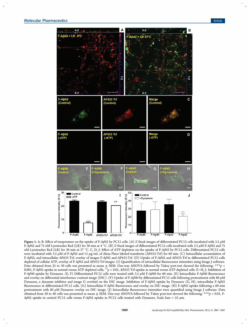
Figure 1. A, B: Effect of temperature on the uptake of F-Aβ42 by PC12 cells. (A) Z-Stack images of differentiated PC12 cells incubated with 3.5 μMF-Aβ42 and 75 nM Lysotracker Red (LR) for 30 min at 4 °C. (B) Z-Stack images of differentiated PC12 cells incubated with 3.5 μM F-Aβ42 and 75nM Lysotracker Red (LR) for 30 min at 37 °C. C, D, J: Effect of ATP depletion on the uptake of F-Aβ42 by PC12 cells. Differentiated PC12 cellswere incubated with 3.5 μM of F-Aβ42 and 15 μg/mL of Alexa Fluor labeled transferrin (AF633-Trf) for 60 min. (C) Intracellular accumulation ofF-Aβ42; and intracellular AF633-Trf, overlay of images F-Aβ42 and AF633-Trf. (D) Uptake of F-Aβ42 and AF633-Trf in differentiated PC12 cellsdepleted of cellular ATP, overlay of F-Aβ42 and AF633-Trf images. (I) Quantification of intracellular fluorescence intensities using Image J software.Data obtained from 25 to 30 cells was presented as mean ± SEM. One-way ANOVA followed by Tukey post-test showed the following: ***p <0.001, F-Aβ42 uptake in normal versus ATP depleted cells; ††p < 0.01, AF633 Trf uptake in normal versus ATP depleted cells. E−H, J: Inhibition ofF-Aβ40 uptake by Dynasore. (E, F) Differentiated PC12 cells were treated with 3.5 μM F-Aβ40 for 60 min. (E) Intracellular F-Aβ40 fluorescenceand overlay on differential interference contrast image (DIC). (F) Uptake of F-Aβ40 by differentiated PC12 cells following pretreatment with 80 μMDynasore, a dynamin inhibitor and image G overlaid on the DIC image. Inhibition of F-Aβ42 uptake by Dynasore (G, H): intracellular F-Aβ42fluorescence in differentiated PC12 cells. (G) Intracellular F-Aβ42 fluorescence and overlay on DIC image. (H) F-Aβ42 uptake following a 60 minpretreatment with 80 μM Dynasore overlay on DIC image. (J) Intracellular fluorescence intensities were quantified using Image J software. Dataobtained from 30 to 40 cells was presented as mean ± SEM. One-way ANOVA followed by Tukey post-test showed the following: ***p < 0.01, F-Aβ42 uptake in control PC12 cells versus F-Aβ42 uptake in PC12 cells treated with Dynasore. Scale bars = 25 μm.
Molecular Pharmaceutics Article
dx.doi.org/10.1021/mp200530q | Mol. Pharmaceutics 2012, 9, 1887−18971889
Then the cells were thoroughly washed, and the intracellular Aβwas quantified by flow cytometry or by Western blots.3. Effect of AF647-CT on the Uptake of F-Aβ40 and F-
Aβ42. PC12 cells grown on the delta T dishes werecoincubated with 3.5 μM F-Aβ40 or F-Aβ42 and 5 μg/mLAF647-CT for 60 min. The cells were washed and imaged usinglive cell microscopy. Similar treatments were performed onPC12 cells grown on 6-well culture plates, and cellularfluorescence was quantified by flow cytometry.Intracellular Itinerary of F-Aβ40 and F-Aβ42. 1. Accu-
mulation in Early Endosomes. Differentiated PC12 cellsgrown on delta T dishes were incubated with 3.5 μM F-Aβ40 orF-Aβ42 for 60 min. An early endosomal marker AF633-Trf (20μg/mL) was added 20 min prior to the termination of theexperiment. The cells were washed thoroughly and imaged bylive cell microscopy.2. Accumulation in Secondary Endosomes. The cells were
treated with 3.5 μM F-Aβ40 or F-Aβ42 and Dil-LDL (Dil-complexed low density lipoprotein) (15 μg/mL) for 30 or 60min. Then the cells were thoroughly washed and imaged livewith confocal microscopy.3. Lysosomal Accumulation. Differentiated PC12 cells that
stably express m-CFP/LAMP-1 (m-Cherry fluorescent proteinfused to lysosomal-associated membrane protein-1) wereincubated with either F-Aβ40 or F-Aβ42 for 60, 120, and 180min respectively. Then the cells were washed with DMEM andimaged by live cell microscopy. The fluorescein and m-CFPsignals in the images were superimposed, and the extent ofcolocalization was estimated by Pearson’s correlation coef-ficients. Approximately 50−100 cells were evaluated for eachtime point.
4. Influence of F-Aβ40 or F-Aβ42 Accumulation on theLysosomal Integrity. PC12 cells that stably express m-CFP/LAMP-1 were incubated with 3.5 μM F-Aβ40 or F-Aβ42 for 15,30, 60, 90, 120, and 180 min. The cells were washed withDMEM without phenol red and imaged using live cellmicroscopy. The areas of individual lysosomes in each cellwere measured using Image J software. At least 50 cells wereassayed for each time point. Change in lysosomal area wasplotted against time.
Live Cell Imaging and Quantification of DigitalImages. Each reported image is a composite of three imagesobtained at low, medium, and high exposure times, whichensure complete definition of the intracellular fluorescencesignal. Caution was exercised to set the maximum exposuretimes below the signal saturation limits. Replicates in each studywere run simultaneously and imaged at the same exposuresetting. For inhibition studies, cells treated with inhibitors wereimaged first with autoexposure settings and controls wereimaged later with the same settings.The cellular fluorescence in micrographs was quantified using
Image J software (National Institute of Mental Health,Bethesda, Maryland). A minimum of 25 cells was quantifiedfor each group in an experiment. From the gray imagesobtained directly from the microscope, individual cells wereselected using polygon selection tool, and parameters such asminimum and maximum intensities, area, integrated density,and mean gray value were obtained. The minimum andmaximum intensities of a typical image varied between 0 and255 RU. The mean gray values ranged between 20 and 75, andthe mean background values were between 5 and 15. Thebackground fluorescence was subtracted from the mean grayvalues obtained for various control and treatment groups in a
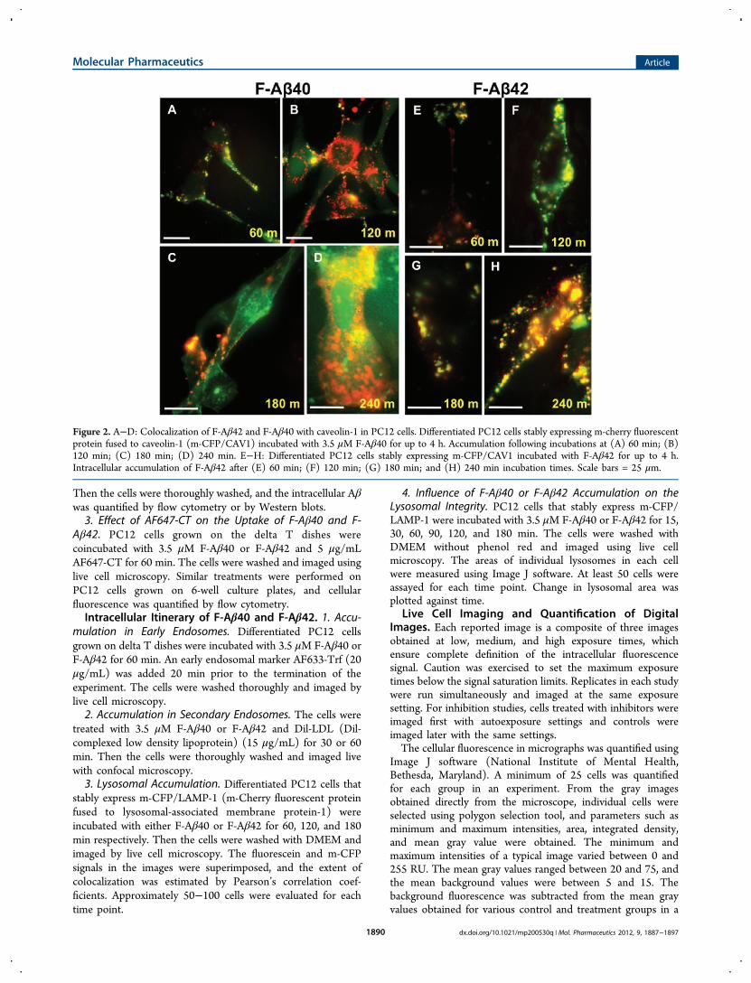
Figure 2. A−D: Colocalization of F-Aβ42 and F-Aβ40 with caveolin-1 in PC12 cells. Differentiated PC12 cells stably expressing m-cherry fluorescentprotein fused to caveolin-1 (m-CFP/CAV1) incubated with 3.5 μM F-Aβ40 for up to 4 h. Accumulation following incubations at (A) 60 min; (B)120 min; (C) 180 min; (D) 240 min. E−H: Differentiated PC12 cells stably expressing m-CFP/CAV1 incubated with F-Aβ42 for up to 4 h.Intracellular accumulation of F-Aβ42 after (E) 60 min; (F) 120 min; (G) 180 min; and (H) 240 min incubation times. Scale bars = 25 μm.
Molecular Pharmaceutics Article
dx.doi.org/10.1021/mp200530q | Mol. Pharmaceutics 2012, 9, 1887−18971890
study, and the differences among them were statisticallyanalyzed using Graphpad Prism software (La Jolla, CA).For the calculation of lysosomal areas, the threshold of gray
images was adjusted to reduce the background. The lysosomalareas were measured using the analyze particle tool. Thevesicles <300 (pixel)2 were considered as small vesicles and thevesicles >300 (pixel)2 were considered as large vesicles whichwere assumed to be formed from the aggregation of smallervesicles.Immunoprecipitation and Western Blot. Following the
uptake experiments the cell pellet was treated with 200 μL ofcell lysis buffer consisting of RIPA buffer (Sigma-Aldrich, St.Louis, MO) and protease inhibitor cocktail (Sigma-Aldrich, St.Louis, MO). Then the cell pellet was subjected to several quickfreeze thaw cycles followed by probe sonication (FisherScientific, Pittsburgh, PA). To a 50 μL aliquot of the celllysate was added 250 μg of IgG 4.1 antibody raised againsthuman fibrillar Aβ42, and this was incubated for 2 h.20 Thenthe IgG 4.1−Aβ complex was separated from the cell lysatewith 100 μL of immobilized protein A bead slurry (ThermoFisher Scientific Inc., Rockford IL). Aβ protein was separatedfrom the beads by boiling for 5 min at 95 °C in tris tricinesample buffer (Bio-Rad Hercules, CA) containing 2% v/v 6-mercaptoethanol. A 30 μL aliquot of this supernatant wasloaded onto 10−20% gradient tris tricine peptide precast gels(Bio-Rad, Hercules, CA). The separated protein bands weretransferred onto a 0.2 μm nitrocellulose membrane (Bio-Rad,
Hercules, CA). Subsequently, the membrane was blocked with5% nonfat milk and incubated with 6E10 monoclonal antibody(Coavance, Dedham, MA) overnight and further incubatedwith goat anti-mouse IgG secondary antibody (Santa CruzBiotechnology, Santa Cruz, CA). The membrane was brieflyincubated with SuperSignal West Pico ChemiluminescentSubstrate (Thermo Scientific Rockford, IL) and imaged.
■ RESULTS
Energy Dependent Internalization of F-Aβ42 inDifferentiated PC12 Cells. The Z-stack image of the PC12cells incubated with F-Aβ42 for 30 min at 37 °C showed clearintracellular accumulation (Figure 1B), which reduced sig-nificantly when the incubation temperature was changed to 4°C (Figure 1A). Upon incubation with F-Aβ42 and AF633-Trf(a clathrin-mediated endocytosis marker) at 37 °C, PC12 cellsaccumulated green (F-Aβ42) and red fluorescence (AF633-Trf)in the perinuclear region (Figure 1C). However, theaccumulation of either F-Aβ42 or AF633-Trf (Figure 1D)decreased in the PC12 cells depleted of cellular ATP. Theintracellular fluorescence intensities quantified using ImageJconfirmed these observations and demonstrated that thedifferences in the uptake of F-Aβ42 and AF633-Trf betweennormal and ATP depleted PC12 cells are statistically significant(Figure1I).
Dynasore Inhibits the Uptake of F-Aβ42 but Not F-Aβ40. Dynasore is a potent dynamin inhibitor and interferes
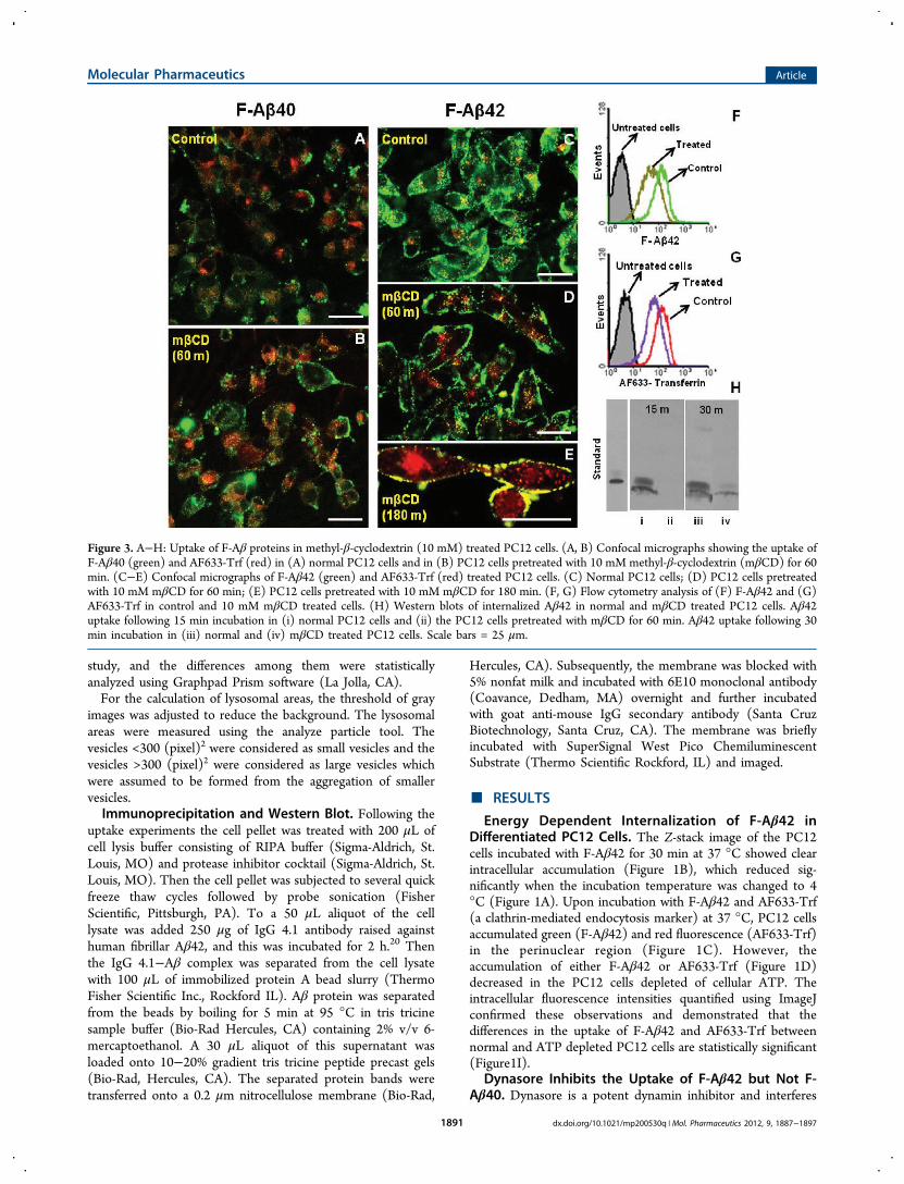
Figure 3. A−H: Uptake of F-Aβ proteins in methyl-β-cyclodextrin (10 mM) treated PC12 cells. (A, B) Confocal micrographs showing the uptake ofF-Aβ40 (green) and AF633-Trf (red) in (A) normal PC12 cells and in (B) PC12 cells pretreated with 10 mM methyl-β-cyclodextrin (mβCD) for 60min. (C−E) Confocal micrographs of F-Aβ42 (green) and AF633-Trf (red) treated PC12 cells. (C) Normal PC12 cells; (D) PC12 cells pretreatedwith 10 mM mβCD for 60 min; (E) PC12 cells pretreated with 10 mM mβCD for 180 min. (F, G) Flow cytometry analysis of (F) F-Aβ42 and (G)AF633-Trf in control and 10 mM mβCD treated cells. (H) Western blots of internalized Aβ42 in normal and mβCD treated PC12 cells. Aβ42uptake following 15 min incubation in (i) normal PC12 cells and (ii) the PC12 cells pretreated with mβCD for 60 min. Aβ42 uptake following 30min incubation in (iii) normal and (iv) mβCD treated PC12 cells. Scale bars = 25 μm.
Molecular Pharmaceutics Article
dx.doi.org/10.1021/mp200530q | Mol. Pharmaceutics 2012, 9, 1887−18971891
with the endocytotic processes that involve dynamin in clathin-coated pits. Uptake of F-Aβ40 by the PC12 cells was notaffected by Dynasore treatment (Figure 1E,F,J), further provingfact that the uptake is nonendocytotic as demonstrated in ourearlier study.9 However, F-Aβ42 uptake in the PC12 cellspretreated with 80 μM Dynasore decreased significantlycompared to that in the normal cells (Figure 1G,H,J).Role of Caveolae Mediated Endocytosis in the
Intracellular Uptake of F-Aβ Proteins. PC12 cells stablyexpressing m-CFP fused caveolin-1 (m-CFP/CAV1) wereemployed to examine the endosomal pathway involved in theinternalization of F-Aβ42. In these cells, F-Aβ42 (Figure 2E−H) but not F-Aβ40 (Figure 2A−D) colocalized with m-CFP/CAV1 at various incubation times ranging between 60 and 240min.In PC12 cells treated with MβCD (depletes membrane
cholesterol required for maintaining the integrity of lipid raftsand also for caveolae formation) for 60 (Figure 3D) or 180 min(Figure 3E), F-Aβ42 remained mostly at the cell periphery butthe untreated cells showed punctate deposition of F-Aβ42 inthe perinuclear region (Figure 3C). Flow cytometry analysis
showed lower accumulation of F-Aβ42 (Figure 3F) as well asAF633-Trf (Figure 3G) in MβCD treated PC12 cells than inthe normal cells. Moreover, the Western blots of theimmunoprecipitated lysates obtained from the PC12 cellsincubated with F-Aβ42 for 15 or 30 min following a 1 hpretreatment with MβCD showed substantially lower F-Aβ42uptake than the normal cells (Figure 3H).
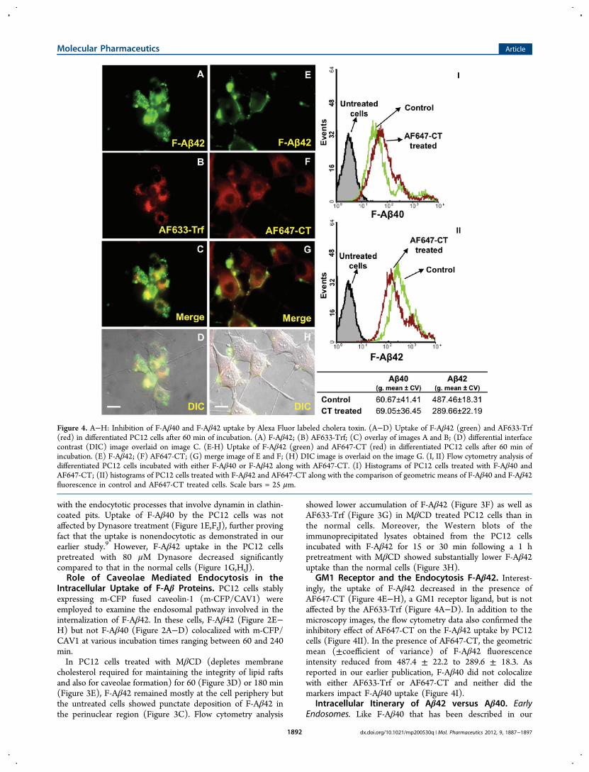
GM1 Receptor and the Endocytosis F-Aβ42. Interest-ingly, the uptake of F-Aβ42 decreased in the presence ofAF647-CT (Figure 4E−H), a GM1 receptor ligand, but is notaffected by the AF633-Trf (Figure 4A−D). In addition to themicroscopy images, the flow cytometry data also confirmed theinhibitory effect of AF647-CT on the F-Aβ42 uptake by PC12cells (Figure 4II). In the presence of AF647-CT, the geometricmean (±coefficient of variance) of F-Aβ42 fluorescenceintensity reduced from 487.4 ± 22.2 to 289.6 ± 18.3. Asreported in our earlier publication, F-Aβ40 did not colocalizewith either AF633-Trf or AF647-CT and neither did themarkers impact F-Aβ40 uptake (Figure 4I).
Intracellular Itinerary of Aβ42 versus Aβ40. EarlyEndosomes. Like F-Aβ40 that has been described in our
Figure 4. A−H: Inhibition of F-Aβ40 and F-Aβ42 uptake by Alexa Fluor labeled cholera toxin. (A−D) Uptake of F-Aβ42 (green) and AF633-Trf(red) in differentiated PC12 cells after 60 min of incubation. (A) F-Aβ42; (B) AF633-Trf; (C) overlay of images A and B; (D) differential interfacecontrast (DIC) image overlaid on image C. (E-H) Uptake of F-Aβ42 (green) and AF647-CT (red) in differentiated PC12 cells after 60 min ofincubation. (E) F-Aβ42; (F) AF647-CT; (G) merge image of E and F; (H) DIC image is overlaid on the image G. (I, II) Flow cytometry analysis ofdifferentiated PC12 cells incubated with either F-Aβ40 or F-Aβ42 along with AF647-CT. (I) Histograms of PC12 cells treated with F-Aβ40 andAF647-CT; (II) histograms of PC12 cells treated with F-Aβ42 and AF647-CT along with the comparison of geometric means of F-Aβ40 and F-Aβ42fluorescence in control and AF647-CT treated cells. Scale bars = 25 μm.
Molecular Pharmaceutics Article
dx.doi.org/10.1021/mp200530q | Mol. Pharmaceutics 2012, 9, 1887−18971892

earlier studies,9 F-Aβ42 did not show any appreciablelocalization in the early endosomes labeled with AF633-Trf(Figure 3C).Late Endosomes. When coincubated with Dil-LDL, a
marker for late endosomes, F-Aβ42 showed remarkablecolocalization as evidenced by the punctate yellow stain onthe X−Z and Y−Z images of the PC12 cells incubated with thefluorophores for 60 min (Figure 5A). F-Aβ42 exhibited asimilar trend following 30 min incubation with the Dil-LDL(Figure 5B), while F-Aβ40 did not show colocalization with themarker under similar incubation conditions (Figure 5C).Lysosomes. Differentiated PC12 cells stably expressing m-
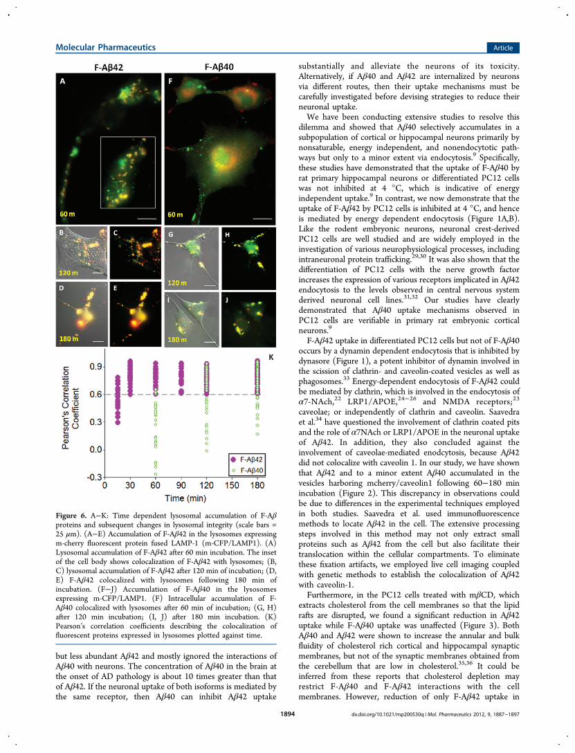
CFP/LAMP1 were incubated with F-Aβ42 or F-Aβ40 toexamine their lysosomal accumulation patterns for up to 180min. F-Aβ42 showed distinct lysosomal accumulation (Figure6A−E) whereas F-Aβ40 showed only a partial localization inthe lysosomes (Figure 6F−J). Pearson's correlation coefficientsof F-Aβ42 colocalization with m-CFP/LAMP1 were greaterthan 0.6 even at incubation times as short as 30 min (Figure6K). Pearson’s correlation coefficient 1 is considered as 100%colocalization. In contrast, F-Aβ40 was unable to colocalizewith lysosomes as much as F-Aβ42 did during the initial timepoints. The Pearson’s correlation coefficients describing thecolocalization of F-Aβ40 with lysosomal m-CFP/LAMP1 variedbetween 0.3 and 0.6, but increased above 0.6 only at later timepoints (Figure 6K).The damage caused to lysosomal integrity due to the
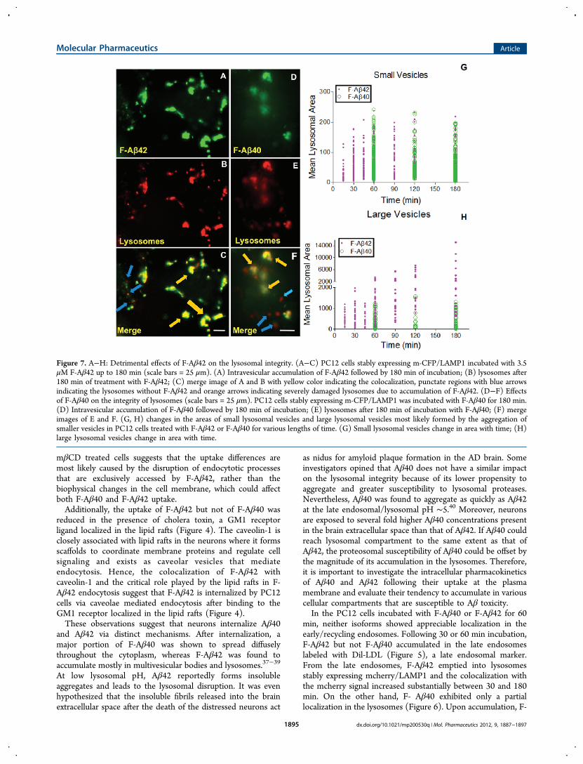
accumulation of F-Aβ proteins was assessed by the following:tracking disrupted lysososmes that show up as red blotches duethe leakage of lysosomal contents into cytoplasm; monitoringchanges in the mean lysosomal areas; and evaluating vesicleslarger than 300 (pixel)2 that are indicative of clumped
lysosomes. More red blotches were found in the cells incubatedwith F-Aβ42 (Figure 7A−C) than those incubated with F-Aβ40(Figure 7D−F). The areas of smaller lysosomal vesicles in F-Aβ42 or F-Aβ40 treated PC12 cells showed a gradual increaseup to 60 min and reached a plateau thereafter (Figure 7G). Butthe appearance of clumped lysosomes increased linearly withtime in F-Aβ42 treated cells but not in the cells treated with F-Aβ40 (Figure 7H).
■ DISCUSSIONWhile parenchymal amyloid plaques and intraneuronal tangles,the most visible pathological hall marks of AD, are consideredas the mere downstream reflections of AD pathology,21 theneurodegeneration in AD is believed to be actually triggered bythe intraneuronal accumulation of Aβ proteins. UnderstandingAβ accumulation in the neurons is important so that treatmentsto prevent neurodegeneration could be identified. Endocytoticpathways involving α7 nicotinic acetylcholine (α7-NAch)receptor,22 N-methyl D-aspartate (NMDA) receptor,23 lowdensity lipoprotein receptor-related protein 1 (LRP1)/apolipoprotein E (APOE),24−26 or receptor for advancedglycosylated end products (RAGE) were implicated in theneuronal uptake of soluble Aβ. In addition, nonsaturable andnonendocytotic uptake of both Aβ40 and Aβ42 in PC12 cells8
and in human neuroblastoma cells27 was also proposed. Such aconfusing array of targets renders the discovery of therapeuticstrategies to prevent intraneuronal Aβ accumulation untenable.The reduction in the Aβ40:Aβ42 ratio in the AD brain, which
correlates with the severity of neurodegeneration, makes itimperative to understand the relative interactions of eachisoform with neurons.28 Many of the aforementioned studieswere focused on the neuronal uptake of more amyloidogenic
Figure 5. A−C: Confocal micrographs depicting the extent of colocalization of F-Aβ proteins with Dil-LDL in differentiated PC12 cells. (A) Z-Seriesimage of F-Aβ42 colocalization with Dil-LDL, a late endosome marker, following 60 min incubation. (B) F-Aβ42 and Dil-LDL colocalizationfollowing 60 min incubation. (C) Absence of F-Aβ40 and Dil-LDL colocalization after 60 min incubation. Scale bars = 25 μm.
Molecular Pharmaceutics Article
dx.doi.org/10.1021/mp200530q | Mol. Pharmaceutics 2012, 9, 1887−18971893
but less abundant Aβ42 and mostly ignored the interactions ofAβ40 with neurons. The concentration of Aβ40 in the brain atthe onset of AD pathology is about 10 times greater than thatof Aβ42. If the neuronal uptake of both isoforms is mediated bythe same receptor, then Aβ40 can inhibit Aβ42 uptake
substantially and alleviate the neurons of its toxicity.Alternatively, if Aβ40 and Aβ42 are internalized by neuronsvia different routes, then their uptake mechanisms must becarefully investigated before devising strategies to reduce theirneuronal uptake.We have been conducting extensive studies to resolve this
dilemma and showed that Aβ40 selectively accumulates in asubpopulation of cortical or hippocampal neurons primarily bynonsaturable, energy independent, and nonendocytotic path-ways but only to a minor extent via endocytosis.9 Specifically,these studies have demonstrated that the uptake of F-Aβ40 byrat primary hippocampal neurons or differentiated PC12 cellswas not inhibited at 4 °C, which is indicative of energyindependent uptake.9 In contrast, we now demonstrate that theuptake of F-Aβ42 by PC12 cells is inhibited at 4 °C, and henceis mediated by energy dependent endocytosis (Figure 1A,B).Like the rodent embryonic neurons, neuronal crest-derivedPC12 cells are well studied and are widely employed in theinvestigation of various neurophysiological processes, includingintraneuronal protein trafficking.29,30 It was also shown that thedifferentiation of PC12 cells with the nerve growth factorincreases the expression of various receptors implicated in Aβ42endocytosis to the levels observed in central nervous systemderived neuronal cell lines.31,32 Our studies have clearlydemonstrated that Aβ40 uptake mechanisms observed inPC12 cells are verifiable in primary rat embryonic corticalneurons.9
F-Aβ42 uptake in differentiated PC12 cells but not of F-Aβ40occurs by a dynamin dependent endocytosis that is inhibited bydynasore (Figure 1), a potent inhibitor of dynamin involved inthe scission of clathrin- and caveolin-coated vesicles as well asphagosomes.33 Energy-dependent endocytosis of F-Aβ42 couldbe mediated by clathrin, which is involved in the endocytosis ofα7-NAch,22 LRP1/APOE,24−26 and NMDA receptors;23
caveolae; or independently of clathrin and caveolin. Saavedraet al.34 have questioned the involvement of clathrin coated pitsand the role of α7NAch or LRP1/APOE in the neuronal uptakeof Aβ42. In addition, they also concluded against theinvolvement of caveolae-mediated enodcytosis, because Aβ42did not colocalize with caveolin 1. In our study, we have shownthat Aβ42 and to a minor extent Aβ40 accumulated in thevesicles harboring mcherry/caveolin1 following 60−180 minincubation (Figure 2). This discrepancy in observations couldbe due to differences in the experimental techniques employedin both studies. Saavedra et al. used immunofluorescencemethods to locate Aβ42 in the cell. The extensive processingsteps involved in this method may not only extract smallproteins such as Aβ42 from the cell but also facilitate theirtranslocation within the cellular compartments. To eliminatethese fixation artifacts, we employed live cell imaging coupledwith genetic methods to establish the colocalization of Aβ42with caveolin-1.Furthermore, in the PC12 cells treated with mβCD, which
extracts cholesterol from the cell membranes so that the lipidrafts are disrupted, we found a significant reduction in Aβ42uptake while F-Aβ40 uptake was unaffected (Figure 3). BothAβ40 and Aβ42 were shown to increase the annular and bulkfluidity of cholesterol rich cortical and hippocampal synapticmembranes, but not of the synaptic membranes obtained fromthe cerebellum that are low in cholesterol.35,36 It could beinferred from these reports that cholesterol depletion mayrestrict F-Aβ40 and F-Aβ42 interactions with the cellmembranes. However, reduction of only F-Aβ42 uptake in
Figure 6. A−K: Time dependent lysosomal accumulation of F-Aβproteins and subsequent changes in lysosomal integrity (scale bars =25 μm). (A−E) Accumulation of F-Aβ42 in the lysosomes expressingm-cherry fluorescent protein fused LAMP-1 (m-CFP/LAMP1). (A)Lysosomal accumulation of F-Aβ42 after 60 min incubation. The insetof the cell body shows colocalization of F-Aβ42 with lysosomes; (B,C) lysosomal accumulation of F-Aβ42 after 120 min of incubation; (D,E) F-Aβ42 colocalized with lysosomes following 180 min ofincubation. (F−J) Accumulation of F-Aβ40 in the lysosomesexpressing m-CFP/LAMP1. (F) Intracellular accumulation of F-Aβ40 colocalized with lysosomes after 60 min of incubation; (G, H)after 120 min incubation; (I, J) after 180 min incubation. (K)Pearson’s correlation coefficients describing the colocalization offluorescent proteins expressed in lysosomes plotted against time.
Molecular Pharmaceutics Article
dx.doi.org/10.1021/mp200530q | Mol. Pharmaceutics 2012, 9, 1887−18971894
mβCD treated cells suggests that the uptake differences aremost likely caused by the disruption of endocytotic processesthat are exclusively accessed by F-Aβ42, rather than thebiophysical changes in the cell membrane, which could affectboth F-Aβ40 and F-Aβ42 uptake.Additionally, the uptake of F-Aβ42 but not of F-Aβ40 was
reduced in the presence of cholera toxin, a GM1 receptorligand localized in the lipid rafts (Figure 4). The caveolin-1 isclosely associated with lipid rafts in the neurons where it formsscaffolds to coordinate membrane proteins and regulate cellsignaling and exists as caveolar vesicles that mediateendocytosis. Hence, the colocalization of F-Aβ42 withcaveolin-1 and the critical role played by the lipid rafts in F-Aβ42 endocytosis suggest that F-Aβ42 is internalized by PC12cells via caveolae mediated endocytosis after binding to theGM1 receptor localized in the lipid rafts (Figure 4).These observations suggest that neurons internalize Aβ40
and Aβ42 via distinct mechanisms. After internalization, amajor portion of F-Aβ40 was shown to spread diffuselythroughout the cytoplasm, whereas F-Aβ42 was found toaccumulate mostly in multivesicular bodies and lysosomes.37−39
At low lysosomal pH, Aβ42 reportedly forms insolubleaggregates and leads to the lysosomal disruption. It was evenhypothesized that the insoluble fibrils released into the brainextracellular space after the death of the distressed neurons act
as nidus for amyloid plaque formation in the AD brain. Someinvestigators opined that Aβ40 does not have a similar impacton the lysosomal integrity because of its lower propensity toaggregate and greater susceptibility to lysosomal proteases.Nevertheless, Aβ40 was found to aggregate as quickly as Aβ42at the late endosomal/lysosomal pH ∼5.40 Moreover, neuronsare exposed to several fold higher Aβ40 concentrations presentin the brain extracellular space than that of Aβ42. If Aβ40 couldreach lysosomal compartment to the same extent as that ofAβ42, the proteosomal susceptibility of Aβ40 could be offset bythe magnitude of its accumulation in the lysosomes. Therefore,it is important to investigate the intracellular pharmacokineticsof Aβ40 and Aβ42 following their uptake at the plasmamembrane and evaluate their tendency to accumulate in variouscellular compartments that are susceptible to Aβ toxicity.In the PC12 cells incubated with F-Aβ40 or F-Aβ42 for 60
min, neither isoforms showed appreciable localization in theearly/recycling endosomes. Following 30 or 60 min incubation,F-Aβ42 but not F-Aβ40 accumulated in the late endosomeslabeled with Dil-LDL (Figure 5), a late endosomal marker.From the late endosomes, F-Aβ42 emptied into lysosomesstably expressing mcherry/LAMP1 and the colocalization withthe mcherry signal increased substantially between 30 and 180min. On the other hand, F- Aβ40 exhibited only a partiallocalization in the lysosomes (Figure 6). Upon accumulation, F-
Figure 7. A−H: Detrimental effects of F-Aβ42 on the lysosomal integrity. (A−C) PC12 cells stably expressing m-CFP/LAMP1 incubated with 3.5μM F-Aβ42 up to 180 min (scale bars = 25 μm). (A) Intravesicular accumulation of F-Aβ42 followed by 180 min of incubation; (B) lysosomes after180 min of treatment with F-Aβ42; (C) merge image of A and B with yellow color indicating the colocalization, punctate regions with blue arrowsindicating the lysosomes without F-Aβ42 and orange arrows indicating severely damaged lysosomes due to accumulation of F-Aβ42. (D−F) Effectsof F-Aβ40 on the integrity of lysosomes (scale bars = 25 μm). PC12 cells stably expressing m-CFP/LAMP1 was incubated with F-Aβ40 for 180 min.(D) Intravesicular accumulation of F-Aβ40 followed by 180 min of incubation; (E) lysosomes after 180 min of incubation with F-Aβ40; (F) mergeimages of E and F. (G, H) changes in the areas of small lysosomal vesicles and large lysosomal vesicles most likely formed by the aggregation ofsmaller vesicles in PC12 cells treated with F-Aβ42 or F-Aβ40 for various lengths of time. (G) Small lysosomal vesicles change in area with time; (H)large lysosomal vesicles change in area with time.
Molecular Pharmaceutics Article
dx.doi.org/10.1021/mp200530q | Mol. Pharmaceutics 2012, 9, 1887−18971895
Aβ42 caused more damage to lysosomes than F-Aβ40.However, it is hard to ignore the correlation between theextent of lysosomal damage and the magnitude of F-Aβaccumulation in the lysosomes (Figure 7). In F-Aβ42 or F-Aβ40 treated PC12 cells, only those lysosomes that harboredthe proteins were disrupted and lysosomes devoid of any greenfluorescence that signifies the absence of F-Aβ accumulationremained intact. Notably, intact lysosomes are more prevalentin F-Aβ40 treated cells than in F-Aβ42 treated cells, which ismost likely due to the inability of F-Aβ40 to accumulate in thelysosomes as much as F-Aβ42.In summary, this study outlines differences between the
mechanisms of uptake and intracellular itinerary of Aβ42 andAβ40 following their acute exposure in differentiated PC12cells, whereas the cortical and hippocampal neurons in ADpatients are subjected to chronic exposure of Aβ proteins atonly 0.1−2% of the concentrations used in this study.41 Evenunder the chronic exposure, the endosomal−lysosmal system inthe neurons is disrupted,42 thereby suggesting that the primarymechanisms through which Aβ proteins are internalized by theneurons and exhibit their toxicity are most likely to be the samewith acute or chronic exposures. Hence, the observations madein the current in vitro study could be extrapolated tounderstand pathophysiological processes driving neurodegen-eration in AD patients and transgenic animals in vivo.By maintaining a mechanism of uptake and cellular
pharmacokinetics different from Aβ42, the question arises asto whether Aβ40 plays a protective role in counteracting thetoxic effects of Aβ42 on the neurons. Or is Aβ40 just a less toxicversion of Aβ42? If the former is true, an ideal drug intended totreat neurodegeneration in AD is expected to specifically targetneuronal uptake of Aβ42 without interfering with Aβ40. If thelatter scenario were to be true, then the potential drug would beexpected to inhibit the neuronal uptake of both Aβ42 andAβ40. More studies are being conducted in our lab to clarifythese questions.
■ AUTHOR INFORMATION
Corresponding Author*Department of Pharmaceutics, College of Pharmacy, 308Harvard Street SE, 9-149A Weaver-Densford Hall, University ofMinnesota, Minneapolis, MN. E-mail: [email protected].
NotesThe authors declare no competing financial interest.
■ ACKNOWLEDGMENTS
The authors acknowledge the financial assistance provided byAlzheimer’s Association Grant NIRG-09-133017 (K.K.K.) andNIH/NCRR/RCMI Grant G12RR03020 (K.K.K.). Thesponsors had no role in study design; data collection, analysis,and interpretation; and played no role in the decision to submitthis paper for publication.
■ REFERENCES(1) Christensen, D.; Kraus, S.; Flohr, A.; Cotel, M.-C.; Wirths, O.;Bayer, T. Transient intraneuronal Aβ rather than extracellular plaquepathology correlates with neuron loss in the frontal cortex of APP/PS1KI mice. Acta Neuropathol. 2008, 116 (6), 647−655.(2) LaFerla, F. M.; Green, K. N.; Oddo, S. Intracellular amyloid-[beta] in Alzheimer’s disease. Nat. Rev. Neurosci. 2007, 8 (7), 499−509.
(3) Wirths, O.; Multhaup, G.; Bayer, T. A. A modified β-amyloidhypothesis: intraneuronal accumulation of the β-amyloid peptidethefirst step of a fatal cascade. J. Neurochem. 2004, 91 (3), 513−520.(4) Gouras, G. K.; Tsai, J.; Naslund, J.; Vincent, B.; Edgar, M.;Checler, F.; Greenfield, J. P.; Haroutunian, V.; Buxbaum, J. D.; Xu, H.;Greengard, P.; Relkin, N. R. Intraneuronal Abeta42 accumulation inhuman brain. Am. J. Pathol. 2000, 156 (1), 15−20.(5) Oddo, S.; Billings, L.; Kesslak, J. P.; Cribbs, D. H.; LaFerla, F. M.A[beta] Immunotherapy Leads to Clearance of Early, but Not Late,Hyperphosphorylated Tau Aggregates via the Proteasome. Neuron2004, 43 (3), 321−332.(6) Jicha, G. A. Is passive immunization for Alzheimer’s disease 'aliveand well' or 'dead and buried'? Expert Opin. Biol. Ther. 2009, 9 (4),481−491.(7) Poduslo, J. F.; Gilles, E. J.; Ramakrishnan, M.; Howell, K. G.;Wengenack, T. M.; Curran, G. L.; Kandimalla, K. K. HH domain ofAlzheimer’s disease Abeta provides structural basis for neuronalbinding in PC12 and mouse cortical/hippocampal neurons. PLoS One2010, 5 (1), e8813.(8) Burdick, D.; Kosmoski, J.; Knauer, M. F.; Glabe, C. G.Preferential adsorption, internalization and resistance to degradationof the major isoform of the Alzheimer’s amyloid peptide, A beta 1−42,in differentiated PC12 cells. Brain Res. 1997, 746 (1−2), 275−284.(9) Kandimalla, K. K.; Scott, O. G.; Fulzele, S.; Davidson, M. W.;Poduslo, J. F. Mechanism of neuronal versus endothelial cell uptake ofAlzheimer’s disease amyloid beta protein. PLoS One 2009, 4 (2),e4627.(10) Yan, Y.; Wang, C. A[beta]40 Protects Non-toxic A[beta]42Monomer from Aggregation. J. Mol. Biol. 2007, 369 (4), 909−916.(11) Kim, J.; Onstead, L.; Randle, S.; Price, R.; Smithson, L.;Zwizinski, C.; Dickson, D. W.; Golde, T.; McGowan, E. A{beta}40Inhibits Amyloid Deposition In Vivo. J. Neurosci. 2007, 27 (3), 627−633.(12) Kumar-Singh, S.; Theuns, J.; Van Broeck, B.; Pirici, D.;Vennekens, K. l.; Corsmit, E.; Cruts, M.; Dermaut, B.; Wang, R.; VanBroeckhoven, C. Mean age-of-onset of familial alzheimer diseasecaused by presenilin mutations correlates with both increased Aβ42and decreased Aβ40. Hum. Mutat. 2006, 27 (7), 686−695.(13) McGowan, E.; Pickford, F.; Kim, J.; Onstead, L.; Eriksen, J.; Yu,C.; Skipper, L.; Murphy, M. P.; Beard, J.; Das, P.; Jansen, K.; DeLucia,M.; Lin, W.-L.; Dolios, G.; Wang, R.; Eckman, C. B.; Dickson, D. W.;Hutton, M.; Hardy, J.; Golde, T. A[beta]42 Is Essential forParenchymal and Vascular Amyloid Deposition in Mice. Neuron2005, 47 (2), 191−199.(14) Zou, K.; Kim, D.; Kakio, A.; Byun, K.; Gong, J.-S.; Kim, J.; Kim,M.; Sawamura, N.; Nishimoto, S.-i.; Matsuzaki, K.; Lee, B.;Yanagisawa, K.; Michikawa, M. Amyloid β-protein (Aβ)1−40 protectsneurons from damage induced by Aβ1−42 in culture and in rat brain.J. Neurochem. 2003, 87 (3), 609−619.(15) Shoji, M. Cerebrospinal fluid Abeta40 and Abeta42: naturalcourse and clinical usefulness. Front. Biosci. 2002, 7, d997−1006.(16) Hansson, O.; Zetterberg, H.; Buchhave, P.; Andreasson, U.;Londos, E.; Minthon, L.; Blennow, K. Prediction of Alzheimer’sDisease Using the CSF Aβ42/Aβ40 Ratio in Patients with MildCognitive Impairment. Dementia Geriatr. Cognit. Disord. 2007, 23 (5),316−320.(17) Herzig, M. C.; Winkler, D. T.; Burgermeister, P.; Pfeifer, M.;Kohler, E.; Schmidt, S. D.; Danner, S.; Abramowski, D.; Sturchler-Pierrat, C.; Burki, K.; van Duinen, S. G.; Maat-Schieman, M. L. C.;Staufenbiel, M.; Mathews, P. M.; Jucker, M. A[beta] is targeted to thevasculature in a mouse model of hereditary cerebral hemorrhage withamyloidosis. Nat. Neurosci. 2004, 7 (9), 954−960.(18) Fryer, J. D.; Simmons, K.; Parsadanian, M.; Bales, K. R.; Paul, S.M.; Sullivan, P. M.; Holtzman, D. M. Human Apolipoprotein E4 Altersthe Amyloid-β 40:42 Ratio and Promotes the Formation of CerebralAmyloid Angiopathy in an Amyloid Precursor Protein TransgenicModel. J. Neurosci. 2005, 25 (11), 2803−2810.
Molecular Pharmaceutics Article
dx.doi.org/10.1021/mp200530q | Mol. Pharmaceutics 2012, 9, 1887−18971896
(19) Klein, W. L. A[beta] toxicity in Alzheimer’s disease: globularoligomers (ADDLs) as new vaccine and drug targets. Neurochem. Int.2002, 41 (5), 345−352.(20) Poduslo, J. F.; Ramakrishnan, M.; Holasek, S. S.; Ramirez-Alvarado, M.; Kandimalla, K. K.; Gilles, E. J.; Curran, G. L.;Wengenack, T. M. In vivo targeting of antibody fragments to thenervous system for Alzheimer’s disease immunotherapy and molecularimaging of amyloid plaques. J. Neurochem. 2007, 102 (2), 420−33.(21) Fein, J. A.; Sokolow, S.; Miller, C. A.; Vinters, H. V.; Yang, F.;Cole, G. M.; Gylys, K. H. Co-localization of amyloid beta and taupathology in Alzheimer’s disease synaptosomes. Am. J. Pathol. 2008,172 (6), 1683−1692.(22) Nagele, R. G.; D’Andrea, M. R.; Anderson, W. J.; Wang, H. Y.Intracellular accumulation of beta-amyloid(1−42) in neurons isfacilitated by the alpha 7 nicotinic acetylcholine receptor inAlzheimer’s disease. Neuroscience 2002, 110 (2), 199−211.(23) Jellinger, K. A. Alzheimer disease and cerebrovascular pathology:an update. J. Neural Transm. 2002, 109 (5), 813−836.(24) Fuentealba, R. A.; Liu, Q.; Zhang, J.; Kanekiyo, T.; Hu, X.; Lee,J. M.; LaDu, M. J.; Bu, G. Low-density lipoprotein receptor-relatedprotein 1 (LRP1) mediates neuronal Abeta42 uptake and lysosomaltrafficking. PLoS One 2010, 5 (7), e11884.(25) LaFerla, F. M.; Troncoso, J. C.; Strickland, D. K.; Kawas, C. H.;Jay, G. Neuronal cell death in Alzheimer’s disease correlates with apoEuptake and intracellular Abeta stabilization. J Clin Invest 1997, 100 (2),310−320.(26) Zerbinatti, C. V.; Wahrle, S. E.; Kim, H.; Cam, J. A.; Bales, K.;Paul, S. M.; Holtzman, D. M.; Bu, G. Apolipoprotein E and lowdensity lipoprotein receptor-related protein facilitate intraneuronalAbeta42 accumulation in amyloid model mice. J. Biol. Chem. 2006, 281(47), 36180−36186.(27) Morelli, L.; Prat, M. I.; Castano, E. M. Differential accumulationof soluble amyloid beta peptides 1−40 and 1−42 in human monocyticand neuroblastoma cell lines. Implications for cerebral amyloido-genesis. Cell Tissue Res. 1999, 298 (2), 225−232.(28) Kuperstein, I.; Broersen, K.; Benilova, I.; Rozenski, J.;Jonckheere, W.; Debulpaep, M.; Vandersteen, A.; Segers-Nolten, I.;Van Der Werf, K.; Subramaniam, V.; Braeken, D.; Callewaert, G.;Bartic, C.; D’Hooge, R.; Martins, I. C.; Rousseau, F.; Schymkowitz, J.;De Strooper, B. Neurotoxicity of Alzheimer’s disease A[beta] peptidesis induced by small changes in the A[beta]42 to A[beta]40 ratio.EMBO J. 2010, 29 (19), 3408−3420.(29) Xu, H.; Sweeney, D.; Greengard, P.; Gandy, S. Metabolism ofAlzheimer beta-amyloid precursor protein: regulation by proteinkinase A in intact cells and in a cell-free system. Proc. Natl. Acad. Sci.U.S.A. 1996, 93 (9), 4081−4084.(30) Fukuda, M.; Yamamoto, A. Effect of forskolin on synaptotagminIV protein trafficking in PC12 cells. J. Biochem. 2004, 136 (2), 245−253.(31) Nery, A. A.; Resende, R. R.; Martins, A. H.; Trujillo, C. A.;Eterovic, V. A.; Ulrich, H. Alpha 7 nicotinic acetylcholine receptorexpression and activity during neuronal differentiation of PC12pheochromocytoma cells. J. Mol. Neurosci. 2010, 41 (3), 329−339.(32) Bu, G.; Sun, Y.; Schwartz, A. L.; Holtzman, D. M. Nerve GrowthFactor Induces Rapid Increases in Functional Cell Surface LowDensity Lipoprotein Receptor-related Protein. J. Biol. Chem. 1998, 273(21), 13359−13365.(33) Abban, C. Y.; Bradbury, N. A.; Meneses, P. I. HPV16 and BPV1infection can be blocked by the dynamin inhibitor dynasore. Am. J.Ther. 2008, 15 (4), 304−311.(34) Saavedra, L.; Mohamed, A.; Ma, V.; Kar, S.; de Chaves, E. P.Internalization of beta-amyloid peptide by primary neurons in theabsence of apolipoprotein E. J. Biol. Chem. 2007, 282 (49), 35722−35732.(35) Chochina, S. V.; Avdulov, N. A.; Igbavboa, U.; Cleary, J. P.;O’Hare, E. O.; Wood, W. G. Amyloid β-peptide1−40 increasesneuronal membrane fluidity: role of cholesterol and brain region. J.Lipid Res. 2001, 42 (8), 1292−1297.
(36) Avdulov, N. A.; Chochina, S. V.; Igbavboa, U.; O’Hare, E. O.;Schroeder, F.; Cleary, J. P.; Wood, W. G. Amyloid β-Peptides IncreaseAnnular and Bulk Fluidity and Induce Lipid Peroxidation in BrainSynaptic Plasma Membranes. J. Neurochem. 1997, 68 (5), 2086−2091.(37) Shirwany, N. A.; Payette, D.; Xie, J.; Guo, Q. The amyloid betaion channel hypothesis of Alzheimer’s disease. Neuropsychiatr. Dis.Treat. 2007, 3 (5), 597−612.(38) Takuma, K.; Fang, F.; Zhang, W.; Yan, S.; Fukuzaki, E.; Du, H.;Sosunov, A.; McKhann, G.; Funatsu, Y.; Nakamichi, N.; Nagai, T.;Mizoguchi, H.; Ibi, D.; Hori, O.; Ogawa, S.; Stern, D. M.; Yamada, K.;Yan, S. S. RAGE-mediated signaling contributes to intraneuronaltransport of amyloid-beta and neuronal dysfunction. Proc. Natl. Acad.Sci. U.S.A. 2009, 106 (47), 20021−20026.(39) Almeida, C. G.; Takahashi, R. H.; Gouras, G. K. beta-AmyloidAccumulation Impairs Multivesicular Body Sorting by Inhibiting theUbiquitin-Proteasome System. J. Neurosci. 2006, 26 (16), 4277−4288.(40) Overly, C. C.; Lee, K. D.; Berthiaume, E.; Hollenbeck, P. J.Quantitative measurement of intraorganelle pH in the endosomal-lysosomal pathway in neurons by using ratiometric imaging withpyranine. Proc. Natl. Acad. Sci. U.S.A. 1995, 92 (8), 3156−3160.(41) Kim, J.; Onstead, L.; Randle, S.; Price, R.; Smithson, L.;Zwizinski, C.; Dickson, D. W.; Golde, T.; McGowan, E. Aβ40 InhibitsAmyloid Deposition In Vivo. J. Neurosci. 2007, 27 (3), 627−633.(42) Belinson, H.; Lev, D.; Masliah, E.; Michaelson, D. M. Activationof the amyloid cascade in apolipoprotein E4 transgenic mice induceslysosomal activation and neurodegeneration resulting in markedcognitive deficits. J. Neurosci. 2008, 28 (18), 4690−4701.
Molecular Pharmaceutics Article
dx.doi.org/10.1021/mp200530q | Mol. Pharmaceutics 2012, 9, 1887−18971897
Related Documents