Atherosclerosis 180 (2005) 271–276 Dietary salt restriction accelerates atherosclerosis in apolipoprotein E-deficient mice Ognen Ivanovski a,∗ , Dorota Szumilak a , Thao Nguyen-Khoa a,b , Michele Dechaux c , Ziad A. Massy a,d , Olivier Phan a , Nadya Mothu a , Bernard Lacour b , Tilman B. Drueke a , Martin Muntzel e a INSERM Unit 507, Necker Hospital, 161 Rue de S` evres, 75015 Paris, France b Laboratory of Biochemistry A, Necker Hospital, Paris, France c Department of Physiology, Necker Hospital, Paris, France d Divisions of Clinical Pharmacology and Nephrology, Faculty of Pharmacy, University of Picardie and Centre Hospitalier Universitaire Amiens, Amiens, France e Department of Biological Sciences, Lehman College, Bronx, NY, USA Received 30 August 2004; received in revised form 30 November 2004; accepted 9 December 2004 Available online 19 January 2005 Abstract Background: Whether a general reduction in salt intake reduces or actually enhances cardiovascular mortality in man remains an issue of controversy. Low sodium diets may lead to adverse side effects by stimulating the renin–angiotensin and sympathetic nervous systems. The present study was designed to investigate the effects of low dietary salt on atherosclerotic lesion progression in apolipoprotein E deficient (apoE −/− ) mice. Methods and results: We fed 7-week-old apoE −/− mice on low (0.036% NaCl; n = 28) or regular (0.64% NaCl; n = 26) salt diets for 16 weeks. At the age of 23 weeks, the cross-section surface area of atherosclerotic plaques was measured in aortic root and thoracic aorta. Serum total cholesterol, triglycerides, plasma angiotensin levels and urinary protein/creatinine concentrations were assessed. Exposure to low salt caused significant increases in atherosclerotic lesion surface area in thoracic aorta, but did not alter lesion area in aortic root. Low-salt mice also had higher serum total cholesterol and higher plasma angiotensin II (ANG-II) concentrations. Atherosclerotic lesion area was correlated with ANG-II levels in low-salt but not in regular-salt animals, and with total cholesterol concentration in all mice. Mean arterial pressure was comparable in both groups. Conclusions: Dietary salt restriction accelerated atherosclerotic lesion formation in apoE −/− mice through a mechanism that is probably related to ANG-II formation. Whether these findings are relevant to human cardiovascular disease remains to be evaluated. © 2004 Elsevier Ireland Ltd. All rights reserved. Keywords: Atherosclerosis; Sodium; Diet; Angiotensin 1. Introduction The question of whether reductions in sodium intake in the general population cause decreases in blood pressure (BP) which thereby reduce the incidence of stroke and myocardial infarction is a major controversy in preventive medicine. This debate continues in part because of the lack of long-term con- ∗ Corresponding author. Tel.: +33 1 44 49 71 67; fax: +33 1 45 66 51 33. E-mail address: [email protected] (O. Ivanovski). trolled studies examining whether reductions in dietary salt can persistently lower BP and associated end-organ compli- cations. Available observational studies in the general popu- lation have yielded considerable heterogeneity in terms of the BP effects [1,2], as well as the cardiovascular morbidity and mortality effects of reduced dietary salt [3–6]. In agreement with this, meta-analyses of interventional studies in humans have shown conflicting effects of salt restriction on BP [7–9]. Although very low salt diets frequently produce mod- erate decreases in BP, this effect is associated with in- 0021-9150/$ – see front matter © 2004 Elsevier Ireland Ltd. All rights reserved. doi:10.1016/j.atherosclerosis.2004.12.020

Welcome message from author
This document is posted to help you gain knowledge. Please leave a comment to let me know what you think about it! Share it to your friends and learn new things together.
Transcript

Atherosclerosis 180 (2005) 271–276
Dietary salt restriction accelerates atherosclerosis in apolipoproteinE-deficient mice
Ognen Ivanovskia,∗, Dorota Szumilaka, Thao Nguyen-Khoaa,b, Michele Dechauxc, Ziad A.Massya,d, Olivier Phana, Nadya Mothua, Bernard Lacourb, Tilman B. Druekea, Martin Muntzele
a INSERM Unit 507, Necker Hospital, 161 Rue de S`evres, 75015 Paris, Franceb Laboratory of Biochemistry A, Necker Hospital, Paris, France
c Department of Physiology, Necker Hospital, Paris, Franced Divisions of Clinical Pharmacology and Nephrology, Faculty of Pharmacy, University of Picardie and Centre
Hospitalier Universitaire Amiens, Amiens, Francee Department of Biological Sciences, Lehman College, Bronx, NY, USA
Received 30 August 2004; received in revised form 30 November 2004; accepted 9 December 2004Available online 19 January 2005
Abstract
B n issue ofc ystems. Thep E deficient(M s.A . Serum totalc salt causeds t mice alsoh elated withA sure wascC blyr©
K
1
gwid
saltmpli-opu-f thendtans
od-h in-
0d
ackground:Whether a general reduction in salt intake reduces or actually enhances cardiovascular mortality in man remains aontroversy. Low sodium diets may lead to adverse side effects by stimulating the renin–angiotensin and sympathetic nervous sresent study was designed to investigate the effects of low dietary salt on atherosclerotic lesion progression in apolipoproteinapoE−/−) mice.ethods and results:We fed 7-week-old apoE−/− mice on low (0.036% NaCl;n= 28) or regular (0.64% NaCl;n= 26) salt diets for 16 weekt the age of 23 weeks, the cross-section surface area of atherosclerotic plaques was measured in aortic root and thoracic aortaholesterol, triglycerides, plasma angiotensin levels and urinary protein/creatinine concentrations were assessed. Exposure to lowignificant increases in atherosclerotic lesion surface area in thoracic aorta, but did not alter lesion area in aortic root. Low-salad higher serum total cholesterol and higher plasma angiotensin II (ANG-II) concentrations. Atherosclerotic lesion area was corrNG-II levels in low-salt but not in regular-salt animals, and with total cholesterol concentration in all mice. Mean arterial presomparable in both groups.onclusions:Dietary salt restriction accelerated atherosclerotic lesion formation in apoE−/− mice through a mechanism that is proba
elated to ANG-II formation. Whether these findings are relevant to human cardiovascular disease remains to be evaluated.2004 Elsevier Ireland Ltd. All rights reserved.
eywords:Atherosclerosis; Sodium; Diet; Angiotensin
. Introduction
The question of whether reductions in sodium intake in theeneral population cause decreases in blood pressure (BP)hich thereby reduce the incidence of stroke and myocardial
nfarction is a major controversy in preventive medicine. Thisebate continues in part because of the lack of long-term con-
∗ Corresponding author. Tel.: +33 1 44 49 71 67; fax: +33 1 45 66 51 33.E-mail address:[email protected] (O. Ivanovski).
trolled studies examining whether reductions in dietarycan persistently lower BP and associated end-organ cocations. Available observational studies in the general plation have yielded considerable heterogeneity in terms oBP effects[1,2], as well as the cardiovascular morbidity amortality effects of reduced dietary salt[3–6]. In agreemenwith this, meta-analyses of interventional studies in humhave shown conflicting effects of salt restriction on BP[7–9].
Although very low salt diets frequently produce merate decreases in BP, this effect is associated wit
021-9150/$ – see front matter © 2004 Elsevier Ireland Ltd. All rights reserved.oi:10.1016/j.atherosclerosis.2004.12.020

272 O. Ivanovski et al. / Atherosclerosis 180 (2005) 271–276
creases of other potentially adverse cardiovascular actions,such as stimulation of sympathetic activity and of therenin–angiotensin–aldosterone system (RAAS), as well asincreases in serum cholesterol, triglycerides, and insulin re-sistance[8].
The success of angiotensin converting enzyme (ACE) in-hibitors in the treatment of cardiovascular diseases has ledto the recognition that angiotensin II (ANG II) plays a ma-jor role in cardiovascular disease. A wide range of studieshas demonstrated direct and indirect adverse effects of ex-cess ANG II on vascular, myocardial and renal tissues[10].Because of this, it has been hypothesized that stimulation ofthe RAAS observed during dietary salt restriction may off-set the potentially beneficial blood pressure effects and evenincrease cardiovascular risk[11].
Pro-atherosclerotic actions of ANG II have been demon-strated in apolipoprotein E-knockout (ApoE−/−) mice, awidely used model of atherosclerosis that spontaneously gen-erates atherosclerotic lesions within weeks after birth that aresimilar to the lesions found in humans[12]. Both ACE in-hibitors and ANG II receptor antagonists markedly attenu-ated the development of atherosclerotic lesions in these mice[13,14]. More important, infusion of ANG II into apoE−/−mice substantially accelerated the atherosclerotic processleading to the development of extensive plaque formationand abdominal aortic aneurysms[15].
weh t po-t scle-r
2
2
m iversB kerM d inp con-t dw untilo ancew l an-i iona r andh
2
lt( diet( hed en-t om-
ponents of the diet as listed by the manufacturer were: 61%dextrose, 6% cellulose, 20% casein, 2.5% colza oil, 2.5%corn oil, 0.44% potassium, 0.79% magnesium, 6% otherminerals.
After 16 weeks on these diets mice were sacrificed in thenon-fasting state and blood was collected via cardiac punctureinto EDTA-containing tubes. Urine was taken prior to sacri-fice and urinary protein and creatinine concentrations weremeasured using the pyrogallol red-colorimetric and Jaffemethods, respectively (Hitachi 917, Roche, Meylan, France).Serum total cholesterol and triglycerides were assessed us-ing Hitachi autoanalyzer. Plasma ANG II concentration wasquantified using Euria-Angiotensin II kit (Euro-Diagnostica,Malmo, Sweden). Since the amount of blood available forbiochemical determinations is very limited in mice and theamount of blood required for angiotensin II measurementswas 500�l per animal these determinations could only bedone in a total of 30 mice. This also explains why for themeasurement of the other plasma parameters the amount ofblood available was not sufficient to perform them in all an-imals.
2.3. Blood pressure measurements
Mean arterial blood pressure (MAP) was measured at theday of sacrifice in ketamine/xylazine (100 and 20 mg/kg)a s pre-v tor ins distale trans-d mins cingsw fivem asure-m d of 7dI ep-a ltd ei on-t
2
oldp ertedi ci-s iont ion.
rox-i . Ther leftv nd-i dingm
Given these profound vasculotoxic actions of ANG II,ypothesised that dietary salt restriction, one of the mos
ent stimulators of ANG II release, may accelerate atherootic lesion development in apoE−/− mice.
. Methods
.1. Animals
All experiments were performed in apoE−/− mice (23ales, 31 females), which were obtained from Charles Rreeding Laboratories (Wilmington, MA) and bred at Necedical Faculty (Paris, France). The mice were houseolycarbonate cages in a pathogen free, temperature
rolled (25◦C) facility, with a strict 12 h light–dark cycle, anere given free access to standard chow diet and waternset of the diet study. All procedures were in accordith NIH guidelines for the care and use of experimenta
mals (NIH publication no. 85-23). After sexual maturatt 4 weeks of age, the mice were separated by gendeoused in groups up to 5.
.2. Diets and experimental procedure
Seven-week-old apoE−/− mice were fed a low sa0.036% NaCl,n= 12 males, 16 females) or a regular salt0.64% NaCl,n= 11 males, 15 females) for 16 weeks. Tiets, supplied by UAR (Villemoisson, France), were id
ical in composition except for the NaCl content. The c
nesthetized mice using direct intra-arterial recordings aiously described[16]. In brief, a PE50 catheter, stretchededuce the tip diameter, was filled with 20 U/ml heparinaline and was inserted into the abdominal aorta. Thend of the catheter was attached to a blood pressureucer (Gould Instrument Systems Inc., USA). After a 5tabilisation period, five subsequent blood pressure traere obtained at 5 min intervals, and the mean of theseeasurements was calculated. We validated these meents by using the same method to assess MAP at the enays ofN-nitro-l-arginine methyl ester (l-NAME) (400�g/g
P daily, Sigma Chemical Co., St. Louis, MO, USA) in a srate group of 20-week-old apoE−/− mice on a regular saiet. Since the administration ofl-NAME leads to an increas
n blood pressure[29] this maneuver served as a positive crol.
.4. Preparation of aorta
The aortic tree was perfused with 20 ml ice-chosphate-buffered saline (PBS) via a 26G cannula ins
n the left ventricle, allowing unrestricted reflux from an inion into the right atrium. To further minimize cell adheso the arterial wall, the lungs were tied off prior to perfus
For dissection of the aortic root, the heart with the pmal aorta was dissected, removed and placed in PBSight ventricle was cut off completely and the remainingentricle (upper half) with a small portion of the asceng aorta was dissected, cryo-mounted in OCT embed
edium and stored at−80◦C.

O. Ivanovski et al. / Atherosclerosis 180 (2005) 271–276 273
For dissection of the thoracic aorta, the remaining aortaand its main branches were dissected from the subclavianbranch down to the renal arteries. The adventitia was removedas much as possible in situ to prevent misinterpretation re-sulting from Sudan staining of the vessels. Because the ves-sels are very thin, Sudan stained adventitial tissue may beobserved through the vessel and affect the quantification ofatherosclerosis. The remaining branches were then cut off,the aorta (from the heart to approximately 5 mm above therenal arteries) was removed and fixed with 4% paraformalde-hyde for further investigations. After a minimum 24 h of ini-tial fixation, the aortas were again cleaned and were openedlongitudinally. The incision followed the inner side of thethoracic part and the inner curvature of the arch. To obtaina flat preparation for imaging, a second incision was madealong the outer curvature of the arch. The aortas were thenstained with oil red O and a third “cleaning” was performed,by carefully dissecting the remaining adventitial tissues fromthe outer wall of the aorta. All procedures were made underdissecting microscope.
2.5. Quantification of atherosclerotic lesions
Aortic atherosclerotic lesion area was assessed using twoindependent methods. Aortic root lesion area was determinedfrom serial sections of the root followed by oil red O stain-iB or-t thea ctionw Thel esw olabs d byc fromn cross-s
face”m ex-t urfacea on ofi ndedf
2
l-y d byS ues<
3
ets,l wo
Fig. 1. Percent intimal area covered by atherosclerotic lesions in entire aortain apoE−/− mice fed either regular salt (0.64% NaCl) or low-salt diet (0.036%NaCl) for 16 weeks (age 23 weeks) (n= 26 andn= 28, respectively, malesand females combined).
groups (low-salt mice, 126± 22.1 versus regular-salt mice,134± 17.2× 10³ �m2, p= NS). This represented extensivelesion formation that covered most of the area examined, asmay be expected from the age of these mice. Thus, salt re-striction did not alter the extent of the established diseasein the aortic root. In contrast, lesion area in thoracic aortawas significantly higher in low-salt apoE−/− mice than inregular-salt apoE−/− mice (p= 0.036) (Fig. 1). Representa-tive microscopic findings of thoracic aorta (aortic arch region)are demonstrated inFig. 2. Low-salt mice had higher serumtotal cholesterol (p= 0.029) (Table 1) and higher plasmaANG-II concentrations than regular-salt mice (p= 0.032)(Fig. 3). Serum triglycerides did not differ between the groups(Table 1). Plasma ANG II concentrations were significantlycorrelated with lesion area in thoracic aorta in the low-salt(n= 14, r2 = 0.5, p= 0.04) but not the regular-salt animals.By simple linear regression analysis, the total aortic plaquearea was positively associated with serum cholesterol (n= 10,r2 = 0.6, p< 0.001). MAP was comparable across the dietgroups (Fig. 4). Intraperitoneal injections ofl-NAME causedlarge and highly significant elevations in MAP (Fig. 4). Therewere no differences in urinary protein/creatinine ratios be-tween the low-salt and regular-salt groups (Table 1).
4
tric-t ns ina sivei thed or-r icei tiont ovidee ayc ast int
ng and analysis of intimal area as previously described[17].riefly, serial 10�m sections (60–80 per mouse) of the a
ic root with valves were cut in a cryostat starting fromppearance of the first valve (point zero). Every 10th seas stained with oil red O for analysis of lesion area.
esions were examined at 5× magnification and the imagere captured on a microcomputer equipped with Histoftware (Microvision Instruments, France) and analyzeomputerized image analysis. The average lesion sizeine sections per mouse was taken to represent theection surface area of the lesion for each animal.
Thoracic aorta lesion area was assessed by the “enethod[17] using the same image analysis system. The
ent of atherosclerosis was expressed as the percent of srea of the entire aorta covered by lesions. The acquisiti
mages and analysis of lesions were performed in a bliashion.
.6. Statistical analysis
Results are expressed as means± S.E.M. Data were anased by ANOVA, simple linear regression analyses, antudent’s unpairedt-tests, as appropriate. Probability val0.05 were considered as significant.
. Results
After 16 weeks of exposure to the two sodium diesion sizes in the aortic root were similar in the t
. Discussion
In the present study, we found that dietary salt resion accelerated the development of atherosclerotic lesiopoE−/− mice. Thoracic aorta lesions were more exten
n low-salt than in regular-salt mice after 16 weeks oniets. The finding that ANG II levels were significantly celated with the extent of atherosclerosis in the low-salt ms consistent with the hypothesis that links sodium restrico adverse effects on vascular tissue. These results prvidence that low salt-induced activation of the RAAS montribute to the pathogenesis of atherosclerosis, at lehis experimental mouse model.

274 O. Ivanovski et al. / Atherosclerosis 180 (2005) 271–276
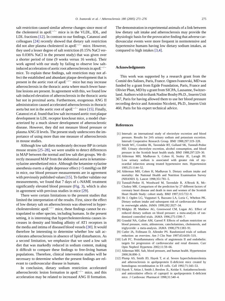
Fig. 2. Representative Oil red O stained aortas of apoE−/− mice fed either regular salt (0.64% NaCl) or low-salt diet (0.036% NaCl) for 16 weeks, showingred lipid-rich atherosclerotic lesions. En face presentation of the aortic arch with its main branches: brachiocephalic artery, right common carotid artery andleft subclavian artery. Magnification×2.5.
Table 1Influence of respective diets on body weight, serum total cholesterol,serum triglycerides, mean arterial blood pressure (MAP) and urinary pro-tein/creatinine ratio in 23-week-old apoE−/− mice
Regular salt Low salt p
Body weight (g) 24.9± 0.5a 26.7± 0.7a NSCholesterol (mM) 9.5± 1.0b 12.8± 0.5b 0.029Triglycerides (mM) 0.5± 0.05b 0.4 ± 0.05b NSMAP (mmHg) 73.0± 3.1c 67.8± 2.2c NSProt/creat ratio 1.2± 0.2c 1.4 ± 0.3c NS
Values are means± S.E.M. Prot/Creat ratio, urinary protein/creatinine ratio.a n= 26 and 28 mice, respectively.b n= 5–7 mice.c n= 13–15 mice, respectively.
The elevated ANG II levels observed in the low salt groupmay exert several direct (blood pressure independent) ef-fects that are relevant to the development of atherosclerosis[18], including stimulation of monocyte recruitment[19], ac-tivation of macrophages[20] and enhanced oxidative stress[20]. ANG II also stimulates arterial deposition of oxidizedLDL, enhances the rate at which modified LDL interacts withmacrophage scavenger receptors[21] and increases activa-tion of circulating monocytes, adhesion molecules, and cy-
Fig. 3. Angiotensin II levels in apoE−/− mice fed either regular-salt diet(0.64% NaCl) or low-salt diet (0.036% NaCl) for 16 weeks (n= 15 and 15,r
tokines[19]. These effects were attenuated by ACE inhibi-tion and by ANG II receptor blockade, further implicating adirect role for the RAAS in the pathogenesis of this process[22]. Pro-atherosclerotic actions of ANG II have been demon-strated in apoE−/− mice. Both ACE inhibitors and ANG IIreceptor antagonists markedly attenuated the development ofatherosclerotic lesions in these mice[13,14]. Furthermore,administration of exogenous ANG II caused an accelera-tion of lesion formation, demonstrating direct effects of theoctapeptide on the atherogenic process in this experimentalmodel[15,23].
In addition to excess ANG II, other metabolic effects ofsodium restriction, including increases in cholesterol, nore-pinephrine, and insulin resistance, may accelerate plaque de-velopment. In the present setting, low dietary salt causedelevations in total cholesterol, a finding that has been welldocumented in previous studies[8,24]. In humans, adverselipid effects of severe salt restriction include elevations intotal and LDL cholesterol but no change in HDL[8]. Al-though we evaluated only total cholesterol, it is likely that
F( lt diet( -s .p
espectively, males and females combined).ig. 4. Mean arterial blood pressure (MAP) in anesthetized apoE−/− micemales and females combined) after 16-week on either regular-san= 15) or low-salt diet (n= 14) and female apoE−/− mice (n= 7) on regularalt diet treated withN-nitro-l-arginine methyl ester (l-NAME) for 7 days< 0.0001 vs. both respective diets.
O. Ivanovski et al. / Atherosclerosis 180 (2005) 271–276 275
salt restriction caused similar adverse changes since most ofthe cholesterol in apoE−/− mice is in the VLDL, IDL, andLDL fractions[12]. In contrast to our findings, Catanozi andcolleagues[24] recently showed that dietary salt restrictiondid not alter plasma cholesterol in apoE−/− mice. However,they used a lesser degree of salt restriction (0.15% NaCl ver-sus 0.036% NaCl in the present study) that was given overa shorter period of time (9 weeks versus 16 weeks). Theirwork agreed with our study by failing to observe low salt-induced acceleration of aortic root atherosclerosis in apoE−/−mice. To explain these findings, salt restriction may not af-fect the established and abundant plaque development that ispresent in the aortic root of apoE−/− mice but may increaseatherosclerosis in the thoracic aorta where much fewer base-line lesions are present. In agreement with this, we found lowsalt-induced elevation of atherosclerosis in the thoracic aortabut not in proximal aorta. Furthermore, exogenous ANG IIadministration caused accelerated atherosclerosis in thoracicaorta but not in the aortic root of apoE−/− mice[15]. Finally,Catanozi et al. found that low salt increased aortic root plaquedevelopment in LDL receptor knockout mice, a model char-acterized by a much slower development of atheroscleroticdisease. However, they did not measure blood pressure orplasma ANG II levels. The present study underscores the im-portance of using more than one measure of atherosclerosisin mouse studies.
rtainm esi di-r ine-x zinea n BPi menw rm tsi
thatl ffecto per-c ex-t esents s in-c s intt t ac-c s. Asa saltd kingi anp l ben e rel-e
teda sa tion.
The demonstration in experimental animals of a link betweenlow dietary salt intake and atherosclerosis may provide thephysiologic basis for the provocative finding that adverse car-diovascular events were more frequent in normotensive andhypertensive humans having low dietary sodium intakes, ascompared to high intakes[3,4].
Acknowledgments
This work was supported by a research grant from theComite des Salines, Paris, France. Ognen Ivanovski, MD wasfunded by a grant from Egide Foundation, Paris, France andOlivier Phan, MD by a grant from SICPA, Lausanne, Switzer-land. Authors wish to thank Nadine Bouby Ph.D., Inserm Unit367, Paris for having allowed them to use her blood pressurerecording device and Antonino Nicoletti, Ph.D., Inserm Unit460, Paris for his expert technical advice.
References
[1] Intersalt: an international study of electrolyte excretion and bloodpressure. Results for 24 h urinary sodium and potassium excretion.Intersalt Cooperative Research Group. BMJ 1988;297:319–328.
doelood–30.JH.y-
nsion
andvey
c-s ofcottish
PK.sease
off ran-
onls, and
ium
[ elin:. Curr
[ nsion
[ emiad by
[ roticient
Although low salt diets moderately decrease BP in ceouse strains[25–28], we were unable to detect differenc
n MAP between the normal and low salt diet groups. Weectly measured MAP from the abdominal aorta in ketamylazine anesthetized mice. Although the ketamine-xylanesthesia exerts a slight depressor effect (<5 mmHg) o
n mice, our blood pressure measurements are in agreeith previously published values[15]. To further validate oueasurements, we found that 7 days ofl-NAME treatmen
ignificantly elevated blood pressure (Fig. 3), which is alson agreement with previous studies in mice[29].
There were certain limitations to the present studiesimited the interpretation of the results. First, since the ef low dietary salt on atherosclerosis was observed in hyholesterolemic apoE−/− mice, these findings cannot berapolated to other species, including humans. In the pretting, it is interesting that hypercholesterolemia causereases in density and binding affinity of AT1 receptorhe media and intima of diseased blood vessels[30]. It wouldherefore be interesting to determine whether low salelerates atherosclerosis in absence of lipid disturbancesecond limitation, we emphasize that we used a low
iet that was markedly reduced in sodium content, mat difficult to compare these findings to free-living humopulations. Therefore, clinical intervention studies wilecessary to determine whether the present findings arvant to cardiovascular disease in humans.
In conclusion, dietary sodium restriction acceleratherosclerotic lesion formation in apoE−/− mice, and thicceleration may be related to increased ANG II forma
t
[2] Smith WC, Crombie IK, Tavendale RT, Gulland SK, Tunstall-PeHD. Urinary electrolyte excretion, alcohol consumption, and bpressure in the Scottish heart health study. BMJ 1988;297:329
[3] Alderman MH, Madhavan S, Cohen H, Sealey JE, LaraghLow urinary sodium is associated with greater risk of mocardial infarction among treated hypertensive men. Hyperte1995;25:1144–52.
[4] Alderman MH, Cohen H, Madhavan S. Dietary sodium intakemortality: the National Health and Nutrition Examination Sur(NHANES I). Lancet 1998;351:781–5.
[5] Tunstall-Pedoe H, Woodward M, Tavendale R, A’Brook R, MCluskey MK. Comparison of the prediction by 27 different factorcoronary heart disease and death in men and women of the SHeart Health Study: cohort study. BMJ 1997;315:722–9.
[6] He J, Ogden LG, Vupputuri S, Bazzano LA, Loria C, WheltonDietary sodium intake and subsequent risk of cardiovascular diin overweight adults. JAMA 1999;282:2027–34.
[7] Midgley JP, Matthew AG, Greenwood CM, Logan AG. Effectreduced dietary sodium on blood pressure: a meta-analysis odomized controlled trials. JAMA 1996;275:1590–7.
[8] Graudal NA, Galloe AM, Garred P. Effects of sodium restrictionblood pressure, renin, aldosterone, catecholamines, cholesterotriglyceride: a meta-analysis. JAMA 1998;279:1383–91.
[9] Cutler JA, Follmann D, Allender PS. Randomized trials of sodreduction: an overview. Am J Clin Nutr 1997;65:643S–51S.
10] Luft FC. Proinflammatory effects of angiotensin II and endothtargets for progression of cardiovascular and renal diseasesOpin Nephrol Hypertens 2002;11:59–66.
11] Alderman MH. Salt, blood pressure, and human health. Hyperte2000;36:890–3.
12] Plump AS, Smith JD, Hayek T, et al. Severe hypercholesteroland atherosclerosis in apolipoprotein E-deficient mice createhomologous recombination in ES cells. Cell 1992;71:343–53.
13] Hayek T, Attias J, Smith J, Breslow JL, Keidar S. Antiatheroscleand antioxidative effects of captopril in apolipoprotein E-deficmice. J Cardiovasc Pharmacol 1998;31:540–4.

276 O. Ivanovski et al. / Atherosclerosis 180 (2005) 271–276
[14] Keidar S, Attias J, Smith J, Breslow JL, Hayek T. The angiotensin-II receptor antagonist, losartan, inhibits LDL lipid peroxidation andatherosclerosis in apolipoprotein E-deficient mice. Biochem BiophysRes Commun 1997;236:622–5.
[15] Daugherty A, Manning MW, Cassis LA. Angiotensin II promotesatherosclerotic lesions and aneurysms in apolipoprotein E-deficientmice. J Clin Invest 2000;105:1605–12.
[16] Meneton P, Ichikawa I, Inagami T, Schnermann J. Renal physi-ology of the mouse. Am J Physiol Renal Physiol 2000;278:339–51.
[17] Mallat Z, Gojova A, Marchiol-Fournigault C, et al. Interleukin-18/interleukin-18 binding protein signaling modulates atheroscleroticlesion development and stability. Circ Res 2001;89(7):E41–5.
[18] Daugherty A, Roselaar SE. Lipoprotein oxidation as a mediator ofatherogenesis: insights from pharmacological studies. Cardiovasc Res1995;29:297–311.
[19] Kim JA, Berliner JA, Nadler JL. Angiotensin II increases mono-cyte binding to endothelial cells. Biochem Biophys Res Commun1996;226:862–8.
[20] Yanagitani Y, Rakugi H, Okamura A, et al. Angiotensin II type 1receptor-mediated peroxide production in human macrophages. Hy-pertension 1999;33:335–9.
[21] Keidar S, Heinrich R, Kaplan M, Hayek T, Aviram M. AngiotensinII administration to atherosclerotic mice increases macrophage up-take of oxidized LDL: a possible role for interleukin-6. ArteriosclerThromb Vasc Biol 2001;21:1464–9.
[22] Warnholtz A, Nickenig G, Schulz E, et al. Increased NADH-oxidase-mediated superoxide production in the early stages of atherosclerosis:
evidence for involvement of the renin–angiotensin system. Circula-tion 1999;99:2027–33.
[23] Weiss D, Kools JJ, Taylor WR. Angiotensin II-induced hyperten-sion accelerates the development of atherosclerosis in apoE-deficientmice. Circulation 2001;103:448–54.
[24] Catanozi S, Rocha JC, Passarelli M, et al. Dietary sodium chlo-ride restriction enhances aortic wall lipid storage and raises plasmalipid concentration in LDL receptor knockout mice. J Lipid Res2003;44:727–32.
[25] Cholewa BC, Mattson DL. Role of the renin–angiotensin systemduring alterations of sodium intake in conscious mice. Am J PhysiolRegul Integr Comp Physiol 2001;281:E987–R993.
[26] Melo LG, Veress AT, Chong CK, Pang SC, Flynn TG, SonnenbergH. Salt-sensitive hypertension in ANP knockout mice: potential roleof abnormal plasma renin activity. Am J Physiol 1998;274:R255–61.
[27] Pradervand S, Barker PM, Wang Q, et al. Salt restriction inducespseudohypoaldosteronism type 1 in mice expressing low levels ofthe beta-subunit of the amiloride-sensitive epithelial sodium channel.Proc Natl Acad Sci USA 1999;96:1732–7.
[28] Oliverio MI, Best CF, Smithies O, Coffman TM. Regulation ofsodium balance and blood pressure by the AT(1A) receptor for an-giotensin II. Hypertension 2000;35:550–4.
[29] Peotta VA, Vasquez EC, Meyrelles SS. Cardiovascular neural re-flexes in l-NAME-induced hypertension in mice. Hypertension2001;38:555–9.
[30] Yang BC, Phillips MI, Mohuczy D, et al. Increased angiotensin IItype 1 receptor expression in hypercholesterolemic atherosclerosis inrabbits. Arterioscler Thromb Vasc Biol 1998;18:1433–9.
Related Documents