ISSN 2234-3806 • eISSN 2234-3814 https://doi.org/10.3343/alm.2017.37.6.465 www.annlabmed.org 465 Ann Lab Med 2017;37:465-474 https://doi.org/10.3343/alm.2017.37.6.465 Review Article Diagnostic Hematology Diagnostics and Prognostication of Myelodysplastic Syndromes Gina Zini, M.D. Department of Oncology and Hematology, Blood Transfusion Service, Policlinico Gemelli Foundation, Catholic University of Sacred Heart, Rome, Italy MDS are a heterogeneous and complex group of clonal hematological neoplasms arising from a hematopoietic stem cell, and characterized by ineffective hematopoiesis, resulting in increased apoptosis in the bone marrow and peripheral cytopenia, which involves one or more lineages. Epigenetic changes are reported as ‘founder’ mutations in the case of MDS. Its incidence in the general population has been reported as five new MDS diagno- ses per 100,000 people. It affects men more frequently than it does women, and its inci- dence increases with age. The diagnostic classification, now in use, is the one of the World Health Organization, revised in August 2016. It recognizes six distinct entities in addition to a provisional entity of childhood. In most of the cases, diagnosis is based on the mor- phologic quantitative and qualitative evaluation of the peripheral blood and bone marrow using basic hematological techniques. Bone marrow biopsy and flow cytometric immuno- phenotyping also offer support for further diagnostic elucidation, while cytogenetics and molecular genetics are presently fully integrated into prognostication, treatment processes, and decision-making. Key Words: Myelodysplastic syndromes (MDS), WHO 2016 classification, Diagnosis, Prog- nostication Received: April 25, 2017 Revision received: June 7, 2017 Accepted: August 2, 2017 Corresponding author: Gina Zini Department of Oncology and Hematology, Blood Transfusion Service, Policlinico Gemelli Foundation, Catholic University of Sacred Heart, L.go Gemelli 8, Rome 00168, Italy Tel: +39-06-30153262 Fax: +39-06-3055153 E-mail: [email protected] © Korean Society for Laboratory Medicine This is an Open Access article distributed under the terms of the Creative Commons Attribution Non-Commercial License (http://creativecom- mons.org/licenses/by-nc/4.0) which permits unrestricted non-commercial use, distribution, and reproduction in any medium, provided the original work is properly cited. DEFINITION AND ETIOLOGY MDS are a heterogeneous and complex group of clonal hemato- logical neoplasms arising from a hematopoietic stem cell (HSC). Epigenetic changes, such as DNA methylation/hydroxymethyl- ation, histone demethylation/modifications, and transcription co- regulation, are reported as ‘founder’ mutations in the case of MDS, and their key roles in the differentiation and aging of HSCs drive stable clonal changes in gene expression, thereby leading to maturation pathway dysfunctions [1]. MDS are characterized by ineffective hematopoiesis, resulting in increased apoptosis of the bone marrow (BM) and peripheral blood (PB) cytopenia in- volving one or more lineages. The common features of MDS are: i) morphological dysplasia in one or more lineages; ii) a blast per- centage less than 20% in the PB and BM; iii) the presence of cytogenetic and molecular genetic abnormalities in up to 90% of de novo cases; and iv) the variable risk of evolution to acute leukemia, mainly in the absence of peripheral leukocytosis. In a majority of the cases, MDS are acquired diseases related to ag- ing ( de novo cases) or are secondary to environmental/occupa- tional exposure to toxic compounds, benzene, smoking, ionizing radiation, or antineoplastic or immunosuppressive therapy (ther- apy-related MDS, t-MDS). Rare, inherited predispositions to pri- mary MDS associated with BM failure syndromes, aplastic ane- mia, Fanconi anemia, dyskeratosis congenita, Diamond–Black- fan anemia, Shwachman–Diamond syndrome, and paroxysmal nocturnal hemoglobinuria are widely described in the literature, mainly in pediatric settings; these are not included within the MDS group. Multiple hereditary predispositions to MDS have been discovered (familial MDS) [2, 3]; a mutation in at least one

Welcome message from author
This document is posted to help you gain knowledge. Please leave a comment to let me know what you think about it! Share it to your friends and learn new things together.
Transcript
ISSN 2234-3806 • eISSN 2234-3814
https://doi.org/10.3343/alm.2017.37.6.465 www.annlabmed.org 465
Ann Lab Med 2017;37:465-474https://doi.org/10.3343/alm.2017.37.6.465
Review ArticleDiagnostic Hematology
Diagnostics and Prognostication of Myelodysplastic SyndromesGina Zini, M.D. Department of Oncology and Hematology, Blood Transfusion Service, Policlinico Gemelli Foundation, Catholic University of Sacred Heart, Rome, Italy
MDS are a heterogeneous and complex group of clonal hematological neoplasms arising from a hematopoietic stem cell, and characterized by ineffective hematopoiesis, resulting in increased apoptosis in the bone marrow and peripheral cytopenia, which involves one or more lineages. Epigenetic changes are reported as ‘founder’ mutations in the case of MDS. Its incidence in the general population has been reported as five new MDS diagno-ses per 100,000 people. It affects men more frequently than it does women, and its inci-dence increases with age. The diagnostic classification, now in use, is the one of the World Health Organization, revised in August 2016. It recognizes six distinct entities in addition to a provisional entity of childhood. In most of the cases, diagnosis is based on the mor-phologic quantitative and qualitative evaluation of the peripheral blood and bone marrow using basic hematological techniques. Bone marrow biopsy and flow cytometric immuno-phenotyping also offer support for further diagnostic elucidation, while cytogenetics and molecular genetics are presently fully integrated into prognostication, treatment processes, and decision-making.
Key Words: Myelodysplastic syndromes (MDS), WHO 2016 classification, Diagnosis, Prog-nostication
Received: April 25, 2017Revision received: June 7, 2017Accepted: August 2, 2017
Corresponding author: Gina ZiniDepartment of Oncology and Hematology, Blood Transfusion Service, Policlinico Gemelli Foundation, Catholic University of Sacred Heart, L.go Gemelli 8, Rome 00168, ItalyTel: +39-06-30153262Fax: +39-06-3055153E-mail: [email protected]
© Korean Society for Laboratory MedicineThis is an Open Access article distributed under the terms of the Creative Commons Attribution Non-Commercial License (http://creativecom-mons.org/licenses/by-nc/4.0) which permits unrestricted non-commercial use, distribution, and reproduction in any medium, provided the original work is properly cited.
DEFINITION AND ETIOLOGY
MDS are a heterogeneous and complex group of clonal hemato-
logical neoplasms arising from a hematopoietic stem cell (HSC).
Epigenetic changes, such as DNA methylation/hydroxymethyl-
ation, histone demethylation/modifications, and transcription co-
regulation, are reported as ‘founder’ mutations in the case of MDS,
and their key roles in the differentiation and aging of HSCs drive
stable clonal changes in gene expression, thereby leading to
maturation pathway dysfunctions [1]. MDS are characterized by
ineffective hematopoiesis, resulting in increased apoptosis of
the bone marrow (BM) and peripheral blood (PB) cytopenia in-
volving one or more lineages. The common features of MDS are:
i) morphological dysplasia in one or more lineages; ii) a blast per-
centage less than 20% in the PB and BM; iii) the presence of
cytogenetic and molecular genetic abnormalities in up to 90%
of de novo cases; and iv) the variable risk of evolution to acute
leukemia, mainly in the absence of peripheral leukocytosis. In a
majority of the cases, MDS are acquired diseases related to ag-
ing (de novo cases) or are secondary to environmental/occupa-
tional exposure to toxic compounds, benzene, smoking, ionizing
radiation, or antineoplastic or immunosuppressive therapy (ther-
apy-related MDS, t-MDS). Rare, inherited predispositions to pri-
mary MDS associated with BM failure syndromes, aplastic ane-
mia, Fanconi anemia, dyskeratosis congenita, Diamond–Black-
fan anemia, Shwachman–Diamond syndrome, and paroxysmal
nocturnal hemoglobinuria are widely described in the literature,
mainly in pediatric settings; these are not included within the
MDS group. Multiple hereditary predispositions to MDS have
been discovered (familial MDS) [2, 3]; a mutation in at least one
1 / 1CROSSMARK_logo_3_Test
2017-03-16https://crossmark-cdn.crossref.org/widget/v2.0/logos/CROSSMARK_Color_square.svg
Zini GMDS diagnostics and prognostication
466 www.annlabmed.org https://doi.org/10.3343/alm.2017.37.6.465
of seven well-defined single-gene loci is reported as predispos-
ing one to an increased lifetime risk of primary MDS [4].
Due to the heterogeneity of the clinical presentation of this
group of hematological neoplasms, particularly in the cases of
lower-risk MDS, differential diagnosis should exclude drug-in-
duced cytopenias, vitamin B12/folate/zinc/copper deficiency, ex-
cessive alcohol intake, exposure to heavy metals (lead, arsenic),
infections (HIV, Epstein-Barr virus, hepatitis C virus, parvovirus,
leishmaniasis), hemophagocytic lymphohistiocytosis, anemia of
chronic disorders (infection, inflammation, cancer), autoimmune
cytopenia, and metabolic disorders (liver failure, kidney failure).
The 2001 WHO classification [5] has recognized groups of he-
matological neoplasms with dysplasia that nevertheless are not
classified as MDS; these include MDS/myeloproliferative neo-
plasms (MPN), AML with myelodysplasia/dysplasia-related chan-
ges, and therapy–related AML/MDS. Finally, it is noted that a
low number of dysplastic erythroid, granulocytic, or megakaryo-
cytic cells can be recognized in the BM of healthy subjects [6].
EPIDEMIOLOGY
The incidence of MDS in the general population is reported as
five new MDS diagnoses per 100,000 people, with a higher in-
cidence among men [7]. In Western countries, among individu-
als older than 70 yr, the incidence is reported as between 22 and
45 per 100,000 people, and this incidence further increases
with age [8, 9]. The occurrences of MDS at a younger age have
been more frequently reported in Asian countries, including Ja-
pan, China, Korea, India, Thailand, India, and Turkey, with the
median age of patients reported between 40 and 50 yr; this is
one to two decades younger than that of patients in Western coun-
tries. Environmental pollutions and/or other factors, including
uncontrolled pesticide use, may contribute to these differences
[10].
However, in a report from a single institution in Italy, about 10%
of patients with MDS were younger than 50 yr (median age 43
yr), with a female predominance [11]. MDS may also affect chil-
dren and adolescents, rarely, with an incidence of less than 5%
of hematopoietic malignancies [12]. Familial cases of MDS are
rare; remarkably, a recent increase in the reported cases in the
literature testifies the higher knowledge and consciousness of
clinicians in the investigation and identification process of famil-
ial cases of MDS [13]. Therapy-related myeloid neoplasms, in-
cluding t-MDS, account for 10–20% of all the cases of AML, MDS,
and MDS/MPN [14].
CLASSIFICATION
First described in 1900 by von Leube [15] as a ‘leukanemia’, on
the basis of an alleged co-existence of pernicious anemia and
leukemia, MDS were named and described in a variety of ways
until 1976, when the French-American-British (FAB) classifica-
tion named them ‘dysmyelopoietic syndromes’ and categorized
them separately from AML [16]. In 1982, the FAB group refined
the proposal, changed the designation to ‘myelodysplastic syn-
dromes’, and provided the modern basis for the diagnosis and
classification of this group of disorders [17]. Five subtypes were
identified, on the basis of quantitative (peripheral cytopenia[s]
involving one or more hematopoietic lineages, the blast percent-
age in PB and BM, monocytes in PB) as well as qualitative ab-
normalities, (ineffective hematopoiesis and morphological dys-
plasia affecting one to three lineages): refractory anemia (RA),
refractory anemia with ring sideroblasts (RARS), refractory ane-
mia with an excess of blasts (RAEB), refractory anemia with an
excess of blasts in transformation (RAEB-t), and chronic myelo-
monocytic leukemia (CMML). From this classification, the intro-
duction of new diagnostic techniques, mainly cytogenetics and
molecular genetics, has made the correlation between the sub-
types of MDS and other new variables possible. This has been
very useful for the prognostication and development of new ther-
apies, as well as for the redefinition of subtypes. After the publi-
cation of the first FAB proposal, more than 20,000 scientific ar-
ticles on the diverse diagnostic, prognostic, and therapeutic as-
pects of MDS have been published, attesting the enormous in-
terest developed by the scientific community in studying these
diseases. Morphology remains a cornerstone in the diagnosis of
MDS. Nevertheless, in terms of the accuracy of diagnosis and
the ability to compare different series of cases, a gray area still
exists owing to the impossibility of experts reaching a unanimous
opinion, particularly when the disease is in its early stages, mainly
because of the lack of specificity of many morphological aspects
of dysplasia in the case of MDS.
The diagnostic classification now in use is the one proposed
by the working group of the WHO, starting from the first edition
in 2001 [5], moving to the second edition in 2008 [18] which
was an expanded and updated version of the previous one, and
now using a further revised classification of myeloid neoplasms
and acute leukemia which was published in August 2016 [19].
This updated classification introduces refinements in the nomen-
clature, morphologic interpretation, and cytopenia assessment.
It also addresses the influence of rapidly accumulating genetic
information in MDS diagnosis and classification; for the first time,
Zini GMDS diagnostics and prognostication
https://doi.org/10.3343/alm.2017.37.6.465 www.annlabmed.org 467
the molecular test for SF3B1 mutation has been included among
the diagnostic tools [20]. Six distinct entities within the MDS group
are nowadays recognized, and defined at diagnosis by precise
criteria including i) the number of lineages presenting dysplastic
features, ii) number of cytopenias in the PB, iii) presence/absence
and percentage of ring sideroblasts, iv) percentage of blasts in
the PB and BM, and v) karyotype and molecular genetics, when
needed [19]. They are:
1) MDS with single lineage dysplasia (MDS-SLD): one dysplas-
tic lineage, one or two PB cytopenias, less than 5% of blasts
in BM and less than 1% blasts in PB, non Auer rods, less
than 15% ring sideroblasts in BM or less than 5%, if SF3B1
mutation is present, (includes the 2008 subtypes refractory
cytopenia with unilineage dysplasia, or RCUD);
2) MDS with multilineage dysplasia (MDS-MLD): two or three
dysplastic lineages, one to three PB cytopenias, less than
15% ring sideroblasts in BM or less than 5%, if SF3B1 mu-
tation is present, less than 5% of blasts in BM and less than
1% blasts in PB, no Auer rods (includes the 2008 subtype
of refractory cytopenia with multilineage dysplasia, or RCMD);
3) MDS with ring sideroblasts (MDS-RS) (previously named as
RARS). It includes the two subtypes i) with single lineage
dysplasia (MDS-RS-SLD): one dysplastic lineage, one or
two PB cytopenias, 15% or more ring sideroblasts in BM
or 5% or more if SF3B1 mutation is present, less than 5%
of blasts in BM and less than 1% blasts in PB, no Auer rods;
and ii) with multilineage dysplasia (MDS-RS-MLD): two to
three dysplastic lineages, one to three PB cytopenias, 15%
or more ring sideroblasts in BM or 5% or more if SF3B1
mutation is present, less than 5% of blasts in BM and less
than 1% blasts in PB, no Auer rods;
4) MDS with isolated del(5q): one to three dysplastic lineages,
one or two PB cytopenias, less than 5% of blasts in BM and
less than 1% of blasts in PB, no Auer rods, none or any
ring sideroblasts, presence of del(5q) alone or with one ad-
ditional abnormality except -7 or del(7q) (this last is a new
compared to the 2008 WHO classification);
5) MDS with excess blasts (MDS-EB): none to three dysplas-
tic lineages, one or three PB cytopenias, none or any ring
sideroblasts; it includes the two subtypes MDS-EB-1 (5 to
9% blasts in BM and/or 2 to 4% blasts in PB, no Auer rods)
and MDS-EB-2 (10 to 19% blasts in BM or 5 to 19% blasts
in PB, and/or presence of Auer rods);
6) MDS, unclassifiable (MDS-U) including three categories: i)
with 1% PB blasts, ii) with single lineage dysplasia and pan-
cytopenia, and iii) with a defining cytogenetic abnormality
related to myelodysplasia.
An additional provisional entity within this category is refrac-
tory cytopenia of childhood, characterized by one to three dys-
plastic lineages, one to three PB cytopenias, less than 5% blasts
in BM and less than 2% blasts in PB.
DIAGNOSIS
1. Peripheral blood and bone marrow aspirate The diagnosis of MDS is based on the quantitative and qualita-
tive evaluation of the cytological composition of the PB and BM,
using basic hematological techniques, such as hemocytometry,
optical microscopy on PB and BM films, fixed and stained with
panoptical stains, and cytochemistry for the detection of iron in
the BM. The presence of at least one cytopenia is a “sine qua non” for any MDS diagnosis; the thresholds are hemoglobin <10
g/dL, platelets <100×109/L, and absolute neutrophil count (ANC)
<1.8×10⁹/L. PB monocytes must be less than 1×10⁹/L. Differ-
ent potential disorders should be accurately excluded as primary
etiologies of cytopenia. Cytopenia should be stable for ≥six mon-
ths, unless it is associated with a specific karyotype or bilineage
dysplasia, in which case only two months of stable cytopenia
are required [21]. It is to be noted that some ethnic groups may
have a reference range of ANC<1.8×10⁹/L; caution should be
exercised in interpreting neutropenia, if it is the only cytopenia
[22].
2. Cytomorphology of dysplasiaOn PB examination, the observation of the presence of morpho-
logical abnormalities in the red blood cells (RBC) is quite usual,
including the occurrence of circulating nucleated RBC (NRBC)
with morphological stigmata of dyserythropoiesis, which is not
rare. Another characteristic finding at the time of diagnosis is
the detection of two RBC populations, one of which is usually
normal, while the other, being a direct expression of the anoma-
lous neoplastic clone, is microcytic or macrocytic. Dysgranulo-
poiesis in neutrophils is variably observed, from absent to se-
vere, and can involve both the nucleus and cytoplasm, and/or
abnormalities in size. The nucleus can show abnormal lobula-
tion and/or an abnormal chromatin pattern, as well as the pres-
ence of more than four nuclear projections, while the cytoplasm
can show abnormalities in granule size (pseudo-Chédiak-Higashi)
and/or content (reduction of two thirds to absence) [23]. The
evaluation of granularity requires optimally stained smears. The
visibility of granules, in fact, is the morphological characteristic
that can most easily be compromised by an inadequate staining
Zini GMDS diagnostics and prognostication
468 www.annlabmed.org https://doi.org/10.3343/alm.2017.37.6.465
technique, either manual or automated. Morphological altera-
tions of the MDS cells at diagnosis are less frequent in eosino-
phils and rare in basophils. The presence of even <1% blast
cells in the PB is a crucial feature in MDS diagnosis, and usually
indicates cases with an unfavorable prognosis. In patients pre-
senting with marked leukopenia, it can be very useful to prepare
buffy coat PB smears after centrifugation.
3. Blast cell identification and countThe upper blast cell threshold for the diagnosis of MDS is <20%
in the PB and/or BM, on a PB differential count performed on
200 nucleated cells, and/or on a myelogram performed on 500
nucleated cells. Blasts are generally small to medium in size,
with a high nucleo-cytoplasmic ratio. The nucleus is usually nu-
cleolated with a finely dispersed chromatin pattern. The cyto-
plasm is relatively scanty and basophilic. Primary (azurophilic)
granules may be absent (agranular blasts), scanty or, sometimes,
more in number (granular blasts); in the latter, the cytoplasm is
more plentiful. The absence of a clear Golgi area is considered
a key morphological feature to differentiate granular blasts from
normal or dysplastic promyelocytes [24]. The recognition of Auer
rods in the cytoplasm, either in circulating or BM blast cells, en-
tails an unfavorable prognosis and leads to the classification of
the patient as MDS-EB2. The cytochemical stain for peroxidase
and the study of specific immunophenotypic markers are use-
ful, when positive, to confirm the myeloid nature of blast cells,
particularly when the blasts are agranular. The exclusion of an
expansion of monocytic lineage should always be taken into ac-
count, in the diagnostic pathway of MDS [25].
The 2016 WHO classification introduces a change in the eval-
uation of blast percentage; now, it should simply be calculated
as the percentage of all the nucleated cells of the BM, irrespec-
tive of the percentage of the erythroid precursors (EP). Originally
introduced by the FAB group, the “50% rule” (the blast percent-
age in patients presenting with EP ≥50% <80% of BM nucle-
ated cells, when less than 20%, should be recalculated and re-
ported as the percentage of non-erythroid BM cells, excluding
lymphocytes and plasma cells) was adopted till the 2008 WHO
edition, to allow for the differential diagnosis between MDS pre-
senting with erythroid hyperplasia versus acute erythroid/myeloid
leukemia (AEL). Different potential disorders as a primary etiol-
ogy of erythroid hyperplasia should be preliminarily excluded.
There is evidence in the literature that suggests that the percent-
age of EP does not impact prognosis, overall survival or leuke-
mia-free survival in these patients [26]. This change determines
that cases previously diagnosed as erythroid/myeloid AML are
now included in the MDS group [27]. It is to be noted that, in
the literature, cases of MDS developing in patients with untreated
CLL are reported [28, 29]. It is also important to come to an agree-
ment on how to evaluate the myeloid blast percentage in suspect
myeloid neoplasms presenting with the infiltration of lymphoma
cells in the BM, using microscopy, to avoid the risk of the un-
derestimation of myeloblasts in BM infiltrated by lymphoid cells
and/or plasma cells. The increased heterogeneity of the platelet
size and abnormalities in the granule content and/or size are com-
mon findings in the PB of MDS. The presence of micromegakary-
ocytes and/or bare megakaryocyte nuclei in the PB is associ-
ated with MDS, but is not specific, because these cells are also
found in other hematology neoplasms. They are easier to find in
buffy coat films and are more frequently observed in high-risk
cases of MDS.
4. Bone marrow cellularityBM cellularity is most often increased in cases of MDS, at diag-
nosis, with hyperplasia of the erythroid or granulocytic series, or
both. In 30 to 40% of the cases, cellularity is quantitatively nor-
mal, while in about 10% of the patients, the BM aspirates ap-
pear hypocellular. In such cases, other diagnostic techniques
should be used, such as histology, immunohistochemistry, and
cytogenetic studies, to distinguish MDS from other hypoplastic
myeloid disorders [30]. Finally, it is not unusual to find non-spe-
cific reactive alterations, such as incre ased lymphocytes, plasma
cells, or mast cells or an increase in hemosiderin-laden macro-
phages with some hemophagocytosis in the BM of MDS cases,
at diagnosis.
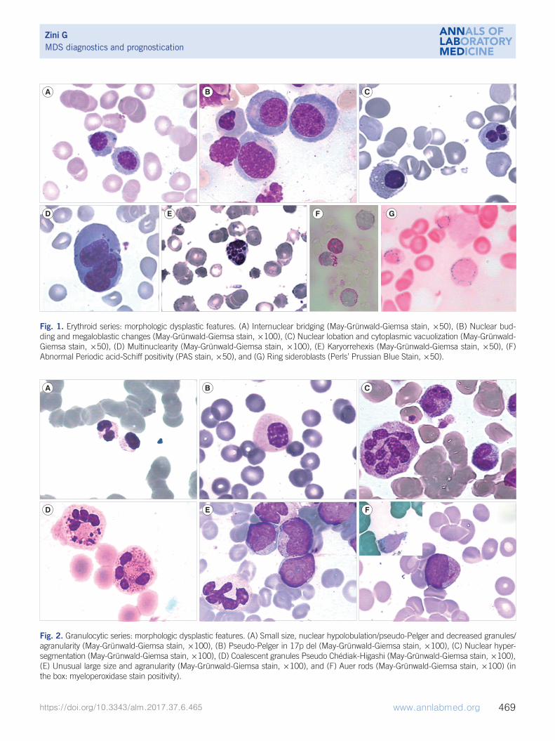
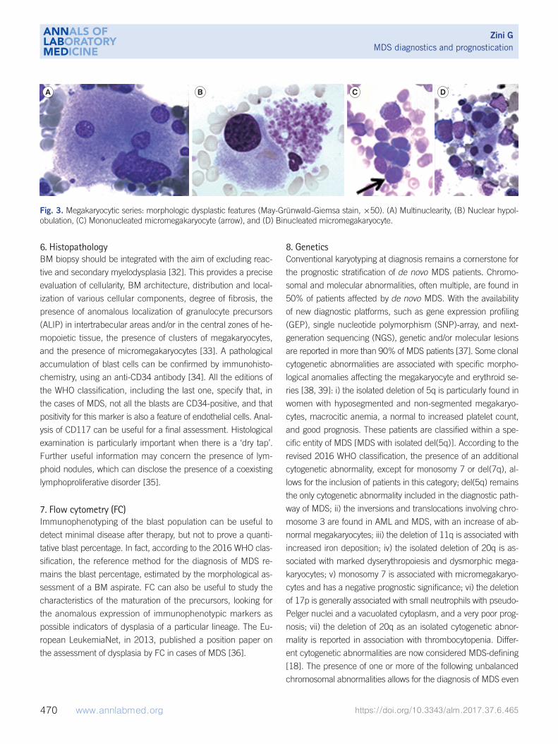
5. Quantification of dysplasiaPrecise morphological criteria, both quantitative and qualitative,
have been identified for each lineage for the definition of mor-
phological dysplasia, with the aim of harmonizing microscopic
diagnosis, in the form of the minimal criteria necessary for an
unequivocal recognition of dysplasia; in particular, to recognize
dysplasia within a specified lineage in the BM, it is necessary
that dysplastic features are present in at least 10% of the EP
(not taking into account mature erythrocytes) and/or 10% of the
granulocytic cells (in this case, also including mature cells) out
of a count of at least 200 cells of each lineage, and/or in a mini-
mum of 10% of megakaryocytes out of at least 30 cells of the
megakaryocyte lineage [31]. The characteristic morphologic fea-
tures that allow the inclusion of the cells into the dysplastic group,
according to the WHO classification, are listed and illustrated in
Fig. 1 to 3.
Zini GMDS diagnostics and prognostication
https://doi.org/10.3343/alm.2017.37.6.465 www.annlabmed.org 469
Fig. 1. Erythroid series: morphologic dysplastic features. (A) Internuclear bridging (May-Grünwald-Giemsa stain, ×50), (B) Nuclear bud-ding and megaloblastic changes (May-Grünwald-Giemsa stain, ×100), (C) Nuclear lobation and cytoplasmic vacuolization (May-Grünwald-Giemsa stain, ×50), (D) Multinuclearity (May-Grünwald-Giemsa stain, ×100), (E) Karyorrehexis (May-Grünwald-Giemsa stain, ×50), (F) Abnormal Periodic acid-Schiff positivity (PAS stain, ×50), and (G) Ring sideroblasts (Perls’ Prussian Blue Stain, ×50).
A
D E F G
B C
Fig. 2. Granulocytic series: morphologic dysplastic features. (A) Small size, nuclear hypolobulation/pseudo-Pelger and decreased granules/agranularity (May-Grünwald-Giemsa stain, ×100), (B) Pseudo-Pelger in 17p del (May-Grünwald-Giemsa stain, ×100), (C) Nuclear hyper-segmentation (May-Grünwald-Giemsa stain, ×100), (D) Coalescent granules Pseudo Chédiak-Higashi (May-Grünwald-Giemsa stain, ×100), (E) Unusual large size and agranularity (May-Grünwald-Giemsa stain, ×100), and (F) Auer rods (May-Grünwald-Giemsa stain, ×100) (in the box: myeloperoxidase stain positivity).
A
D
B
E
C
F
Zini GMDS diagnostics and prognostication
470 www.annlabmed.org https://doi.org/10.3343/alm.2017.37.6.465
6. HistopathologyBM biopsy should be integrated with the aim of excluding reac-
tive and secondary myelodysplasia [32]. This provides a precise
evaluation of cellularity, BM architecture, distribution and local-
ization of various cellular components, degree of fibrosis, the
presence of anomalous localization of granulocyte precursors
(ALIP) in intertrabecular areas and/or in the central zones of he-
mopoietic tissue, the presence of clusters of megakaryocytes,
and the presence of micromegakaryocytes [33]. A pathological
accumulation of blast cells can be confirmed by immunohisto-
chemistry, using an anti-CD34 antibody [34]. All the editions of
the WHO classification, including the last one, specify that, in
the cases of MDS, not all the blasts are CD34-positive, and that
positivity for this marker is also a feature of endothelial cells. Anal-
ysis of CD117 can be useful for a final assessment. Histological
examination is particularly important when there is a ‘dry tap’.
Further useful information may concern the presence of lym-
phoid nodules, which can disclose the presence of a coexisting
lymphoproliferative disorder [35].
7. Flow cytometry (FC)Immunophenotyping of the blast population can be useful to
detect minimal disease after therapy, but not to prove a quanti-
tative blast percentage. In fact, according to the 2016 WHO clas-
sification, the reference method for the diagnosis of MDS re-
mains the blast percentage, estimated by the morphological as-
sessment of a BM aspirate. FC can also be useful to study the
characteristics of the maturation of the precursors, looking for
the anomalous expression of immunophenotypic markers as
possible indicators of dysplasia of a particular lineage. The Eu-
ropean LeukemiaNet, in 2013, published a position paper on
the assessment of dysplasia by FC in cases of MDS [36].
8. GeneticsConventional karyotyping at diagnosis remains a cornerstone for
the prognostic stratification of de novo MDS patients. Chromo-
somal and molecular abnormalities, often multiple, are found in
50% of patients affected by de novo MDS. With the availability
of new diagnostic platforms, such as gene expression profiling
(GEP), single nucleotide polymorphism (SNP)-array, and next-
generation sequencing (NGS), genetic and/or molecular lesions
are reported in more than 90% of MDS patients [37]. Some clonal
cytogenetic abnormalities are associated with specific morpho-
logical anomalies affecting the megakaryocyte and erythroid se-
ries [38, 39]: i) the isolated deletion of 5q is particularly found in
women with hyposegmented and non-segmented megakaryo-
cytes, macrocitic anemia, a normal to increased platelet count,
and good prognosis. These patients are classified within a spe-
cific entity of MDS [MDS with isolated del(5q)]. According to the
revised 2016 WHO classification, the presence of an additional
cytogenetic abnormality, except for monosomy 7 or del(7q), al-
lows for the inclusion of patients in this category; del(5q) remains
the only cytogenetic abnormality included in the diagnostic path-
way of MDS; ii) the inversions and translocations involving chro-
mosome 3 are found in AML and MDS, with an increase of ab-
normal megakaryocytes; iii) the deletion of 11q is associated with
increased iron deposition; iv) the isolated deletion of 20q is as-
sociated with marked dyserythropoiesis and dysmorphic mega-
karyocytes; v) monosomy 7 is associated with micromegakaryo-
cytes and has a negative prognostic significance; vi) the deletion
of 17p is generally associated with small neutrophils with pseudo-
Pelger nuclei and a vacuolated cytoplasm, and a very poor prog-
nosis; vii) the deletion of 20q as an isolated cytogenetic abnor-
mality is reported in association with thrombocytopenia. Differ-
ent cytogenetic abnormalities are now considered MDS-defining
[18]. The presence of one or more of the following unbalanced
chromosomal abnormalities allows for the diagnosis of MDS even
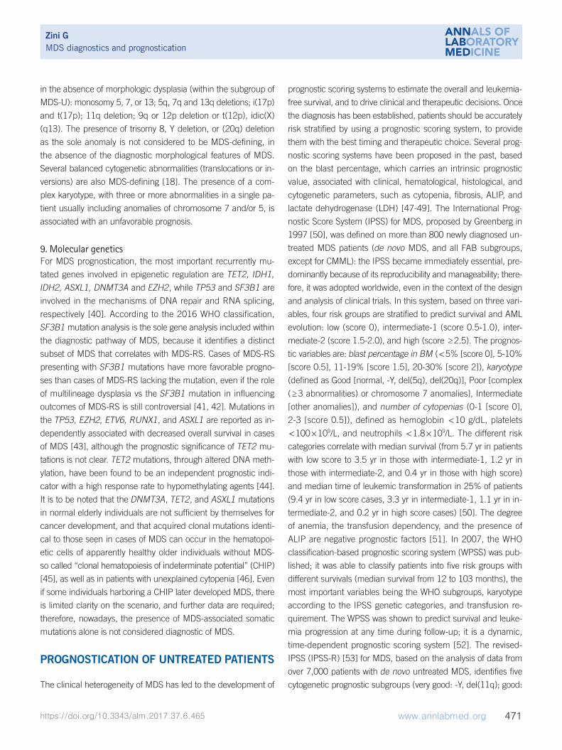
Fig. 3. Megakaryocytic series: morphologic dysplastic features (May-Grünwald-Giemsa stain, ×50). (A) Multinuclearity, (B) Nuclear hypol-obulation, (C) Mononucleated micromegakaryocyte (arrow), and (D) Binucleated micromegakaryocyte.
A B C D
Zini GMDS diagnostics and prognostication
https://doi.org/10.3343/alm.2017.37.6.465 www.annlabmed.org 471
in the absence of morphologic dysplasia (within the subgroup of
MDS-U): monosomy 5, 7, or 13; 5q, 7q and 13q deletions; i(17p)
and t(17p); 11q deletion; 9q or 12p deletion or t(12p), idic(X)
(q13). The presence of trisomy 8, Y deletion, or (20q) deletion
as the sole anomaly is not considered to be MDS-defining, in
the absence of the diagnostic morphological features of MDS.
Several balanced cytogenetic abnormalities (translocations or in-
versions) are also MDS-defining [18]. The presence of a com-
plex karyotype, with three or more abnormalities in a single pa-
tient usually including anomalies of chromosome 7 and/or 5, is
associated with an unfavorable prognosis.
9. Molecular geneticsFor MDS prognostication, the most important recurrently mu-
tated genes involved in epigenetic regulation are TET2, IDH1, IDH2, ASXL1, DNMT3A and EZH2, while TP53 and SF3B1 are
involved in the mechanisms of DNA repair and RNA splicing,
respectively [40]. According to the 2016 WHO classification,
SF3B1 mutation analysis is the sole gene analysis included within
the diagnostic pathway of MDS, because it identifies a distinct
subset of MDS that correlates with MDS-RS. Cases of MDS-RS
presenting with SF3B1 mutations have more favorable progno-
ses than cases of MDS-RS lacking the mutation, even if the role
of multilineage dysplasia vs the SF3B1 mutation in influencing
outcomes of MDS-RS is still controversial [41, 42]. Mutations in
the TP53, EZH2, ETV6, RUNX1, and ASXL1 are reported as in-
dependently associated with decreased overall survival in cases
of MDS [43], although the prognostic significance of TET2 mu-
tations is not clear. TET2 mutations, through altered DNA meth-
ylation, have been found to be an independent prognostic indi-
cator with a high response rate to hypomethylating agents [44].
It is to be noted that the DNMT3A, TET2, and ASXL1 mutations
in normal elderly individuals are not sufficient by themselves for
cancer development, and that acquired clonal mutations identi-
cal to those seen in cases of MDS can occur in the hematopoi-
etic cells of apparently healthy older individuals without MDS-
so called “clonal hematopoiesis of indeterminate potential” (CHIP)
[45], as well as in patients with unexplained cytopenia [46]. Even
if some individuals harboring a CHIP later developed MDS, there
is limited clarity on the scenario, and further data are required;
therefore, nowadays, the presence of MDS-associated somatic
mutations alone is not considered diagnostic of MDS.
PROGNOSTICATION OF UNTREATED PATIENTS
The clinical heterogeneity of MDS has led to the development of
prognostic scoring systems to estimate the overall and leukemia-
free survival, and to drive clinical and therapeutic decisions. Once
the diagnosis has been established, patients should be accurately
risk stratified by using a prognostic scoring system, to provide
them with the best timing and therapeutic choice. Several prog-
nostic scoring systems have been proposed in the past, based
on the blast percentage, which carries an intrinsic prognostic
value, associated with clinical, hematological, histological, and
cytogenetic parameters, such as cytopenia, fibrosis, ALIP, and
lactate dehydrogenase (LDH) [47-49]. The International Prog-
nostic Score System (IPSS) for MDS, proposed by Greenberg in
1997 [50], was defined on more than 800 newly diagnosed un-
treated MDS patients (de novo MDS, and all FAB subgroups,
except for CMML): the IPSS became immediately essential, pre-
dominantly because of its reproducibility and manageability; there-
fore, it was adopted worldwide, even in the context of the design
and analysis of clinical trials. In this system, based on three vari-
ables, four risk groups are stratified to predict survival and AML
evolution: low (score 0), intermediate-1 (score 0.5-1.0), inter-
mediate-2 (score 1.5-2.0), and high (score ≥2.5). The prognos-
tic variables are: blast percentage in BM (<5% [score 0], 5-10%
[score 0.5], 11-19% [score 1.5], 20-30% [score 2]), karyotype (defined as Good [normal, -Y, del(5q), del(20q)], Poor [complex
(≥3 abnormalities) or chromosome 7 anomalies], Intermediate
[other anomalies]), and number of cytopenias (0-1 [score 0],
2-3 [score 0.5]), defined as hemoglobin <10 g/dL, platelets
<100×109/L, and neutrophils <1.8×10⁹/L. The different risk
categories correlate with median survival (from 5.7 yr in patients
with low score to 3.5 yr in those with intermediate-1, 1.2 yr in
those with intermediate-2, and 0.4 yr in those with high score)
and median time of leukemic transformation in 25% of patients
(9.4 yr in low score cases, 3.3 yr in intermediate-1, 1.1 yr in in-
termediate-2, and 0.2 yr in high score cases) [50]. The degree
of anemia, the transfusion dependency, and the presence of
ALIP are negative prognostic factors [51]. In 2007, the WHO
classification-based prognostic scoring system (WPSS) was pub-
lished; it was able to classify patients into five risk groups with
different survivals (median survival from 12 to 103 months), the
most important variables being the WHO subgroups, karyotype
according to the IPSS genetic categories, and transfusion re-
quirement. The WPSS was shown to predict survival and leuke-
mia progression at any time during follow-up; it is a dynamic,
time-dependent prognostic scoring system [52]. The revised-
IPSS (IPSS-R) [53] for MDS, based on the analysis of data from
over 7,000 patients with de novo untreated MDS, identifies five
cytogenetic prognostic subgroups (very good: -Y, del(11q); good:
Zini GMDS diagnostics and prognostication
472 www.annlabmed.org https://doi.org/10.3343/alm.2017.37.6.465
normal, del(5q), and del(20q) as sole or double abnormalities
including del(5q); intermediate: del(7q), +8, +19, i(17q), or any
other not listed in the other risk groups; poor: -7, inv(3)/ t(3q)/
del(3q), double abnormalities including -7/del(7q) or complex
with 3 abnormalities; very poor: complex with more than three
abnormalities) and splits the low marrow blast percentage value
into four groups (2%, >2 but <5%, 5 to 10%, >10%) and depth
of cytopenias (anemia into three groups, thrombocytopenia in
three, neutropenia in two). The different IPSS-R risk categories
also show a strict correlation with median survival (from 8.8 yr
in patients with very low score, 5.3 yr in those with low score, 3.0
yr in those with intermediate score, 1.6 yr in those with high score,
to 0.8 yr in those with very high score), as well as with the me-
dian time of leukemic transformation in 25% of patients (more
than 14.5 yr in patients with very low score, 10.8 yr in those with
low score, 3.2 yr in those with intermediate score, 1.2 yr in those
with high score, and 0.7 yr in those with very high score) [53].
All these scoring systems have been developed on data from MDS
de novo untreated patients.
Global prognosticationThe MD Anderson Cancer Center group, in 2008 [54], proposed
the Global MDACC-MDS prognostic model- a new risk model
applicable to cases of de novo MDS, treated MDS, t-MDS, and
CMML. In this, the age, BM blast percentage, PB anemia, pres-
ence of thrombocytopenia and leukocytosis, prior treatment(s)
for and transfusions of platelets and/or RBC, adverse cytoge-
netic abnormalities, and performance status are the evaluated
prognostic variables. The same group developed the MDACC-
LR Prognostic Scoring System [55], analyzing 856 patients, to
identify patients classified as having a lower risk for MDS but
having a poor prognosis; patients with CMML and t-MDS were
also included. Unfavorable cytogenetics, Hb levels, platelet counts,
and BM blasts percentages, together with higher ferritin and β2-
microglobulin levels, are the analyzed variables that allow for the
stratification of patients into three categories, thereby identifying
patients with a median survival of 14.2 months, who require early
treatment.
CONCLUSION
Owing to the availability of high-throughput molecular techniques
over the last decade, a significant number of studies have dem-
onstrated that new diagnostic pathways are fundamental for the
comprehension of the aberrant mechanisms, which underlie
the pathogenesis and development of MDS. The newly discov-
ered molecular aberrations do have a great impact on the diag-
nosis, risk stratification, and choice of treatment approach. Sev-
eral classification and prognostic scoring systems have been
developed in recent years to incorporate new information and
data. Each discovery leads to further diagnostic and prognostic
refinements, thereby leading to improved knowledge and more
informed attempts to treat this heterogeneous group of diseases.
Author’s Disclosure of Potential Conflicts of Interest
No potential conflicts of interest relevant to this article were re-
ported.
REFERENCES
1. ItzyksonR and Fenaux P. Epigenetics of myelodysplastic syndromes. Leukemia 2014; 28:497-506.
2. Hahn CN, Chong CE, Carmichael CL, Wilkins EJ, Brautigan PJ, Li XC, et al. Heritable GATA2 mutations associated with familial myelodysplastic syndrome and acute myeloid leukemia. Nat Genet 2011;43:1012-7.
3. Lewinsohn M, Brown AL, Weinel LM, Phung C, Rafidi G, Lee MK, et al. Novel germ line DDX41 mutations define families with a lower age of MDS/AML onset and lymphoid malignancies. Blood 2016;127:1017-23.
4. Bannon SA and DiNardo CD. Hereditary predispositions to myelodys-plastic syndrome. Int J Mol Sci 2016;17:838-49.
5. Vardiman JW, Harris NL, Brunning RD. The World Health Organization (WHO) classification of the myeloid neoplasms. Blood 2002;100:2292-302.
6. Bain BJ. The bone marrow aspirate of healthy subjects. Br J Haematol 1996;94:206-9.
7. Rollison DE, Howlader N, Smith MT, Strom SS, Merritt WD, Ries LA, et al. Epidemiology of myelodysplastic syndromes and chronic myelopro-liferative disorders in the United States, 2001-2004, using data from the NAACCR and SEER programs. Blood 2008;112:45-52.
8. Dinmohamed AG, Visser O, van Norden Y, Huijgens PC, Sonneveld P, van de Loosdrecht AA, et al. Trends in incidence, initial treatment and survival of myelodysplasticsyndromes: a population-based study of 5144 patients diagnosed in the Netherlands from 2001 to 2010. Eur J Cancer 2014;50:1004-12.
9. Ma X. Epidemiology of myelodysplastic syndromes. Am J Med 2012; 125(S7):S2-5.
10. Paydas S.Young age MDS: differences between Western and Eastern countries. Leuk Res 2006;30:362.
11. Breccia M, Finsinger P, Loglisci G, Santopietro M, Salaroli A, Serrao A, et al. Prognostic features of patients with myelodysplastic syndromes aged <50 years: update of a single-institution experience. Leuk Lym-phoma 2012;53:2439-43.
12. Niemeyer CM and Baumann I. Myelodysplastic syndrome in children and adolescents. Semin Hematol 2008;45:60-70.
13. Liew E and Owen C. Familial myelodysplastic syndromes: a review of the literature. Haematologica 2011;96:1536-42.
14. Bueso-Ramos CE, Kanagal-Shamanna R, Routbort MJ, Hanson CA. Ther-apy-related myeloid neoplasms. Am J Clin Pathol 2015;144:207-18.
Zini GMDS diagnostics and prognostication
https://doi.org/10.3343/alm.2017.37.6.465 www.annlabmed.org 473
15. Von Leube W. Rapid verlaufende schwere Anämie mit gleichzeitiger leu-kämischer Veränderung del Blutbildes. Klin Wochenschr 1900;37:85.
16. Bennett JM, Catovsky D, Daniel MT, Flandrin G, Galton DA, Gralnick HR, et al. Proposals for the classification of the acute leukaemias. French-American-British (FAB) cooperative group. Br J Haematol 1976;33:451-8.
17. Bennett JM, Catovsky D, Daniel MT, Flandrin G, Galton DA, Gralnick HR, et al. Proposals for the classification of the myelodysplastic syndromes. Br J Haematol 1982;51:189-99.
18. Vardiman JW, Thiele J, Arber DA, Brunning RD, Borowitz MJ, Porwit A, et al. The 2008 revision of the World Health Organization (WHO) classi-fication of myeloid neoplasms and acute leukemia: rationale and impor-tant changes. Blood 2009;114:937-51.
19. Arber DA, Orazi A, Hasserjian R, Thiele J, Borowitz MJ, Le Beau MM, et al. The 2016 revision to the World Health Organization classification of myeloid neoplasms and acute leukemia. Blood 2016;127:2391-405.
20. Malcovati L, Papaemmanuil E, Bowen DT, Boultwood J, Della Porta MG, Pascutto C, et al. Clinical significance of SF3B1 mutations in myelodys-plastic syndromes and myelodysplastic/myeloproliferative neoplasms. Blood 2011;118:6239-46.
21. Greenberg PL, Stone RM, Al-Kali A, Barta SK, Bejar R, Bennett JM, et al. Myelodysplastic syndromes, Version 2.2017, NCCN Clinical practice guidelines in oncology. J Natl Compr Canc Netw 2017;15:60-87.
22. Lim EM, Cembrowski G, Cembrowski M, Clarke G. Race-specific WBC and neutrophil count reference intervals. Int J Lab Hemat 2010;32:590-7.
23. Goasguen JE, Bennett JM, Bain BJ, Brunning R, Vallespi MT, Tomona-ga M, et al. Proposal for refining the definition of dysgranulopoiesis in acute myeloid leukemia and myelodysplastic syndromes. Leuk Res 2014; 38:447-53.
24. Mufti GJ, Bennett JM, Goasguen J, Bain BJ, Baumann I, Brunning R, et al. Diagnosis and classification of myelodysplastic syndrome: Interna-tional Working Group on Morphology of myelodysplastic syndrome (IW-GM-MDS) consensus proposals for the definition and enumeration of myeloblasts and ring sideroblasts. Haematologica 2008;93:1712-7.
25. Goasguen JE, Bennett JM, Bain BJ, Vallespi T, Brunning R, Mufti GJ. Morphological evaluation of monocytes and their precursors. Haemato-logica 2009;94:994-7.
26. Bennett JM, Tuechler H, Aul C, Strupp C, Germing U. Dysplastic ery-throid precursors in the myelodysplastic syndromes and the acute my-eloid leukemias: Is there biologic significance? (How should blasts be counted?). Leuk Res 2016;47:63-9.
27. Zini G and d’Onofrio G. Epitaph for erythroleukemia. Haematologica 2004; 89:ELT11.
28. Lai R, Arber DA, Brynes RK, Chan O, Chang KL. Untreated chronic lym-phocytic leukemia concurrent with or followed by acute myelogenous leukemia or myelodysplastic syndrome. A report of five cases and re-view of the literature. Am J Clin Pathol 1999; 111:373-8.
29. Al Mussaed E, Osman H, Elyamany G. Simultaneous existence of acute myeloid leukemia and chronic lymphocytic leukemia: a case report. BMC Cancer 2016;16:739.
30. Bennett JM and Orazi A. Diagnostic criteria to distinguish hypocellular acute myeloid leukemia from hypocellular myelodysplastic syndromes and aplastic anemia: recommendations for a standardized approach. Haematologica 2009;94:264-8.
31. Goasguen JE, Bennett JM, Bain BJ, Brunning RD, Vallespí MT, Tomona-ga M, et al. Quality control initiative on the evaluation of the dysmegakaryo-poiesis in myeloid neoplasms: difficulties in the assessment of dyspla-sia. Leuk Res 2016;45:75-81.
32. Bartl R, Frisch B, Baumgart R. Morphologic classification of the myelo-dysplastic syndromes (MDS): combined utilization of bone marrow as-pirates and trephine biopsies. Leuk Res 1992;16:15-33.
33. Orazi A. Histopathology in the diagnosis and classification of acute my-eloid leukemia, myelodysplastic syndrome and myelodysplastic/myelo-proliferative diseases. Pathobiology 2007;74:97-114.
34. Della Porta MG, Malcovati L, Boveri E, Travaglino E, Pietra D, Pascutto C, et al. Clinical relevance of bone marrow fibrosis and CD34-positive cell clusters in primary myelodysplastic syndromes. J Clin Oncol 2009;27: 754-62.
35. Charafeddine KM, Ibrahim GY, Mahfouz RA, Zaatari GS, Salem ZM. Chro-nic lymphocytic leukemia associated with myelodysplastic syndrome with ring sideroblasts. South Med J 2010;103:823-7.
36. van de Loosdrecht AA, Ireland R, Kern W, Della Porta MG, Alhan C, Bal-leisen JS, et al. Rationale for the clinical application of flow cytometry in patients with myelodysplastic syndromes: position paper of an Interna-tional Consortium and the European LeukemiaNet Working Group. Leuk Lymphoma 2013;54:472-5.
37. Haferlach T, Nagata Y, Grossmann V, Okuno Y, Bacher U, Nagae G, et al. Landscape of genetic lesions in 944 patients with myelodysplastic syndromes. Leukemia 2014;28:241-7.
38. Vila L, Charrin C, Archimbaud E, Treille-Ritouet D, Fraisse J, Felman P, et al. Correlations between cytogenetics and morphology in myelodys-plastic syndromes. Blut 1990;60:223-7.
39. Gupta R, Soupir CP, Johri V, Hasserjian RP. Myelodysplastic syndrome with isolated deletion of chromosome 20q: an indolent disease with mini-mal morphologic dysplasia and frequent thrombocytopenic presentation. Br J Haematol 2007;139:265-8.
40. Cargo CA, Rowbotham N, Evans PA, Barrans SL, Bowen DT, Crouch S, et al. Targeted sequencing identifies patients with preclinical MDS at high risk of disease progression. Blood 2015;126:2362-5.
41. Malcovati L, Karimi M, Papaemmanuil E, Ambaglio I, Jädersten M, Jans-son M, et al. SF3B1 mutation identifies a distinct subset of myelodys-plastic syndrome with ring sideroblasts. Blood 2015;126:233-41.
42. Patnaik MM, Hanson CA, Sulai NH, Hodnefield JM, Knudson RA, Ket-terling RP, et al. Prognostic irrelevance of ring sideroblast percentage in World Health Organization-defined myelodysplastic syndromes without excess blasts. Blood 2012;119:5674-7.
43. Bejar R, Stevenson K, Abdel-Wahab O, Galili N, Nilsson B, Garcia-Mane-ro G, et al. Clinical effect of point mutations in myelodysplastic syndromes. N Engl J Med 2011; 364:2496-506.
44. Ganguly BB and Kadam NN. Mutations of myelodysplastic syndromes (MDS): an update. Mutat Res Rev Mutat Res 2016;769:47-62.
45. Steensma DP, Bejar R, Jaiswal S, Lindsley RC, Sekeres MA, Hasserjian RP, et al. Clonal hematopoiesis of indeterminate potential and its dis-tinction from myelodysplastic syndromes. Blood 2015;126:9-16.
46. Kwok B, Hall JM, Witte JS, Xu Y, Reddy P, Lin K, et al. MDS-associated somatic mutations and clonal hematopoiesis are common in idiopathic cytopenias of undetermined significance. Blood 2015;126:2355-61.
47. Worsley A, Oscier DG, Stevens J, Darlow S, Figes A, Mufti GJ, et al. Prog-nostic features of chronic myelomonocytic leukaemia: a modified Bour-nemouth score gives the best prediction of survival. Br J Haematol 1988; 68:17-21.
48. Goasguen JE, Garand R, Bizet M, Bremond JL, Gardais J, Callat MP, et al. Prognostic factors of myelodysplastic syndromes: a simplified 3-D scoring system. Leuk Res 1990;14:255-62.
49. Aul C, Gattermann N, Heyll A, Germing U, Derigs G, Schneider W. Pri-mary myelodysplastic syndromes: analysis of prognostic factors in 235 patients and proposal for an improved scoring system. Leukemia 1992; 6:52-9.
50. Greenberg P, Cox C, LeBeau MM, Fenaux P, Morel P, Sanz G, et al. In-ternational scoring system for evaluating prognosis in myelodysplastic syndromes. Blood 1997;89:2079-88.
Zini GMDS diagnostics and prognostication
474 www.annlabmed.org https://doi.org/10.3343/alm.2017.37.6.465
51. Malcovati L, Porta MG, Pascutto C, Invernizzi R, Boni M, Travaglino E, et al. Prognostic factors and life expectancy in myelodysplastic syndromes classified according to WHO criteria: a basis for clinical decision mak-ing. J Clin Oncol 2005;23:7594-603.
52. Malcovati L, Germing U, Kuendgen A, Della Porta MG, Pascutto C, In-vernizzi R, et al. Time-dependent prognostic scoring system for predict-ing survival and leukemic evolution in myelodysplastic syndromes. J Clin Oncol 2007;25:3503-10.
53. Greenberg PL, Tuechler H, Schanz J, Sanz G, Garcia-Manero G, Solé F,
et al. Revised international prognostic scoring system for myelodysplas-tic syndromes. Blood 2012;120:2454-65.
54. Kantarjian H, O’Brien S, Ravandi F, Cortes J, Shan J, Bennett JM, et al. Proposal for a new risk model in myelodysplastic syndrome that accounts for events not considered in the original International Prognostic Scoring System. Cancer 2008;113:1351-61.
55. Garcia-Manero G, Shan J, Faderl S, Cortes J, Ravandi F, Borthakur G, et al. A prognostic score for patients with lower risk myelodysplastic syn-drome. Leukemia 2008;22:538-43.
Related Documents