International Scholarly Research Network ISRN Surgery Volume 2011, Article ID 721525, 5 pages doi:10.5402/2011/721525 Research Article Diagnostic Accuracy of Fine Needle Aspiration Cytology in Parotid Lesions Naeem Sultan Ali, 1 Shabbir Akhtar, 1 Montasir Junaid, 1 Sohail Awan, 1 and Kanwal Aftab 2 1 Division of Otorhinolaryngology-Head and Neck Surgery, Aga Khan University Hospital, Karachi 74800, Pakistan 2 Department of Pathology, Aga Khan University Hospital, Karachi 74800, Pakistan Correspondence should be addressed to Naeem Sultan Ali, [email protected] Received 9 March 2011; Accepted 12 April 2011 Academic Editors: G. I. Salti and J. P. Wei Copyright © 2011 Naeem Sultan Ali et al. This is an open access article distributed under the Creative Commons Attribution License, which permits unrestricted use, distribution, and reproduction in any medium, provided the original work is properly cited. Objective. Histopathology of parotid gland tumors is extremely varied and complex due to heterogeneous cellular composition. Preoperative diagnostic tools include fine needle aspiration cytology, the role of which remains controversial. The aim of this paper is to evaluate the usefulness and accuracy of fine needle aspiration cytology (FNAC) in the diagnosis of parotid gland tumors. Methods. We retrospectively reviewed charts of 129 patients who underwent parotidectomy for parotid lesions at Aga Khan University Hospital from 2002 to 2010. We compared the results of preoperative FNAC with final histopathological diagnosis. Results. Concordance with histological results was observed in 86%, specificity was 98%, sensitivity was 84%, and diagnostic accuracy was 94%. Conclusion. Our results demonstrate that preoperative cytology in parotid lesions is fairly accurate and useful in diagnosing benign from malignant and in planning appropriate approach for treatment. 1. Introduction A lump in the salivary gland region often presents as a diagnostic challenge with regards to its site of origin, benign or malignant. Most of these occur in the parotid glands; small percentages occur in the submandibular, sublingual, and minor salivary glands. Parotid gland lesions are a histologically diverse group. Tumors of this region comprise 3% of all head and neck and 0.6% of all tumors of human body [1]. The history of fine needle aspiration cytology (FNAC) goes back to the 1920s where it came into use simultaneously in Europe and the United states [2, 3]. FNAC is a cyto- diagnostic method based on the morphological findings of individual cells, group of cells, and microparticles of tissue, acquired using a needle. The role of FNAC for the diagnosis of salivary gland masses is well documented. The traditional open biopsy is no longer justified because of the risk of tumor spillage and damage to the facial nerve [4]. The method has a high degree of sensitivity in distinguishing the tumors from nonneoplastic lesions of salivary gland [5, 6]. FNAC is an easily done procedure with minimal inci- dence of complication and no risk of implantation of tumor cells (<1%). The complications are rare and bleeding or inflammatory reaction in the region of the puncture. The impairment of the involved nerves has been reported as a very rare complication [7, 8]. FNAC has its proponents and opponents. It is regarded as a diagnostic procedure to be superior to the combination of physical examination and radiological evaluation [9, 10]. Many authors claimed that it is accurate, safe and cost effective [11–13]. It can also be performed in children older than seven years [14]. However, Batsakis and colleagues were of the opinion that preoperative FNAC has little influence on the clinical management since most of the parotid masses ultimately require surgery [15]. This point of view ignores a considerable number of benign salivary tumors that do not necessitate surgery. Because of the distinct morphology of parotid gland, the effectiveness of FNAC in its interpretation is still considered controversial. However, the use of FNAC helps to distinguish between reactive inflammatory process, which may not

DiagnosticAccuracyofFineNeedleAspiration ...downloads.hindawi.com/archive/2011/721525.pdfCorrespondence should be addressed to Naeem Sultan Ali, [email protected] Received
Mar 13, 2020
Welcome message from author
This document is posted to help you gain knowledge. Please leave a comment to let me know what you think about it! Share it to your friends and learn new things together.
Transcript

International Scholarly Research NetworkISRN SurgeryVolume 2011, Article ID 721525, 5 pagesdoi:10.5402/2011/721525
Research Article
Diagnostic Accuracy of Fine Needle AspirationCytology in Parotid Lesions
Naeem Sultan Ali,1 Shabbir Akhtar,1 Montasir Junaid,1
Sohail Awan,1 and Kanwal Aftab2
1 Division of Otorhinolaryngology-Head and Neck Surgery, Aga Khan University Hospital, Karachi 74800, Pakistan2 Department of Pathology, Aga Khan University Hospital, Karachi 74800, Pakistan
Correspondence should be addressed to Naeem Sultan Ali, [email protected]
Received 9 March 2011; Accepted 12 April 2011
Academic Editors: G. I. Salti and J. P. Wei
Copyright © 2011 Naeem Sultan Ali et al. This is an open access article distributed under the Creative Commons AttributionLicense, which permits unrestricted use, distribution, and reproduction in any medium, provided the original work is properlycited.
Objective. Histopathology of parotid gland tumors is extremely varied and complex due to heterogeneous cellular composition.Preoperative diagnostic tools include fine needle aspiration cytology, the role of which remains controversial. The aim of thispaper is to evaluate the usefulness and accuracy of fine needle aspiration cytology (FNAC) in the diagnosis of parotid glandtumors. Methods. We retrospectively reviewed charts of 129 patients who underwent parotidectomy for parotid lesions at Aga KhanUniversity Hospital from 2002 to 2010. We compared the results of preoperative FNAC with final histopathological diagnosis.Results. Concordance with histological results was observed in 86%, specificity was 98%, sensitivity was 84%, and diagnosticaccuracy was 94%. Conclusion. Our results demonstrate that preoperative cytology in parotid lesions is fairly accurate and usefulin diagnosing benign from malignant and in planning appropriate approach for treatment.
1. Introduction
A lump in the salivary gland region often presents as adiagnostic challenge with regards to its site of origin, benignor malignant. Most of these occur in the parotid glands;small percentages occur in the submandibular, sublingual,and minor salivary glands. Parotid gland lesions are ahistologically diverse group. Tumors of this region comprise3% of all head and neck and 0.6% of all tumors of humanbody [1].
The history of fine needle aspiration cytology (FNAC)goes back to the 1920s where it came into use simultaneouslyin Europe and the United states [2, 3]. FNAC is a cyto-diagnostic method based on the morphological findings ofindividual cells, group of cells, and microparticles of tissue,acquired using a needle. The role of FNAC for the diagnosisof salivary gland masses is well documented. The traditionalopen biopsy is no longer justified because of the risk of tumorspillage and damage to the facial nerve [4]. The method has ahigh degree of sensitivity in distinguishing the tumors fromnonneoplastic lesions of salivary gland [5, 6].
FNAC is an easily done procedure with minimal inci-dence of complication and no risk of implantation of tumorcells (<1%). The complications are rare and bleeding orinflammatory reaction in the region of the puncture. Theimpairment of the involved nerves has been reported as avery rare complication [7, 8]. FNAC has its proponents andopponents. It is regarded as a diagnostic procedure to besuperior to the combination of physical examination andradiological evaluation [9, 10]. Many authors claimed thatit is accurate, safe and cost effective [11–13]. It can also beperformed in children older than seven years [14]. However,Batsakis and colleagues were of the opinion that preoperativeFNAC has little influence on the clinical management sincemost of the parotid masses ultimately require surgery [15].This point of view ignores a considerable number of benignsalivary tumors that do not necessitate surgery.
Because of the distinct morphology of parotid gland, theeffectiveness of FNAC in its interpretation is still consideredcontroversial. However, the use of FNAC helps to distinguishbetween reactive inflammatory process, which may not

2 ISRN Surgery
require surgery, and neoplastic lesions, between benign andmalignant thus allowing proper planning before embarkingany treatment. Surgical planning is dependent on clinicaland radiological investigations; however, if the pathology isknown preoperatively it is easy to counsel the patient andplan surgery. It is here where FNAC is of help specially formalignant lesions. In this article we describe our experienceof using FNAC in the preoperative evaluation of parotidlesions.
2. Material and Methods
A retrospective review was carried out of medical records ofpatients that underwent parotidectomy for various patholo-gies between 2002 to 2010. A total of 205 parotid surgerieswere performed during this period. Of these, only 129patients were included in this study in which preoperativeFNAC was done at our institution exclusively. Rest of thepatients, in which we only performed surgery but FNAC wasnot carried out at our centre, were excluded.
FNAC was performed at our pathology department usinga 22-gauge needle attached to a 10 mL syringe holder by a freehand technique. A minimum of two needle passes were madein each case. The specimens were expelled onto two or threeslides, and thin smears were prepared between two slidesand immediately fixed. The slides were generally stained withPapanicolaou and occasionally with May-Grunwald Giemsamethods. None of the FNAC was carried out with ultrason-ography.
We classified our FNAC results into the followingcategories: true-negative (absence of malignancy correctlydiagnosed), true-positive (presence of malignancy correctlydiagnosed), false-negative (cytological specimen failed todiagnose a malignancy), and false-positive (cytological spec-imen was incorrectly considered or suspect for malignancy).
We also compared the histopathology of the surgicalspecimens with the preoperative cytology of the FNACspecimens and evaluated the sensitivity, specificity, positivepredictive value (PPV), negative predictive value (NPV), andoverall accuracy of FNAC to differentiate between benign andmalignant disease.
3. Results
There were 73 males and 56 females patients. The age rangewas 15 to 78 years with a mean age of 44 years. The finalhistological diagnoses of the included cases are listed inTable 1.
There were 98 benign lesions (89 neoplastic and 9nonneoplastic) while 31 (24%) were malignant tumors(Table 2). Pleomorphic adenoma was the most common inbenign tumor group (60%) while mucoepidermoid carci-noma (12%) in the malignant group. The FNAC smears werenondiagnostic in 5 (3.8%) cases, of which 4 were reportedbenign (2 neoplastic and 2 nonneoplastic) and 1 malignantlesion on final histopathology.
Table 1: Histological diagnosis.
Pleomorphic adenoma 77
Warthin’s tumor 11
Sialadenitis 6
Monomorphic adenoma 1
Tuberculosis 1
Cyst 1
Lymph nodes 1
Myoepithelial carcinoma 1
Squamous cell carcinoma 1
Salivary duct carcinoma 1
Carcinosarcoma 1
Metastatic melanoma 1
Mucoepidermoid carcinoma 16
Adenoid cystic carcinoma 4
Acinic cell carcinoma 3
Lymphoma 2
Malignant mixed tumor 1
Total 129
Table 2: Histological and cytological diagnosis.
Histology Cytology discordant
Pleomorphic adenoma 77 4 (5%)
Warthin’s tumors 11 2 (2%)
Sialadenitis 6 3 (50%)
Monomorphic adenoma 1 1
Tuberculosis 1 1
Cyst 1
Lymph nodes 1 1
Total (Benign) 98 (76%) 12 (12%)
Myoepithelial carcinoma 1
Sq cell carcinoma 1
Salivary duct carcinoma 1 1
Carcinosarcoma 1 1
Metastatic melanoma 1
Mucoepidermoid carcinoma 16 2 (1%)
Adenoid cystic carcinoma 4
Acinic cell carcinoma 3 1
Lymphoma 2 1 (50%)
Malignant mixed tumor 1 1
Total (Malignant) 31 (24%) 7 (22%)
Total 129 19 ( 14%)
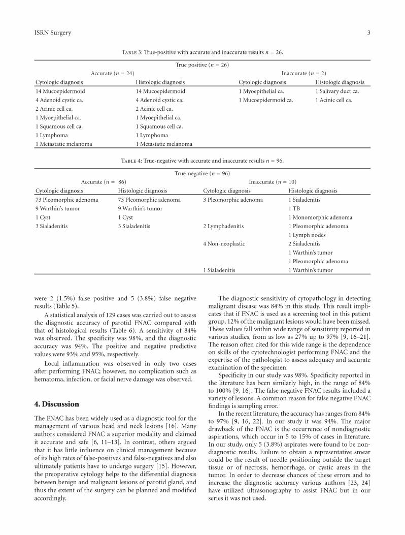
An overall of eighty-five percent (110/129) concordancebetween FNAC and final histological diagnosis was estab-lished, 78% with malignant and 88% with benign caseson break up (Table 2). The cytological diagnosis was true-positive in 26 (20%) cases and true-negative in 96 (74%)cases. Twenty-four of the 26 (92%) true-positive neoplasmsand 86 of the 96 (90%) true-negative benign lesions weredetermined as an accurate results (Tables 3 and 4). There
ISRN Surgery 3
Table 3: True-positive with accurate and inaccurate results n = 26.
True positive (n = 26)
Accurate (n = 24) Inaccurate (n = 2)
Cytologic diagnosis Histologic diagnosis Cytologic diagnosis Histologic diagnosis
14 Mucoepidermoid 14 Mucoepidermoid 1 Myoepithelial ca. 1 Salivary duct ca.
4 Adenoid cystic ca. 4 Adenoid cystic ca. 1 Mucoepidermoid ca. 1 Acinic cell ca.
2 Acinic cell ca. 2 Acinic cell ca.
1 Myoepithelial ca. 1 Myoepithelial ca.
1 Squamous cell ca. 1 Squamous cell ca.
1 Lymphoma 1 Lymphoma
1 Metastatic melanoma 1 Metastatic melanoma
Table 4: True-negative with accurate and inaccurate results n = 96.
True-negative (n = 96)
Accurate (n = 86) Inaccurate (n = 10)
Cytologic diagnosis Histologic diagnosis Cytologic diagnosis Histologic diagnosis
73 Pleomorphic adenoma 73 Pleomorphic adenoma 3 Pleomorphic adenoma 1 Sialadenitis
9 Warthin’s tumor 9 Warthin’s tumor 1 TB
1 Cyst 1 Cyst 1 Monomorphic adenoma
3 Sialadenitis 3 Sialadenitis 2 Lymphadenitis 1 Pleomorphic adenoma
1 Lymph nodes
4 Non-neoplastic 2 Sialadenitis
1 Warthin’s tumor
1 Pleomorphic adenoma
1 Sialadenitis 1 Warthin’s tumor
were 2 (1.5%) false positive and 5 (3.8%) false negativeresults (Table 5).
A statistical analysis of 129 cases was carried out to assessthe diagnostic accuracy of parotid FNAC compared withthat of histological results (Table 6). A sensitivity of 84%was observed. The specificity was 98%, and the diagnosticaccuracy was 94%. The positive and negative predictivevalues were 93% and 95%, respectively.
Local inflammation was observed in only two casesafter performing FNAC; however, no complication such ashematoma, infection, or facial nerve damage was observed.
4. Discussion
The FNAC has been widely used as a diagnostic tool for themanagement of various head and neck lesions [16]. Manyauthors considered FNAC a superior modality and claimedit accurate and safe [6, 11–13]. In contrast, others arguedthat it has little influence on clinical management becauseof its high rates of false-positives and false-negatives and alsoultimately patients have to undergo surgery [15]. However,the preoperative cytology helps to the differential diagnosisbetween benign and malignant lesions of parotid gland, andthus the extent of the surgery can be planned and modifiedaccordingly.
The diagnostic sensitivity of cytopathology in detectingmalignant disease was 84% in this study. This result impli-cates that if FNAC is used as a screening tool in this patientgroup, 12% of the malignant lesions would have been missed.These values fall within wide range of sensitivity reported invarious studies, from as low as 27% up to 97% [9, 16–21].The reason often cited for this wide range is the dependenceon skills of the cytotechnologist performing FNAC and theexpertise of the pathologist to assess adequacy and accurateexamination of the specimen.
Specificity in our study was 98%. Specificity reported inthe literature has been similarly high, in the range of 84%to 100% [9, 16]. The false negative FNAC results included avariety of lesions. A common reason for false negative FNACfindings is sampling error.
In the recent literature, the accuracy has ranges from 84%to 97% [9, 16, 22]. In our study it was 94%. The majordrawback of the FNAC is the occurrence of nondiagnosticaspirations, which occur in 5 to 15% of cases in literature.In our study, only 5 (3.8%) aspirates were found to be non-diagnostic results. Failure to obtain a representative smearcould be the result of needle positioning outside the targettissue or of necrosis, hemorrhage, or cystic areas in thetumor. In order to decrease chances of these errors and toincrease the diagnostic accuracy various authors [23, 24]have utilized ultrasonography to assist FNAC but in ourseries it was not used.

4 ISRN Surgery
Table 5: False Positive and Negative results.
False-negative (n = 5) False-positive (n = 2)
Cytologic diagnosis Histologic diagnosis Cytologic diagnosis Histologic diagnosis
1 Pleomorphic adenoma 1 Malignant mixed tumor 1 Myoepithelial ca. 1 Pleomorphic adenoma
1 Cyst 1 Mucoepidermoid ca. 1 Mucoepidermoid ca. 1 Pleomorphic adenoma
1 Warthin’s tumor 1 Carcinosarcoma
1 Non-neoplastic 1 Mucoepidermoid ca.
1 Lymphadenitis 1 Lymphoma
Table 6: Comparison of histological results in 129 cases withpreoperative cytology results.
Histological diagnosis
Benign Malignant Total
FNAC DiagnosisBenign 96 TN 5 FN 101
Malignant 2 FP 26 TP 28
Total 98 31 129
TN = true-negative, TP = true positive, FN = false negative, and FP = falsepositive.
Because of the varied, complex and overlapping morpho-logical features, the diagnosis of pleomorphic, monomorphicadenoma, and adenoid cystic carcinoma can be sometimesproblematic. We had one case of monomorphic adenomawhich was reported pleomorphic on FNAC. Similarly a caseof carcinosarcoma was labelled as Warthin’s tumor by FNAC(Table 5). The rate of false negative results was 3.8% in ourreview which is significantly less than what Zurrida et al.[9] and others have reported [16, 25]. Two pleomorphicadenoma were misinterpreted by FNAC and diagnosed asmucoepidermoid and myoepithelial carcinoma. This couldhave happened because of lack of typical features and thepresence of atypical squamous cells on FNAC [26].
We correctly typed pleomorphic adenoma in 73 of 77(95%) cases, better than the reported ranges from 82 to94% in the literature [9, 27]. Mucoepidermoid carcinoma,according to Cohen et al. [28] is one of the most challenginglesions to diagnose and type cytologically. However, in ourseries 14 out of 16 were correctly classified. The two aciniccell and 4 adenoid cystic carcinoma were also correctlyrecognized in our series.
According to the experience of Shaha et al. [29] thediagnosis of the malignant lymphoma using the aspiratedcytology is possible. Zurrida et al. [9] reported correctidentification of only 2 of 7 cases of parotid lymphomaby FNAC while in a series of Al-Khafaji et al. [30], all tenlymphomas were accurately identified. In our study we wereable to diagnose correct only 1 lymphoma of two cases.The fact that it was not detected as malignant on FNAC isconsistent with other reports that lymphoma is difficult todiagnose by FNAC [31] and if a lymphoma is suspected onclinical grounds, flow cytometry of FNAC can be an adjunctin diagnosing it.
Still, to some authors, role of FNAC in the diagnosis ofparotid lesions has not been well taken [32]. The only relative
contraindication of the performing the FNAC could be thehemorrhagic disease. Many authors exclude the possibilityof the implantation of the malignant cells or its recurrencecaused by FNAB [33]. The phenomenon of tumor cellsseeding has become a rare complication with the currentuse of small-bored needles [34]. We only had two casesof FNAC postprocedure local inflammation which resolvedsubsequently. No other complications such as hematoma,nerve damage, or infection was observed in our series.
The most important questions to be answered by thisstudy is whether results gained from FNAC can be useful inthe clinical management of patient with parotid lesions. Ourexperience has demonstrated a variety of circumstances inwhich such data may be valuable. The usual recommenda-tion for the neoplastic lesions regardless of the preoperativecytological diagnosis is excision but recognition of benignlesion, beforehand like in case of Warthin’s tumor, in poorrisk patients may be of benefit in avoiding inappropriatesurgery.
5. Conclusion
Our study shows that preoperative FNAC plays an importantrole in the accurate diagnosis of parotid tumors. It is asafe and effective modality for the treatment of patientswith parotid lesion. This office based procedure is reliable,well tolerated, easy to perform and cost effective. Moreoverpreoperative differentiation of tumors may help prepareboth the surgeon and patient for an appropriate surgicalprocedure.
Conflict of Interests
The authors declare that they have no conflict of interest.
Funding
The authors received no funding for this paper.
References
[1] N. E. Hugo, P. McKinney, and B. H. Griffith, “Management oftumors of the parotid gland,” Surgical Clinics of North America,vol. 53, no. 1, pp. 105–111, 1973.
[2] L. S. Dudheon and C. V. Patrick, “A new method for the rapidmicroscopical diagnosis of tumors,” British Journal of Surgery,vol. 15, no. 58, pp. 250–261, 1927.

ISRN Surgery 5
[3] H. E. Martin and E. B. Ellis, “Biopsy by needle puncture andaspiration,” Annals of Surgery, vol. 92, no. 2, pp. 169–181, 1930.
[4] W. F. McGuirt and B. F. McCabe, “Significance of node biopsybefore definitive treatment of cervical metastatic carcinoma,”Laryngoscope, vol. 88, no. 4, pp. 594–597, 1978.
[5] A. Costas, P. Castro, R. Martın-Granizo, F. Monje, C. Marron,and A. Amigo, “Fine needle aspiration biopsy (FNAB) forlesions of the salivary glands,” British Journal of Oral andMaxillofacial Surgery, vol. 38, no. 5, pp. 539–542, 2000.
[6] C. J. Stewart, K. MacKenzie, G. W. McGarry, and A. Mowat,“Fine-needle aspiration cytology of salivary gland a review of341 cases,” Diagnostic Cytopathology, vol. 22, no. 3, pp. 139–146, 2000.
[7] E. A. Allen, S. Z. Ali, and S. Mathew, “Lymphoid lesions of theparotid,” Diagnostic Cytopathology, vol. 21, no. 3, pp. 170–173,1999.
[8] U. Schroder, H. E. Eckel, V. Rasche, G. Arnold, M. Ortmann,and E. Stennert, “Value of fine needle aspiration biopsy in thediagnosis of the parotid gland neoplasm,” HNO, vol. 48, no. 6,pp. 421–429, 2000.
[9] S. Zurrida, L. Alasio, N. Tradati, C. Bartoli, F. Chiesa, and S.Pilotti, “Fine needle aspiration of parotid masses,” Cancer, vol.72, no. 8, pp. 2306–2311, 1993.
[10] E. R. Owen, A. K. Banerjee, A. J. N. Prichard, E. A. Hudson,and A. E. Kark, “Role of fine-needle aspiration cytology andcomputed tomography in the diagnosis of parotid swellings,”British Journal of Surgery, vol. 76, no. 12, pp. 1273–1274, 1989.
[11] M. A. S. Frable and W. J. Frable, “Fine-needle aspiration bi-opsy revisited,” Laryngoscope, vol. 92, no. 12, pp. 1414–1418,1982.
[12] P. O’Dwyer, W. B. Farrar, A. G. James, W. Finkelmeir, and D.P. McCabe, “Needle aspiration biopsy of major gland tumors.Its value,” Cancer, vol. 57, no. 3, pp. 554–557, 1986.
[13] C. Lussier, J. Klijanienko, and P. Vielh, “Fine-needle aspirationof metastatic nonlymphomatous tumors to the major salivaryglands: a clinicopathologic study of 40 cases cytologicallydiagnosed and histologically correlated,” Cancer, vol. 90, no.6, pp. 350–356, 2000.
[14] E. S. Liu, J. M. Bernstein, N. Sculerati, and H. C. Wu, “Fineneedle aspiration biopsy of pediatric head and neck masses,”International Journal of Pediatric Otorhinolaryngology, vol. 60,no. 2, part 1, pp. 135–140, 2001.
[15] J. G. Batsakis, N. Sneige, and A. K. El-Naggar, “Fine needleaspiration in salivary glands: its utility and tissue effects,”Annals of Otology, Rhinology and Laryngology, vol. 101, no. 2pt I, pp. 185–188, 1992.
[16] T. Atula, R. Grenman, P. Laippala, and P. J. Klemi, “Fine-needle aspiration biopsy in the diagnosis of parotid glandlesions: evaluation of 438 biopsies,” Diagnostic Cytopathology,vol. 15, no. 3, pp. 185–190, 1996.
[17] D. S. Y. Wong and G. K. H. Li, “The role of fine-needleaspiration cytology in the management of parotid tumors: acritical clinical appraisal,” Head and Neck, vol. 22, no. 5, pp.469–473, 2000.
[18] G. Jayaram, A. K. Verma, N. Sood, and N. Khurana, “Fineneedle aspiration cytology of salivary gland lesions,” Journalof Oral Pathology and Medicine, vol. 23, no. 6, pp. 256–261,1994.
[19] P. Boccato, G. Altavilla, and S. Blandamura, “Fine needleaspiration biopsy of salivary gland lesions: a repraisal of pitfallsand problems,” Acta Cytologica, vol. 42, no. 4, pp. 888–898,1998.
[20] R. S. Cajulis, S. T. Gokaslan, G. H. Yu, and D. Frias-Hidvegi,“Fine-needle aspiration of salivary glands,” Acta Cytologica,vol. 41, no. 5, pp. 1412–1420, 1997.
[21] C. G. Que Hee and C. F. Perry, “Fine-needle aspirationcytology of parotid tumors: is it useful?” ANZ Journal ofSurgery, vol. 71, no. 6, pp. 345–348, 2001.
[22] S. Aversa, C. Ondolo, E. Bollito, G. Fadda, and S. Conticello,“Preoperative cytology in the management of parotid neo-plasms,” American Journal of Otolaryngology, vol. 27, no. 2, pp.96–100, 2006.
[23] Y. Akbas, E. U. Tuna, A. Demireller, H. Ozcan, and C. Ekinci,“Ultrasonography guided fine needle aspiration biopsy ofparotid gland masses,” Kulak Burun Bogaz Ihtisas Dergisi, vol.13, no. 1-2, pp. 15–18, 2004.
[24] R. Pratap, A. Qayyum, N. Ahmed, P. Jani, and L. H. Berman,“Ultrasound-guided core needle biopsy of parotid glandswellings,” Journal of Laryngology and Otology, vol. 123, no. 4,pp. 449–452, 2009.
[25] D. B. Pitts, R. L. Hilsinger Jr., E. Karandy, J. C. Ross, and J.E. Caro, “Fine-needle aspiration in the diagnosis of salivarygland disorder in the community hospital setting,” Archivesof Otolaryngology Head and Neck Surgery, vol. 118, no. 5, pp.479–482, 1992.
[26] O. David, S. Blaney, and M. Hearp, “Parotid gland fine-needleaspiration aspiration cytology: an approach to differentialdiagnosis,” Diagnostic Cytopathology, vol. 35, no. 1, pp. 47–56,2007.
[27] S. R. Orell, “Diagnostic difficulties in the interpretation of fineneedle aspirates of salivary gland lesions,” Cytopathology, vol.6, no. 5, pp. 285–300, 1995.
[28] M. B. Cohen, P. E. Fisher, E. A. Holly, B. M. Ljung, T.Lowhagen, and K. Bottles, “Fine-needle aspiration biopsydiasgnosis of mucoepedermoid carcinoma,” Acta Cytologica,vol. 34, no. 1, pp. 43–49, 1990.
[29] A. R. Shaha, C. Webber, T. Di Maio, and B. M. Jaffe, “Needleaspiration biopsy in salivary gland lesions,” American Journalof Surgery, vol. 160, no. 4, pp. 373–376, 1990.
[30] B. M. Al-Khafaji, B. R. Nestok, and R. L. Katz, “Fine needleaspiration of 154 parotid masses with histologic correlation,”Cancer, vol. 84, no. 3, pp. 153–159, 1998.
[31] J. A. Tworek and C. W. Michael, “Parotid gland mucosaassociated lymphomas and their cytologic mimics,” Seminarsin Diagnostic Pathology, vol. 18, no. 2, pp. 147–150, 2001.
[32] H. Bayramoglu, E. Duzcan, M. Akbulut, and B. Topuz,“Infarction after fine-needle aspiration biopsy of pleomorphicadenoma of the parotid gland,” Acta Cytologica, vol. 45, no. 6,pp. 1008–1010, 2001.
[33] M. D. Glant, “Cytopathology of lymph nodes in nonspecificreactive hyperplasia. Prognostication and differential diag-noses,” American Journal of Clinical Pathology, vol. 108, no. 4,supplement 1, pp. S31–S55, 1997.
[34] R. G. Amedee and N. R. Dhurandhar, “Fine-needle aspirationbiopsy,” Laryngoscope, vol. 111, no. 9, pp. 1551–1557, 2001.

Submit your manuscripts athttp://www.hindawi.com
Stem CellsInternational
Hindawi Publishing Corporationhttp://www.hindawi.com Volume 2014
Hindawi Publishing Corporationhttp://www.hindawi.com Volume 2014
MEDIATORSINFLAMMATION
of
Hindawi Publishing Corporationhttp://www.hindawi.com Volume 2014
Behavioural Neurology
EndocrinologyInternational Journal of
Hindawi Publishing Corporationhttp://www.hindawi.com Volume 2014
Hindawi Publishing Corporationhttp://www.hindawi.com Volume 2014
Disease Markers
Hindawi Publishing Corporationhttp://www.hindawi.com Volume 2014
BioMed Research International
OncologyJournal of
Hindawi Publishing Corporationhttp://www.hindawi.com Volume 2014
Hindawi Publishing Corporationhttp://www.hindawi.com Volume 2014
Oxidative Medicine and Cellular Longevity
Hindawi Publishing Corporationhttp://www.hindawi.com Volume 2014
PPAR Research
The Scientific World JournalHindawi Publishing Corporation http://www.hindawi.com Volume 2014
Immunology ResearchHindawi Publishing Corporationhttp://www.hindawi.com Volume 2014
Journal of
ObesityJournal of
Hindawi Publishing Corporationhttp://www.hindawi.com Volume 2014
Hindawi Publishing Corporationhttp://www.hindawi.com Volume 2014
Computational and Mathematical Methods in Medicine
OphthalmologyJournal of
Hindawi Publishing Corporationhttp://www.hindawi.com Volume 2014
Diabetes ResearchJournal of
Hindawi Publishing Corporationhttp://www.hindawi.com Volume 2014
Hindawi Publishing Corporationhttp://www.hindawi.com Volume 2014
Research and TreatmentAIDS
Hindawi Publishing Corporationhttp://www.hindawi.com Volume 2014
Gastroenterology Research and Practice
Hindawi Publishing Corporationhttp://www.hindawi.com Volume 2014
Parkinson’s Disease
Evidence-Based Complementary and Alternative Medicine
Volume 2014Hindawi Publishing Corporationhttp://www.hindawi.com
Related Documents