Diagnostic Techniques Diagnostic Techniques Michael Del Core, M.D. Michael Del Core, M.D.

Diagnostic Techniques Michael Del Core, M.D.. History Skills History Symptoms. You need to ascertain when the problem started, what if anything brought.
Dec 25, 2015
Welcome message from author
This document is posted to help you gain knowledge. Please leave a comment to let me know what you think about it! Share it to your friends and learn new things together.
Transcript
Diagnostic TechniquesDiagnostic Techniques
Michael Del Core, M.D.Michael Del Core, M.D.
History SkillsHistory Skills
HistoryHistory SymptomsSymptoms. You need to ascertain when the . You need to ascertain when the problem started, what if anything brought it problem started, what if anything brought it on, how frequently it happens, how long it on, how frequently it happens, how long it lasts, what makes it better, what makes it lasts, what makes it better, what makes it worse and what other symptoms are worse and what other symptoms are associated. associated.

History SkillsHistory Skills
SymptomsSymptoms– Chest pain or discomfortChest pain or discomfort– Dyspnea, orthopnea, paroxismal nocturnal Dyspnea, orthopnea, paroxismal nocturnal
dyspnea, wheezing, cough, hemoptysisdyspnea, wheezing, cough, hemoptysis– Peripheral findings: Edema, pain in the Peripheral findings: Edema, pain in the
extremities (claudication)extremities (claudication)– PalpitationsPalpitations– Lightheadedness, dizziness, syncopeLightheadedness, dizziness, syncope– Fatique and weaknessFatique and weakness

History SkillsHistory Skills
Chest PainChest Pain– Myocardial IschemiaMyocardial Ischemia– Pericardial PainPericardial Pain– Aortic DissectionAortic Dissection– Pulmonary EmbolismPulmonary Embolism– MusculoskeletalMusculoskeletal
History SkillsHistory Skills
Chest PainChest Pain– LocationLocation– RadiationRadiation– NatureNature– DurationDuration– Associated symptomsAssociated symptoms
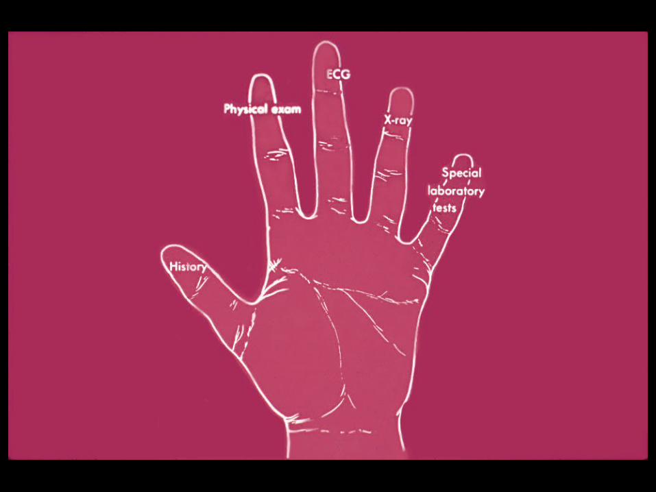
Physical Examination SkillsPhysical Examination Skills
Essential ComponentsEssential Components– General appearance of the patientGeneral appearance of the patient– Vital signsVital signs– PulsesPulses – Jugular venous pressureJugular venous pressure– Cardiac and pulmonary examinationCardiac and pulmonary examination
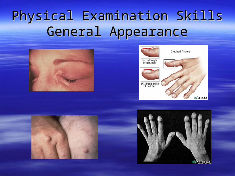
Physical Examination SkillsPhysical Examination SkillsGeneral AppearanceGeneral Appearance
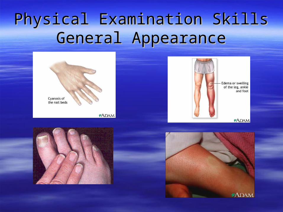
Physical Examination SkillsPhysical Examination SkillsGeneral AppearanceGeneral Appearance
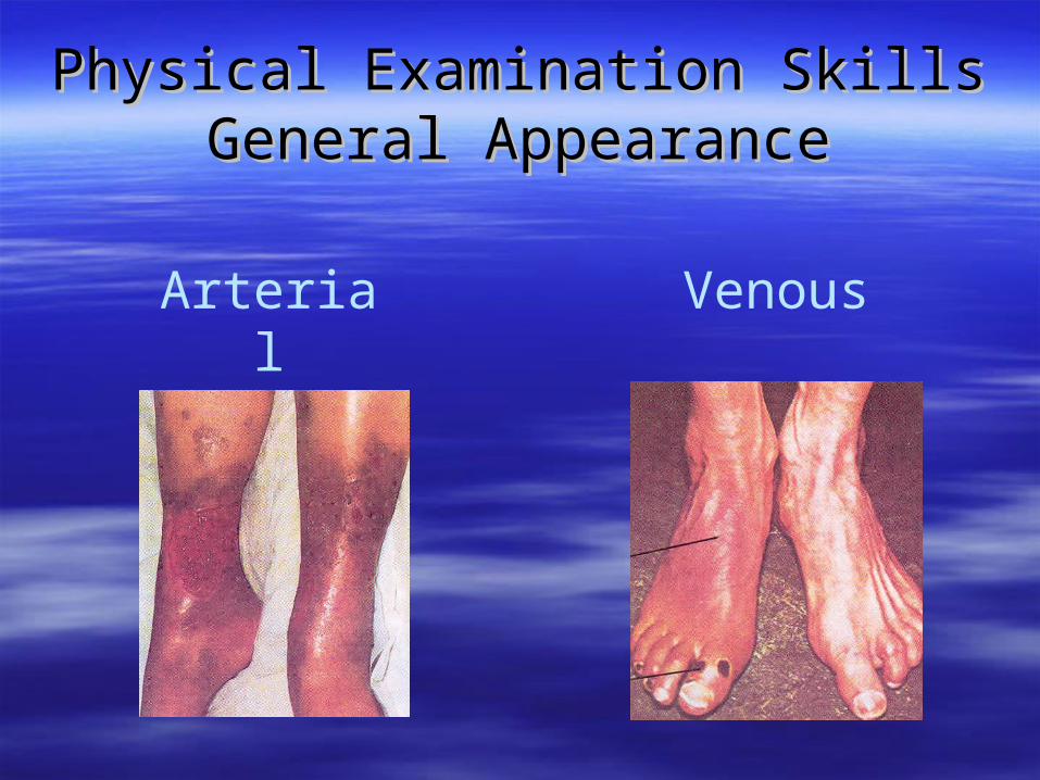
Physical Examination SkillsPhysical Examination SkillsGeneral AppearanceGeneral Appearance
Arterial Venous

Physical Examination SkillsPhysical Examination SkillsJugular Venous PulseJugular Venous Pulse

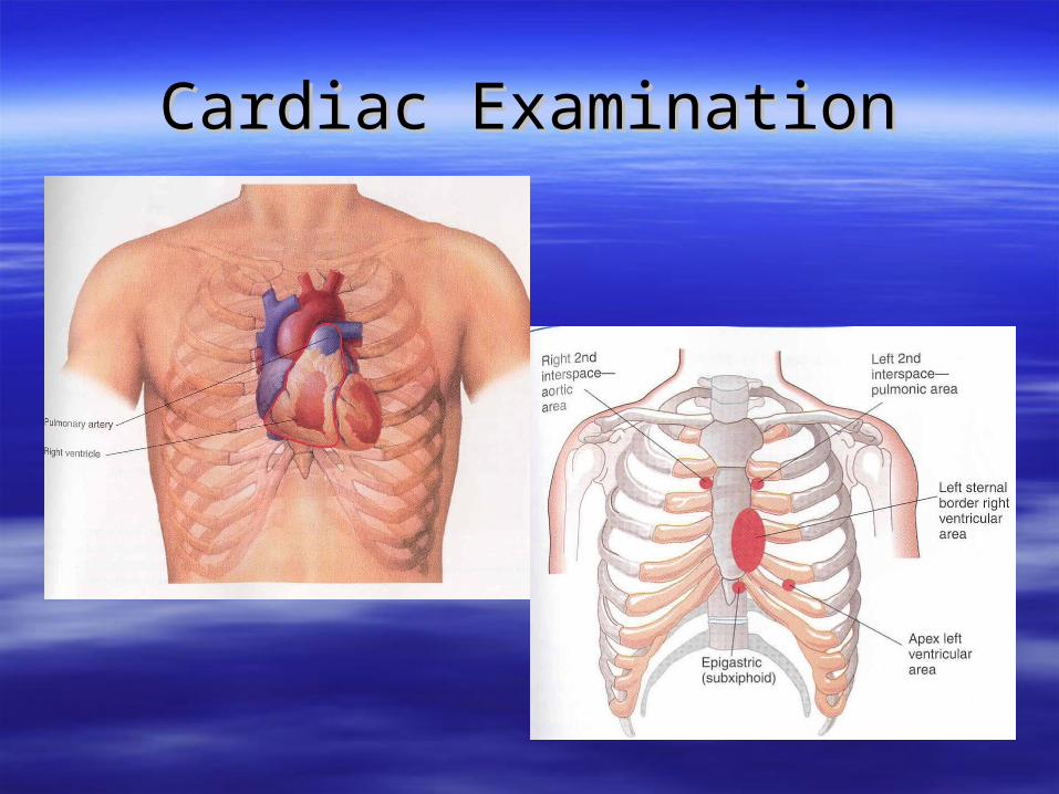
Cardiac ExaminationCardiac Examination
S1 and S2 are valve closure soundsS1 and S2 are valve closure sounds S3 and S4 are ventricular soundsS3 and S4 are ventricular sounds Murmurs are generated whenever there is Murmurs are generated whenever there is
turbulent flow in the heart. Blood flow is generally turbulent flow in the heart. Blood flow is generally laminar and uniform as it moves through the heart. laminar and uniform as it moves through the heart. Whenever there is obstruction to flow (stenotic Whenever there is obstruction to flow (stenotic valves) or abnormal flow (regurgitation) or high valves) or abnormal flow (regurgitation) or high velocities (children, thyroid abnormalities, etc.) velocities (children, thyroid abnormalities, etc.) there will be turbulent flow and a murmur.there will be turbulent flow and a murmur.
Friction rubs are caused by an inflamed Friction rubs are caused by an inflamed pericardium that rubs against each other.pericardium that rubs against each other.
Cardiac ExaminationCardiac Examination
The ElectrocardiogramThe Electrocardiogram
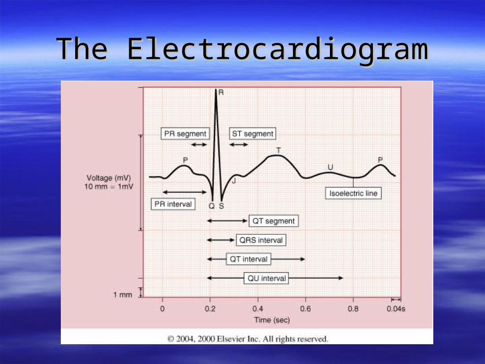
The ECG (EKG) is a recording of the The ECG (EKG) is a recording of the electrical potentials produced by cardiac electrical potentials produced by cardiac tissue. These electrical potentials are what tissue. These electrical potentials are what causes the fibers to contract. These causes the fibers to contract. These electrical impulses spread throughout the electrical impulses spread throughout the body and can be recorded on the skin by body and can be recorded on the skin by applying electrodes at various points on the applying electrodes at various points on the surface.surface.
The ElectrocardiogramThe Electrocardiogram
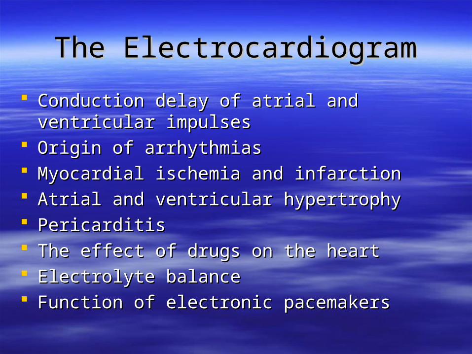
Conduction delay of atrial and ventricular impulsesConduction delay of atrial and ventricular impulses Origin of arrhythmias Origin of arrhythmias Myocardial ischemia and infarctionMyocardial ischemia and infarction Atrial and ventricular hypertrophyAtrial and ventricular hypertrophy PericarditisPericarditis The effect of drugs on the heartThe effect of drugs on the heart Electrolyte balanceElectrolyte balance Function of electronic pacemakersFunction of electronic pacemakers
The ElectrocardiogramThe Electrocardiogram
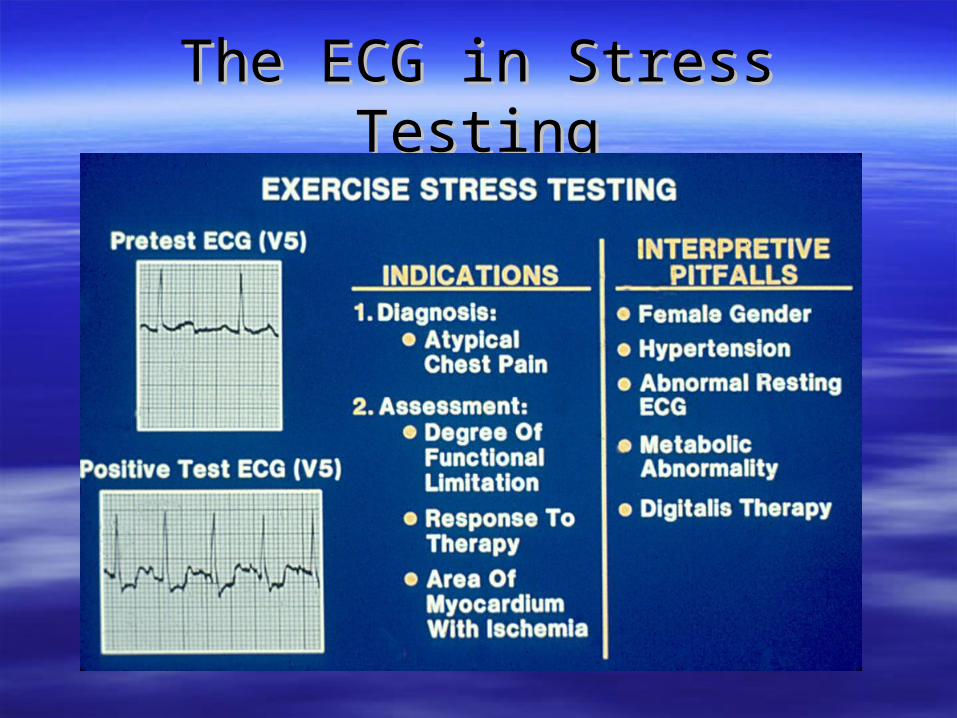
The ECG in Stress TestingThe ECG in Stress Testing
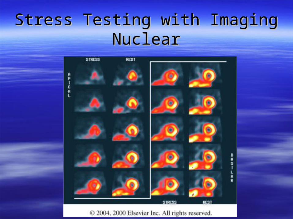
Stress Testing with ImagingStress Testing with ImagingNuclearNuclear

Stress Testing with ImagingStress Testing with ImagingEchocardiographyEchocardiography

Specialized Electrocardiographic Specialized Electrocardiographic TechniquesTechniques

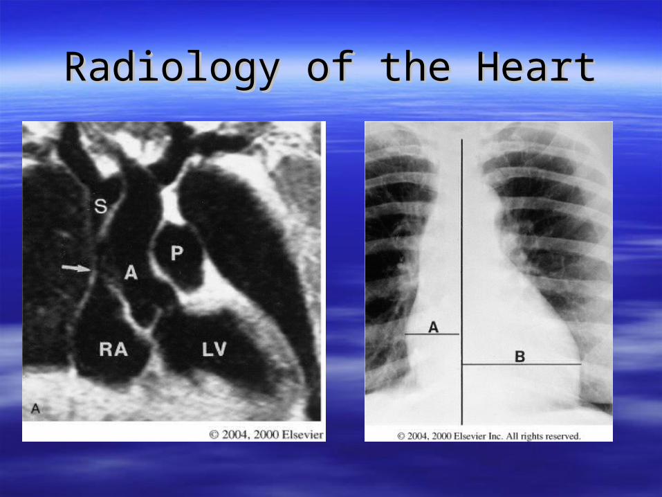
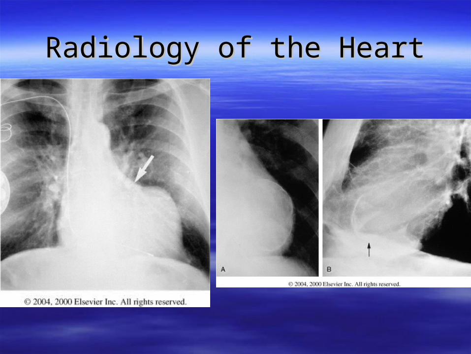
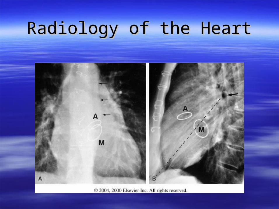
Radiology of the HeartRadiology of the Heart
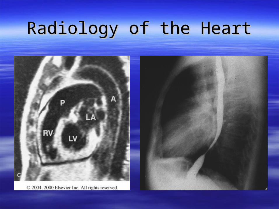
Radiology of the HeartRadiology of the Heart
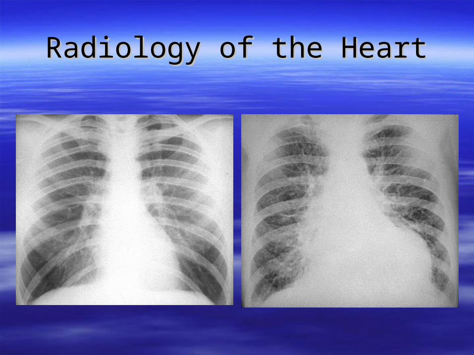
Radiology of the HeartRadiology of the Heart
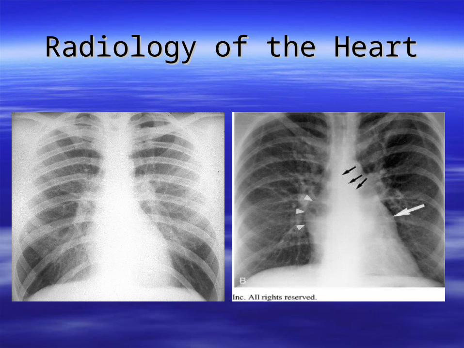
Radiology of the HeartRadiology of the Heart
Radiology of the HeartRadiology of the Heart
Radiology of the HeartRadiology of the Heart

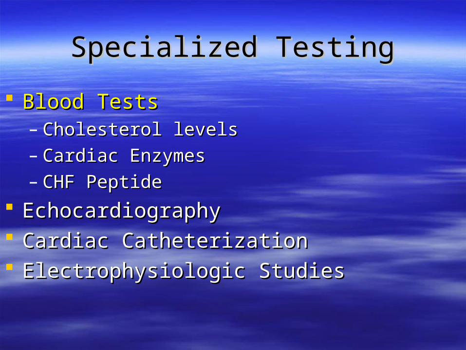
Specialized TestingSpecialized Testing
Blood TestsBlood Tests– Cholesterol levelsCholesterol levels– Cardiac EnzymesCardiac Enzymes– CHF PeptideCHF Peptide
EchocardiographyEchocardiography Cardiac CatheterizationCardiac Catheterization Electrophysiologic StudiesElectrophysiologic Studies
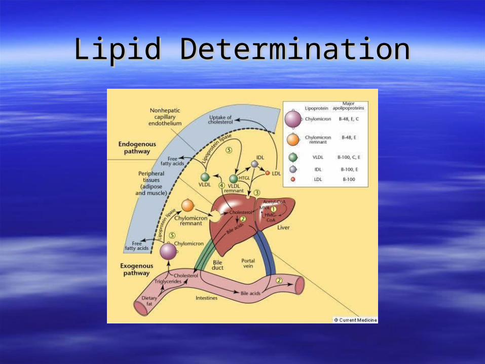
Lipid DeterminationLipid Determination
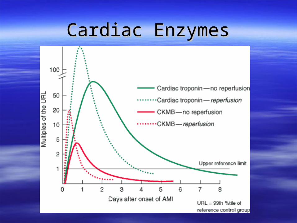
Cardiac EnzymesCardiac Enzymes
Specialized TestingSpecialized Testing
Blood TestsBlood Tests– Cholesterol levelsCholesterol levels– Cardiac EnzymesCardiac Enzymes– CHF PeptideCHF Peptide
EchocardiographyEchocardiography Cardiac CatheterizationCardiac Catheterization Electrophysiologic StudiesElectrophysiologic Studies

EchocardiographyEchocardiography
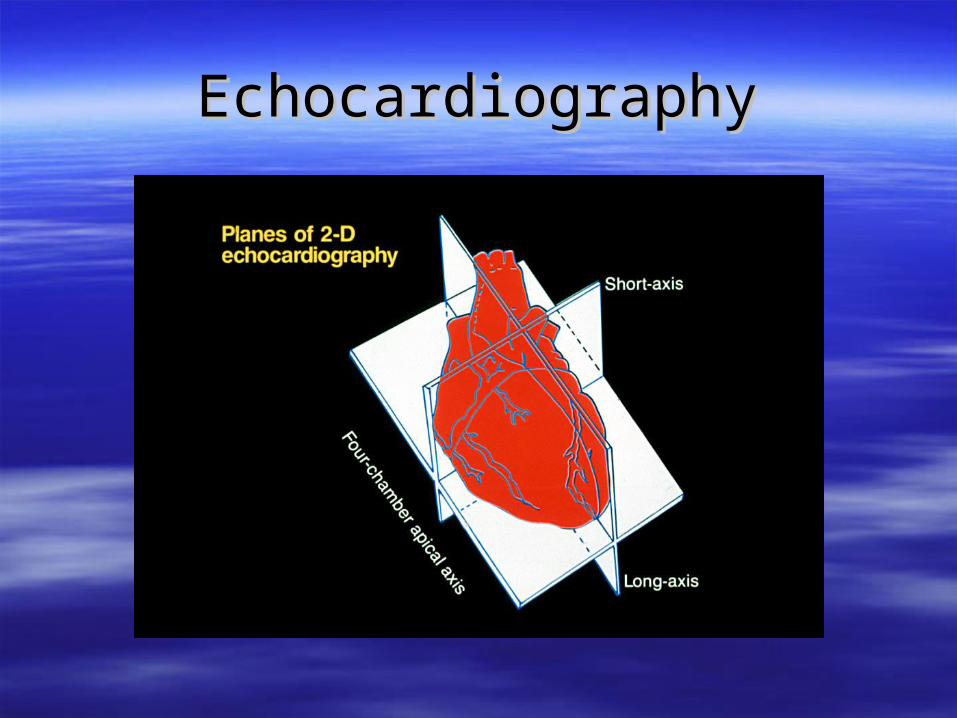
EchocardiographyEchocardiography
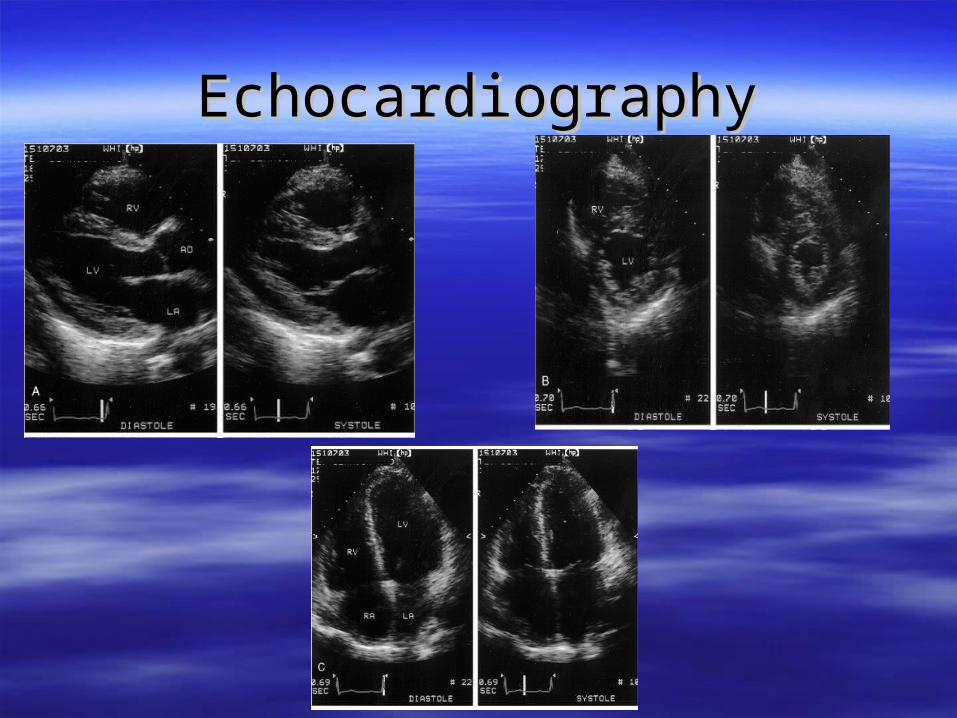
EchocardiographyEchocardiography
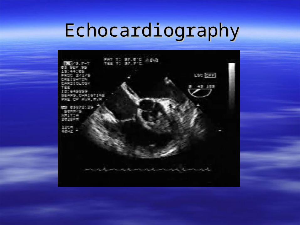
EchocardiographyEchocardiography
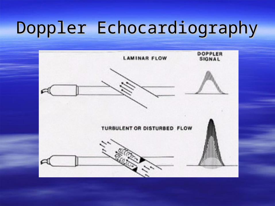
Doppler EchocardiographyDoppler Echocardiography
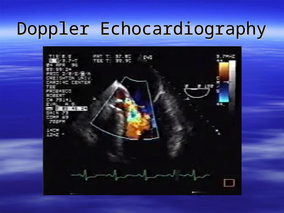
Doppler EchocardiographyDoppler Echocardiography

Specialized TestingSpecialized Testing
Blood TestsBlood Tests– Cholesterol levelsCholesterol levels– Cardiac EnzymesCardiac Enzymes– CHF PeptideCHF Peptide
EchocardiographyEchocardiography Cardiac CatheterizationCardiac Catheterization Electrophysiologic StudiesElectrophysiologic Studies
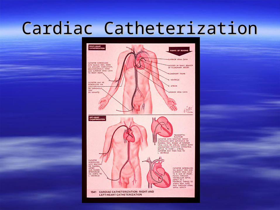
Cardiac CatheterizationCardiac Catheterization
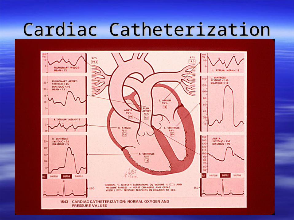
Cardiac CatheterizationCardiac Catheterization
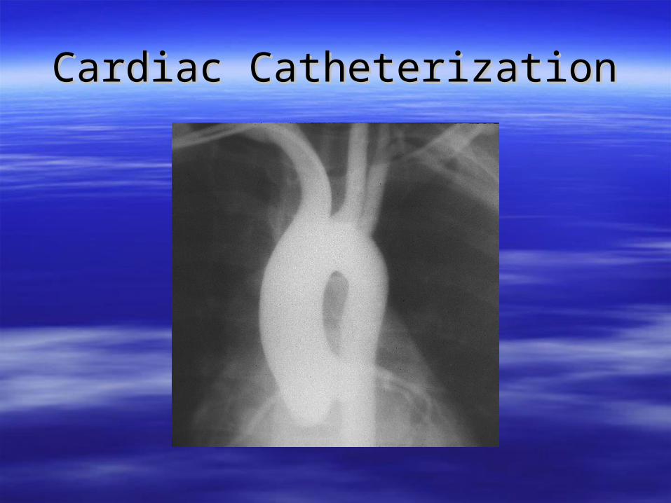
Cardiac CatheterizationCardiac Catheterization
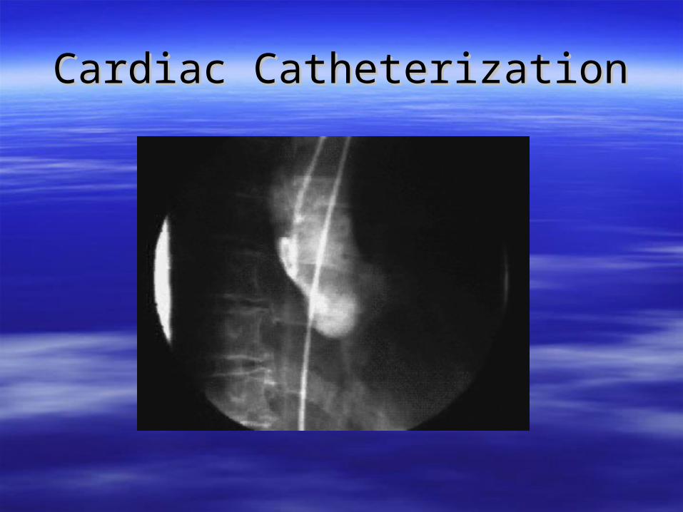
Cardiac CatheterizationCardiac Catheterization
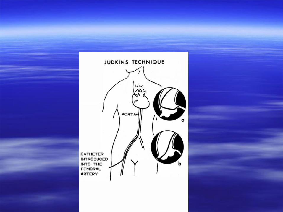
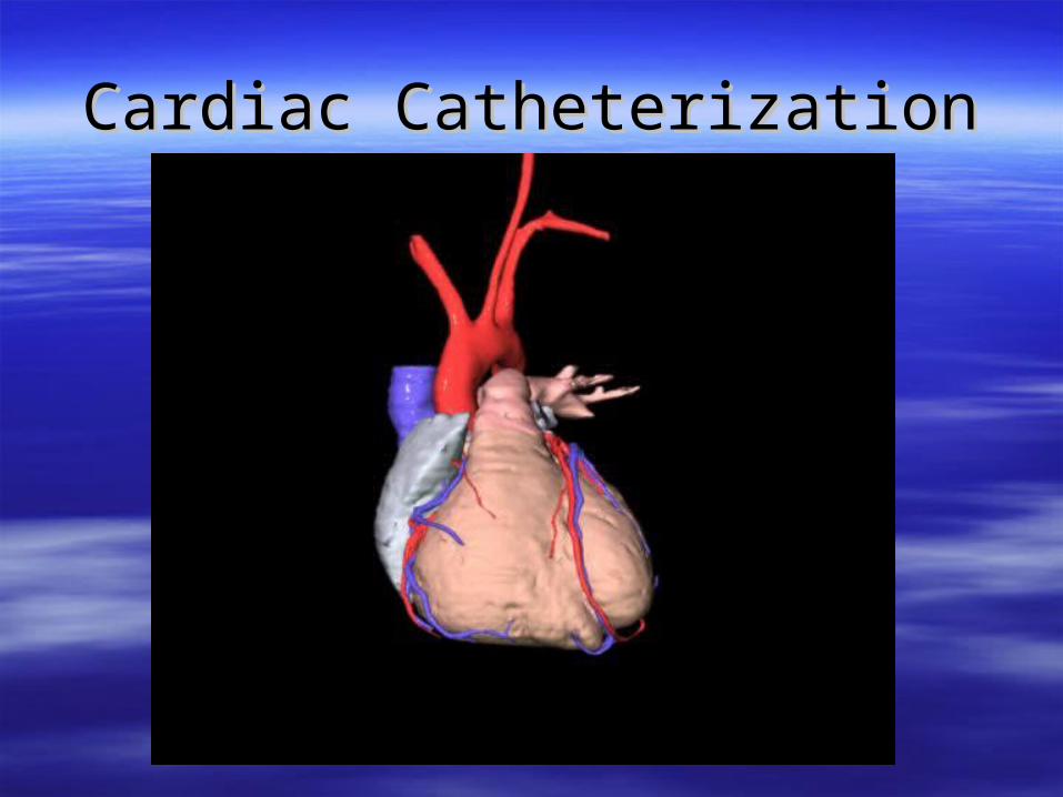
Cardiac CatheterizationCardiac Catheterization
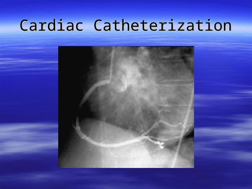
Cardiac CatheterizationCardiac Catheterization
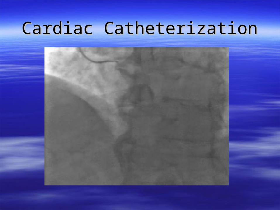
Cardiac CatheterizationCardiac Catheterization
Specialized TestingSpecialized Testing
Blood TestsBlood Tests– Cholesterol levelsCholesterol levels– Cardiac EnzymesCardiac Enzymes– CHF PeptideCHF Peptide
EchocardiographyEchocardiography Cardiac CatheterizationCardiac Catheterization Electrophysiologic StudiesElectrophysiologic Studies
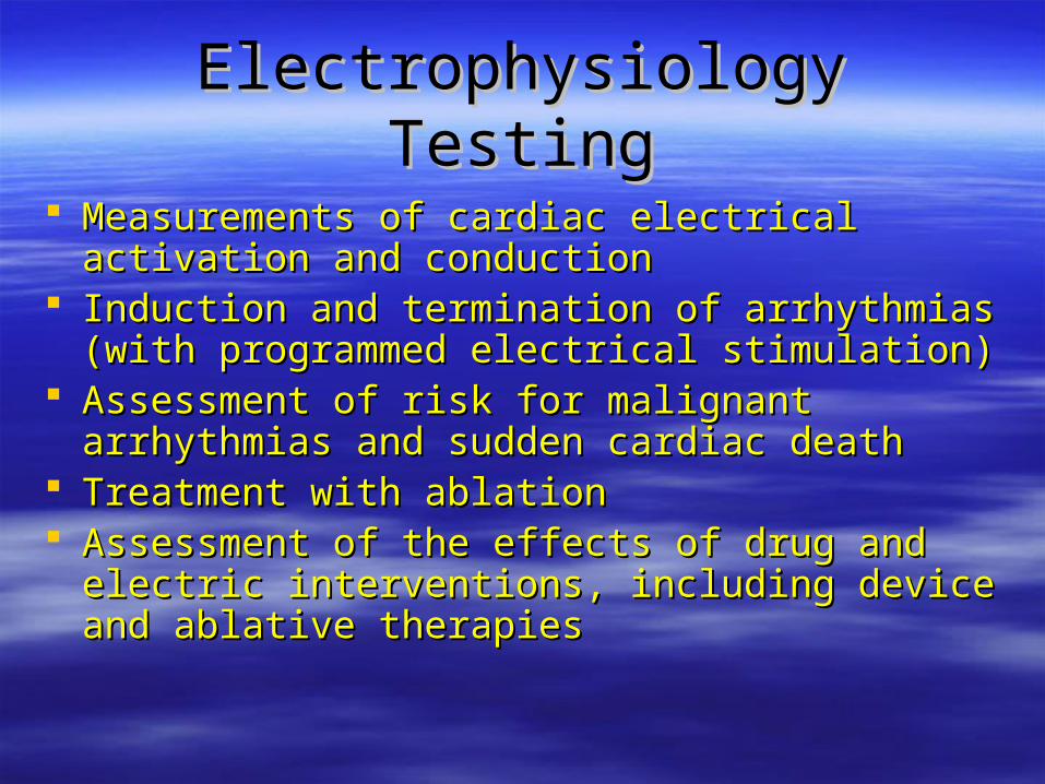
Electrophysiology TestingElectrophysiology Testing
Measurements of cardiac electrical activation and Measurements of cardiac electrical activation and conductionconduction
Induction and termination of arrhythmias (with Induction and termination of arrhythmias (with programmed electrical stimulation)programmed electrical stimulation)
Assessment of risk for malignant arrhythmias and Assessment of risk for malignant arrhythmias and sudden cardiac deathsudden cardiac death
Treatment with ablationTreatment with ablation Assessment of the effects of drug and electric Assessment of the effects of drug and electric
interventions, including device and ablative interventions, including device and ablative therapiestherapies

ElectrophysiologyElectrophysiology
Related Documents