DIAGNOSTIC ROLE OF PROCALCITONIN IN NEONATAL SEPSIS IN TERTIARY CARE HOSPITAL DISSERTATION SUBMITTED TO In partial fulfillment of the requirement for the degree of DOCTOR OF MEDICINE IN MICROBIOLOGY (Branch IV) M. D. (MICROBIOLOGY) of THE TAMIL NADU DR. M. G. R MEDICAL UNIVERSITY CHENNAI- 600032 DEPARTMENT OF MICROBIOLOGY TIRUNELVELI MEDICAL COLLEGE TIRUNELVELI- 11 MAY 2018

Welcome message from author
This document is posted to help you gain knowledge. Please leave a comment to let me know what you think about it! Share it to your friends and learn new things together.
Transcript
DIAGNOSTIC ROLE OF PROCALCITONIN IN NEONATAL SEPSIS IN
TERTIARY CARE HOSPITAL
DISSERTATION SUBMITTED TO
In partial fulfillment of the requirement for the degree of
DOCTOR OF MEDICINE IN MICROBIOLOGY
(Branch IV) M. D. (MICROBIOLOGY)
of
THE TAMIL NADU DR. M. G. R MEDICAL UNIVERSITY
CHENNAI- 600032
DEPARTMENT OF MICROBIOLOGY
TIRUNELVELI MEDICAL COLLEGE
TIRUNELVELI- 11
MAY 2018
BONAFIDE CERTIFICATE
This is to certify that the dissertation entitled “Diagnostic role of
Procalcitonin in neonatal sepsis in tertiary care hospital” submitted by
Dr.D.Jeyaganguli to the Tamilnadu Dr. M.G.R Medical University, Chennai, in
partial fulfillment of the requirement for the award of M.D. Degree Branch – IV
(Microbiology) is a bonafide research work carried out by her under direct
supervision & guidance.
Head of the Department,Department of MicrobiologyTirunelveli Medical College,
Tirunelveli.
CERTIFICATE
This is to certify that the Dissertation “Diagnostic role of procalcitonin in
neonatal sepsis in tertiary care hospital” presented here in by Dr.D.Jeyaganguli
is an original work done in the Department of Microbiology, Tirunelveli Medical
College Hospital,Tirunelveli for the award of Degree of M.D. (Branch IV)
Microbiology under my guidance and supervision during the academic period of
2015 -2018.
The DEANTirunelveli Medical College,
Tirunelveli - 627011.
DECLARATION
I solemnly declare that the dissertation titled“Diagnostic role of procalcitonin
in neonatal sepsis in tertiary care hospital” is done by me at Tirunelveli Medical
College hospital, Tirunelveli. I also declare that this bonafide work or a part of this
work was not submitted by me or any others for any award, degree, or diploma to
any other University, Board, either in or abroad.
The dissertation is submitted to The Tamilnadu Dr. M.G.R.Medical
University towards the partial fulfilment of requirements for the award of M.D.
Degree (Branch IV) in Microbiology.
Place: Tirunelveli Dr. D.Jeyaganguli,Date: Postgraduate Student,
M.D Microbiology,Department of Microbiology,Tirunelveli Medical College
Tirunelveli.

ACKNOWLEDGEMENT
I am grateful to the Dean, Dr. Siddhi athiya munaivara M.D.,Tirunelveli Medical College, Tirunelveli for all the facilities provided for thestudy.
I take this opportunity to express my profound gratitude toDr.C.Revathy,M.D., Professor and Head, Department of Microbiology,Tirunelveli Medical College, whose kindness, guidance and constantencouragement enabled me to complete this study.
I wish to thank Dr. V.Ramesh Babu, M.D., Professor ,Department ofMicrobiology, Tirunelveli Medical College, for his valuable guidance for thestudy.
I am deeply indebted to Dr. S. Poongodi@ Lakshmi,M.D., Professor,Department of Microbiology, Tirunelveli Medical College, who helped meoffering most helpful suggestions and corrective comments.
I am very grateful to Dr.B.Sorna jeyanthi,M.D., Professor, Departmentof Microbiology, Tirunelveli Medical College, for the constant support renderedthroughout the period of study and encouragement in every stage of this work.
I am highly obliged to Senior Assistant ProfessorsDr.B.Cinthujah,M.D., Dr. G.Velvizhi, M.D., Dr. G.Sucila Thangam, M.D, DrV.P.Amudha M.D.and Assistant Professors Dr I.M Regitha M.D., Dr.Gowri,M.D ,Dr.Kanagapriya,M.D. , Department of Microbiology, Tirunelveli MedicalCollege, for their evincing keen interest, encouragement, and corrective commentsduring the research period.
Special thanks are due to my co-postgraduate colleagues Dr. S. Punitharanjitham, Dr.R.P.R.Suyambu Meenakshi, Dr.V.Uma Maheswari andDr.Ambuja Sekhar for never hesitating to lend a helping hand throughout thestudy.
I would also wish to thank my junior post-graduate colleagues,Dr.M.SaiShruti , Dr.E.Manimala, Dr. Maya Kumar, Dr. L.Gracia Paul andDr.R.Uma Maheswari for their help and support.

Thanks are due to the, Messer V.Parthasarathy, V.Chandran,S.Pannerselvam, S.Santhi, S.Venkateshwari, S.Arifal Beevi, S.Abul Kalam,A.Kavitha, ,T.Jeya, K.Sindhu, Mangai,Manivannan, K.Umayavel, Sreelakshmi andother supporting staffs for their services Rendered.
I am indebted to my husband, my parents and my family for not onlytheir moral support but also for tolerating my dereliction of duty during the periodof my study.
And of course, I thank the Almighty for His presence throughout mywork. Without the Grace of God nothing would have been possible.
CERTIFICATE - II
This is certify that this dissertation work title “Diagnostic role of
procalcitonin in neonatal sepsis in tertiary care hospital”of the candidate
Dr.D.Jeyaganguli with registration Number 201514302 for the award of M.D.
Degree in the branch of MICROBIOLOGY(IV). I personally verified the
urkund.com website for the purpose of plagiarism check. I found that the uploaded
thesis file contains from introduction to conclusion page and result shows
1percentage of plagiarism in the dissertation.
Guide & Supervisor sign with Seal.

ABBREVIATIONS
ADCC – Antibody depended cell mediated cytotoxicity
ANC – Absolute neutrophil count
CRP – C-reactive protein
ELISA – Enzyme linked immuosorbent assay
EONS – Early onset neonatal sepsis
ESBL – Extended spectrum beta lactamase
ESR - Erythrocyte sedimentation rate
IL – Interleukin
LONS – Late onset neonatal sepsis
LBW – Low birth weight
MRSA – Methicillin resistant Staphylococcus aureus
NBW – Normal birth weight
PCT – Procalcitonin
TNF-α – Tumour necrosis factor α

CONTENTS
Sl. No. Title Page No.
1 INTRODUCTION
2 AIMS & OBJECTIVES
3 REVIEW OF LITERATURE
4 MATERIALS AND METHODS
5 RESULTS
6 DISCUSSION
7 SUMMARY
8 CONCLUSION
9 BIBLIOGRAPHY
10 ANNEXURE
i. Data Collection Proforma
ii. Master Chart
1. INTRODUCTION
Neonatal sepsis is a clinical syndrome characterized by systemic signs of infection,
accompanied by bacteremia within the first four weeks of life (28 days). Neonatal
sepsis is the most common cause of morbidity and mortality in neonatal
period.1Every year 135 million babies are born alive worldwide. Statistical data in
2011 estimated 3.0 million of these died during the first four weeks of life.1
Neonatal sepsis is classified into early onset neonatal sepsis (EONS) and late onset
neonatal sepsis (LONS) according to time of onset of signs and symptoms. Early
onset neonatal sepsis is defined as the onset of signs and symptoms within the first
72 hours of life.2In late onset neonatal sepsis (LONS) clinical signs and symptoms
occurs after 72 hours of age.3
EONS, which occurs within the first 72 hours of life, usually presents with
respiratory distress and pneumonia. In severe cases, the neonate may be
symptomatic at birth.4Infection may be acquired through the transplacental route
during in utero period or transcervical route during birth. Ascending infection
through the cervix, with or without rupture of the amniotic fluid membranes may
result in amnionitis, funisitis (infection of the umbilical cord), congenital
pneumonia and sepsis.4
LONS usually presents after 72 hours of age and can either be nosocomial
(hospital-acquired) or community-acquired infections. The most common cause of
late onset sepsis is nosocomial infection from neonatal intensive care unit. Preterm
babies and low birth weight infants are mainly affected. The risk factors for late
onset sepsis are prematurity, low birth weight, male sex, low serum Ig G levels,
low Apgar scores, mechanical ventilation, prolonged use of intravascular catheters,
total parenteral nutrition and delayed enteral feedings. These neonates are mainly
diagnosed to have septicemia, pneumonia or meningitis.5
Initial diagnosis of neonatal sepsis based on clinical signs and symptoms which are
non-specific as other non-infective condition like aspiration, asphyxia and
metabolic disorders may also present with similar signs mimicking sepsis. The
problem of symptom wise false positivity in diagnosing sepsis resulting in
unwarranted initiation of empirical antibiotic therapy may lead to development of
drug resistance, prolonged hospital stay, increased treatment cost and the
separation of the neonates from their mothers.
Diagnosis of neonatal sepsis is broadly classified into direct method and indirect
methods.
Direct method: Isolation of causative microorganisms of sepsis from blood.
Indirect method: Over the last decade, a variety of laboratory tests have been
developed to enhance the early and accurate identification and treatment of
neonates with suspected sepsis. Those are haematological markers, serological
sepsis markers and radiological evidences.
The gold standard method for diagnosis of neonatal sepsis is isolation of
microorganism from blood. It is time consuming procedure usually takes more
than three days for complete result and also requires well equipped laboratory and
trained personnel for better results. Hence alternative fast diagnostic test of
serological markers enabling earlier detection of neonatal sepsis might be
beneficial.
Increased neonatal mortality in neonatal sepsis scenario necessiates need of rapid
and effective diagnostic test with 100% sensitivity and 100% specificity. Such an
ideal test is not available at this point of time and yet to be invented.
Hence combinations of clinical signs, haematological and serological markers have
been proved to be a useful strategy in the diagnosis of neonatal sepsis in resource
poor settings.6 Commonly used diagnostic haematological markers such as total
leucocyte count, immature: total neutrophil ratio, platelet count, absolute
neutrophil count and micro erythrocyte sedimentation rate are less sensitive and
specific for diagnosing neonatal sepsis.
In recent days screening of serological markers such as C-reactive protein (CRP),
and various cytokines have been suggested as being useful and more sensitive
indicators for early identification of sepsis in neonates. The biomarkers are
classified into early phase marker (Interleukin-6, Interleukin-8, Tumour Necrosis
factor-α and Interferon-γ) mid phase marker (Procalcitonin) and late phase marker
(C-reactive protein).7
C-reactive protein (CRP) is an acute-phase protein synthesized by the liver within
six hours after the onset of inflammation and tissue necrosis. Its rapid synthesis,
short half-life and rapid decline with recovery, together with its increase in serious
bacterial infections, have made the CRP test popular in the diagnosis of infections.8
But it may also rise in systemic inflammatory conditions and giving rise to false
positive results which limits its usage as specific diagnostic test.
Serum procalcitonin (PCT) is one of the most promising9. PCT, a 116-amino-acid
protein with amolecular weight of 13 KDa, is the precursor in the synthesis of
calcitonin (CT)secreted by the C cells of thyroid glandin normal situation but its
levels may increaseduring septicemia, meningitis, pneumonia andurinary tract
infection. This acute phase reactant has the characters of acute phase proteins,
hormones and cytokines.10In sepsis, macrophages and the monocytic cells of the
liver are involved in the synthesis of PCT.
Serum PCT concentration rises 2-4 hours after endotoxin injection, reaches its
peak level right after 6 hours, maintains a plateau through 8 to 24 hours11 and
decreases to its normal level if the infection stimulus stops. It has been reported to
be a reliable marker for severe bacterial infections and sepsis.10PCT levels increase
in severesepsis and its plasma concentration is related to the
patient’s clinical condition. Serum PCT levelsappeared to correlate with the
severity of microbialinvasionSeveral studies have reported on the usefulness of the
quantitative measurement of PCT for an early diagnosis of sepsis in
newborns.12Actually, definitive data are lacking, which can validate CRP and PCT
as screening tools in the Emergency Department.

The aim of this study was to determine the diagnostic performance of PCT and
CRP as early diagnostic markers for the detection of neonatal sepsis in the
intensive neonatal care unit.
2.AIMS AND OBJECTIVES
To detect the bacteriological profile of neonatal sepsis and antibiotic
susceptibility pattern of the isolates in Tirunelveli Medical College.
To determine the value of C-reactive protein and Procalcitonin in
establishing the diagnosis of neonatal sepsis.
To compare the efficacy of C-reactive protein and Procalcitonin with
conventional blood culture method for the diagnosis of neonatal sepsis.
3. REVIEW OF LITRATURE
Neonatal sepsis is a clinical syndrome characterized by systemic signs of infection
and isolation of a pathogen from blood, cerebral spinal fluid and from any other
sterile site with in the first 28 days of life. Bacterial infection is a main cause of
neonatal sepsis.
It includes various systemic infections of the newborn such as septicemia,
meningitis, pneumonia, arthritis, osteomyelitis, and urinary tract infections.
Superficial infections like conjunctivitis and oral thrush are not included under
neonatal sepsis.13
In developed countries the mortality from neonatal sepsis is found to be in
declining trend due to improved health care and appropriate use of antibiotics.
But, the mortality due to neonatal sepsis is still high in developing countries
accounting up to 50% of neonatal deaths.14
Rapid diagnosis and appropriate treatment is very essential in reducing morbidity
and mortality associated with neonatal sepsis.15 The empiric use of antimicrobial
treatment to all neonates presenting with clinical symptoms of sepsis practiced in
resource poor settings in developing countries exposes neonates to adverse drug
effects and promotes the development of drug resistant strains.4
3.1 Classification of neonatal sepsis
Neonatal sepsis is classified into early onset neonatal sepsis and late onset neonatal
sepsis according to time of onset of signs and symptoms.
3.1.1 Risk factors for early onset neonatal sepsis are16
Low birth weight – less than 2500 grams or prematurity
Febrile illness in the mother with evidence of bacterial infection within two
weeks prior to delivery.
Foul smelling liquor and / or meconium stained liquor.
Prolonged rupture of membranes - more than 24 hours.
More than three vaginal examinations during lobour.
Prolonged labour – more than 24 hours.
Perinatal asphyxia.
3.1.2 Risk factors for late onset neonatal sepsis are5
Low birth weight – less than 2500 grams or prematurity.
Low Apgar scores.
Low Immuoglobulin G (IgG) levels.
Prolonged use of intravascular catheters.
Treatment with steroids.
Total parenteral nutrition.
Delayed enteral feedings.
Male sex.
The incidences of neonatal sepsis are more common in male infants compared to
female infants. This male predominance may be due to X-linked
immunoregulatory gene factor contributing to increased host’s susceptibility to
infections in male neonates.
A study conducted in Nepal during period of two years from July 2007 to June
2009 by YR Khinchi AK et al, found that among 175 neonatal sepsis cases, 65.1%
were male infants and 34.9% were female infants.17
In a study conducted by RekhaSriramet al , Sri DevarajUrs Medical College,
Karnataka, India, out of 115 clinically suspected sepsis cases, 76 (66.1%) were
males and 39 (33.9%) were females. In this study 48 cases were positive for blood
culture. Among culture positive cases, male infants were 35 (60.3%) and female
were 23 (39.7%). Male infants were predominantely affected compared to female
infants with a ratio of 1.5:1.18
A study conducted by SucilaThangamet al found that among 50
clinicallysuspected neonatal cases, EONS cases were 58% and remaining 42%
were undercategory of LONS.19
Another study from Tanzaia, during March to November 2009 done by
NeemaKayangeet al reported that 60% cases were LONS and 40% cases were
EONS among 300 clinically suspected neonatal sepsis cases.20
Recent study done by Flora Chachaet al at Catholic University of Health and
Allied Sciences, Tanzania during October 2013 to April 2014 revealed among
305clinically suspected sepsis cases, 224 cases (73.4%) were under the age group
of 0-3 days ( less than 72 hours).21
The incidence of neonatal sepsis is inversely associated to gestational age. A study
conducted by Seoet al. revealed that increased sepsis incidence of 16.6% in
preterm neonates with a gestational age less than 28 weeks and only 0.6% of
incidence of neonatal sepsis in term neonates.22
A study conducted by Rabindra N Misraet al over a period of one year from
October 2010 to October 2011 highlighted that out of 115 clinically suspected
neonatal cases, 75 were found to be culture positive cases. Among these, 75% of
proved sepsis cases were preterm and low birth weight neonates. Higher incidence
of sepsis in preterm and low birth weight neonates are due to inherent deficiency of
both humoral and cellular immunity during the first week of life.23
Long term complications are more in neonatal sepsis of preterm infants compared
to term infant with neonatal sepsis. A study of Rachel E et al in Brazil (study
period 2012-2013) reported that neurological complication are 2.5 times more in
preterm infants with very low birth weight compared to term infants.24
3.2 Causative organisms:
The causative organisms include a wide variety of gram positive and gram
negative organisms. These include Staphylococcus aureus, Coagulase negative
Staphylococcus(CONS),Escherichia coli (Ecoli), Listeria monocytogenes,
Klebsiellapneumoniae, Group B streptococcus(GBS), Acinetobacter, Serratia,
Pseudomonas, Haemophilusinfluenzae, Enterobacter, Candida and anaerobes. The
common bacterial isolates for early neonatal sepsis are Klebsiellapneumoniae,
Staphylococcus aureus and Escherichia coli. In developing countries,
Staphylococcus aureus is found to be a significant neonatal pathogen isolated from
8-22% of blood culture.25The etiological bacterial agents differ from developed to
developing countries. In 1990, Group B Streptococci was the commonest organism
for EONS in developed countries. Escherichia coli was second common
organism.26 Following Group B Streptococci prophylaxis, developed countries
identified gram negative enteric pathogens as the most common causative
organism.27
A study done by Dar es Salaam et al at Muhimbili National Hospital in Tanzania
found Staphylococcus aureus and Klebsiellaspecies to be the most common causes
of neonatal sepsis.28
A study carried out in China by Xin-Chuan Chen et al found gram positive
organism (77.4%) was predominant in neonatal sepsis in Sichuan University.29
NeelamKaisthaet al conducted a study at Government Medical College Hospital at
Chandigarh (study period July 2003 to October 2007) which revealed that gram
negative organism were responsible for 80.40% neonatal sepsis among 296 blood
culture positive cases.30
A study conducted by RajlakshmiViswanathanet al, in Institute of Post graduate
Education and Research and Seth SukhlalKarnani Memorial Hospital Kalkata
reported gram negative organism (58%) mainly enteric gram negative bacteria as
causative organism for neonatal sepsis among 216 neonatal blood culture
samples.31
A study done by NeemaKayangeet al found that among 149 culture positive cases,
61.1% of sepsis were due to gram negative bacteria. Common bacterial isolates
were Klebsiellapneumoniae, Escherichia coli and Staphylococcus aureus. More
than 50% of Klebsiellapneumoniae, Escherichia coli and other gram negative
organism were resistant to third generation cephalosporins with majority of them
Extended Spectrum Beta Lactamase (ESBL) producer. Among 32 Staphylococcus
aureus , 28% of isolates were Methicillin Resistant Staphylococcus aureus.20
Based on study conducted in North India by Kaistha N et al found that 88% gram
negative isolates were resistant to gram negative cephalosporin.32
Based on the study conducted by Emily J Welsonet al at Centres for Disease
Control and Prevention, Atlanta and various centre in USA, reported that most
common organisms were group B Streptococci (37.8%), Escherichia coli (24%),
viridans Streptococci (18%), Haemophilusinfluenzae(4%) and Staphylococcus
aureus (4%). This study showed that group B Streptococci as the commonest
pathogen of early onset neonatal sepsis.33
3.3 Pathogenesis of neonatal sepsis:
The normal amniotic membrane, placenta and amniotic fluid by itself serves as a
natural protective barrier against infection for inutero fetus by exhibiting anti-
microbial effects.34 Fatal systemic bacterial infection may occur in preterm and
also in term infants with the neonates presenting symptomatic at birth itself. This
infers bacterial infection may occur at inutero stage. Bacterial infection like
Listeria monocytogenesis an example for transplacental route of infection affecting
neonates via maternal circulation.35
3.3.1Pathogenesis of intrauterine infection
Clinical or subclinical maternal infection with agent like cytomegalovirus,
Treponema pallidum, Toxoplasma gondii, rubella virus, varicella virus and
parvovirus B19 are transmitted to fetus by transplacental transmission.
Transplacental infection may occur at any time during pregnancy. Signs of
transplacental infection may be noticed immediately after delivery or at later
periods variable from months to years. Transplacental infection may result in early
spontaneous abortion, congenital anomalies, intra uterine growth retardation,
premature birth, stillbirth or asymptomatic persistent infection with sequelae later
in life.
First trimester infection may affect organogenesis causing congenital anomalies.
Example is congenital rubella. Last trimester infection frequently results in intra
partum acquirement of pathogens. In third trimester infection, clinical
manifestation occurs sometime after birth.
3.3.2 Pathogenesis of ascending bacterial infection:
Intact amniotic membrane serves as natural protective barrier against the inutero
stage infection of fetus. Some anaerobic and aerobic microbes are found as normal
inhabitants of birth passage and those microbes may at times cause ascending
bacterial infection.
Chorioamnionitis is microbial infection of amniotic fluid and chorioamniotic
membrane. Maternal fever, lower abdominal pain, foul smelling vaginal
discharge/amniotic fluid, maternal leukocytosis, maternal and/or fetal tachycardia
are main signs and symptoms of chorioamnioitis. Prolonged rupture of membrane
(more than 24 hour)may cause chorioamionitis and inturn ascending infection.
Bacterial colonization only does not cause neonatal infection. Prematurity,
underlying illness, inoculum size, virulence of infecting organism, the innate
immune system and transplacental antibodies are main factors for early neonatal
infection. Aspiration or ingestion of bacteria in amniotic fluid, endotracheal
intubation, insertion of an umbilical vessels catheter are other factors result in early
neonatal sepsis.
A study conducted by Pyatiet al detected Group B Streptococci sepsis among 3000
newborn infants. Nearly all with early neonatal sepsis and a birth weight less than
2000 gram presented with symptoms less than one hour after birth, whereas more
than two-thirds of those with a higher birth weight developed symptoms later than
four hours.36 These findings indicate that preterm neonates may be exposed to GBS
in utero, whereas term neonates often may be exposed during the passage through
the birth canal and by aspiration of contaminated amniotic fluid, or by bacteria
penetrating through injured skin or natural body openings. In most cases this
colonisation proceeds without causing disease. The mechanism by which bacterial
colonisation converts to invasive disease is not fully understood, but it is mainly
depends upon bacterial virulence, maternal immunological factors, and the
competence of the neonatal immune system.37,34
In the study of Seoet al, on EONS (culture proven or clinical) found clinical
chorioamnionitis in 34.7% of preterms with a gestational age less than 28 weeks,
among 22.2% with gestational age 31-33 weeks, and 9.1% of term neonates.
Compared to noninfected deliveries, clinical chorioamnionitis increased the risk of
early-onset neonatal sepsis 8 to 10 times.22
3.3.3 Pathogenesis of late onset post natal infection:
In late-onset neonatal sepsis, cases are less likely to have a history of obstetric
complications, and may be infected mainly with nosocomial acquired organisms.
Post natal infection occurred by direct contact with hospital personnel, the mother,
and other family members or from inanimate sources such as contaminated
equipment.34
3.4 Immunity:
Both term and pre term infants have decreased function of neutrophils, and
decreased complement level. These factors are mainly responsible for neonatal
infection. Preterm babies have low immunoglobulin concentration compared to
term babies. Maternal IgG antibody is actively transported to fetus through the
placenta. These IgG levels are directly proportional to gestational age. Ig M and
IgA are not transferred across the placenta, although a fetus can synthesize IgM
and IgA in response to intrauterine infection. Neonatal tetanus and Group B
Streptococci infections are prevented by maternal Ig G antibody. IgM antibodies
are mainly synthesized against gram negative enteric pathogens. Usually newborn
infants lack antibody mediated protection against Escherichia coli and other
Enterobacteriaceae.
3.5 Complement:
The complement system facilitates bactericidal activity against certain organism
such as Escherichia coli and functions as an opsonin with antibody in the
phagocytosis of bacteria such as Group B Streptococci. Maternal complement does
not cross placenta. A fetus begins to synthesis complement components in first
trimester itself. Usually term newborn infants have slightly diminished classical
pathway complement activity and moderately diminished alternative pathway
activity. In comparison to term infant, preterm infants have low levels of
complement components and less compliment activities. These deficiencies
contribute to diminished complement derived chemotactic activity and to a
diminished ability to opsonize certain organisms in the absence of antibody. In
neonatal infant, opsonization of Staphylococcus aureus is normal, but various
degrees of impairment have been noted against Escherichia coli and Group B
Streptococci.
3.6 Neutrophils:
Qualitative and quantitative deficiencies of the phagocyte system mainly contribute
to neonatal sepsis.
Causes for increased susceptibility of neonates to infection are stated as
1. Abnormal Neutrophil migration at birth in both term and preterm infants.
Specifically telling decreased adhesion, aggregation and deformability of
neutrophils may delay prompt response to infection.
2. Abnormal expression of cell membrane adhesion molecules β2 integrins and
selectins and abnormalities in neonatal neutrophil cytoskeleton leads to abnormal
chemotaxis and hence impair response to infection.
3. Impaired oxidative respiratory burst of neonatal neutrophils aids in increased
risk of sepsis.
4. Decreased storage pool of neutrophil - Frequently noticed neutropenia in
preterm and intra uterine growth retarded infants attributes to the increased risk for
sepsis.
3.7 Monocyte –Macrophage system:
Prime functions of activated macrophages include antigen presentation,
phagocytosis and immune modulation. Although count of monocyte in neonatal
blood is normal, function of macrophages such as impaired chemotaxis increases
risk of sepsis.
3.8 Natural killer cells:
Natural killer cells (lymphocyte sub group) are cytolytic against cells infected with
pathogens. These cells also lyse antibody coated cells and this action is called
antibody depended cell mediated cytotoxicity (ADCC). Natural killer cells appear
early in gestation and are of equivalent numbers as in adults. However diminished
cytotoxic activity and ADCC predispose to increased susceptibility of neonates to
sepsis.
3.9 Cytokines and acute phase proteins:
Cytokines are endogenous chemical mediators that carry information between
different cells and are important factors in the human inflammatory response. They
are regulated by a complicated web of regulatory mechanisms including several
different cell types. In case of infection, both pro-inflammatory and anti-
inflammatory cytokines are upregulated according to a specific time schedule, and
so by studying this upregulation in blood samples we can conclude whether
systemic inflammation is present or not. Tumor necrosis factor-α (TNFα),
Interleukin-1 (IL-1), IL-4, IL-6, IL-8, IL-10, IL-12 and platelet-activating factor
are important chemical mediators released in various inflammatory reactions.
Potential marker for bacterial neonatal sepsis, pneumonia and necrotizing
enterocolitis are TNFα, IL-6 and IL-8.
Innate immunity also plays an important role against an infectious agent which is
due to nonspecific cellular and humoral response. Recognition of pathogens is
initiated by soluble components in plasma (including mannose binding lectin) and
by recognition of receptors monocyte and other cells.
3.10 C-Reactive Protein:
Tillet and Franchis of Rockefeller University were firstly described C – reactive
protein. They demonstrated a precipitation reaction with polysaccharide fraction C
from the peumococcal cell wall and serum of patient suffering from peumococcal
pneumonia. Serum of healthy controls and some pneumococcal pneumonia
recovered patients does not show this precipitation. In interpretation of the fact that
the polysaccharide fraction was a protein, the C-reactive component in the serum
was named C-reactive protein.38 In 1950, CRP had been detected in more than 70
various disorders including acute bacterial, viral, and other infections, as well as
noninfectious diseases such as, rheumatic disorders, acute myocardial infarction,
and various malignancies. All of these disorders of different etiology had in
common the factor of inflammation and/or tissue injury. Increased serum level of
CRP is very early and sensitive response to most of microbial infections.39
C- reactive protein (CRP) is one of the acute-phase proteins. It belongs to the
pentraxin family of ligand-binding and calcium-dependent plasma proteins. In
acute infection, serum level of CRP increased up to 50 to 100 mg/L. But in chronic
condition like rheumatoid arthritis and atherosclerosis its level generally remains
below 10 mg/L.40
3.10.1 Synthesis and metabolism:
Hepatocytes are main site for production of CRP. CRP synthesis and secreted
depends upon various response to cytokines such as Interleukin-6, Interleukin-1
and Tumour Necrosis Factor-α (TNF-α). CRP is primarily derived via IL 6-
dependent hepatic biosynthesis. Increased CRP level in neonate always represents
endogenous synthesis. CRP passes through the placenta is very very minimal. Only
single stimuli enough to hepatic synthesis of CRP, that increase the serum
concentration above 6mg/l by about 6 hours and peaking at around 48 hours.41
The stimulatory effects of cytokines on the production of acute phase proteins
increased by glucocorticoids.42 Insulin, on the other hand, decreases their effects on
the production of some acute phase proteins.43,44
3.10.2 CRP detection method:
A large number of methods are available for the detection of CRP and estimation
of CRP level in the serum. Even though, nephrometry, electroimmunoprecipitation
assay and immunometric assay are sensitive and quantitative method for the
estimation of CRP. These methods have complicated procedure, so these tests done
only well-equipped laboratories only.39 Latex agglutination test is the alternative
test, it have a quiet sensitive, rapid method to detect serum CRP in qualitatively
and semi quantitatively. The serum CRP concentration of 6mg/L or more was
considered as positive.
3.10.3 Function of C-reactive protein:
The main receptor to CRP is phosphocholine. Phosphocholine is found in
lipopolysaccharide of bacterial cell wall and in most of biological membrane. CRP
and phosphocholine binds first, after that CRP is recognized by the complement
system. CRP activates the complements system and promotes the phagocytosis by
neutrophils and macrophages. Then CRP initiates release of proinflammatory
cytokines.45,46
The sensitivities and specificities of CRP assay in the detection of neonatal sepsis
using culture as a gold standard. Sensitivity of CRP is more important than
specificity in detection of EONS and LONS.
3.10.4 Condition where CRP is elevated:
CRP level is increased in some acute conditions such as bacterial infection,
bacterial endocarditis, pneumococcal pneumonia and acute rheumatic fever. CRP
level is increased in more number of chronic condition like polyarthritis nodosa,
rheumatoid arthritis, systemic lupus erythematosis, Inflammatory bowel disease,
acute myocardial ischemia and malignancies etc.44
A study conducted by Benitz MD et al from Stanford University of Medicine
shows 54.6% of sensitivity on proven neonatal sepsis and 65.5% of sensitivity in
probable neonatal sepsis among 1002 infants. The positive predictive value was
99.7% and negative predictive s for CRP was 98.7% for conformed neonatal
sepsis. 47
Laborada G et al revealed that during study (2003), out of 105 neonatal sepsis
cases blood culture tested 48 cases were positive by automated technique. This
study also shows 69% sensitivity , 96% specificity, 93% positive predictive value
and 80% negative predictive value.48 A similar study by Doellner H et al reported
that CRP sensitivity 63%, specificity 97%, positive predictive 83% and negative
predictive value 91%. Doellner H et al study also include 36 samples are positive
among 253 blood culture.49
According to a study conducted by Franz A R et al revealed that 46 cases are
culture positive among 162 neonates with suspected sepsis and also reported 28%
of CRP sensitivity, 97% of specificity, 81% of positive predictive and 77% of
negative predictive value.50
Neonates with septicemia due to gram negative organism have higher serum CRP
level than gram positive organism.51
Recent study by Flora Chacha et al highlighted that out of 305 samples received
from clinically suspected cases of neonatal over a period of 2 years, 104 cases
showed CRP positive. This study also revealed CRP sensitivity 90% , among these
75% higher sensitivity for gram negative septicemia compare to 50% sensitivity
for gram positive septicemia.
A study conducted in Thailand by Nuntnarumit P et al found that serial
quantitative CRP measurement were found to have better predictive than complete
blood count with 100% sensitivity, 94% specificity, 91% positive predictive and
100% negative predictive. 52
A study was done in Rawalpindi Pakistan by Khassawneh M et al, comparing
CRP, absolute neutrophil count and I/T ratio, CRP was found to have a specificity
of 95% in diagnosing neonatal sepsis followed by absolute neutrophil count.53
A study conducted by Kohli-Kochhar R et al, in Port Harcourt Nigeria, the
sensitivity, specificity, positive predictive value and negative predictive of serial
CRP measurements were found to be 74.0%, 74.1%, 68.0% and 79.0%
respectively in the diagnosis of neonatal sepsis using blood culture as the gold
standard.54
In a study done by Hofer N et al, comparing CRP, interleukin 6 and
immunoglobulin M; revealed that CRP was the best among the three with 95%
sensitivity and 98% NPV in the diagnosis of early gram negative sepsis.55
Serial serum CRP measurements taken between 24 and 48 hours after the onset of
infection have been found to have high sensitivity for probable septicemia. Hence
serial CRP is suggested for the diagnosis of neonatal sepsis to predict early
infection.56 The neonates who were admitted with clinical features of neonatal
sepsis and started on empirical treatment with antibiotics following the negative
results of CRP, the physician can stop the antibiotics thus can minimize antibiotic
exposure and shorten hospital stays. The diagnostic value of serial measurements
of serum CRP levels can also be used for monitoring the severity of sepsis and
improvement after initiation of treatment.57
3.11 Procalcitonin:
Procalcitonin is an another acute phase protein which is made up of 116 amino
acids. It is a precursor of calcitonin. Within 6-8 hours of bacterial infection,
bacterial endotoxin stimulates monocytes and hepatocytes which produce
procalcitonin. Its level reaches peak at 6 – 8 hours, and stays minimum for a day.
Its half-life is upto 30hours. Procalcitonin level is increased during infection in
neonates, children and adults. Serum procalcitonin level is more increased in
bacterial infection than in viral infection. In early neonatal bacterial infection,
procalcitonin is more sensitive than CRP.58
3.11.1 Structure and production of proclcitonin:
PCT is one of a group of peptides in the calcitonin super-family of peptides. The
PCT peptide has an approximate molecular weight of 14.5 kDa and consists of a
sequence of 116 amino acids. PCT is encoded by the Calc-1 gene located on
chromosome 11p15.59 The peptide has three regions: the PCT amino terminus, the
mature calcitonin segment, and the carboxyl-terminus called katacalcin.
In the absence of infection, the production of PCT outside of the neuroendocrine
cells of the thyroid and the lung is suppressed. In the presence of sepsis, all tissues
produce PCT. Because of this dual role, PCT is considered a “homokine”.
Homokines can either act as a hormone as in the normal physiologic state or as a
cytokine during inflammatory processes.60
As with other cytokines, there is little intracellular storage of PCT during
sepsis. While synthesis of PCT is necessary for the production of calcitonin,
animal studies have shown that increased concentrations of PCT may have lethal
effects during sepsis. Administration of PCT to septic hamsters with peritonitis
doubled the death rate to over 90%. Immunoneutralization of PCT by the
administration of antiserum in septic hamster and pig studies led to increased
survival of these animals60
3.11.2 Procalcitonin detection methods:
Quantitative and qualitative (semi-quantitative) assays available for measuring
PCT
1. Qualitative tests: rapid test strips for point-of-care testing (results available in <
30 minutes)
2. Quantitative tests: use luminescence immunoassay, ELISA (results available in a
few hours).
3.11.3 Procalcitonin levels and interpretation:
Normal:˂0.1ng/mL ( infants˃72 h – adults)
Suspected lower respiratory tract infection
0.1–0.25 ng/mL – Low likelihood for bacterial infection; antibiotics discouraged.
>0.25 ng/mL – Increased likelihood for bacterial infection; antibiotics encouraged.
Suspected sepsis: Strongly consider initiating antibiotics in all unstable patients.
0.1–0.5 ng/mL – Low likelihood for sepsis.
>0.5 ng/mL – Increased likelihood for sepsis.
>2.0 ng/mL – High risk of sepsis.
>10 ng/mL – Septic shock .
A study done by Cetinkaya M et al, during the period of 2008-2009 found that the
serum procalcitonin levels were higher in the neonatal septic cases compared with
the non-septic cases. This also revealed procalcitonin and CRP had sensitivity of
97%, specicity of 91%, positive predictive value 96% and negative predictive
value of 87%. The inference was procalcitonin more than 2.3 ng/ml or CRP more
than 30 mg/l denotes a possibility of EONS and LONS. In such condition
antimicrobial treatment need to be carried over in the absence of positive culture.51In a recent study Koksal et al concluded that serum procalcitonin level wassuperior to serum CRP level in terms of early diagnosis of neonatal sepsis, indetecting the severity of the illness and in evaluation of the response toantibiotic treatment.Athhan et al in their study revealed that at 7th day of therapy neonates whohad achieved clinical recovery had a significant decrease of procalcitoninlevels compared to the initial values.Carol et al in their study showed that procalcitonin is more sensitive than theCRP in the diagnosis of septicemia, meningitis and urinary tract infection.Kawezynski and Piotrowski analyzed inflammatory parameters in 48newborn infants suffering nosocomial sepsis admitted to the intensive care.They obtained samples for PCT and CRP levels just at time of onset of the signsand 24 hours later. At the onset of gram negative sepsis 14 of 17 contaminatednewborns had significantly increased PCT and CRP levels, but at the onset ofgram positive sepsis only 18 from 31 neonates with positive blood culture had
increased CRP level and 28 of them had elevated concentration of serum PCT.These differences were statistically significant3.12 Clinical features of neonatal sepsis:
Clinical features of neonatal sepsis are mainly variable. Clinical features of
neonatal sepsis is divided into non-specific features and specific features.
Non-specific features: The earliest signs of sepsis are frequently subtle and
nonspecific.
Clinical diagnosis needs a high index of suspicion for early diagnosis.
Clinical features are “hypothermia or fever, lethargy, poor cry, refusal to suck,
poor perfusion, prolonged capillary refill time, hypotonia, absent neonatal reflexes,
brady/tachycardia, respiratory distress, apnea and gasping respiration,
hypo/hyperglycemia and metabolic acidosis”.
Early manifestation of neonatal sepsis may involve only one system and
present with limited symptomatology. Initial signs and symptoms of neonatal
sepsis are temperature instability (hypothermia or fever), refusal of feeding and
edema.Signs and symptoms related to respiratory system are apnea, tachypnea,
grunting, cyanosis, retractions of chest wall and nasal flaring. Main signs and
symptoms related to cardio vascular system are pallor, cold and clammy skin,
tachycardia (more than 160 beats /min) or bradycardia (less than 100 beats /min)
and hypotension. Signs and symptoms related to central nervous systems are
lethargy, irritability tremors, convulsion, abnormal moro reflex and hypotonia.
Abdominal distension, vomiting, diarrhea and hepatomegaly are main signs and
symptoms related to gastro intestinal tract. Oliguria is main symptom related to
renal system. Signs and symptoms related to haematologic systems are jaundice,
splenomegaly, pallor, petechial purpura and bleeding. Signs and symptoms related
to skin and soft tissue are impetigo, omphalitis, scalp abscess, fascilitis, adenitis
and abscess of cystic hygroma. Most of the times, various non-infectious diseases
can coexist with neonatal sepsis, which in turn makes sepsis diagnosis a tough one.
Surfactant deficiency leading to respiratory distress syndrome can coexist with
bacterial pneumonia.61
3.13 Clinical criteria for neonatal sepsis:
3.13.1 Integrated Management of Childhood Illness criteria:
“Tachypnea (> 60 breath per minute), nasal flaring, increased chest retraction,
grunting, nasal flaring, bulging fontanel, pus draining from ear, redness around
umbilicus, temperature instability (>37.7˚C or <35.5˚C), lethargic, reduced
movements, not able to feed and convulsions” are components of integrated
management of childhood illness criteria for neonatal sepsis.
3.13.2 WHO criteria:
“Convulsion, tachypnea (> 60 breath per minute), severe chest retraction,
temperature instability (>37.7˚C or <35.5˚C), lethargic, reduced movements, not
able to feed, crepitation and cyanosis” are main components of WHO criteria for
neonatal sepsis.62
A study conducted by Jaswal RS et al, in Shimla medical collages India, found that
the most frequent clinical presentation of neonatal sepsis were respiratory distress,
lethargy and jaundice with combined frequency of 40% followed by fever and poor
feeding.63
Tanzania based study by Arif SH et al, stated the most common clinical
presentation found in neonatal sepsis were fever reported in 91% of neonates,
inability to breast feed, bulging anterior fontanelle , dyspnea, jaundice, and
seizures. Few other clinical features of neonatal sepsis included abdominal
distension, tachycardia, tachypnea, disseminated intravascular coagulopathy and
abscesses.64
3.14 Diagnosis of neonatal sepsis:
Neonatal sepsis is a potentially dangerous and serious condition. That can cause
increased mortality and morbidity very rapidly, if not treated correctly and quickly.
The ideal test to confirm neonatal sepsis should have 100% sensitivity and 100%
specificity. But such a test is unlikely to be discovered till date, due to non-specific
signs of neonatal sepsis. Many signs and symptoms of neonatal sepsis are also
present in some of noninfectious conditions like acute respiratory distress,
aspiration of amniotic fluid and hypoglycemia etc.65 Hence it remains a challenge
for physicians to correctly diagnose neonatal sepsis in a timely manner. So a rapid
reliable diagnostic test for neonatal sepsis is essential in order to initiate treatment
in suspected neonates on time so as to reduce associated morbidity and mortality.
In routine clinical practice, the recommended approach is to liberally start
intravenous antibiotics and then perform a ruling out procedure that normally lasts
for several days. If all the tests for neonatal sepsis are negative and neonate has
recovered fully, the antimicrobials can be discontinued and infant can be
discharged from the neonatal intensive care unit. This rule out procedure normally
includes various cultures such as blood, cerebrospinal fluid, urine etc, x-rays and
various markers of sepsis. If itwere possible to decrease the time taken by this
investigation, the benefits would beobvious in terms of reduced costs of treatment,
reduced infants suffering and reduced duration of antibiotics. So, there is a great
need for specific and less time consuming diagnostic methods.
A study conducted by Ng PC et al in 2004, presented a list of 58 different
laboratory tests that had already been evaluated as diagnostic tests for neonatal
sepsis.66 Another study of review article by Pierrakoset al reviewed 3370
references covering 178 biomarkers.67
3.14.1. Blood culture:
Gold standard diagnostic test for suspected neo natal sepsis is blood culture. A
small blood volume is enough for isolation of bacteriological agents as low as 0.2
to 0.5mls. But, increased blood volume 1 to 2mls is required for detection of low
bacteraemia particularly where there is history of prior use of antibiotics. Venous
blood is routinely used for blood culture. The skin should be prepared with
disinfectant solution before veupuncture. But care must be taken; disinfectant
solution does not harm skin of newborn infants.68
A study conducted in 1997 by Kellogg et al revealed that low level bacteremia was
very common in infants. So they recommended a sample volume of 6 ml for to get
optimize sensitivity. Though, this would represent around 4.5% of an infant’s
blood volume.69
Volume of blood needed for culture depends up methods. Automated blood culture
systems such as BacT/Alert required small volume of blood such as 0.5ml only.
But 1-2 ml needed for conventional method.
Schelonka RL et al found that, if one or two viable colony-forming units are in the
blood inoculated into culture media, the BacT/Alert system will detect growth
rapidly. Since there appears to be a sizable subset of neonates who are at risk of
sepsis with a colony count less than 4 CFU/ml, then a 0.5 ml inoculum of blood
into the culture media is insufficient for sensitive and timely detection of
bacteremia. One to two milliliters of blood should increase microorganism retrieval
in the face of low-colonycount sepsis by conventional blood culture method.70
In developing countries, the conventional type of blood culture method is
commonly used. Because it is less expensive to do when compared to automated
culture systems. But the procedure is labour intensive and the yield is significantly
low sensitivity than that of an automated system.71 In developed countries,
automated systems are mostly used. Main advantages of these techniques are the
blood culture to be monitored continuously and resulting in a shorter time to
identify a positive culture.72
A study conducted by Baltimore RS et al in Yale University of Medicine, New
Haevan, USA “found ninety-four cases of non-GBS early-onset sepsis were
detected between 1996 and 1999. The rate of GBS-related early-onset infection
reduced from 0.61/1000 to 0.23/1000 births, but the annual rate of non-GBS sepsis
remained steady, ranging from 0.65 to 0.68/1000 during the surveillance period.
There was an increase in the proportion of Escherichia coli infections that were
ampicillin resistant between 1996 and 1998, but the proportion decreased in
1999”.62
A study (during 1998 to 2004) conducted by Ramesh Bhat et al highlighted that
out of 2182 samples received from clinically suspected cases of early onset
neonatal sepsis 389 (17.8%) showed positive blood culture.26
A study by SubhranshuSekarKaret al (2007–2010) revealed that, among 160 blood
culture samples tested, 60(16.2%) were blood culture positive. This study was
done at Hi-Tech medical college, Bhubaneshwar, Odisha.73
A study done by SucilaThangamet al in Tamiladu during April – September 2010
revealed 28% of positive blood culture in 50 samples. Based on the study
conducted by Shrestha R K et al in Nepal medical college, Kathmandu, out of 120
suspected cases, 37 (30.8%) were found to be blood culture positive during the
period of July 2011 to January 2012.74
Even though, if ideal blood volumes are used, blood culture has obvious
limitations in sensitivity. A negative blood culture report alone cannot support
withdrawal of antibiotic treatment if the neonate’s clinical condition indicates
ongoing sepsis. So blood cultures have limited sensitivity and this method is time
consuming, and most microbiology laboratories will take one week for complete
report.75
3.14.2 Haematological marker scoring system:
This test can be used for screening for neonatal sepsis. It can be performed easily
and it is readily available in most of the settings. It is usually a combination of
various parameters from complete blood picture. Various parameters are
“Total white blood cell count
Absolute Neutrophil count (ANC)
Immature Neutrophil : Total Neutrophil Ratio
Platelet count
Micro erythrocyte sedimentation rate
C-reactive protein”.75
In complete blood count, total leukocyte count, neutrophils and platelets are
predictors of ongoing infection. Ongoing infection interpreted by extreme value of
these parameters.75,76
Non-infectious causes such as asphyxia, maternal fever and post gestational age
are elevating these parameters. This factors causes difficult in interpretation of
results.77
Total white cell count alone could not be considered valid for confirmation of
neonatal sepsis. The reasons being are its grossly varied values and narrow
transition between normal and abnormal values. Neutropenia is seen more in sepsis
rather than neutrophila. This neutropenia results from significantly raised
adherence of neutrophils to surface of endothelial cells and its increased
consumption at the area of infection.
Neutropenia also being noted in certain other condition such as inborn errors of
metabolism and asphyxia neonatorum makes it of limited value as a sole marker of
sepsis. Frequently neonatal sepsis is associated with low absolute neutrophils
count, and high I/T ratio. Low white blood cell count are more helpful if obtained
after 4 hours of life due to normal increase of white blood cell and neutrophil count
after 6 hours of life.78,79
Hematological marker scoring interpretation:
“Total leucocyte count: less than 5000/mm3 or more than 30000/mm3
Absolute Neutrophil Count (ANC): less than 1000/mm3
Immature / Total neutrophil ratio: more than 0.2
Micro Erythrocyte sedimentation rate: more than 15 mm in hour.
C-reactive protein: more than 1mg/dl”.
If two or more abnormal hematological markers are present, it should be
considered as a positive screen for neonatal sepsis. If the hematological marker
score is negative, but clinical suspicion persists, it should be repeated within 12
hours. If the hematological markers are still negative, neonatal sepsis can be
excluded. Presence of two abnormal hematological markers are associated with 93-
100% sensitivity, 83% specificity, 27% positive predictive value and 100%
negative predictive value in neonatal sepsis.78
Neonatal sepsis also causes thrombocytopenia, because of disseminated
intravascular coagulation and the damaging effects of endotoxin on platelets.
Therefore combining the parameter of the complete blood count is the form of a
Hematological markers Scoring System that has been suggested and can serve as a
screening tools.80
3.15 Prevention strategies for EONS:
Prior administration of parental antibiotics to antenatal mothers prevents EONS by
group B Streptococci to a great extent. Ampicillin or cefazolin are used as
prophylactically four hour before delivery. In mother who have mild pencillin
allergy, cefazolin may be administrated. In case of serious pencillin allergy,
clinadamycin is the drug of choice. In clindamycin resistant cases, vancomycin is
used as an alternative drug for prophylaxis.81
Indications for intranatal antibiotics are
Positive antenatal culture for group B streptococci
Premature rupture of membranes. (>18 hours)
Previous infection with group B Streptococci.
3.16 Prevention strategies for LONS:
To provide practical training for hand washing technique for entire health care
team.
To provide adequate soap and running water facilities.
To prepare standard operating procedure for all invasive methods.
To carry out regular meeting with infection control committee to monitor
infection rate.
To ensure the adequate of physicians and nurses per bed according to current
recommendations.81
4. MATERIALS AND METHODS:
The present study was conducted at the Department of Microbiology , Tirunelveli
Medical College, Tirunelveli from June 2016 to May 2017
4.1 Study group
A total of 100 clinically suspected sepsis cases in neonates (0 day to 28 days)
4.2 Inclusion criteria
Neonates who were admitted in Neonatal Intensive Care Unit at Tirunelveli
Medical College with signs suggestive of sepsis, or those who developed signs of
sepsis while they were in the ward.
4.3 Exclusion criteria:
Neonates who were on antibiotics,
Neonates who had birth asphyxia and aspiration syndromes,
Neonates who had congenital anomalies and inborn errors of metabolism.
4.4 Ethical clearance
Ethical clearance was obtained from the college ethical committee before the
commencement of the study.
4.5 Consent
Informed consent was obtained from reliable informants of neonates who
participated in the study.
4.6 Proforma:
The proforma was filled with the details like name of mother of neonates, age (in
hours or days), sex, weight of neonates at time of birth, gestational age (in weeks),
mode of delivery and clinical diagnosis and other parameters relevant to the
present study.
4.7 METHODS
Blood samples were taken from 100 clinically suspected neonatal sepsis and were
processed for blood culture, detection of serum level of CRP by latex agglutination
test and detection of serum level of PROCALCITONIN by ELISA.
4.7.1 Blood collection method:
Ideal blood sample collection should be done before initiation of anti-microbial
agents.
Volume of blood needed for culture:
Amount of blood needed for cultures for neonates is significantly lower than that
needed for adults because neonates tend to have a higher concentration of bacteria
in their bloodstream than adults. Hence 2ml of blood was usually considered as the
standard volume of blood adequate to detect bacteremia in neonates.
Proper aseptic precautions were undertaken during blood specimen collection
to avoid sample contamination.
With clean gloved hands, preliminary aseptic precautionary steps like
cleansing the venipuncture site with 70% ethanol and 2% tincture iodine and
proper drying were followed.
Then using a 2ml syringe with a 28G needle about 2-3 ml of blood was
aspirated. Immediately and without changing or contaminating the needle 2 ml of
blood sample was transferred into the top of the blood culture bottle that contains
20ml brain heart infusion broth. (HiMedia, India)
Another 1 ml of blood was collected in serum separating vial.
Sharps were disposed in a sharps container.
The culture bottle was gently mixed and labeled.
The inoculated bottles were sent to the laboratory immediately.
The collected samples were subjected to various laboratory studies.
4.7.2 Storage of serum sample:
Blood samples were centrifuged within 30 minutes of collection. Serum samples
were immediately tested for CRP by latex agglutination method and then stored for
Procalcitonin ELISA at -20°C.
4.7.3 Blood culture processing procedure:
Inoculated culture bottle was incubated at 37˚C for up to 7 days. Subsequent sub
culture was done in solid agar plates such as blood agar plate, chocolate agar plate,
Mac conkey agar plate and nutrient agar plate after 24 hours and 72 hours with last
subculture being done after seven days. The subcultured blood agar plate, Mac
Conkey agar plate and nutrient agar plates were incubated aerobically and
chocolate agar plate was incubated in carbon dioxide atmosphere for 24 hours.
The isolates were routinely identified by standard bacteriological techniques.
4.7.4 ANTIBIOTIC SUSCEPTIBILITY TESTING:
The antibiotic susceptibility testing was done in all isolates by Kirby Bauer disc
diffusion method according to the CLSI guideline.
Kirby-Bauer’s disc diffusion method:
About 3-5 colonies of the test organism were inoculated in 2 ml of peptone water
and incubated for 2-4 hours at 37˚C. The turbidity of the inoculum was adjusted to
0.5 McFarland standards (1.5x10⁸ CFU/ ml). A sterile cotton swab was soaked in
the inoculum and a lawn culture was made on to the Muller-Hinton agar (MHA).
By rotating the swab against the inner side of the test tube, excess broth was
expressed. The panel of antibiotic discs was applied and incubated at 37˚C for 18-
24 hours. The zone size was recorded and interpreted as per the CLSI guidelines.
The three interpretive categories are described as follows.
Susceptible:
This indicates that the recommended antibiotic in appropriate dose for
recommended period is the appropriate agent for treating the infection.
Intermediate:
This indicates that the tested organism may be inhibited by possible concentrations
of certain drugs if higher concentrations of the drug can be used safely.
Resistant:
The antibiotic tested may not be an appropriate choice for the infection against the
tested organisms either they are not inhibited by the concentration of the drug
normally achievable with the recommended dose or because the test result vastly
correlates with a resistance mechanism.
4.8 Serum CRP level detection by latex agglutination test:
All the 100 samples were tested for CRP detection by latex agglutination test
with the help CRP test kit of AGAPPE DIAGNOSTICS, INDIA
4.8.1 Principle:
Specially selected polystyrene latex particles are coated with monospecific goat
anti human CRP antibodies. When a serum positive for C - reactive protein is
mixed with the latex reagent, a positive result is indicated by a distinctly visible
agglutination of the latex particles in the test cell of the slide used. In specimen
negative for C –Reactive Protein, the latex remains in a smooth suspension form in
the test cell.
4.8.2 Materials provided:
Latex reagent for tests – Suspension of polystyrene latex particles, coated
with monospecific goat anti-human CRP antibodies.
Positive Control serum – 0.5ml
Negative Control serum – 0.5 ml
Reaction slide – 1no
Applicator sticks- 50 nos
Serum droppers – 50 nos
Rubber teat – 1 no
4.8.3 Storage
C-Reactive Protein latex agglutination kit was stored at 2-8 ˚C.
4.8.4 Test procedure:
The latex reagent, controls and serum specimens were brought to room
temperature. The antigen suspension was mixed thoroughly prior to use.
One drop each of patient serum, positive and negative control sera were
placed in respective cells of the test plate.
Then one drop each of CRP latex reagent was added to each of these sera.
The sera and latex reagent were mixed with separate mixing sticks and the
Fluid spread over the entire area of the particular cells.
The test slide was tilted back and forth for two minutes so that the mixture
rotates slowly inside the cells .
At the end of two minutes the results were read under bright light.
4.8.5 Interpretation of results:
Strong Positive – Distinct coarse agglutination occurs within 0.5 minute.
Weakly Positive – Fine agglutination usually taking full 2 minutes.
Negative - No agglutination.
Distinct agglutination indicates CRP content of more than 6 mg/litre in undiluted
serum specimen.
4.9 Serum Procalcitonin level detection by ELISA:
All the 100 samples were tested for Procalcitonin using ELISA with the help of
HUMAN PROCALCITOIN ELISA KIT (SINCERE BIOTECH, Beijing, China).
4.9.1 Principle of the method:
Standard and Samples are aspirated into the wells and Human PCT present in
them is bound to Human PCT monoclonal immobilized antibodies ,which pre-
coated onto 96-well plate.The Biotinylated detection antibodies are added to the
wells and then followed by washing with PBS or TBS buffer. After washing away
unbound Biotinylated antibody, Avidin-Biotin-Peroxidase Complex is added to the
wells.The wells are washed again, a TMB substrate solution is added to the wells
and the color changes after adding acidic TMB Stop solution. The intensity of the
color is proportional to the amount of Human HPV bound in samples and
measured at 450nm±10nm. The absorbance of thecolour complex is then measured
and the generated OD values for each standard areplotted against expected
concentration forming a standard curve. This standard curve canthen be used to
accurately determine the concentration of Procalcitonin in any sample.
4.9.2 Materials provided:
Precoatedmicrotitre late 8×12
Standards (frozen dried) 2vials
Biotinylated detection antibodies 1 vial*120ml
Avidin biotin peroxidase complex 1 vial*120ml
TMB colour developing reagent A 1vial*10ml
TMB colour developing reagent B 1vial*1.5ml
Sample diluent buffer 1vial*14ml
ABC diluent buffer 2vial*12ml
Antibody diluent buffer 1vial*12ml

TMB stop solution 1vial*10ml
TMB wash buffer 1vial*20ml
4.9.3 Materials required:
1)Microplate reader (450nm detection wavelength filter, 570nm or 630nm
correction wavelength filters)
2)Beakers, flasks, cylinders necessary for preparation of reagents
3)Clean benches, Incubator(37), Refrigerators (4, -20), Low Temperature
Centrifuge
4)High-precision single-channel and multi-channel Pipette and disposable Tips.
5)Polypropylene tubes for diluting and aliquoting Standards
6)Distilled or de-ionized water
7)Absorbent paper for blotting the microtiter plate
8)Automated or manual microplate washer
4.9.4 Kit storage:
All the components in the kit should be stored up to 1 year at -20 and 3 months
at 2-8.
4.9.5 Assay procedure:
Add 100μl of each, standard and diluted sample to appropriate number of
wells.
Plates were sealed and incubated at 37°C for 90min
The wells were washed 3 times with diluted washing solution using an
automatic washer.
100μl of Biotinylated anti- Human PCT antibody working solution was
added to into each well
Plates were sealed and incubated at 37°C for 60min
The wells were washed 3 times with diluted washing solution using an
automaticwasher.
100μl of TMB working solution was added to each wells and incubated at
37°C for 15min away from the light
100μl of TMB stop solution was added to each wells to stop the reaction.
The absorbance of each well was read within 30 minutes at a wavelength of
450nm.
4.9.6 Data analysis:
Absorbance values of standards, controls and samples were calculated. Linear
standard curve was generated by bloating the OD value of each standard on the
vertical axis versus the corresponding Procalcitonin standard concentration on the

horizontal axis. The amount of Procalcitoninin each sample was determined by
extrapolating OD values against Procalcitoninstandardconcentrations using the
standard curve.
5. RESULTS
5.1 The Study Group
A total of 100 neonates (0 to 28 days) who fulfilled the criteria of
clinicallysuspected sepsis were analyzed. This study was conducted at the
Department ofMicrobiology , Tirunelveli Medical College Hospital, Tirunelveli
over a period of
one year from June 2016 to May 2017
5.2 Statistical Analysis
All the results obtained were analyzed statistically for their completeness
,consistency and accuracy by the parameters like mean and percentages. Kappa
value was calculated to measure the degree of agreement between three diagnosis
methods- Blood culture with CRP and Blood culture with Procalcitonin. The
correlation of serum CRP level and Procalcitonin level with blood culture for
neonatal sepsis was comparedstatistically and results were analyzed by IBM SPSS
Statistics 20. Chi-square test and Fisher Exact test were used in calculating the P-
value. The P Values of less than 0.05were considered as statistically significant
(P<0.05).
Result Analysis:
The selected 100 study subjects were analyzed based on age and sex. The
results of the analysis are tabulated in Table 1
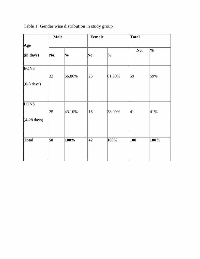
Table 1: Gender wise distribution in study group
Age
(in days)
Male Female Total
No. % No. %No. %
EONS
33 56.86% 26 61.90% 59 59%
(0-3 days)
LONS
25 43.10% 16 38.09% 41 41%
(4-28 days)
Total 58 100% 42 100% 100 100%
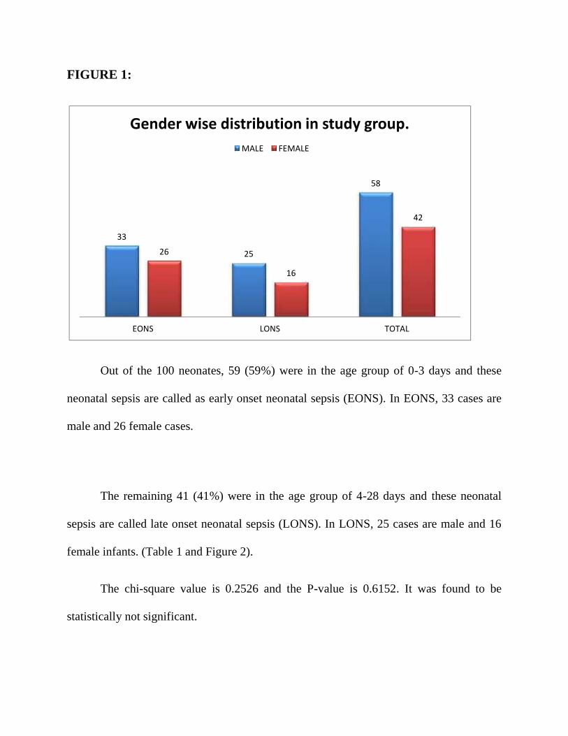
FIGURE 1:
Out of the 100 neonates, 59 (59%) were in the age group of 0-3 days and these
neonatal sepsis are called as early onset neonatal sepsis (EONS). In EONS, 33 cases are
male and 26 female cases.
The remaining 41 (41%) were in the age group of 4-28 days and these neonatal
sepsis are called late onset neonatal sepsis (LONS). In LONS, 25 cases are male and 16
female infants. (Table 1 and Figure 2).
The chi-square value is 0.2526 and the P-value is 0.6152. It was found to be
statistically not significant.
33
25
58
26
16
42
EONS LONS TOTAL
Gender wise distribution in study group.MALE FEMALE
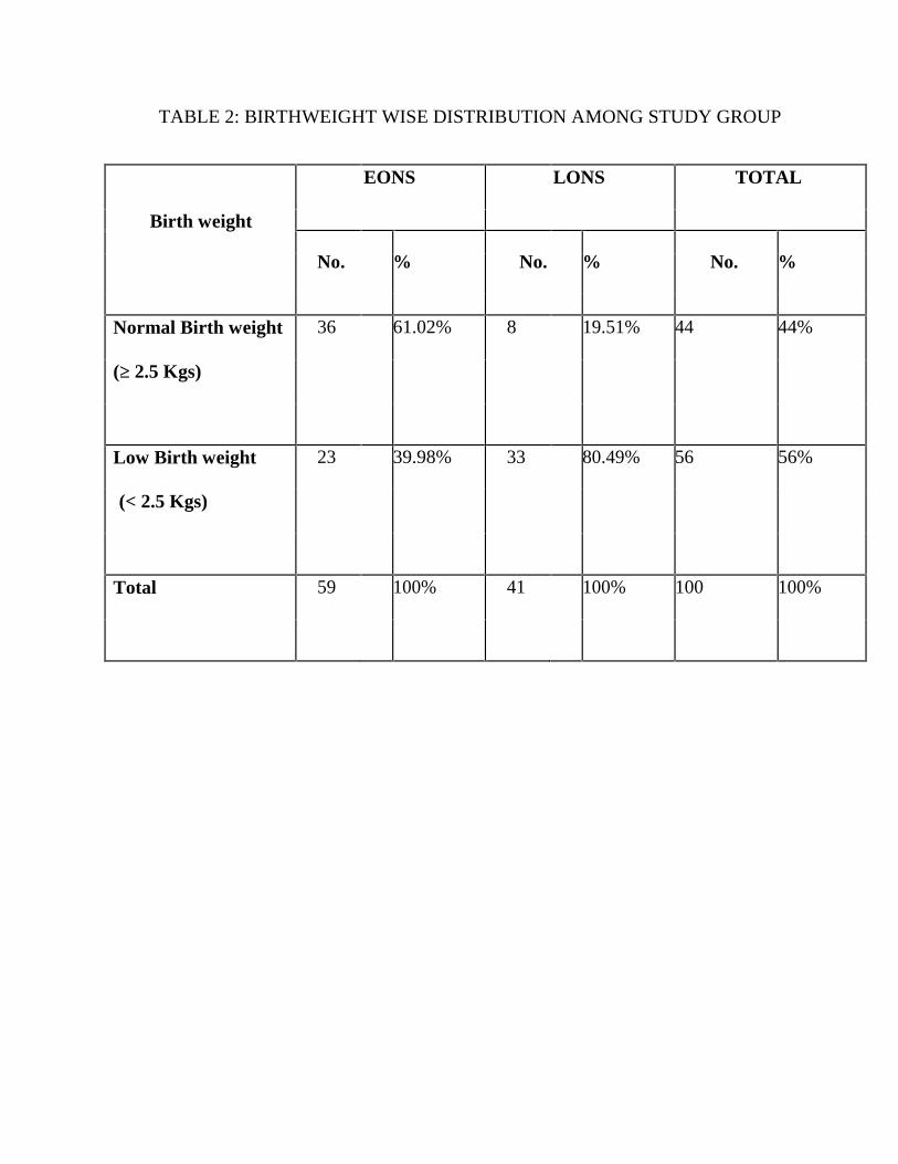
TABLE 2: BIRTHWEIGHT WISE DISTRIBUTION AMONG STUDY GROUP
EONS LONS TOTAL
Birth weight
No. % No. % No. %
Normal Birth weight 36 61.02% 8 19.51% 44 44%
(≥ 2.5 Kgs)
Low Birth weight 23 39.98% 33 80.49% 56 56%
(< 2.5 Kgs)
Total 59 100% 41 100% 100 100%
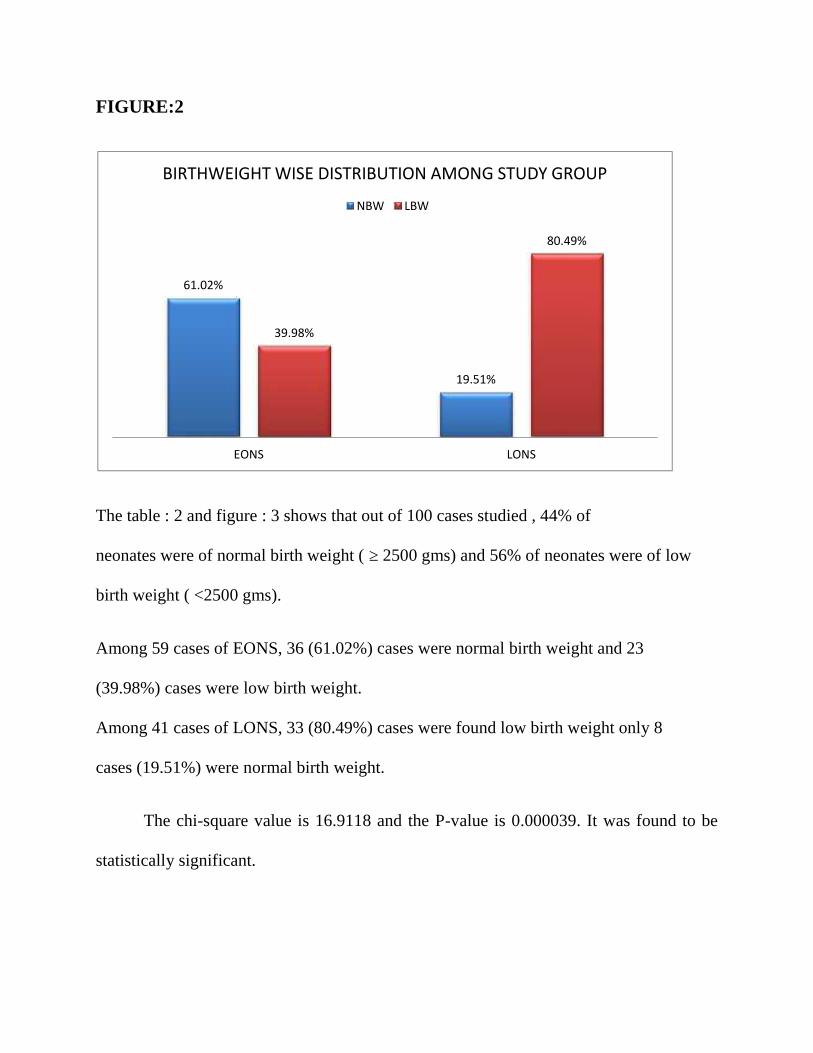
FIGURE:2
The table : 2 and figure : 3 shows that out of 100 cases studied , 44% of
neonates were of normal birth weight ( ≥ 2500 gms) and 56% of neonates were of low
birth weight ( <2500 gms).
Among 59 cases of EONS, 36 (61.02%) cases were normal birth weight and 23
(39.98%) cases were low birth weight.
Among 41 cases of LONS, 33 (80.49%) cases were found low birth weight only 8
cases (19.51%) were normal birth weight.
The chi-square value is 16.9118 and the P-value is 0.000039. It was found to be
statistically significant.
61.02%
19.51%
39.98%
80.49%
EONS LONS
BIRTHWEIGHT WISE DISTRIBUTION AMONG STUDY GROUP
NBW LBW
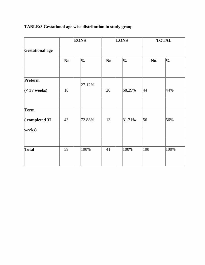
TABLE:3 Gestational age wise distribution in study group
EONS LONS TOTAL
Gestational age
No. % No. % No. %
Preterm27.12%
(< 37 weeks) 16 28 68.29% 44 44%
Term
( completed 37 43 72.88% 13 31.71% 56 56%
weeks)
Total 59 100% 41 100% 100 100%
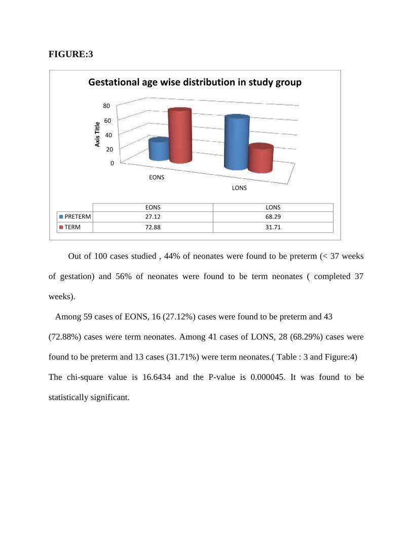
FIGURE:3
Out of 100 cases studied , 44% of neonates were found to be preterm (< 37 weeks
of gestation) and 56% of neonates were found to be term neonates ( completed 37
weeks).
Among 59 cases of EONS, 16 (27.12%) cases were found to be preterm and 43
(72.88%) cases were term neonates. Among 41 cases of LONS, 28 (68.29%) cases were
found to be preterm and 13 cases (31.71%) were term neonates.( Table : 3 and Figure:4)
The chi-square value is 16.6434 and the P-value is 0.000045. It was found to be
statistically significant.
0
20
40
60
80
EONSLONS
Axis
Title
EONS LONSPRETERM 27.12 68.29TERM 72.88 31.71
Gestational age wise distribution in study group
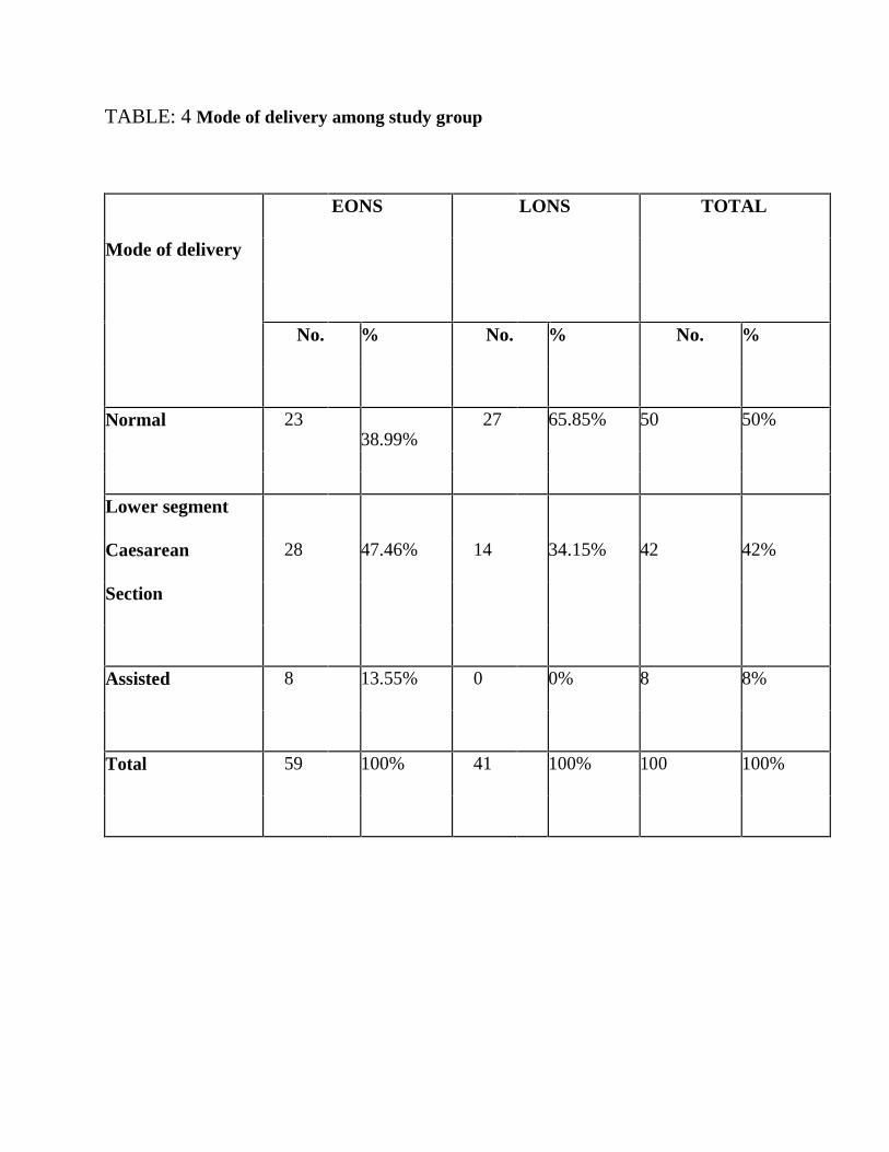
TABLE: 4 Mode of delivery among study group
EONS LONS TOTAL
Mode of delivery
No. % No. % No. %
Normal 2338.99%
27 65.85% 50 50%
Lower segment
Caesarean 28 47.46% 14 34.15% 42 42%
Section
Assisted 8 13.55% 0 0% 8 8%
Total 59 100% 41 100% 100 100%
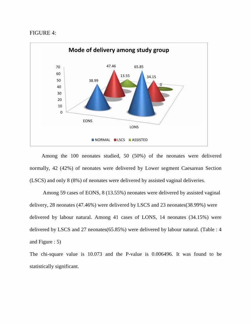
FIGURE 4:
Among the 100 neonates studied, 50 (50%) of the neonates were delivered
normally, 42 (42%) of neonates were delivered by Lower segment Caesarean Section
(LSCS) and only 8 (8%) of neonates were delivered by assisted vaginal deliveries.
Among 59 cases of EONS, 8 (13.55%) neonates were delivered by assisted vaginal
delivery, 28 neonates (47.46%) were delivered by LSCS and 23 neonates(38.99%) were
delivered by labour natural. Among 41 cases of LONS, 14 neonates (34.15%) were
delivered by LSCS and 27 neonates(65.85%) were delivered by labour natural. (Table : 4
and Figure : 5)
The chi-square value is 10.073 and the P-value is 0.006496. It was found to be
statistically significant.
010203040
50
60
70
EONSLONS
38.99
65.8547.46
34.1513.55
0
Mode of delivery among study group
NORMAL LSCS ASSISTED
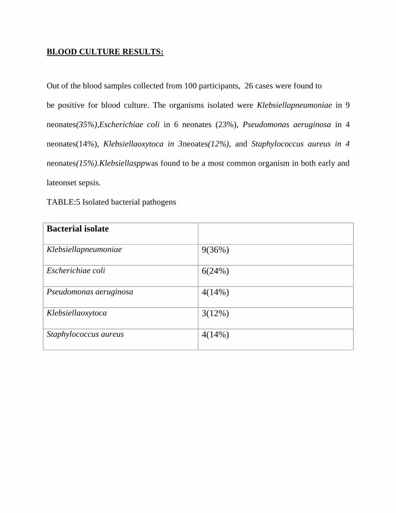
BLOOD CULTURE RESULTS:
Out of the blood samples collected from 100 participants, 26 cases were found to
be positive for blood culture. The organisms isolated were Klebsiellapneumoniae in 9
neonates(35%),Escherichiae coli in 6 neonates (23%), Pseudomonas aeruginosa in 4
neonates(14%), Klebsiellaoxytoca in 3neoates(12%), and Staphylococcus aureus in 4
neonates(15%).Klebsiellasppwas found to be a most common organism in both early and
lateonset sepsis.
TABLE:5 Isolated bacterial pathogens
Bacterial isolate
Klebsiellapneumoniae 9(36%)
Escherichiae coli 6(24%)
Pseudomonas aeruginosa 4(14%)
Klebsiellaoxytoca 3(12%)
Staphylococcus aureus 4(14%)
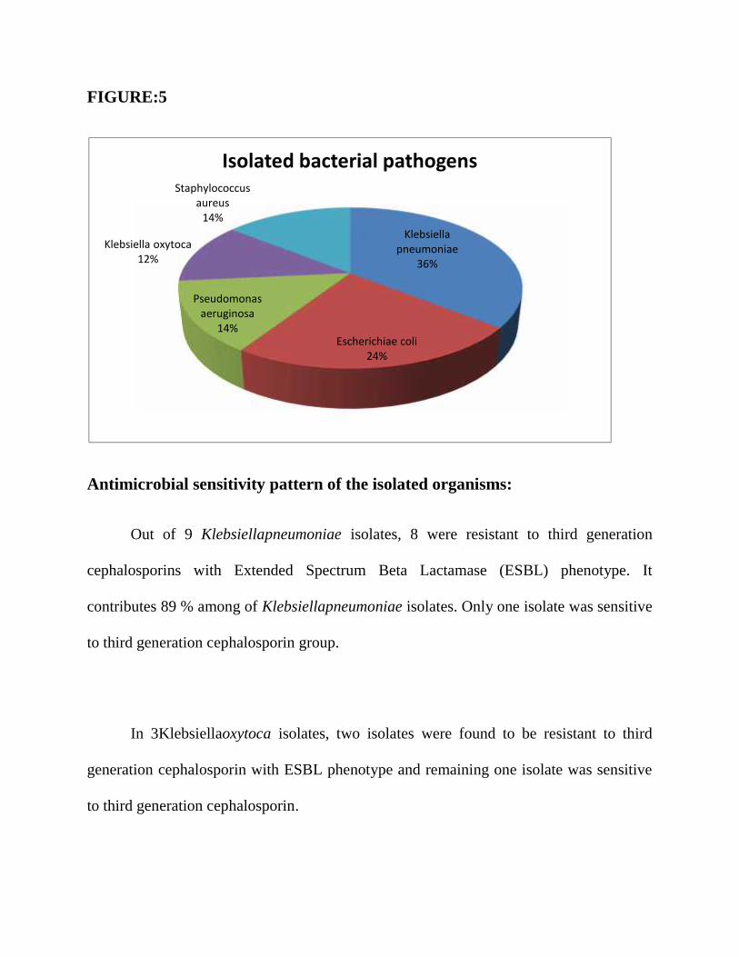
FIGURE:5
Antimicrobial sensitivity pattern of the isolated organisms:
Out of 9 Klebsiellapneumoniae isolates, 8 were resistant to third generation
cephalosporins with Extended Spectrum Beta Lactamase (ESBL) phenotype. It
contributes 89 % among of Klebsiellapneumoniae isolates. Only one isolate was sensitive
to third generation cephalosporin group.
In 3Klebsiellaoxytoca isolates, two isolates were found to be resistant to third
generation cephalosporin with ESBL phenotype and remaining one isolate was sensitive
to third generation cephalosporin.
Klebsiellapneumoniae
36%
Escherichiae coli24%
Pseudomonasaeruginosa
14%
Klebsiella oxytoca12%
Staphylococcusaureus
14%
Isolated bacterial pathogens
Out of 6 Escherichiae coli isolates, five (80%) were sensitive to third generation
cephalosporin. Only one case (20%) was resistant to third generation cephalosporin with
ESBL phenotype. But all 22 gram negative isolates were sensitive to imipenem.
Among four Staphylococcus aureus, 3 isolates (75%) were methicillin resistant
strain (MRSA).
Out of 26 bacterial isolates, 21 isolates (81%) were sensitive to amikacin , 20
isolates (76.9%) were sensitive to gentamicin , 21 isolates (81%) were
sensitive to ciprofloxacin and 12isolates (46.1%) were sensitive to
cotrimoxaxole.
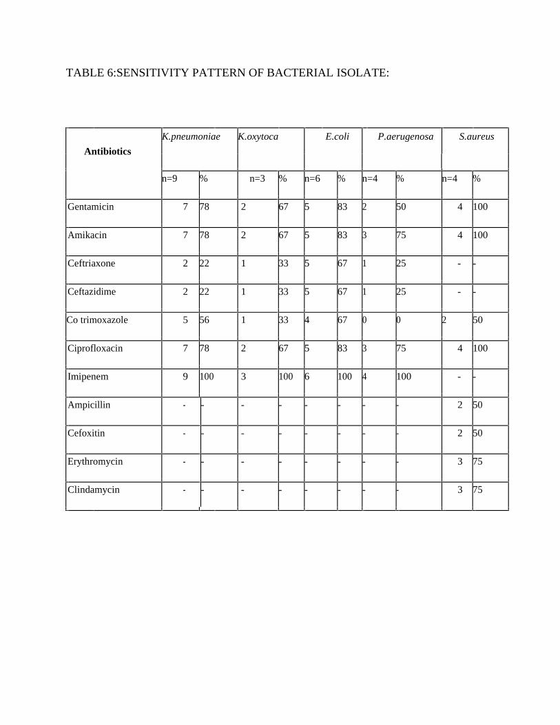
TABLE 6:SENSITIVITY PATTERN OF BACTERIAL ISOLATE:
Antibiotics
K.pneumoniae K.oxytoca E.coli P.aerugenosa S.aureus
n=9 % n=3 % n=6 % n=4 % n=4 %
Gentamicin 7 78 2 67 5 83 2 50 4 100
Amikacin 7 78 2 67 5 83 3 75 4 100
Ceftriaxone 2 22 1 33 5 67 1 25 - -
Ceftazidime 2 22 1 33 5 67 1 25 - -
Co trimoxazole 5 56 1 33 4 67 0 0 2 50
Ciprofloxacin 7 78 2 67 5 83 3 75 4 100
Imipenem 9 100 3 100 6 100 4 100 - -
Ampicillin - - - - - - - - 2 50
Cefoxitin - - - - - - - - 2 50
Erythromycin - - - - - - - - 3 75
Clindamycin - - - - - - - - 3 75
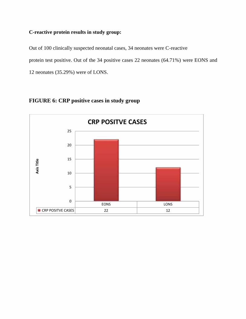
C-reactive protein results in study group:
Out of 100 clinically suspected neonatal cases, 34 neonates were C-reactive
protein test positive. Out of the 34 positive cases 22 neonates (64.71%) were EONS and
12 neonates (35.29%) were of LONS.
FIGURE 6: CRP positive cases in study group
EONS LONS CRP POSITVE CASES 22 12
0
5
10
15
20
25
Axis
Title
CRP POSITVE CASES
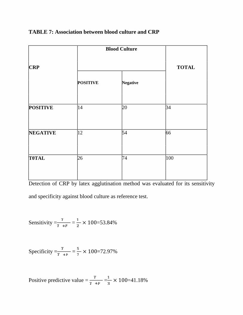
TABLE 7: Association between blood culture and CRP
CRP
Blood Culture
TOTAL
POSITIVE Negative
POSITIVE 14 20 34
NEGATIVE 12 54 66
T0TAL 26 74 100
Detection of CRP by latex agglutination method was evaluated for its sensitivity
and specificity against blood culture as reference test.
Sensitivity = = × 100=53.84%
Specificity = = × 100=72.97%
Positive predictive value = = × 100=41.18%
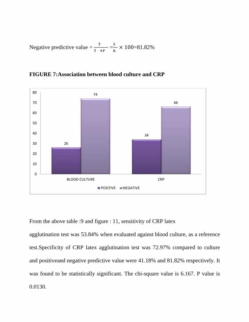
Negative predictive value = = × 100=81.82%
FIGURE 7:Association between blood culture and CRP
From the above table :9 and figure : 11, sensitivity of CRP latex
agglutination test was 53.84% when evaluated against blood culture, as a reference
test.Specificity of CRP latex agglutination test was 72.97% compared to culture
and positiveand negative predictive value were 41.18% and 81.82% respectively. It
was found to be statistically significant. The chi-square value is 6.167. P value is
0.0130.
26
34
74
66
0
10
20
30
40
50
60
70
80
BLOOD CULTURE CRP
POSITIVE NEGATIVE
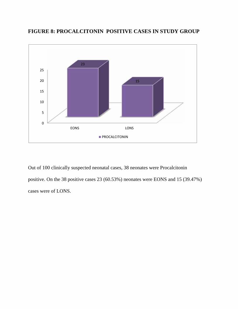
FIGURE 8: PROCALCITONIN POSITIVE CASES IN STUDY GROUP
Out of 100 clinically suspected neonatal cases, 38 neonates were Procalcitonin
positive. On the 38 positive cases 23 (60.53%) neonates were EONS and 15 (39.47%)
cases were of LONS.
0
5
10
15
20
25
EONS LONS
23
15
PROCALCITONIN
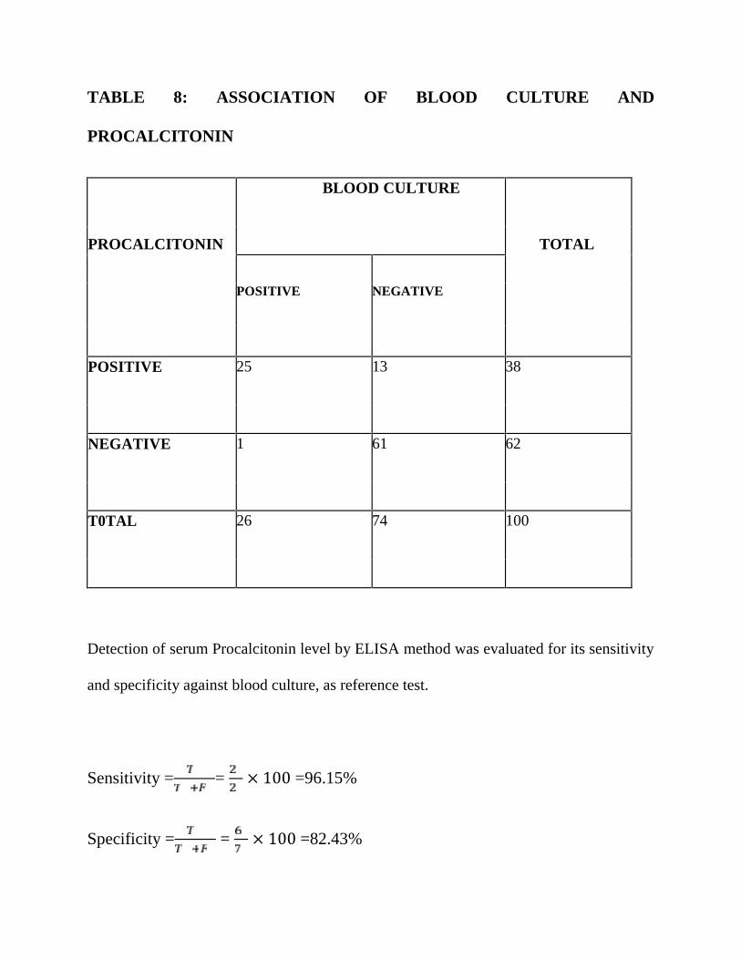
TABLE 8: ASSOCIATION OF BLOOD CULTURE AND
PROCALCITONIN
PROCALCITONIN
BLOOD CULTURE
TOTAL
POSITIVE NEGATIVE
POSITIVE 25 13 38
NEGATIVE 1 61 62
T0TAL 26 74 100
Detection of serum Procalcitonin level by ELISA method was evaluated for its sensitivity
and specificity against blood culture, as reference test.
Sensitivity = = × 100 =96.15%
Specificity = = × 100 =82.43%
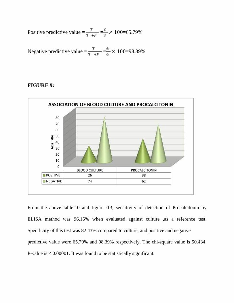
Positive predictive value = = × 100=65.79%
Negative predictive value = = × 100=98.39%
FIGURE 9:
From the above table:10 and figure :13, sensitivity of detection of Procalcitonin by
ELISA method was 96.15% when evaluated against culture ,as a reference test.
Specificity of this test was 82.43% compared to culture, and positive and negative
predictive value were 65.79% and 98.39% respectively. The chi-square value is 50.434.
P-value is < 0.00001. It was found to be statistically significant.
0
10
20
30
40
50
60
70
80
BLOOD CULTURE PROCALCITONINPOSITIVE 26 38NEGATIVE 74 62
Axis
Title
ASSOCIATION OF BLOOD CULTURE AND PROCALCITONIN
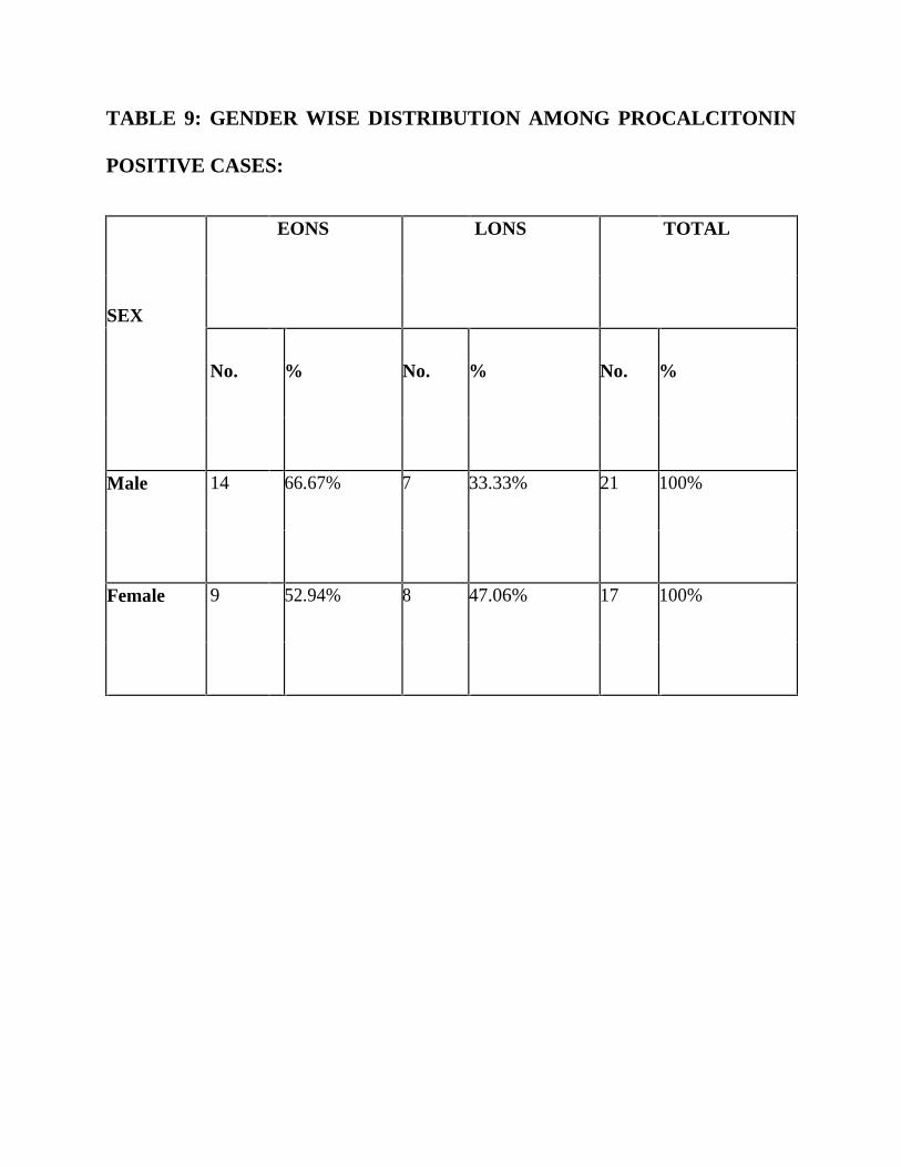
TABLE 9: GENDER WISE DISTRIBUTION AMONG PROCALCITONIN
POSITIVE CASES:
EONS LONS TOTAL
SEX
No. % No. % No. %
Male 14 66.67% 7 33.33% 21 100%
Female 9 52.94% 8 47.06% 17 100%
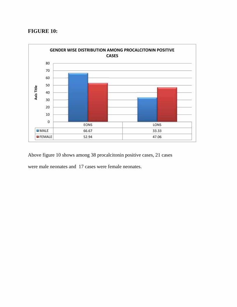
FIGURE 10:
Above figure 10 shows among 38 procalcitonin positive cases, 21 cases
were male neonates and 17 cases were female neonates.
EONS LONSMALE 66.67 33.33FEMALE 52.94 47.06
0
10
20
30
40
50
60
70
80
Axis
Title
GENDER WISE DISTRIBUTION AMONG PROCALCITONIN POSITIVECASES
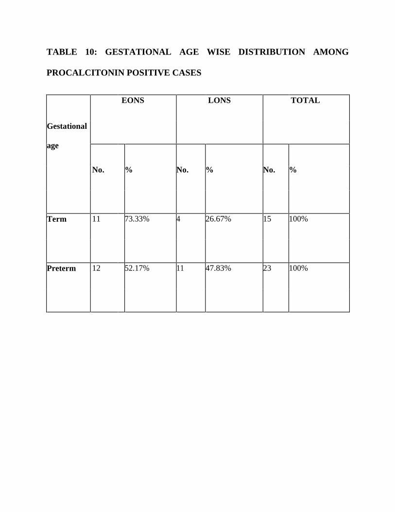
TABLE 10: GESTATIONAL AGE WISE DISTRIBUTION AMONG
PROCALCITONIN POSITIVE CASES
EONS LONS TOTAL
Gestational
age
No. % No. % No. %
Term 11 73.33% 4 26.67% 15 100%
Preterm 12 52.17% 11 47.83% 23 100%
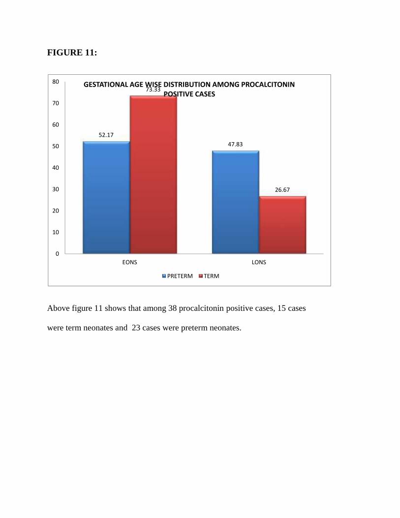
FIGURE 11:
Above figure 11 shows that among 38 procalcitonin positive cases, 15 cases
were term neonates and 23 cases were preterm neonates.
52.1747.83
73.33
26.67
0
10
20
30
40
50
60
70
80
EONS LONS
GESTATIONAL AGE WISE DISTRIBUTION AMONG PROCALCITONINPOSITIVE CASES
PRETERM TERM

TABLE 11: BIRTH WEIGHT WISE DISTRIBUTION AMONG
PROCALCITONIN POSITIVE CASES
EONS LONS TOTAL
BIRTH
WEIGHT
No. % No. % No. %
NBW 9 81.82% 2 18.18% 11 100%
LBW 14 51.85% 13 48.15% 27 100%
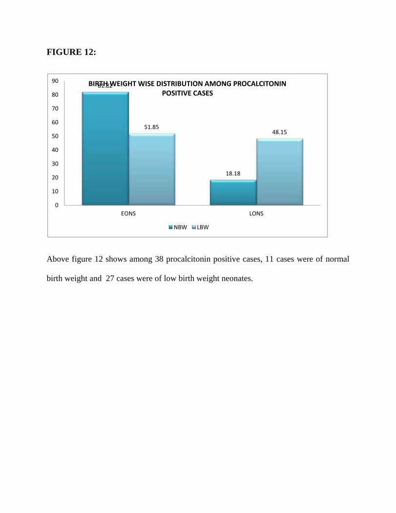
FIGURE 12:
Above figure 12 shows among 38 procalcitonin positive cases, 11 cases were of normal
birth weight and 27 cases were of low birth weight neonates.
81.82
18.18
51.8548.15
0
10
20
30
40
50
60
70
80
90
EONS LONS
BIRTH WEIGHT WISE DISTRIBUTION AMONG PROCALCITONINPOSITIVE CASES
NBW LBW
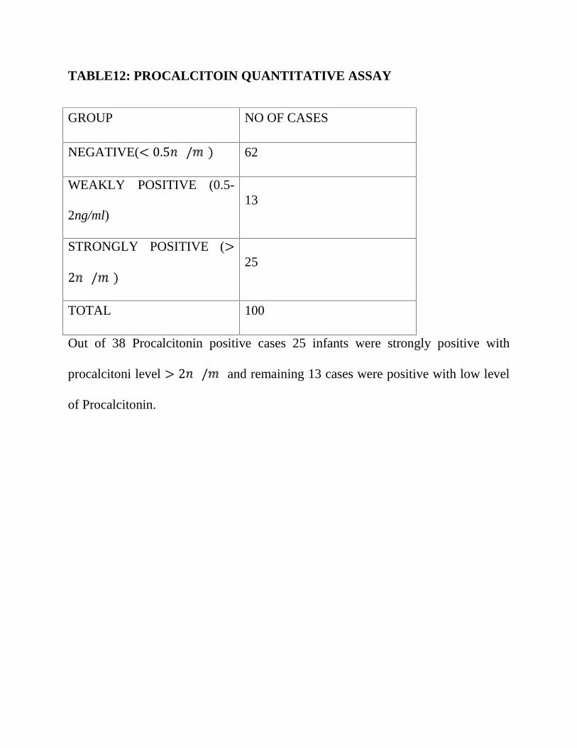
TABLE12: PROCALCITOIN QUANTITATIVE ASSAY
GROUP NO OF CASES
NEGATIVE(< 0.5 / ) 62
WEAKLY POSITIVE (0.5-
2ng/ml)13
STRONGLY POSITIVE (>2 / ) 25
TOTAL 100
Out of 38 Procalcitonin positive cases 25 infants were strongly positive with
procalcitoni level > 2 / and remaining 13 cases were positive with low level
of Procalcitonin.
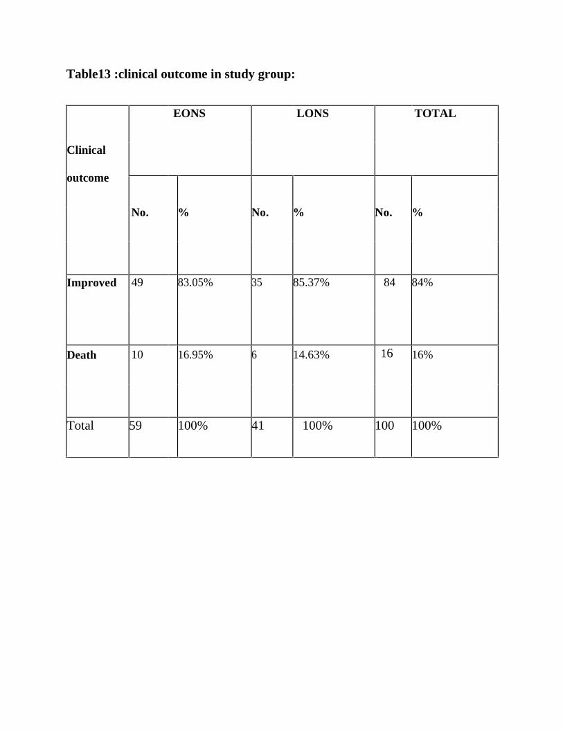
Table13 :clinical outcome in study group:
EONS LONS TOTAL
Clinical
outcome
No. % No. % No. %
Improved 49 83.05% 35 85.37% 84 84%
Death 10 16.95% 6 14.63% 16 16%
Total 59 100% 41 100% 100 100%
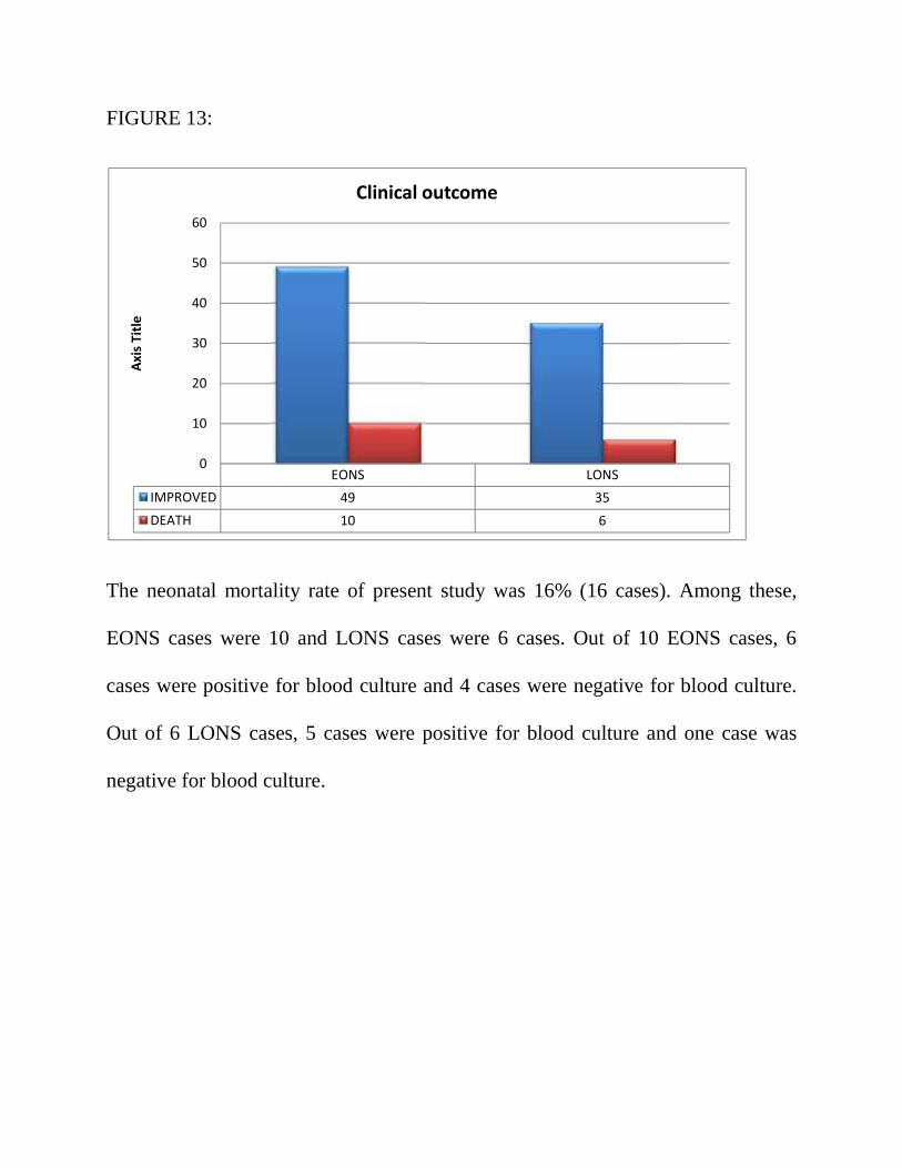
FIGURE 13:
The neonatal mortality rate of present study was 16% (16 cases). Among these,
EONS cases were 10 and LONS cases were 6 cases. Out of 10 EONS cases, 6
cases were positive for blood culture and 4 cases were negative for blood culture.
Out of 6 LONS cases, 5 cases were positive for blood culture and one case was
negative for blood culture.
EONS LONSIMPROVED 49 35DEATH 10 6
0
10
20
30
40
50
60
Axis
Title
Clinical outcome
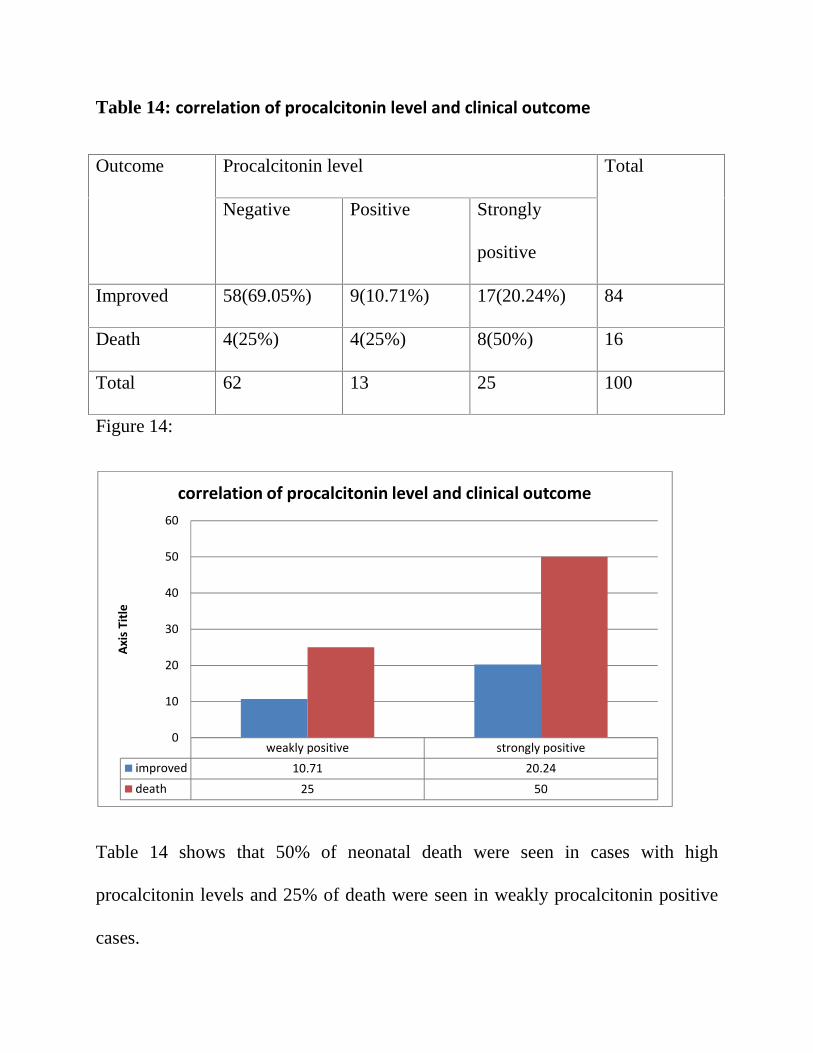
Table 14: correlation of procalcitonin level and clinical outcome
Outcome Procalcitonin level Total
Negative Positive Strongly
positive
Improved 58(69.05%) 9(10.71%) 17(20.24%) 84
Death 4(25%) 4(25%) 8(50%) 16
Total 62 13 25 100
Figure 14:
Table 14 shows that 50% of neonatal death were seen in cases with high
procalcitonin levels and 25% of death were seen in weakly procalcitonin positive
cases.
weakly positive strongly positiveimproved 10.71 20.24death 25 50
0
10
20
30
40
50
60
Axis
Title
correlation of procalcitonin level and clinical outcome
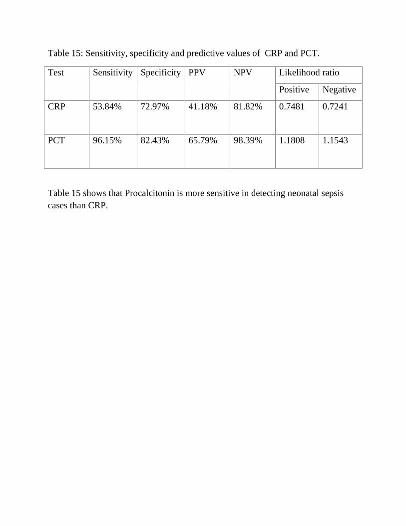
Table 15: Sensitivity, specificity and predictive values of CRP and PCT.
Test Sensitivity Specificity PPV NPV Likelihood ratio
Positive Negative
CRP 53.84% 72.97% 41.18% 81.82% 0.7481 0.7241
PCT 96.15% 82.43% 65.79% 98.39% 1.1808 1.1543
Table 15 shows that Procalcitonin is more sensitive in detecting neonatal sepsiscases than CRP.
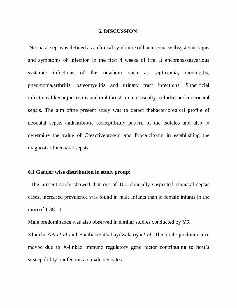
6. DISCUSSION:
Neonatal sepsis is defined as a clinical syndrome of bacteremia withsystemic signs
and symptoms of infection in the first 4 weeks of life. It encompassesvarious
systemic infections of the newborn such as septicemia, meningitis,
pneumonia,arthritis, osteomyelitis and urinary tract infections. Superficial
infections likeconjunctivitis and oral thrush are not usually included under neonatal
sepsis. The aim ofthe present study was to detect thebacteriological profile of
neonatal sepsis andantibiotic susceptibility pattern of the isolates and also to
determine the value of Creactiveprotein and Procalcitonin in establishing the
diagnosis of neonatal sepsis.
6.1 Gender wise distribution in study group:
The present study showed that out of 100 clinically suspected neonatal sepsis
cases, increased prevalence was found in male infants than in female infants in the
ratio of 1.38 : 1.
Male predominance was also observed in similar studies conducted by YR
Khinchi AK et al and BambalaPuthattayilZakariyaet al. This male predominance
maybe due to X-linked immune regulatory gene factor contributing to host’s
susceptibility toinfections in male neonates.
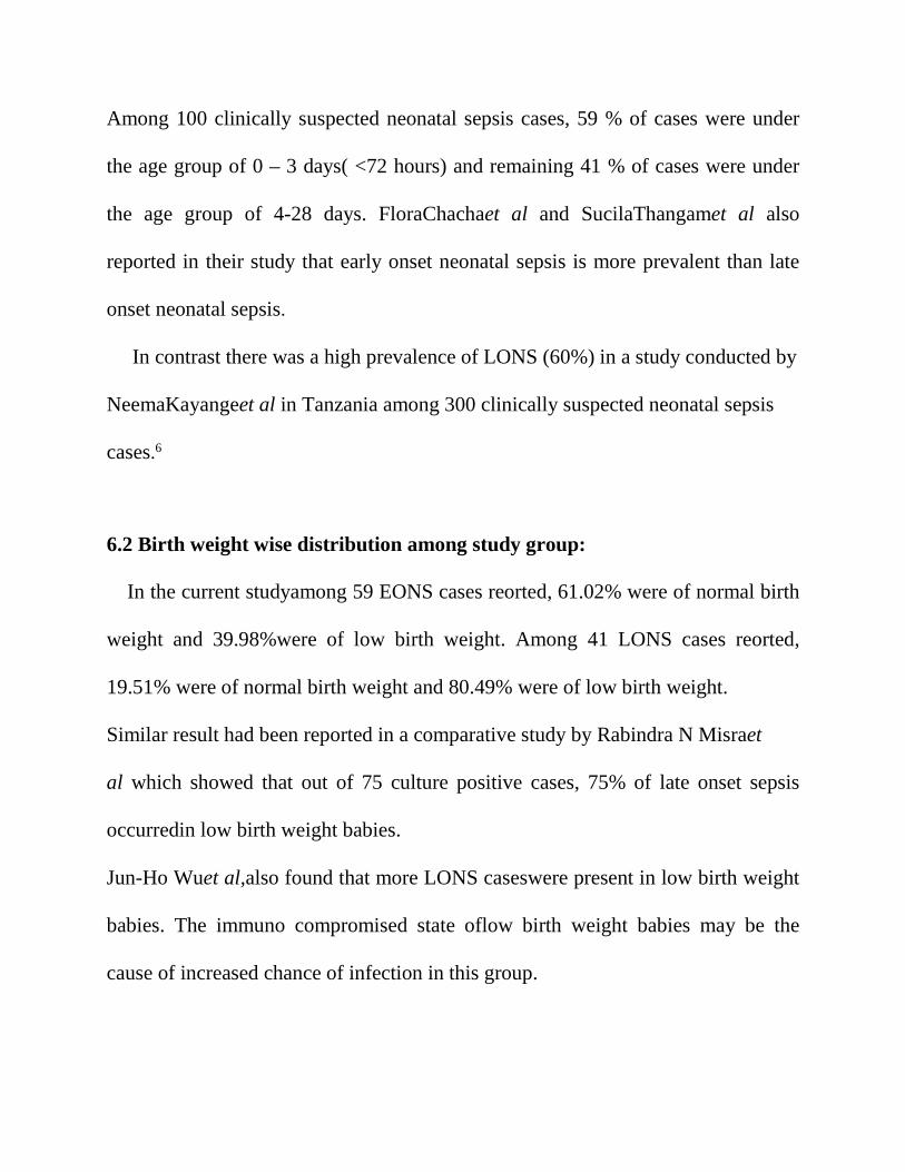
Among 100 clinically suspected neonatal sepsis cases, 59 % of cases were under
the age group of 0 – 3 days( <72 hours) and remaining 41 % of cases were under
the age group of 4-28 days. FloraChachaet al and SucilaThangamet al also
reported in their study that early onset neonatal sepsis is more prevalent than late
onset neonatal sepsis.
In contrast there was a high prevalence of LONS (60%) in a study conducted by
NeemaKayangeet al in Tanzania among 300 clinically suspected neonatal sepsis
cases.6
6.2 Birth weight wise distribution among study group:
In the current studyamong 59 EONS cases reorted, 61.02% were of normal birth
weight and 39.98%were of low birth weight. Among 41 LONS cases reorted,
19.51% were of normal birth weight and 80.49% were of low birth weight.
Similar result had been reported in a comparative study by Rabindra N Misraet
al which showed that out of 75 culture positive cases, 75% of late onset sepsis
occurredin low birth weight babies.
Jun-Ho Wuet al,also found that more LONS caseswere present in low birth weight
babies. The immuno compromised state oflow birth weight babies may be the
cause of increased chance of infection in this group.
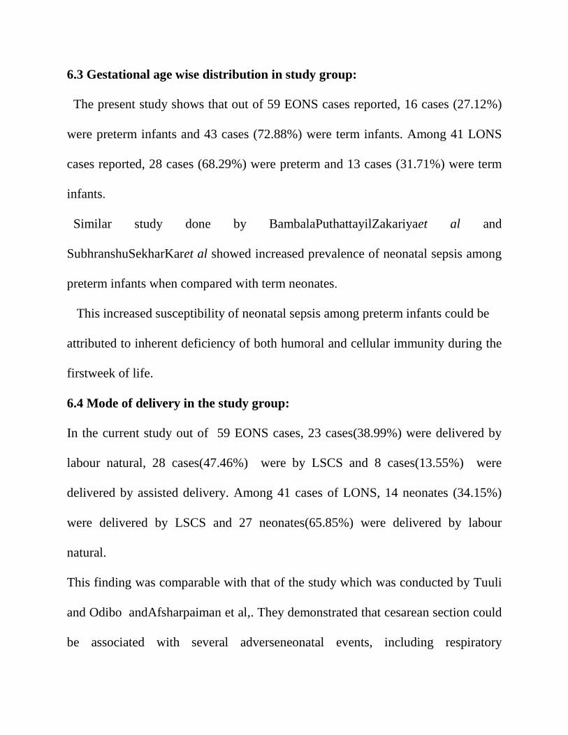
6.3 Gestational age wise distribution in study group:
The present study shows that out of 59 EONS cases reported, 16 cases (27.12%)
were preterm infants and 43 cases (72.88%) were term infants. Among 41 LONS
cases reported, 28 cases (68.29%) were preterm and 13 cases (31.71%) were term
infants.
Similar study done by BambalaPuthattayilZakariyaet al and
SubhranshuSekharKaret al showed increased prevalence of neonatal sepsis among
preterm infants when compared with term neonates.
This increased susceptibility of neonatal sepsis among preterm infants could be
attributed to inherent deficiency of both humoral and cellular immunity during the
firstweek of life.
6.4 Mode of delivery in the study group:
In the current study out of 59 EONS cases, 23 cases(38.99%) were delivered by
labour natural, 28 cases(47.46%) were by LSCS and 8 cases(13.55%) were
delivered by assisted delivery. Among 41 cases of LONS, 14 neonates (34.15%)
were delivered by LSCS and 27 neonates(65.85%) were delivered by labour
natural.
This finding was comparable with that of the study which was conducted by Tuuli
and Odibo andAfsharpaiman et al,. They demonstrated that cesarean section could
be associated with several adverseneonatal events, including respiratory
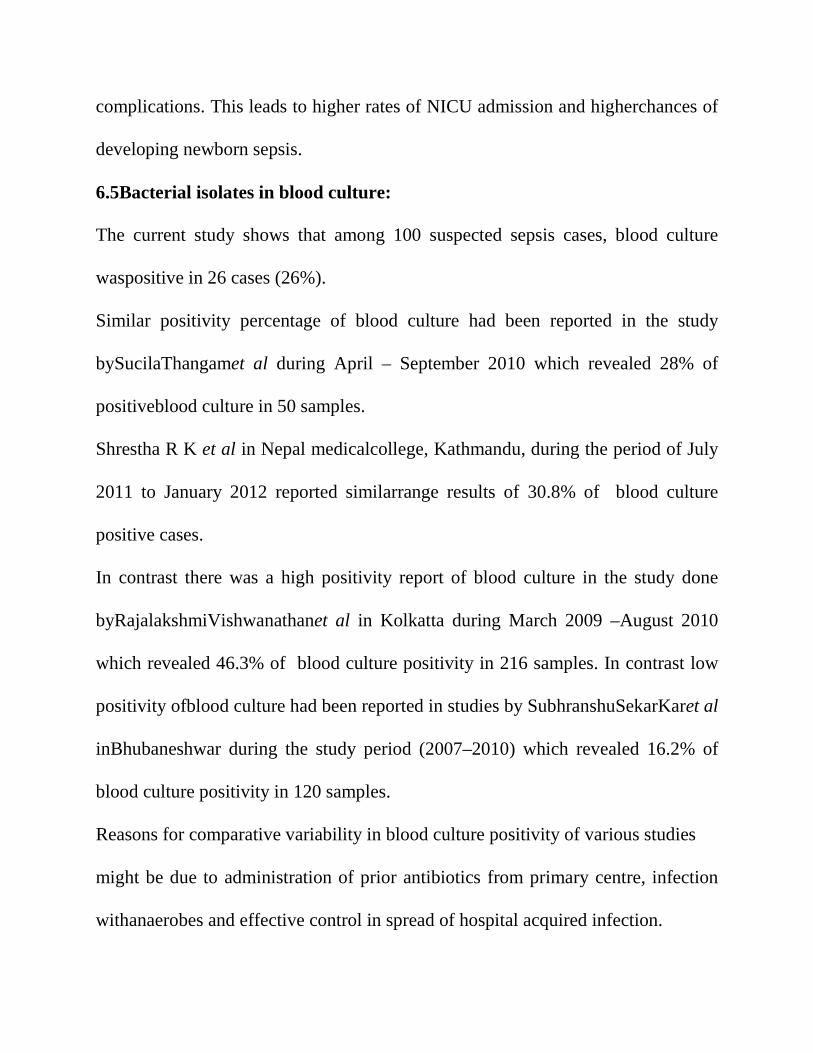
complications. This leads to higher rates of NICU admission and higherchances of
developing newborn sepsis.
6.5Bacterial isolates in blood culture:
The current study shows that among 100 suspected sepsis cases, blood culture
waspositive in 26 cases (26%).
Similar positivity percentage of blood culture had been reported in the study
bySucilaThangamet al during April – September 2010 which revealed 28% of
positiveblood culture in 50 samples.
Shrestha R K et al in Nepal medicalcollege, Kathmandu, during the period of July
2011 to January 2012 reported similarrange results of 30.8% of blood culture
positive cases.
In contrast there was a high positivity report of blood culture in the study done
byRajalakshmiVishwanathanet al in Kolkatta during March 2009 –August 2010
which revealed 46.3% of blood culture positivity in 216 samples. In contrast low
positivity ofblood culture had been reported in studies by SubhranshuSekarKaret al
inBhubaneshwar during the study period (2007–2010) which revealed 16.2% of
blood culture positivity in 120 samples.
Reasons for comparative variability in blood culture positivity of various studies
might be due to administration of prior antibiotics from primary centre, infection
withanaerobes and effective control in spread of hospital acquired infection.
Another reason attributed to the variable positive result may depend on the mode
of test employed for culture. Commonly used conventional test is time consuming
and itsyield is of significantly low sensitivity when compared to fully automated
system whichis less time consuming and has the benefit of continuous monitoring
with highsensitivity.
Out of the 26 isolates, 22 were gram negative bacteria and only 4 isolates were
gram positive bacteria. Among gram negative organisms
Klebsiellapneumoniaewas found to be the mostcommon organism in both early
and late onset sepsis.
Similar results were observed in studies done by BambalaPuthattayilZakariya et al
,NeemaKayangeet aland Iregu KC et al.
Klebsiellapneumoniae is one of most common pathogen profoundly found in
neonatal intensive care ward environment and various equipment in neonatal ward.
This could be the cause for increased incidence of Klebsiellapneumoniaeas a
causative organism for neonatal sepsis.
In contrast, in a study conducted by Shrestha R K et al in Nepal medical college,
Kathmandu, during the period of July 2011 to January 2012, Staphylococcus
aureus(56.8%) had been reported as main etiological agent in neonatal sepsis and
followed byKlebsiellapneumoniae(21.7%), Pseudomonas aeruginosa (13.4%) and
others
6.6 Antibiotic susceptibility pattern of isolated pathogens:
The present study showed that among 12 Klebsiellaspp, 9 isolates were
Klebsiellapneumoniaespecies and remaining 3 isolates were Klebsiellaoxytoca.
Out of 12 isolates of Klebsiellaspp, 83% (10 isolates) were resistant to third
generation cephalosporin with ESBL phenotype.
A similar study done by Iregu KC et al found that 87% of Klebsiella
pneumoniaeisolates were found to be ESBL producers. Another similar study done
in North India by Kaistha N et al found that 88% gram negative isolates were
resistant tothird generation cephalosporin.
But this was contrary to the observations of BambalaPuthattayilZakariyaet al in
Jawaharlal NehruInstitute of Post graduate, Medical Education and Research
Puducherry, in which 32% of Klebsiellapneumoniaeisolates were found to be
ESBL producers.
The thirdgeneration cephalosporin are commonly used empirically in all cases of
clinicallysuspected sepsis in present study centre. It could be the main cause for
increasedincidence of ESBL in the present study.
But all 12 isolates of Klebsiellasppwere sensitive to imipenem. Similar result
wasobtained in the study done by NeemaKayangeet al with about 95% of
Klebsiella spp sensitive to imipenem.
The present study shows that among four Pseudomonas aeruginosa, 2 isolates
(50%) were resistant to gentamicin, 3 isolates (75%) were resistant to third
generationcephalosporin and one isolate was resistant to ciprofloxacin. But all four
isolates were sensitive to imipenem and Piperacillin-Tazobactum. This finding
similar to study of BambalaPuthattayilZakariyaet al.
The current study shows that among four Staphylococcus aureus, 3 isolates
(75%)were resistant to methicillin (MRSA), which is comparable with Ramesh
Bhat Yet al study results.
Increased prevalence of MRSA may be due to healthy carriers in hospital staffs
and relatives.
6.7 Comparison of C-reactive protein with blood culture:
The current study showed that out of 100 clinically suspected sepsis cases 34 were
positive for CRP. Out of the 34 positive cases 22 neonates (64.71%) were EONS
and 12 neonates (35.29%) were of LONS. The sensitivity of CRP for proven sepsis
(more than 6 μg/ mL)was 53.84%, its specificity was 72.95%, its positive
predictive value was 41.18% and its negative predictive value was 81.82%.
Similar observation was reported in study done by SucilaThangamet al, that
sensitivity of CRP were 50%, its specificity was 69.4%, its positivepredictive value
was 38.8% and its negative predictive value was 78.1%.
Contrary results were observed in a study done by Doeller H et al, reported that
CRP was 63% sensitive, 97% specific, its positive predictive value was 83% and
negative predictivevalue was 91%.
A study done by Franz A R et al found that 28% of CRP sensitivity, 97% of
specificity, 81% of positive predictive value and 77% of negative predictive value.
This study showed lower sensitivity compared to the present study.
6.8 Comparison of Procalcitonin with blood culture:
In the current study, totally 38 neonates were Procalcitonin
positive. Among them 23 (60.53%) neonates were EONS and 15 (39.47%)cases
were of LONS. The sensitivity of procalcitonin for proven sepsis was 96.15%,
itsspecificity was 82.43%, its positive predictive valve was 65.79% and its
negative predictive value was 98.39%.
In a similar study by Sakha et al. showed that the sensitivity, specificity, positive
predictive valueand the negative predictive value of PCTwere 66.7%, 50%,
28.6%and 83.3% which is lower than that of the current study.
Chiesa et al., studied the reliability of the PCT concentration in 28 infants who had
a severe early onset of neonatal sepsis. They found that the sensitivity, specificity,
PPV and NPV were 92.6, 97.5, 94.3 and 96.8%, respectively. In this study the
specificity is higher than that of current study.
Vazzalwar et al., assessed PCT for the diagnosis of neonatal sepsis in 67 neonates
was found that the sensitivity and specificity were of 97% and 80% respectively.
6.9 Gender wise distribution among procalcitonin positive cases:
The current study shows among 38 Procalcitonin positive cases, male neonates
were of 55.26% and female neonates were of 44.74%. Similar studies by
RekhaSriramet al, Kuruvillaet al and Shrestha et al also showed that male neonates
werepredominance in culture positive cases. This male predominance maybe due
to X-linked immune regulatory gene factor contributing to host’s susceptibility
toinfections in male neonates
6.10 Gestational age wise distribution in procalcitoin positive cases:
The present study shows that out of 38procalcitonin positive cases, 23
cases (60.53%) were preterm babies and remaining 15 cases (39.47) were term
babies.
In similar study done by Bomaet al, BambalaPuthattayilZakariyaet al, Shrestha et
al and Kuruvillaet al observed increasedprevalence of neonatal sepsis among
preterm neonates when compared with termneonates. The immuno compromised
state of low birth weight and preterm babies may bethe cause for more infection in
these group
6.11Birth weight wise distribution in Procalcitonin positive cases:
In the current study out of 38 Procalcitonin positive cases, 27 (71.05%) cases
were low birth weight (less than 2500 gm) neonates and 11 cases(28.95%) were of
normal birth ( weight above 2500 gms)
In similar study by NeemaKayangeet al , Rabindra N Misraet al and
SubhranshuSekharKaret al., it was found that increased prevalence of neonatal
sepsis among low birthweight neonates when compared with normal birth weight
neonates.
6.12 Clinical outcome and procalcitonin levels:
The neonatal mortality rate of present study was 16% (16 cases). Among these,
EONS cases were 10 and LONS cases were 6 cases.
Current study showed that higher mortality rate was present in neonates with
higherProcalcitonin level (more than 2ng/ml). Similarly, Brunkhorst et al
have even demonstrated that very high PCT values were related to septic shock and
death. Jensen JU et al also demonstrated that patients with septic process present
lessmortality risk with PCT levels less than 1mg/dl and had more mortality risk
with PCT value more than 1mg/dl.
In the current study, the PCT levels were remarkable high in neonateswith proven
sepsis and also in suspected sepsis cases.It is comparable withthe study conducted
by YadollaZahedpasha et al., andMonneretet al,. In the present study PCT
detected nearly all blood culture positive cases.
In a study by Carolet al., showed that procalcitonin is moresensitive than the
CRP in the diagnosis of septicemia, meningitis andurinary tract infection. In our
study proclacitonin was positive in almost all culture positive cases when
compared to CRP. PCT also detects more culture negative clinically suspected
sepsis cases than CRP. These findingssupport the usefulness of the PCT to
establish an early diagnosis of neonatalsepsis.
The current study confirmed the findings of various authors that Procalcitonin was
more sensitive than CRP in detection of neonatal sepsis. In neonatal sepsis, serum
Procalcitonin level rise earlier than the CRP level.So serum Procalcitonin level
detection is mainly useful in earlydetection of neonatal sepsis. Detection of serum
Procalcitonin level also detects the severity ofinfection and evaluation of the
response to antibiotic treatment.
Early diagnosis of neonatal sepsis by Procalcitonin evaluation helps to prevent
neonatalmortality and morbidity and avoid unnecessary use of empirical antibiotics
whichin turn helps in preventing drug resistance.
7. SUMMARY
The present study aimed at detecting the bacteriological profile in neonatal sepsis
and antibiotic susceptibility pattern of isolates in 100 clinically suspected cases and
alsodetect the role of CRP and Procalcitonin in the diagnosis of neonatal sepsis.
Out of 100 clinically suspected neonatal sepsis cases, 58% were male babies
and 42% were female babies. Male and Female ratio was 1.38 : 1.
Out of 100 cases, Early Onset Neonatal Sepsis was 59% and Late Onset
Neonatalsepsis was 41%.
Out of 100 study cases, 26% cases were blood culture positive.
Among 26 clinical isolates there was increased incidence of gram negative
Organisms in which Klebsiella pneumoniae (36%) was the most common
followed by, Escherichia coli(6%), Pseudomonas aeruginosa(14%) and Klebsiella
oxytoca(12%).
Among 18 Enterobacteriaceae isolated , 72%were resistant to
third generation cephalosporin with ESBL phenotype.
Among the four Staphylococcus aureus, 3 isolates (75%) were resistant to
methicillin(MRSA).
Serum CRP level detected by latex agglutination method had 53.84% of
sensitivity, 72.97% of specificity, 41.18% of Positive predictive value and 81.82%
of negative predictive value against blood culture as a reference test.

Serum PCT level detected by ELISA method had 96.15% of
sensitivity, 82.43% of specificity, 65.79% of Positive predictive value and 98.39%
of negative predictive value against blood culture as a reference test.
8. CONCLUSION:
Early recognition and diagnosis of neonatal sepsis are difficult due to the variable
and nonspecific clinical presentation. It is important to make an early diagnosis of
sepsis, because prompt initiation of antimicrobial therapy improves
outcomes.
The findings of the present study confirm that the serum level of Procalcitonin is
a more reliable marker than the serum levels of CRP. Serum procalcitonin level
was superior to serum CRP level in terms of early diagnosis of neonatal sepsis and
in detecting the severity of the illness.
The benefit of measuring serum PCT routinely in the diagnosis and follow-up of
neonatal sepsis, is that it reduces the hospital costs. Such a benefit might support a
wider acceptance of the test in the routine practice.
9. BIBLIOGRAPHY
1. Blenclowe H, Cousens S, Addressing the challenge of neonatal mortality.
Tropical medicine of international health. March 2013; Vol 18(3): 303-312.
2. Polin RA: Management of neonates with suspected or proven early-onset
bacterial sepsis. Pediatrics 2012, 129(5):1006-1015.
3. West BA, Peterside O: Sensitivity pattern among bacterial isolates in neonatal
septicaemia in port Harcourt. Annals of clinical microbiology and antimicrobials
2012, 11(1):7.
4. West BA, Peterside O, Ugwu RO, Eneh AU: Prospective evaluation of the
usefulness of C-reactive protein in the diagnosis of neonatal sepsis in a sub-
Saharan African region. Antimicrob Resist Infect Control 2012, 1(1):22.7.
5. Bizzarro MJ, Raskind C, Baltimore RS, Gallagher PG. Seventy-five years of
neonatal sepsis at Yale: 1928-2003. Paediatrics. 2005;116(3):595-602.
6.Aparna Narasimha M.L, Harendra Kumar N. Significance of Hematological
Scoring System (HSS) in Early diagnosis of neonatal sepsis. Indian J Hematol
Blood Transfus (Jan-Mar 2011) 27(1):14–17.
7.Mikkel Zahle Oestergaard1, Mie Inoue, Sachiyo Yoshida, Wahyu Retno
Mahanani, Fiona M Gore, Simon Cousens, Joy E Lawn, Colin Douglas Mathers.
Neonatal Mortality Levels for 193 Countries in 2009 with trends since 1990: A
Systematic Analysis of Progress, Projections, and Priorities. PLoS Medicine;
August 2011 ; Volume 8(8): 1-13.
8.Mishra U K, Jacobs S E, Doyle L W, Garland S M. Newer approaches to the
diagnosis of early onset neonatal sepsis. Arch Dis Child Fetal Neonatal Ed
2006;91:F208–F212.
9. Christ-Crain, M, Muller, B. Procalcitonin in bacterial infections – hype, hope,more or less? Swiss Med Wkly. 2005;135:451–460.
10.Assicot, M, Gendrel, D, Carsin, H, Raymond, J, Guilbaud, J,Bohuon, C. Highserum procalcitonin concentrations in patients with sepsis and infection. Lancet.1993;341:515- 518
11. Dandona, P, Nix, D, Wilson, MF, Aljada, A, Love, J, Assicot, M. Procalcitoninincrease after endotoxin injection in normal subjects. J Clin Endo Met.1994;79:1605-1608
12.Gendrel D, Assicot M, Raymond J. Procalcitonin as a marker for the earlydiagnosis of neonatal infections. J Paediatr 1996;128:570-73.
13. Bryce J, Boschi-Pinto C, Shibuya K, Black RE. WHO estimates of the causesof death in children. Lancet. 2005 Mar 26-Apr 1;365(9465):1147-52.
14. Africa S AW, European WHO: Reducing child mortality to achieve MDG 4.2011
15. Tsering DC, Chanchal L, Pal R, Kar S: Bacteriological profile of septicemiaand the risk factors in neonates and infants in sikkim. J Glob Infect Dis 2011,3(1):42-45.
16. Sankar MJ, Agarwal R, Deorari AK, Paul VK. Sepsis in the newborn. Indian JPediatr. 2008 Mar;75(3):261-6.
17. YR Khinchi AK, Satish Yadav. Profile of Neonatal sepsis. Journal of college ofMedical SciencesNepal. 2010; Vol.6( No2):p16.
18.Rekha Sriram IJBMR. Correlation of blood culture results with the sepsis scoreand the sepsis screen in the diagnosis of neonatal septicemia. Bio Med Sci Directpublications;2011. p.3608.
19. . Sucilathangam G., Amuthavalli K., Velvizhi G., Ashihabegum M.A.,Jeyamurugan T., Palaniappan N. Early Diagnostic Markers for Neonatal Sepsis:
Comparing Procalcitonin (PCT) and C-Reactive Protein (CRP). Journal of Clinicaland Diagnostic Research. 2012 May (Suppl-2), Vol-6(4): 627-631.
20. Neema Kayange1, Erasmus Kamugisha2, Damas L Mwizamholya1, SeniJeremiah3 and Stephen E Mshana*3. RPerseeardchi acrtticolers of positive bloodculture and deaths among neonates with suspected neonatal sepsis in a tertiaryhospital, Mwanza- Tanzania.BMC Pediatrics 2010, 10:39.
21. Flora Chacha1, Mariam M Mirambo, Martha F Mushi, Neema Kayange1,Antke Zuechner, Benson R Kidenya and Stephen E Mshana. Utility of qualitativeC-reactive protein assay and white blood cells counts in the diagnosis of neonatalsepticaemia at Bugando Medical Centre, Tanzania. BMC Pediatrics 2014, 14:248.
22. Seo K, McGregor JA, French JI. Preterm birth is associated with increased riskof maternal and neonatal infection. Obstet.Gynecol. 1992; 79:75-80.
23. . Rabindra N. Misra, Savita V. Jadhav, Purbasha Ghosh, Nageswari Gandham,Kalpana Angadi, Chanda Vyawahare. Role of sepsis screen in the diagnosis ofneonatal sepsis. Medical Journal of Dr. D.Y. Patil University July-September2013; Vol 6(3):254-257.
24. Rachel C. Ferreira, Rosane R. Mello∗, Kátia S. Silva. Neonatal sepsis as a riskfactor for neurodevelopmental changes in preterm infants with very low birthweight. J Pediatr (Rio J). 2014;90(3):293---299.
25. Downie L, Armiento R, Subhi R, Kelly J, Clifford V, Duke T. Community-acquired neonatal and infant sepsis in developing countries: efficacy of WHO'scurrently re commended antibiotics--systematic review and meta-analysis. ArchDis Child. 2013 Feb;98(2):146-54.
26. Ramesh Bhat Y, Leslie Edward S, Vandana KE. Bacterial isolates of early –onset neonatal sepsis and their antibiotic susceptibility pattern between 1998 and2004: an audit from a centre in India. Italian j of paed 2011, 37:32.
27. Kuruvilla K A, Pillai S, Jesudas M, Jana AK: Bacterial profile of sepsis in aneonatal unit in south India. Indian Pediatr 1998, 35:851-858.
28. Dar es Salaam , Vallance H, Lockitch G: Rapid, semi-quantitative assay ofCreactive protein evaluated. Clin Chem 1991, 37(11):1981-1982.
29. Xin-Chuan Chen, Yun-Fan Yang, Rui Wang, Hong-Feng Go, Xin-Zu Chen:Epidemiology and microbiology of sepsis in mainland China in the first decade ofthe 21st century. International Journal of Infectious Diseases 31 (2015) 9–14.
30. Neelam Kaistha, Manjula Mehta, Nidhi Singla, Ritu Garg, Jagdish Chander.Neonatal septicemia isolates and resistance patterns in a tertiary care hospital ofNorth India. J Infect Dev Ctries 2010; 4(1):055-057.
31. Rajlakshmi Viswanathan, Arun K. Sing, Chiranjib Ghosh, Sudipta Dasgupta,Suchandra Mukherje1, and Sulagna Basu: Profile of Neonatal Septicaemia at aDistrict-level Sick Newborn Care Unit. J Health Popul Nutr 2012 Mar;30(1):41-48
32. Kaistha N, Mehta M, Singla N, Garg R, Chander J. Neonatal septicemiaisolates and resistance patterns in a tertiary care hospital of North India. J InfectDev Ctries. 2009;4:55–7.
33. Emily J. Weston, MPH, Tracy Pondo, MSPH, Melissa M. Lewis, MPH, PatMartell- Cleary,MSW, LPN, Craig Morin, MPH, Brenda Jewell, Pam Daily, MPH,Mirasol Apostol, MPH, Sue Petit, MPH, Monica Farley, MD, Ruth Lynfield, MD,Art Reingold, MD, Nellie I.Hansen, MPH, Barbara J. Stoll, MD, Andi L. Shane,MD, MPH, MSC, Elizabeth Zell,MSTAT, and Stephanie J. Schrag, DPHIL. TheBurden of Invasive Early-Onset Neonatal Sepsis in the United States, 2005–2008.Pediatr Infect Dis J. 2011 November ; 30(11): 937–941.
34. Eichenwald EC. Perinatally transmitted neonatal bacterial infections.Infect.Dis.Clin.North Am. 1997; 11:223-239.
35. Silver HM, Siler-Khodr T, Prihoda TJ, Gibbs RS. The effects of pH andosmolality on bacterial growth in amniotic fluid in a laboratory model.Am.J.Perinatol. 1992; 9:69-74.
36. Pyati SP, Pildes RS, Jacobs NM et al. Penicillin in infants weighing twokilograms or less with early-onset Group B streptococcal disease. N.Engl.J.Med.1983; 308:1383- 1389.
37. Pierce JR, Merenstein GB, Stocker JT. Immediate postmortem cultures in anIntensive care nursery. Pediatr.Infect.Dis. 1984; 3:510-513.
38. Tillett WS, Francis T: Serological reactions in pneumonia with a non-proteinsomatic fraction of pneumococcus. J Exp Med 1930;52: 561–571.
39. Pepys MB: C-reactive protein fifty years on.Lancet 1981; 1: 653–657.
40. De Ferranti S, Rifai N. C-reactive protein and cardiovascular disease: a reviewof risk prediction and interventions’, Clinica Chimica Acta 2002; 317 :1.
41. Kääpä P, Koistinen E: Maternal and neonatal C-reactive protein afterinterventions during delivery. Acta Obstet Gynecol Scand 1993; 72: 543–546.
42.Baumann H, Richards C, Gauldie J Interaction among hepatocyte-stimulatingfactors, interleukin-1, and glucocorticoids for regulation of acute phase plasmaproteins in human hepatoma (Hep G2)cells. J Immunol 1987;139 :4122– 4128. 26.
43.Campos SP, Wang Y, Koj A, Baumann H Insulin cooperates with IL-1 inregulating expression of alpha1-acid glycoprotein gene in rat hepatoma cells.Cytokine 1994;6: 485–492.
44. ParvinV, Ingle, Devang M and Patel. C-reactive protein I various diseasecondition – An over view. Asian J of Pharmaceutical and Clinical Research Vol. 4,Issue 1, 2011.
45. Volanakis JE: Human C-reactive protein: expression, structure, and function.Mol Immunol 2001; 38: 189–197.
46. Du Clos TW: Function of C-reactive protein. Ann Med 2000; 32: 274–278.
47.Benitz WE, Han MY, Madan A, Ramachandra P: Serial serum C-reactiveprotein levels in the diagnosis of neonatal infection. Pediatrics 1998; 102:E41
48. Laborada G, Stavola J, Ballabh P, et al: Diagnostic value of cytokines and C-reactive protein in the first 24 h of neonatal sepsis. Am J Perinatol 2003; 20: 491–501.
49. Doellner H, Arntzen KJ, Haereid PE, Aag S, Austgulen R: Interleukin-6concentrations in neonates evaluated for sepsis. J Pediatr 1998; 132: 295–299.
50. Franz AR, Kron M, Pohlandt F, Steinbach G: Comparison of procalcitoninwith interleukin 8, C-reactive protein and differential white blood cell count for theearly diagnosis of bacterial infections in newborn infants. Pediatr Infect Dis J1999; 18: 666–671.
51.Kalra K, Sunder S, Ethence SR, Dayal RS.C-reactive protein in neonatalinfections.IndPediatr. 1985;22:215.
52. Nuntnarumit P, Pinkaew O, Kitiwanwanich S: Predictive values of serial C-reactive protein in neonatal sepsis. J Med Assoc Thai 2002, 85 Suppl 4:S1151-1158.
53. Khassawneh M, Hayajneh WA, Kofahi H, Khader Y, Amarin Z, Daoud A:Diagnostic markers for neonatal sepsis: comparing C-reactive protein, interleukin-6 and immunoglobulin M. Scand J Immunol 2007, 65(2):171-175.
54. Kohli-Kochhar R, Omuse G, Revathi G: A ten-year review of neonatalbloodstream infections in a tertiary private hospital in Kenya. Journal of infectionin developing countries 2011, 5(11).
55. Hofer N, Zacharias E, Müller W, Resch B: An update on the use of C-reactiveprotein in early-onset neonatal sepsis. Neonatology 2012, 102(1):25-36.
56. Bipin K: Evaluation Of Serum C-reactive Protein In Diagnosis And PrognosisOf Neonatal Septicemia. WebmedCentralPAEDIATRICS 2013,4(7):WMC001643.
57. Chiesa C, Panero A, Osborn JF, Simonetti AF, Pacifico L: Diagnosis ofneonatal sepsis:a clinical and laboratory challenge. Clin Chem 2004, 50(2):279-287
58. Meem M, Modak JK, Mortuza R, Morshed M, Islam MS, Saha SK. 2011.Biomarkers for diagnosis of neonatal infections: a systematic analysis of theirpotential as a pointof- care diagnostics. J. Glob. Health1:201–209.
59. Christ-Crain & Muller, 2008; Jin & Khan, 2010; Russwurm, Oberhoffer,Zipfelk, & Reinhart, 1999
60. (Muller & Becker, 2001; Nylen et al., 1998; Nystrom, 1998; Whang et al.,1999)
61. Nelson Textbook of Paediatrics 18 th edition. Page 794-810.
62. Baltimore RS, Huie SM, Meek JI, Schuchat A, O’Brei KL. Early onset ofneonatal sepsis in the era of group B Streptococcal prevention. Paediatrics 2001,108:1094- 1098.
63. Jaswal RS, Kaushal RK, Goel A, Pathania K: Role of C-reactive protein indeciding duration of antibiotic therapy in neonatal septicemia. Indian Pediatr 2003,40(9):880- 883.
64. . Arif SH, Ahmad I, Ali SM, Khan HM: Thrombocytopenia and bacterial sepsisin neonates. Indian J Hematol Blood Transfus 2012, 28(3):147-151.
65. Mishra UK, Jacobs SE, Doyle LW, Garland SM: Newer approaches to thediagnosis of early onset neonatal sepsis. Arch Dis Child Fetal Neonatal Ed 2006,91(3):F208- 212.
66. Ng PC. Diagostic markers of infection in neonates. Arch Dis child FetalNeonatal Ed. 2004;89(3):F229-235.
67. Pierrakos C, Vincent JL. Sepsis biomarkers:a review. Crit care.14(1):R 15.

68. Dietzman DE, Fischer GW, Schoenknecht FD. Neonatal Escherichia colisepticemia-- bacterial counts in blood. J Pediatr. 1974 Jul;85(1):128-30.
69. Kellog JA, Ferrentino FL, Goodstei MH, Liss J, Shapiro SL, Bankert DA.Frequency of low level bactermia in infats from birth to two months of age. PediatrInfect Dis J. 1997;16(4):381-385.
70. Schelonka RL, Chai MK, Yoder BA, Hensley D, Brockett RM, Ascher DP.Volumeof blood required to detect common neonatal pathogens. J Pediatr.1996;129(2):275- 278.
71. Viswanathan R SA, Ghosh C, Dasgupta S, Mukherjee S, Basu S: Profile ofneonatal septicaemia at a district-level sick newborn care unit. Journal of health,population, and nutrition 2012 30(1):41-48.
72. Pacifico L, Panero A, Colarizi P, Matrunola M, Simonetti AF, Chiesa C:Neonatal Candida albicans septic thrombosis of the portal vein followed bycavernoustransformation of the vessel. J Clin Microbiol 2004, 42(9):4379-4382.
73. . Subhrashu Sekar Kar, Rajani Dube, Samarendra Mahapatro, Sitanshu SekarKar. The role of clinical signs in the diagnosis of late onset neonatal sepsis andformation of clinical score. Indian J clin Prac, 2003 Mar; Vol.23, No 23: 654-660.
74. Shrestha R K, Rai S K and Mandhal P K. Bacteriological study of neo natalsepsis and antibiotic susceptibility pattern of isolates in Kathmandu, Nepal. NepalMed Coll J 2013; 15(1): 71-73.
75. Benitz WE. Adjunct laboratory testsin the diagnosis of early-onset neonatalsepsis. Clin perinato.37(2):421-438.
76. Mishra UK, Jacobs SE, Doyle LW, Garland SM. Newer approaches to thediagnosis of early onset neonatal sepsis. Arch Dis Child Fetal Neonatal Ed. 2006May;91(3):F208-12.
77. Chiesa C, Panero A, Osborn JF, Simonetti AF, Pacifico L. Diagnosis ofneonatal sepsis: a clinical and laboratory challenge. Clin chem. 2004;279-287. 106
78. Hornik CP, Benjamin DK, Becker KC, Li J, Clark RH, Cohen-Wolkowiez M,Smith PB: Use of the complete blood cell count in early-onset neonatal sepsis. ThePediatric infectious disease journal 2012, 31(8):799-802.
79. Labib AZ, Mahmoud AB, Eissa N, El Gendy FM, Soliman MA, Aly AA: EarlyDiagnosis of Neonatal Sepsis: A Molecular Approach and Detection of DiagnosticMarkers Versus Conventional Blood Culture. Intl J 2013, 4:77-85.

80. Manucha V, Rusia U, Sikka M, Faridi MM, Madan N. Utility ofhaematological parameters and C-reactive protein in the detection of neonatalsepsis. J Paediatr Child Health. 2002 Oct;38(5):459-64.
81.Centers for Disease Control and Prevention. Prevention of perinatal group Bstreptococcal disease—revised guidelines from CDC, 2010. MMWR RecommRep. 2010;59 (RR-10):1–36.
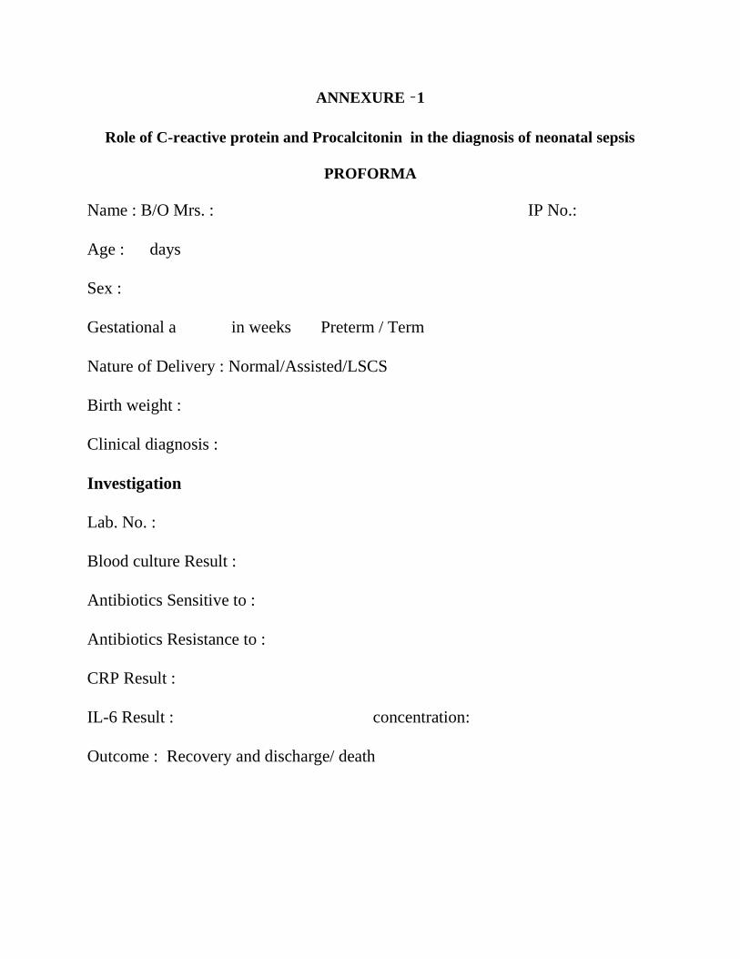
ANNEXURE –1
Role of C-reactive protein and Procalcitonin in the diagnosis of neonatal sepsis
PROFORMA
Name : B/O Mrs. : IP No.:
Age : days
Sex :
Gestational a in weeks Preterm / Term
Nature of Delivery : Normal/Assisted/LSCS
Birth weight :
Clinical diagnosis :
Investigation
Lab. No. :
Blood culture Result :
Antibiotics Sensitive to :
Antibiotics Resistance to :
CRP Result :
IL-6 Result : concentration:
Outcome : Recovery and discharge/ death
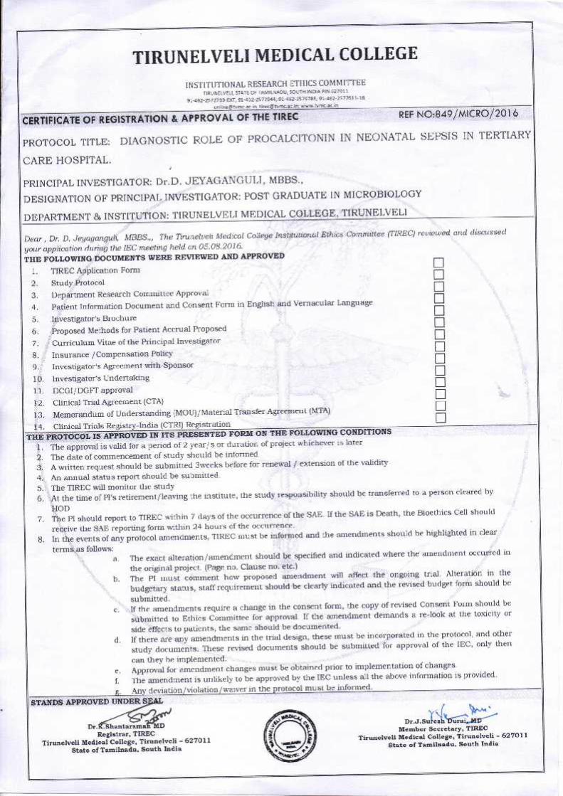
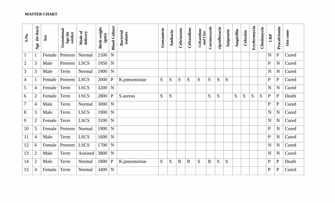
MASTER CHART
S.N
o
Age
(in
day
s)
Sex
Ges
tati
onal
Age
(in
wee
ks)
Mod
e of
deliv
ery
Bir
th w
eigh
t(g
ms)
Blo
od C
ultu
re
Bac
teri
alis
olat
es
Gen
tam
icin
Am
ikac
in
Cef
tria
xone
Cef
tazi
dim
e
Cef
tazi
dim
ean
d C
lav
Cot
rim
oxaz
ole
cipr
oflo
xaci
n
Imip
enem
Am
pici
llin
Cef
oxit
in
Ery
thro
myc
in
Clin
dam
ycin
CR
P
Pro
calc
iton
in
Out
com
e
1 1 Female Preterm Normal 2100 N N P Cured
2 3 Male Preterm LSCS 1950 N P N Cured
3 3 Male Term Normal 1900 N N N Cured
4 1 Female Preterm LSCS 2000 P K.pneumoniae S S S S S S S S P P Cured
5 4 Female Term LSCS 3200 N N N Cured
6 2 Female Term LSCS 2800 P S.aureus S S S S S S S S P P Death
7 4 Male Term Normal 3000 N P P Cured
8 3 Male Term LSCS 1900 N N N Cured
9 2 Female Term LSCS 3100 N N N Cured
10 5 Female Preterm Normal 1900 N P N Cured
11 4 Male Term LSCS 1600 N P N Cured
12 6 Female Preterm LSCS 1700 N N N Cured
13 2 Male Term Assisted 3800 N N N Cured
14 2 Male Term Normal 1800 P K.pneumoniae S S R R S R S S P P Death
15 4 Female Term Normal 3400 N P P Cured
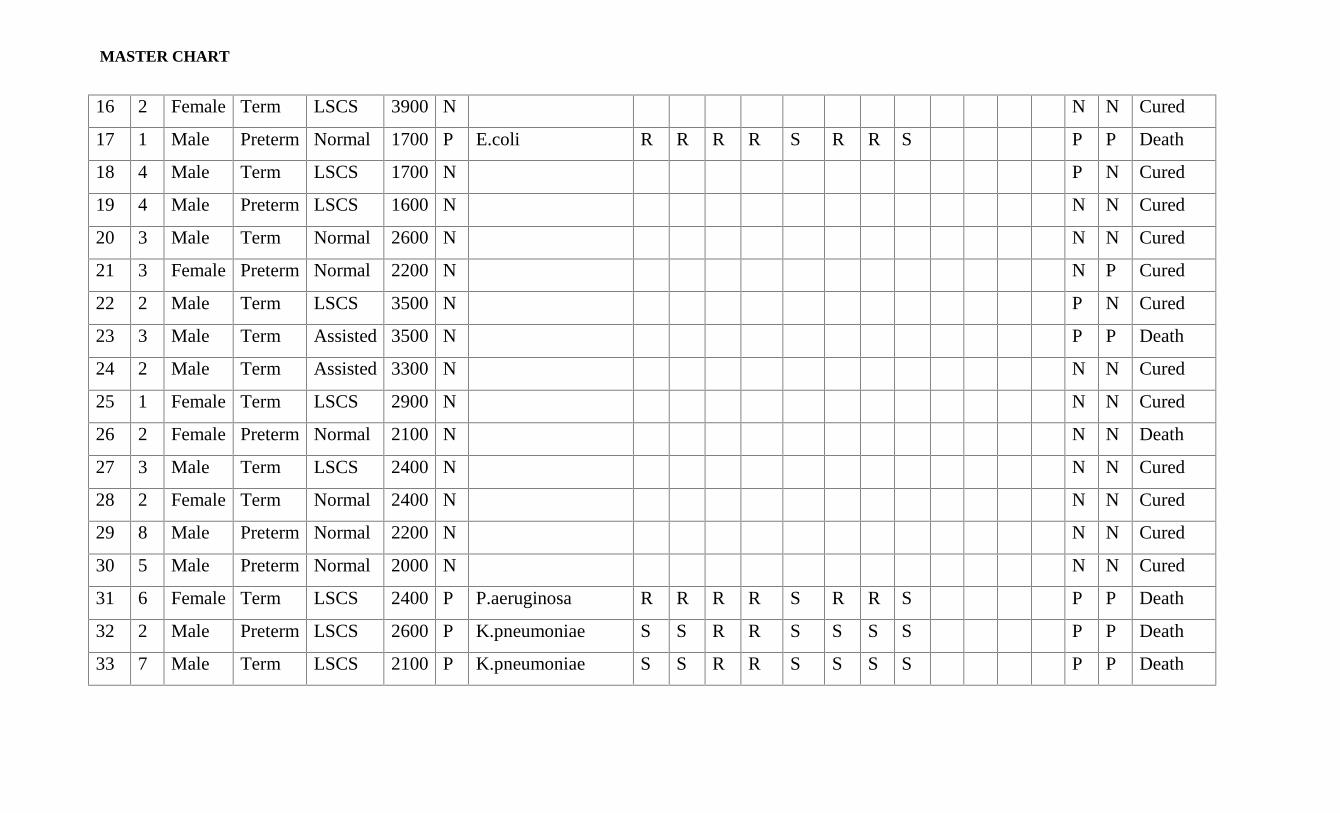
MASTER CHART
16 2 Female Term LSCS 3900 N N N Cured
17 1 Male Preterm Normal 1700 P E.coli R R R R S R R S P P Death
18 4 Male Term LSCS 1700 N P N Cured
19 4 Male Preterm LSCS 1600 N N N Cured
20 3 Male Term Normal 2600 N N N Cured
21 3 Female Preterm Normal 2200 N N P Cured
22 2 Male Term LSCS 3500 N P N Cured
23 3 Male Term Assisted 3500 N P P Death
24 2 Male Term Assisted 3300 N N N Cured
25 1 Female Term LSCS 2900 N N N Cured
26 2 Female Preterm Normal 2100 N N N Death
27 3 Male Term LSCS 2400 N N N Cured
28 2 Female Term Normal 2400 N N N Cured
29 8 Male Preterm Normal 2200 N N N Cured
30 5 Male Preterm Normal 2000 N N N Cured
31 6 Female Term LSCS 2400 P P.aeruginosa R R R R S R R S P P Death
32 2 Male Preterm LSCS 2600 P K.pneumoniae S S R R S S S S P P Death
33 7 Male Term LSCS 2100 P K.pneumoniae S S R R S S S S P P Death
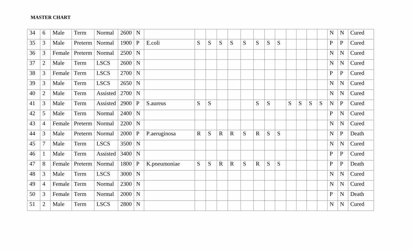
MASTER CHART
34 6 Male Term Normal 2600 N N N Cured
35 3 Male Preterm Normal 1900 P E.coli S S S S S S S S P P Cured
36 3 Female Preterm Normal 2500 N N N Cured
37 2 Male Term LSCS 2600 N N N Cured
38 3 Female Term LSCS 2700 N P P Cured
39 3 Male Term LSCS 2650 N N N Cured
40 2 Male Term Assisted 2700 N N N Cured
41 3 Male Term Assisted 2900 P S.aureus S S S S S S S S N P Cured
42 5 Male Term Normal 2400 N P N Cured
43 4 Female Preterm Normal 2200 N N N Cured
44 3 Male Preterm Normal 2000 P P.aeruginosa R S R R S R S S N P Death
45 7 Male Term LSCS 3500 N N N Cured
46 1 Male Term Assisted 3400 N P P Cured
47 8 Female Preterm Normal 1800 P K.pneumoniae S S R R S R S S P P Death
48 3 Male Term LSCS 3000 N N N Cured
49 4 Female Term Normal 2300 N N N Cured
50 3 Female Term Normal 2000 N P N Death
51 2 Male Term LSCS 2800 N N N Cured
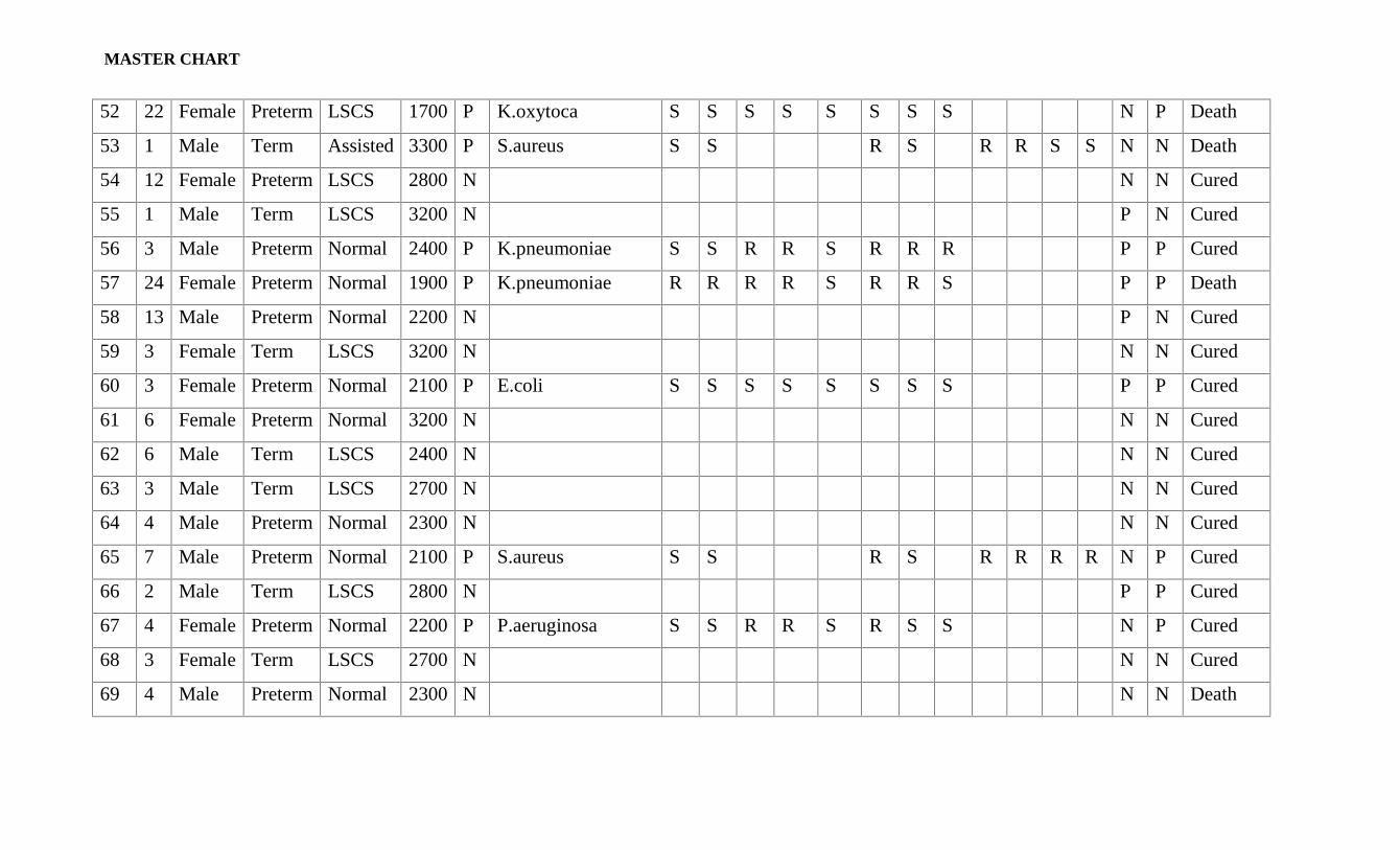
MASTER CHART
52 22 Female Preterm LSCS 1700 P K.oxytoca S S S S S S S S N P Death
53 1 Male Term Assisted 3300 P S.aureus S S R S R R S S N N Death
54 12 Female Preterm LSCS 2800 N N N Cured
55 1 Male Term LSCS 3200 N P N Cured
56 3 Male Preterm Normal 2400 P K.pneumoniae S S R R S R R R P P Cured
57 24 Female Preterm Normal 1900 P K.pneumoniae R R R R S R R S P P Death
58 13 Male Preterm Normal 2200 N P N Cured
59 3 Female Term LSCS 3200 N N N Cured
60 3 Female Preterm Normal 2100 P E.coli S S S S S S S S P P Cured
61 6 Female Preterm Normal 3200 N N N Cured
62 6 Male Term LSCS 2400 N N N Cured
63 3 Male Term LSCS 2700 N N N Cured
64 4 Male Preterm Normal 2300 N N N Cured
65 7 Male Preterm Normal 2100 P S.aureus S S R S R R R R N P Cured
66 2 Male Term LSCS 2800 N P P Cured
67 4 Female Preterm Normal 2200 P P.aeruginosa S S R R S R S S N P Cured
68 3 Female Term LSCS 2700 N N N Cured
69 4 Male Preterm Normal 2300 N N N Death

MASTER CHART
70 3 Male Preterm Normal 2100 P K.pneumoniae R R R R S S S S N P Cured
71 2 Female Term Assisted 3000 N N N Cured
72 2 Female Term LSCS 2900 N P P Death
73 4 Male Preterm Normal 2100 P E.coli S S S S S S S S N P Cured
74 4 Male Preterm Normal 2200 N N N Cured
75 17 Female Preterm Normal 2000 N N P Cured
76 4 Female Term LSCS 2100 N N N Cured
77 10 Male Preterm Normal 2400 N N N Cured
78 3 Male Term LSCS 2300 N P P Cured
79 2 Female Term Normal 2700 N N N Cured
80 3 Female Term Normal 2800 N N N Cured
81 5 Male Preterm LSCS 2400 N N N Cured
82 2 Male Preterm Normal 2000 P K.pneumoniae S S S S S S S S N P Cured
83 3 Male Term LSCS 2900 N P P Cured
84 1 Male Term Normal 3100 N N N Cured
85 1 Female Term Normal 2500 N N N Cured
86 2 Female Term LSCS 2400 P E.coli S S S S S S S S P P Cured
87 2 Female Preterm Normal 1900 N N N Cured
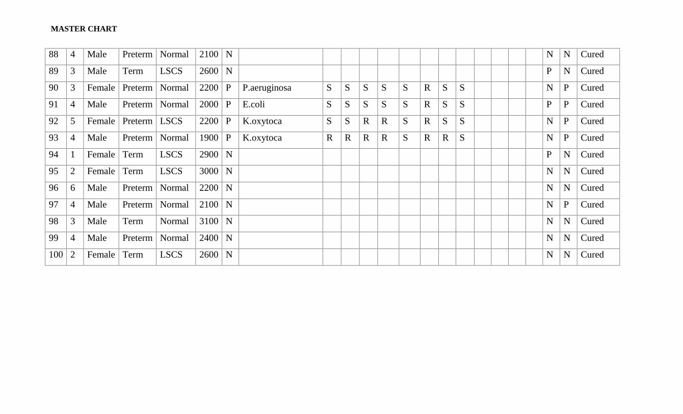
MASTER CHART
88 4 Male Preterm Normal 2100 N N N Cured
89 3 Male Term LSCS 2600 N P N Cured
90 3 Female Preterm Normal 2200 P P.aeruginosa S S S S S R S S N P Cured
91 4 Male Preterm Normal 2000 P E.coli S S S S S R S S P P Cured
92 5 Female Preterm LSCS 2200 P K.oxytoca S S R R S R S S N P Cured
93 4 Male Preterm Normal 1900 P K.oxytoca R R R R S R R S N P Cured
94 1 Female Term LSCS 2900 N P N Cured
95 2 Female Term LSCS 3000 N N N Cured
96 6 Male Preterm Normal 2200 N N N Cured
97 4 Male Preterm Normal 2100 N N P Cured
98 3 Male Term Normal 3100 N N N Cured
99 4 Male Preterm Normal 2400 N N N Cured
100 2 Female Term LSCS 2600 N N N Cured
Related Documents