Diagnostic protocol for the identification and detection of Candidatus Liberibacter solanacearum, the causal agent of zebra chip of potatoes PEST STATUS Not present in Australia PROTOCOL NUMBER NDP 18 VERSION NUMBER V1.2 PROTOCOL STATUS Endorsed ISSUE DATE 2012 REVIEW DATE 2017 ISSUED BY SPHDS

Welcome message from author
This document is posted to help you gain knowledge. Please leave a comment to let me know what you think about it! Share it to your friends and learn new things together.
Transcript
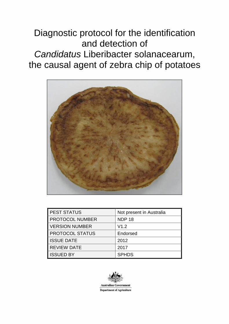
Diagnostic protocol for the identification and detection of
Candidatus Liberibacter solanacearum, the causal agent of zebra chip of potatoes
PEST STATUS Not present in Australia PROTOCOL NUMBER NDP 18 VERSION NUMBER V1.2 PROTOCOL STATUS Endorsed ISSUE DATE 2012 REVIEW DATE 2017 ISSUED BY SPHDS

Prepared for the Subcommittee on Plant Health Diagnostic Standards (SPHDS)
This version of the National Diagnostic Protocol (NDP) for Candidatus Liberibacter solanacearum is current as at the date contained in the version control box on the
front of this document. NDPs are updated every 5 years or before this time if required (i.e. when new
techniques become available). The most current version of this document is available from the SPHDS website:
http://plantbiosecuritydiagnostics.net.au/resource-hub/priority-pest-diagnostic-resources/

Contents
1. Introduction .................................................................................................................................... 1
1.1 Hosts ....................................................................................................................................... 1
1.2 Alternative host plants ............................................................................................................ 1
2. Taxonomy ...................................................................................................................................... 2
3. Detection ........................................................................................................................................ 3
3.1 Symptoms associated with Candidatus Liberibacter solanacearum ....................................... 3
3.1.1. Potato .............................................................................................................................. 3
3.1.2. Tomato ............................................................................................................................ 8
3.1.3. Capsicum ....................................................................................................................... 11
3.1.4. Tamarillo ....................................................................................................................... 13
3.1.5. Carrots ........................................................................................................................... 17
3.2 Diseases with similar symptoms ........................................................................................... 19
3.2.1. Psyllid yellows in potatoes ............................................................................................ 19
3.2.2. Potato purple top and other phytoplasmas in potatoes .................................................. 19
3.2.3. Carrots ........................................................................................................................... 19
4. Identification ................................................................................................................................ 20
4.1 Summary of the recommended Ca. L. solanacearum identification method ........................ 20
4.2 Sample selection ................................................................................................................... 20
4.3 DNA extraction procedure .................................................................................................... 21
4.4 Polymerase Chain Reaction .................................................................................................. 24
4.4.1. Housekeeping PCR ....................................................................................................... 24
4.4.2. Conventional PCR......................................................................................................... 25
4.4.3. Quantitative PCR .......................................................................................................... 28
4.5 Electrophoresis for conventional PCR .................................................................................. 29
4.6 Interpretation of results ......................................................................................................... 29
5. Contacts ........................................................................................................................................ 30
6. Acknowledgements ...................................................................................................................... 30
7. References .................................................................................................................................... 31
8. Appendix ...................................................................................................................................... 35
8.1 Nucleic acid cleanup ............................................................................................................. 35
8.2 Additional information .......................................................................................................... 36
8.3 Diseases associated with Candidatus Liberibacter solanacearum ........................................ 36
8.3.1. Zebra chip in potatoes ................................................................................................... 36
8.3.2. Psyllid yellows in tomatoes and Capsicum annuum ..................................................... 37
8.3.3. Ca. L. solanacearum in tamarillo .................................................................................. 38
8.3.4. Ca. L. solanacearum and an associated disease of carrots ............................................ 38

1. INTRODUCTION
Candidatus Liberibacter species are obligate intracellular parasites that inhabit the phloem cells of infected plant host species and are transmitted by insect vectors.
Ca. L solanacearum is a bacterium that infects several solanaceaous plant species and is transmitted by Bactericera cockerelli (the tomato potato psyllid). In Finland, Ca. L solanacearum has been detected in the carrot psyllid Trioza apicalis suggesting that this psyllid is also a potential vector (Munyaneza et al., 2010b).
Ca. L solanacearum may also be transmitted through infected seed potato (Henne et al., 2010). While plants produced from infected tubers were often weak, infected tubers could produce asymptomatic plants in which the bacterium could still be detected in the foliage (Andrew Pitman Crop and Food NZ, pers. comm.; Henne et al., 2010). The rate of bacterial transmission and expression of symptoms in the tubers of these asymptomatic plants was low. However infected foliage of potato plants can act as an acquisition source for the bacterium by the psyllid vector (Henne et al., 2010; Andrew Pitman, pers. comm.).
1.1 Hosts
Ca. L. solanacearum is associated with a disease complex rather than a single disease. Diseases associated with Ca. L. solanacearum include zebra chip in potatoes (Liefting et al., 2008a; Hansen et al., 2008; Secor et al., 2009; Abad et al., 2009; Rehman et al., 2010) and psyllid yellows in tomato and capsicum (Liefting 2008a; Hansen et al., 2008; Munyaneza et al., 2009b; Munyaneza et al., 2009c). Ca. L. solanacearum has also been reported causing dieback and leaf curling in tamarillo (Solanum betaceum) (Liefting et al., 2008b; Watson, 2009) and secondary root proliferation in carrots (Munyaneza et al., 2010a).
1.2 Alternative host plants
Ca. L solanacearum was detected in apparently healthy Cape gooseberry plants from a home garden in Auckland New Zealand (Liefting et al., 2009b). No disease has been reported and Cape gooseberries may be a symptomless host. Ca. L. solanacearum has also been detected in several solanaceous weeds with psyllid yellows symptoms including Solanum ptychanthum (black nightshade), Solanum elaeagnifolium (silver leaf nightshade) and Lycium barbarum (wolfberry) (Wen et al., 2009).
1

2. TAXONOMY
The bacteria are phloem-limited, non-culturable, filamentous, gram-negative which belong to the order Rhizobiales, in the class Alphaproteobacteria of the phylum Proteobacteria (Jagoueix et al., 1994; Bové, 2006).
Candidatus Liberibacter solanacearum is also referred to by the synonym Candidatus Liberibacter psyllaurous (Hansen et al., 2008). While both scientific names occur in the literature, Ca. L. solanacearum should be used in reference to this bacterium as the name and a description of the bacterium was published in the “International Journal of Systematic and Evolutionary Microbiology”, which is the preferred journal for descriptions of novel bacterial species (Liefting et al., 2009b). Reference material of the original isolate is also available from the authors (Liefting et al., 2009b). The bacteria will be referred to as Ca. L. solanacearum throughout this document.
Ca. Liberibacter species have a triple layered cell envelope with an outer wall membrane and an inner cytoplasmic membrane (Jagoueix et al., 1996; Bové 2006). Ca. L. solanacearum is approximately 0.2 µm in width and 4 µm in length and has a genome size approximately 1.26 Mbp (Lin et al., 2009).
2

3. DETECTION
Diseases associated with Ca. L solanacearum can be identified by the presence of symptoms, however diagnosis should be confirmed through PCR detection and sequencing of the amplified product, particularly as other organisms may cause similar symptoms. Most symptoms, particularly if they are observed on their own, may be caused by other biotic and abiotic factors.
3.1 Symptoms associated with Candidatus Liberibacter solanacearum
3.1.1. Potato • Yellowing or purpling of potato leaves and shoots (Figures 1 and 2) • Curling or rolling of leaves (Figures 1 and 2) • Stunted shoots with shortened and swollen internodes (Figure 2) • Aerial tuber formation (Figure 3) • Scorched potato tops that collapse prematurely (Figure 4) • Early senescence • Tubers may have enlarged lenticels, necrotic flecking of the vascular tissue and
streaks along the medullary rays (Figure 5) that are enhanced when slices of the potatoes are fried (Figure 6)
Figure 1. Shoots of “zebra chip” affected potato plants infected with Candidatus Liberibacter
solanacearum. Leaves on younger shoots display purpling, and older leaves are chlorotic. The leaves are also rolled (Images from New Zealand, courtesy of Dr Lia Liefting, Ministry for Primary Industries, New Zealand (MPI)).
3

Figure 2. Shoots of a “zebra chip” affected potato plant infected with Candidatus Liberibacter solanacearum. Leaves are chlorotic, curled and rolled. Leaves on younger shoots display mild purpling. The shoots are stunted and swollen (arrows) and swelling is occurring at the nodes (Image from New Zealand, courtesy of Dr Lia Liefting, MPI).
4

Figure 3. Aerial tuber formation on “zebra chip” affected potato plant infected with Candidatus Liberibacter solanacearum (Images from New Zealand, courtesy of Dr Lia Liefting, MPI).
Figure 3. Aerial tuber formation on “zebra chip” affected potato plant that was also infected with Candidatus Liberibacter solanacearum. (Images courtesy of Dr Lia Liefting, Ministry of Agriculture and Forestry, New Zealand). 5

Figure 4. Shoots of a “zebra chip” affected potato plant infected with Candidatus Liberibacter solanacearum. Leaves are chlorotic and rolled and some are necrotic or scorched (Image from New Zealand, courtesy of Dr Lia Liefting, MPI).
Figure 5. Fresh tuber slices of “zebra chip” affected potato plants infected with Candidatus Liberibacter solanacearum. The images show mild (a), moderate (b) and severe (c and d) discolouration /flecking of the vascular tissue and flecking and streaking of the medullary rays (arrows), which may be observed in fresh tubers before frying (Images from New Zealand, courtesy of Dr Lia Liefting, MPI).
a. b.
c. d.
6

Figure 6. Fried tuber slices of “zebra chip” affected potato plants infected with Candidatus Liberibacter solanacearum. The images show the enhanced discolouration and flecking of the vascular tissue and streaking of the medullary rays of tuber slices after frying (Images from New Zealand, courtesy of Dr Lia Liefting, MPI).
7
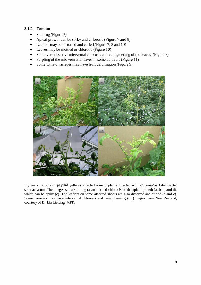
3.1.2. Tomato • Stunting (Figure 7) • Apical growth can be spiky and chlorotic (Figure 7 and 8) • Leaflets may be distorted and curled (Figure 7, 8 and 10) • Leaves may be mottled or chlorotic (Figure 10) • Some varieties have interveinal chlorosis and vein greening of the leaves (Figure 7) • Purpling of the mid vein and leaves in some cultivars (Figure 11) • Some tomato varieties may have fruit deformation (Figure 9)
Figure 7. Shoots of psyllid yellows affected tomato plants infected with Candidatus Liberibacter solanacearum. The images show stunting (a and b) and chlorosis of the apical growth (a, b, c, and d), which can be spiky (c). The leaflets on some affected shoots are also distorted and curled (a and c). Some varieties may have interveinal chlorosis and vein greening (d) (Images from New Zealand, courtesy of Dr Lia Liefting, MPI).
a
b
c
d
8
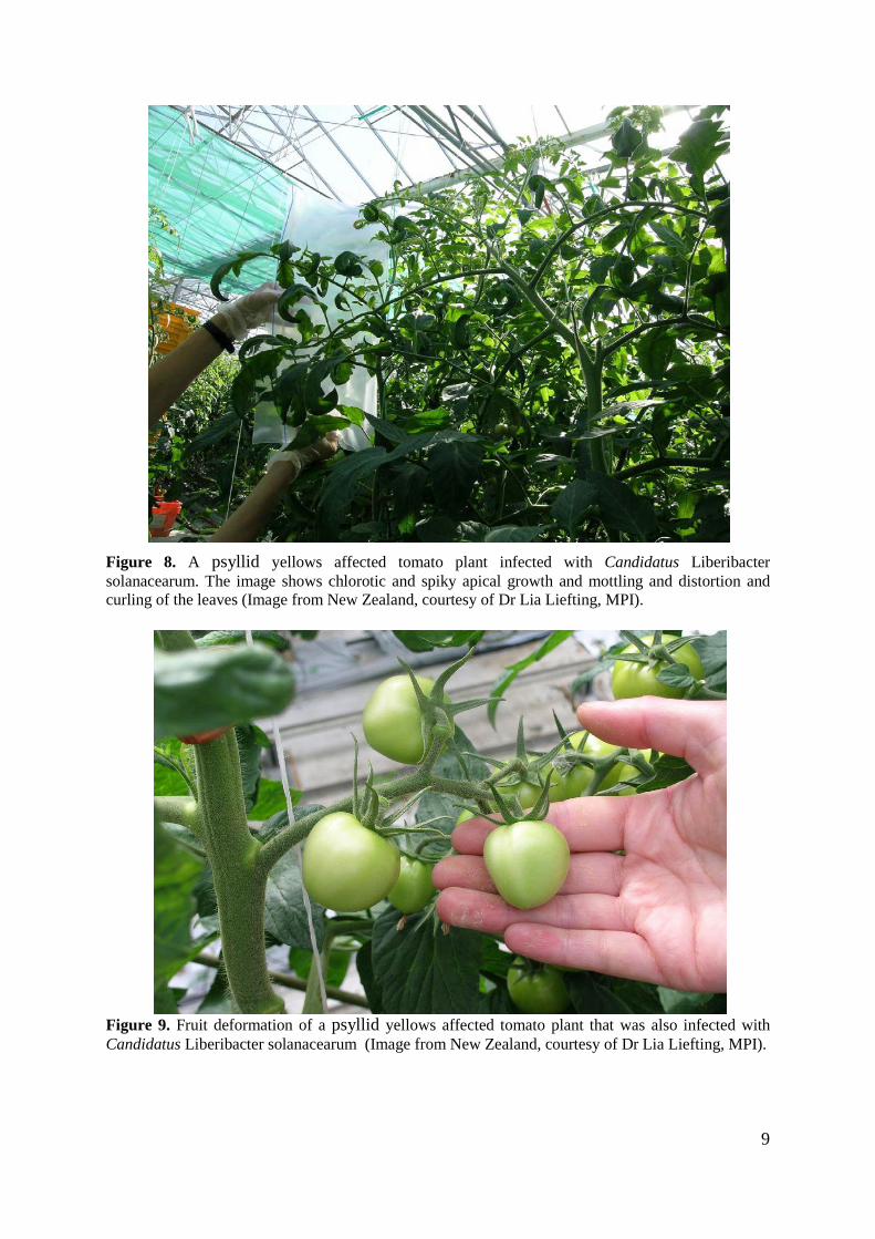
Figure 8. A psyllid yellows affected tomato plant infected with Candidatus Liberibacter solanacearum. The image shows chlorotic and spiky apical growth and mottling and distortion and curling of the leaves (Image from New Zealand, courtesy of Dr Lia Liefting, MPI).
Figure 9. Fruit deformation of a psyllid yellows affected tomato plant that was also infected with Candidatus Liberibacter solanacearum (Image from New Zealand, courtesy of Dr Lia Liefting, MPI).
9

Figure 10. Leaves of psyllid yellows affected tomato plants infected with Candidatus Liberibacter solanacearum. The images show chlorosis and mottling (a and b). The leaflets may also be distorted and curled (c) (Image from New Zealand, courtesy of Dr Lia Liefting, MPI).
Figure 11. Shoot tip of psyllid yellows affected tomato plant infected with Candidatus Liberibacter solanacearum showing purpling of the leaflets and petioles (Image from New Zealand, courtesy of Dr Lia Liefting, MPI).
a b
c
10

3.1.3. Capsicum • Stunting (Figures 12, 13 and 15) • Shortened internodes of the stems • Leaves can be pale green or chlorotic (Figures 12, 13, 14 and 15) • Leaves may have shortened petioles • Leaves may be cupped (Figures 12 and 14) • Leaf apices may taper giving plants a spiky appearance (Figures 12 and 13) • Flower abortion may be observed • Tip necrosis • Parts of the plant may die back
Figure 12. A stunted shoot of psyllid yellows affected Capsicum annuum plant infected with Candidatus Liberibacter solanacearum. The leaves are pale green and tapered and some are slightly cupped (Image from New Zealand, courtesy of Dr Lia Liefting, MPI).
Figure 13. A shoot of a psyllid yellows affected Capsicum annuum plant infected with Candidatus Liberibacter solanacearum. The leaves are pale green/chlorotic and tapered to give a spiky appearance (Image from New Zealand, courtesy of Dr Lia Liefting, MPI).
11

Figure 14. A stunted shoot of a psyllid yellows affected Capsicum annuum plant infected with Candidatus Liberibacter solanacearum. The leaves are pale green/chlorotic and the apical leaves are cupped (Image from New Zealand, courtesy of Dr Lia Liefting, MPI).
Figure 15. Stunted shoots of a psyllid yellows affected Capsicum annuum plant infected with Candidatus Liberibacter solanacearum. The leaves are chlorotic and curled (Image from New Zealand, courtesy of Dr Lia Liefting, MPI).
12
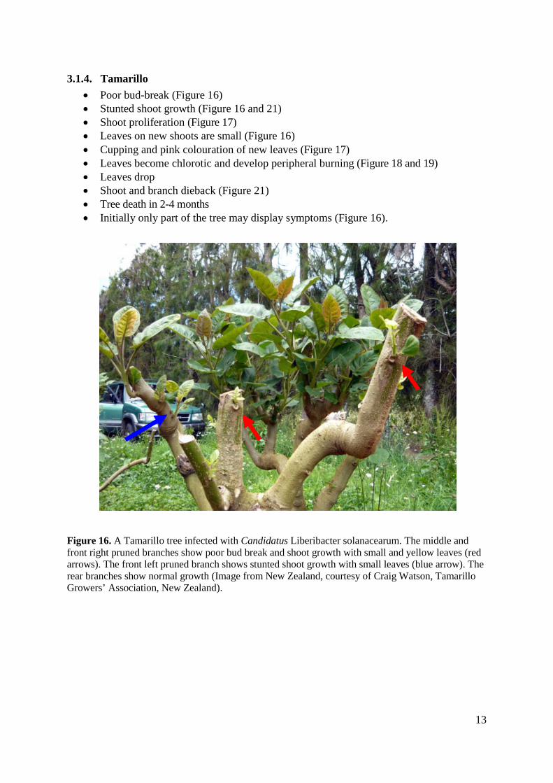
3.1.4. Tamarillo • Poor bud-break (Figure 16) • Stunted shoot growth (Figure 16 and 21) • Shoot proliferation (Figure 17) • Leaves on new shoots are small (Figure 16) • Cupping and pink colouration of new leaves (Figure 17) • Leaves become chlorotic and develop peripheral burning (Figure 18 and 19) • Leaves drop • Shoot and branch dieback (Figure 21) • Tree death in 2-4 months • Initially only part of the tree may display symptoms (Figure 16).
Figure 16. A Tamarillo tree infected with Candidatus Liberibacter solanacearum. The middle and front right pruned branches show poor bud break and shoot growth with small and yellow leaves (red arrows). The front left pruned branch shows stunted shoot growth with small leaves (blue arrow). The rear branches show normal growth (Image from New Zealand, courtesy of Craig Watson, Tamarillo Growers’ Association, New Zealand).
13

Figure 17. A Tamarillo tree infected with Candidatus Liberibacter solanacearum. The new leaves show an abnormal pink colouration and shoot proliferation (Image from New Zealand, courtesy of Craig Watson, Tamarillo Growers’ Association, New Zealand).
Figure 18. A Tamarillo tree infected with Candidatus Liberibacter solanacearum. The leaves have interveinal chlorosis, cupping and leaf scorching. Note the pink colouration of the new leaf in the centre of the image (Image from New Zealand, courtesy of Craig Watson, Tamarillo Growers’ Association, New Zealand).
14
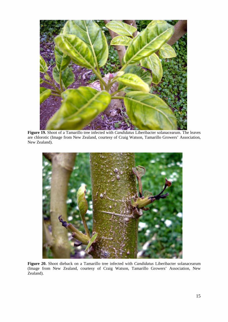
Figure 19. Shoot of a Tamarillo tree infected with Candidatus Liberibacter solanacearum. The leaves are chlorotic (Image from New Zealand, courtesy of Craig Watson, Tamarillo Growers’ Association, New Zealand).
Figure 20. Shoot dieback on a Tamarillo tree infected with Candidatus Liberibacter solanacearum (Image from New Zealand, courtesy of Craig Watson, Tamarillo Growers’ Association, New Zealand).
15
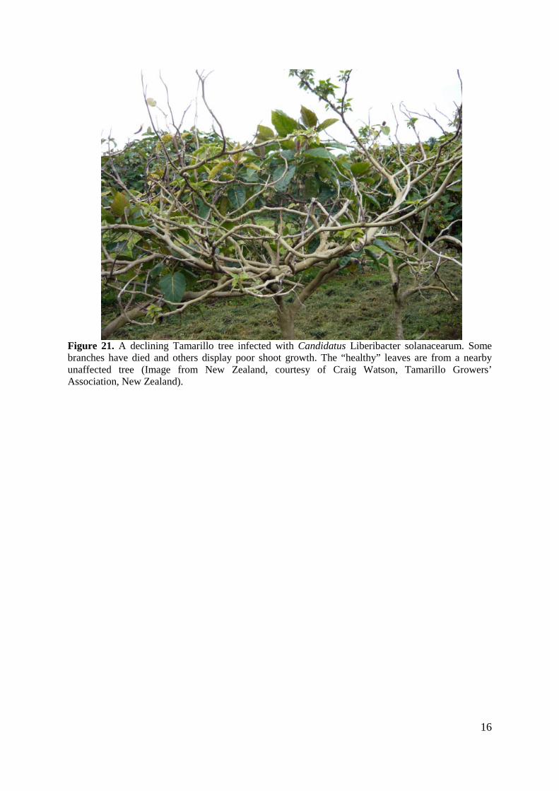
Figure 21. A declining Tamarillo tree infected with Candidatus Liberibacter solanacearum. Some branches have died and others display poor shoot growth. The “healthy” leaves are from a nearby unaffected tree (Image from New Zealand, courtesy of Craig Watson, Tamarillo Growers’ Association, New Zealand).
16
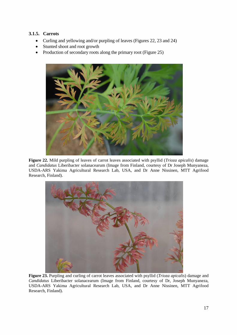
3.1.5. Carrots • Curling and yellowing and/or purpling of leaves (Figures 22, 23 and 24) • Stunted shoot and root growth • Production of secondary roots along the primary root (Figure 25)
Figure 22. Mild purpling of leaves of carrot leaves associated with psyllid (Trioza apicalis) damage and Candidatus Liberibacter solanacearum (Image from Finland, courtesy of Dr Joseph Munyaneza, USDA-ARS Yakima Agricultural Research Lab, USA, and Dr Anne Nissinen, MTT Agrifood Research, Finland).
Figure 23. Purpling and curling of carrot leaves associated with psyllid (Trioza apicalis) damage and Candidatus Liberibacter solanacearum (Image from Finland, courtesy of Dr, Joseph Munyaneza, USDA-ARS Yakima Agricultural Research Lab, USA, and Dr Anne Nissinen, MTT Agrifood Research, Finland).
17
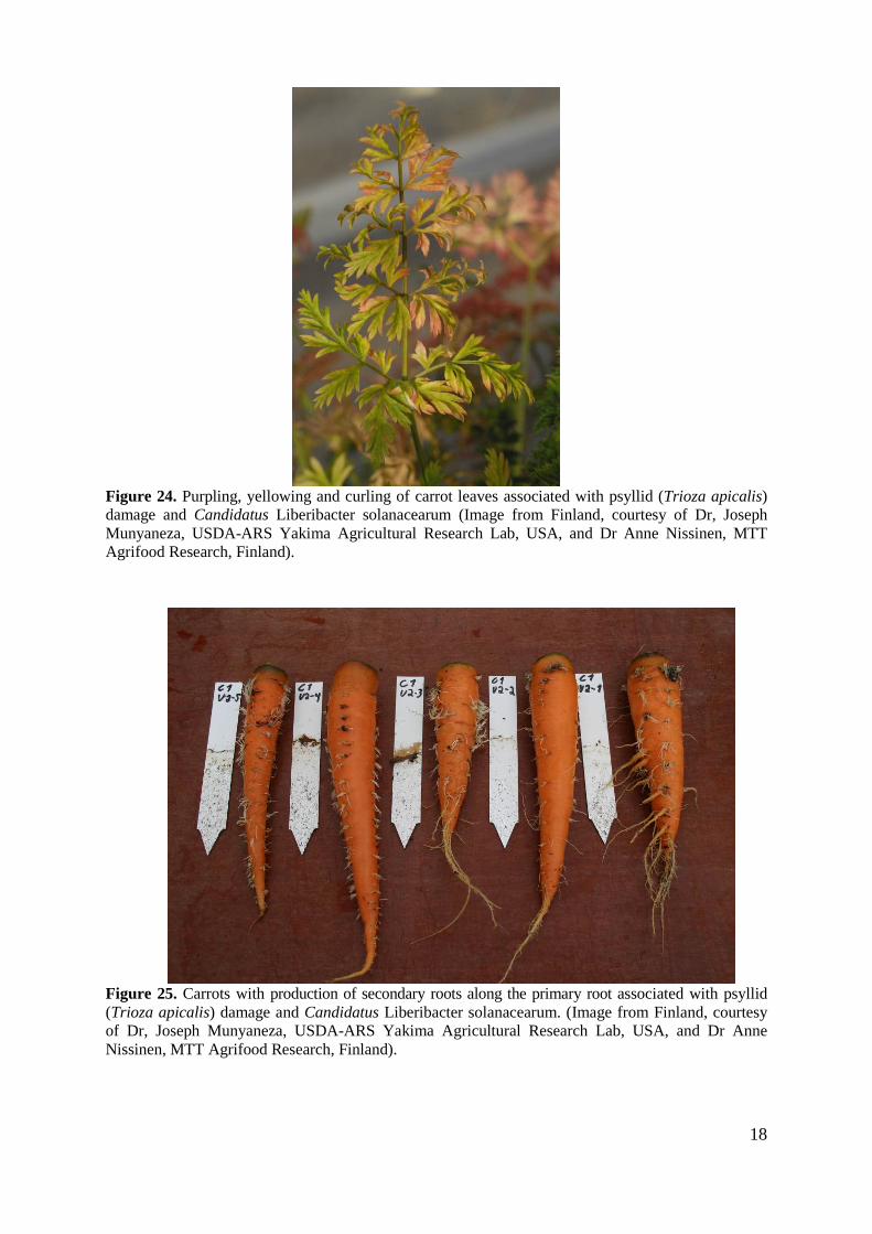
Figure 24. Purpling, yellowing and curling of carrot leaves associated with psyllid (Trioza apicalis) damage and Candidatus Liberibacter solanacearum (Image from Finland, courtesy of Dr, Joseph Munyaneza, USDA-ARS Yakima Agricultural Research Lab, USA, and Dr Anne Nissinen, MTT Agrifood Research, Finland).
Figure 25. Carrots with production of secondary roots along the primary root associated with psyllid (Trioza apicalis) damage and Candidatus Liberibacter solanacearum. (Image from Finland, courtesy of Dr, Joseph Munyaneza, USDA-ARS Yakima Agricultural Research Lab, USA, and Dr Anne Nissinen, MTT Agrifood Research, Finland).
18

3.2 Diseases with similar symptoms
3.2.1. Psyllid yellows in potatoes Psyllid yellows disease of potatoes has aerial symptoms similar to zebra chip disease and was first observed in the USA in 1927 (Richards, 1928). Psyllid yellows is associated with feeding of the tomato potato psyllid (Sengoda et al., 2010). It was suggested that zebra chip and psyllid yellows diseases could be differentiated on the basis of symptomatology in the tubers: psyllid yellows affected plants do not exhibit the tuber symptoms and survive longer than zebra chip affected plants (Sengoda et al., 2010). However the bacterium is not necessarily evenly distributed throughout a plant and it is possible that not all tubers of infected plants are affected. Timing of infection may also play an important role in the effect of the bacteria on tubers and some tubers may only display mild symptoms of disease. It is also possible that there is a difference in symptom expression between varieties. Consequently psyllid yellows disease of potatoes could be confused with zebra chip disease.
3.2.2. Potato purple top and other phytoplasmas in potatoes Several phytoplasma species representing six taxonomically distinct phytoplasma groups (16SrI, 16SrII, 16SrIII, 16SrVI, 16SrXII and 16SrXIII) have also been found in association with diseases of potatoes that have similar symptoms to zebra chip disease. These diseases include haywire disease, potato purple top, or potato purple top wilt, and stolbur disease (Semancik and Peterson 1971; Harding and Teakle, 1985; Lee et al., 2004; Lee et al., 2006; Leyva-Lopez et al., 2002; Secor, 2007; Munyaneza et al., 2007; Lee et al., 2009). Symptoms associated with these diseases include stunting, leaf curl, chlorosis, purpling of the apical leaves, swollen nodes, proliferation of axillary buds, aerial tubers and scattered light-brown discolouration of tubers which are enhanced when tuber tissue is fried. (Munyaneza et al., 2009d). The phytoplasmas and the associated diseases have been reported from Mexico (16SrI, II, III and XIII), USA (16SrIII, VI and XII), Europe (16SrI and XII), South America (16SrI and II), Asia (16SrINew Zealand (16SrXII) and Australia (16SrII)(Norris, 1954; Harding and Teakle 1985, Lee et al., 2009; Liefting et al., 2009c; Secor, 2007)
3.2.3. Carrots Similar symptoms have been found on carrots in association with other organisms including a phytoplasma and an undescribed bacteria-like organism occurring in Australia (Gibb et al., 2003) and stolbur phytoplasma in Spain (Font et al., 1999).
19

4. IDENTIFICATION
The most reliable method for Ca. L. solanacearum detection is polymerase chain reaction (PCR), which is used to detect the DNA of the bacterium. The efficiency of this test is dependent on appropriate sampling of plant tissue, reliable nucleic acid extraction methods and species-specific primers used in the PCR test.
4.1 Summary of the recommended Ca. L. solanacearum identification method • Sample vascular tissue containing phloem cells from candidate plants. • Extract total DNA. • Perform a housekeeping PCR with the rP1/fD2 primers. The rP1/fD2 primers amplify
the 16S rRNA gene from most prokaryotes as well as from chloroplasts (Weisberg et al., 1991). If this test is negative then there is no DNA present or there are DNA polymerase inhibitors co-extracted with the nucleic acid. In this situation, try cleaning the nucleic acid (Appendix I) or repeat the extraction.
• Perform PCR using the following procedure: Use a conventional nested PCR on the purified DNA. Or Use the quantitative PCR with a Taqman probe if there is access to a suitable real-time PCR machine.
• Analyse the conventional PCR products by agarose gel electrophoresis. • To confirm Ca. L. solanacearum identity, direct sequence the PCR product. If direct
sequencing is problematic, the PCR product can be cloned and then sequenced using standard cloning and sequencing procedures. Sequence data can be analysed using the Basic Local Alignment Search Tool (BLAST) available at: http://blast.ncbi.nlm.nih.gov/Blast.cgi.
4.2 Sample selection
Ca. L. solanacearum is phloem limited and 0.5g of vascular tissue should be sampled for successful PCR detection. Symptomatic tissue provides the best opportunity to detect Ca. L. solanacearum in infected plants (Lia Liefting, MPI New Zealand, pers. comm.). Higher concentrations of the bacterium were observed in root, tuber and stolon tissue compared to aerial tissue of potato plants (Li et al., 2009; Wen et al., 2009). Fried symptomatic potato tissue may improve the chance of detection compared to fresh symptomatic tuber tissue (Lia Liefting, MPI, pers.comm.).
Symptomatic potato tubers are most reliable and the stems of ZC symptomatic plants can also be used. For tomato, tissue from symptomatic shoots should be used. Tissue can include stems, leaf petioles, the peduncles attached to fruit and the portion of affected fruit to which the peduncle is attached.
Symptomless infections may occur and if this is suspected it is important to thoroughly sample phloem tissue from different aerial and subterranean tissue such as roots and tubers of the one plant for Ca. L solanacearum detection.
There is very little information available on the best time for detection. The translocation and titre of Ca. L. solanacearum and symptom development in any host may be dependent on the time of infection, the abundance of the insect vector and on environmental factors that affect replication of the bacterium and symptom expression.
20
4.3 DNA extraction procedure
This method uses the QIAGEN DNeasy® Plant mini kit (Green et al., 1999). DNA extraction methods using other column-based or magnetic-bead based protocols can be used if they have been validated for use by the laboratory performing the diagnostic test.
Materials and equipment 1. QIAGEN DNeasy® Plant mini kit
2. 1.5 ml centrifuge tubes
3. 20-200 µl and 200-1000 µl pipettes
4. 20-200 µl and 200-1000 µl sterile filter pipette tips 5. Autoclave
6. Balance 7. Bench top centrifuge 8. Distilled water 9. Ice machine
10. Freezer
11. Sterile mortars and pestles or “Homex” grinder (Bioreba) and grinding bags (Agdia or Bioreba) or hammer and grinding bags (Agdia or Bioreba)
If using mortar and pestles, ensure they are thoroughly cleaned prior to use to prevent cross-contamination from previous extractions. To clean thoroughly, soak mortars and pestles in 2% bleach for 1 hour. Rinse with tap water then soak in 0.2 M HCl or 0.4 M NaOH for 1 hour. Rinse thoroughly with distilled water.
12. Scalpel handle
13. Sterile scalpel blades
14. Vortex
15. Water bath or heating block at 55-65°C
16. Latex or nitrile gloves
17. Buffers:
• CTAB grinding buffer (Table 1)
• Absolute ethanol The 2% cetyltrimethylammonium bromide (CTAB) buffer (Table 1) is required for all extraction procedures:
21

Table 1. 2.5% cetyltrimethylammonium bromide (CTAB) extraction buffer for DNA purification.
Reagent* Final
Amount needed for 1 L
CTAB (cetyltrimethylammonium bromide) 2.5% 25 g
Sodium chloride 1.4 M 56 g
1 M Tris-HCl, pH 8.0 (sterile) 100 mM 100 ml
0.5 M EDTA, pH8.0 (sterile) 20 mM 40 ml
Polyvinylpyrrolidone (PVP-40) 1% 10 g
*Make up to volume with sterile distilled water. Store at room temperature. Just before use, add 0.2% 2-mercaptoethanol (v/v) to the required volume of buffer.
If a fume hood is unavailable β – mercaptoethanol can be omitted but the quality of the extract from some plant species may be affected.
Method 1. Grind 0.5 g of tissue in 5 ml of CTAB extraction buffer (room temperature) containing
0.2% β – mercaptoethanol.
2. Transfer 500 µl of extract to a 1.5 ml microfuge tube and add 4 µl of RNase A (Supplied with the DNeasy kit), cap tube and incubate at 65°C for 25-35 min, mixing gently several times.
3. Add 130 µl of QIAGEN buffer AP2 to extract. Invert 3 times to mix and place on ice for 5 minutes.
4. Apply lysate onto a QIAshredder™ column and centrifuge at 20,000 x g (14,000 rpm or maximum speed) for 2 minutes.
5. Transfer 450 µl of flowthrough from the QIAshredder™ column to a 1.5 ml centrifuge tube containing 675 µl QIAGEN buffer AP3/E. Mix by pipetting.
6. Transfer 650 µl of extract onto a DNeasy column and spin at 6,000 × g (8000 rpm) for 1 minute.
7. Discard flow-through and add the rest of the sample to the column and spin at 10000 rpm for 1 minute.
8. Place DNeasy column in a new 2 ml collection tube and add 500 µl of QIAGEN buffer AW (wash buffer) and spin at 10000 rpm for one minute.
9. Discard flowthrough and add another 500 µl of QIAGEN buffer AW and spin at maximum speed for 2 minutes.
10. Discard flowthrough and collection tube. Ensure that the base of the column is dry (blot on tissue if it is not) and place in an appropriately labelled microfuge tube. Add 100 µl of pre-warmed 65°C AE buffer directly to the filter (don’t apply down the side of the tube) and spin at 10000 rpm for 1 minute. Discard column and store DNA in Freezer.
22

Table 2. PCR primers used for phytoplasma detection and internal control primers.
PCR test† Primer name
(direction) Primer sequence (5´-3´)
Tm
Product
size (bp) Reference
Candidatus Liberibacter solanacearum
Nested PCR - First stage
OA2 GCGCTTATTTTTAATAGGAGCGGCA 60°C
(use 66°C for capsicum samples)
1,160 bp Jagoueix et al., 1996 Liefting et al., 2009a Liefting et al., 2009b OI2c GCCTCGCGACTTCGCAACCCAT
Nested PCR –second stage
Lib16S01F TTCTACGGGATAACGCACGG 58°C 580 bp Liefting et al., 2009b
Lib16S01R CGTCAGTATCAGGCCAGTGAG
Real Time PCR
LsoF GTCGAGCGCTTATTTTTAATAGGA
58°C 68 bp Li et al., 2009 HLBr GCGTTATCCCGTAGAAAAAGGTAG
HLBp (hydrolysis probe)* FAM-AGACGGGTGAGTAACGCG-BHQ1
Internal control
16S bacterial and plant chromosomal
FD2 AGAGTTTGATCATGGCTCAG 55ºC
approx. 1400-1500 bp.
Weisberg et al., 1991 RP1 ACG GTT ACC TTG TTA CGA CTT
* Hydrolysis probes are often referred to as Taqman; “Taqman” is the proprietary name held by Applied Biosystems. Hydrolysis probes can be purchased from a number of biotechnology companies. FAM= 6-carboxy-fluorescein reporter; BHQ = Black hole quencher
23

4.4 Polymerase Chain Reaction
To reduce the risk of contamination and possible false positive results, particularly when nested PCR is used for Ca. L solanacearum detection, it is desirable to set up PCR reactions in a different lab to where nucleic acid extractions have been done. It is also desirable to handle PCR reagent stocks and to set up PCR reactions in a clean room or bio-safety cabinet with dedicated pipettes, PCR tubes and tips that have not been exposed to nucleic acid extracts and PCR products. Use a separate pipette for the addition of nucleic acids to the PCR reactions. Do not add nucleic acid to reactions in the same clean room or bio-safety cabinet in which PCR stocks are handled.
PCR materials and equipment 1. PCR reagents of choice
2. Primers (Table 4)
3. PCR grade water
4. 0-2 µl, 2-20 µl, 20-200 µl and 200-1000 µl pipettes
5. 0-2 µl, 2-20 µl, 20-200 µl and 200-1000 µl sterile filter pipette tips 6. 1.5 ml centrifuge tubes to store reagents
7. PCR tubes (volume depends on thermal cycler)
8. Bench top centrifuge – with adapters for small tubes
9. Freezer
10. Ice machine
11. Latex or nitrile gloves
12. Conventional thermal cycler
13. Real time thermal cycler
14. DNA molecular weight marker
4.4.1. Housekeeping PCR The housekeeping PCR should be conducted to determine if the nucleic extract is of sufficient quality for Ca. L. solanacearum detection. For the house keeping PCR the primers and the expected size of the PCR product are listed in Table 2, the components and concentrations are listed in Table 3 and the cycling times are listed in Table 4. Run the PCR products on a gel as described below. The house keeping PCR is successful if a product of the expected size is observed, indicating the presence of quality DNA in the nucleic acid extract. If the housekeeping PCR is successful continue with the nested PCR assay for Ca. L solanacearum detection. The housekeeping PCR is not successful if no PCR product is observed. If this occurs, the nucleic acid extract should be cleaned up or the sample should be re-extracted and a housekeeping PCR conducted on these extracts. It may also be useful to dilute some of the original DNA extract 1:5 or 1:10 and repeat the housekeeping PCR. Should this work the diluted sample could be used for the nested PCR for Ca. L solanacearum detection.
24

Controls Positive control: DNA of known good quality No template control: Sterile distilled water
4.4.2. Conventional PCR For Ca. L. solanacearum detection the primers and the expected size of the PCR product are listed in Table 2, the components and concentrations for the first stage PCR are listed in Table 3 and the cycling conditions are given in Table 4. The 16S rRNA gene, to which all the primers and probe were designed is highly conserved amongst the four known Candidatus Liberibacter species, of the primers used for detection in the nested PCR and real-time PCR only OA2 and LsoF are specific for Ca. L. solanacearum.
For nested PCR, the first-stage PCR products are diluted 1:25 (v/v) in water prior to re-amplification using the second-stage PCR primers.
If a positive result is obtained the PCR product should be sequenced to confirm the identity of the organism that is detected.
When establishing the test initially, it is advised that a negative control (DNA extracted from healthy plant tissue) is included. The PCR will only be considered valid if no amplicons are produced in this negative control.
Controls Positive control: DNA extracted from Ca. L. solanacearum infected tissue or
plasmid containing Ca. L. solanacearum first stage PCR product
Negative control: DNA extracted from plant tissue not known to be infected with Ca. L. solanacearum
No template control: Sterile distilled water
25

Table 3. Conventional PCR reaction master mix – housekeeping gene PCR and first stage and second stages of the Ca. L. solanacearum nested PCR
Reagent First stage PCR for Ca. L. solanacearum and House keeping PCR
Volume per reaction
Second Stage for Ca. L. solanacearum
Volume per reaction
Sterile (RNase, DNase free) water
13.9 µl 14.9 µl
10 × reaction buffer 2 µl 2 µl
50 mM MgCl2 0.6 µl 0.6 µl
10 mM dNTP mixture 0.4 µl 0.4 µl
10 µM Forward primer 0.5 µl 0.5 µl
10 µM Reverse primer 0.5 µl 0.5 µl
5 units/µl Platinum Taq DNA polymerase (Invitrogen 10966-026)
0.1 µl 0.1 µl
DNA template or control 2 µl 1 µl
Total reaction volume 20 µl 20 µl
For the house keeping PCR and first stage of the nested PCR for Ca. L. solanacearum pipette 18 µl of reaction mix into each PCR tube then add 2 µl of DNA template.
For the second stage of the nested PCR for Ca. L. solanacearum pipette 19 µl of reaction mix into each PCR tube then add 1 µl of first stage PCR product as the template.
26

Table 4. PCR cycling conditions for the housekeeping PCR, and the first and second stages of the nested PCR for detection of Ca. L. solanacearum.
Step
Housekeeping PCR assay
Ca. L. solanacearum – Nested PCR assay
First stage Second stage
Temp. Time No. of cycles Temp. Time No. of
cycles Temp. Time No. of cycles
Initial denaturation 94oC 5
min 1 94oC 5 min 1 94oC 5
min 1
Denaturation 94oC 45 s
40
94oC 45 s
40
94oC 30 s
40 Annealing 60ºC 45 s 60ºC
66ºC for Capsicum)
45 s 58ºC 30 s
Elongation 72oC 1 min 72oC 1
min 72oC 45 s
Final elongation 72oC 7
min 1 72oC 7 min 1 72oC 7
min 1
27

4.4.3. Quantitative PCR Quantitative PCR can be used to confirm detection of Ca. L solanacearum in samples with high titres of the bacterium. The primers and probe for detection of Ca. L. solanacearum are given in Table 2, the components and their concentrations for the real time PCR are given in Table 5 and the cycling conditions are given in Table 6. Like the conventional nested assay this protocol is based on the 16S rRNA gene; however the use of a probe in addition to the primers increases the specificity of the assay. The production of a smaller amplicon over 50 cycles should increase the sensitivity of the assay compared to single (first) stage PCR but is not as sensitive as the nested PCR.
Table 5. Quantitative PCR reaction master mix for detection of Ca. L. solanacearum.
Reagent* Volume per reaction
Sterile (RNase, DNase free) water 6.75 µl
Platinum® Quantitative PCR SuperMix-UDG 10 µl
10 µM LsoF primer 0.5 µl
10 µM HLBr primer 0.5 µl
10 µM HLBp hydrolysis probe 0.25 µl
DNA template or control 2 µl
Total reaction volume 20 µl
*0.8 µl of 10 mg/ml BSA can be added to the reaction mix and may reduce the risk of inhibition of the DNA polymerase – if BSA is used, adjust the volume of water so that the final reaction volume remains at 20 µl.
28

Table 6. PCR cycling conditions for the quantitative PCR detection of Ca. L. solanacearum.
Step Temperature Time No. of cycles Initial Hold 50oC 2 min 1 Initial Denaturation 95oC 2 min 1 Denaturation 95ºC 10 s
50 Annealing and Elongation 58oC 45 s
4.5 Electrophoresis for conventional PCR
Separate the PCR products (5-10 µl) on a 1% agarose gel containing ethidium bromide or SybR-Safe and visualise using an UV transilluminator (ethidium bromide staining) or blue light box (SybR-Safe staining). Use a DNA molecular weight marker to determine the size of the products. Table 4 lists the expected PCR product size for each primer pair.
4.6 Interpretation of results
Failure of the samples to amplify with the housekeeping primers suggests that the DNA extraction has failed, compounds inhibitory to PCR are present in the DNA extract or the DNA has degraded.
The Ca. L. solanacearum conventional PCR tests will only be considered valid if:
(a) the positive control produces the correct size product as indicated in Table 4; and
(b) no bands are produced in the negative control (if used) and the no template control.
The Ca. L. solanacearum quantitative PCR tests will only be considered valid if a Ct value above the threshold automatically set by the real time PCR instrument occurs.
29

5. CONTACTS
Dr Fiona Constable Department of Primary Industries - Knoxfield Private Bag 15 Ferntree Gully Delivery Centre Victoria 3156 AUSTRALIA Ph + 61 3 92109222
6. ACKNOWLEDGEMENTS
This protocol was developed and compiled by Dr Fiona Constable, Department of Primary Industries, Victoria. The following people provided training, information, advice and images for this protocol:
Dr Lia Liefting MPI, New Zealand Dr Nadine Berry, Crop and Food, New Zealand Dr Andrew Pitman, Crop and Food, New Zealand Dr Ian Scott, Crop and Food, New Zealand Craig Watson, Tamarillo Grower’s Association, New Zealand Dr Joe Munyaneza, USDA-ARS, Yakima Agricultural Research Laboratory, USA This protocol was reviewed and verified by Dr Lia Liefting, (Senior Scientist, Virology & Post-Entry Quarantine), Plant Health and Environment Laboratory, Ministry for Primary industries (MPI), New Zealand.
30

7. REFERENCES
Abad, J.A., Bandla, M., French-Monar, R.D., Liefting, L.W. and Clover, G.R.G. 2009. First report of the detection of 'Candidatus Liberibacter' species in zebra chip disease-infected potato plants in the United States. Plant Disease 93, 108-109.
Binkley, A.M. 1929. Transmission Studies with the new Psyllid-Yellows Disease of solanaceous Plants. Science 70, 615.
Blood, H.L., Richards B.L. and Wann F.B. 1933. Studies of psyllid yellows of tomato. Phytopathology 23, 930.
Bové, J.M. 2006. Huanglongbing: a destructive, newly-emerging, century-old disease of citrus. Journal of Plant Pathology 88, 7-37.
Brown, J.K. Rehman, M., Rogan, D., Martin, R.R. and Idri, A. M. 2009. First report of "Candidatus Liberibacter psyllaurous" (synonym "Ca. L. solanacearum") associated with 'tomato vein-greening' and 'tomato psyllid yellows' diseases in commercial greenhouses in Arizona. Plant Disease 94, 376.
Crosslin, J.M. and Munyaneza, J.E. 2009. Evidence that the zebra chip disease and the putative causal agent can be maintained in potatoes by grafting and in vitro. American Journal of Potato Research 86, 183-187.
Font, I., Abad, P., Albinana, M., Espino, A.I., Dally, E.L., Davis, R.E. and Jorda, C. 1999. Yellowing and reddening of carrots: disease diagnosis. Boletin de Sanidad Vegetal, Plagas 25, 405-416.
French-Monar, R. D., Patton, III, A.F., Douglas, J. M., Abad, J.A., Schuster, G., Wallace, R.W. and Wheeler, T.A. 2010. First report of "Candidatus Liberibacter solanacearum" on field tomatoes in the United States. Plant Disease 94, 481.
Garnier M., Jagoueix-Eveillard S., Cronje P., Le Roux H. and Bové J.M. 2000. Genomic characterisation of a liberibacter present in an ornamental Rutaceous tree, Calodendrum capense, in the Western Cape province of South Africa. Proposal for a “Candidatus Liberibacter africanus subsp. capensis”. International Journal of Systematic Evolution Microbiology 50, 2119-2125.
Gibb, K., Streten, C., Tran-Nguyen, L. and Davison, E. 2003. Carrots with hairy roots are associated with a phytoplasma and a new bacteria-like organism. 8th International Congress of Plant Pathology (ICPP2003) Christchurch, New Zealand.p. 283.
Hansen, A.K. Trumble, J.T. Stouthamer, R. and Paine, T.D. 2008. A new huanglongbing species, "Candidatus Liberibacter psyllaurous," found to infect tomato and potato, is vectored by the psyllid Bactencera cockerelli (Sulc). Applied and Environmental Microbiology 74, 5862-5865.
Harding, R.M. and Teakle, D.S. 1985. Mycoplasma-like organisms as causal agents of potato purple top wilt in Queensland. Australian Journal of Agricultural Research 36, 443-449.
Henne, D.C., Workneh, F., Wen, A., Price, J. A., Pasche, J. S., Gudmestad, N. C., and Rush, C. M. 2010. Characterization and epidemiological significance of potato
31
plants grown from seed tubers affected by Zebra Chip disease. Plant Disease 94, 659-665.
Jagoueix S., Bové J.M., and Garnier M., 1994. The phloem-limited bacterium of greening disease of citrus is a member of the α subdivision of the Proteobacteria. International Journal of Systematic Bacteriology 44, 397-386.
Jagoueix, S., Bové, J. M., and Garnier, M. 1996. PCR detection of the two ‘Candidatus’ Liberobacter species associated with greening disease of citrus. Molecular and Cellular Probes 10, 43-50.
Lee, I.-M., Bottner, K. D., Munyaneza, J. E., Secor, G. A. and Gudmestad, N. C. 2004. Clover proliferation group (16SrVI) subgroup A (16SrVI-A) phytoplasma is a probable causal agent of potato purple top disease in Washington and Oregon. Plant Disease 88, 429.
Lee, I.-M., Bottner, K.D., Secor, G.A., and Rivera-Varas V. 2006. ‘Candidatus Phytoplasma americanum’, a phytoplasma associated with a potato purple top wilt disease complex. International Journal of Systematic and Evolutionary Microbiology 56, 1593–1597.
Lee, I.-M., Bottner, K.D. and Sun, M. 2009 An emerging potato purple top disease associated with a new 16SrIII group phytoplasma in Montana. Plant Disease 93, 970.
Leyva-López, N.E., J.C. Ochoa-Sánchez, D.S. Leal-Klevezas, and J.P. Martínez-Soriano. 2002. Multiple phytoplasmas associated with potato diseases in Mexico. Canadian Journal of Microbiology 48, 1062–1068.
Li W. Abad J.A., French-Monar, R.D., Rascoe J., Wen A., Gudmestad N.C., Secor G.A., Lee I.-M., Duan Y. and Levy, L. 2009. Multiplex real-time PCR for detection, identification and quantification of ‘Candidatus Liberibacter solanacearum’ in potato plants with zebra chip. Journal of Microbiological Methods 78, 59–65
Liefting, L. 2008. New 'Candidatus liberabacter' species infecting solanaceous crops. Biosecurity 85, 21.
Liefting, L.W., Perez-Egusquiza, Z.C., Clover, G.R.G. and Anderson, J.A.D. 2008a. A new 'Candidatus Liberibacter' species in Solanum tuberosum in New Zealand. Plant Disease 92, 1474.
Liefting, L.W., Ward, L.I., Shiller, J.B. and Clover, G.R.G. 2008b. A new 'Candidatus Liberibacter' species in Solanum betaceum (Tamarillo) and Physalis peruviana (Cape Gooseberry) in New Zealand. Plant Disease 92, 1588.
Liefting, L.W., Sutherland, P.W., Ward, L.I., Paice, K.L., Weir, B.S. and Clover, G.R.G. 2009a. A new ‘Candidatus Liberibacter’ species associated with diseases of solanaceous crops. Plant Disease 93, 208-214.
Liefting, L.W., Weir, B.S., Pennycook, S.R. and Clover, G.R.G. 2009b. 'Candidatus Liberibacter solanacearum', associated with plants in the family Solanaceae. International Journal of Systematic and Evolutionary Microbiology 59, 2274-2276.
32

Liefting L.W., Veerakone S., Ward L.I. and Clover G. R. G. 2009c. First Report of ‘Candidatus Phytoplasma australiense’ in Potato. Plant Disease 93, 969.DOI: 10.1094/PDIS-93-9-0969A
Lin, H., Doddapaneni, H., Chen, C., Duan, Y., Zhou, L., Civerolo, E.L. and Stenger, D.C. 2009. Draft Genome Sequence of Potato `Zebra Chip" Associated Bacterium `Candidatus Liberibacter solanacearum". American Phytopathological Society Annual Meeting, Portland, OR (07/09). Phytopathology 99, S73.
Markkula, M., Laurema, S. and Tiittanen, K. 1976. Systemic damage caused by Trioza apicalis on carrot. Symposia Biologica Hungarica 16, 153–155.
Munyaneza, J.E., J.M. Crosslin, and J.E. Upton. 2007. Association of Bactericera cockerelli (Homoptera: Psyllidae) with “zebra chip”, a new potato disease in southwestern United States and Mexico. Journal of Economic Entomology 100, 656-663.
Munyaneza, J.E., Sengoda, V.G. Crosslin, J. M. Rosa-Lozano, G. and de la Sanchez, A. 2009a. First report of 'Candidatus Liberibacter psyllaurous' in potato tubers with zebra chip disease in Mexico. Plant Disease 93, 552.
Munyaneza, J.E., Sengoda, V.G., Crosslin, J.M., Garzon-Tiznado, J.A. and Cardenas-Valenzuela, O.G. 2009b. First report of "Candidatus Liberibacter solanacearum" in pepper plants in Mexico. Plant Disease. 93, 1076.
Munyaneza, J.E., Sengoda, V.G., Crosslin, J.M., Garzon-Tiznado, J.A. and Cardenas-Valenzuela, O.G. 2009c. First report of "Candidatus Liberibacter solanacearum" in pepper plants in Mexico. Plant Disease 93, 1076.
Munyaneza, J.E., J.M. Crosslin, and Buchman, J.L. 2009d. Susceptibility of Different Potato Cultivars to Purple Top Disease. American Journal of Potato Research 86, 499–503.
Munyaneza, J.E., Fisher, T.W., Sengoda, V.G., Garczynski, S.F., Nissinen, A. and Lemmetty, A. 2010a. First report of "Candidatus Liberibacter solanacearum" associated with psyllid-affected carrots in Europe. Plant Disease 94, 639.
Munyaneza, J.E., Fisher, T.W., Sengoda, V.G., Garczynski, S.F., Nissinen, A. and Lemmetty, A. 2010b. Association of “Candidatus Liberibacter solanacearum" with the carrot psyllid, Trioza apicalis (Homoptera: Triozidae) in Europe. Journal of Economic Entomology 103:1060-1070.
Nissinen, A., Vanhala, P., Holopainen, J.K. and Tiilikkala, K. 2007. Short feeding period of carrot psyllid (Trioza apicalis) females at early growth stages of carrot reduces yield and causes leaf discolouration. Entomologia Experimentalis et Applicata 125, 277–283.
Norris, D.O. 1954. Purple-top wilt, a disease of potato caused by tomato big bud virus. Australian Journal of Agricultural Research. 5, 1-11.
Planet P., Jagoueix S., Bové J.M. and Garnier M. 1995. Detection and characterization of the African citrus greening liberobacter by amplification, cloning and sequencing of the rplKAJL-rpoBC operon. Current Microbiology 30, 137-141.
33
Rehman, M., Melgar, J.C., Rivera C., J.M., Idris, A.M. and Brown, J.K. 2010. First report of "Candidatus Liberibacter psyllaurous" or "Ca. Liberibacter solanacearum" associated with severe foliar chlorosis, curling, and necrosis and tuber discoloration of potato plants in Honduras. Plant Disease 94, 376-377.
Richards, B.L. 1928. A new and destructive disease of the potato in Utah and its relation to potato psylla. Phytopathology 18,140-141
Secor, G.A. 2007. The Canon of Potato Science: 13. Phytoplasma Diseases. Potato Research 50, 255–257
Secor, G.A. and Rivera-Varas, V.V. 2004. Emerging diseases of cultivated potato and their impact on Latin America. Revista Latinoamericana de la Papa 1 (Suppl.), 1–8.
Secor, G. A., Rivera, V.V., Abad, J.A., Lee, I.-M., Clover, G.R.G., Liefting, L.W., Li, X. and Boer, S.H. de. 2009. Association of 'Candidatus Liberibacter solanacearum' with zebra chip disease of potato established by graft and psyllid transmission, electron microscopy, and PCR. Plant Disease 93, 574-583.
Sengoda, V.G., Munyaneza, J.E., Crosslin, J.M., Buchman, J.L. and Pappu, H.R. 2010. Phenotypic and etiological differences between psyllid yellows and zebra chip diseases of potato. American Journal of Potato Research 87, 41–49
Teixeira D. do C., Saillard C., Eveillard S., Danet J.L., Ayres A.J. and Bové J.M. 2005 “Candidatus Liberibacter Huanglongbing of Citrus americanus”, associated with citrus huanglongbing (greening disease) in São Paulo State, Brazil. International Journal of Systematic Evolution Microbiology 55, 1857-1862.
Watson C. 2009. Industry perspectives and observations: Tamarillo growers. Solanaceous Crops – Psyllids & Liberibacter Proceedings of the workshop held 16 March 2009, Christchurch, New Zealand 7th World Potato Congress. p50-53.
Weisberg, WG, Barns, S, Pelletier, DA and Lane DJ. 1991. 16S ribosomal DNA amplification for phylogenetic study. Journal of Bacteriology 173, 697-703
Wen, A., Mallik, I., Alvarado, V.Y., Pasche, J.S., Wang, X., Li, W., Levy, L., Lin, H., Scholthof, H.B., Mirkov, T.E., Rush, C.M. and Gudmestad, N. C. 2009. Detection, distribution, and genetic variability of 'Candidatus Liberibacter' species associated with zebra complex disease of potato in North America. Plant Disease 93, 1102-1115.
34
8. APPENDIX
8.1 Nucleic acid cleanup
Materials and equipment
1. 1.5 ml centrifuge tubes
2. 20-200 µl and 200-1000 µl pipettes
3. 20-200 µl and 200-1000 µl sterile filter pipette tips 4. Autoclave
5. Balance 6. Bench top centrifuge 7. Distilled water 8. Freezer
9. Vortex 10. Latex or nitrile gloves
11. Reference:
12. Buffers/solutions:
• Chloroform:iso-amyl alcohol (24:1 v/v)
• Ice-cold isopropanol
• 70% (v/v) ethanol
• Sterile distilled water
• TE buffer (10 mM Tris-HCl, 1 mM EDTA, pH 7.5 or 8.0)
Method
1. Add an additional 100-200 µl of sterile water or TE to the nucleic extract to assist ease of handling.
2. Add an equal volume of chloroform:isoamyl alcohol (24:1) and mix thoroughly by vortexing. Centrifuge in a microfuge at room temperature for 15 minutes at 13000 rpm.
3. Transfer the epiphase into a new 1.5ml microcentrifuge tube and add an equal volume of isopropanol (stored at -20°C). Mix immediately by inversion. Centrifuge for 15 minutes at 13000rpm.
4. Discard the supernatant and wash the pellet once with 70% ethanol.
5. Air dry the pellet and resuspend in 20-50 µl of water.
Alternatively the DNA may be purified through a MicroSpin™ S-300 HR column (GE Healthcare Cat. No 27-5130-01) according to the manufacturer’s instructions.
35

8.2 Additional information
Candidatus Liberibacter species are obligate intracellular parasites that inhabit the phloem cells of infected plant host species and are transmitted by insect vectors. They were first described in 1994 and were associated with citrus greening (Huanglongbing) disease (Jagoueix et al.,, 1994).
Since their discovery, four Candidatus Liberibacter species have been described, including three which are reported from citrus, “Candidatus Liberibacter asiaticus” (Jagoueix et al., 1994, Jagoueix et al., 1996), “Candidatus Liberibacter africanus” (Jagoueix et al., 1994; Planet et al., 1995; Jagoueix et al., 1996; Garnier et al., 2000), and “Candidatus Liberibacter americanus” (Teixeira et al., 2005) and one species, Candidatus Liberibacter solanacearum (syn. Ca. L. psyllaurous), associated with diseases of solanaceaous plants and carrots (Liefting 2008; Liefting et al., 2008a; Liefting et al., 2008b; Liefting et al., 2009a; Hansen et al., 2008; Munyaneza et al., 2010a).
The association between Ca. L. solanacearum and diseases of solanaceaous hosts, including Solanum lycopersicum (tomato), Solanum tuberosum (potato) and Capsicum annuum (pepper and chilli) was reported independently by two research groups in New Zealand and the USA in 2008 (Liefting et al., 2008a; Liefting et al., 2008b; Hansen et al., 2008). Preliminary microscopy studies indicated the presence of a bacteria-like organism in the phloem of affected plants (Liefting et al., 2009). Prior to this discovery the cause of the associated diseases, including zebra chip in potatoes and psyllid yellows of tomatoes and capsicums, was unknown. A phytoplasma aetiology was suspected for zebra chip disease and phytoplasmas have been associated with similar symptoms (Munyaneza et al., 2009d). In tomatoes and capsicums no potential pathogen had been identified.
Although Koch’s postulates for cause of disease have not been fulfilled, Ca. L. solanacearum and the associated diseases have been successfully transmitted to potatoes and tomatoes by grafting infected material onto unaffected plants (Crosslin and Munyaneza, 2009; Secor et al., 2009) The bacterium and diseases have also been transmitted to healthy plants by B cockerelli (Hansen et al., 2008; Secor et al., 2009). Both experiments provide further evidence for an association between the bacterium and disease (Secor et al., 2009).
Ca. L. solanacearum was also reported in symptomless Solanum betaceum (tamarillo) and Physalis peruviana (Cape gooseberry) (Liefting et al., 2008b). Subsequent observations indicate that infection by the bacterium may lead to death of affected tamarillo trees (Watson, 2009). Symptoms in Cape gooseberry have not been reported. Ca. L solanacearum was detected in carrots with symptoms associated with carrot psyllid (Trioza apicalis) damage in Europe (Munyaneza et al., 2010a). The bacterium has also been detected in the carrot psyllid (Munyaneza et al., 2010b).
8.3 Diseases associated with Candidatus Liberibacter solanacearum
8.3.1. Zebra chip in potatoes Zebra chip disease in potato was first reported in Mexico in 1994, where it was called “papa manchada” (Secor and Rivera-Varas, 2004; Secor et al., 2009). The disease has since been found in the USA, Guatemala, where it is known as “papa rayada”,
36

Honduras and New Zealand and is associated with Ca. L. solanacearum (Liefting et al., 2008a; Hansen et al., 2008; Secor et al., 2009; Abad et al., 2009; Rehman et al., 2010). Zebra chip disease has caused substantial economic damage in the regions where it is found. The disease is named for the darkened streaks that formed along the medullary rays of the potato tuber slices after frying.
Symptoms in the aerial parts of Zebra chip affected potato plants include: yellowing or purpling of potato leaves and shoots, interveinal chlorosis, vein-greening and downward curling of leaves. Stunted shoots are associated with shortened and thickened internodes and potato tops can appear scorched and collapse prematurely. Affected plants may form aerial tubers. Early senescence may also be observed (Liefting et al., 2008a; Liefting et al., 2009a; Liefting et al., 2009b; Hansen et al., 2008; Li et al., 2009; Rehman et al., 2010).
Tubers of Zebra chip affected plants may have necrotic flecking and streaks that are enhanced when slices of the potatoes are fried (Munyaneza et al., 2007). Affected fried potato slices are darker than unaffected potatoes and have a burnt taste which makes them unsaleable and the presence of zebra chip symptoms can result in crops being rejected for use by processors. There may be significant yield loss and affected tubers may have less dry matter (13%) than unaffected potatoes (Munyaneza et al., 2007; Liefting et al., 2008a).
8.3.2. Psyllid yellows in tomatoes and Capsicum annuum Tomatoes and Capsicum annuum (peppers and chilli) have long been known to be affected by psyllid yellows (Binkley, 1929; Blood et al., 1933) but no pathogen had been associated with the disease in either crop. In 2008 Ca. L solanacearum was first reported on green house grown tomatoes and capsicums (bell peppers) with psyllid yellows symptoms from New Zealand and independently from tomatoes with psyllid yellows symptoms in the USA (Liefting 2008; Hansen et al., 2008). It has subsequently been reported in C. annuum (Bell pepper and jalapeno chilli) and in tomatoes in Mexico (Munyaneza et al., 2009b; Munyaneza et al., 2009c) and chillies and in field grown tomatoes in Texas, USA (Wen et al., 2009; French-Monar et al., 2010). Infection results in loss of quality and yield in C. annuum and tomatoes (Liefting et al., 2009b).
Psyllid yellows affected tomato plants may appear stunted and the apical growth can be spiky and chlorotic. Leaves may be mottled or chlorotic and leaves of some varieties have interveinal chlorosis and vein greening (Liefting et al., 2009c; Brown et al., 2009). Some cultivars have purpling of the mid-vein and the leaflets may be curled. Some tomato varieties may have fruit deformation resulting in a strawberry-shaped appearance and some varieties may have no fruit (Liefting et al., 2009c; Brown et al., 2009).
The stems of C. annuum plants may have shortened internodes and plants may be stunted. Leaves can be pale green or chlorotic leaves with shortened petioles, the leaves may be cupped and the leaf apices may taper giving plants a spiky appearance. Flower abortion may be observed. Tip necrosis can occur and parts of the plant may die back. Disease severity is dependent on cultivar (Liefting et al., 2009c).
37
8.3.3. Ca. L. solanacearum in tamarillo Ca. L. solanacearum was first reported in symptomless Tamarillo plants from a home garden in Auckland, New Zealand (Liefting et al., 2009a). Subsequent observations indicate that the bacterium is associated with a significant disease of Tamarillo and may lead to tree death in 2-4 months (Watson, 2009). Early symptoms on Tamarillo include cupping and pink colouration of new leaves. As the disease progresses leaves become yellow and develop peripheral burning. Eventually the leaves drop, branches dieback and the tree dies. Early in the season there is poor bud-break and the shoots and leaves that develop are small. Proliferation of shoots may be observed from a single bud. Fruit may not be produced on affected shoots. It is possible that initially only part of the tree may display symptoms. The disease and the aerial symptoms are similar to those caused by phytophthora, however no root rot is observed in Ca. L. solanacearum infected trees and the trees remain stable in the ground (Watson, 2009).
8.3.4. Ca. L. solanacearum and an associated disease of carrots In Europe a disease of Daucus carota (carrot) thought to be caused by feeding of the carrot psyllid (Trioza apicalis) was first reported in 1977 (Markkula et al., 1976, Nissinen 2007). In 2009, Ca. L. solanacearum was detected, by two PCR assays for the 16S rRNA gene and rplJ/rplL ribosomal protein genes respectively, in similarly diseased carrot plants and some asymptomatic carrot plants that were infested with the carrot psyllid in Finland (Munyaneza et al., 2010a). Symptoms of the disease include curling and yellowing and/or purpling of leaves. Affected carrots also have stunted shoot and root growth and production of secondary roots along the primary root. Up to 100% yield loss has been observed in psyllid infested and diseased crops (Nissinen et al., 2007). Like potatoes similar symptoms have been found in association with other organisms including a phytoplasma and an undescribed bacteria-like organism occurring in Australia (Gibb et al., 2003) and stolbur phytoplasma in Spain (Font et al., 1999).
38
Related Documents