Genito-Urinary System Adrenal Glands

Welcome message from author
This document is posted to help you gain knowledge. Please leave a comment to let me know what you think about it! Share it to your friends and learn new things together.
Transcript

Genito-Urinary System
Adrenal Glands

Mohamed Zaitoun
Assistant Lecturer-Diagnostic Radiology Department , Zagazig University Hospitals
EgyptFINR (Fellowship of Interventional
Neuroradiology)[email protected]


Knowing as much as possible about your enemy precedes successful battle
and learning about the disease process precedes successful management

Adrenal Glandsa) Incidental Adrenal Massb) Bilateral Adrenal Massesc) Adrenal Calcification
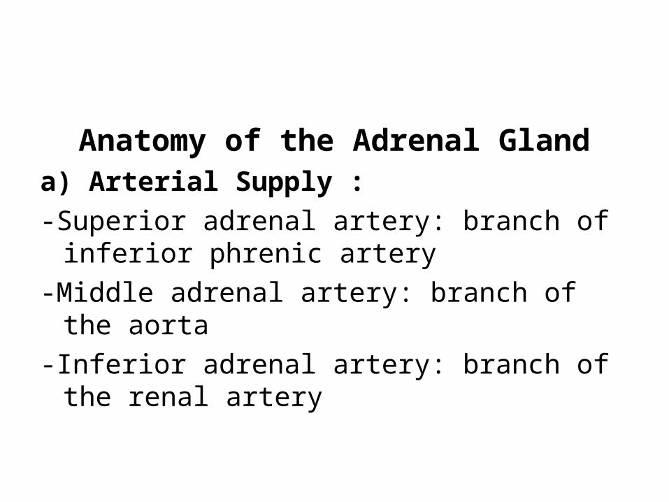
Anatomy of the Adrenal Glanda) Arterial Supply :-Superior adrenal artery: branch of inferior
phrenic artery-Middle adrenal artery: branch of the aorta-Inferior adrenal artery: branch of the renal
artery

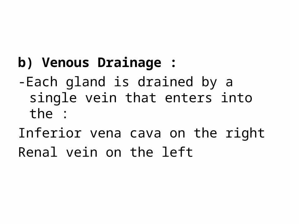
b) Venous Drainage :-Each gland is drained by a single vein that
enters into the :Inferior vena cava on the rightRenal vein on the left

c) Physiology :-Cortex divided into 3 zones :Zona glomerulosa (aldosterone)Zona fasciculata (ACTH dependent)Zona reticularis (cortisol)-Medulla (epinephrine, norepinephrine)

d) Imaging Appearance :-Y configuration : each adrenal gland consists of an
anteromedial ridge (body) and two posterior limbs best seen by CT/MR
-Posterior limbs are close together superiorly but spread out inferiorly
-Right adrenal lies adjacent to IVC throughout its extent-Left adrenal lies adjacent to splenic vessels at its cephalad
margin-Size :Limbs: 3 to 6 mm thickLength of entire adrenal: 4 to 6 cmWidth of entire adrenal: <1 cmWeight: 4 to 5 g/gland


Coronal T1-weighted, three-dimensional, GRE MR image obtained with VIBE shows the normal inverted Y shape of the right adrenal gland (arrow)

a) Incidental Adrenal Mass :1-Functioning Tumors2-Malignant Tumors3-Benign
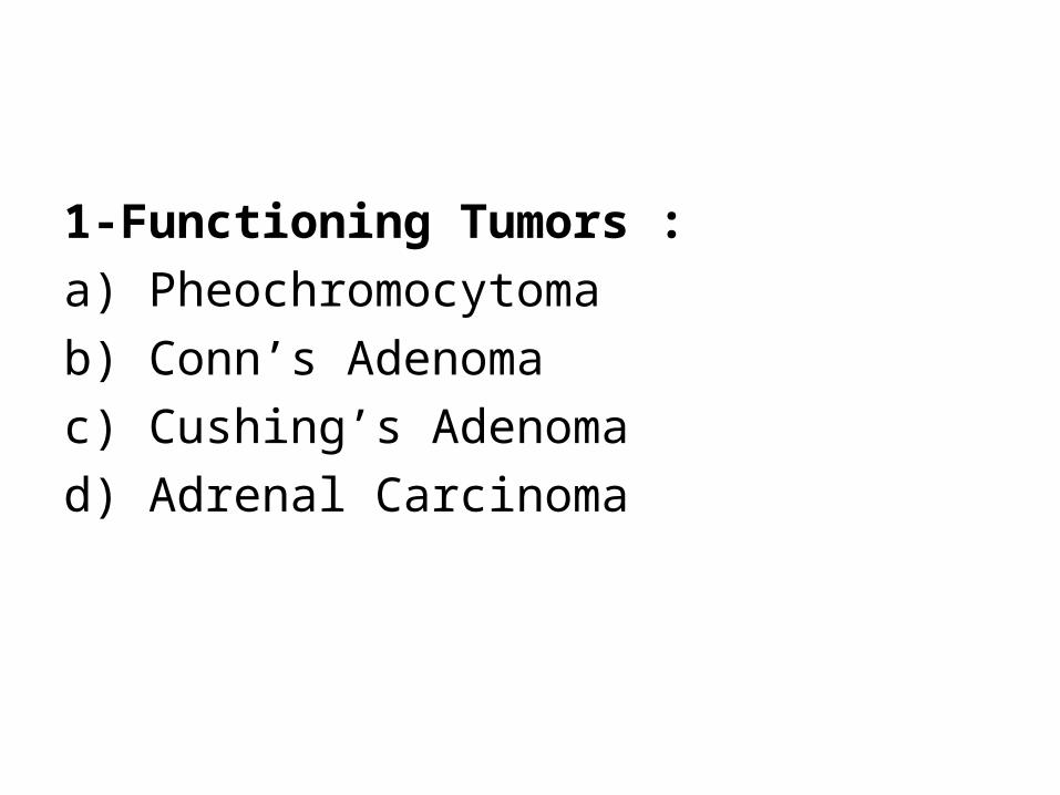
1-Functioning Tumors :a) Pheochromocytomab) Conn’s Adenomac) Cushing’s Adenomad) Adrenal Carcinoma
a) Pheochromocytoma :1-Incidence2-Associations3-Radiographic Features

1-Incidence :-Is an uncommon tumor of the adrenal gland-The tumors are said to follow a 10% rule :10% are extra-adrenal 10% are bilateral10% are malignant10% are found in children10% are familial10 % are not associated with hypertension

2-Associations :-The majority of cases are sporadic-In 5-10% of cases , a pheochromocytoma is a
manifestation of an underlying condition including :a) MEN II (both MEN IIa and MEN IIb) :-MEN IIa : medullary thyroid carcinoma ,
pheochromocytoma & parathyroid adenoma-MEN IIb : medullary thyroid carcinoma ,
pheochromocytoma , oral ganglioneuromas & other soft tissue tumors
b) VHLc) Sturge-Weber Syndromed) TS
3-Radiographic Features :-Usually large > 5 cm with marked contrast
enhancement-It should be noted that in patients with suspected
pheochromocytoma contrast may be contraindicated as it could precipitate a hypertensive crisis
a) CT :-On CT pheochromocytomas are large usually
heterogeneous masses with areas of necrosis and cystic change with marked contrast enhancement
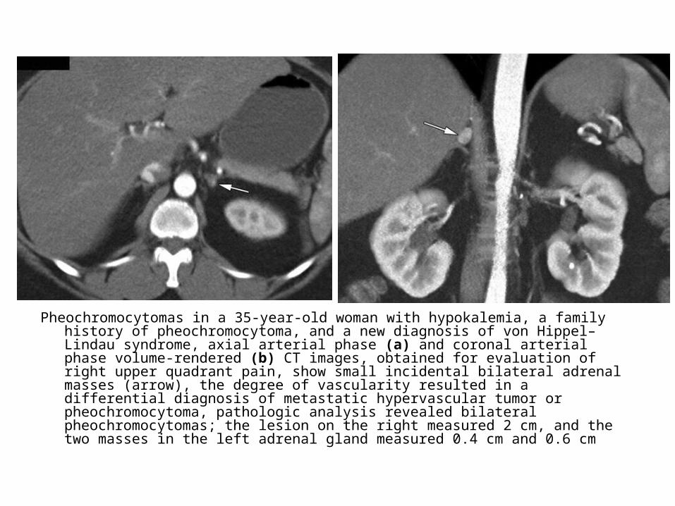
Pheochromocytomas in a 35-year-old woman with hypokalemia, a family history of pheochromocytoma, and a new diagnosis of von Hippel–Lindau syndrome, axial arterial phase (a) and coronal arterial phase volume-rendered (b) CT images, obtained for evaluation of right upper quadrant pain, show small incidental bilateral adrenal masses (arrow), the degree of vascularity resulted in a differential diagnosis of metastatic hypervascular tumor or pheochromocytoma, pathologic analysis revealed bilateral pheochromocytomas; the lesion on the right measured 2 cm, and the two masses in the left adrenal gland measured 0.4 cm and 0.6 cm

Pheochromocytoma with pathologically proved hemorrhage and necrosis in a 39-year-old woman, coronal precontrast volume-rendered (a) and axial postcontrast (b) CT images show a large, well-defined mass with higher attenuation inferiorly that compresses the liver and right kidney, on the contrast-enhanced image, enhancing septa and multiple cystic areas are seen. Classically a vascular mass, pheochromocytoma can also be cystic, particularly when the tumor is large, as in this case
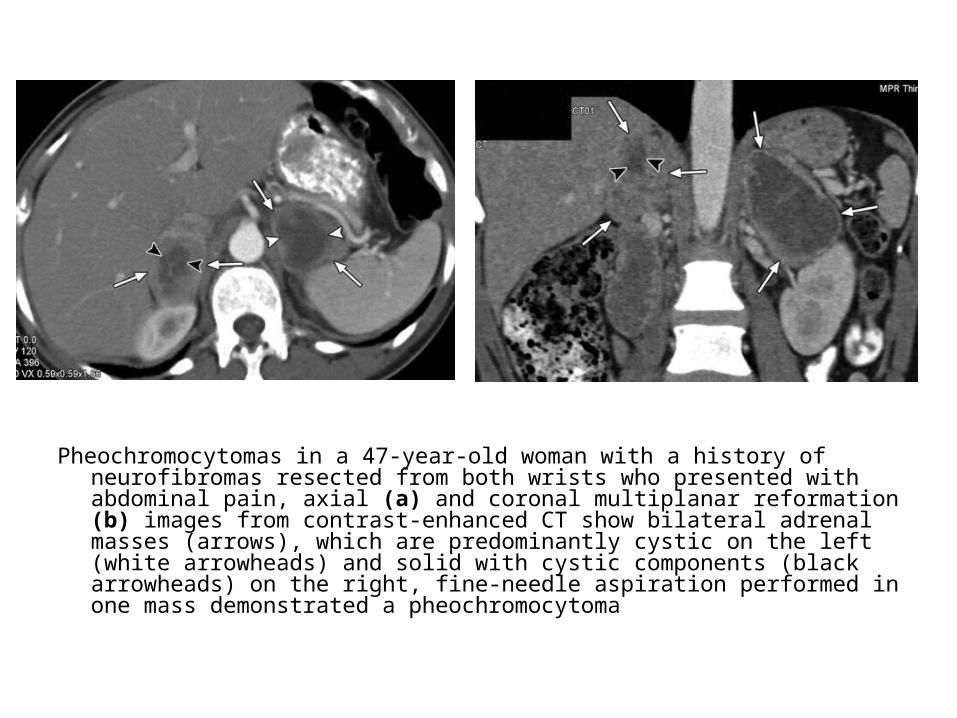
Pheochromocytomas in a 47-year-old woman with a history of neurofibromas resected from both wrists who presented with abdominal pain, axial (a) and coronal multiplanar reformation (b) images from contrast-enhanced CT show bilateral adrenal masses (arrows), which are predominantly cystic on the left (white arrowheads) and solid with cystic components (black arrowheads) on the right, fine-needle aspiration performed in one mass demonstrated a pheochromocytoma
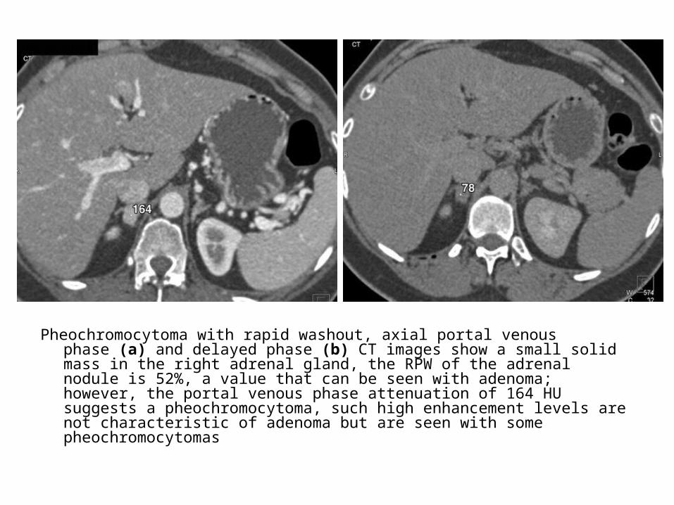
Pheochromocytoma with rapid washout, axial portal venous phase (a) and delayed phase (b) CT images show a small solid mass in the right adrenal gland, the RPW of the adrenal nodule is 52%, a value that can be seen with adenoma; however, the portal venous phase attenuation of 164 HU suggests a pheochromocytoma, such high enhancement levels are not characteristic of adenoma but are seen with some pheochromocytomas

Necrotic pheochromocytoma in a 42-year-old man, coronal arterial phase (a) and venous phase (b) volume-rendered images from contrast-enhanced CT show a large (>20 cm) hypervascular right suprarenal mass, the mass has central necrosis and compresses the right kidney inferiorly

b) MRI :*T1 :-Slightly hypointense to the remainder of the adrenal*T2 :-Markedly hyperintense (lightbulb sign) , this is a
helpful feature*T1+C :-Heterogenous enhancementc) Nuclear Medicine : MIBG-Abnormal uptake
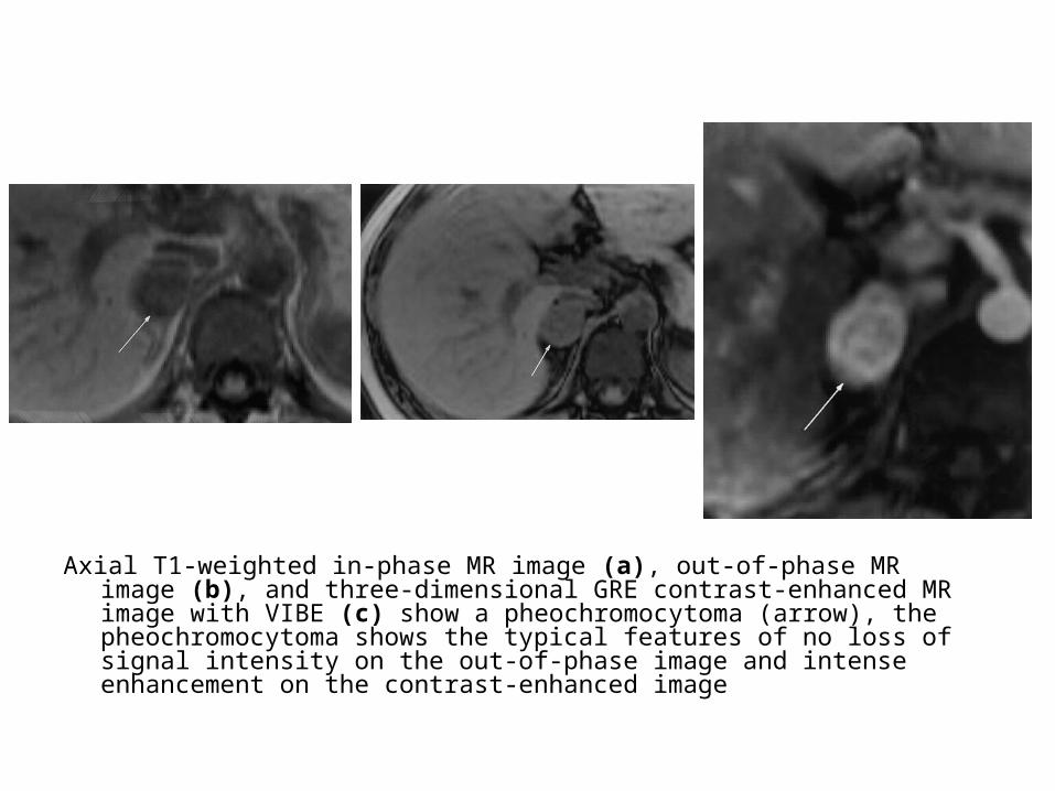
Axial T1-weighted in-phase MR image (a), out-of-phase MR image (b), and three-dimensional GRE contrast-enhanced MR image with VIBE (c) show a pheochromocytoma (arrow), the pheochromocytoma shows the typical features of no loss of signal intensity on the out-of-phase image and intense enhancement on the contrast-enhanced image

b) Conn’s Adenoma :1-Incidence2-Radiographic Features

1-Incidence :-Accounts for 70 % of Conn’s syndrome-30 % of Conn’s syndrome due to
hyperplasia which can be occasionally nodular and mimic an adenoma
2-Radiographic Features :-Usually small < 2 cm-Relatively low dense
c) Cushing’s Adenoma :1-Incidence2-Radiographic Features

1-Incidence :-Accounts for 20 % of Cushing syndrome-80 % of Cushing syndrome is due to excess
ACTH from pituitary tumor or ectopic source (small cell carcinoma , pancreatic islet cell , carcinoid medullary carcinoma of the thyroid & thymoma)
2-Radiographic Features :-Usually > 2 cm in diameter

d) Adrenal Carcinoma :-50 % are present as functioning tumor-Cushing's syndrome most common clinical
manifestation
2-Malignant Tumors :a) Metastasesb) Carcinomac) Lymphomad) Neuroblastoma

a) Metastases :1-Primary sites2-Radiographic Features

1-Primary sites :-Lung :Small cell carcinoma : 90% of adrenal masses
detected by CT screening represent metastasesNon–small cell carcinoma : 60% of adrenal masses-Breast-Kidney-Bowel-Ovary-Melanoma
2-Radiographic Features :-Adrenal mass usually > 2-3 cm with
irregular margins-Bilateral adrenal masses-Heterogeneous enhancement-In the presence of a known primary
malignant tumor many adrenal masses are benign (40 % are metastases)
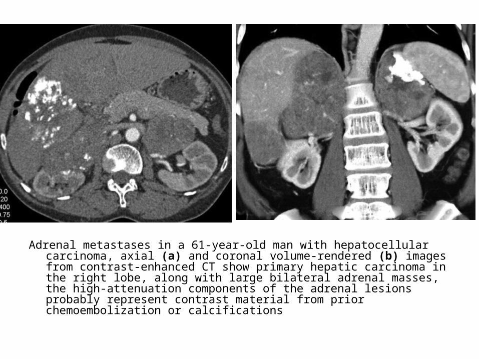
Adrenal metastases in a 61-year-old man with hepatocellular carcinoma, axial (a) and coronal volume-rendered (b) images from contrast-enhanced CT show primary hepatic carcinoma in the right lobe, along with large bilateral adrenal masses, the high-attenuation components of the adrenal lesions probably represent contrast material from prior chemoembolization or calcifications
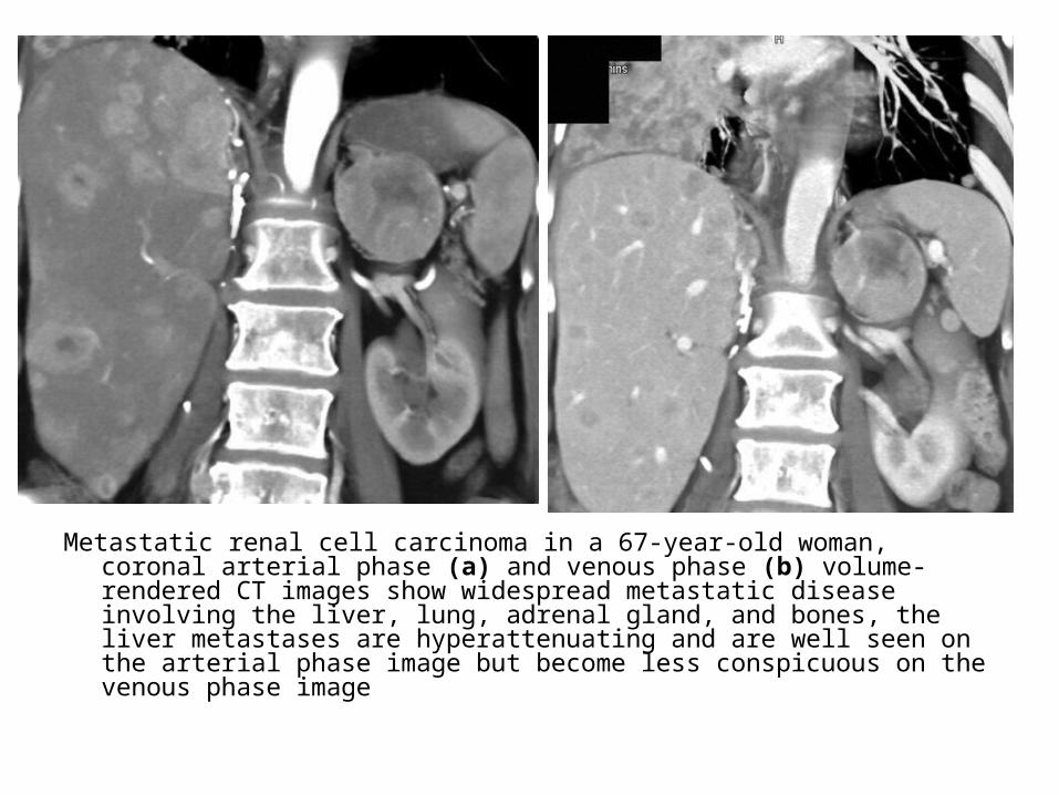
Metastatic renal cell carcinoma in a 67-year-old woman, coronal arterial phase (a) and venous phase (b) volume-rendered CT images show widespread metastatic disease involving the liver, lung, adrenal gland, and bones, the liver metastases are hyperattenuating and are well seen on the arterial phase image but become less conspicuous on the venous phase image
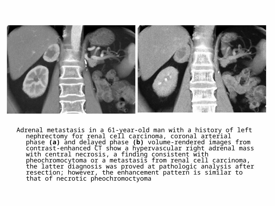
Adrenal metastasis in a 61-year-old man with a history of left nephrectomy for renal cell carcinoma, coronal arterial phase (a) and delayed phase (b) volume-rendered images from contrast-enhanced CT show a hypervascular right adrenal mass with central necrosis, a finding consistent with pheochromocytoma or a metastasis from renal cell carcinoma, the latter diagnosis was proved at pathologic analysis after resection; however, the enhancement pattern is similar to that of necrotic pheochromoctyoma
b) Carcinoma :1-Incidence2-Radiographic Features

1-Incidence :-50 % are present as functioning tumor-Cushing's syndrome most common clinical
manifestation
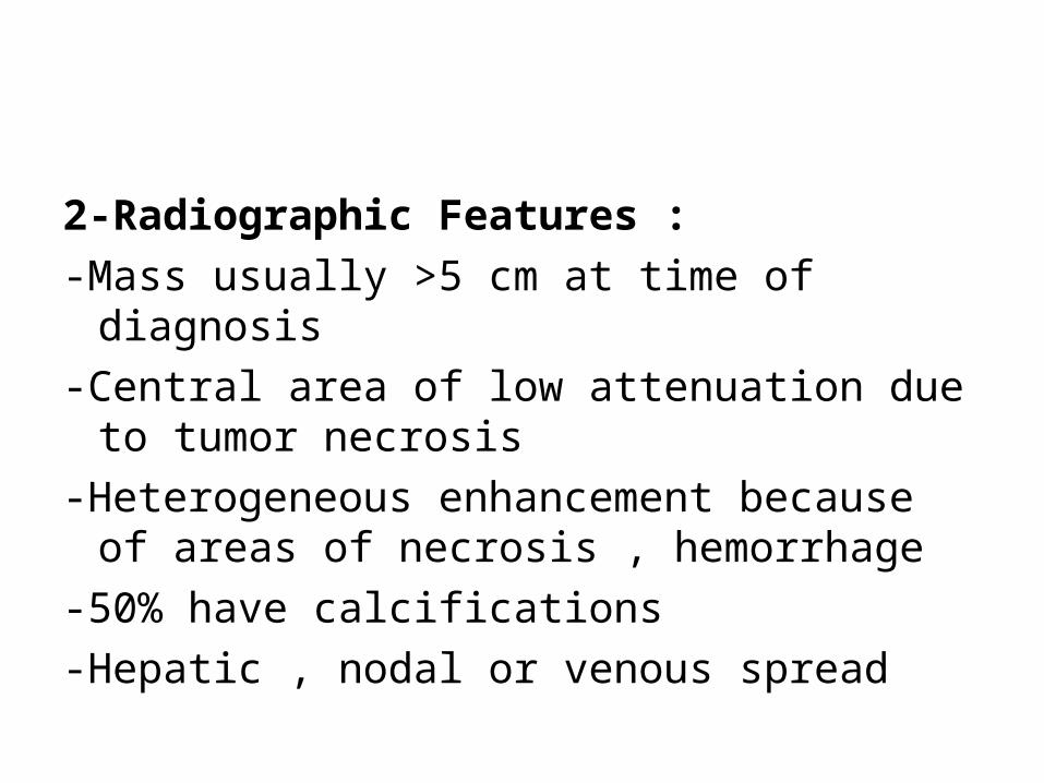
2-Radiographic Features :-Mass usually >5 cm at time of diagnosis-Central area of low attenuation due to
tumor necrosis-Heterogeneous enhancement because of
areas of necrosis , hemorrhage-50% have calcifications-Hepatic , nodal or venous spread
Adrenocortical carcinoma in a 62-year-old woman with hypertension, virilization, and an enlarging abdominal mass, coronal arterial phase (a) and venous phase (b) volume-rendered CT images show a large left suprarenal mass with hypervascularity and necrosis on the arterial phase image and some areas of mild enhancement on the venous phase image, the mass abuts the left hemidiaphragm, with left pleural effusion and left lung atelectasis, and is inseparable from the left kidney, at surgery, which included left nephrectomy, a portion of the left hemidiaphragm was resected and the left lower lobe was partially decorticated, pathologic analysis revealed a malignant adrenocortical neoplasm
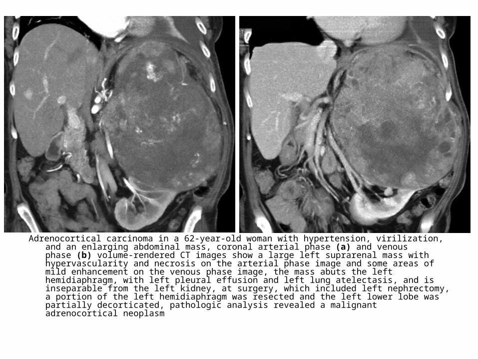
Primary adrenocortical carcinoma in a 55-year-old woman, coronal volume-rendered images from contrast-enhanced CT show a nearly 15-cm right adrenal mass that displaces the right kidney inferolaterally and invades the inferior vena cava (IVC) medially (arrowheads in a), tumor thrombus extends into the intrahepatic IVC (arrows in b)

61-year-old woman who presented with left lower quadrant pain, arterial phase (A), portal venous phase axial (B), and coronal (C) images show well-encapsulated large 13.5-cm mass lesion arising from left adrenal gland with internal calcifications but containing no focal fat, pancreas and left kidney are displaced by mass, but there is no evidence of invasion into adjacent vascular structures, on resection this mass represented adrenocortical carcinoma
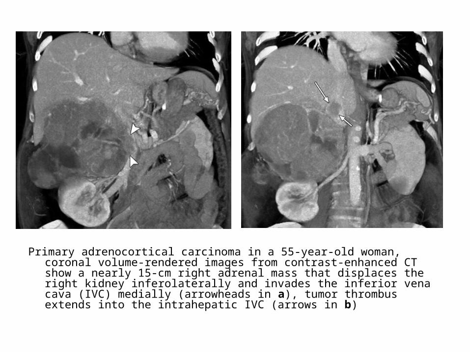
Sagittal T1-weighted three-dimensional contrast-enhanced GRE MR image obtained with VIBE (a) and coronal T2 obtained with half-Fourier RARE (b) show a large mass involving the right adrenal gland, the mass exhibits heterogeneous low signal intensity on the T1 and high signal intensity with a heterogeneous pattern of contrast enhancement and areas of necrosis (arrow in b) on the T2
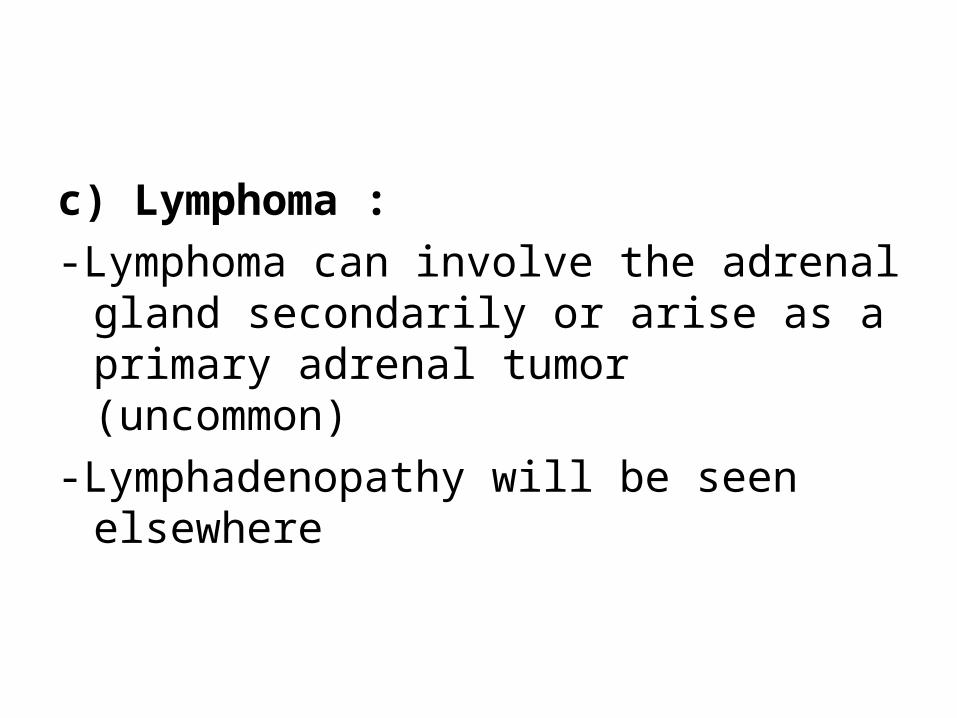
c) Lymphoma :-Lymphoma can involve the adrenal gland
secondarily or arise as a primary adrenal tumor (uncommon)
-Lymphadenopathy will be seen elsewhere

Adrenal lymphoma in a 67-year-old man with an adrenal mass, imaging was performed for diagnosis and staging, axial arterial phase (a) and coronal arterial phase volume-rendered (b) CT images show an 11-cm mass in the left adrenal bed, the mass invades the left hemidiaphragm, encases the celiac and renal arteries, and displaces the aorta, the mild degree of organ displacement despite the size of the mass and the infiltrative appearance are suggestive of lymphoma; the diagnosis was confirmed at core biopsy

Axial T1-weighted in-phase (a) and out-of-phase (b) MR images show bilateral lymphomatous deposits, the deposits have low signal intensity, and the signal intensity does not decrease on the out-of-phase compared with the in-phase image

d) Neuroblastoma :-> 5 cm-Calcification in 90 %-Extends across midline

Neuroblastoma with disease crossing midline

Neuroblastoma (a, b) Coronal unenhanced T1 (a) and axial T2 obtained with inversion recovery (b) show a right adrenal tumor, the tumor is predominantly hypointense on the T1 and has areas of high-signal-intensity hemorrhage (arrow in a), the tumor is hyperintense on the T2

3-Benign :a) Non-Functioning Adenomab) Myelolipomac) AMLd) Cyste) Adrenal Hemorrhagef) Granulomatous Disease
a) Non-Functioning Adenoma :-The majority of lesions are not functioning. Although CT
does not allow differentiation of functioning from nonfunctioning masses, the presence of contralateral adrenal atrophy suggests that a lesion may be functioning, because pituitary adrenocorticotropic hormone secretion is suppressed by elevated cortisol levels
-The precontrast attenuation varies according to the presence or absence of lipid, with mean attenuation in the range of −2 to 16 HU in lipid-rich adenomas and higher attenuation (20–25 HU) seen in the setting of lipid-poor adenomas
-Lipid-poor adenomas represent 10%-40% of adenomas-Regardless of lipid content, adenomas typically
demonstrate rapid washout, which is defined as an APW of more than 60% and an RPW of more than 40% on delayed images

-Radiographic Features :1-CT :-Mass 1 to 5 cm-< 0 HU : diagnostic of adenoma (due to fat)-0 to 10 HU : diagnosis almost certain (follow-up or MRI)-Calcification rare-Slight enhancement with IV contrast-Rarely, an adenoma can hemorrhage, usually in a patient
receiving anticoagulant therapy, the presence of hemorrhage results in regions of higher attenuation and heterogeneity, at CT, heterogeneity and regions of increased attenuation have been shown to correlate with hemorrhage at pathologic analysis, before liquefaction, the precontrast attenuation will be higher than 10 HU
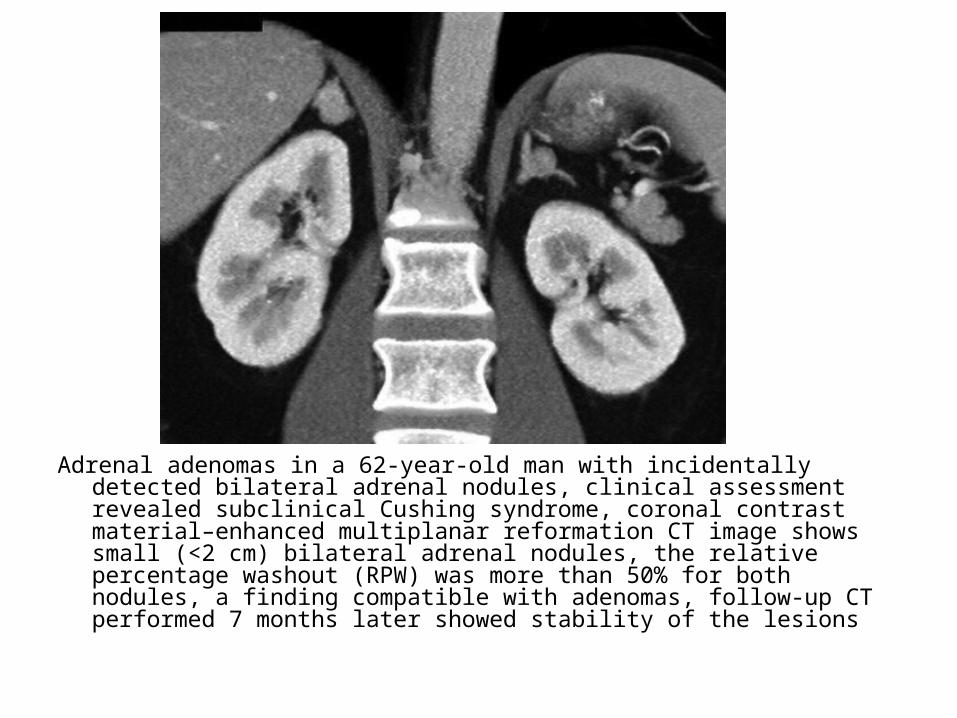
Adrenal adenomas in a 62-year-old man with incidentally detected bilateral adrenal nodules, clinical assessment revealed subclinical Cushing syndrome, coronal contrast material–enhanced multiplanar reformation CT image shows small (<2 cm) bilateral adrenal nodules, the relative percentage washout (RPW) was more than 50% for both nodules, a finding compatible with adenomas, follow-up CT performed 7 months later showed stability of the lesions

2.1-cm left adrenal mass was discovered incidentally on contrast-enhanced computed tomography (CT), because the mass could not be characterized on the contrast-enhanced CT, this unenhanced CT was performed. It shows that the lesion (arrows) is of low attenuation (6 HU), which is consistent with a lipid-rich adenoma
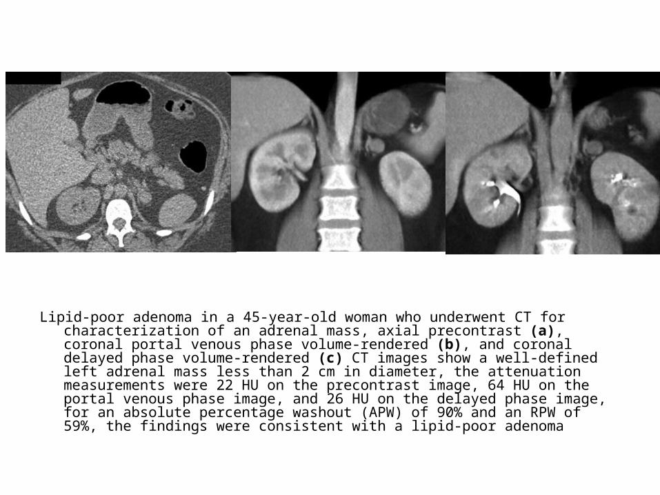
Lipid-poor adenoma in a 45-year-old woman who underwent CT for characterization of an adrenal mass, axial precontrast (a), coronal portal venous phase volume-rendered (b), and coronal delayed phase volume-rendered (c) CT images show a well-defined left adrenal mass less than 2 cm in diameter, the attenuation measurements were 22 HU on the precontrast image, 64 HU on the portal venous phase image, and 26 HU on the delayed phase image, for an absolute percentage washout (APW) of 90% and an RPW of 59%, the findings were consistent with a lipid-poor adenoma
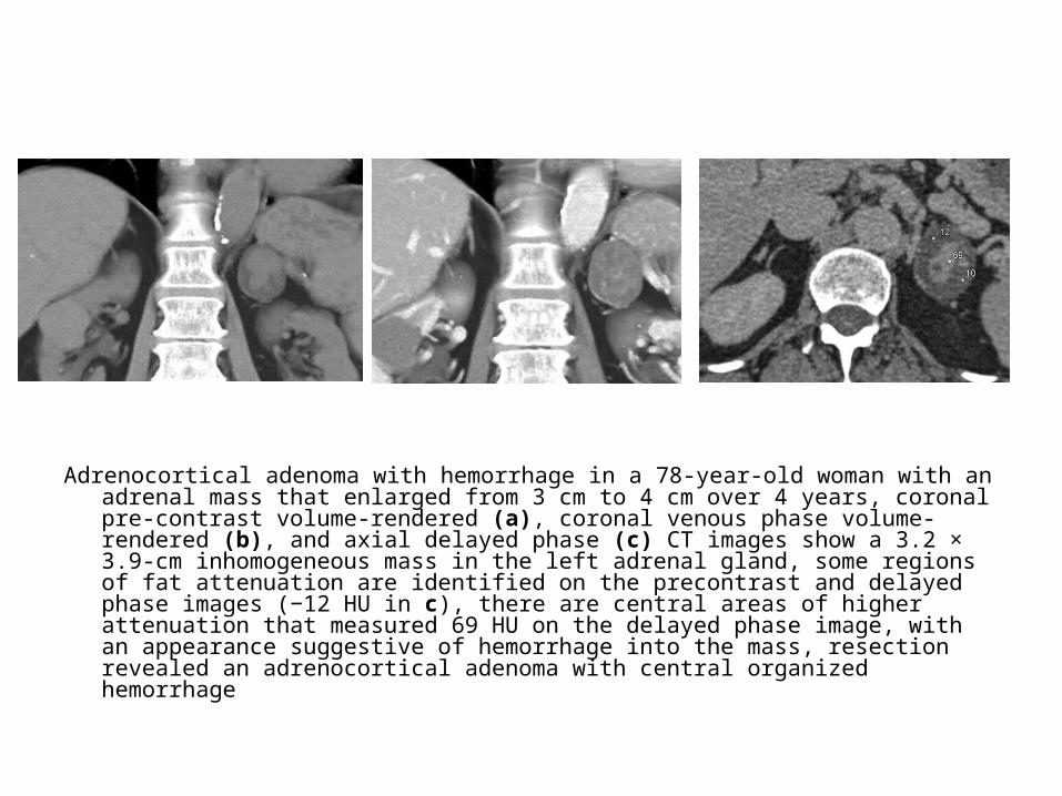
Adrenocortical adenoma with hemorrhage in a 78-year-old woman with an adrenal mass that enlarged from 3 cm to 4 cm over 4 years, coronal pre-contrast volume-rendered (a), coronal venous phase volume-rendered (b), and axial delayed phase (c) CT images show a 3.2 × 3.9-cm inhomogeneous mass in the left adrenal gland, some regions of fat attenuation are identified on the precontrast and delayed phase images (−12 HU in c), there are central areas of higher attenuation that measured 69 HU on the delayed phase image, with an appearance suggestive of hemorrhage into the mass, resection revealed an adrenocortical adenoma with central organized hemorrhage
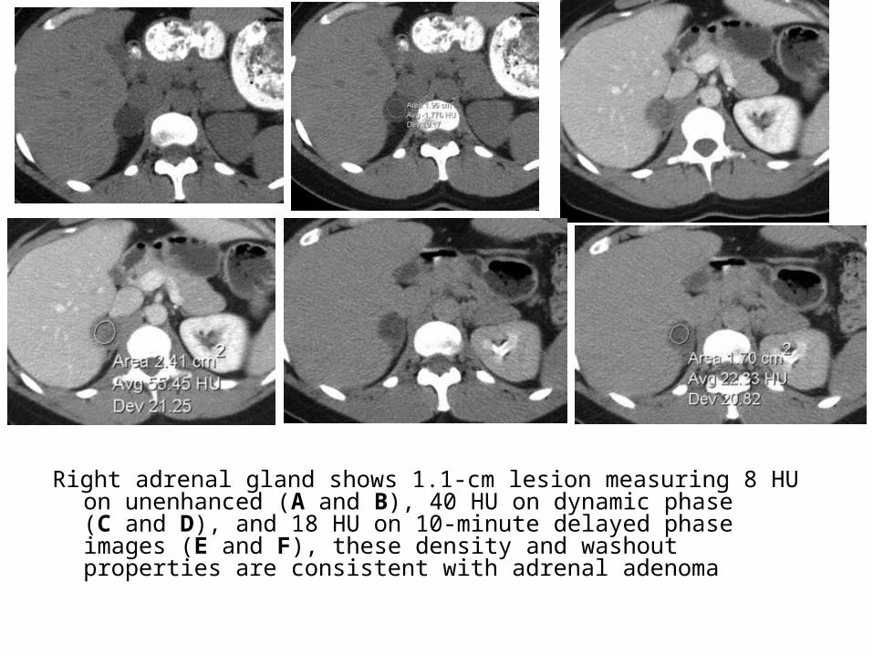
Right adrenal gland shows 1.1-cm lesion measuring 8 HU on unenhanced (A and B), 40 HU on dynamic phase (C and D), and 18 HU on 10-minute delayed phase images (E and F), these density and washout properties are consistent with adrenal adenoma

2-MRI :-Fat-suppression techniques are used to
determine if a given lesion contains fat (e.g., in phase/out of phase imaging , spin-echo fat-suppression imaging) , if a lesion contains fat , it is considered an adenoma
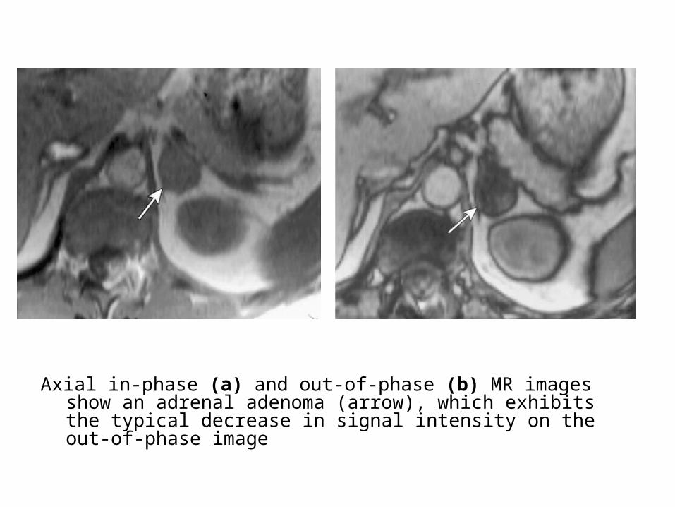
Axial in-phase (a) and out-of-phase (b) MR images show an adrenal adenoma (arrow), which exhibits the typical decrease in signal intensity on the out-of-phase image

Axial T1-weighted out-of-phase MR image shows an adrenal adenoma (black arrow) with a focal area of high-signal-intensity hemorrhage (white arrow)
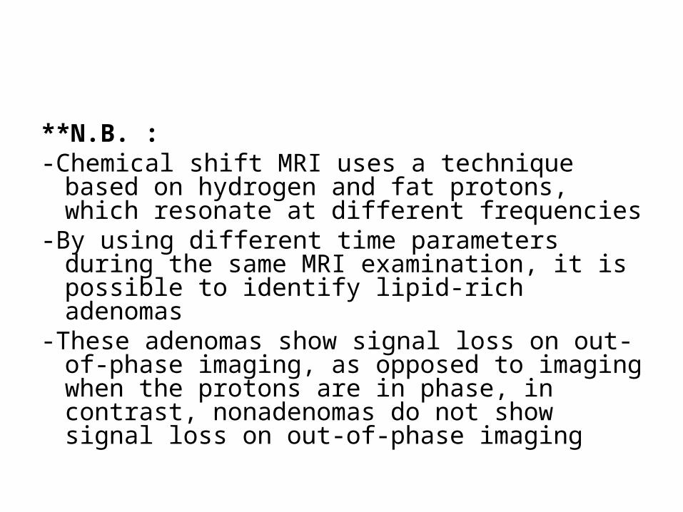
**N.B. :-Chemical shift MRI uses a technique based on
hydrogen and fat protons, which resonate at different frequencies
-By using different time parameters during the same MRI examination, it is possible to identify lipid-rich adenomas
-These adenomas show signal loss on out-of-phase imaging, as opposed to imaging when the protons are in phase, in contrast, nonadenomas do not show signal loss on out-of-phase imaging
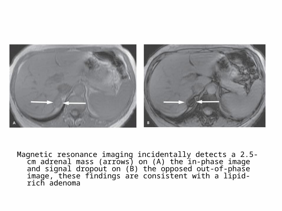
Magnetic resonance imaging incidentally detects a 2.5-cm adrenal mass (arrows) on (A) the in-phase image and signal dropout on (B) the opposed out-of-phase image, these findings are consistent with a lipid-rich adenoma

b) Myelolipoma :-Very rare-Area of obvious fat mass (low negative
attenuation)-May enhance with contrast administration-Calcification , 20%

Myelolipoma in a 40-year-old man with metastatic medullary carcinoma of the thyroid, coronal multiplanar reformation image from contrast-enhanced CT shows a 5-cm left adrenal mass predominantly composed of fat (arrows), an appearance diagnostic of a myelolipoma
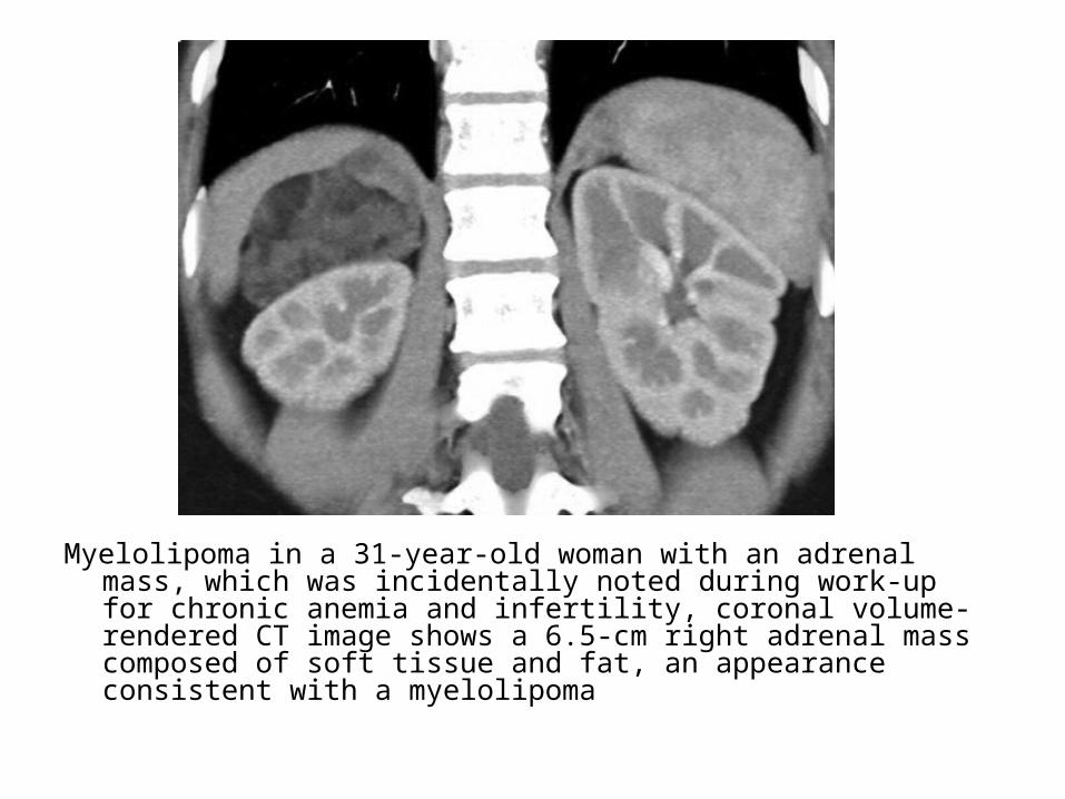
Myelolipoma in a 31-year-old woman with an adrenal mass, which was incidentally noted during work-up for chronic anemia and infertility, coronal volume-rendered CT image shows a 6.5-cm right adrenal mass composed of soft tissue and fat, an appearance consistent with a myelolipoma

Myelolipoma in a 59-year-old woman with a history of long-standing hypertension, a normal urinary metanephrine level, and no clinical evidence of hypercortisolism. Coronal precontrast (a) and arterial phase (b) multiplanar reformation images from contrast-enhanced CT show an 8-cm left adrenal mass containing multiple foci of fat and punctate calcifications; there was mild enhancement on venous phase images, after resection, pathologic analysis revealed a benign vascular lesion with adipose tissue, findings consistent with a myelolipoma
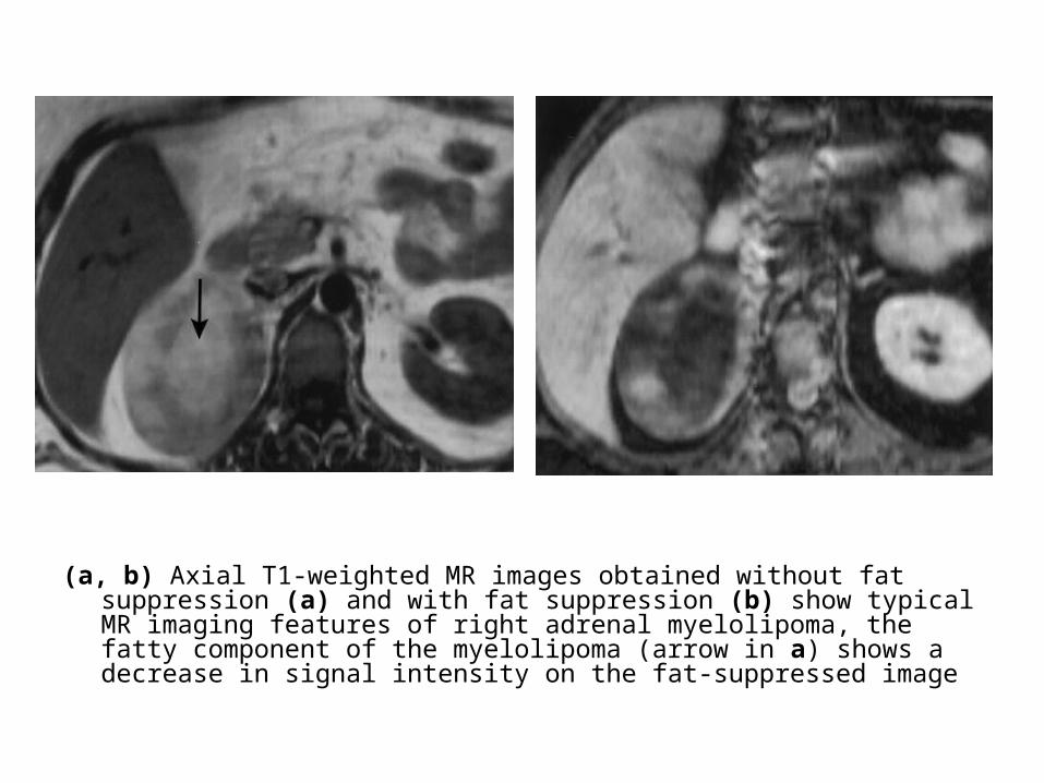
(a, b) Axial T1-weighted MR images obtained without fat suppression (a) and with fat suppression (b) show typical MR imaging features of right adrenal myelolipoma, the fatty component of the myelolipoma (arrow in a) shows a decrease in signal intensity on the fat-suppressed image
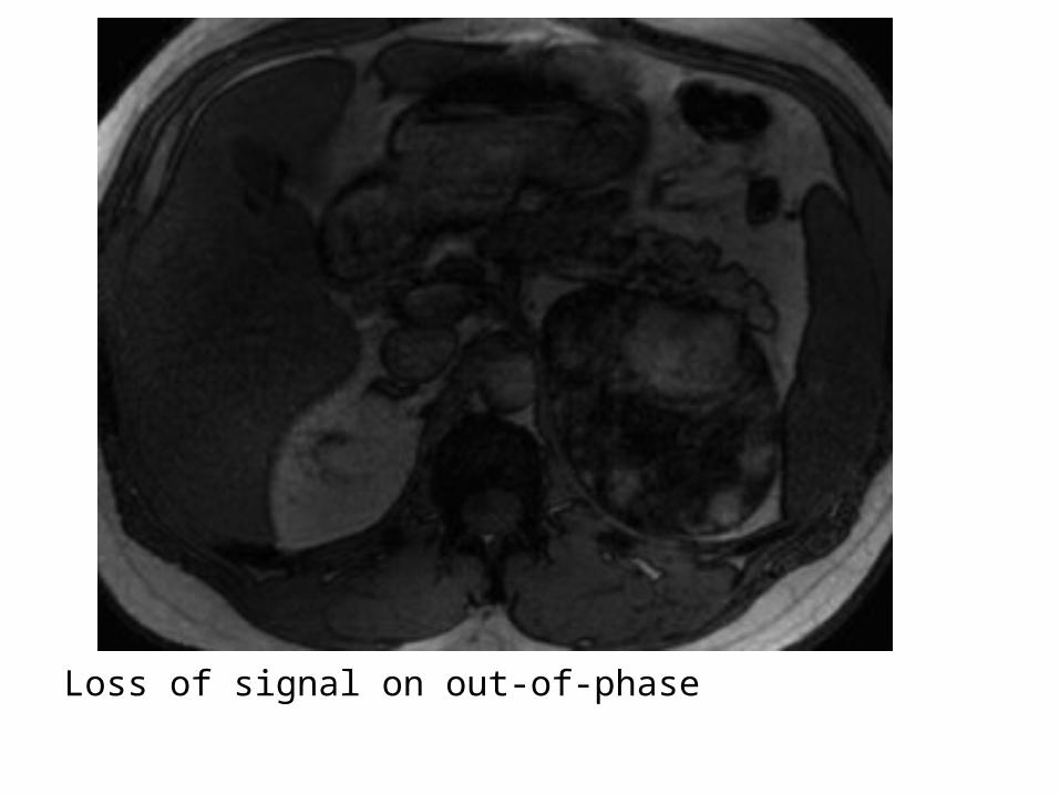
Loss of signal on out-of-phase

c) AML :-Very rare
d) Cyst :1-Classification2-Radiographic Features

1-Classification :a) Endothelial cyst , 40%b) Pseudocyst (hemorrhage) , 40% , may
contain calcified rimc) Epithelial cyst , 10%d) Parasitic cysts (Echinococcus) , 5%
2-Radiographic Features :-Well defined water density-Mural calcification (15%) , especially in
pseudocysts and parasitic cysts
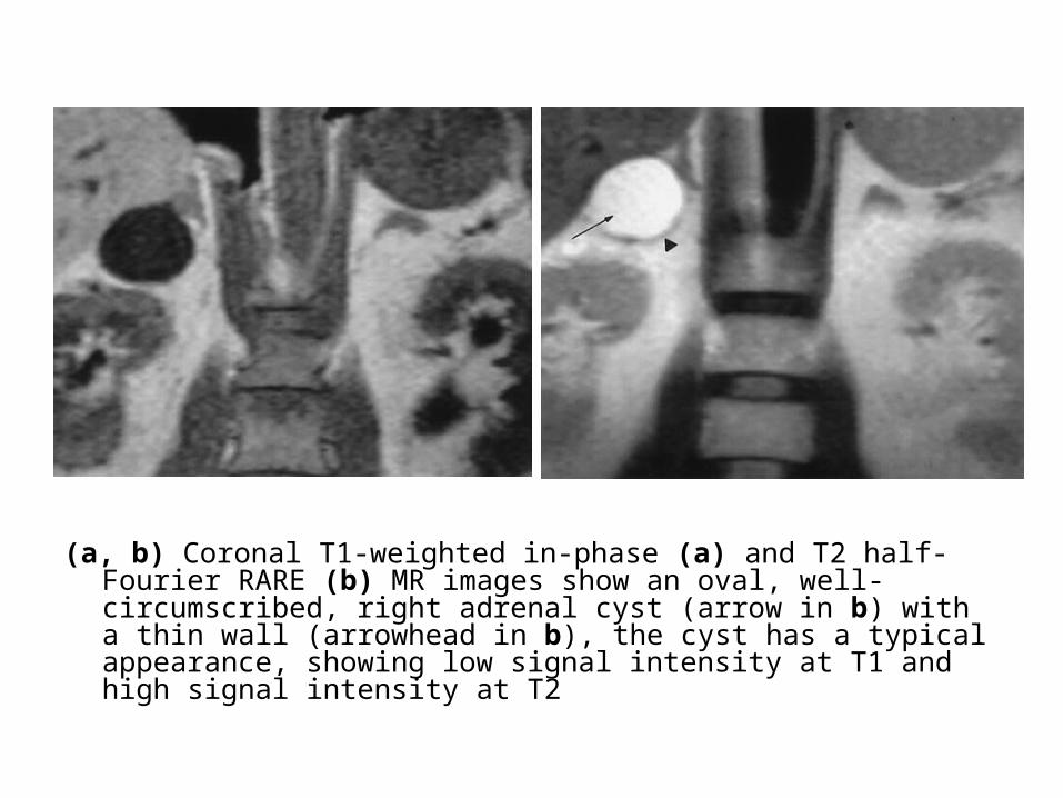
(a, b) Coronal T1-weighted in-phase (a) and T2 half-Fourier RARE (b) MR images show an oval, well-circumscribed, right adrenal cyst (arrow in b) with a thin wall (arrowhead in b), the cyst has a typical appearance, showing low signal intensity at T1 and high signal intensity at T2
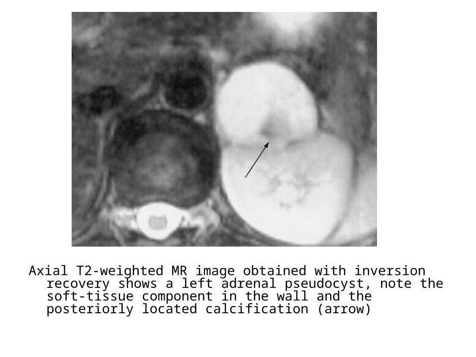
Axial T2-weighted MR image obtained with inversion recovery shows a left adrenal pseudocyst, note the soft-tissue component in the wall and the posteriorly located calcification (arrow)

Hemorrhagic complicated adrenal cyst, (a, b) Coronal T2 obtained with half-Fourier RARE (a) and axial contrast-enhanced VIBE image (b) show a left adrenal mass with areas of signal intensity similar to that of blood
e) Adrenal Hemorrhage :1-Incidence2-Etiology3-Radiographic Features

1-Incidence :-More common in neonates than adults
2-Etiology :a) Hemorrhagic tumorsb) Severe trauma c) Anticoagulation d) Severe stress (surgery , sepsis , burns &
hypotension)
3-Radiographic Features :a) Acute hematoma :-High CT density (>40 HU)-Enlarged adrenal glandb) Old hematoma :-Liquefaction-Fluid-fluid level-May evolve into pseudocyst
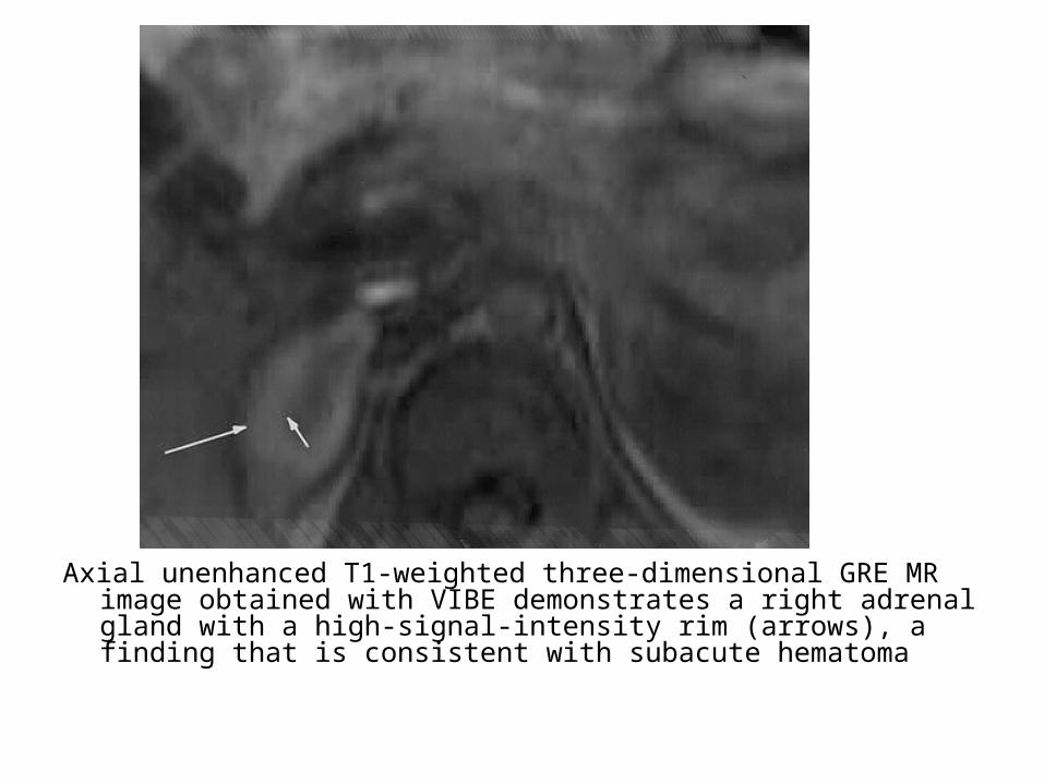
Axial unenhanced T1-weighted three-dimensional GRE MR image obtained with VIBE demonstrates a right adrenal gland with a high-signal-intensity rim (arrows), a finding that is consistent with subacute hematoma

f) Granulomatous Disease :-Most common causes are TB ,
histoplasmosis , blastomycosis , meningococcus and echinococcus
-Present as diffuse enlargement or as discrete mass
-Can have a central cystic component with or without calcifications


b) Bilateral Adrenal Masses :1-Metastases , in 15 %2-Pheochromocytoma , in 10 %3-Hyperplasia :-Bilateral adrenal enlargement but usually not seen
on CT4-Spontaneous Adrenal Hemorrhage5-Lymphoma6-Granulomatous Disease
c) Adrenal Calcification :1-Pseudocyst , Parasitic cyst2-Carcinoma3-Addison Disease :-If caused by TB , calcification is a common finding4-Neuroblastoma5-Granulomatous Disease6-Pheochromocytoma7-Myelilipoma

Related Documents