ORIGINAL ARTICLE Diagnostic accuracy of 68 Ga-DOTANOC PET/CT imaging in pheochromocytoma Punit Sharma & Varun Singh Dhull & Saurabh Arora & Poonam Gupta & Rajeev Kumar & Prashant Durgapal & Arun Malhotra & Sunil Chumber & Ariachery C. Ammini & Rakesh Kumar & Chandrasekhar Bal Received: 29 June 2013 /Accepted: 1 October 2013 # Springer-Verlag Berlin Heidelberg 2013 Abstract Purpose The purpose of the present study was to evaluate the diagnostic accuracy of 68 Ga-DOTANOC positron emission tomography (PET)/CT in patients with suspicion of pheochromocytoma. Methods Data of 62 patients [age 34.3±16.1 years, 14 with multiple endocrine neoplasia type 2 (MEN2)] with clinical/ biochemical suspicion of pheochromocytoma and suspicious adrenal lesion on contrast CT (n =70), who had undergone 68 Ga-DOTANOC PET/CT, were retrospectively analyzed. PET/CT images were analyzed visually as well as semiquantitatively, with measurement of maximum standardized uptake value (SUV max ), SUV mean , SUV max / SUV liver , and SUV mean /SUV liver . Results of PET/CT were compared with 131 I-metaiodobenzylguanidine (MIBG) imaging, which was available in 40 patients (45 lesions). Histopathology and/or imaging/clinical/biochemical follow- up (minimum 6 months) was used as reference standard. Results The sensitivity, specificity, and accuracy of 68 Ga- DOTANOC PET/CT was 90.4, 85, and 88.7 %, respectively, on patient-based analysis and 92, 85, and 90 %, respectively, on lesion-based analysis. 68 Ga-DOTANOC PET/CT showed 100 % accuracy in patients with MEN2 syndrome and malignant pheochromocytoma. On direct comparison, lesion- based accuracy of 68 Ga-DOTANOC PET/CT for pheochromocytoma was significantly higher than 131 I-MIBG imaging (91.1 vs 66.6 %, p =0.035). SUV max was higher for pheochromocytomas than other adrenal lesions (p =0.005), MEN2-associated vs sporadic pheochromocytoma (p =0.012), but no difference was seen between benign vs malignant pheochromocytoma (p =0.269). Conclusion 68 Ga-DOTANOC PET/CT shows high diagnostic accuracy in patients with suspicion of pheochromocytoma and is superior to 131 I-MIBG imaging for this purpose. Best results of 68 Ga-DOTANOC PET/CT are seen in patients with MEN2- associated and malignant pheochromocytoma. Keywords Pheochromocytoma . 68 Ga-DOTANOC . PET/CT . MIBG Introduction Pheochromocytomas are rare tumors arising from the chromaffin cells of the adrenal medulla [1]. They represent a rare (<1 %) but potentially curable cause of hypertension [2]. While the majority of pheochromocytomas are sporadic, in about a quarter of unselected cases they may be associated with hereditary syndromes like multiple endocrine neoplasia (MEN), von Hippel-Lindau syndrome (VHL), neurofibromatosis 1, and tuberous sclerosis [3]. The diagnosis of pheochromocytoma is established by measurement of P. Sharma : V. S. Dhull : S. Arora : R. Kumar : A. Malhotra : R. Kumar : C. Bal (*) Department of Nuclear Medicine, All India Institute of Medical Sciences, Ansari Nagar, New Delhi 110029, India e-mail: [email protected] P. Gupta : A. C. Ammini Department of Endocrinology and Metabolism, All India Institute of Medical Sciences, New Delhi, India P. Durgapal Department of Pathology, All India Institute of Medical Sciences, New Delhi, India S. Chumber Department of Surgical Disciplines, All India Institute of Medical Sciences, New Delhi, India Eur J Nucl Med Mol Imaging DOI 10.1007/s00259-013-2598-1

Welcome message from author
This document is posted to help you gain knowledge. Please leave a comment to let me know what you think about it! Share it to your friends and learn new things together.
Transcript
ORIGINAL ARTICLE
Diagnostic accuracy of 68Ga-DOTANOC PET/CTimaging in pheochromocytoma
Punit Sharma & Varun Singh Dhull & Saurabh Arora & Poonam Gupta & Rajeev Kumar &
Prashant Durgapal & Arun Malhotra & Sunil Chumber & Ariachery C. Ammini &Rakesh Kumar & Chandrasekhar Bal
Received: 29 June 2013 /Accepted: 1 October 2013# Springer-Verlag Berlin Heidelberg 2013
AbstractPurpose The purpose of the present study was to evaluate thediagnostic accuracy of 68Ga-DOTANOC positron emissiontomography (PET)/CT in patients with suspicion ofpheochromocytoma.Methods Data of 62 patients [age 34.3±16.1 years, 14 withmultiple endocrine neoplasia type 2 (MEN2)] with clinical/biochemical suspicion of pheochromocytoma and suspiciousadrenal lesion on contrast CT (n =70), who had undergone68Ga-DOTANOC PET/CT, were retrospectively analyzed.PET/CT images were analyzed visually as well assemiquantitatively, with measurement of maximumstandardized uptake value (SUVmax), SUVmean, SUVmax/SUVliver, and SUVmean/SUVliver. Results of PET/CT werecompared with 131I-metaiodobenzylguanidine (MIBG)imaging, which was available in 40 patients (45 lesions).Histopathology and/or imaging/clinical/biochemical follow-up (minimum 6 months) was used as reference standard.
Results The sensitivity, specificity, and accuracy of 68Ga-DOTANOC PET/CT was 90.4, 85, and 88.7 %, respectively,on patient-based analysis and 92, 85, and 90%, respectively, onlesion-based analysis. 68Ga-DOTANOC PET/CT showed100 % accuracy in patients with MEN2 syndrome andmalignant pheochromocytoma. On direct comparison, lesion-based accuracy of 68Ga-DOTANOC PET/CT forpheochromocytoma was significantly higher than 131I-MIBGimaging (91.1 vs 66.6 %, p =0.035). SUVmax was higher forpheochromocytomas than other adrenal lesions (p =0.005),MEN2-associated vs sporadic pheochromocytoma (p =0.012),but no difference was seen between benign vs malignantpheochromocytoma (p =0.269).Conclusion 68Ga-DOTANOC PET/CTshows high diagnosticaccuracy in patients with suspicion of pheochromocytoma andis superior to 131I-MIBG imaging for this purpose. Best resultsof 68Ga-DOTANOC PET/CTare seen in patients withMEN2-associated and malignant pheochromocytoma.
Keywords Pheochromocytoma .68Ga-DOTANOC .PET/CT .
MIBG
Introduction
Pheochromocytomas are rare tumors arising from thechromaffin cells of the adrenal medulla [1]. They represent arare (<1 %) but potentially curable cause of hypertension [2].While the majority of pheochromocytomas are sporadic, inabout a quarter of unselected cases they may be associatedwith hereditary syndromes like multiple endocrine neoplasia(MEN) , von Hippe l -L indau synd rome (VHL) ,neurofibromatosis 1, and tuberous sclerosis [3]. The diagnosisof pheochromocytoma is established by measurement of
P. Sharma :V. S. Dhull : S. Arora : R. Kumar :A. Malhotra :R. Kumar :C. Bal (*)Department of Nuclear Medicine, All India Institute of MedicalSciences, Ansari Nagar, New Delhi 110029, Indiae-mail: [email protected]
P. Gupta :A. C. AmminiDepartment of Endocrinology and Metabolism, All India Institute ofMedical Sciences, New Delhi, India
P. DurgapalDepartment of Pathology, All India Institute of Medical Sciences,New Delhi, India
S. ChumberDepartment of Surgical Disciplines, All India Institute of MedicalSciences, New Delhi, India
Eur J Nucl Med Mol ImagingDOI 10.1007/s00259-013-2598-1
blood and urinary catecholamines or their metabolites,followed by demonstration of an adrenal mass on anatomical(CT/MRI) imaging. However, biochemical markers have highsensitivity (96–100 %), but low specificity (69 %) for thedetection of pheochromocytomas [4]. Similarly, thespecificity of CT or MRI is also limited for differentiatingpheochromocytoma from other adrenal lesions [5]. Theselimitations have led to reliance on functional imaging methodsin order to avoid unnecessary surgery.
131/123I-Metaiodobenzylguanidine (MIBG) scintigraphy iscurrent ly the func t ional imaging of choice forpheochromocytoma [6], but suffers from drawbacks likelimited spatial resolution, difficulty in detection of smalltumors (<1.5–2.0 cm), or large tumors with extensivenecrosis/hemorrhage, lack of tracer uptake in some tumors,and interference with certain medications, all of which canlead to false-negative results [7]. Addition of single photonemission computed tomography (SPECT) improves thesensitivity [8], but even then the spatial resolution is low(∼1.5 cm) and the detection of small lesions is often difficult.Given the superior resolution of positron emissiontomography (PET) over SPECT, functional imaging ofpheochromocytoma has been attempted with a wide array ofPET tracers like 18F-fluorodeoxyglucose (FDG) [9], 18F-fluorodopa (FDOPA) [10], 18F-fluorodopamine (FDA) [11],11C-hydroxyephedrine (HED) [12], and 68Ga-DOTA-peptides[13] with encouraging results. Expression of somatostatinreceptors (SSTR) by pheochromocytomas [14] facilitatestargeted PET imaging with 68Ga-DOTA-peptides (68Ga-DOTATOC, 68Ga-DOTANOC, and 68Ga-DOTATATE).However, to date very few studies have evaluated 68Ga-DOTA-peptide PET/CT (only one with 68Ga-DOTANOC) inpheochromocytomas and only in small and mixed patientpopulations [15–19]. Therefore, the purpose of the presentstudy was to comprehensively evaluate the diagnosticaccuracy of 68Ga-DOTANOC PET/CT in a large number ofpatients with suspected pheochromocytoma and compare theresults with 131I-MIBG imaging when available (123I-MIBG isnot available in our country).
Materials and methods
The present retrospective study was approved by theInstitutional Ethics Committee and because of theretrospective nature written informed consent was waived.The inclusion criteria were clinical and/or biochemicalsuspicion of pheochromocytoma along with adrenalabnormality on contrast CT. The biochemical suspicion wasbased on the presence of one or more of the following: urinaryepinephrine (>20 μg/day), urinary norepinephrine (>90 μg/day), urinary vanillylmandelic acid (>7.9 mg/day), urinarytotal metanephrine (>1.2 mg/day), plasma epinephrine
(>85 pg/ml), plasma norepinephrine (500 pg/ml), plasmametanephrine (>400 pg/ml), plasma normetanephrine(>676 pg/ml), and plasma methoxytyramine (>1,800 pg/ml).A total of 62 patients who underwent 68Ga-DOTANOC PET/CT between March 2007 and December 2012 satisfied theinclusion criteria. Data of these 62 patients were retrievedfrom departmental registries and analyzed. In addition, 40 ofthese 62 patients also had undergone 131I-MIBG scintigraphy.Of these 62 patients, 10 had been previously included in thepilot study by Naswa et al. [18].
68Ga-DOTANOC synthesis
68Ga-DOTANOC synthesis was carried out using the methodpreviously detailed by Zhernosekov et al. [20] and is brieflydescribed here: 30–50 mCi 68Ge/68Ga generator (CyclotronCo Ltd., Obninsk, Russia) was eluted using 0.1 Mhydrochloric acid (HCl). The eluent was loaded on aminiaturized column of organic cation-exchange resin topre-concentrate and pre-purify (using 80 % acetone/0.15 MHCl). The processed 68Ga was directly eluted with 97.7 %acetone/0.05 M HCl into the reaction vial containing 30–50 μg of DOTANOC. Synthesis was carried out atapproximately 126 °C for 10–15 min. This was followed byremoval of labeled peptide from unlabeled peptide using areverse-phase C-18 column, using 400 μl of ethanol. This wasfurther diluted with normal saline and passed through a0.22-μm filter to obtain a sterile preparation for injection. Alabeling yield of >95 % and specific activity (>15 GBq/μmol)were achieved after 10–15 min of heating.
68Ga-DOTANOC PET/CT imaging
Fasting was not required. A dose of 132–222 MBq (4–6 mCi)of 68Ga-DOTANOCwas injected intravenously. After a 45- to60-min uptake period PET/CT acquisition was started on adedicated PET/CT scanner (Biograph 2, Siemens MedicalSolutions, Erlangen, Germany). First, CT acquisition wasperformed on a spiral dual-slice CT with a slice thickness of4 mm, pitch of 1, a matrix of 512×512 pixels, and pixel size of1 mm. No oral or intravenous contrast was used in CT. AfterCT acquisition, 3-D PET acquisition of the same axial rangewas done from the base of the skull (including pituitary fossa)to mid thighs, using a matrix of 128×128 pixels and slicethickness of 1.5 mm. CT-based attenuation correction of theemission images was then applied. The PET images werereconstructed by iterative method ordered subset expectationmaximization (OSEM) with two iterations and eight subsets.The reconstructed attenuation-corrected PET images, CTimages, and fused images of matching pairs of PET and CTimages were then available for review in axial, coronal, andsagittal planes, as well as in maximum intensity projections(MIP), 3-D cine mode.
Eur J Nucl Med Mol Imaging
131I-MIBG imaging
In 40 patients 131I-MIBG scintigraphy with/without SPECT/CTwas donewithin ±2 weeks of PET/CT. None of the patientshad received any drugs that would interfere with MIBGuptake, such as tricyclic antidepressants or sympathomimeticamines. Following the intravenous injection of a mean dose of37±12 MBq of 131I-MIBG (GE Healthcare, Braunschweig,Germany), a planar scintigraphy image was obtained with alarge field of view dual-head gamma camera (Symbia E,Siemens Medical Solutions, Hoffman Estates, IL, USA) anda high energy-collimator. Whole-body images in the ventraland dorsal planes as well as target images of the abdomenwereacquired 48 h after injection. In 20 patients, additional SPECT/CTof the adrenals was also performed. SPECTwas done withthe following parameters: 128×128 matrix, 120 projections in3° angle increments, and an acquisition time of 40 s perprojection. SPECT was followed by CT examination withacquisition parameters of 130 kV, 100 mAs, pitch of 1, and512×512 matrix using standard filters. All SPECT/CT imageswere uniformly processed with commercially available e.softsoftware (Siemens Medical Solutions, Knoxville, TN, USA)on a Syngo nuclear medicine workstation (Siemens MedicalSolutions, Hoffman Estates, IL, USA).
Image analysis
68Ga-DOTANOC PET/CT studies were evaluated by twoexperienced nuclear medicine physicians. To provide a“clean” analysis of PET/CT results they were blinded to thepatient’s clinical, biochemical, and previous imaging findings.Their findings were concordant for 60 of the 62 patients. Forthe remaining two patients a consensus diagnosis was reachedafter discussion. For qualitative evaluation of PET/CT imagesthe presence of an adrenal lesion (nodule/mass) on CTshowing visual 68Ga-DOTANOC uptake was taken aspositive for pheochromocytoma. Care was taken not toconfuse the physiological uptake in the adrenal gland fortumor uptake. In the absence of a definite adrenal lesion onPET/CT, asymmetrically increased 68Ga-DOTANOC uptakein an adrenal was taken as posi t ive. Any othernonphysiological focal area of increased 68Ga-DOTANOCuptake was looked for and was classified as a second primaryor metastasis. For the purpose of quantitative analysis themaximum standardized uptake values (SUVmax) and averageSUV (SUVmean) were measured for all adrenal lesions, normalliver, and the unaffected adrenal (if present). The SUVmean andSUVmax were measured by drawing an isocontour to delineate50 % of the highest radioactivity concentration as a 50 %cutoff region of interest (ROI). If a primary tumor was notfound, the highest SUV in the adrenal glands was recorded. Alarge circular ROI was drawn in the posterior part of the rightliver lobe, and the SUVmean of the liver (SUVliver) was
recorded. SUVmax was also recorded for the second primaryor metastatic lesions, if present. All SUV calculation was donevia the default method by body weight [SUV=mean ROIactivity (MBq/g)/injected dose (MBq)/body weight (g)].
131I-MIBG images were evaluated by two experiencednuclear medicine physicians independently, blinded toclinical, biochemical, and other imaging findings. Any focalaccumulation of 131I-MIBG in the adrenal glands or extra-adrenal regions that exceeded the normal regional traceruptake was considered abnormal. They were in agreementfor 37 of 40 patients. For the remaining three patients aconsensus was reached after discussion.
Reference standard
Histopathology was used as reference standard for 52 lesions(48 pheochromocytomas, 3 adenomas, and 1 adrenocorticalcarcinoma). For the remaining 18 lesions, a combination ofclinical, biochemical, and imaging follow-up of at least 6monthswas used as reference standard (2 pheochromocytomas and 16adenomas/normal adrenals) as tumor biopsy is contraindicatedin these groups of neoplasms. Themedian duration of follow-upwas 9 months (range 6–36 months). Based on these above-mentioned reference standards, 50/70 (71.4 %) lesions wereproven to be pheochromocytoma. The diagnosis of malignantpheochromocytoma was considered if there was cleardocumentation of metastasis on histopathology and/orimaging. Medullary thyroid carcinoma (MTC) in all fivepatients (four thyroidal and one nodal) was histologically/cytologically confirmed.
Statistical analysis
All statistical analysis was done using MedCalc 11.3.0.0(MedCalc Software, Acacialaan, Ostend, Belgium). TheKolmogorov-Smirnov test was used to check the normalityof the data. The Wilcoxon rank sum test was used to comparepaired data and Mann-Whitney U test for unpaired data.Spearman’s correlation coefficient analysis was used to testthe association of two variables. Receiver-operatingcharacteristic curve (ROC) analysis was done to derive cutofflevels for SUV parameters. McNemar’s test was used tocompare the diagnostic accuracy of 68Ga-DOTANOC PET/CT with 131I-MIBG scintigraphy. A p value<0.05 wasconsidered significant.
Results
Patient characteristics
Patient characteristics are detailed in Table 1. In 30 patientsonly one of the biochemical markers detailed in the “Materials
Eur J Nucl Med Mol Imaging
and methods” section was elevated. In the remaining 32patients, variable combination of these markers was elevated.As different biochemical markers were available for differentpatients, these are not detailed.
68Ga-DOTANOC PET/CT diagnostic accuracy
Patient-based analysis
68Ga-DOTANOC PET/CT was interpreted as positive forpheochromocytoma in 41/62 (66.1 %) and negative in 21/62(33.9 %) patients. On a patient-based analysis it was true-positive (TP) in 38, true-negative (TN) in 17, false-positive(FP) in 3, and false-negative (FN) in 4 patients (Table 2).
Lesion-based analysis
Of the total 70 lesions seen on CT, 68Ga-DOTANOC PET/CTwas interpreted as positive for pheochromocytoma in 49/70(70 %) and negative in 21/70 (30 %). On a lesion-basedanalysis it was TP in 46, TN in 17, FP in 3, and FN in 4patients (Table 2). All three FP lesions on PET/CTwere foundto be adenoma at histopathology. In three of the four FNlesions, no mass/nodule was seen on the non-contrast CT part
of PET/CT. While one patient had a definite nodule (1.1 cm),the remaining three showed only asymmetrical thickening.
MEN2 syndrome
Among 14 patients with MEN2, 68Ga-DOTANOC PET/CTwas positive for pheochromocytoma in 9 and negative in 5. Inthese patients with 18 suspicious adrenal lesions on CT, PET/CTwas TP for 13 and TN for 5 lesions. There were no FP orFN lesions (accuracy 100 %). In addition, 68Ga-DOTANOCPET/CTalso demonstratedMTC in four patients (two primaryand two recurrent) and nodal metastasis from MTC in onepatient.
Malignant pheochromocytoma
6 8 G a - DOTANOC PET / C T w a s p o s i t i v e f o rpheochromocytoma in all seven patients with malignantpheochromocytoma (nine lesions, accuracy 100 %). In allthese seven patients 68Ga-DOTANOC PET/CT alsodemonstrated metastases. The metastatic sites were node insix patients, bone in three, liver in three, and lung in one. Inthree patients there was more than one site of metastasis. Inone patient, 68Ga-DOTANOC PET/CT also demonstrated asynchronous thoracic paraganglioma.
Semiquantitative analysis
Overall
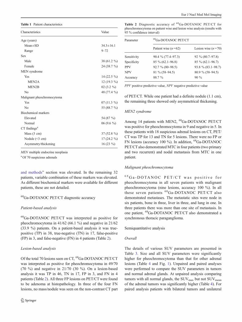
The details of various SUV parameters are presented inTable 3. Size and all SUV parameters were significantlyhigher for pheochromocytoma than that for other adrenallesions (Table 4 and Fig. 1). Unpaired and paired analyseswere performed to compare the SUV parameters in tumorsand normal adrenal glands. At unpaired analysis comparingtumors with all normal glands, the SUVmax but not SUVmean
of the adrenal tumors was significantly higher (Table 4). Forpaired analysis patients with bilateral tumors and unilateral
Table 1 Patient characteristics
Characteristics Value
Age (years)
Mean±SD 34.3±16.1
Range 9–72
Sex
Male 38 (61.2 %)
Female 24 (38.7 %)
MEN syndrome
Yes 14 (22.5 %)
MEN2A 12 (19.3 %)
MEN2B 02 (3.2 %)
No 48 (77.4 %)
Malignant pheochromocytoma
Yes 07 (11.3 %)
No 55 (88.7 %)
Biochemical markers
Elevated 54 (87 %)
Normal 06 (9.6 %)
CT findingsa
Mass (3 cm) 37 (52.8 %)
Nodule (<3 cm) 17 (24.2 %)
Asymmetry/thickening 16 (23 %)
MEN multiple endocrine neoplasiaa Of 70 suspicious adrenals
Table 2 Diagnostic accuracy of 68Ga-DOTANOC PET/CT forpheochromocytoma on patient wise and lesion wise analysis (results with95 % confidence interval)
Parameter 68Ga-DOTANOC PET/CT
Patient wise (n =62) Lesion wise (n =70)
Sensitivity 90.4 % (77.4–97.3) 92 % (80.7–97.8)
Specificity 85 % (62.1–96.8) 85 % (62.1–96.7)
PPV 92.7 % (80–98.5) 93.8 % (83.1–98.7)
NPV 81 % (58–94.5) 80.9 % (58–94.5)
Accuracy 88.7 % 90 %
PPV positive predictive value, NPV negative predictive value
Eur J Nucl Med Mol Imaging
adrenals were excluded, and tumor SUV parameters werecompared with contralateral normal adrenal. On pairedanalysis both SUVmax and SUVmean of adrenal tumors wassignificantly higher (Table 4). On ROC analysis (Table 5) asignificant cutoff could be derived for all SUV parameters.Among these parameters, a tumor SUVmax/SUVliver cutoff of>2.5 showed the highest diagnostic accuracy (AUC 0.741,95 % confidence interval 0.622–0.838).
MEN2 syndrome
Comparison of size and SUV parameters of the tumors amongpatients with and without MEN2 is detailed in Table 4. Whilethe SUVmax and SUVmean of tumors were significantly higherin patients with MEN2, no difference was noted for otherparameters.
Malignant pheochromocytoma
The comparison of size and SUV parameters of the tumorsamo n g p a t i e n t s w i t h b e n i g n a n d ma l i g n a n t
pheochromocytoma is detailed in Table 4. No significantdifference was found between benign and malignantpheochromocytoma for any of these parameters.
Correlation with tumor size
Themean tumor sizewas 4.1±2.4 cm (median 3.8, range 0.6–11).We assessed the relationship between lesion size and therespective SUV parameters to see whether tumor sizecorrelated with SSTR expression. No significant correlationwas noted between tumor size and tumor SUVmax (ρ=0.227,p =0.099), tumor SUVmean (ρ=0.155, p =0.262), tumorSUVmax/SUVliver (ρ=0.131, p =0.347), and tumor SUVmean/SUVliver (ρ=0.092, p =0.510).
Comparison with 131I-MIBG imaging
Overall
Both68Ga-DOTANOC PET/CT and 131I-MIBG imaging wereavailable for 45 adrenal lesions in 40 patients (Figs. 2–5). Thesensitivity, specificity, positive predictive value (PPV),negative predictive value (NPV), and accuracy of bothimaging methods for these 45 lesions are detailed in Table 6.On a lesion-based comparison 68Ga-DOTANOC PET/CTwas superior to 131I-MIBG imaging for pheochromocytoma(p =0.035). Results of 68Ga-DOTANOC PET/CT and 131I-MIBG imaging were discordant for 15/45 (33.3 %) lesions.
MEN2 syndrome
In patients with MEN2, both68Ga-DOTANOC PET/CT and131I-MIBG were available for 14/18 adrenal lesions. 68Ga-DOTANOC PET/CT was TP in ten and TN in four, while
Table 3 Results of semiquantitative analysis of 68Ga-DOTANOCPET/CT
Parameter Mean±SD Median Range
Tumor SUVmax 10.1±9.7 6.7 0.4–44.4
Tumor SUVmean 6.4±6.2 3.9 0.2–26.4
SUVliver 2.1±1.6 1.7 0.2–7.8
Tumor SUVmax/SUVliver 5±3.1 4.7 0.5–15
Tumor SUVmean/SUVliver 3.2±2 3.1 0.4–10.3
Normal adrenal SUVmax 6.7±6.3 4.1 0.3–30.4
Normal adrenal SUVmean 4.8±4.6 2.9 0.2–20.7
Metastasis SUVmax 5.7±4.2 5.1 0.7–16
Table 4 Comparison of size and SUV parameters between subgroups
Parameter Mean±SD
Tumor size (cm) Tumor SUVmax Tumor SUVmean Tumor SUVmax/SUVliver
Tumor SUVmax/SUVliver
Pheochromocytoma vs other lesionsa 4.4±2.4 vs 2.3±1.4 11.9±10.7 vs 5.5±4 7.5±6.8 vs 3.7±2.9 5.7±3.1 vs 3.2±2.5 3.5±2 vs 2.2±1.9
p=0.030 p =0.005 p =0.022 p =0.001 p=0.003
MEN2 vs sporadic tumors 3.8±2.5 vs 4.3±2.4 17±14.3 vs 7.7±6 10.5±8.6 vs 5±4.4 5.5±3 vs 4.8±3.2 3.4±1.9 vs 3±2.1
p=0.395 p =0.012 p =0.011 p =0.316 p=0.367
Benign vs malignant pheochromocytoma 3.9±2.4 vs 5.5±2.2 9.3±8.4 vs 15.3±15.6 6±5.4 vs 9.2±10 4.9±3.3 vs 5.5±2.1 3.2±2.2 vs 3±1.1
p=0.073 p =0.269 p =0.533 p =0.380 p=0.909
Tumor vs unpaired adrenal – 10.1±9.7 vs 6.7±6.3 6.4±6.2 vs 4.8±4.6 – –
p =0.026 p =0.137
Tumor vs paired adrenal – 10.1±9.7 vs 6.4±6 6.4±6.2 vs 4.6±4.3 – –
p =0.002 p =0.012
a Based on reference standard
Eur J Nucl Med Mol Imaging
131I-MIBG was TP in eight, TN in four, and FN in two. Onlesion-based analysis no significant difference was seenbetween 68Ga-DOTANOC PET/CT and 131I-MIBG (p =0.500). 131I-MIBG imaging demonstrated MTC in only oneof four patients with MEN syndrome and failed to show necknode metastasis from MTC in one patient. All these MTClesions were positive on 68Ga-DOTANOC PET/CT.
Malignant pheochromocytoma
Comparable 68Ga-DOTANOC PET/CT and 131I-MIBGimages were available for five of seven patients (five adrenallesions) with malignant pheochromocytoma. While 68Ga-DOTANOC PET/CT was positive for pheochromocytoma inall, 131I-MIBG was positive in three of five (60 %). Inaddition, 131I-MIBG imaging failed to show metastaticdisease in three of five patients (60 %) with malignant
pheochromocytoma, all of which were picked up by 68Ga-DOTANOC PET/CT.
Discussion
Pheochromocytomas rou t ine ly express SSTRs ,predominantly SSTR3 and to some extent SSTR2 [14]. Thisforms the basis of SSTR-based SPECT and PET imaging inpheochromocytoma. Results of 111In-octreotide SPECTimaging in pheochromocytoma have generally been poorand inferior to MIBG imaging, with better results reportedfor malignant than benign pheochromocytoma [21]. Apartfrom the issue of limited resolution, this might have also beenbecause of predominant affinity of 111In-octreotide towardsSSTR2, rather than SSTR3 [22]. More recently SSTR-basedPET/CT imaging of pheochromocytoma has been attemptedusing 68Ga-DOTA-peptides with encouraging results [15–19].PET/CT imaging with these agents has already been shown tobe superior to 111In-octreotide SPECT [23], though datapertaining specifically to pheochromocytoma are lacking.Among the 68Ga-DOTA-peptides, 68Ga-DOTANOC appearsto be of particular interest because of its broader SSTRaffinity, including intermediate to low affinity for SSTR3[ 2 4 , 2 5 ] , t h e p r e d om i n a n t s u b t y p e i n mo s tpheochromocytomas [14]. Surprisingly, most of the previousstudies have employed 68Ga-DOTATOC [17] or 68Ga-DOTATATE [15, 16, 19] for imaging of pheochromocytoma,
Fig. 1 Box and whisker plotscomparing the SUV parametersbetween non-pheochromocytomaadrenal lesions (solid boxes) andpheochromocytoma (dashedboxes). The circles represent theoutliers
Table 5 Results of ROC analysis
Parameters Cutoff Sensitivity Specificity AUC p value
Tumor SUVmax 5.4 70 % 70 % 0.712 0.0009
Tumor SUVmean 2.4 80 % 50 % 0.695 0.009
Tumor SUVmax/SUVliver
2.5 86 % 60 % 0.741 0.0001
Tumor SUVmean/SUVliver
2.53 72 % 75 % 0.727 0.0003
Eur J Nucl Med Mol Imaging
both of which have no significant affinity for SSTR3.Moreover, these studies suffered from the drawbacks of smallsample sizes and mixed patient populations. To address theselimitations, the present study assessed the diagnostic accuracyof 68Ga-DOTANOC PET/CT in a large population of patientswith suspected pheochromocytoma. The present study differsfrom the pilot study conducted at our center by Naswa et al.[18] in certain important aspects. First is the larger sample
size. Second, the present study evaluated an exclusivepopulation of pheochromocytoma, while that by Naswae t a l . [ 1 8 ] e v a l u a t e d a m ix ed popu l a t i o n o fpheochromocytoma and paraganglioma. Third, we have alsodone in-depth analysis of SUV parameters, which was notattempted in the pilot study.
In the present study, 68Ga-DOTAOC PET/CT showed highdiagnostic accuracy on both patient-based and lesion-basedanalysis (88.7 and 90%, respectively). It was FP only for threeadrenal lesions (size <1.5 cm), all of which turned out to bebenign adenoma at histopathology. Two factors probablyaccounted for the FP results. First, since high 68Ga-DOTANOC uptake is seen in normal adrenals [26],differentiation of tumor uptake from tracer uptake in normaladrenal medulla is sometimes difficult, especially if theadrenal lesion is small. Second, some of the cortical adenomascan also demonstrate low-grade SSTR expression [27],resulting in FP findings. Because we did not have the SSTRprofile of the resected specimens, we were not able to confirmthese hypotheses in the present study. 68Ga-DOTANOC PET/CT was FN for three lesions. Definite adrenal nodule on CTwas seen in only one of these four lesions, with the remainingthree showing asymmetrical thickening. In fact, 68Ga-DOTANOC PET/CT was interpreted as negative for all 16lesions without adrenal nodule/mass on CT. This is because ofthe limitation that it is not always possible to differentiatephysiological from pathological uptake at visual PET analysisalone if CT does not show a definite mass or nodule.Therefore, 68Ga-DOTANOC PET/CTseems to be most usefulwhen there is a definite adrenal mass or nodule at CT in theclinical setting of pheochromocytoma. The values ofdiagnostic accuracy reported in this study are lower than thoseof Naswa et al. [18]. The main reason for this difference isprobably the preponderance of paraganglioma in their studypopulation. On 68Ga-DOTANOC PET/CT, evaluation and
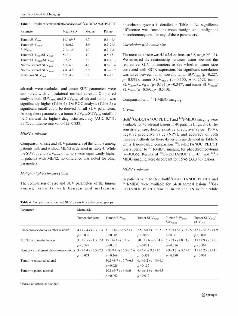
Fig. 2 A 37-year-old man with headache, paroxysmal hypertension,elevated 24-h urinary metanephrine, and left adrenal mass on CT. MIP68Ga-DOTANOC PET image (a) shows abnormal radiotracer uptake inthe left suprarenal region (arrow), corresponding to the necrotic left
adrenal mass on PET/CT image (c , arrow ). Anterior 131I-MIBGscintigraphy image (b) is normal and on SPECT/CT image no 131I-MIBGuptake is seen in the adrenal mass (arrow). The patient underwent surgeryand the diagnosis of pheochromocytoma was confirmed
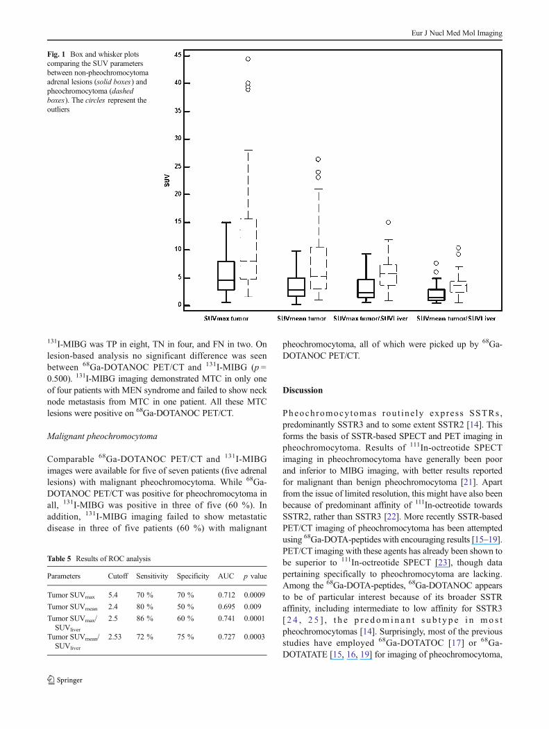
Fig. 3 A 67-year-old man with uncontrolled hypertension and rightadrenal mass on CT. The 24-h urinary metanephrines were elevated.MIP 68Ga-DOTANOC PET (a ) image showed mildly increasedabnormal tracer uptake in the right suprarenal region (arrow). Axialnon-contrast CT (b) image of the abdomen shows a right adrenal mass(arrow) with areas of calcification and cystic degeneration. The massshows mild 68Ga-DOTANOC uptake (SUVmax 1.6) on PET (c) and PET/CT (d) images (arrow). The patient underwent surgery and postoperativehistopathology confirmed the diagnosis of pheochromocytoma
Eur J Nucl Med Mol Imaging
identification of lesions at extra-adrenal sites is easier due tothe lack of physiological tracer uptake. In addition, it is knownthat paragangliomas show higher levels of SSTR expressionthan pheochromocytomas. At semiquantitative analysis, widevariation was seen in SUV parameters for the tumors as wellas normal adrenals. However, a major inference that could bederived is that pheochromocytomas have higher SUVparameters than other adrenal lesions, reconfirming highin vivo SSTR expression. Although there was significantdifference between the SUVmax of tumors with paired andunpaired normal adrenal glands (p =0.002 and 0.026,respectively), considerable overlap was seen between thesevalues. Using ROC analysis we were able to find significantcutoffs for all SUV parameters for differentiatingpheochromocytoma from other adrenal lesions. Among these,tumor SUVmax/SUVliver showed the highest diagnosticaccuracy. However, the sensitivity and specificity of theseSUV parameter cutoffs is only modest and inferior to visual
analysis. Hence, the proposed thresholds might not be suitablefor routine application in clinical practice.
In patients with MEN2 68Ga-DOTANOC PET/CT showedexcellent results for detection of pheochromocytoma. Therewere no FP or FN results, thus showing the utility of 68Ga-DOTANOC PET/CT in MEN2 wi th su spec t edpheochromocytoma. However, all these patients had clinical/biochemical suspicion along with suspicious adrenal lesions atCT. Hence, whether the same success can be replicated inscreening of asymptomatic cases with MEN2 needs to beestablished. 68Ga-DOTANOC PET/CT also detectedassociatedMTC in five patients, for which it has known utility[28]. The SUVmax and SUVmean of the adrenal tumors inpatients with MEN2 were significantly higher than sporadictumors. Although the exact cause of this finding is unclear,this finding has also been previously reported in a rat model[29]. 68Ga-DOTANOC PET/CT also showed excellent resultsin patients with malignant pheochromocytoma. Primary
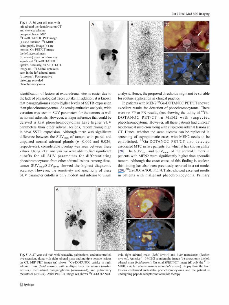
Fig. 5 A 27-year-old man with headache, palpitations, and uncontrolledhypertension, along with right adrenal mass and multiple hepatic lesionson CT. MIP PET image (a) shows 68Ga-DOTANOC uptake in rightadrenal mass (bold arrow ), with multiple liver metastases (brokenarrows ), mediastinal paraganglioma (arrowhead ), and pulmonarymetastases (arrows). Axial PET/CT image (c) shows 68Ga-DOTANOC
avid right adrenal mass (bold arrow ) and liver metastases (brokenarrows). Anterior 131I-MIBG scintigraphy image (b) shows only the leftadrenal mass (bold arrow). On axial SPECT/CT image (d) only the 131I-MIBG avid left adrenal mass is seen (bold arrow). Biopsy from the liverlesions confirmed metastatic pheochromocytoma and the patient isundergoing peptide receptor radionuclide therapy
Fig. 4 A 56-year-old man withleft adrenal incidentaloma on CTand elevated plasmanorepinephrine. MIP68Ga-DOTANOC PET image(a) and anterior 131I-MIBGscintigraphy image (b) arenormal. On PET/CT imagethe left adrenal mass(c , arrow) does not show anysignificant 68Ga-DOTANOCuptake. Similarly, on SPECT/CTimage no 131I-MIBG uptake isseen in the left adrenal mass(d , arrow). Postoperativehistology revealedpheochromocytoma
Eur J Nucl Med Mol Imaging
tumor as well as metastasis was identified in all seven patients.Similar results were also reported by Kroiss et al. [17] in theirstudy in patients with metastatic pheochromocytoma andneuroblastoma. We found no significant difference betweenmalignant and benign pheochromocytoma with respect toSUV parameters, probably reflecting overlap of SSTRexpression.
On a lesion-based comparison, 68Ga-DOTANOC PET/CTshowed a significantly higher accuracy as compared to 131I-MIBG (91.1 vs 66.6 %, p =0.035). These findings are similarto those reported by Naswa et al. [18] and Maurice et al. [19].However, in patients with MEN2, no significant differencewas found between 68Ga-DOTANOC PET/CT and 131I-MIBG for adrenal lesions (p =0.500). This equality isprobably the result of high accuracy of MIBG imaging fordetection of pheochromocytoma in MEN2 [30] coupled withthe small number of MEN2 patients in the present study.While further studies are required to make definitivecomment, given the fact that 68Ga-DOTANOC PET/CT isalso useful for imaging MTC, it may be preferred over MIBGimaging in patients with MEN2. In patients with malignantpheochromocytoma 68Ga-DOTANOC PET/CT showedprimary tumor and metastasis in all patients, while 131I-MIBGscintigraphy was negative for pheochromocytoma in two offive patients and metastasis in three of five patients. Given thesmall number of patients statistical comparison was notattempted. We would like to highlight that 131I-MIBG, not123I-MIBG, was used in the present study. The latter allows ahigher dose, gives a high photon flux, and provides betterimage quality. Unfortunately, 123I-MIBG is not available inour country and cannot be imported because of the short half-life (T1/2 13 h) and high cost. One might argue that the use of123I-MIBG might have altered our results. However, thesupe r i o r i t y o f 1 2 3 I -MIBG ove r 1 3 1 I -MIBG inpheochromocytoma is only marginal [31]. In addition, thesensitivity of 131I-MIBG for pheochromocytoma in thepresent study is similar to those reported for 123I-MIBG byKroiss et al. (61.2 and 63.3 %, respectively) [17]. Therefore, itis unlikely that use of 123I-MIBG would have significantlyaltered the results. On the other hand, a recent study has
demonstrated the superiority of 131I-MIBG SPECT/CT overSPECTand planar imaging in pheochromocytoma [8]. Hence,the lack of SPECT/CT in all patients might have compromisedthe performance of 131I-MIBG imaging in the present study.
Other PET tracers which can be employed for imaging ofpheochromocytoma include 18F-FDG to assess glucosemetabolism, 11C-HED and 18F-FDA for catecholaminergicphenotype, and 18F-FDOPA for assessing amine uptake anddecarboxylation. The most commonly used PET tracer 18F-FDG has shown low sensitivity but high specificity,comparable to 123I-MIBG imaging in pheochromocytoma[9]. It is particularly useful in metastatic pheochromocytomaand might have played a complementary role to 68Ga-DOTANOC PET/CT in the present population. 18F-FDOPAPET/CT has demonstrated high sensitivity and specificity forpheochromocytoma [10]. Minimal uptake of 18F-FDOPA innormal adrenal glands is advantageous when interpretingPET/CT images for pheochromocytoma. 18F-FDA PET/CTa l so ha s demons t r a t ed encourag ing re su l t s i npheochromocytoma and it is superior to both 18F-FDOPAPET/CT and 123I-MIBG for metastatic lesions [11]. 11C-HED is another PET/CT tracer showing high accuracy forpheochromocytoma [12]. Given the high diagnostic accuracyshown by 68Ga-DOTANOC PET/CT in the present study, ahead to head comparison with these other PET tracers iswarranted to find the optimum agent for different subgroupsof pheochromocytoma. The points in favor of 68Ga-DOTANOC are its easy generator-based synthesis ascompared to difficult synthesis (18F-FDOPA, 18F-FDA) orneed for on-site cyclotron (11C-HED) for other tracers.Additionally, 68Ga-DOTANOC PET/CT can help in selectionof patients (inoperable/metastatic) for peptide receptorradionuclide therapy, which is currently not possible withother PET tracers.
A few limitations of the present study must beacknowledged. First is the retrospective design. Second,histopathological diagnosis was not available for all theadrenal lesions. Although this would have been ideal, it wasnot ethically feasible. Hence, we employed a combination ofclinical, biochemical, and imaging follow-up of at least6 months to confirm or rule out pheochromocytoma, whichis an accepted method. Third, 131I-MIBG SPECT/CT imageswere not available for all patients, as at our center, like mostothers, the decision to perform SPECT/CT is guided by planarimaging. Finally, the lack of knowledge regarding thesuccinate dehydrogenase (SDH) status, which is pivotal tounderstanding the results of functional imaging ofpheochromocytomas, was a major limitation of the presentstudy. Germline mutations in SDH enzyme gene (especiallysubun i t B) occur in a subse t o f pa t i en t s wi thpheochromocytoma. SDH status appears to be a criticalparameter determining the prognosis as well as selecting thebest functional imaging agent for pheochromocytomas and
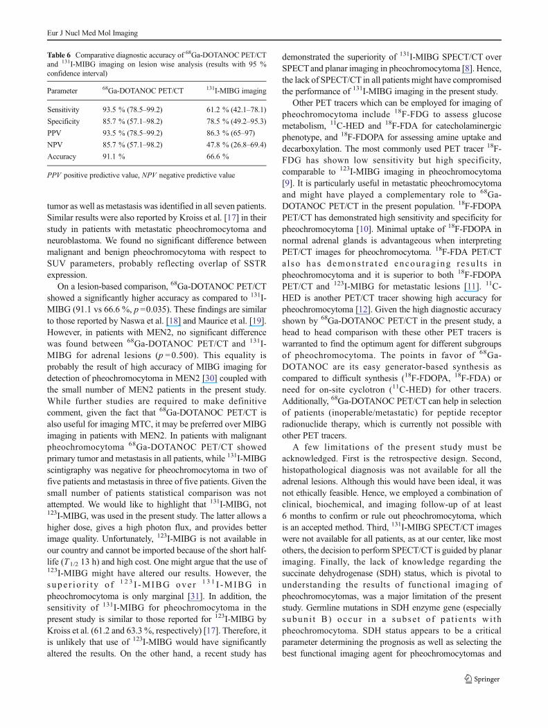
Table 6 Comparative diagnostic accuracy of 68Ga-DOTANOC PET/CTand 131I-MIBG imaging on lesion wise analysis (results with 95 %confidence interval)
Parameter 68Ga-DOTANOC PET/CT 131I-MIBG imaging
Sensitivity 93.5 % (78.5–99.2) 61.2 % (42.1–78.1)
Specificity 85.7 % (57.1–98.2) 78.5 % (49.2–95.3)
PPV 93.5 % (78.5–99.2) 86.3 % (65–97)
NPV 85.7 % (57.1–98.2) 47.8 % (26.8–69.4)
Accuracy 91.1 % 66.6 %
PPV positive predictive value, NPV negative predictive value
Eur J Nucl Med Mol Imaging
paragangliomas [32, 33]. Future prospective studiesaddressing these shortcomings and comparing 68Ga-DOTANOC with other PET tracers are warranted.
Conclusion
68Ga-DOTANOC PET/CT demonstrates high diagnosticaccuracy in patients with clinical/biochemical suspicion ofpheochromocytoma and adrenal abnormality at contrast CTand is superior to 131I-MIBG imaging for this purpose. Bestresults of 68Ga-DOTANOC PET/CT are seen in patients withMEN2-associated and malignant pheochromocytoma.
Conflicts of interest None.
References
1. Werbel SS, Ober KP. Pheochromocytoma. Update on diagnosis,localization, and management. MedClin North Am 1995;79:131–53.
2. Manger WM, Gifford RW. Pheochromocytoma: a clinical overview.In: Laragh JH, Brenner BM, editors. Hypertension: pathophysiology,diagnosis and management. New York: Raven; 1995. p. 225–44.
3. NeumannHPH, Berger DP, Sigmund G, BlumU, Schmidt D, ParmerRJ, et al. Pheochromocytomas, multiple endocrine neoplasia type 2,and von Hippel-Lindau disease. N Engl J Med 1993;329:1531–8.
4. Lenders JW, Pacak K, Walther MM, Linehan WM, Mannelli M,Friberg P, et al. Biochemical diagnosis of pheochromocytoma: whichtest is best? JAMA 2002;287:1427–34.
5. Quint LE, Glazer GM, Francis IR, Shapiro B, Chenevert TL.Pheochromocytoma and paraganglioma: comparison of MR imagingwith CT and I-131 MIBG scintigraphy. Radiology 1987;165:89–93.
6. Bombardieri E, Giammarile F, Aktolun C, Baum RP, BischofDelaloye A, Maffioli L, et al. 131I/123I-metaiodobenzylguanidine(mIBG) scintigraphy: procedure guidelines for tumour imaging. EurJ Nucl Med Mol Imaging 2010;37:2436–46.
7. Feggi L, Degli Uberti E, Pansini GC, Transforini G, Prandini N,Ambrosio MR, et al. Pitfalls in scintigraphic detection ofneuroendocrine tumours. Eur J Nucl Med 1992;19:214–8.
8. Sharma P, Dhull VS, Jeph S, Reddy RM, Singh H, Naswa N, et al.Can hybrid SPECT-CTovercome the limitations associated with poorimaging properties of 131I-MIBG?: comparison with planarscintigraphy and SPECT in pheochromocytoma. Clin Nucl Med2013;38:e346–53.
9. Timmers HJ, Chen CC, Carrasquillo JA, Whatley M, Ling A,Eisenhofer G, et al. Staging and functional characterization ofpheochromocytoma and paraganglioma by 18F-fluorodeoxyglucose(18F-FDG) positron emission tomography. J Natl Cancer Inst2012;104:700–8.
10. Fiebrich HB, Brouwers AH, KerstensMN, Pijl ME, Kema IP, de JongJR, et al. 6-[F-18]Fluoro-L-dihydroxyphenylalanine positronemission tomography is superior to conventional imaging with(123)I-metaiodobenzylguanidine scintigraphy, computer tomography,and magnetic resonance imaging in localizing tumors causingcatecholamine excess. J Clin Endocrinol Metab 2009;94:3922–30.
11. Ilias I, Yu J, Carrasquillo JA, Chen CC, Eisenhofer G, Whatley M,et al. Superiority of 6-[18F]-fluorodopamine positron emissiontomography versus [131I]-metaiodobenzylguanidine scintigraphy inthe localization of metastatic pheochromocytoma. J Clin EndocrinolMetab 2003;88:4083–7.
12. Trampal C, Engler H, Juhlin C, Bergström M, Långström B.Pheochromocytomas: detection with 11C hydroxyephedrine PET.Radiology 2004;230:423–8.
13. Naji M, AL-Nahhas A. 68Ga-labelled peptides in the management ofneuroectodermal tumours. Eur JNuclMedMol Imaging 2012;39:S61–7.
14. Mundschenk J, Unger N, Schulz S, Höllt V, Schulz S, Steinke R, et al.Somatostatin receptor subtypes in human pheochromocytoma:subcellular expression pattern and functional relevance for octreotidescintigraphy. J Clin Endocrinol Metab 2003;88:5150–7.
15. Win Z, Al-Nahhas A, Towey D, Todd JF, Rubello D, Lewington V,et al. 68Ga-DOTATATE PET in neuroectodermal tumours: firstexperience. Nucl Med Commun 2007;28:359–63.
16. Naji M, Zhao C, Welsh SJ, Meades R, Win Z, Ferrarese A, et al.68Ga-DOTA-TATE PET vs. 123I-MIBG in identifying malignantneural crest tumours. Mol Imaging Biol 2011;13:769–75.
17. Kroiss A, Putzer D, Uprimny C, Decristoforo C, Gabriel M, SantnerW, et al. Functional imaging in phaeochromocytoma andneuroblastoma with 68Ga-DOTA-Tyr 3-octreotide positron emissiontomography and 123I-metaiodobenzylguanidine. Eur J Nucl MedMol Imaging 2011;38:865–73.
18. Naswa N, Sharma P, Nazar AH, Agarwal KK, Kumar R, AmminiAC, et al. Prospective evaluation of 68Ga-DOTA-NOC PET-CT inphaeochromocytoma and paraganglioma: preliminary results from asingle centre study. Eur Radiol 2012;22:710–9.
19. Maurice JB, Troke R, Win Z, Ramachandran R, Al-Nahhas A, NajiM, et al. A comparison of the performance of 68Ga-DOTATATEPET/CT and 123I-MIBG SPECT in the diagnosis and follow-up ofphaeochromocytoma and paraganglioma. Eur J Nucl Med MolImaging 2012;39:1266–70.
20. Zhernosekov KP, Filosofov DV, Baum RP, Aschoff P, Bihl H,Razbash AA, et al. Processing of generator-produced 68Ga formedical application. J Nucl Med 2007;48:1741–8.
21. van der Harst E, de Herder WW, Bruining HA, Bonjer HJ, de KrijgerRR, Lamberts SW, et al. [(123)I]metaiodobenzylguanidine and[ (111) In]oc t reo t ide uptake in benign and mal ignantpheochromocytomas. J Clin Endocrinol Metab 2001;86:685–93.
22. Hofland LJ, Lamberts SW, van Hagen PM, Reubi JC, Schaeffer J,Waaijers M, et al. Crucial role for somatostatin receptor subtype 2 indetermining the uptake of [111In-DTPA-D-Phe1]octreotide insomatostatin receptor-positive organs. J NuclMed 2003;44:1315–21.
23. Srirajaskanthan R, Kayani I, Quigley AM, Soh J, Caplin ME,Bomanji J. The role of 68Ga-DOTATATE PET in patients withneuroendocrine tumors and negative or equivocal findings on111In-DTPA-octreotide scintigraphy. J Nucl Med 2010;51:875–82.
24. Wild D, Mäcke HR, Waser B, Reubi JC, Ginj M, Rasch H, et al.68Ga-DOTANOC: a first compound for PET imaging with highaffinity for somatostatin receptor subtypes 2 and 5. Eur J Nucl MedMol Imaging 2005;32:724.
25. Wild D, Schmitt JS, Ginj M, Mäcke HR, Bernard BF, Krenning E,et al. DOTA-NOC, a high-affinity ligand of somatostatin receptorsubtypes 2, 3 and 5 for labelling with various radiometals. Eur J NuclMed Mol Imaging 2003;30:1338–47.
26. Pettinato C, Sarnelli A, Di Donna M, Civollani S, Nanni C, MontiniG, et al. 68Ga-DOTANOC: biodistribution and dosimetry in patientsaffected by neuroendocrine tumors. Eur J Nucl Med Mol Imaging2008;35:72–9.
27. Unger N, Serdiuk I, Sheu SY, Walz MK, Schulz S, Saeger W, et al.Immunohistochemical localization of somatostatin receptor subtypesin benign and malignant adrenal tumours. Clin Endocrinol (Oxf)2008;68:850–7.
28. Naswa N, Sharma P, Suman Kc S, Lata S, Kumar R, Malhotra A,et al. Prospective evaluation of 68Ga-DOTA-NOC PET-CT inpatients with recurrent medullary thyroid carcinoma: comparisonwith 18F-FDG PET-CT. Nucl Med Commun 2012;33:766–74.
29. Miederer M, Molatore S, Marinoni I, Perren A, Spitzweg C, Reder S,et al. Functional imaging of pheochromocytoma with Ga-DOTATOC
Eur J Nucl Med Mol Imaging
and C-HED in a genetically defined rat model of multiple endocrineneoplasia. Int J Mol Imaging 2011;2011:175352.
30. Rodriguez JM, Balsalobre M, Ponce JL, Ríos A, Torregrosa NM,Tebar J, et al. Pheochromocytoma inMEN 2A syndrome. Study of 54patients. World J Surg 2008;32:2520–6.
31. Furuta N, Kiyota H, Yoshigoe F, HasegawaN, Ohishi Y. Diagnosis ofpheochromocytoma using [123I]-compared with [131I]-metaiodobenzylguanidine scintigraphy. Int J Urol 1999;6:119–24.
32. Timmers HJ, Kozupa A, Eisenhofer G, RaygadaM, Adams KT, SolisD, et al. Clinical presentations, biochemical phenotypes, andgenotype-phenotype correlations in patients with succinatedehydrogenase subunit B-associated pheochromocytomas andparagangliomas. J Clin Endocrinol Metab 2007;92:779–86.
33. Havekes B, King K, Lai EW, Romijn JA, Corssmit EP, Pacak K. Newimaging approaches to phaeochromocytomas and paragangliomas.Clin Endocrinol (Oxf) 2010;72:137–45.
Eur J Nucl Med Mol Imaging
Related Documents