1 Diagnosis, staging and treatment of patients with gestational trophoblastic disease National Clinical Guideline No. 13 Draft Update – For consultation July 2021

Welcome message from author
This document is posted to help you gain knowledge. Please leave a comment to let me know what you think about it! Share it to your friends and learn new things together.
Transcript

1
Diagnosis, staging and treatment of patients with gestational trophoblastic disease
National Clinical Guideline No. 13 D Draft Update – For consultation
D
July 2021

ii
The National Clinical Guideline on the diagnosis, staging and treatment of patients with gestational trophoblastic disease (GTD) in Ireland has been developed by the National Cancer Control Programme (NCCP), in collaboration with clinicians, librarians and stakeholder groups.
Using this National Clinical Guideline This National Clinical Guideline applies to adults (over 18 years old) that have a suspected diagnosis of Gestational Trophoblastic Disease (GTD). This guideline is intended for all health professionals involved in the diagnosis, staging and treatment of patients with GTD. While the Chief Executive Officer (CEO), General Manager and the Clinical Director of the hospital have corporate responsibility for the implementation of the recommendations in this Clinical Guideline, each member of the multidisciplinary team is responsible for the implementation of the individual guideline recommendations relevant to their discipline. This guideline is also relevant to those involved in clinical governance, in both primary and secondary care, to help ensure that arrangements are in place to deliver appropriate care for the population covered by this guideline. Whilst the guideline is focused on clinical care, it is expected to be of interest to patients with GTD and their significant others. Effort has been made to make this document more user friendly. A list of medical abbreviations used throughout the guideline can be found in Appendix 8: Glossary of terms and abbreviations.
Disclaimer The Guideline Development Group’s expectation is that health professionals will use clinical knowledge and judgment in applying the principles and recommendations contained in this guideline. These recommendations may not be appropriate in all circumstances and it may be necessary to deviate from this guideline. Clinical judgment in such a decision must be clearly documented. Care options should be discussed with the patient, his/her significant other(s), and the multidisciplinary team on a case-by-case basis as necessary.

iii
Membership of the Guideline Development Group
The Clinical Chairmen of the Guideline Development Group was by Dr John Coulter, Consultant Gynaecologist and Clinical Lead of the National Gestational Trophoblastic Disease Registry, Monitoring and Advisory Centre. Dr Eve O’Toole, Head of the Evidence and Quality Hub, NCCP acted as the methodology chair. This National Clinical Guideline is supported by the National Cancer Control Programme. Membership nominations were sought from a variety of clinical and non-clinical backgrounds so as to be representative of all key stakeholders within the Health Service Executive. Guideline Development Group members included patient representatives, those involved in clinical practice, hospital administration, project management, and research and librarian services. The NCCP would like to acknowledge the guideline development group responsible for the development of ‘National Clinical Guideline No. 13 Diagnosis, staging and treatment of patients with gestational trophoblastic disease’ in 2015, on which this guideline is based.
Name Job title and affiliation
Patient Representatives
Ms Evelyn Mythen Patient representative
Ms Deirdre Cronin O’Driscoll Patient representative
Surgery
Dr John Coulter
(Clinical Chair)
Consultant Gynaecologist and Clinical Lead of the National Gestational Trophoblastic Disease Registry, Monitoring and Advisory Centre, CUMH
Dr Karen Flood Consultant Obstetrician & Gynaecologist, Rotunda
Medical Oncology
Prof. Seamus O’Reilly Consultant Medical Oncologist, CUH
Dr Dearbhaile O’Donnell Consultant Medical Oncologist, SJH
Radiology
Dr Tony Geoghegan Consultant Radiologist, MMUH
Pathology
Dr Brendan Fitzgerald Consultant Histopathologist, Cork University Hospital
Dr Paul Downey Consultant Pathologist, National Maternity Hospital Dublin
Nursing
Ms Caitriona Kenneally Clinical Nurse Specialist, National GTD Registry, Monitoring and Advisory Centre, CUMH
Ms Clare Buckley Clinical Nurse Specialist, National GTD Registry, Monitoring and Advisory Centre, CUMH Until
Ms Joanne Higgins Clinical Nurse Specialist, GUH until March 2020
Ms Michelle Brady Clinical Nurse Specialist, MMUH until March 2020
Biochemistry
Ms Caroline Joyce Principal Clinical Biochemist, CUMH

iv
Dr Paula O’Shea Consultant Chemical Biochemist, CUH
Dr Sean Costello Locum Consultant Biochemist, CUH
Ms Ruth O’Kelly Principal Clinical Biochemist, Coombe until August 2020
Hospital Administration
Ms Miriam Lyons Business Manager, CUMH
Project Management
Ms Catherine Duffy Project Manager, NCCP
Research
Dr Eve O’Toole
(Methodology Chair)
Head of Evidence and Quality Hub, NCCP
Ms Louise Murphy Research Officer, NCCP
Library
Mr Brendan Leen Regional Librarian, HSE South

v
Acknowledgments
The Clinical Chair of the Guideline Development Group (GDG) Dr John Coulter and the Methodology Chair Dr Eve O’Toole wish to acknowledge all members of the Guideline Development Group as full contributors credited with having given substantial intellectual leadership to the National Clinical Guideline. Ms Catherine Duffy and Dr Eve O’ Toole successfully submitted the guideline for NCEC prioritisation. The Guideline Development Group clinical members, methodology chair, research members and project manager agreed the scope and developed the clinical questions. The Guideline Development Group librarians and research members carried out the systematic searches for evidence. The Guideline Development Group research members reviewed the evidence, appraised the literature and performed the data extraction. The Guideline Development Group led by Dr Eve O’Toole carried out the evidence synthesis including formulation of the evidence summaries and recommendations. We would like in addition to thank Ms Louise Murphy for her editorial support during preparation for publication. A full list of members of the Guideline Development Group is available in the previous page/s.
Signed by the Chair(s) __________________________________ Date: _________________

vi
Table of contents
Page
Section 1: Background
1.1 Impact of Gestational Trophoblastic Disease 8
1.2 National Gestational Trophoblastic Disease Registry, Monitoring and Advisory Centre
8
1.3 Context and Scope of this National Clinical Guideline 9
Section 2: National Clinical Guideline
2.1 Summary of clinical questions, recommendations and summary of budget impact analysis
10
2.2 Diagnosis 19
2.3 Staging 35
2.4 Treatment 40
Section 3: Development of a National Clinical Guideline
3.1 Epidemiology 59
3.2 Rationale for this National Clinical Guideline 59
3.3 Aim and objectives 59
3.4 Financial impact of GTD 59
3.5 Guideline scope 60
3.6 Conflict of interest statement 60
3.7 Sources of funding 60
3.8 Guideline methodology and literature review 60
3.9 Consultation process 60
3.10 External review 60
3.11 Implementation 61
3.12 Monitoring and audit 61
3.13 Recommendations for research 61
3.14 Systematic review of cost-effectiveness 61
3.15 Budget impact analysis 62
3.16 Plan to update this National Clinical Guideline 62
Section 4: Appendices
1 Guideline Development Group terms of reference and logic model 63 2 Systematic Literature Review Protocol 65 3 Details of consultation process 70
4 Economic assessment
Part A: Economic evidence summary Part B: Budget impact analysis
71
5 Implementation plan 72 6 Supporting tools 73 7 Monitoring and audit 74 8 Glossary of terms and abbreviations 75 9 Clinical question in PICO format 78
10 Levels of evidence and grading systems 79 References 80

vii
List of tables Table 1 FIGO Anatomical Staging as adapted by FIGO (2009) .............................................................. 38 Table 2 Modified WHO prognostic scoring system as adapted by FIGO (2009) ................................... 38 Table 3 Quality of evidence adapted from GRADE working group 2013 .............................................. 79 Table 4 Strength of recommendation adapted from GRADE working group 2013 .............................. 79
List of figures Figure 1 The current protocol for monitoring hCG levels in women with complete hydatidiform molar pregnancy.............................................................................................................................................. 30 Figure 2 The current protocol for monitoring hCG levels in women with partial hydatidiform mole . 31

8
1 Background 1.1 Impact of Gestational Trophoblastic Disease Gestational trophoblastic disease (GTD) is a spectrum of diseases that can occur during or after pregnancy, each having a varying propensity for local invasion and metastasis. GTD has been defined as a continuum of a neoplastic process that arises from the trophoblastic cells that during pregnancy are involved in the development of the placenta. Its pathogenesis is unique as it arises from gestational rather than maternal tissue (Berkowitz et al., 2020). The World Health Organisation (WHO) has classified GTD as two premalignant diseases, consisting of complete hydatidiform mole (CHM) and partial hydatidiform mole (PHM), and as four malignant disorders, consisting of invasive mole, choriocarcinoma, placental site trophoblastic tumour (PSTT) and epithelioid trophoblastic tumour (ETT). The last four conditions are often collectively referred to as gestational trophoblastic neoplasia (GTN) (Kumar & Kumar, 2011). GTD is the most curable of all gynaecologic malignancies. It represents an oncologic success story attributable primarily to early disease recognition, chemotherapy regimens, and accurate and reliable assessment of disease status with sensitive assays for the measurement of human chorionic gonadotropin (hCG) levels. Its importance as a disease status cannot be overstated to the general gynaecologist, who is initially responsible for the diagnosis and management of GTD as well as the timely registration of the patient at the National Gestational Trophoblastic Disease Registry, Monitoring & Advisory Centre and referral to a gynaecological oncologist. The management of these women is specialised and, in many countries, is undertaken by gynaecological and medical oncologists with special expertise in treating this disease. A structured approach to diagnosis and management will result in a cure for most patients, even in the setting of advanced disease, without adversely affecting future fertility (McGee & Covens, 2012). 1.2 National Gestational Trophoblastic Disease Registry, Monitoring and Advisory Centre The National Gestational Trophoblastic Disease Registry, Monitoring and Advisory Centre was established in May 2017 to monitor and co-ordinate the follow-up of women who have been diagnosed with a molar pregnancy. It is a service established by the Health Service Executive, the National Cancer Control Programme and Cork University Maternity Hospital and is the only such centre in Ireland. GTD is a rare disease, and registration of all patients with GTD is recommended as a minimum standard of care. The GTD centre provides diagnosis, monitoring, treatment and advice to patients and clinicians on the care of patients with GTD. The outcome for more than 98% of women with GTN is excellent however a small number of women will die from the disease, mainly due to late presentation and diagnosis or drug resistance (Seckl et al., 2010). In 2019, 123 women with suspected GTD were registered with the National Gestational Trophoblastic Disease Registry, Monitoring & Advisory Centre. As not all patients are registered with the GTD centre this underestimates the incidence of this disease. A 2019 laboratory study estimated that 42% of women with suspected GTD/GTN were not registered with the National Gestational Trophoblastic Disease Registry, Monitoring & Advisory Centre.

9
1.3 Context and Scope of this National Clinical Guideline This guideline aims to improve the standard of clinical practice to ensure that young women affected by GTD and GTN are diagnosed promptly and receive the best available care. The diagnosis, staging, and treatment of patients with GTN requires multidisciplinary care in an acute hospital setting. The majority of patients will require diagnostic tests (radiology, pathology) and depending on the treatment plan may require surgery and chemotherapy.

10
2 National Clinical Guideline 2.1 Summary of clinical questions, recommendations and summary of budget impact analysis Here follows a list of all the recommendations in this guideline, along with the quality of evidence and strength of that recommendation. The quality of evidence and strength of recommendation system used is defined in Appendix 10 Levels of evidence and grading systems. Clinical question 2.2.1 Should all women undergoing medical management of miscarriage have histopathology of products of conception to exclude trophoblastic disease? Recommendation 2.2.1.1: The histological assessment of material obtained from the surgical management of all failed pregnancies is recommended to exclude trophoblastic disease. Quality of Evidence: Low Grade of recommendation: Strong Recommendation 2.2.1.2: All women undergoing medical management of miscarriage should be
advised to perform a follow-up urinary pregnancy test two to three weeks after miscarriage or
ultrasound as per local protocol.
Quality of Evidence: Very low Grade of recommendation: Strong
Good Practice Point: In women undergoing a miscarriage communication should be sensitive and in line with local hospital policy.
Practical considerations around patient care: The patient should be provided with relevant information (e.g. a leaflet) explaining why the sample is sent to the laboratory.
Clinical question 2.2.2 For women with suspected molar pregnancy (suspected partial hydatidifrom mole (PHM), complete hydatidifrom mole (CHM) or in patients where molar pregnancy cannot be excluded), what diagnostic tests should be done to accurately diagnose partial or complete molar pregnancy? Recommendation 2.2.2.1: Ultrasound examination is helpful in making pre-evacuation diagnosis of partial or complete molar pregnancy but the definitive diagnosis is made by histological examination of the products of conception. Quality of Evidence: Moderate Grade of recommendation: Strong Recommendation 2.2.2.2: Laboratories examining products of conception should have access to p57KIP2 immunohistochemistry to aid in the differential diagnosis of complete and partial molar pregnancies. Quality of Evidence: Low Grade of recommendation: Weak
Good Practice Point: GTD may be diagnosed in the absence of histopathological proof based on clinical, radiological, or biochemical suspicion (raised hCG). In these circumstances early expert referral to the National GTD Registry, Monitoring and Advisory Centre is recommended.

11
Practical considerations around patient care:
All patients registered with the National GTD Registry, Monitoring and Advisory Centre should have access to a specialist nurse for information, counselling and support.
Written information, guidance and support for health professionals and GPs should be available including a link to the National GTD Registry, Monitoring and Advisory Centre website.
Clinical question 2.2.3 For women where there is suspicion of partial or complete molar pregnancy who have an evacuation performed, in what time frame should the pathology report (post-evacuation) be available to the clinician? Recommendation 2.2.3.1: In cases of suspected molar pregnancy, a pathology report should be available to the clinician within 14 calendar days. Quality of Evidence: Very low Grade of recommendation: Weak Recommendation 2.2.3.2: If molar pregnancy is suspected the requesting clinician should indicate their clinician suspicion on the pathology request form and/or inform pathologist. Quality of Evidence: Very low Grade of recommendation: Weak
Good Practice Point: GTD may be diagnosed in the absence of histopathological proof based on clinical, radiological, or biochemical suspicion (raised hCG). In these circumstances early expert referral to the National GTD Registry, Monitoring and Advisory Centre is recommended. In certain cases where ancillary laboratory testing is needed additional time may be necessary for a diagnosis. Written information guidance and support on GTD for health professionals including GPs should be available including a link to the National GTD centre website
Practical considerations around patient care
Patients should be informed that results from the pathology test will take two weeks.
Clinical question 2.2.4 Which patients with confirmed or suspected GTD should be registered with the National GTD Registry, Monitoring and Advisory Centre? Recommendation 2.2.4.1: The guideline development group recommends that all women with suspected GTD should be registered with the National GTD Registry, Monitoring and Advisory Centre. Quality of Evidence: Very low Grade of recommendation: Strong Good Practice Point: The registration of women with suspected GTD with the National GTD Registry, Monitoring and Advisory Centre represents a minimum standard of care.

12
Clinical question 2.2.5 In patients with suspected GTD, how should hCG be measured? Recommendation 2.2.5.1: hCG serum should be tested using an assay that is CE marked for oncology. Quality of Evidence: Low Grade of recommendation: Strong Good Practice Point: The registration of women with suspected GTD with the National GTD Registry, Monitoring and Advisory Centre represents a minimum standard of care. hCG testing should be performed in a laboratory that is accredited to medical testing standard ISO 15189 (2012). Clinical question 2.2.6 For women with partial and complete molar pregnancy, what clinical and human chorionic gonadotropin monitoring protocol should be carried out to ensure they have been fully followed up and require no further therapy or monitoring? Recommendation 2.2.6.1: For patients with complete hydatidiform mole serum hCG is monitored weekly until normalisation is achieved for three weeks.
If this occurs within eight weeks post evacuation then monitor monthly for six months from the time of evacuation.
If normalisation occurs greater than eight weeks post evacuation then monitoring continues monthly for six months post normalisation.
Quality of Evidence: Moderate Grade of recommendation: Strong Recommendation 2.2.6.2: For patients with partial hydatidiform mole the serum hCG should be
monitored weekly until normalisation and one further confirmatory hCG measurement should be
performed four weeks later. If that confirmatory hCG is normal then follow up is complete.
Quality of Evidence: Moderate Grade of recommendation: Strong
Good Practice Point: For all women with a previous diagnosis of GTD early fetal ultrasound is standard practice to ensure a normal intrauterine pregnancy and to rule out recurrence of a molar pregnancy. If a normal intrauterine pregnancy is confirmed there are no extra investigations necessary during the pregnancy. hCG should be monitored on an assay that is approved for use in oncology. For all women with a previous diagnosis of GTD any subsequent pregnancy should be followed with a serum hCG measurement at six and ten weeks post-natally regardless of the outcome of pregnancy.

13
Clinical question 2.2.7 In women with confirmed GTD should monitoring of hCG be centralised? Recommendation 2.2.7.1: hCG testing should be centralised in women with confirmed GTD who have been registered with the National GTD Registry, monitoring and advisory centre. Quality of Evidence: Very Low Grade of recommendation: Strong Good Practice Point: The registration of women with suspected GTD with the National GTD Registry, Monitoring and Advisory Centre represents a minimum standard of care. Clinical question 2.3.1: For women with Gestational Trophoblastic Neoplasia (GTN), what investigations should be done to accurately stage GTN? Recommendation 2.3.1.1: Women with a diagnosis of GTN should have serum hCG monitoring, endovaginal ultrasound and a CT scan of thorax, abdomen & pelvis performed within one week of diagnosis. Quality of Evidence: Low Grade of recommendation: Strong Recommendation 2.3.1.2: If clinically significant lung metastases are present on a CT scan of the
thorax a contrast enhanced MRI of the brain should be performed.
Quality of Evidence: Low Grade of recommendation: Strong
Good Practice Point: Investigation and management decisions should be performed by experienced professionals in the management of GTD. Clinical question 2.3.2 For women with gestational trophoblastic neoplasia (GTN), what risk scoring system should be used to stage GTN? [Retained from 2015] Recommendation 2.3.2.1: Women with GTN (invasive mole, choriocarcinoma) should be assigned a FIGO score to direct management decisions of chemotherapy regimens. Grade of recommendation: Grade B Good Practice Point: Placental site trophoblastic tumour and epithelioid trophoblastic tumour should not be scored using the FIGO system. They require separate classification in consultation with international experts.
14
Clinical question 2.4.1 For women with gestational trophoblastic neoplasia, what are the clinical indicators to diagnose GTN warranting chemotherapy? Recommendation 2.4.1.1: Indications for chemotherapy following diagnosis of GTN: • Plateaued or rising hCG after evacuation, • Heavy vaginal bleeding or evidence of gastrointestinal or intraperitoneal haemorrhage, • Histological evidence of choriocarcinoma, except in exceptional circumstances, • Evidence of metastases in the brain, liver, or gastrointestinal tract, or radiological opacities of >2cm on chest x-ray. Quality of Evidence: Moderate Grade of recommendation: Strong Recommendation 2.4.1.2: For women who have a raised hCG six months after evacuation with a falling hCG should have their treatment plan discussed with the National Gestational Trophoblastic Disease Registry, Monitoring and Advisory Centre. Quality of Evidence: Low Grade of recommendation: Strong
Recommendation 2.4.1.3: For women with serum hCG of ≥20,000 IU/l more than four weeks after evacuation should have their treatment plan discussed with the National Gestational Trophoblastic Disease Registry, Monitoring and Advisory Centre. Quality of Evidence: Low Grade of recommendation: Strong
Good Practice Point: For women with histological evidence of choriocarcinoma primary surgery may be considered. The treating physician should ensure that the patient is registered with the National GTD Registry, Monitoring and Advisory Centre. Clinical question 2.4.2 For patients with low-risk (FIGO 0-6) GTN, what is the optimal first-line chemotherapy regimen? Recommendation 2.4.2.1: Patients with a FIGO score of 0-6 can be treated with either single-agent methotrexate with or without folinic acid, or actinomycin-D. Taking into account the treatment cycles, potential complications and quality of life the guideline development group agreed that methotrexate is the preferred first line chemotherapy. Quality of Evidence: Moderate Grade of recommendation: Strong Recommendation 2.4.2.2: Chemotherapy for low-risk disease should be continued for three cycles
of maintenance treatment after hCG normalisation.
Quality of Evidence: Moderate Grade of recommendation: Strong
Good Practice Point: In patients with low risk GTN serum hCG/blood should be done on day one of each chemotherapy cycle or more frequently if required.

15
Practical considerations around patient care
Patients diagnosed with GTN should have access to counselling and support from the nurses in the National GTD Registry, Monitoring and Advisory Centre.
Patients diagnosed with GTN should have access to a liasion nurse or designated key contact in their treatment centre who should also be in contact with the National GTD Registry, Monitoring and Advisory Centre.
Clinical question 2.4.3 For women with high-risk (FIGO ≥7) GTN, what is the optimal first-line chemotherapy regimen? Recommendation 2.4.3.1:Patients with a FIGO score of ≥7 should receive multi-agent chemotherapy and most centres now use EMA/CO, as it is highly effective. Quality of Evidence: Moderate Grade of recommendation: Strong Recommendation 2.4.3.2: Early deaths in ultra-high-risk GTN (FIGO score ≥12) can be reduced by induction therapy with etoposide and cisplatin. Such patients may also benefit from substitution of EMA/CO with EP/EMA. Quality of Evidence: Moderate Grade of recommendation: Strong
Good Practice Point For women with high-risk GTN, decisions should be made on an individual patient basis following discussion with clinicians experienced in high-risk GTN management at a GTD Centre. Registration of patients at the National GTD Registry, Monitoring and Advisory Centre is a minimum standard of care.
Practical considerations around patient care
Patients should be informed that their treatment will require in-patient care.
Patients with high risk GTN (FIGO score of ≥7) should have access to a liaison nurse or designated key contact locally who is in contact with the National GTD Registry, Monitoring and Advisory Centre.
Patients diagnosed with GTN should have access to a to a liaison nurse or designated key contact in their treatment centre who should also be in contact with the National GTD Registry, Monitoring and Advisory Centre.
Clinical question 2.4.4 For women with low-risk gestational trophoblastic neoplasia undergoing chemotherapy (first-course), what is the recommended course of action for observing and managing bleeding? Recommendation 2.4.4.1: For women with low-risk GTN undergoing first-line chemotherapy, the first ± second courses of chemotherapy may need to be administered as an in-patient at a centre with medical oncology, gynaecological services and interventional radiology. Grade of recommendation: Grade C
16
Clinical question 2.4.5: For women with gestational trophoblastic neoplasia, what are the appropriate investigations to monitor response to chemotherapy and follow-up? Recommendation 2.4.5.1: Monitoring during treatment low-risk: Patient should have human chorionic gonadotropin (hCG) levels prior to their next chemotherapy cycle. Treatment is continued until hCG is normal and for three further consolidation cycles. Quality of Evidence: Low Grade of recommendation: Strong Recommendation 2.4.5.2:
Monitoring during treatment high-risk: Patient should have human chorionic gonadotropin (hCG) levels prior to their next chemotherapy cycle. Patients with high-risk disease should have maintenance therapy for three cycles after hCG normalisation extended to four cycles for patients with poor prognostic features such as liver metastases with or without brain metastases. Quality of Evidence: Low Grade of recommendation: Strong
Recommendation 2.4.5.3: Follow-up post treatment: After remission is achieved, serum hCG should be measured fortnightly for six months then monthly for a further six months and every two months for two years. Quality of Evidence: Low Grade of recommendation: Strong
Practical considerations around patient care
Patients should have access to a nurse or designated key contact with experience treating GTN that can provide advice, written information and support to patients before commencing treatment.
Patients should be provided with clear written information in patient friendly language that they can share with family.
Clinical question 2.4.6: For women with gestational trophoblastic neoplasia what are the indicators to determine switching treatments from first-line chemotherapy? [Retained from 2015] Recommendation 2.4.6.1 : For patients with low-risk GTN the clinical indicators for a change in treatment from first-line chemotherapy include: treatment related toxicity, a rise in hCG values over two successive treatments. Grade of recommendation: Grade C Good Practice Point: Consideration could be given to re-staging patients prior to the initiation of a new regimen (particularly high-risk patients).

17
Clinical question 2.4.7: For women with low-risk gestational trophoblastic neoplasia who have not responded or have relapsed from single agent treatment (methotrexate or actinomycin D) or have relapsed following normalisation of hCG after completion of single agent treatment, what is the next line treatment? Recommendation 2.4.7.1: For women with low-risk GTN who have not responded to methotrexate with a hCG <1,000 IU/L the next line of treatment is actinomycin D. Quality of Evidence: Low Grade of recommendation: Strong Recommendation 2.4.7.2: For women with low-risk GTN who have not responded to methotrexate with a hCG >1,000 IU/L the next line of treatment is EMA/CO. Quality of Evidence: Low Grade of recommendation: Strong Recommendation 2.4.7.3
For women with low-risk GTN who have not responded or have relapsed from sequential single-agent treatment the next line of treatment is combination chemotherapy with EMA/CO. Quality of Evidence: Low Grade of recommendation: Strong
Good Practice Point: Once normalistion of hCG has occured on EMA/CO treatment etoposide can be discontinued from the regimen to reduce the risk of secondary malignancies. Clinical question 2.4.8 For women with high-risk GTN who have not responded to first-line treatment, what is second-line treatment? Recommendation 2.4.8.1: For women with high-risk GTN who have not responded to first-line treatment second line treatment is EP/EMA, if patients do not respond to EP/EMA TE/TP is the recommmended third line treatment. Quality of Evidence: Low Grade of recommendation: Strong Recommendation 2.4.8.2
In women with high-risk GTN who have not responded to first-line treatment, discussions of each individual case at a GTD MDM should be considered. Quality of Evidence: Very Low Grade of recommendation: Strong
Good Practice Point: All women with GTN should be registered at the National GTD Registry, monitoring and advisory centre. Given the rarity of this condition consideration should be given to discussing each individual case with international experts.

18
Clinical question 2.4.9 For women with GTN, who are acutely ill with liver, brain or lung metastasis at presentation, what is the optimum chemotherapy regimen?
Recommendation 2.4.9.1: Emergency treatment Patients who are acutely unwell from liver or CNS disease and particularly those with large lung metastases who are at risk of respiratory failure should be admitted and emergency chemotherapy commenced as soon as possible. Quality of Evidence: Very Low Grade of recommendation: Strong
Recommendation 2.4.9.2: Hepatic metastases Patients with hepatic metastases at presentation should commence therapy using EP/EMA protocol. Quality of Evidence: Very Low Grade of recommendation: Strong
Recommendation 2.4.9.3: Cerebral metastases Patients with cerebral metastases should be treated with EMA(CNS)/CO. Quality of Evidence: Very Low Grade of recommendation: Strong
Recommendation 2.4.9.4: Hepatic and synchronous cerebral metastases Patients with liver and brain metastases should be treated with a combination of EMA (CNS) and EP. Quality of Evidence: Very Low Grade of recommendation: Strong
Practical considerations around patient care
Patients should be provided with reassurance that they are being managed in co-operation with the National GTD Registry, Monitoring and Advisory Centre.
Patients should be counselled and reassured of the high cure rate of this patient cohort.

19
2.2 Diagnosis The following are responsible for the implementation of the recommendations regarding diagnosis: While the CEO, General Manager and the Clinical Director of the hospital have corporate responsibility for the implementation of the recommendations in this National Clinical Guideline, each member of the multidisciplinary team is responsible for the implementation of the individual guideline recommendations relevant to their discipline.

20
Clinical question 2.2.1 (D1) Should all women undergoing medical management of miscarriage have histopathology of products of conception to exclude trophoblastic disease?
Quality of evidence The evidence to address this question comes from an international guideline (Tidy et al., 2020). Histopathology of products of conception should be performed in all cases of surgical management of miscarriage (Tidy et al., 2020). Histopathology of products of conception enables earlier accurate diagnosis of trophoblastic disease. Women who miscarry at home should be advised to perform a follow-up urinary pregnancy test two to three weeks after miscarriage as per local protocol (Tidy et al., 2020). Alternatively, they may be advised to return for an ultrasound based on clinical presentation as per local protocol. If tissue is available the attending practitioner should arrange for the appropriate examination. Benefit and Harm The guideline development group agreed that in all cases of surgical management of miscarriage histopathological testing of products of conception would benefit patients as it would identify trophoblastic disease if present. The estimated risk of missing a molar pregnancy that requires treatment from not having tissue for histopathology is ~ 1 in 9,000. This is based on the number of miscarriages, molar pregnancies and the number of molar pregnancies that require treatment. Therefore the benefit of having tissue is outweighed by the psychological stress that may be caused to the patient. In women who miscarry at home it was agreed that histopathological testing of products of conception had the potential to distress patients as retrieving and transferring the products of conception may cause emotional and psychological distress to the patient and the quality of the tissue sample may be poor However, in women who miscarry at home a follow-up urinary pregnancy test would benefit patients as it would ensure that a complete miscarriage has taken place and in patients where a complete miscarriage has not taken place further investigations are needed. Preferences and values The guideline development group considered the emotional distress of a miscarriage and agreed that the benefit of having tissue for histology is outweighed by the psychological distress and dignity of the women. For patients undergoing surgical management of miscarriage the patient is going through the emotional distress of a miscarriage and the issue of processing tissue samples should be dealt with in a sensitive manner. The patient should be counselled around why histopathology is required.
21
Resources and other considerations No relevant cost-effectiveness literature was identified to address this clinical question. The following resources and other considerations were discussed in detail by the guideline development group: Access to histopathological assessment In addition to routine pathological resourcing, access to specialised pathological investigations may be required in certain cases e.g. P57kip2 immunohistochemistry/ploidy assessment/molecular analysis. These requirements are detailed further in Clinical question 2.2.2 (D2). Recommendation 2.2.1.1: The histological assessment of material obtained from the surgical management of all failed pregnancies is recommended to exclude trophoblastic disease. Quality of Evidence: Low
Grade of recommendation: Strong
Recommendation 2.2.1.2: All women undergoing medical management of miscarriage should be advised to perform a follow-up urinary pregnancy test two to three weeks after miscarriage or ultrasound as per local protocol. Quality of Evidence: Very low
Grade of recommendation: Strong
Good Practice Point In women undergoing a miscarriage communication should be sensitive and in line with local hospital policy.
Practical considerations around patient care The patient should be provided with relevant information (e.g. a leaflet) explaining why the sample is sent to the laboratory.
22
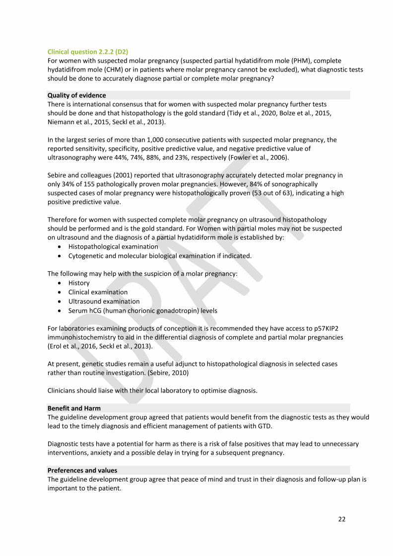
Clinical question 2.2.2 (D2) For women with suspected molar pregnancy (suspected partial hydatidifrom mole (PHM), complete hydatidifrom mole (CHM) or in patients where molar pregnancy cannot be excluded), what diagnostic tests should be done to accurately diagnose partial or complete molar pregnancy?
Quality of evidence There is international consensus that for women with suspected molar pregnancy further tests should be done and that histopathology is the gold standard (Tidy et al., 2020, Bolze et al., 2015, Niemann et al., 2015, Seckl et al., 2013). In the largest series of more than 1,000 consecutive patients with suspected molar pregnancy, the reported sensitivity, specificity, positive predictive value, and negative predictive value of ultrasonography were 44%, 74%, 88%, and 23%, respectively (Fowler et al., 2006). Sebire and colleagues (2001) reported that ultrasonography accurately detected molar pregnancy in only 34% of 155 pathologically proven molar pregnancies. However, 84% of sonographically suspected cases of molar pregnancy were histopathologically proven (53 out of 63), indicating a high positive predictive value. Therefore for women with suspected complete molar pregnancy on ultrasound histopathology should be performed and is the gold standard. For Women with partial moles may not be suspected on ultrasound and the diagnosis of a partial hydatidiform mole is established by:
Histopathological examination
Cytogenetic and molecular biological examination if indicated. The following may help with the suspicion of a molar pregnancy:
History
Clinical examination
Ultrasound examination
Serum hCG (human chorionic gonadotropin) levels For laboratories examining products of conception it is recommended they have access to p57KIP2 immunohistochemistry to aid in the differential diagnosis of complete and partial molar pregnancies (Erol et al., 2016, Seckl et al., 2013). At present, genetic studies remain a useful adjunct to histopathological diagnosis in selected cases rather than routine investigation. (Sebire, 2010) Clinicians should liaise with their local laboratory to optimise diagnosis. Benefit and Harm The guideline development group agreed that patients would benefit from the diagnostic tests as they would lead to the timely diagnosis and efficient management of patients with GTD. Diagnostic tests have a potential for harm as there is a risk of false positives that may lead to unnecessary interventions, anxiety and a possible delay in trying for a subsequent pregnancy. Preferences and values The guideline development group agree that peace of mind and trust in their diagnosis and follow-up plan is important to the patient.

23
Further reassurance can be provided to the patient by registering them with the National GTD Registry, Monitoring and Advisory Centre where they can be provided with consistent up to date information, support and advice. Resources and other considerations No relevant cost-effectiveness literature was identified to address this clinical question. The following resources and other considerations were discussed in detail by the guideline development group: Access to histopathological assessment Histopathological assessment of molar pregnancy should include p57KIP2. In addition to routine pathological resourcing, access to specialised pathological investigations may be required in certain cases e.g. P57kip2 immunohistochemistry/ploidy assessment/molecular analysis. Serum hCG testing hCG follow-up may have a financial implication for patients as a small number of women attend their GP for follow-up blood tests incurring personal cost. The guideline development group agreed that it would be useful for the GP to receive information along with a letter from the GTD centre regarding their patients care. Recommendation 2.2.2.1: Ultrasound examination is helpful in making pre-evacuation diagnosis of partial or complete molar pregnancy but the definitive diagnosis is made by histological examination of the products of conception. Quality of Evidence: Moderate
Grade of recommendation: Strong
Recommendation 2.2.2.2: Laboratories examining products of conception should have access to p57KIP2 immunohistochemistry to aid in the differential diagnosis of complete and partial molar pregnancies. Quality of Evidence: Low
Grade of recommendation: Weak
Good Practice Point
GTD may be diagnosed in the absence of histopathological proof based on clinical, radiological, or biochemical suspicion (raised hCG). In these circumstances early expert referral to the National GTD Registry, Monitoring and Advisory Centre is recommended.
Practical considerations around patient care
All patients registered with the National GTD Registry, Monitoring and Advisory Centre should have access to a specialist nurse for information, counselling and support.
Written information, guidance and support for health professionals and GPs should be available including a link to the National GTD Registry, Monitoring and Advisory Centre website.

24
Clinical question 2.2.3 (D3) For women where there is suspicion of partial or complete molar pregnancy who have an evacuation performed, in what time frame should the pathology report (post-evacuation) be available to the clinician? Quality of evidence The evidence that informs this question comes from the fact that most women who develop persistent GTD do so within 12 weeks of evacuation (Soto-Wright et al., 1995). Soto-Wright et al. (1995) and Sun et al. (2015) observed that the diagnosis of complete hydatidiform mole was being made earlier in gestation, the median gestational age of complete molar pregnancy at the time of evacuation was reduced from 16 weeks (1965 -1975) to 12 weeks (1988 -1993) to 9 weeks (1994-2013). The use of ultrasound in early pregnancy has probably led to the earlier diagnosis of molar pregnancy. Some women present acutely unwell and require chemotherapy less than two weeks post evacuation. Laboratory tests should be prioritised by histopathology departments attached to maternity hospitals in cases of suspected GTD. If complete molar pregnancy is suspected on ultrasound the pathology department should be informed at the time of the uterine evacuation. Benefit and Harm The guideline development group agreed that the timeframe of two weeks for a pathology report would benefit the patient as it would lead to an earlier confirmed diagnosis which would allow for prompt patient management. A delay in pathology report has a potential for harm as it may cause a delay in diagnosis and treatment and an increase in patient anxiety. Preferences and values The guideline development group agreed that a timeframe of two weeks for a pathology report provides clarity for the patient and reduces anxiety. Communication around timeframes and the potential diagnosis are important in managing patients and clinicians expectations and maintaining trust. Resources and other considerations No relevant cost-effectiveness literature was identified to address this clinical question. No barriers were identified to implementing the recommendations. Recommendation 2.2.3.1: In cases of suspected molar pregnancy, a pathology report should be available to the clinician within 14 calendar days. Quality of Evidence: Very low
Grade of recommendation: Weak

25
Recommendation 2.2.3.2: If molar pregnancy is suspected the requesting clinician should indicate their clinician suspicion on the pathology request form and/or inform pathologist. Quality of Evidence: Very low
Grade of recommendation: Weak
Good Practice Point
GTD may be diagnosed in the absence of histopathological proof based on clinical, radiological, or biochemical suspicion (raised hCG). In these circumstances early expert referral to the National GTD Registry, Monitoring and Advisory Centre is recommended.
Good Practice Point
In certain cases where ancillary laboratory testing is needed additional time may be necessary for a diagnosis.
Good Practice Point
Written information guidance and support on GTD for health professionals including GPs should be available including a link to the National GTD centre website
Practical considerations around patient care
Patients should be informed that results from the pathology test will take two weeks.
Patients with a suspected complete hydatidiform mole should be offered a follow-up appointment two weeks from the date of evacuation.
In patients where a complete hydatidiform mole is suspected on ultrasound, the patient should be informed and counselled of the suspected diagnosis and the follow–up that may be required.
Clinicians and patients should refer to the following website for information: https://irelandsouthwid.cumh.hse.ie/gynaecology/gtd-centre/
26
Clinical question 2.2.4 (D5) Which patients with confirmed or suspected GTD should be registered with the National GTD Registry, Monitoring and Advisory Centre? Quality of evidence The evidence discusses the United Kingdom model of centralisation, which has led to excellent historical outcomes and ongoing improvement. The low rate of relapse and high subsequent cure rate supports a policy of informing treated patients that they are almost certainly cured (97%), but that they should take part in a structured hCG follow-up programme because of the small (3%) chance of relapse. (Sita-Lumsden et al., 2012) This is supported by a recent worldwide survey that demonstrated that mortality for patients with GTN primarily treated at a trophoblastic centre was 2.1% (59 of 2859 patients) compared to 8% (149 of 1854 patients) among those referred after failure of primary treatment (P < 0.001 by X2 )(Kohorn, 2014). A National GTD Registry, Monitoring and Advisory Centre for patients with GTD was established in Ireland in 2017 to monitor and co-ordinate care of all women in Ireland with GTD. All patients with GTD should be registered with the national GTD centre to allow centralised monitoring of hCG levels and co-ordination of care. The National Clinical Lead will notify the patients’ treating clinician if further intervention/treatment is needed following hCG monitoring. Based on the Royal College of Obstetrics and Gynaecology guideline (Tidy et al., 2020) the guideline development group recommend that women with the following diagnoses should be registered and require follow-up:
CHM
PHM
twin pregnancy with CHM or PHM
limited macroscopic or microscopic molar change suggesting possible early CHM or PHM
choriocarcinoma
PSTT or ETT
atypical placental site nodule
atypical GTD suspected
p57KIP2 discordant villi Benefit and Harm The guideline development group identified the following benefits of being registered at the centre for patients:
Standardisation and optimisation of care for women
Emotional support is provided to patients by the centre and it is reassuring for the patient to have a point of contact with a CNS
The centre advocates and communicates with the treating hospital on behalf of the patient
Reduced inequity for patients who are not being treated at the centre
The gathering of prospective data allows treatment to be tailored to the Irish population. The guideline development group did not identify any harm in registering the patient at the centre. Preferences and values It was agreed that the reassurance and understanding provided by the centre is important to the patient. All women with suspected GTD should be registered with the National GTD Registry, Monitoring and Advisory Centre allowing them to have equitable access to the support provided by the centre.

27
Resources and other considerations No relevant cost-effectiveness literature was identified to address this question. Designated point of contact The guideline development group identified that each maternity hospital should have a designated point of contact/person to advocate registration of patients at the National GTD Registering, Monitoring and Advisory centre. Recommendation 2.2.4.1: The guideline development group recommends that all women with suspected GTD should be registered with the National GTD Registry, Monitoring and Advisory Centre. Quality of Evidence: Very low
Grade of recommendation: Strong
Good Practice Point
The registration of women with suspected GTD with the National GTD Registry, Monitoring and Advisory Centre represents a minimum standard of care.

28
Clinical question 2.2.5 (N3) In patients with suspected GTD, how should hCG be measured? Quality of evidence Two retrospective studies and international guidelines addressed this clinical question (de Souza et al., 2017, Lertkhachonsuk, 2015, National Comprehensive Cancer Network (NCCN), 2021, Santaballa et al., 2018, Seckl et al., 2013, Harvey et al., 2021). The hCG assay used for women with GTD is different from that used in the hCG pregnancy test. To differentiate both hCG assays, the test code TM hCG should be used when measuring hCG as a tumour marker in women with GTD. hCG serum or plasma should be tested using an assay that can detect all forms of hCG and is approved for use in oncology. hCG testing should be performed in a laboratory that is accredited to medical testing standard ISO 15189 (2012). Benefit and Harm Benefit
Co-ordination and standardisation of care
Use of an approved hCG assay for appropriate treatment, management and follow-up of patients
Standardisation of hCG assays will facilitate accurate monitoring of patients.
Standardisation of hCG measurement across the country will facilitate understanding the impact of treatment on hCG levels.
To facilitate national audit in this cohort.
Standardisation of hCG measurement will help to define future monitoring pathways. Harm
Potential for false negative or false positives Preferences and values The guideline development group considered the use of a hCG assay that is CE marked for oncology and agreed that it would provide reassurance and certainty to patients and clinicians involved in their care. Resources and other considerations No relevant cost-effectiveness literature was identified to address this clinical question. The following resources and other considerations were identified by the guideline development group: Communication of guideline recommendations Development of a dissemination and communication plan to ensure all women with suspected GTD are registered with the National GTD Registry, Monitoring and Advisory Centre. Recommendation 2.2.5.1: hCG serum should be tested using an assay that is CE marked for oncology. Quality of Evidence: Low
Grade of recommendation: Strong

29
Good Practice Point
The registration of women with suspected GTD with the National GTD Registry, Monitoring and Advisory Centre represents a minimum standard of care.
Good Practice Point
hCG testing should be performed in a laboratory that is accredited to medical testing standard ISO 15189 (2012).

30
Clinical question 2.2.6 (D4) For women with partial and complete molar pregnancy, what clinical and human chorionic gonadotropin monitoring protocol should be carried out to ensure they have been fully followed up and require no further therapy or monitoring? Quality of evidence There are a number of different protocols for the follow-up of hCG levels (Charring Cross, 2020, Bagshawe et al., 1986, Alazzam et al., 2011). If hCG levels normalise within 56 days of the uterine evacuation risk of persistent subsequent disease is almost negligible (Seckl et al., 2010, Coyle et al., 2018). Complete hydatidiform mole For complete molar pregnancy serum hCG is monitored weekly until normalisation for three weeks. If this occurs within eight weeks then monitor monthly for six months post evacuation. If normalisation occurs more than eight weeks post evacuation the monitoring continues monthly for six months post normalisation (Figure 1). The current protocol is consistent with international best practice and is chosen for consistency.
Figure 1 The current protocol for monitoring hCG levels in women with complete hydatidiform molar pregnancy
Partial hydatidiform mole For partial hydatidiform mole, stopping hCG surveillance after normalisation in more than 500 patients did not result in GTN being missed. In a prospective cohort of 1,980 patients diagnosed pathologically with GTD, the risk of developing GTN (239 patients) in patients with a normalised hCG was shown to be 0.36% (4/1,122) for complete hydatidiform mole and 0% (0/593) for partial hydatidiform mole (Schmitt et al., 2013). Similarly, in a retrospective study carried out in Charing Cross Hospital Trophoblast Disease Centre including 9,586 patients with partial hydatidiform mole, three patients went on to develop post-molar gestational trophoblastic neoplasia. The study found for women with partial hydatidiform mole the risk of post-molar gestational trophoblastic neoplasia developing at the point of hCG normalisation was very low at 1 in 3195. This risk of pGTN developing was reduced three-fold after six months to 1 in 9584 (Coyle et al., 2018). Although these concordant data do not definitely exclude the possibility of GTN, they do suggest that the risk is too low

31
to justify follow-up after hCG normalisation in patients with partial hydatidiform mole (Coyle et al., 2018, Schmitt et al., 2013). Pending further research, it may be reasonable to recommend stopping surveillance in PHM patients from the date of normalisation of hCG. Based on suggestions from external reviewers and the guideline development group, it was agreed that patients with PHM should have their serum hCG monitored weekly until normalisation and one further confirmatory hCG measurement is performed four weeks later. If that confirmatory hCG is normal then follow up is complete (Figure 2).
Figure 2 The current protocol for monitoring hCG levels in women with partial hydatidiform mole
Benefit and Harm The guideline development group identified that patients with a partial hydatidiform molar pregnancy would benefit from not having to continue monitoring following confirmation of normalisation of hCG as this means fewer blood tests and the potential to try for a future pregnancy sooner. However, patients with a complete hydatidiform molar pregnancy are at a higher risk of recurrence following normalisation of hCG. These patients may need chemotherapy and should be monitored. Preferences and values It is important for the patient’s peace of mind to know that they are being followed up appropriately. The guideline development group agreed that the value of autonomy around starting a family outweighs the benefit of follow up for a rare recurrence. Patients with CHM will be reassured to continue follow-up. Resources and other considerations No cost-effectiveness literature was identified to address this clinical question. The following resources and other considerations were discussed in detail by the guideline development group: Dissemination of this National Clinical Guideline Dissemination of this National Clinical Guideline to relevant stakeholders is important to ensure patients are not over/under managed or monitored. This dissemination plan is detailed in section Appendix 5:

32
Implementation plan. Centralisation of serum hCG testing Centralisation of serum hCG testing is discussed in further detail in Clinical question 2.2.7 (N2). A business case for centralisation of serum hCG testing has been developed and is available here as an Annex (link). Recommendation 2.2.6.1: For patients with complete hydatidiform mole serum hCG is monitored weekly until normalisation is achieved for three weeks.
If this occurs within eight weeks post evacuation then monitor monthly for six months from the time of evacuation.
If normalisation occurs greater than eight weeks post evacuation then monitoring continues monthly for six months post normalisation.
Quality of Evidence: Moderate
Grade of recommendation: Strong
Recommendation 2.2.6.2: For patients with partial hydatidiform mole the serum hCG should be monitored weekly until normalisation and one further confirmatory hCG measurement should be performed four weeks later. If that confirmatory hCG is normal then follow up is complete. Quality of Evidence: Moderate
Grade of recommendation: Strong
Good Practice Point
For all women with a previous diagnosis of GTD early fetal ultrasound is standard practice to ensure a normal intrauterine pregnancy and to rule out recurrence of a molar pregnancy.
Good Practice Point
If a normal intrauterine pregnancy is confirmed there are no extra investigations necessary during the pregnancy.
Good Practice Point
hCG should be monitored on an assay that is approved for use in oncology.
Good Practice Point
For all women with a previous diagnosis of GTD any subsequent pregnancy should be followed with a serum hCG measurement at six and ten weeks post-natally regardless of the outcome of pregnancy.

33
Clinical question 2.2.7 (N2) In women with confirmed GTD should monitoring of hCG be centralised? Quality of evidence A number of international guidelines agree that hCG should be performed in the same laboratory and on the same platform to ensure consistency of results (Tidy et al., 2020, Bolze et al., 2015, Seckl et al., 2013, Goff, 2019). Benefit and Harm The guideline development group identified the following benefits of centralising hCG testing and monitoring: Benefit
A standardised approach to disease management that would provide clinical staff and patients with hCG results from a fully governanced GTD centre.
It is of benefit to clinicians and all patients with GTD to offer more efficient hCG testing to improve patient management.
It would enable investigation of low level hCG persistence by scientists with expertise in GTD management.
Troubleshooting hCG results that do not fit with clinical assesment to exclude analytical error (eg. antibody interference, high dose hook effect) and to infrom multidisciplinary team discussions.
All results will be available from a single accrediated laboratory that will use assays that are CE marked for oncology.
Equity of access for all patients and faster turnaround time of results. Harms The guideline development group identified the following harms of centralising hCG testing and monitoring:
Logistics of transporting samples to a centralised laboratory. Preferences and values Centralisation of hCG testing provides patients with more confidence and timely information providing reassurance. Measurement of hCG at a centralised laboratory would facilitate equity of access to the expertise in the National GTD Registry, Monitoring and Advisory Centre Resources and other considerations No relevant cost-effectiveness literature was identified to address this clinical question. The following resources and other considerations were identified by the guideline development group: Capacity of the National GTD registry, monitoring and advisory centre It is time and resource intensive on the specialist nursing staff in the GTD Centre to gather and follow up patient’s hCG results from laboratories across the country. Centralisation of serum hCG testing A business case for centralisation of serum hCG testing has been developed and is available here as an Annex (link). It details cost and funding estimates for centralisation of serum hCG testing which has the potential to be cost saving for other laboratories. It would enable international comparison of GTD centres and clinical audit to assess the benefits of a centralised service to patients.

34
hCG tumour marker code New proposed code for hCG tumour marker testing (TMHCG) to distinguish routine hCG pregnancy testing from oncology use, to facilitate referral testing and future service audits. Research to facilitate international comparison of GTD centres and clinical audit to assess the benefits of a centralised service to patients.
Recommendation 2.2.7.1: hCG testing should be centralised in women with confirmed GTD who have been registered with the National GTD Registry, monitoring and advisory centre. Quality of Evidence: Very Low
Grade of recommendation: Strong
Good Practice Point
The registration of women with suspected GTD with the National GTD Registry, Monitoring and Advisory Centre represents a minimum standard of care.

35
2.3 Staging
The following are responsible for the implementation of the recommendations regarding staging: While the CEO, General Manager and the Clinical Director of the hospital have corporate responsibility for the implementation of the recommendations in this National Clinical Guideline, each member of the multidisciplinary team is responsible for the implementation of the individual guideline recommendations relevant to their discipline.
36
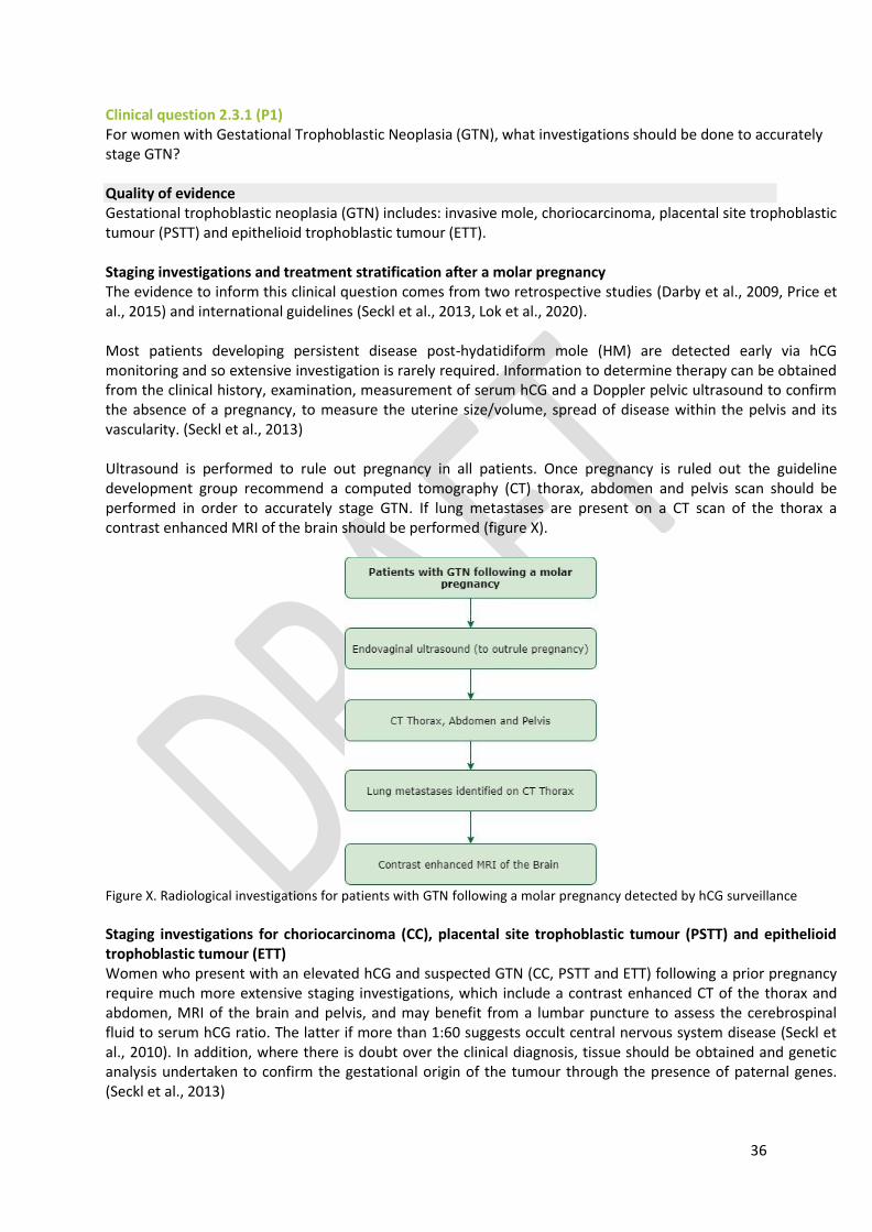
Clinical question 2.3.1 (P1) For women with Gestational Trophoblastic Neoplasia (GTN), what investigations should be done to accurately stage GTN? Quality of evidence Gestational trophoblastic neoplasia (GTN) includes: invasive mole, choriocarcinoma, placental site trophoblastic tumour (PSTT) and epithelioid trophoblastic tumour (ETT). Staging investigations and treatment stratification after a molar pregnancy The evidence to inform this clinical question comes from two retrospective studies (Darby et al., 2009, Price et al., 2015) and international guidelines (Seckl et al., 2013, Lok et al., 2020). Most patients developing persistent disease post-hydatidiform mole (HM) are detected early via hCG monitoring and so extensive investigation is rarely required. Information to determine therapy can be obtained from the clinical history, examination, measurement of serum hCG and a Doppler pelvic ultrasound to confirm the absence of a pregnancy, to measure the uterine size/volume, spread of disease within the pelvis and its vascularity. (Seckl et al., 2013) Ultrasound is performed to rule out pregnancy in all patients. Once pregnancy is ruled out the guideline development group recommend a computed tomography (CT) thorax, abdomen and pelvis scan should be performed in order to accurately stage GTN. If lung metastases are present on a CT scan of the thorax a contrast enhanced MRI of the brain should be performed (figure X).
Figure X. Radiological investigations for patients with GTN following a molar pregnancy detected by hCG surveillance
Staging investigations for choriocarcinoma (CC), placental site trophoblastic tumour (PSTT) and epithelioid trophoblastic tumour (ETT) Women who present with an elevated hCG and suspected GTN (CC, PSTT and ETT) following a prior pregnancy require much more extensive staging investigations, which include a contrast enhanced CT of the thorax and abdomen, MRI of the brain and pelvis, and may benefit from a lumbar puncture to assess the cerebrospinal fluid to serum hCG ratio. The latter if more than 1:60 suggests occult central nervous system disease (Seckl et al., 2010). In addition, where there is doubt over the clinical diagnosis, tissue should be obtained and genetic analysis undertaken to confirm the gestational origin of the tumour through the presence of paternal genes. (Seckl et al., 2013)
37
Benefit and Harm The guideline development group identified that the patient would benefit from staging and appropriate treatment. It was agreed that CT TAP as a baseline can be useful as a comparator and that the benefit of the information gained through carrying out a CT TAP outweighs the potential harm of additional radiation dose. Preferences and values The guideline development group agreed that CT TAP gives the patients peace of mind and provide reassurance that they have been accurately staged and will receive appropriate treatment. Patients may prefer to have a CT TAP as it provides reassurance. Resources and other considerations No relevant cost-effectiveness literature was identified to address this question. The guideline development group identified the following barriers to implementing the recommendations. Timely access to diagnostics The guideline development group identified the importance of access to timely diagnostics in patients with GTN as an issue. It was agreed to include the timeframe of one week from diagnosis to staging investigation in the clinical recommendation to ensure patients have access to diagnostics in a timely manner. Recommendation 2.3.1.1: Women with a diagnosis of GTN should have serum hCG monitoring, endovaginal ultrasound and a CT scan of thorax, abdomen & pelvis performed within one week of diagnosis. Quality of Evidence: Low
Grade of recommendation: Strong
Recommendation 2.3.1.2: If clinically significant lung metastases are present on a CT scan of the thorax a contrast enhanced MRI of the brain should be performed. Quality of Evidence: Low
Grade of recommendation: Strong
Good Practice Point
Investigation and management decisions should be performed by experienced professionals in the management of GTD.

38
Clinical question 2.3.2
For women with gestational trophoblastic neoplasia (GTN), what risk scoring system should be used to stage GTN? Quality of evidence The International Federation of Gynecology and Obstetrics (FIGO) reports data on GTN using anatomic staging systems (Table 2) and prognostic scoring (Table 3) (FIGO, 2009). Since 2002, all physicians treating GTN should use this system to enable the comparison of data. The prognostic score predicts the potential for developing resistance to single-drug chemotherapy with methotrexate or actinomycin D. A score of 0–6 and ≥7 indicates a low- and high-risk of resistance, respectively. The latter has almost no chance of being cured with single drug therapy and requires multi-agent treatment. The anatomical staging not only helps with determining therapy, but provides additional information to help clinicians who compare results between centres. Table 1 FIGO Anatomical Staging as adapted by FIGO (2009)
Stage I Disease confined to the uterus
Stage II GTN extends outside of the uterus, but is limited to the genital structures
Stage III GTN extends to the lungs, with or without known genital tract involvement
Stage IV All other metastatic sites
Table 2 Modified WHO prognostic scoring system as adapted by FIGO (2009)
Scores Prognostic factor 0 1 2 4
Age
<40 ≥40 - -
Antecedent pregnancy
Mole Abortion Term
Interval months from index pregnancy
<4 4-6 7-12 >12
Pre-treatment serum hCG IU/l
<103 103-104 104-105 >105
Largest tumour size (including uterus)
<3 cm 3-4 cm ≥5cm -
Site of metastases
Lung Spleen Kidney
Gastrointestinal Liver Brain
Number of metastases
- 1-4 5-8 >8
Prior failed chemotherapy - - 1 drug 2 or more drugs
Staging notation uses a Roman numeral followed by an Arabic numeral that indicate FIGO anatomic staging and the WHO modified score, respectively. Placental site trophoblastic tumour (PSTT) and Epithelioid trophoblastic tumour (ETT) are classified separately (Biscaro et al., 2015). The total score for a patient is obtained by adding the individual scores for each prognostic factor: Low-risk 0-6; high-risk ≥7. Decision making based on the risk score (i.e. choosing and administering chemotherapy) should be made by experienced professionals in this area.
Retained 2015
39
PSTT and ETT should not be scored and instead require separate classification in consultation with international experts (Biscaro et al., 2015, Seckl et al., 2013). Consideration should be given to discussing borderline patients with international experts. Some reports suggest that patients with prognostic scores of 5 or 6 may be at an increased risk of resistance to single-agent chemotherapy. In a study by Taylor et al. (2013), over half the patients defined by FIGO/WHO score as low-risk (score 0–6) had a complete response to first-line treatment with methotrexate/folinic acid (60%). However, patients with a total FIGO/WHO score of 6 or hCG level of >100,000 IU/l had significantly higher rates of resistance. Only 19% of patients with a FIGO/WHO low-risk score of 6 and 16% with an hCG level of >100,000 IU/l achieved a complete response to methotrexate/folinic acid. Research is ongoing to try to better define which “low-risk” patients may particularly benefit from primary combination chemotherapy (Sita-Lumsden et al., 2012, Taylor et al., 2013). Recommendation 2.3.2.1: Women with GTN (invasive mole, choriocarcinoma) should be assigned a FIGO score to direct management decisions of chemotherapy regimens. Quality of Evidence:
Grade of recommendation: Grade B
Good Practice Point
Placental site trophoblastic tumour and epithelioid trophoblastic tumour should not be scored using the FIGO system. They require separate classification in consultation with international experts.

40
2.4 Treatment The following are responsible for the implementation of the recommendations regarding treatment: While the CEO, General Manager and the Clinical Director of the hospital have corporate responsibility for the implementation of the recommendations in this National Clinical Guideline, each member of the multidisciplinary team is responsible for the implementation of the individual guideline recommendations relevant to their discipline.

41
Clinical question 2.4.1 (T1) For women with gestational trophoblastic neoplasia, what are the clinical indicators to diagnose GTN warranting chemotherapy? Quality of evidence Three retrospective studies (Agarwal et al., 2012, Braga et al., 2018, Braga et al., 2016) and an international guideline (ESMO - Seckl et al., 2013) addressed this clinical question. The United Kingdom indications for commencing chemotherapy are listed below and are broadly similar to those of the International Federation of Gynecology and Obstetrics (FIGO) (Kohorn, 2002). The commonest is a plateaued or rising human chorionic gonadotropin (hCG), but others include a tissue diagnosis of choriocarcinoma (CC) and spread to other organs. The United Kingdom (UK) experience indicates that the disease is also unlikely to spontaneously remit if the hCG is >20,000 IU/l one month after hydatidiform mole (HM) evacuation (also associated with an increased risk of uterine perforation) or there are lung or vaginal metastasis of >2 cm (smaller lesions may spontaneously regress) (Seckl et al., 2010). In addition, in the UK, chemotherapy is started to help stop heavy bleeding that requires transfusion even if the hCG is falling. (ESMO - Seckl et al., 2013) Recent data have suggested that surveillance is adequate for some women who continue to have a falling hCG six months after evacuation (Agarwal et al., 2014, Braga et al., 2016). However these decisions must be made on an individual patient basis following consultation with clinicians experienced in GTN management. Indications for chemotherapy following the diagnosis of GTN:
Plateaued or rising hCG after evacuation1,
Heavy vaginal bleeding or evidence of gastrointestinal or intraperitoneal haemorrhage,
Histological evidence of choriocarcinoma (except in exceptional circumstances),
Evidence of metastases in the brain, liver, or gastrointestinal tract, or radiological opacities larger than 2 cm on chest radiograph.
The following patients should be discussed on an individual basis with experienced professionals:
Women with a serum hCG of 20,000 IU/l or more, four weeks or more after evacuation, because of the risk of uterine perforation (Braga et al., 2018)
Women with a raised hCG six months after evacuation, even when hCG is still decreasing as approximately 80.2% patients will achieve spontaneous remission (Braga et al., 2016).
Benefit and Harm The guideline development group identified that patients would benefit from decisions on their treatment being made by professionals experienced in this disease. This includes the continued follow-up of patients with a decreasing hCG for six months after evacuation which may allow for avoidance of chemotherapy. Continuous follow-up of hCG may be difficult for patients as they have to continue attending their hospital/GP for regular blood tests which may have a financial implication for patients. Preferences and values In women who do need chemotherapy there is certainty that the decision is being made with clinicians experienced in this disease. In women who’s hCG continues to fall after six months continous follow-up may be difficult (emotionally,
1 * Plateaued or rising is defined as four or more equivalent values of hCG over at least three weeks (days 1, 7, 14, and 21) and three consecutive rises in hCG of 10% or greater over at least two weeks (days 1, 7, and 14), respectively.

42
logistically and financially) but avoiding chemotherapy and its associated complications is of greater value to the patient. Resources and other considerations No relevant cost-effectiveness literature was identified to address this question. Serum hCG testing hCG follow-up may have a financial implication for patients as a small number of women attend their GP for follow-up blood tests incurring personal cost. Patients may also attend an early pregnancy unit for blood tests, which patients find very distressing. The guideline development group agreed that it would be useful for the GP to receive a booklet along with a letter from the GTD centre regarding their patients care and that patients attending a regular phlebotomy unit should be facilitated where possible. Recommendation 2.4.1.1: Indications for chemotherapy following diagnosis of GTN: • Plateaued or rising hCG after evacuation, • Heavy vaginal bleeding or evidence of gastrointestinal or intraperitoneal haemorrhage, • Histological evidence of choriocarcinoma, except in exceptional circumstances, • Evidence of metastases in the brain, liver, or gastrointestinal tract, or radiological opacities of >2cm on chest x-ray. Quality of Evidence: Moderate
Grade of recommendation: Strong
Recommendation 2.4.1.2: For women who have a raised hCG six months after evacuation with a falling hCG should have their treatment plan discussed with the National Gestational Trophoblastic Disease Registry, Monitoring and Advisory Centre. Quality of Evidence: Low
Grade of recommendation: Strong
Recommendation 2.4.1.3: For women with serum hCG of ≥20,000 IU/l more than four weeks after evacuation should have their treatment plan discussed with the National Gestational Trophoblastic Disease Registry, Monitoring and Advisory Centre. Quality of Evidence: Low
Grade of recommendation: Strong
Good Practice Point
For women with histological evidence of choriocarcinoma primary surgery may be considered.
Good Practice Point
The treating physician should ensure that the patient is registered with the National GTD Registry, Monitoring and Advisory Centre.
43
Clinical question 2.4.2 (T2) For patients with low-risk (FIGO 0-6) GTN, what is the optimal first-line chemotherapy regimen? Quality of evidence Three retrospective studies (Lybol et al., 2012, Hasanzadeh et al., 2014, Taylor et al., 2013) an international guideline (Seckl et al., 2013) and experience from an expert centre (Charring Cross, 2020) addressed this clinical question. Low-risk disease is characterised by any one of the following:
FIGO stage I GTN – This is characterised as a persistently elevated human chorionic gonadotropin (hCG) level and/or tumour confined to the uterus
Stage II or III GTN with a WHO risk score 0-6. For nearly all low-risk GTN patients, single-agent chemotherapy with either methotrexate or actinomycin D is the standard treatment. A variety of regimens have been developed. The variability in regimens reflects differences in dose, frequency and route of administration as well as criteria used to select patients for therapy (Berkowitz and Goldstein, 2009). Some investigators have argued that more intense therapies given daily over 5–8 days every two weeks are superior to treatments given once every two weeks (Kohorn, 2002). Others have suggested that actinomycin D is more likely to induce remission than methotrexate. The few randomised studies to address some of these issues (Osborne et al., 2011) have been underpowered and compared regimens that are not frequently used internationally (Alazzam et al., 2011). (ESMO - Seckl et al., 2013) Importantly, in patients with persistent disease after first line therapy, usually because of resistance, can be easily treated with second and occasionally third-line chemotherapy so that the overall survival (OS) is ~100% (Lurain et al., 2012, McNeish et al., 2002, Sita-Lumsden et al., 2012). As survival is so high, it seems sensible to start with the least toxic therapy first to minimise the exposure of patients to more harmful treatments. (ESMO - Seckl et al., 2013) The methotrexate with folinic acid rescue regimen developed at Charing Cross hospital is effective, well tolerated and unlike actinomycin-D, does not induce hair loss, so methotrexate with folinic acid has been widely adopted (McNeish et al., 2002). (ESMO - Seckl et al., 2013) Non-randomised data suggest that reducing the consolidation therapy by just one cycle doubles the risk of relapse (Lybol et al., 2012). This provides justification for the current regimen of three consolidation cycles of methotrexate after hCG normalisation. In a study by Hasanzadeh et al. (2014) the efficacy of weekly IM methotrexate regimen with dose escalation in low-risk GTN was 74.3%, which is the highest rate among present studies. Additionally, this study showed that the mentioned methotrexate regimen was less effective in patients with score 5 and 6, especially score 6. Therefore, more schedules should be performed to make changes in management, therapeutic protocols, and also classification of this group. Similarly in a retrospective study carried out by Taylor et al. (2013) 173/289 patients (60%) treated with methotrexate/folinic acid achieved a complete biochemical response, while 116 patients (40%) developed resistance. Central nervous system (CNS) prophylaxis Charing Cross hospital’s policy is to give prophylaxis to low-risk patients with lung metastases. Treatment is intrathecal methotrexate (12.5mg) followed by oral folinic acid (15mg at 24 hrs) on three occasions during the first three methotrexate courses.

44
Benefit and Harm The guideline development group identified that survival rates are high with both methotrextate and actinomycin D. Treatment of patients with methotrexate is less toxic than actinomycin D and does not result in hair loss. Patients receiving actinomycin D require admission as an in-patient. Preferences and values While disease progression will occur in up to a third of patients, the guideline development group agree that it is preferred to start with the less toxic therapy, reduced side effects, hair loss and complications whilst undergoing treatment. Resources and other considerations One cost-effectiveness analysis was identified to address this question (Miller et al., 2017). Miller et al. (2017) constructed a cost effectiveness decision model from the third party payer perspective comparing weekly intramuscular methotrexate with biweekly pulsed intravenous dactinomycin (Act-D) as single-agent chemotherapy for low-risk gestational trophoblastic neoplasia (GTN). The analysis included clinical data from the Gynecologic Oncology Group (GOG) 0174 randomised trial (Osborne et al., 2011) of 240 women with low risk GTN. Costs for each arm were in 2014 US dollars and were obtained through publicly available databases. One-way sensitivity analyses were performed by varying treatment costs, rates of cure, and QOL-related utility scores. The study found that Act-D ($18,505) was more expensive compared to weekly methotrexate ($8,950) with an ICER of $56,215 per first-line treatment success compared to weekly methotrexate. Small decreases in QOL dramatically increased the ICER during sensitivity analysis. Models with multi-day methotrexate regimens were also more cost-effective than Act-D. If effectiveness was redefined as avoidance of multi-agent chemotherapy, weekly MTX was more effective. The guideline development group identified the following barriers to implementation of the recommendations: Patients diagnosed with GTN should have access to a designated point of contact in the treatment centre who should also be in contact with the National GTD Registry, Monitoring and Advisory Centre. Recommendation 2.4.2.1: Patients with a FIGO score of 0-6 can be treated with either single-agent methotrexate with or without folinic acid, or actinomycin-D. Taking into account the treatment cycles, potential complications and quality of life the guideline development group agreed that methotrexate is the preferred first line chemotherapy. Quality of Evidence: Moderate
Grade of recommendation: Strong
Recommendation 2.4.2.2: Chemotherapy for low-risk disease should be continued for three cycles of maintenance treatment after hCG normalisation. Quality of Evidence: Moderate
Grade of recommendation: Strong

45
Good Practice Point
In patients with low risk GTN serum hCG/blood should be done on day one of each chemotherapy cycle or more frequently if required. Normalisation of hCG values should be assay specific.
Practical considerations around patient care
Patients diagnosed with GTN should have access to counselling and support from the nurses in the National GTD Registry, Monitoring and Advisory Centre.
Patients diagnosed with GTN should have access to a liasion nurse or designated key contact in their treatment centre who should also be in contact with the National GTD Registry, Monitoring and Advisory Centre.

46
Clinical question 2.4.3 (T3) For women with high-risk (FIGO ≥7) GTN, what is the optimal first-line chemotherapy regimen? Quality of evidence High-risk gestational trophoblastic neoplasia (GTN) is characterised by any one of the following: • Stage IV disease • Stage II and III with risk score ≥7. A Cochrane meta-analysis (Deng et al., 2013), a retrospective study (Alifrangis et al., 2013) and experience from an international centre Charing Cross (2019) addresed this clinical question. EMA/CO (etoposide, methotrexate, actinomycin-D plus cyclophosphamide and vincristine) is currently the most widely used first-line combination chemotherapy for high-risk GTN, although this regimen has not been rigorously compared to other combinations such as MAC (methotrexate, actinomycin-D, cyclophosphamide or chlorambucil) or FAV (5-FU, actinomycin D, and vincristine) in randomised controlled trials. Other regimens may be associated with less acute toxicity than EMA/CO; however, proper evaluation of these combinations in high quality RCTs that include long-term surveillance for secondary cancers is required. Given the low incidence of GTN, RCTs in this field are difficult to conduct, hence multi-centre collaboration is necessary. CHAMOCA (cyclophosphamide, hydroxyurea, actinomycin D, methotrexate, doxorubicin, melphalan and vincristine) is not recommended for GTN treatment as it is more toxic and not more effective than MAC. (Deng et al., 2013) A recent retrospective study by Alifrangis et al. (2013) demonstrated that during the period 1995 to 2010, overall survival for all patients with GTN treated with EMA/CO chemotherapy significantly increased from 86.2% before 1995 to 97.9%. EP induction chemotherapy was given to 23.1% of high-risk patients (33 of 140 patients) with a large disease burden, and the early death rate was only 0.7% (n = 1; 95% CI, 0.1% to 3.7%) compared with 7.2% (n = 11 of 151 patients; 95% CI, 4.1% to 12.6%) in the pre-1995 cohort. However, high-risk patients receiving EP, compared with patients not receiving EP, did have a higher but not statistically significant relapse rate (9% v 6%, respectively; P = .44) and death rate (12% v 4%, respectively; P = .088). Central nervous system (CNS) prophylaxis Charing Cross hospital’s policy is to give prophylaxis to all high-risk patients. Treatment is intrathecal methotrexate (12.5mg) followed by oral folinic acid (15mg at 24 hrs) on three occasions during the first three methotrexate courses, which usually coincides with the CO treatment. (Charring Cross, 2020). Benefit and Harm The guideline development group identified that commencing treatment with EMA/CO provides a greater opportunity for curative treatment and is consistent with Charing Cross Hospital and other European GTD centres, this treatment regimen does not have a significant impact on fertility. Following the use of EMA/CO there can be treatment related toxicity including inducing menopause on average three years early. Preferences and values The guideline development group agreed that patients would choose the multi-agent chemotherapy due to the greater opportunity for curative treatment. Resources and other considerations No relevant cost-effectiveness literature was identified to address this clinical question
47
Recommendation 2.4.3.1: Patients with a FIGO score of ≥7 should receive multi-agent chemotherapy and most centres now use EMA/CO, as it is highly effective. Quality of Evidence: Moderate
Grade of recommendation: Strong
Recommendation 2.4.3.2: Early deaths in ultra-high-risk GTN (FIGO score ≥12) can be reduced by induction therapy with etoposide and cisplatin. Such patients may also benefit from substitution of EMA/CO with EP/EMA. Quality of Evidence: Moderate
Grade of recommendation: Strong
Good Practice Point
For women with high-risk GTN, decisions should be made on an individual patient basis following discussion with clinicians experienced in high-risk GTN management at a GTD Centre.
Good Practice Point
Registration of patients at the National GTD Registry, Monitoring and Advisory Centre is a minimum standard of care.
Practical considerations around patient care
Patients should be informed that their treatment will require in-patient care.
Patients with high risk GTN (FIGO score of ≥7) should have access to a liaison nurse or designated key contact locally who is in contact with the National GTD Registry, Monitoring and Advisory Centre.
Patients diagnosed with GTN should have access to a to a liaison nurse or designated key contact in their treatment centre who should also be in contact with the National GTD Registry, Monitoring and Advisory Centre.

48
Clinical question 2.4.4 For women with low-risk gestational trophoblastic neoplasia undergoing chemotherapy (first-course), what is the recommended course of action for observing and managing bleeding? Quality of evidence The guideline development group recommends that the first one/two courses of chemotherapy should be administered as an inpatient at a centre with medical oncology, gynaecological services and interventional radiology. Subsequent courses in uncomplicated patients are administered at a medical oncology day ward facility. If hCG levels are very high, the uterine mass large or there is evidence of vaginal metastases, patients may be kept in for two complete courses or longer due to the risk of haemorrhage (Seckl and Savage, 2012). Per vaginal or intraperitoneal bleeding can occur. Moderate bleeding usually responds to bed rest and chemotherapy. Torrential bleeding may require treatment with a vaginal pack, blood products, anti-fibrinolytics, emergency embolisation and very rarely with hysterectomy. In Charing Cross experience, less than 1.5% of GTN patients have required one of these interventions over the past 25 years (Charring Cross, 2020). Recommendation 2.4.4.1: For women with low-risk GTN undergoing first-line chemotherapy, the first ± second courses of chemotherapy may need to be administered as an in-patient at a centre with medical oncology, gynaecological services and interventional radiology. Grade of recommendation: Grade C
Retained 2015

49
Clinical question 2.4.5 (T4) For women with gestational trophoblastic neoplasia, what are the appropriate investigations to monitor response to chemotherapy and follow-up? Quality of evidence Monitoring response to chemotherapy – Low-Risk Patients should have hCG levels measured prior to their next chemotherapy cycle (Lok et al., 2020). Treatment is continued until hCG is normal and then usually for three further courses to eliminate any residual tumour cells and to minimise the chances of relapse. Non-randomised data suggest that reducing the consolidation therapy by just one cycle doubles the risk of relapse. Monitoring response to chemotherapy – High-Risk Patients should have hCG levels measured prior to their next chemotherapy cycle (Lok et al., 2020). Therapy is continued for 6 weeks of normal hCG values or 8 weeks if poor prognostic features such as liver or brain metastases are present. Patients are then re-imaged to document the post-treatment appearance for future comparison. Removal of residual masses is unnecessary as it does not reduce the risk of recurrence which is less than 3% (Seckl et al., 2010). (ESMO - Seckl et al., 2013) Follow-up of patients post chemotherapy After remission is achieved, serum hCG should be measured fortnightly for six months after consolidation therapy then monthly for a further six months and every two months for two years (Balachandran et al., 2019) Follow-up for at least 5 years may be considered for those at highest risk. Benefit and Harm The guideline development group agreed that patients would benefit from having their hCG monitored during and following treatment as it allows for timely treatment management. Continuous follow-up of hCG may be difficult for patients as they have to continue attending their hospital/GP for regular blood tests and there may be a delay in trying for a subsequent pregnancy. Preferences and values The guideline development group agreed that the value of autonomy around starting a family outweighs the benefit of follow up for a rare recurrence. Resources and other considerations No relevant cost-effectiveness literature was identified to address this clinical question. The guideline development group identified the following barriers to implementing the recommendations: Centralisation of hCG testing – the group identified that patients may benefit from urine assays as this would allow for a longer follow up without causing patients to undergo blood tests. The issue of centralisation of hCG testing is covered in Clinical question 2.2.7 (N2). The guideline development group agreed that the recommendations were suitable for audit. Recommendation 2.4.5.1: Monitoring during treatment low-risk: Patient should have human chorionic gonadotropin (hCG) levels prior to their next chemotherapy cycle. Treatment is continued until hCG is normal and for three further consolidation cycles. Quality of Evidence: Low
Grade of recommendation: Strong
50
Recommendation 2.4.5.2: Monitoring during treatment high-risk: Patient should have human chorionic gonadotropin (hCG) levels prior to their next chemotherapy cycle. Patients with high-risk disease should have maintenance therapy for three cycles after hCG normalisation extended to four cycles for patients with poor prognostic features such as liver metastases with or without brain metastases. Quality of Evidence: Low
Grade of recommendation: Strong
Recommendation 2.4.5.3: Follow-up post treatment: After remission is achieved, serum hCG should be measured fortnightly for six months then monthly for a further six months and every two months for two years. Quality of Evidence: Low
Grade of recommendation: Strong
Practical considerations around patient care
Patients should have access to a nurse or designated key contact with experience treating GTN that can provide advice, written information and support to patients before commencing treatment.
Patients should be provided with clear written information in patient friendly language that they can share with family.

51
Clinical question 2.4.6 For women with gestational trophoblastic neoplasia what are the indicators to determine switching treatments from first-line chemotherapy? Quality of evidence Chemotherapy should continue until hCG returns to normal, and at least three more chemotherapy cycles should be administered after the first normal hCG result (Lybol et al., 2012). The drug in use should be replaced by another when there is an inadequate response i.e. a rise in hCG values over two successive measurements a week apart or a plateau in three successive weekly measurements a week apart or when toxicity (such as mucositis, pleuritic chest pain or abdominal pain) precludes the use of appropriate doses or treatment frequency. About 5% of patients with low-risk GTN without metastases and 10-15% of those that have metastases develop resistance to first-line chemotherapy (Lurain and Nejad, 2005). (Biscaro et al., 2015) Resistance to chemotherapy and recurrent disease are more frequent in patients with high risk GTN (Berkowitz and Goldstein, 2013). About 20-30% of high-risk patients have an incomplete response to first-line chemotherapy or recurrence after remission and eventually need salvage chemotherapy. (Biscaro et al., 2015) Recommendation 2.4.6.1: For patients with low-risk GTN the clinical indicators for a change in treatment from first-line chemotherapy include: treatment related toxicity, a rise in hCG values over two successive treatments. Grade of recommendation: Grade C
Good Practice Point
Consideration could be given to re-staging patients prior to the initiation of a new regimen (particularly high-risk patients).
Retained 2015

52
Clinical question 2.4.7 (T5) For women with low-risk gestational trophoblastic neoplasia who have not responded or have relapsed from single agent treatment (methotrexate or actinomycin D) or have relapsed following normalisation of hCG after completion of single agent treatment, what is the next line treatment? Quality of evidence First line methotrexate The next line of treatment is determined by the patient’s current hCG levels, with those with hCG levels <1,000 IU/l receiving single-agent Actinomycin-D and those with hCG levels of >1,000 IU/l commencing on EMA–CO (Lok et al., 2020). For women with low-risk GTN if sequential single-agent therapy fails, multi-agent chemotherapy must be used to achieve a cure; this is necessary in 6% to 15% of cases (Covens et al., 2006, Goldstein and Berkowitz, 2012). The multi-agent therapy used most frequently at Charing Cross (one of two treatment centres in the UK) is EMA/CO. The New England Trophoblastic Disease Centre (NETDC, USA) prefers to use MAC before EMA/CO owing to concerns that etoposide may be associated with an increased risk of secondary tumours (Goldstein and Berkowitz, 2012). (Alazzam et al., 2016) In Ireland the guideline development group recommends the use EMA/CO as first-line combination therapy. All patients should be registered at the National GTD Registry, monitoring and advisory centre. Patients should be treated under the care of a medical oncologist with experience in the treatment of GTN. A retrospective study demonstrated an overall survival rate of 99.6%, in 250 low risk patients who received second-line EMA/CO after relapse or resistance to single-agent chemotherapy. Four patients (1.5%) developed resistance and/or experienced relapse after EMA/CO. These patients were all cured with further salvage regimens (Alifrangis et al., 2013). Once normalistion of hCG has occured on EMA/CO treatment with etoposide can be discontinued from the regimen to reduce the risk of secondary malignancies. (Charring Cross, 2020) Central nervous system (CNS) prophylaxis Charing Cross hospital’s policy is to give prophylaxis to all high-risk patients and to the low-risk patients with lung metastases. Treatment is intrathecal methotrexate (12.5mg) followed by oral folinic acid (15mg at 24 hrs) on three occasions during the first three methotrexate courses. For the high-risk patients it usually coincides with the CO treatment (Charring Cross, 2020). Benefit and Harm The guideline development group agreed that the patients who have not responded to methotrexate with a hCG <1,000 IU/L would benefit from treatment with actinomycin-d as they could avoid treatment with multi-agent therapy including etoposide, as etoposide can increase the risk of secondary malignancies. Patients with low-risk GTN who have not responded to methotrexate with a hCG >1,000 IU/L would benefit from treatment with EMA/CO as it reduces the risk of CNS involvement. Preferences and values The use of a less toxic treatment initially reserves the more toxic treatment for the minority of people who may need it. This will avoid unnecessary treatment related toxicities for patients. Resources and other considerations No relevant cost-effectiveness studies were identified to address this clinical question.
53
Recommendation 2.4.7.1: For women with low-risk GTN who have not responded to methotrexate with a hCG <1,000 IU/L the next line of treatment is actinomycin D. Quality of Evidence: Low
Grade of recommendation: Strong
Recommendation 2.4.7.2: For women with low-risk GTN who have not responded to methotrexate with a hCG >1,000 IU/L the next line of treatment is EMA/CO. Quality of Evidence: Low
Grade of recommendation: Strong
Recommendation 2.4.7.3: For women with low-risk GTN who have not responded or have relapsed from sequential single-agent treatment the next line of treatment is combination chemotherapy with EMA/CO. Quality of Evidence: Low
Grade of recommendation: Strong
Good Practice Point
Once normalistion of hCG has occured on EMA/CO treatment etoposide can be discontinued from the regimen to reduce the risk of secondary malignancies.

54
Clinical question 2.4.8 (T6) For women with high-risk GTN who have not responded to first-line treatment, what is second-line treatment? Quality of evidence In women with high-risk GTN who have not responded to first-line treatment, consideration should be given to discussing each individual case with an international expert due to the rarity of this condition. Currently, the most commonly used salvage regimen in North America and the UK for the treatment of resistant or recurrent high-risk GTN is EMA/EP (May et al., 2011). A Cochrane review conducted by Alazzam et al., (2016) stated that approximately 90% of high-risk patients treated initially with EMA/ CO, followed by salvage therapy with a platinum-etoposide combination if required, will survive (Lurain et al., 2010). In three series of EMA/EP salvage treatment following EMA/CO treatment failure, cure rates of 75% (nine out of 12 women; (Newlands et al., 2000)) 66.6% (12 out of 18 women; (Mao et al., 2007)) and 84.9% (11 out of 13 women; (Lu et al., 2008)) were reported; however, EMA/EP was associated with significant myelosuppression and hepatotoxicity, leading to treatment delays and dose reductions. Myelosuppression may be minimised by administering granulocyte-colony stimulating factor (G-CSF) (El-Helw et al., 2005, Lurain and Nejad, 2005, Seckl et al., 2010). (Alazzam et al., 2016) An alternative to EP/EMA is TE/TP (paclitaxel/cisplatin and paclitaxel/etoposide). The taxane containing regimen was found to be associated with comparable cure rates to EMA/EP (70% of 10 patients who had not been exposed to previous EP treatment were cured) but with relatively reduced toxicity and no dose delays or reductions (Alazzam et al., 2016). Central nervous system (CNS) prophylaxis Charing Cross hospital’s policy is to give prophylaxis to all high-risk patients and to the low-risk patients with lung metastases. Treatment is intrathecal methotrexate (12.5mg) followed by oral folinic acid (15mg at 24 hrs) on three occasions during the first three methotrexate courses. For the high-risk patients it usually coincides with the CO treatment. Benefit and Harm The second line treatment for high risk patients is associated with toxicity including myelosuppression and hepatotoxicity. However, salvage treatment has a 90% cure rate, therefore the benefit of this second-line treatment outweighs the harm and toxicities associated with it. Preferences and values The guideline development group agreed that patients would choose the second line chemotherapy due to the greater opportunity for curative treatment. Resources and other considerations No relevant cost-effectiveness literature was identified to address this clinical question. Recommendation 2.4.8.1: For women with high-risk GTN who have not responded to first-line treatment second line treatment is EP/EMA, if patients do not respond to EP/EMA TE/TP is the recommmended third line treatment. Quality of Evidence: Low
Grade of recommendation: Strong

55
Recommendation 2.4.8.2: In women with high-risk GTN who have not responded to first-line treatment, discussions of each individual case at a GTD MDM should be considered. Quality of Evidence: Very Low
Grade of recommendation: Strong
Good Practice Point
All women with GTN should be registered at the National GTD Registry, monitoring and advisory centre.
Good Practice Point
Given the rarity of this condition consideration should be given to discussing each individual case with international experts.

56
Clinical question 2.4.9 (T7) For women with GTN, who are acutely ill with liver, brain or lung metastasis at presentation, what is the optimum chemotherapy regimen? Quality of evidence Given the rarity of this condition consideration should be given to discussing each individual case with international experts. Emergency treatment Patients who are acutely unwell from liver or CNS disease and particularly those with large lung metastases who are at risk of respiratory failure should be admitted and emergency chemotherapy started as soon as possible as these patients can deteriorate rapidly (Charring Cross, 2020). Chemotherapy can be started with two day EP. This can be repeated weekly and then altered to EMA/CO or EP/EMA at a later point. Please refer to the NCCP Chemotherapy Regimen (NCCP regimen code: 00267) for more information.
Hepatic metastases Patients with hepatic metastases at presentation are usually diagnosed with ultra high risk disease (FIGO >12) and therefore should commence therapy using EP/EMA protocol (Charring Cross, 2020). Please refer to the NCCP Chemotherapy Regimen (NCCP regimen code: 00264) for more information. Cerebral metastases The Charing Cross hospital’s treatment for this is the high dose EMA (CNS)/CO, using an increased methotrexate dose (1gm/m2) combined with longer folinic acid (FA) rescue. In CNS disease the EMA (CNS)/ CO chemotherapy is continued for eight weeks after the human chorionic gonadotropin (hCG) normalisation (Charring Cross, 2020). Intrathecal methotrexate is also given as 12.5mg +15mg FA on the EMA week until serum hCG is normal at which point it is discontinued. Please refer to the NCCP Chemotherapy Regimen (NCCP regimen code: 00249) for more information. In emergency situations with cerebral metastases, hi-dose dexamethasone is given followed by two day EP as above (Charring Cross, 2020). Hepatic and synchronous cerebral metastases In patients with liver and brain metastases the treatment used should be as follows:

57
This combines the EMA (CNS) dose with the EP treatment. It misses out the day two of the normal EMA protocol as it is too myelosuppressive when combined with EP to allow for this. We would use G-CSF (granulocyte – colony stimulating factor) for 3-4 days every week in between day 1 and 8 and day 8 and 1. Please refer to the NCCP Chemotherapy Regimen (NCCP regimen code: 00264) for more information. Intrathecal methotrexate is also given 12.5mg + 15mg FA on the EP week until serum hCG is normal at which point it is discontinued (Charring Cross, 2020, Savage et al., 2015) Respiratory failure In patients with large volume pulmonary lung metastases oxygen support can be given but ventilation is contraindicated, due to the risk of traumatic haemorrhage from the tumour vasculature. Respiratory compromise can also result from tumour within the pulmonary vasculature. This can respond promptly to chemotherapy. Consideration can be given to anti-coagulation in these rare patients with tumour emboli (Charring Cross, 2020). Interventional radiology Consideration could be given to radiological embolization of lesions complicated by intractable bleeding. Benefit and Harm The urgent need for therapy outweighs potential side effects of treatment. Preferences and values The guideline development group agreed that patients would choose the opportunity for curative treatment. Resources and other considerations No relevant cost-effectiveness literature was identified to address this clinical question. Recommendation 2.4.9.1: Emergency treatment Patients who are acutely unwell from liver or CNS disease and particularly those with large lung metastases who are at risk of respiratory failure should be admitted and emergency chemotherapy commenced as soon as possible. Quality of Evidence: Very Low
Grade of recommendation: Strong

58
Recommendation 2.4.9.2: Hepatic metastases Patients with hepatic metastases at presentation should commence therapy using EP/EMA protocol. Quality of Evidence: Very Low
Grade of recommendation: Strong
Recommendation 2.4.9.3: Cerebral metastases Patients with cerebral metastases should be treated with EMA(CNS)/CO. Quality of Evidence: Very Low
Grade of recommendation: Strong
Recommendation 2.4.9.4: Hepatic and synchronous cerebral metastases Patients with liver and brain metastases should be treated with a combination of EMA (CNS) and EP. Quality of Evidence: Very Low
Grade of recommendation: Strong
Practical considerations around patient care
Patients should be provided with reassurance that they are being managed in co-operation with the National GTD Registry, Monitoring and Advisory Centre.
Patients should be counselled and reassured of the high cure rate of this patient cohort.

59
3 Development of a National Clinical Guideline 3.1 Epidemiology To be added closer to publication date 3.2 Rationale for this National Clinical Guideline The National Cancer Strategy 2017-2026 (DoH, 2017) recommends: The NCCP will develop further guidelines for cancer care in line with National Clinical Effectiveness Committee (NCEC) standards. In November 2015, the NCEC published the first iteration ‘Diagnosis, staging and treatment of patients with gestational trophoblastic disease - National Clinical Guideline No. 13’. The purpose of updating this guideline is to continue to improve the quality of care delivered to patients.
3.3 Aim and objective The overall objectives of the National Clinical Guideline No. 13 ‘Diagnosis, staging and treatment of patients with GTD’ are:
To improve the quality of clinical care,
To prevent variation in practice,
To address areas of clinical care with new and emerging evidence,
Based on the best research evidence in conjunction with clinical expertise,
Developed using a clear evidence-based internationally used methodology. The guideline is based on the best research evidence in conjunction with clinical expertise, patient preferences and is developed using a clear evidence-based internationally used methodology. 3.4 Financial impact of GTD The diagnosis, staging, and treatment of patients with GTD requires multidisciplinary care in an acute hospital setting. The majority of patients will require diagnostic tests (radiology, pathology) and depending on the treatment plan may require surgery and chemotherapy. The guideline development group identified the issue of serum hCG testing for patients. hCG follow-up may have a financial implication for patients as a small number of women attend their GP for follow-up blood tests incurring personal cost. The guideline development group agreed that it would be useful for the GP to receive a booklet along with a letter from the GTD centre regarding their patients care. This is included in Appendix 4: Economic assessment and Appendix 5: Implementation plan. The establishment of the National GTD Registry, Monitoring and Advisory Centre has aided in the identification of the volume of patients with GTD in the country, which has informed costs as outlined in Appendix 4: Economic assessment.

60
3.5 Guideline scope 3.5.1 Target population Patients that are covered by this guideline are: Women who have had a miscarriage, any woman who has had a molar pregnancy, any woman with unexplained elevated hCG, any woman presenting with metastatic disease of uncertain origin where the hCG is elevated, and any woman with atypical placental site nodules.
3.5.2 Target audience This guideline is intended for all health professionals involved in the diagnosis, staging and treatment of patients with GTD, such as gynaecologists, radiologists, pathologists, biochemists, surgeons, medical oncologists, GPs and nursing staff. While the CEO, General Manager and the Clinical Director of the hospital have corporate responsibility for the implementation of the recommendations in this Clinical Guideline, each member of the multidisciplinary team is responsible for the implementation of the individual guideline recommendations relevant to their discipline.
This guideline is also relevant to those involved in clinical governance, in both primary and secondary care, to help ensure that arrangements are in place to deliver appropriate care for the population covered by this guideline.
Whilst the guideline is focused on clinical care, it is expected to be of interest to patients with GTD and their significant others. A list of medical abbreviations used throughout the guideline can be found in Appendix 8: Glossary of terms and abbreviations. 3.6 Conflict of interest statement A conflict of interest form developed by the NCEC was signed by all Guideline Development Group members and reviewers. The Guideline Development Group was managed by the Chair to promote the highest professional standard in the development of this guideline. Where a conflict arises a Guideline Development Group member absents themselves from discussion pertaining to their area of conflict. 3.7 Source of funding The guideline was commissioned and funded by the NCCP however the guideline content was not influenced by the NCCP or any other funding body. This process was fully independent of lobbying powers. All recommendations were based on the best research evidence integrated with clinical expertise.
3.8 Guideline methodology and literature review To be added closer to publication date
3.9 Consultation process The draft guideline was signed off by the entire Guideline Development Group and the NCCP Guideline Steering Group before going to national stakeholder review. It was placed on the NCCP website and circulated to relevant organisations and individuals for comment between XXXX. A full list of those invited to review this guideline is available in Appendix 3: Details of consultation process. Stakeholders were asked to comment on the comprehensiveness of evidence used to form the recommendations. Stakeholders were required to submit feedback with supporting evidence on a form provided along with a completed conflict of interest form. A time-period of six weeks was allocated to submit comments. 3.10 External review The draft guideline was also submitted for international expert review. The Guideline Development Group nominated Prof. Michael Seckl and Prof. Francois Golfier, as International reviewers to provide feedback on the

61
draft guideline. These reviewers were chosen by the Guideline Development Group based on their in-depth knowledge of the subject area and guideline development processes. The review followed the same procedure as the National Stakeholder Review. The guideline was circulated for comment between the XXXX and XXXX. All feedback received was reviewed by the guideline development group. Suggested amendments and supporting evidence were reviewed by the guideline development group and consensus reached to accept or reject the amendments. All modifications were documented and the report is available upon request.
3.11 Implementation The implementation plan (Appendix 5: Implementation plan) was developed based on the NCEC Implementation guide (DoH, 2018). The implementation plan outlines the actions required to implement the recommendations, who has lead responsibility for delivering the action, the timeframe for completion and the expected outcomes of implementation (Appendix 5: Implementation plan). This National Clinical Guideline including the implementation plan should be reviewed by the multidisciplinary team and senior management in the hospital which outlines the actions required to implement the recommendations. The CEO, General Manager and the Clinical Director of the hospital have corporate responsibility for the implementation of the National Clinical Guideline and to ensure that all relevant staff are appropriately supported to implement the guideline. All medical staff with responsibility for the care of patients with gestational trophoblastic disease are required to:
• Comply with this National Clinical Guideline and any related procedures or protocols. • Adhere to their code of conduct and professional scope of practice guidelines as appropriate to their
role and responsibilities. • Maintain their competency in the management and treatment of patients with gestational
trophoblastic disease. The National Clinical Guideline will be circulated and disseminated through the professional networks who participated in developing and reviewing this document. The guideline will also be available via the NCEC and NCCP websites. A summary of tools to assist in the implementation of this National Clinical Guideline is available in Appendix 6: Supporting tools. The following documents are also available on the NCCP website:
3.12 Monitoring and audit It is important that both the implementation of the guideline and patient outcomes are audited to ensure that this guideline positively impacts on patient care. For audit criteria see Appendix 7: Monitoring and audit. 3.13 Recommendations for research 3.14 Systematic review of cost-effectiveness As part of the systematic literature review any health economic literature identified in the literature search for each clinical question was included and was critically appraised. Critical appraisal was carried out using the Consensus on Health Economic Criteria (CHEC)-list quality appraisal tool (Evers et al., 2005). The studies were also evaluated for applicability to the Irish setting using the International Society for Pharmacoeconomics

62
(ISPOR) questionnaire (Jaime Caro et al., 2014). A narrative synthesis of any cost-effectiveness literature identified was included in the evidence summary under the heading ‘Resources and other considerations’ and were taken into account when formulating recommendations. 3.15 Budget impact analysis Any potential barriers or resource implications of implementing the recommendations were identified by the clinicians during meetings to discuss and develop the clinical recommendations. The potential barriers and resource implications identified for each clinical question is described in detail in Appendix 4: Economic assessment. 3.16 Plan to update this National Clinical Guideline This guideline was issued in xxxxx 2021 and will be considered for review by the NCCP in three years. Surveillance of the literature base will be carried out periodically by the NCCP. Any updates to the guideline in the interim period or as a result of three year review will be subject to the NCEC approval process and noted in the guidelines section of the NCCP and NCEC websites.

63
4 Appendices Appendix 1: Guideline Development Group terms of reference and logic model Membership of the Guideline Development Group is outlined at the beginning of this document. Terms of Reference The terms of reference of the Guideline Development Group was to update National Clinical Guideline No. 13 - Diagnosis, staging and treatment of patients with gestational trophoblastic disease. To integrate the best research evidence with clinical expertise and patient values. To provide guidance to clinicians in relation to the diagnosis, staging and treatment of patients. Full terms of reference are available upon request.
External Reviewers
The guideline development group would like to acknowledge Prof. Michael Seckl and Prof. Francois Golfier for sharing their expertise and their commitment in reviewing this guideline. Conflict of interest declarations
A conflict of interest form developed by the NCEC was signed by all Guideline Development Group members and reviewers. The Guideline Development Group was managed by the Chair to promote the highest professional standard in the development of this guideline. Where a conflict arises a Guideline Development Group member absents themselves from discussion pertaining to their area of conflict. No conflicts were declared in the development of this national clinical guideline.
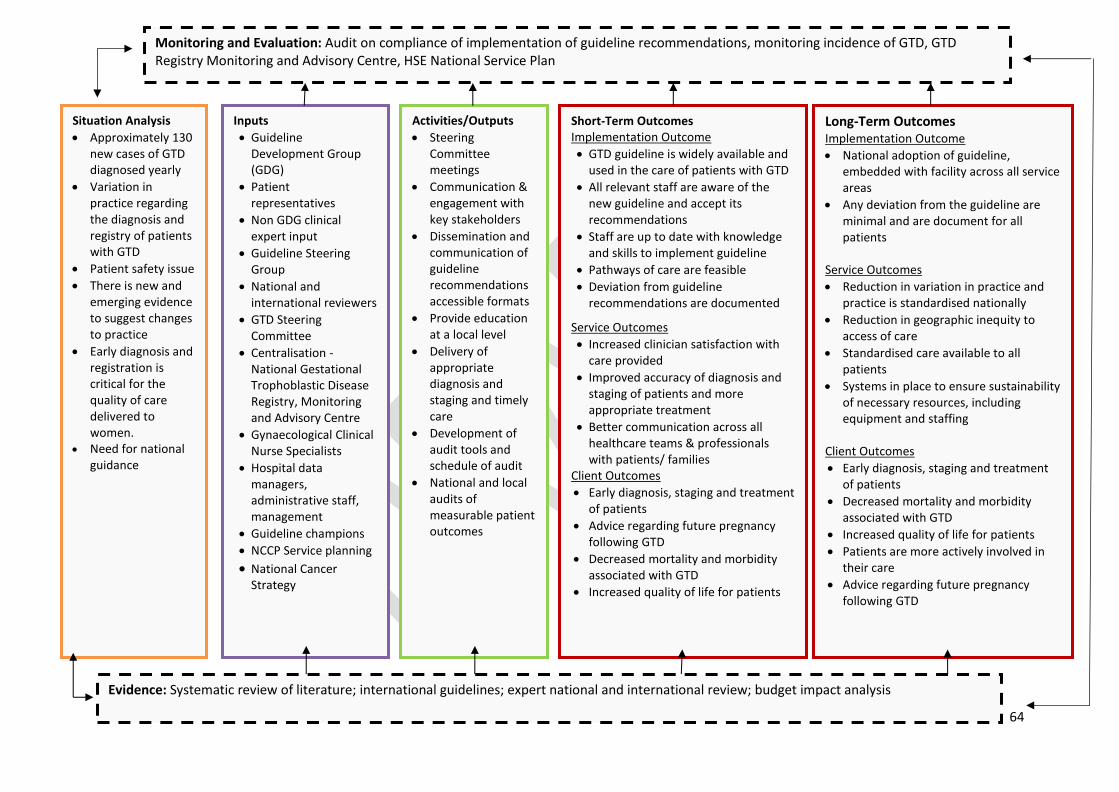
64
Monitoring and Evaluation: Audit on compliance of implementation of guideline recommendations, monitoring incidence of GTD, GTD Registry Monitoring and Advisory Centre, HSE National Service Plan
Situation Analysis
Approximately 130 new cases of GTD diagnosed yearly
Variation in practice regarding the diagnosis and registry of patients with GTD
Patient safety issue
There is new and emerging evidence to suggest changes to practice
Early diagnosis and registration is critical for the quality of care delivered to women.
Need for national guidance
Inputs
Guideline Development Group (GDG)
Patient representatives
Non GDG clinical expert input
Guideline Steering Group
National and international reviewers
GTD Steering Committee
Centralisation - National Gestational Trophoblastic Disease Registry, Monitoring and Advisory Centre
Gynaecological Clinical Nurse Specialists
Hospital data managers, administrative staff, management
Guideline champions
NCCP Service planning
National Cancer Strategy
Activities/Outputs
Steering Committee meetings
Communication & engagement with key stakeholders
Dissemination and communication of guideline recommendations accessible formats
Provide education at a local level
Delivery of appropriate diagnosis and staging and timely care
Development of audit tools and schedule of audit
National and local audits of measurable patient outcomes
Short-Term Outcomes Implementation Outcome
GTD guideline is widely available and used in the care of patients with GTD
All relevant staff are aware of the new guideline and accept its recommendations
Staff are up to date with knowledge and skills to implement guideline
Pathways of care are feasible
Deviation from guideline recommendations are documented
Service Outcomes
Increased clinician satisfaction with care provided
Improved accuracy of diagnosis and staging of patients and more appropriate treatment
Better communication across all healthcare teams & professionals with patients/ families
Client Outcomes
Early diagnosis, staging and treatment of patients
Advice regarding future pregnancy following GTD
Decreased mortality and morbidity associated with GTD
Increased quality of life for patients
Long-Term Outcomes Implementation Outcome
National adoption of guideline, embedded with facility across all service areas
Any deviation from the guideline are minimal and are document for all patients
Service Outcomes
Reduction in variation in practice and practice is standardised nationally
Reduction in geographic inequity to access of care
Standardised care available to all patients
Systems in place to ensure sustainability of necessary resources, including equipment and staffing
Client Outcomes
Early diagnosis, staging and treatment of patients
Decreased mortality and morbidity associated with GTD
Increased quality of life for patients
Patients are more actively involved in their care
Advice regarding future pregnancy following GTD
Evidence: Systematic review of literature; international guidelines; expert national and international review; budget impact analysis

65
Appendix 2: Systematic Literature Review Protocol
Literature searches to answer clinical questions identified by the relevant tumour group will be conducted using the following procedure. Questions should only be submitted if they have not been adequately answered in the guidelines adopted by the tumour group, or where guidelines need to be updated. Guidelines should be identified in consultation with library services.
Tumour Group
1 PICO(T) Analyse the clinical question using PICO(T) and complete a Clinical Query Request form. See below Appendix 1: Clinical Query Request.
Tumour Group
or Library
Services
2 Question Category
Assign a question category, if appropriate:
Therapy/Intervention Aetiology/Risk Factors Diagnosis
Prognosis/Prediction Frequency/Rate Phenomena Other
Library Services
3 Literature Search
Conduct searches of the following bibliographic databases in the order specified below using keywords implicit in the PICO(T) strategy and any identified subject headings:
Cochrane 3.1 Cochrane Library Comprising: the Cochrane Database of Systematic Reviews; the Cochrane Central Register of Controlled Trials (Central); the Database of Abstracts of Reviews of Effects; the Health Technology Assessment Database; the NHS Economic Evaluation Database. Use MeSH and keyword searches to identify systematic reviews and other relevant studies.
Point-of-Care 3.2 Point-of-Care Reference Tools One or more of the following point-of-care reference tools: BMJ Best Practice; DynaMed; UpToDate.
Medline 3.3 Medline Use MeSH and keyword searches. Limit results using the ‘Human’ search filter. Unless otherwise specified by the tumour group or warranted by the specific clinical question, limit results to studies from the previous 5 years. Where appropriate, limit intervention questions according to the following priority: Medline clinical queries; Cochrane systematic reviews; other systematic reviews or meta-analyses; RCTs; systematic reviews of cohort or cross-sectional studies; cohort or cross-sectional studies; general Medline or other sources. Where appropriate, limit diagnosis, prognosis or aetiology questions according to the following priority: Medline clinical queries; systematic reviews of cohort or cross-sectional studies; cohort or cross-sectional studies; general Medline or other sources.
Embase 3.4 Embase Repeat the Medline search strategy above using Embase, if available.
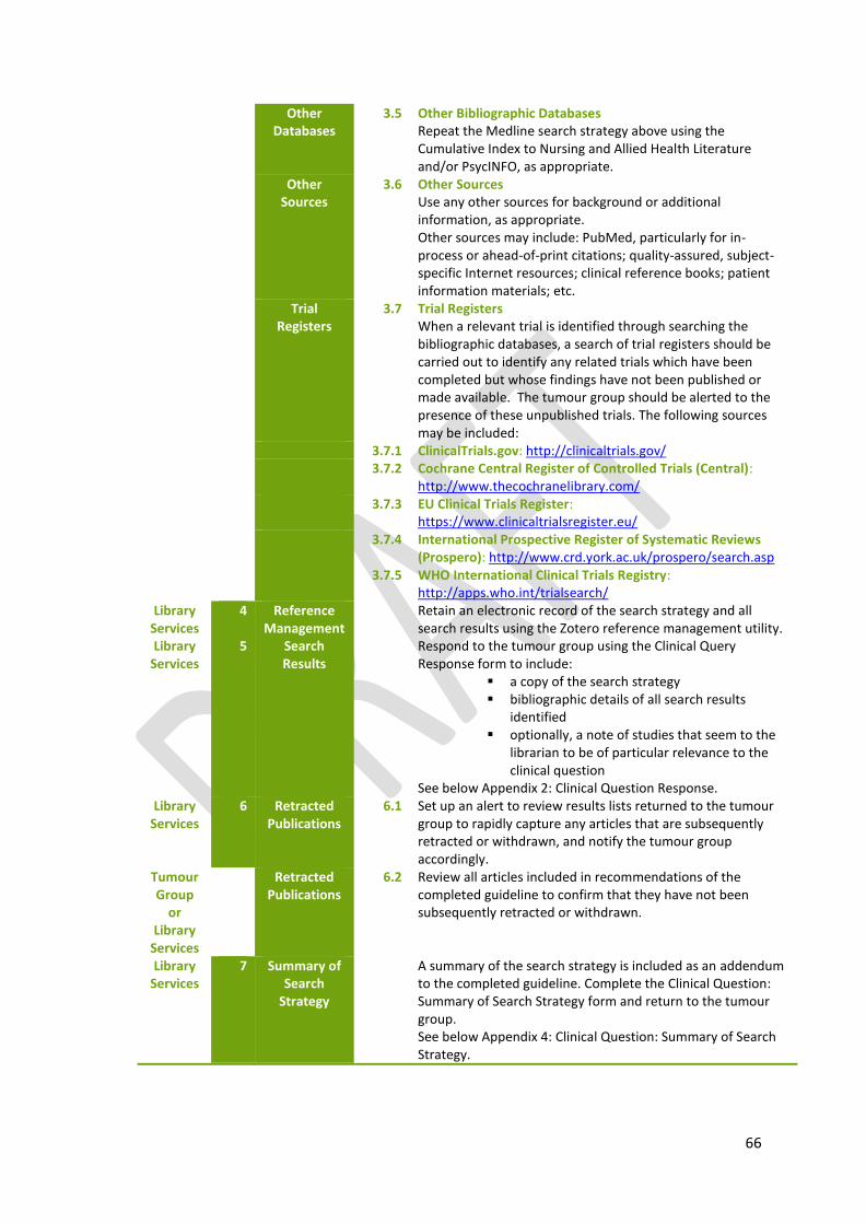
66
Other Databases
3.5 Other Bibliographic Databases Repeat the Medline search strategy above using the Cumulative Index to Nursing and Allied Health Literature and/or PsycINFO, as appropriate.
Other Sources
3.6 Other Sources Use any other sources for background or additional information, as appropriate. Other sources may include: PubMed, particularly for in-process or ahead-of-print citations; quality-assured, subject-specific Internet resources; clinical reference books; patient information materials; etc.
Trial Registers
3.7 Trial Registers When a relevant trial is identified through searching the bibliographic databases, a search of trial registers should be carried out to identify any related trials which have been completed but whose findings have not been published or made available. The tumour group should be alerted to the presence of these unpublished trials. The following sources may be included:
3.7.1 ClinicalTrials.gov: http://clinicaltrials.gov/ 3.7.2 Cochrane Central Register of Controlled Trials (Central):
http://www.thecochranelibrary.com/ 3.7.3 EU Clinical Trials Register:
https://www.clinicaltrialsregister.eu/ 3.7.4 International Prospective Register of Systematic Reviews
(Prospero): http://www.crd.york.ac.uk/prospero/search.asp 3.7.5 WHO International Clinical Trials Registry:
http://apps.who.int/trialsearch/ Library
Services 4 Reference
Management Retain an electronic record of the search strategy and all
search results using the Zotero reference management utility. Library
Services 5 Search
Results Respond to the tumour group using the Clinical Query
Response form to include: a copy of the search strategy bibliographic details of all search results
identified optionally, a note of studies that seem to the
librarian to be of particular relevance to the clinical question
See below Appendix 2: Clinical Question Response. Library
Services 6 Retracted
Publications 6.1 Set up an alert to review results lists returned to the tumour
group to rapidly capture any articles that are subsequently retracted or withdrawn, and notify the tumour group accordingly.
Tumour Group
or Library
Services
Retracted Publications
6.2 Review all articles included in recommendations of the completed guideline to confirm that they have not been subsequently retracted or withdrawn.
Library Services
7 Summary of Search
Strategy
A summary of the search strategy is included as an addendum to the completed guideline. Complete the Clinical Question: Summary of Search Strategy form and return to the tumour group. See below Appendix 4: Clinical Question: Summary of Search Strategy.
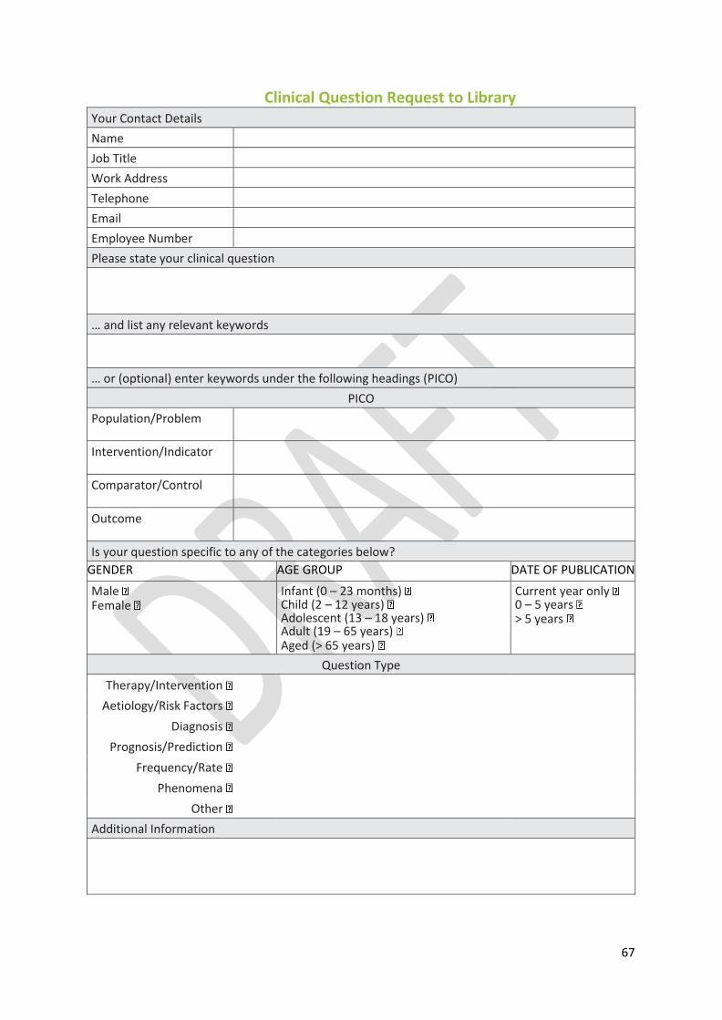
67
Clinical Question Request to Library Your Contact Details
Name
Job Title
Work Address
Telephone
Employee Number
Please state your clinical question
… and list any relevant keywords
… or (optional) enter keywords under the following headings (PICO)
PICO
Population/Problem
Intervention/Indicator
Comparator/Control
Outcome
Is your question specific to any of the categories below?
GENDER AGE GROUP DATE OF PUBLICATION
Male Female
Infant (0 – 23 months) Child (2 – 12 years) Adolescent (13 – 18 years) Adult (19 – 65 years) Aged (> 65 years)
Current year only 0 – 5 years > 5 years
Question Type
Therapy/Intervention
Aetiology/Risk Factors
Diagnosis
Prognosis/Prediction
Frequency/Rate
Phenomena
Other
Additional Information

68
Clinical Question Response Dear _______________, Thank you for your email. Please see attached in response to your clinical query and, below, details of the search strategy applied to your question. If you wish to source any of the references contained in these results, or to search further, please do not hesitate to contact us. Best wishes, _______________.
[ATTACH CLINICAL QUESTION REQUEST HERE]
Search Strategy
Primary Database(s) Searched
Search Strategy
Other/Secondary
Resources
Searched
Comments
Contact
Your Library Staff Contact
Date

69
Clinical Question: Summary of Search Strategy
Clinical Question
PICO
Population/Problem
Intervention/Indicator
Comparator/Control
Outcome
Is your question specific to any of the categories below?
GENDER AGE GROUP DATE OF PUBLICATION
Male
Female
Infant (0 – 23 months)
Child (2 – 12 years)
Adolescent (13 – 18 years)
Adult (19 – 65 years)
Aged (> 65 years)
Current year only
0 – 5 years
> 5 years
Question Type
Therapy/Intervention
Aetiology/Risk Factors
Diagnosis
Prognosis/Prediction
Frequency/Rate
Phenomena
Other
Search Strategy
Primary Database(s) Searched
Search Strategy [Copy of base Medline and/or PubMed search strategy HERE. Include subject headings and search hits].
Other/Secondary Resources Searched
Search Strategy: Other Resources
[Copy of other search strategies HERE. Include subject headings and search hits].
Comments [Short paragraph describing search].
Date

70
Appendix 3: Details of consultation process As part of the consultation process, the draft guideline was circulated for review to this list of groups, committees and organisations. The guideline was also available on the NCCP website so it was accessible to all who wished to comment and provide feedback.
Clinical leaders and healthcare managers
National groups, organisations, faculties & committees
Patient support and advocacy groups
International Expert Review

71
Appendix 4: Economic assessment
Budget Impact Analysis To be added closer to publication date
Economic evidence summary As part of the systematic literature review any health economic literature identified in the literature search for each clinical question was included and was critically appraised. Critical appraisal was carried out using the Consensus on Health Economic Criteria (CHEC)-list quality appraisal tool (Evers et al., 2005). The studies were also evaluated for applicability to the Irish setting using the International Society for Pharmacoeconomics (ISPOR) questionnaire (Jaime Caro et al., 2014). A narrative synthesis of any cost-effectiveness literature identified was included in the evidence summary under the heading ‘Resources and other considerations’.

72
Appendix 5: Implementation plan To be added closer to publication date

73
Appendix 6: Supporting tools Downloading this guideline This National Clinical Guideline will be available to download on the following websites:
NCCP: https://www.hse.ie/eng/services/list/5/cancer/profinfo/guidelines/
NCEC: https://health.gov.ie/national-patient-safety-office/ncec/national-clinical-guidelines/
Guides for health professionals To be added closer to publication date Patient information booklets/leaflets/website To be added closer to publication date Service Quality To be added closer to publication date

74
Appendix 7: Monitoring and audit The National Gestational Trophoblastic Disease Registry, Monitoring and Advisory Centre was established in May 2017 to monitor and co-ordinate the follow-up of women who have been diagnosed with a molar pregnancy. It is a service established by the Health Service Executive, the National Cancer Control Programme and Cork University Maternity Hospital. The Clinical Director of the National Gestational Trophoblastic Disease Registry, Monitoring and Advisory Centre is Dr John Coulter, Consultant Gynaecologist. The primary objectives of the National GTD Registry, Monitoring and Advisory Centre are to:
To register all women with GTD to ensure early detection of malignant change so that appropriate
treatment can be instituted at the earliest possible time.
To monitor GTD patients for resolution, relapse or new episodes of the disease
To provide accurate (expert) diagnosis for women
To facilitate urgent management of patients requiring chemotherapy
To provide Clinical Advice to Patients and Clinicians throughout the country on the management of GTD
To provide regular reports (including an annual report) to the National Cancer Control Programme and
the Health Service Executive (HSE) on GTD cases, treatment and outcome
The registration of patients (following patient consent) has allowed centralised recording of hCG levels, which has ensured consistent monitoring and efficient management decisions, to improve clinical outcomes. The guideline development group recommend that women with the following diagnoses should be registered and require follow-up:
CHM
PHM
twin pregnancy with CHM or PHM
limited macroscopic or microscopic molar change suggesting possible early CHM or PHM
choriocarcinoma
PSTT or ETT
atypical placental site nodule
atypical GTD suspected
p57KIP2 discordant villi Once registered, the patient is contacted by the National GTD Registry, Monitoring and Advisory Centre and given further information/counselling about her diagnosis. A number of supporting tools developed by the National GTD Registry, Monitoring and Advisory and assessed by the National Adult Literary Agency (NALA) are listed in Appendix 6: Supporting tools. The National GTD Registry, Monitoring and Advisory team will audit and report annually according to the KPIs determined by the clinical governance group under the auspices of the NCCP.
75
Appendix 8: Glossary of terms and abbreviations Definitions within the context of this document
Case control study The observational epidemiologic study of persons with the disease (or other outcome variable) of interest and a suitable control (comparison, reference) group of persons without the disease. The relationship of an attribute to the disease is examined by comparing the diseased and non-diseased with regard to how frequently the attribute is present or, if quantitative, the levels of the attribute, in each of the groups. (CEBM website)
Case series A group or series of case reports involving patients who were given similar treatment. Reports of case series usually contain detailed information about the individual patients. This includes demographic information (for example, age, gender, ethnic origin) and information on diagnosis, treatment, response to treatment, and follow-up after treatment. (NCI Dictionary)
Choriocarcinoma A malignant disease characterised by abnormal trophoblastic hyperplasia and anaplasia, absence of chorionic villi, hemorrhage, and necrosis with direct invasion into the myometrium and vascular invasion resulting in spread to distant sites. (Lurain, 2010)
Cohort study A research study that compares a particular outcome (such as lung cancer) in groups of individuals who are alike in many ways but differ by a certain characteristic (for example, female nurses who smoke compared with those who do not smoke). (NCI dictionary)
Complete mole Complete moles are diploid and androgenic in origin, hydatidiform mole with no evidence of fetal tissue. Complete moles usually (75–80%) arise as a consequence of duplication of a single sperm following fertilisation of an ‘empty’ ovum. Some complete moles (20–25%) can arise after dispermic fertilisation of an ‘empty’ ovum. (RCOG, 2010)
Epithelioid trophoblastic tumour
ETT is a rare variant of PSTT. It develops from neoplastic transformation of chorionic-type extra-villous trophoblast. ETT typically presents as a discrete, hemorrhagic, solid, and cystic lesion that is located either in the fundus, lower uterine segment, or endocervix. Like PSTT, it forms tumour nodules in the myometrium. (Berkowitz et al., 2015a)
External validity The extent to which we can generalise the results of a study to the population of interest.
Internal validity The extent to which a study properly measures what it is meant to measure.
Invasive mole A benign tumour that arises from myometrial invasion of a hydatidiform mole via direct extension through tissue or venous channels. (Lurain, 2010)
Meta-analysis A process that analyses data from different studies done about the same subject. The results of a meta-analysis are usually stronger than the results of any study by itself. (NCI dictionary)

76
Partial mole Partial moles are usually (90%) triploid in origin, with two sets of paternal haploid genes and one set of maternal haploid genes. Partial moles occur, in almost all cases, following dispermic fertilisation of an ovum. Ten percent of partial moles represent tetraploid or mosaic conceptions. In a partial mole, there is usually evidence of a fetus or fetal red blood cells. (RCOG, 2010)
Placental site trophoblastic tumor
PSTTs are malignant and develop from extravillous, intermediate trophoblast. They are usually diploid and monomorphic. Microscopically, these tumours show tumour (PSTT) no chorionic villi and are characterised by a proliferation of mononuclear intermediate trophoblast cells with oval nuclei and abundant eosinophilic cytoplasm. (Berkowitz et al., 2015a)
Randomised trial An epidemiological experiment in which subjects in a population are randomly allocated into groups, usually called study and control groups, to receive or not receive an experimental preventive or therapeutic procedure, manoeuvre, or intervention. The results are assessed by rigorous comparison of rates of disease, death, recovery, or other appropriate outcome in the study and control groups. (CEBM website)
Systematic review The application of strategies that limit bias in the assembly, critical appraisal, and synthesis of all relevant studies on a specific topic. Systematic reviews focus on peer-reviewed publications about a specific health problem and use rigorous, standardised methods for selecting and assessing articles. A systematic review differs from a meta-analysis in not including a quantitative summary of the results. (CEBM website)

77
Abbreviations To be added closer to publication date

78
Appendix 9: Clinical questions in PICO format To be added closer to publication date

79
Appendix 10 Levels of evidence and grading systems The Guideline Development Group assigned each recommendation a quality of evidence and strength of recommendation. The Grading of Recommendations Assessment, Development and Evaluation (GRADE) approach provides an explicit system for rating the quality of evidence and whether the recommendation is strong or weak (Guyatt et al., 2008). Quality of evidence It is recognised that in guideline development that just assessing the level of evidence does not take into account the methodological quality of each individual study or the quality of the body of evidence as a whole (Harbour and Miller, 2001). The Guideline Development Group used the GRADE system which considers the following factors when classifying the quality of evidence; high, moderate or low (Guyatt et al., 2008):
• Study design • Study design limitations • Consistency of results • Directness of the evidence • Imprecision of results • Reporting bias
Table 3 Quality of evidence adapted from GRADE working group 2013
High We are very confident that the true effect lies close to that of the estimate of the effect.
Moderate We are moderately confident in the effect estimate: The true effect is likely to be close to the estimate of the effect, but there is a possibility that it is substantially different.
Low Our confidence in the effect estimate is limited: The true effect may be substantially different from the estimate of the effect.
Very Low We have very little confidence in the effect estimate: The true effect is likely to be substantially different from the estimate of effect
Strength of recommendation There are two grades of recommendation: strong or weak. The strength of recommendation reflects the balance of the following items: • The quality of the body of evidence • The balance between benefit and harm to patient • Patient preferences and values • Resources/cost Table 4 Strength of recommendation adapted from GRADE working group 2013
Strong A strong recommendation is one for which the Guideline Development Group is confident that the desirable effects of an intervention outweigh its undesirable effects (strong recommendation for an intervention) or that the undesirable effects of an intervention outweigh its desirable effects (strong recommendation against an intervention). Strong recommendations are not necessarily high priority recommendations. A strong recommendation implies that most or all individuals will be best served by the recommended course of action.
Weak A weak recommendation is one for which the desirable effects probably outweigh

80
the undesirable effects (weak recommendation for an intervention) or undesirable effects probably outweigh the desirable effects (weak recommendation against an intervention) but appreciable uncertainty exists. A weak recommendation implies that not all individuals will be best served by the recommended course of action. There is a need to consider more carefully than usual the individual patient’s circumstances, preferences, and values. When there are weak recommendations caregivers need to allocate more time to shared decision making, making sure that they clearly and comprehensively explain the potential benefits and harms to a patient.
Good practice points Good practice points were based on the clinical expertise of the Guideline Development Group. Practical considerations around patient care Practical considerations around patient care are statements developed with the patients on issues that were important to them with regards to their own experience of the diagnosis and staging of their cancer.

81
References
AGARWAL, R., ALIFRANGIS, C., EVERARD, J., SAVAGE, P. M., SHORT, D., TIDY, J., FISHER, R. A., SEBIRE, N.
J., HARVEY, R., HANCOCK, B. W., COLEMAN, R. E. & SECKL, M. J. 2014. Management and survival of patients with FIGO high-risk gestational trophoblastic neoplasia: the U.K. experience, 1995-2010. The Journal Of Reproductive Medicine, 59, 7-12.
AGARWAL, R., TEOH, S., SHORT, D., HARVEY, R., SAVAGE, P. M. & SECKL, M. J. 2012. Chemotherapy and human chorionic gonadotropin concentrations 6 months after uterine evacuation of molar pregnancy: a retrospective cohort study. The Lancet, 379, 130-135.
ALAZZAM, M., YOUNG, T., COLEMAN, R., HANCOCK, B., DREW, D., WILSON, P. & TIDY, J. 2011. Predicting gestational trophoblastic neoplasia (GTN): is urine hCG the answer? Gynecol Oncol, 122, 595-9.
ALAZZAM, M. I., TIDY, J., OSBORNE, R., COLEMAN, R., HANCOCK, B. W. & LAWRIE, T. A. 2016. Chemotherapy for resistant or recurrent gestational trophoblastic neoplasia. The Cochrane Database Of Systematic Reviews, CD008891.
ALIFRANGIS, C., AGARWAL, R., SHORT, D., FISHER, R. A., SEBIRE, N. J., HARVEY, R., SAVAGE, P. M. & SECKL, M. J. 2013. EMA/CO for high-risk gestational trophoblastic neoplasia: good outcomes with induction low-dose etoposide-cisplatin and genetic analysis. Journal of Clinical Oncology, 31, 280-286.
BAGSHAWE, K., DENT, J. & WEBB, J. 1986. Hydatidiform mole in England and Wales 1973–1983.: Lancet. BALACHANDRAN, K., SALAWU, A., GHORANI, E., KAUR, B., SEBIRE, N. J., SHORT, D., HARVEY, R.,
HANCOCK, B., TIDY, J., SINGH, K., SARWAR, N., WINTER, M. C. & SECKL, M. J. 2019. When to stop human chorionic gonadotrophin (hCG) surveillance after treatment with chemotherapy for gestational trophoblastic neoplasia (GTN): A national analysis on over 4,000 patients. Gynecologic Oncology.
BERKOWITZ, R., HOROWITZ, N. & ELIAS, K. 2020. Hydatidiform mole: Epidemiology, clinical features and diagnosis [Online]. UpToDate, Post TW(Ed) UpToDate, Waltham, MA. [Accessed 15/02/2021].
BERKOWITZ, R. S. & GOLDSTEIN, D. P. 2009. Current management of gestational trophoblastic diseases. Gynecologic Oncology, 112, 654-662.
BERKOWITZ, R. S. & GOLDSTEIN, D. P. 2013. Current advances in the management of gestational trophoblastic disease. Gynecol Oncol, 128, 3-5.
BISCARO, A., BRAGA, A. & BERKOWITZ, R. S. 2015. Diagnosis, classification and treatment of gestational trophoblastic neoplasia. Revista Brasileira De Ginecologia E Obstetricia: Revista Da Federacao Brasileira Das Sociedades De Ginecologia E Obstetricia, 37, 42-51.
BOLZE, P.-A., ATTIA, J., MASSARDIER, J., SECKL, M. J., MASSUGER, L., VAN TROMMEL, N., NIEMANN, I., HAJRI, T., SCHOTT, A.-M. & GOLFIER, F. 2015. Formalised consensus of the European Organisation for Treatment of Trophoblastic Diseases on management of gestational trophoblastic diseases. European Journal Of Cancer (Oxford, England: 1990), 51, 1725-1731.
BRAGA, A., BISCARO, A., DO AMARAL GIORDANI, J. M., VIGGIANO, M., ELIAS, K. M., BERKOWITZ, R. S. & SECKL, M. J. 2018. Does a human chorionic gonadotropin level of over 20,000 IU/L four weeks after uterine evacuation for complete hydatidiform mole constitute an indication for chemotherapy for gestational trophoblastic neoplasia? European Journal Of Obstetrics, Gynecology, And Reproductive Biology, 223, 50-55.
BRAGA, A., TORRES, B., BURLÁ, M., MAESTÁ, I., SUN, S. Y., LIN, L., MADI, J. M., UBERTI, E., VIGGIANO, M., ELIAS, K. M. & BERKOWITZ, R. S. 2016. Is chemotherapy necessary for patients with molar pregnancy and human chorionic gonadotropin serum levels raised but falling at 6months after uterine evacuation? Gynecologic Oncology, 143, 558-564.
CHARING CROSS. 2019. Available: http://www.hmole-chorio.org.uk/ [Accessed 19/11/2019]. CHARRING CROSS 2020. Charing Cross Hospital Trophoblast Disease Clinic Guide. COVENS, A., FILIACI, V. L., BURGER, R. A., OSBORNE, R. & CHEN, M. D. 2006. Phase II trial of pulse
dactinomycin as salvage therapy for failed low-risk gestational trophoblastic neoplasia: a
82
Gynecologic Oncology Group study. Cancer, 107, 1280-6. COYLE, C., SHORT, D., JACKSON, L., SEBIRE, N. J., KAUR, B., HARVEY, R., SAVAGE, P. M. & SECKL, M. J.
2018. What is the optimal duration of human chorionic gonadotrophin surveillance following evacuation of a molar pregnancy? A retrospective analysis on over 20,000 consecutive patients. Gynecologic Oncology, 148, 254-257.
DARBY, S., JOLLEY, I., PENNINGTON, S. & HANCOCK, B. W. 2009. Does chest CT matter in the staging of GTN? Gynecol Oncol, 112, 155-60.
DE SOUZA, J. M. Q., BRAGA, A., SANCHES DOS SANTOS, R., RAMOS, M. M., CORTÉS-CHARRY, R. & MAESTÁ, I. 2017. Comparison of 2 Human Chorionic Gonadotropin Immunoassays Commercially Available for Monitoring Patients With Gestational Trophoblastic Disease. International Journal Of Gynecological Cancer: Official Journal Of The International Gynecological Cancer Society, 27, 1494-1500.
DENG, L., ZHANG, J., WU, T. & LAWRIE, T. A. 2013. Combination chemotherapy for primary treatment of high-risk gestational trophoblastic tumour. Cochrane Database Syst Rev, 1, Cd005196.
EROL, O., SÜREN, D., TUTUŞ, B., TOPTAŞ, T., GÖKAY, A. A., DERBENT, A. U., ÖZEL, M. K. & SEZER, C. 2016. Immunohistochemical Analysis of E-Cadherin, p53 and Inhibin-α Expression in Hydatidiform Mole and Hydropic Abortion. Pathology Oncology Research: POR, 22, 515-521.
EVERS, S., GOOSSENS, M., DE VET, H., VAN TULDER, M. & AMENT, A. 2005. Criteria list for assessment of methodological quality of economic evaluations: Consensus on Health Economic Criteria. Int J Technol Assess Health Care, 21, 240-5.
FOWLER, D. J., LINDSAY, I., SECKL, M. J. & SEBIRE, N. J. 2006. Routine pre-evacuation ultrasound diagnosis of hydatidiform mole: experience of more than 1000 cases from a regional referral center. Ultrasound Obstet Gynecol, 27, 56-60.
GOFF, B. 2019. Human chorionic gonadotropin: Testing in pregnancy and gestational trophoblastic disease and causes of low persistent levels [Online]. [Accessed 28/06/2020].
GOLDSTEIN, D. P. & BERKOWITZ, R. S. 2012. Current management of gestational trophoblastic neoplasia. Hematology/oncology clinics of North America, 26, 111-131.
GUYATT, G. H., OXMAN, A. D., VIST, G. E., KUNZ, R., FALCK-YTTER, Y., ALONSO-COELLO, P. & SCHUNEMANN, H. J. 2008. GRADE: an emerging consensus on rating quality of evidence and strength of recommendations. Bmj, 336, 924-6.
HARBOUR, R. & MILLER, J. 2001. A new system for grading recommendations in evidence based guidelines.
HARVEY, R., GOFF, B. & CHAKRABARTI, A. 2021. Human chorionic gonadotropin: Biochemistry and measurement in pregnancy and disease [Online]. UpToDate, Post TW(Ed) UpToDate, Waltham, MA. [Accessed 15/02/2021].
HASANZADEH, M., TABARI, A., HOMAE, F., SHAKERI, M., BAKHSHANDEH, T. & MADANISANI, F. 2014. Evaluation of weekly intramuscular methotrexate in the treatment of low risk gestational trophoblastic neoplasia. J Cancer Res Ther, 10, 646-50.
INTERNATIONAL FEDERATION OF GYNECOLOGY AND OBSTETRICS 2009. Current FIGO staging for cancer of the vagina, fallopian tube, ovary, and gestational trophoblastic neoplasia. Int J Gynaecol Obstet, 105, 3-4.
JAIME CARO, J., EDDY, D. M., KAN, H., KALTZ, C., PATEL, B., ELDESSOUKI, R. & BRIGGS, A. H. 2014. Questionnaire to assess relevance and credibility of modeling studies for informing health care decision making: an ISPOR-AMCP-NPC Good Practice Task Force report. Value Health, 17, 174-82.
KOHORN, E. I. 2002. Negotiating a staging and risk factor scoring system for gestational trophoblastic neoplasia. A progress report. J Reprod Med, 47, 445-50.
KOHORN, E. I. 2014. Worldwide survey of the results of treating gestational trophoblastic disease. J Reprod Med, 59, 145-53.
LERTKHACHONSUK, R. 2015. Quantitative urine hCG and urine pregnancy test in gestational trophoblastic disease patients with low hCG titer. J Med Assoc Thai, 98 Suppl 1, S36-41.

83
LOK, C., VAN TROMMEL, N., MASSUGER, L., GOLFIER, F. & SECKL, M. 2020. Practical clinical guidelines of the EOTTD for treatment and referral of gestational trophoblastic disease. Eur J Cancer, 130, 228-240.
LURAIN, J. R., CHAPMAN-DAVIS, E., HOEKSTRA, A. V. & SCHINK, J. C. 2012. Actinomycin D for methotrexate-failed low-risk gestational trophoblastic neoplasia. J Reprod Med, 57, 283-7.
LURAIN, J. R. & NEJAD, B. 2005. Secondary chemotherapy for high-risk gestational trophoblastic neoplasia. Gynecol Oncol, 97, 618-23.
LYBOL, C., SWEEP, F., HARVEY, R., MITCHELL, H., SHORT, D., THOMAS, C. M. G., OTTEVANGER, P. B., SAVAGE, P. M., MASSUGER, L. & SECKL, M. J. 2012. Relapse rates after two versus three consolidation courses of methotrexate in the treatment of low-risk gestational trophoblastic neoplasia. Gynecologic oncology, 125, 576-579.
MCNEISH, I. A., STRICKLAND, S., HOLDEN, L., RUSTIN, G. J. S., FOSKETT, M., SECKL, M. J. & NEWLANDS, E. S. 2002. Low-risk persistent gestational trophoblastic disease: outcome after initial treatment with low-dose methotrexate and folinic acid from 1992 to 2000. Journal of Clinical Oncology, 20, 1838-1844.
MILLER, C. R., CHAPPELL, N. P., SLEDGE, C., LEATH, C. A., 3RD, PHIPPEN, N. T., HAVRILESKY, L. J. & BARNETT, J. C. 2017. Are different methotrexate regimens as first line therapy for low risk gestational trophoblastic neoplasia more cost effective than the dactinomycin regimen used in GOG 0174? Gynecologic Oncology, 144, 125-129.
NATIONAL COMPREHENSIVE CANCER NETWORK (NCCN) 2021. Gestational Trophoblastic Neoplasia - NCCN Clinical Practice Guideline in Oncology.
NIEMANN, I., VEJERSLEV, L. O., FRØDING, L., BLAAKÆR, J., MAROUN, L. L., HANSEN, E. S., GROVE, A., LUND, H., HAVSTEEN, H. & SUNDE, L. 2015. Gestational trophoblastic diseases - clinical guidelines for diagnosis, treatment, follow-up, and counselling. Danish Medical Journal, 62, A5082-A5082.
OSBORNE, R. J., FILIACI, V., SCHINK, J. C., MANNEL, R. S., ALVAREZ SECORD, A., KELLEY, J. L., PROVENCHER, D., SCOTT MILLER, D., COVENS, A. L. & LAGE, J. M. 2011. Phase III trial of weekly methotrexate or pulsed dactinomycin for low-risk gestational trophoblastic neoplasia: a gynecologic oncology group study. J Clin Oncol, 29, 825-31.
PRICE, J. M., LO, C., ABDI, S., WINTER, M. C., COLEMAN, R. E., TIDY, J. & HANCOCK, B. W. 2015. The Role of Computed Tomography Scanning of the Thorax in the Initial Assessment of Gestational Trophoblastic Neoplasia. International Journal Of Gynecological Cancer: Official Journal Of The International Gynecological Cancer Society, 25, 1731-1736.
SANTABALLA, A., GARCÍA, Y., HERRERO, A., LAÍNEZ, N., FUENTES, J., DE JUAN, A., RODRIGUEZ FREIXINÓS, V., APARICIO, J., CASADO, A. & GARCÍA-MARTINEZ, E. 2018. SEOM clinical guidelines in gestational trophoblastic disease (2017). Clinical & Translational Oncology: Official Publication Of The Federation Of Spanish Oncology Societies And Of The National Cancer Institute Of Mexico, 20, 38-46.
SAVAGE, P., KELPANIDES, I., TUTHILL, M., SHORT, D. & SECKL, M. J. 2015. Brain metastases in gestational trophoblast neoplasia: an update on incidence, management and outcome. Gynecologic Oncology, 137, 73-76.
SCHMITT, C., DORET, M., MASSARDIER, J., HAJRI, T., SCHOTT, A. M., RAUDRANT, D. & GOLFIER, F. 2013. Risk of gestational trophoblastic neoplasia after hCG normalisation according to hydatidiform mole type. Gynecol Oncol, 130, 86-9.
SEBIRE, N. J. 2010. Histopathological diagnosis of hydatidiform mole: contemporary features and clinical implications. Fetal & Pediatric Pathology, 29, 1-16.
SEBIRE, N. J., REES, H., PARADINAS, F., SECKL, M. & NEWLANDS, E. 2001. The diagnostic implications of routine ultrasound examination in histologically confirmed early molar pregnancies. Ultrasound Obstet Gynecol, 18, 662-5.
SECKL, M. J. & SAVAGE, P. 2012. Gestational Trophoblastic Disease Regimens v3.1. In: LONDON CANCER ALLIANCE (ed.). North West London Cancer Network.

84
SECKL, M. J., SEBIRE, N. J. & BERKOWITZ, R. S. 2010. Gestational trophoblastic disease. The Lancet, 376, 717-729.
SECKL, M. J., SEBIRE, N. J., FISHER, R. A., GOLFIER, F., MASSUGER, L., SESSA, C. & ON BEHALF OF THE, E. G. W. G. 2013. Gestational trophoblastic disease: ESMO Clinical Practice Guidelines for diagnosis, treatment and follow-up. Annals of Oncology, 24, vi39-vi50.
SITA-LUMSDEN, A., SHORT, D., LINDSAY, I., SEBIRE, N. J., ADJOGATSE, D., SECKL, M. J. & SAVAGE, P. M. 2012. Treatment outcomes for 618 women with gestational trophoblastic tumours following a molar pregnancy at the Charing Cross Hospital, 2000-2009. Br J Cancer, 107, 1810-4.
SOTO-WRIGHT, V., BERNSTEIN, M., GOLDSTEIN, D. P. & BERKOWITZ, R. S. 1995. The changing clinical presentation of complete molar pregnancy. Obstetrics & Gynecology, 86, 775-779.
SUN, S. Y., MELAMED, A., GOLDSTEIN, D. P., BERNSTEIN, M. R., HOROWITZ, N. S., MORON, A. F., MAESTÁ, I., BRAGA, A. & BERKOWITZ, R. S. 2015. Changing presentation of complete hydatidiform mole at the New England Trophoblastic Disease Center over the past three decades: does early diagnosis alter risk for gestational trophoblastic neoplasia? Gynecologic Oncology, 138, 46-49.
TAYLOR, F., GREW, T., EVERARD, J., ELLIS, L., WINTER, M. C., TIDY, J., HANCOCK, B. W. & COLEMAN, R. E. 2013. The outcome of patients with low risk gestational trophoblastic neoplasia treated with single agent intramuscular methotrexate and oral folinic acid. Eur J Cancer, 49, 3184-90.
TIDY, J., SECKL, H., HANCOCK, B. & GYNAECOLOGISTS., O. B. O. T. R. C. O. O. A. 2020. Management of Gestational Trophoblastic Disease - Green-top Guideline No. 38.

85
Related Documents