Diagnosis of oral mucous membrane pemphigoid by means of combined serologic testing Taihei Hayakawa, DDS, a,b Minao Furumura, MD, PhD, c Hideo Fukano, DDS, PhD, d Xiaoguang Li, MD, PhD, e Norito Ishii, MD, f Takahiro Hamada, MD, f Chika Ohata, MD, c Daisuke Tsuruta, MD, PhD, g Kazuo Shimozato, DDS, PhD, h and Takashi Hashimoto, MD i Kurume University, Kurume, Japan; Aichi-Gakuin University, Nagoya, Japan; Osaka City University, Osaka, Japan Objective. Mucous membrane pemphigoid (MMP) is a rare autoimmune bullous disease caused by various autoantibodies. This study aimed to evaluate the diagnostic value of MMP-specific autoantibodies in patient sera. Study Design. We analyzed sera from 30 MMP-suspected patients with intractable oral mucosal lesions using a combination of indirect immunofluorescence with 1M NaClesplit skin, immunoblot analysis, and ELISAs. We also analyzed clinical features among different types of MMP. Results. Seventeen, 4, and 3 patients were diagnosed with anti-BP180-type MMP, anti-laminin-332-type MMP, and combined anti-BP180/anti-laminin-332-type MMP, respectively. Conclusions. Our results indicated that a combination of immunologic testing for circulating autoantibodies is useful for the diagnosis of MMP. (Oral Surg Oral Med Oral Pathol Oral Radiol 2014;117:483-496) Autoimmune bullous diseases (ABDs) are a group of heterogeneous blistering disorders that may involve the oral cavity. 1,2 ABDs are divided into the pemphigus group and subepidermal bullous disease group. Current classification of ABDs with their autoantigens is sum- marized in Table I. Major diseases of the pemphigus group are pemphigus vulgaris (PV), pemphigus foliaceus (PF), and paraneoplastic pemphigus (PNP). The subepi- dermal bullous disease group includes bullous pemphi- goid (BP), mucous membrane pemphigoid (MMP), and epidermolysis bullosa acquisita (EBA). ABDs show autoantibodies to various epithelial adhesion molecules or extracellular matrices. Pemphigus diseases are char- acterized by the presence of autoantibodies against components of cell-cell adhesion, whereas pemphigoid diseases have autoantibodies against components at the dermoepidermal junction (Figure 1). Recently developed assays for autoantibodies and autoantigens enable sero- logic diagnosis in 90% of ABD cases. 3-9 Previous studies using biochemical and molecular biologic techniques have identified autoantigens and epitopes in various ABDs. 10-13 These autoantigens were originally examined by immunoprecipitation and immunoblotting (IB). 14-16 In particular, noncollagenous 16a (NC16a) domain, which localizes adjacent to the transmembrane portion in the extracellular region of BP180 (type XVII collagen), has been found to be highly antigenic in BP. 17-19 To facilitate the testing of larger numbers of sera simultaneously, various enzyme-linked immunosorbent assays (ELISAs) were developed. 3-6,8,9 ELISAs of recombinant proteins (RPs) of the extracel- lular domains of desmoglein 1 (Dsg1) and Dsg3, which were produced by the baculovirus-expression system, This study was supported by Grants-in-Aid for Scientific Research and a Strategic Research Basis Formation Supporting Project from the Ministry of Education, Culture, Sports, Science and Technology of Japan, and by Health and Labour Sciences Research Grants and grants for Research on Measures for Intractable Diseases from the Ministry of Health, Labour and Welfare of Japan. a Postgraduate Researcher, Department of Dermatology, Kurume University School of Medicine and Kurume University Institute of Cutaneous Cell Biology. b Postgraduate Student, Department of Maxillofacial Surgery, Aichi- Gakuin University School of Dentistry. c Associate Professor, Department of Dermatology, Kurume University School of Medicine and Kurume University Institute of Cutaneous Cell Biology. d Associate Professor, Department of Maxillofacial Surgery, Aichi- Gakuin University School of Dentistry. e Postdoctoral Researcher, Department of Dermatology, Kurume University School of Medicine and Kurume University Institute of Cutaneous Cell Biology. f Senior Lecturer, Department of Dermatology, Kurume University School of Medicine and Kurume University Institute of Cutaneous Cell Biology. g Professor, Department of Dermatology, Osaka City University Graduate School of Medicine. h Professor, Department of Maxillofacial Surgery, Aichi-Gakuin Uni- versity School of Dentistry. i Professor, Department of Dermatology, Kurume University School of Medicine, and Kurume University Institute of Cutaneous Cell Biology. Received for publication Oct 3, 2013; returned for revision Dec 11, 2013; accepted for publication Dec 14, 2013. Ó 2014 Elsevier Inc. All rights reserved. 2212-4403/$ - see front matter http://dx.doi.org/10.1016/j.oooo.2013.12.402 Statement of Clinical Relevance Mucous membrane pemphigoid (MMP) is a rare autoimmune bullous disease caused by various au- toantibodies. Our results suggest that a combination of multiple immunologic tests can be used in the diagnosis of MMP. 483 Vol. 117 No. 4 April 2014

Welcome message from author
This document is posted to help you gain knowledge. Please leave a comment to let me know what you think about it! Share it to your friends and learn new things together.
Transcript

Vol. 117 No. 4 April 2014
Diagnosis of oral mucous membrane pemphigoid by means ofcombined serologic testingTaihei Hayakawa, DDS,a,b Minao Furumura, MD, PhD,c Hideo Fukano, DDS, PhD,d Xiaoguang Li, MD, PhD,e
Norito Ishii, MD,f Takahiro Hamada, MD,f Chika Ohata, MD,c Daisuke Tsuruta, MD, PhD,g
Kazuo Shimozato, DDS, PhD,h and Takashi Hashimoto, MDi
Kurume University, Kurume, Japan; Aichi-Gakuin University, Nagoya, Japan; Osaka City University, Osaka, Japan
Objective. Mucous membrane pemphigoid (MMP) is a rare autoimmune bullous disease caused by various autoantibodies.
This study aimed to evaluate the diagnostic value of MMP-specific autoantibodies in patient sera.
Study Design. We analyzed sera from 30 MMP-suspected patients with intractable oral mucosal lesions using a combination
of indirect immunofluorescence with 1M NaClesplit skin, immunoblot analysis, and ELISAs. We also analyzed clinical
features among different types of MMP.
Results. Seventeen, 4, and 3 patients were diagnosed with anti-BP180-type MMP, anti-laminin-332-type MMP, and combined
anti-BP180/anti-laminin-332-type MMP, respectively.
Conclusions. Our results indicated that a combination of immunologic testing for circulating autoantibodies is useful for the
diagnosis of MMP. (Oral Surg Oral Med Oral Pathol Oral Radiol 2014;117:483-496)
Autoimmune bullous diseases (ABDs) are a group ofheterogeneous blistering disorders that may involve theoral cavity.1,2 ABDs are divided into the pemphigusgroup and subepidermal bullous disease group. Currentclassification of ABDs with their autoantigens is sum-marized in Table I. Major diseases of the pemphigusgroup are pemphigus vulgaris (PV), pemphigus foliaceus
This study was supported by Grants-in-Aid for Scientific Researchand a Strategic Research Basis Formation Supporting Project from theMinistry of Education, Culture, Sports, Science and Technology ofJapan, and by Health and Labour Sciences Research Grants and grantsfor Research on Measures for Intractable Diseases from the Ministryof Health, Labour and Welfare of Japan.aPostgraduate Researcher, Department of Dermatology, KurumeUniversity School of Medicine and Kurume University Institute ofCutaneous Cell Biology.bPostgraduate Student, Department of Maxillofacial Surgery, Aichi-Gakuin University School of Dentistry.cAssociate Professor, Department of Dermatology, Kurume UniversitySchool of Medicine and Kurume University Institute of Cutaneous CellBiology.dAssociate Professor, Department of Maxillofacial Surgery, Aichi-Gakuin University School of Dentistry.ePostdoctoral Researcher, Department of Dermatology, KurumeUniversity School of Medicine and Kurume University Institute ofCutaneous Cell Biology.fSenior Lecturer, Department of Dermatology, Kurume UniversitySchool of Medicine and Kurume University Institute of CutaneousCell Biology.gProfessor, Department of Dermatology, Osaka City UniversityGraduate School of Medicine.hProfessor, Department of Maxillofacial Surgery, Aichi-Gakuin Uni-versity School of Dentistry.iProfessor, Department of Dermatology, Kurume University School ofMedicine, and Kurume University Institute of Cutaneous Cell Biology.Received for publication Oct 3, 2013; returned for revision Dec 11,2013; accepted for publication Dec 14, 2013.� 2014 Elsevier Inc. All rights reserved.2212-4403/$ - see front matterhttp://dx.doi.org/10.1016/j.oooo.2013.12.402
(PF), and paraneoplastic pemphigus (PNP). The subepi-dermal bullous disease group includes bullous pemphi-goid (BP), mucous membrane pemphigoid (MMP), andepidermolysis bullosa acquisita (EBA). ABDs showautoantibodies to various epithelial adhesion moleculesor extracellular matrices. Pemphigus diseases are char-acterized by the presence of autoantibodies againstcomponents of cell-cell adhesion, whereas pemphigoiddiseases have autoantibodies against components at thedermoepidermal junction (Figure 1). Recently developedassays for autoantibodies and autoantigens enable sero-logic diagnosis in 90% of ABD cases.3-9
Previous studies using biochemical and molecularbiologic techniques have identified autoantigens andepitopes in various ABDs.10-13 These autoantigens wereoriginally examined by immunoprecipitation andimmunoblotting (IB).14-16 In particular, noncollagenous16a (NC16a) domain, which localizes adjacent to thetransmembrane portion in the extracellular region ofBP180 (type XVII collagen), has been found to be highlyantigenic in BP.17-19 To facilitate the testing of largernumbers of sera simultaneously, various enzyme-linkedimmunosorbent assays (ELISAs) were developed.3-6,8,9
ELISAs of recombinant proteins (RPs) of the extracel-lular domains of desmoglein 1 (Dsg1) and Dsg3, whichwere produced by the baculovirus-expression system,
Statement of Clinical Relevance
Mucous membrane pemphigoid (MMP) is a rareautoimmune bullous disease caused by various au-toantibodies. Our results suggest that a combinationof multiple immunologic tests can be used in thediagnosis of MMP.
483
Table I. Classification and autoantigens in variousABDs
Diseases Ig subtypes Autoantigens**Pemphigus vulgaris (PV) IgG
**Mucosal dominant typePV
IgG Dsg3
**Mucocutaneous typePV
IgG Dsg3 + Dsg1
**Pemphigus vegetans IgG Dsg3, Dsg1, (Dsc3)Pemphigus foliaceus IgG Dsg1
*Oral pemphigus foliaceus IgG Dsg1Pemphigus erythematosus IgG Dsg1Herpetiform pemphigus IgG Dsg1, (Dsg3, Dsc1)Drug-induced pemphigus IgG Mainly Dsg1**Paraneoplastic pemphigus IgG Plectin, epiplakin,
desmoplakin, BP230,envoplakin, periplakin,A2ML1, Dsg1, Dsg3,Dsc2, Dsc3
Atypical pemphigus (Anti-Dsc pemphigus)
IgG/IgA Dsc1-3
IgA pemphigusSPD type IgA pemphigus IgA Dsc1IEN type IgA pemphigus IgA Unknown
*Bullous pemphigoid (BP) IgG BP180, BP230BP230-type BP IgG BP230
*Herpes gestationis IgG BP180**Mucous membrane
pemphigoid (MMP)**Anti-BP180-type MMP IgG/IgA BP180**Anti-laminin332-typeMMP
IgG Laminin-332 (laminin-5,epiligrin)
**Anti-p168 MMP IgG 168-kDa unknownprotein
Ocular MMP IgG/IgA b4 integrin, unknown**Oral MMP IgG a6 integrin
Dermatitis herpetiformis IgA Epidermaltransglutaminase
C3 dermatitisherpetiformis
C3 Unknown(no autoantigen?)
Linear IgA dermatosis(LABD)Lamina lucida type LABD IgA 97-kDa/120-kDa LAD-1Sublamina densa typeLABD
IgA Unknown (Type VIIcollagen, Laminin g1)
**Epidermolysis bullosaacquisita
IgG Type VII collagen
Bullous SLE IgG Type VII collagen*Anti-laminin g1
pemphigoidIgG Laminin g1 (p200)
Italic text: recently characterized.Bold text: autoatigens have not been characterized to date.ABDs, autoimmune bullous diseases; Dsc, desmocollin; Dsg, des-moglein; IEN, intraepidermal neutrophilic; Ig, immunoglobulin;LAD-1, linear IgA bullous dermatosis antigen-1; SLE, systemic lupuserythematosus; SPD, subcorneal pustular dermatosis.*Diseases that may show oral lesions.**Diseases that show extensive oral lesions.
ORAL AND MAXILLOFACIAL PATHOLOGY OOOO
484 Hayakawa et al. April 2014
were established for use in the diagnosis of PF and PV.8,9
Subsequently, ELISAs of RP of BP180 NC16a domainand RPs of N- and C-terminal domains of BP230 weredeveloped for diagnosis of BP.20,21 Recently, ELISA of
RP of combined NC1 and NC2 domains of type VIIcollagen was developed for the diagnosis of EBA.22
These 5 ELISAs are now commercially available inJapan. Furthermore, in Japan at least, ELISAs for Dsg1,Dsg3, and BP180 are now covered by the NationalHealth Insurance. Thus, most patients with PV, PF, BP,and EBA can be assessed using commercial ELISAkits.3-6,8,9,22,23
ABDs with predominant mucosal lesions were previ-ously referred to as cicatricial pemphigoid. However,although ocular or laryngeal lesions frequently result inscarring, oral mucosal lesions are not commonly associ-ated with scarring. Therefore, at an international con-sensus meeting on MMP held in 2002, cicatricialpemphigoidwas renamedMMP.24MMPmost commonlyaffects oral mucosa, followed by ocular, nasal, genital,pharyngeal, esophageal, laryngeal, and anal mucosae.24-26
In addition, a variable number of MMP patients mayshow skin lesions. These variable clinical features leadto frequent misdiagnoses of patients with MMP.
On indirect immunofluorescence (IIF) of varioussubstrates, MMP demonstrates the presence of immu-noglobulin G (IgG) antibodies, IgA antibodies, or both(IgG/IgA) against the epithelial basement membranezone (BMZ).26,27 However, MMP sera will occasion-ally show negative or very low reactivity in either IIF ofnormal human skin sections or IIF of 1M NaClesplitskin sections (ss-IIF), because of low titers of circu-lating antibodies.28-31 MMP sera react with a numberof additional BMZ antigens. Among these, 70% ofMMP patients exhibit IgG/IgA antibodies againstthe BP180 C-terminus domain and are diagnosed asanti-BP180-type MMP.24,28,30,31 Most patients withanti-BP180-type MMP are negative by ELISA for theBP180 NC16a domain. In addition, approximately20% of MMP patients show IgG antibodies againstvarious subunits of laminin-332, previously referred toas laminin-5 or epiligrin,32 and thus are diagnosed asanti-laminin-332-type MMP.33-35 Anti-BP180-type andanti-laminin-332-type MMP sera react to the epidermaland dermal sides on ss-IIF, respectively.35,36
Furthermore, there are reports of MMP patientsexhibiting IgG/IgA antibodies against BP230,37 the 97-/120-kDa linear IgA bullous dermatosis antigen-1 (LAD-1; soluble truncated extracellular domain of BP180),38
laminin-331,39 type VII collagen,24 and integrin a6 andintegrin b4.40 Thus, MMP is very heterogeneous, whichmay lead to difficulty in its diagnosis. The relationshipbetween clinical features and different autoantigens hasnot been completely studied, and autoantigens associatedwith several MMP types have not been identified.
As PV and BP patients may show blistering skin lesi-ons in addition to oral lesions, they often present initiallyto dermatology. In contrast, because most MMP patientspresent initially with oral mucosal lesions as their
Fig. 1. Schematic structure and component proteins of desmosomes and hemidesmosomes. Desmosomes connect neighboringkeratinocytes and hemidesmosomes connect the epidermis to the dermis. Keratin intermediate filaments bind to the cell membranecontaining plakoglobins, plakofilins, and desmoplakins at desmosomal attachment plaques. These proteins bind to desmogleins anddesmocollins, cadherin-type desmosomal cell adhesion molecules. Hemidesmosomal cytoplasmic proteins (BP230, plectin) bindtransmembrane proteins BP180 and a6b4-integrin, which connect to laminin-332. Type VII collagen in the lamina densa and in theuppermost dermis composes anchoring fibrils and binds dermal collagens.
OOOO ORIGINAL ARTICLE
Volume 117, Number 4 Hayakawa et al. 485
principal complaint, significant numbers of these patientsinitially present to their dentists. However, because the orallesions are not specific to ABDs, many patients are mis-diagnosed as having other acute or chronic conditions.
Because ABDs are typically intractable and affect thepatient’s quality of life, both dermatologists and dentistsshould always consider ABDs, including PV, MMP, andless commonly PNP or EBA, in patients with chronic oralerosive lesions. Serologic tests for autoantibodies andautoantigens are important in distinguishing ABDs fromother oral mucosal disorders. As mentioned earlier, mostpatients with PV, PNP, and EBA can be diagnosed bymeans of commercially available ELISAs.3-6,8,9,22,23 Inaddition, commercial ELISA of BP180 NC16a domainRP shows negative results in typical MMP cases, becausemost MMP sera contains predominantly antibodiesagainst BP180 C-terminal domain or laminin-332 but notagainst the BP180 NC16a domain.
In this study, we examined 3 immunologic methodsto analyze circulating antibodies in 30 patients withsuspected MMP. Our results suggest that a combinationof multiple immunologic tests for circulating autoanti-bodies is useful in the diagnosis of MMP.
MATERIALS AND METHODSPatientsWe enrolled 30 patients with suspected oral mucosal le-sions ofMMP. In these patients, clinical, histopathologic,
and ELISA studies found no findings suggestive of PVor oral lichen planus. We excluded patients who wereclinically diagnosed as bacterial, viral, or fungalinfection; physical, heat, or chemical injuries; malig-nant tumors; erythema multiforme; Stevens-Johnsonsyndrome; toxic epidermal necrolysis; or systemiclupus erythematosus. Sera from patients with differentABDs, including PV, BP, PF, MMP, PNP, EBA, andlinear IgA bullous dermatosis (LABD), were used aspositive controls. Normal sera were used as negativecontrols.
All studies were performed in accordance with theguidelines of the Medical Ethics Committees of Aichi-Gakuin University School of Dentistry and KurumeUniversity School of Medicine. All patients or their legalguardians provided informed consent before partici-pating in this study. The study was conducted incompliance with the principles of the Declaration ofHelsinki.
Indirect immunofluorescenceIIF of normal human skin sections was performedusing standard methods.41 ss-IIF was also performed,using previously described methods.42 For immuno-staining, skin sections were first blocked with 1%bovine serum albumin for 30 minutes at 4�C, followedby incubation with patient sera, diluted to 1:10 to1:160 for IgG antibodies and to 1:10 to 1:40 for IgA
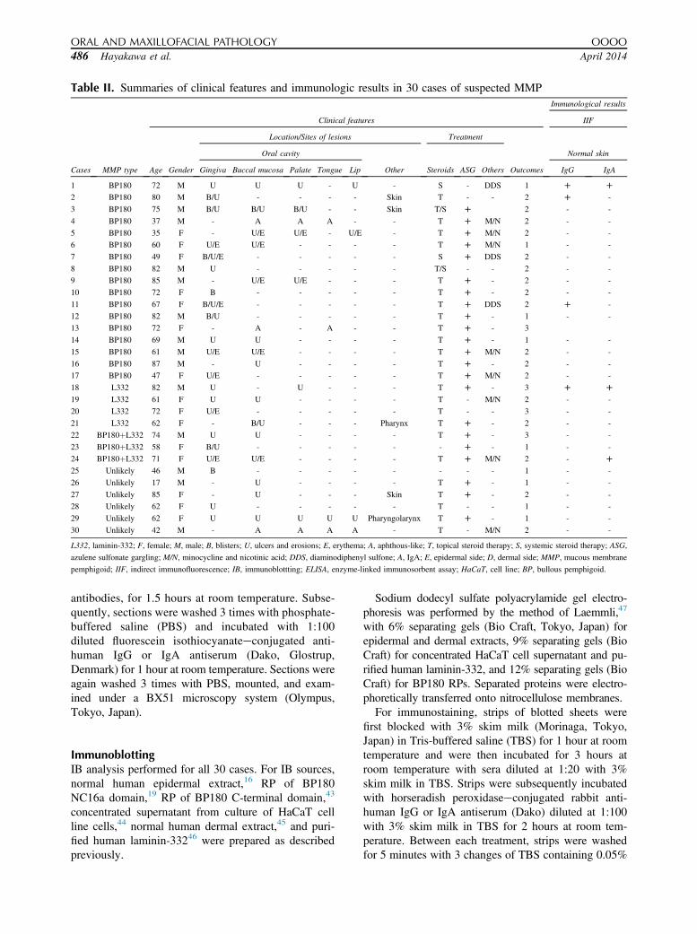
Table II. Summaries of clinical features and immunologic results in 30 cases of suspected MMP
Cases MMP type
Clinical features
Immunological results
IIF
Age Gender
Location/Sites of lesions Treatment
Outcomes
Normal skinOral cavity
Other Steroids ASG OthersGingiva Buccal mucosa Palate Tongue Lip IgG IgA
1 BP180 72 M U U U - U - S - DDS 1 D D
2 BP180 80 M B/U - - - - Skin T - - 2 D -
3 BP180 75 M B/U B/U B/U - - Skin T/S D 2 - -
4 BP180 37 M - A A A - - T D M/N 2 - -
5 BP180 35 F - U/E U/E - U/E - T D M/N 2 - -
6 BP180 60 F U/E U/E - - - - T D M/N 1 - -
7 BP180 49 F B/U/E - - - - - S D DDS 2 - -
8 BP180 82 M U - - - - - T/S - - 2 - -
9 BP180 85 M - U/E U/E - - - T D - 2 - -
10 BP180 72 F B - - - - - T D - 2 - -
11 BP180 67 F B/U/E - - - - - T D DDS 2 D -
12 BP180 82 M B/U - - - - - T D - 1 - -
13 BP180 72 F - A - A - - T D - 3
14 BP180 69 M U U - - - - T D - 1 - -
15 BP180 61 M U/E U/E - - - - T D M/N 2 - -
16 BP180 87 M - U - - - - T D - 2 - -
17 BP180 47 F U/E - - - - - T D M/N 2 - -
18 L332 82 M U - U - - - T D - 3 D D
19 L332 61 F U U - - - - T - M/N 2 - -
20 L332 72 F U/E - - - - - T - - 3 - -
21 L332 62 F - B/U - - - Pharynx T D - 2 - -
22 BP180þL332 74 M U U - - - - T D - 3 - -
23 BP180þL332 58 F B/U - - - - - - D - 1 - -
24 BP180þL332 71 F U/E U/E - - - - T D M/N 2 - D
25 Unlikely 46 M B - - - - - - - - 1 - -
26 Unlikely 17 M - U - - - - T D - 1 - -
27 Unlikely 85 F - U - - - Skin T D - 2 - -
28 Unlikely 62 F U - - - - - T - - 1 - -
29 Unlikely 62 F U U U U U Pharyngolarynx T D - 1 - -
30 Unlikely 42 M - A A A A - T - M/N 2 - -
L332, laminin-332; F, female; M, male; B, blisters; U, ulcers and erosions; E, erythema; A, aphthous-like; T, topical steroid therapy; S, systemic steroid therapy; ASG,
azulene sulfonate gargling; M/N, minocycline and nicotinic acid; DDS, diaminodiphenyl sulfone; A, IgA; E, epidermal side; D, dermal side; MMP, mucous membrane
pemphigoid; IIF, indirect immunofluorescence; IB, immunoblottting; ELISA, enzyme-linked immunosorbent assay; HaCaT, cell line; BP, bullous pemphigoid.
ORAL AND MAXILLOFACIAL PATHOLOGY OOOO
486 Hayakawa et al. April 2014
antibodies, for 1.5 hours at room temperature. Subse-quently, sections were washed 3 times with phosphate-buffered saline (PBS) and incubated with 1:100diluted fluorescein isothiocyanateeconjugated anti-human IgG or IgA antiserum (Dako, Glostrup,Denmark) for 1 hour at room temperature. Sections wereagain washed 3 times with PBS, mounted, and exam-ined under a BX51 microscopy system (Olympus,Tokyo, Japan).
ImmunoblottingIB analysis performed for all 30 cases. For IB sources,normal human epidermal extract,16 RP of BP180NC16a domain,19 RP of BP180 C-terminal domain,43
concentrated supernatant from culture of HaCaT cellline cells,44 normal human dermal extract,45 and puri-fied human laminin-33246 were prepared as describedpreviously.
Sodium dodecyl sulfate polyacrylamide gel electro-phoresis was performed by the method of Laemmli,47
with 6% separating gels (Bio Craft, Tokyo, Japan) forepidermal and dermal extracts, 9% separating gels (BioCraft) for concentrated HaCaT cell supernatant and pu-rified human laminin-332, and 12% separating gels (BioCraft) for BP180 RPs. Separated proteins were electro-phoretically transferred onto nitrocellulose membranes.
For immunostaining, strips of blotted sheets werefirst blocked with 3% skim milk (Morinaga, Tokyo,Japan) in Tris-buffered saline (TBS) for 1 hour at roomtemperature and were then incubated for 3 hours atroom temperature with sera diluted at 1:20 with 3%skim milk in TBS. Strips were subsequently incubatedwith horseradish peroxidaseeconjugated rabbit anti-human IgG or IgA antiserum (Dako) diluted at 1:100with 3% skim milk in TBS for 2 hours at room tem-perature. Between each treatment, strips were washedfor 5 minutes with 3 changes of TBS containing 0.05%

Immunological results
IIF IB ELISA
Split skin
Epidermal extracts BP180 RP
HaCaT (LAD-1) Dermal extracts Laminin -332
BP180
NC16a BP230BP180 BP230 NC16a C-Term
IgG IgA IgG IgA IgG IgA IgG IgA IgG IgA IgG IgA IgG IgA IgG IgA IgG IgA IgG IgA
E E - D D D - - D D D D - - - - - - - -
E E D - - - D - D - D - - - - - D - - -
E - D D þ - þ - þ - þ - - - - - D - - -
E - - - - - - - D D - - - - - - - - - -
E - - - D - - - D - - - - - - - - - - -
- - D - - - - - D D - - - - - - - - - -
- - - - - - - - D - - - - - - - - - - -
E - D - - D D - D - - - - - - - D - - -
E - - - - - - - - D - - - - - - - - - -
E - - - - - D - - - - - - - - - D - - -
E - D - - - D - - - D - - - - - D - - -
E E - - - - D - - - - - - - - - D - - -
E E - - - - þ - - - - - - - - - þ - - -
E - - - - - - - - - - - - - - - - - - -
E - - - - - - - - - - - - - - - - - - -
E - - - - - - - - - - - - - - - - - - -
- E D - - D - - - - - - - - - - D - - -
E/D E - - - - - - - - - - - - g2 - - - - -
E/D - - - - - - - - - - - - - g2 - - - - -
D - - - - - - - - - - - - - a3 - - - - -
- - - - - - - - - - - - - - g2 - - - - -
E/D E - - - - D - D - D - - - g2 - - - - -
- - - - - - - - D D - - - - g2 - - - - -
- E/D - - - - - - D D - - - - - a3 D - - -
- - - - - - - - - - - - - - - - - - - -
- - - - - - - - - - - - - - - - - - - -
- - - - - - - - - - - - - - - - - - - -
- - - - - - - - - - - - - - - - - - - -
- - - - - - - - - - - - - - - - - - - -
- - - - - - - - - - - - - - - - - - - -
OOOO ORIGINAL ARTICLE
Volume 117, Number 4 Hayakawa et al. 487
Tween 20 (Wako, Osaka, Japan) and with 1 change ofTBS. Finally, color was developed with 4-chloro-1-naphthol (Sigma-Aldrich, St Louis, MO, USA) in thepresence of hydrogen peroxide.
Enzyme-linked immunosorbent assayELISA of BP180 NC16a domain RP using MESACUPBP180 test20 (MBL, Nagoya, Japan) and ELISA ofBP230 N- and C-terminal domain RPs using BP230ELISA Kit21 were performed according to protocolsprovided by the supplier (MBL).
Statistical analysisData were statistically analyzed with the Student t testand Pearson c2 test using SigmaPlot (Hulinks Inc,Tokyo, Japan). We assessed the clinical features amongdifferent MMP groups. Differences were consideredsignificant at P < .05.
RESULTSClinical features of 30 patients with suspectedMMPThe present study included 15 male and 15 femalepatients with ages ranging from 17 to 87 years (mean,64.2 years; Table II). Additional complicating diseasesincluded hypertension, diabetes mellitus, chronic renalfailure, angina pectoris, sarcoidosis, drug inducedhepatitis, hepatitis C, ovarian tumor, benign thyroidtumor, collagen disease, bronchial asthma, and severalallergic diseases. Five patients (17%) had significanthistories of malignant tumors: 2 cases (7%) of oralcancer and 1 case (3%) each of stomach, liver, andskin cancer. Three (10%) of these represented priorhistories of cancer, and 2 (7%) developed cancerduring follow-up.
Twenty-five patients (83%) showed bullous, ul-cerative, or erythematous lesions suspicious for MMPinvolving only the oral mucosa. The gingiva (70%

Fig. 2. Clinical appearance of oral mucosal lesions in 4 representative MMP patients. A-B, anti-BP180-type MMP, case 3. C-D,BP180-type MMP, case 10. E-F, Anti-laminin-332-type MMP, case 19. G-H, Concurrence of anti-BP180-type MMP and anti-laminin-332-type MMP, case 24. (MMP, mucous membrane pemphigoid.)
ORAL AND MAXILLOFACIAL PATHOLOGY OOOO
488 Hayakawa et al. April 2014
[21 of 30]) and buccal mucosa (60% [18 of 30]) weremost commonly affected, with 10 patients showingonly gingival lesions. Less commonly affected sitesincluded the palate (27% [8 of 30]), tongue (13% [4of 30]), and lip (13% [4 of 30]). Two patients (7%)had pharyngeal or laryngeal lesions, and 3 patients(10%) had skin lesions (see Table II). No ocularmucosal involvement was seen. The clinical ap-pearances of 4 representative cases are shown inFigure 2.
Topical corticosteroid medication was used as first-line therapy for most patients (87% [26 of 30]) to reduceoral symptoms. For poor responders to topical cortico-steroid therapy, oral minocycline (26.7% [8 of 30];dosage range, 100-300 mg; mean, 175 mg), oral nico-tinic acid (26.7% [8 of 30]; dosage range, 150-600 mg;
mean, 262.5 mg), and oral 4,40-diaminodiphenyl sulfone(DDS) (10% [3 of 30]; dosage range, 50-75 mg; mean,58.3 mg) were administered simultaneously or indepen-dently. For patients with more severe disease, systemiccorticosteroids were administered (13% [4 of 30];dosage range, 5-30mg; mean, 15mg). Two patients weretreated with a combination of oral steroids and DDS.
Indirect immunofluorescenceIIF of normal human skin sections found IgG and IgAanti-BMZ antibodies at titers of 1:10 to > 1:160 in 4and 3 of the 30 cases, respectively (see Table II).Because 2 patients were positive for both IgG andIgA antibodies, IIF of normal human skin sectionsdetected anti-BMZ antibodies in only 5 sera (17%). Incontrast, ss-IIF showed much higher sensitivity, and
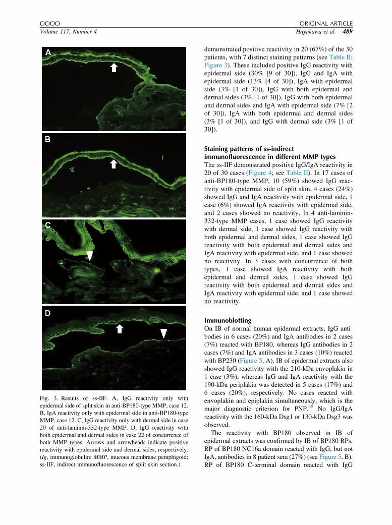
Fig. 3. Results of ss-IIF. A, IgG reactivity only withepidermal side of split skin in anti-BP180-type MMP, case 12.B, IgA reactivity only with epidermal side in anti-BP180-typeMMP, case 12. C, IgG reactivity only with dermal side in case20 of anti-laminin-332-type MMP. D, IgG reactivity withboth epidermal and dermal sides in case 22 of concurrence ofboth MMP types. Arrows and arrowheads indicate positivereactivity with epidermal side and dermal sides, respectively.(Ig, immunoglobulin; MMP, mucous membrane pemphigoid;ss-IIF, indirect immunofluorescence of split skin section.)
OOOO ORIGINAL ARTICLE
Volume 117, Number 4 Hayakawa et al. 489
demonstrated positive reactivity in 20 (67%) of the 30patients, with 7 distinct staining patterns (see Table II;Figure 3). These included positive IgG reactivity withepidermal side (30% [9 of 30]), IgG and IgA withepidermal side (13% [4 of 30]), IgA with epidermalside (3% [1 of 30]), IgG with both epidermal anddermal sides (3% [1 of 30]), IgG with both epidermaland dermal sides and IgA with epidermal side (7% [2of 30]), IgA with both epidermal and dermal sides(3% [1 of 30]), and IgG with dermal side (3% [1 of30]).
Staining patterns of ss-indirectimmunofluorescence in different MMP typesThe ss-IIF demonstrated positive IgG/IgA reactivity in20 of 30 cases (Figure 4; see Table II). In 17 cases ofanti-BP180-type MMP, 10 (59%) showed IgG reac-tivity with epidermal side of split skin, 4 cases (24%)showed IgG and IgA reactivity with epidermal side, 1case (6%) showed IgA reactivity with epidermal side,and 2 cases showed no reactivity. In 4 anti-laminin-332-type MMP cases, 1 case showed IgG reactivitywith dermal side, 1 case showed IgG reactivity withboth epidermal and dermal sides, 1 case showed IgGreactivity with both epidermal and dermal sides andIgA reactivity with epidermal side, and 1 case showedno reactivity. In 3 cases with concurrence of bothtypes, 1 case showed IgA reactivity with bothepidermal and dermal sides, 1 case showed IgGreactivity with both epidermal and dermal sides andIgA reactivity with epidermal side, and 1 case showedno reactivity.
ImmunoblottingOn IB of normal human epidermal extracts, IgG anti-bodies in 6 cases (20%) and IgA antibodies in 2 cases(7%) reacted with BP180, whereas IgG antibodies in 2cases (7%) and IgA antibodies in 3 cases (10%) reactedwith BP230 (Figure 5, A). IB of epidermal extracts alsoshowed IgG reactivity with the 210-kDa envoplakin in1 case (3%), whereas IgG and IgA reactivity with the190-kDa periplakin was detected in 5 cases (17%) and6 cases (20%), respectively. No cases reacted withenvoplakin and epiplakin simultaneously, which is themajor diagnostic criterion for PNP.48 No IgG/IgAreactivity with the 160-kDa Dsg1 or 130-kDa Dsg3 wasobserved.
The reactivity with BP180 observed in IB ofepidermal extracts was confirmed by IB of BP180 RPs.RP of BP180 NC16a domain reacted with IgG, but notIgA, antibodies in 8 patient sera (27%) (see Figure 5, B).RP of BP180 C-terminal domain reacted with IgG
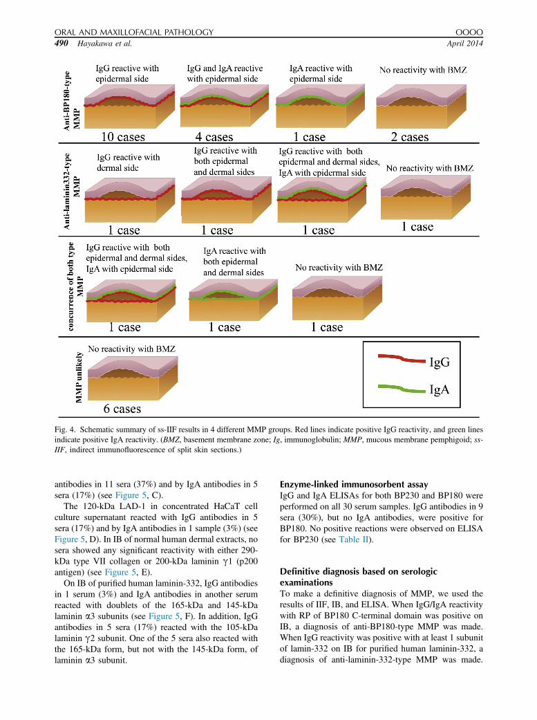
Fig. 4. Schematic summary of ss-IIF results in 4 different MMP groups. Red lines indicate positive IgG reactivity, and green linesindicate positive IgA reactivity. (BMZ, basement membrane zone; Ig, immunoglobulin; MMP, mucous membrane pemphigoid; ss-IIF, indirect immunofluorescence of split skin sections.)
ORAL AND MAXILLOFACIAL PATHOLOGY OOOO
490 Hayakawa et al. April 2014
antibodies in 11 sera (37%) and by IgA antibodies in 5sera (17%) (see Figure 5, C).
The 120-kDa LAD-1 in concentrated HaCaT cellculture supernatant reacted with IgG antibodies in 5sera (17%) and by IgA antibodies in 1 sample (3%) (seeFigure 5, D). In IB of normal human dermal extracts, nosera showed any significant reactivity with either 290-kDa type VII collagen or 200-kDa laminin g1 (p200antigen) (see Figure 5, E).
On IB of purified human laminin-332, IgG antibodiesin 1 serum (3%) and IgA antibodies in another serumreacted with doublets of the 165-kDa and 145-kDalaminin a3 subunits (see Figure 5, F). In addition, IgGantibodies in 5 sera (17%) reacted with the 105-kDalaminin g2 subunit. One of the 5 sera also reacted withthe 165-kDa form, but not with the 145-kDa form, oflaminin a3 subunit.
Enzyme-linked immunosorbent assayIgG and IgA ELISAs for both BP230 and BP180 wereperformed on all 30 serum samples. IgG antibodies in 9sera (30%), but no IgA antibodies, were positive forBP180. No positive reactions were observed on ELISAfor BP230 (see Table II).
Definitive diagnosis based on serologicexaminationsTo make a definitive diagnosis of MMP, we used theresults of IIF, IB, and ELISA. When IgG/IgA reactivitywith RP of BP180 C-terminal domain was positive onIB, a diagnosis of anti-BP180-type MMP was made.When IgG reactivity was positive with at least 1 subunitof lamin-332 on IB for purified human laminin-332, adiagnosis of anti-laminin-332-type MMP was made.
OOOO ORIGINAL ARTICLE
Volume 117, Number 4 Hayakawa et al. 491
Thus, when both tests were positive, the patient wasdiagnosed as having concurrence of both MMP types.
Patients without any positive reactivity on IB testswere diagnosed based on the results of ss-IIF. WhenIgG/IgA reactivity to the epidermal side was seen onss-IIF, a diagnosis of anti-BP180-type MMP was made,as this ss-IIF pattern is characteristic of anti-BP180-type MMP.24,49 When IgG reactivity on the dermal sidewas observed, a diagnosis of anti-laminin-332-typeMMP was made.35,36 When reactivity was positive onboth sides of split skin, the patient was diagnosed asconcurrence of both MMP types. Remaining patientswith negative results in both IB and ss-IIF studies werediagnosed as “MMP-unlikely.”
All 30 patients with suspected MMP were diagnosedby these criteria. Twelve sera were positive for anti-bodies against BP180 C-terminal domain RP in IB, and7 sera were positive for antibodies against anti-laminin-332. Among the 14 IB-negative sera, 8 sera reactedwith epidermal side on ss-IIF. The remaining 6 serashowed no positive reactivity on either IB or ss-IIF.Three sera reacted with both BP180 C-terminal domainRP and laminin-332. Four sera showed reactivity withboth the epidermal and dermal sides on ss-IIF. How-ever, in these 4 samples, which showed reactivity withboth epidermal and dermal sides on ss-IIF, 2 serareacted only with laminin-332, but not with the BP180C-terminal domain, on IB. As 2 sera had very weakreactivity with the epidermal side on ss-IIF and no anti-BP180-antibodies were detected on IB, we diagnosedthese as anti-laminin-332-type MMP. Eventually, wediagnosed 17 cases as anti-BP180-type MMP, 4 casesas anti-laminin-332-type MMP, 3 cases as concurrenceof both MMP types, and 6 cases as MMP-unlikely (seeTable II).
Finally, we analyzed the results for ELISA of theBP180 NC16a domain RP. Eight of the 17 anti-BP180-type MMP cases and 1 of the 3 concurrent MMP cases,but none of the 4 anti-laminin-332-type MMP cases,were positive on BP180 ELISA. These results sug-gested that BP180 ELISA may be useful in the diagno-sis of anti-BP180-type MMP, although to date BP180ELISA has mainly been used for the diagnosis of BP.
Statistical analyses of relationships betweenclinical features and MMP typesWe statistically compared the clinical features, includingage at onset, gender, lesional sites, lesional status,presence of extraoral lesions, presence of malignanttumor, and clinical outcome, among the different MMPtypes. The mean age at onset in MMP-unlikely patientswas 52.3 years, which was significantly younger thanthe 67.2 years in patients diagnosed as any MMP type(P ¼ .0271). Gingiva and buccal mucosa were mostcommonly affected in both MMP and MMP-unlikely
patients, but no significant differences in affected siteswere observed among different MMP types. Frequencyof malignant complications was similar among dif-ferent MMP types (P > .05). Stomach cancer and livercancer were associated with 1 case each of anti-laminin-332-type MMP, although the difference in frequency ofassociation of malignant tumors between anti-laminin-332-type MMP and anti-BP180-type MMP was notstatistically significant (P ¼ .079).
Outcome was determined by improvement of oralsymptoms: grade 0 for deterioration, grade 1 for nochange, grade 2 for moderate improvement, and grade 3for marked improvement. Nine (30%) of the 30 patientswere refractory to topical or systemic therapies andwere rated as grade 0 or 1. Nineteen (79%) patientsshowed good response to topical and systemic treat-ments and were rated as grade 2 or 3. In contrast, only 2of 6 MMP-unlikely patients improved with topicaltreatment. When we compared patients with grades 2and 3 with those with grades 0 and 1, improvement inboth anti-laminin-332-type MMP and all MMP wassignificantly better than in MMP-unlikely patients(P ¼ .035 and P ¼ .028, respectively).
Relationship between immunoglobulin isotypesand BP180 domainsDomain specificity of anti-BP180 antibodies was clas-sified based on the results of IB and ELISA studiesusing RPs of BP180 NC16a and C-terminal domains,and the relationship with immunoglobulin isotypes wasexamined (Table III). Five sera reactive with NC16adomain showed only IgG antibodies. Among the 5 serareactive with both NC16a and C-terminal domains, 4sera showed only IgG antibodies and 1 serum showedboth IgG and IgA antibodies. Among the 7 sera reactiveonly with the C-terminal domain, 1 serum showed onlyIgA antibodies, 2 sera showed only IgG antibodies, and4 sera showed both IgG and IgA antibodies. Statisticalanalysis found that IgA antibodies had significantlyhigher reactivity with BP180 C-terminal domain thanNC16a domain (P ¼ .004678). Furthermore, no pa-tients showed IgA antibodies against BP180 NC16adomain in this study.
DISCUSSIONIn this study, when the results of ss-IIF and IB studieswere combined, 24 patients with suspected MMP(80%) demonstrated anti-BMZ autoantibodies and werediagnosed as 1 of 3 MMP types. In contrast, theremaining 6 cases were negative on all tests and wereconsidered unlikely to have MMP. We detected circu-lating IgG/IgA anti-BMZ antibodies in the sera of 20 of30 patients with suspected MMP by ss-IIF. Further IBstudies found that 21 sera had IgG/IgA reactivity with
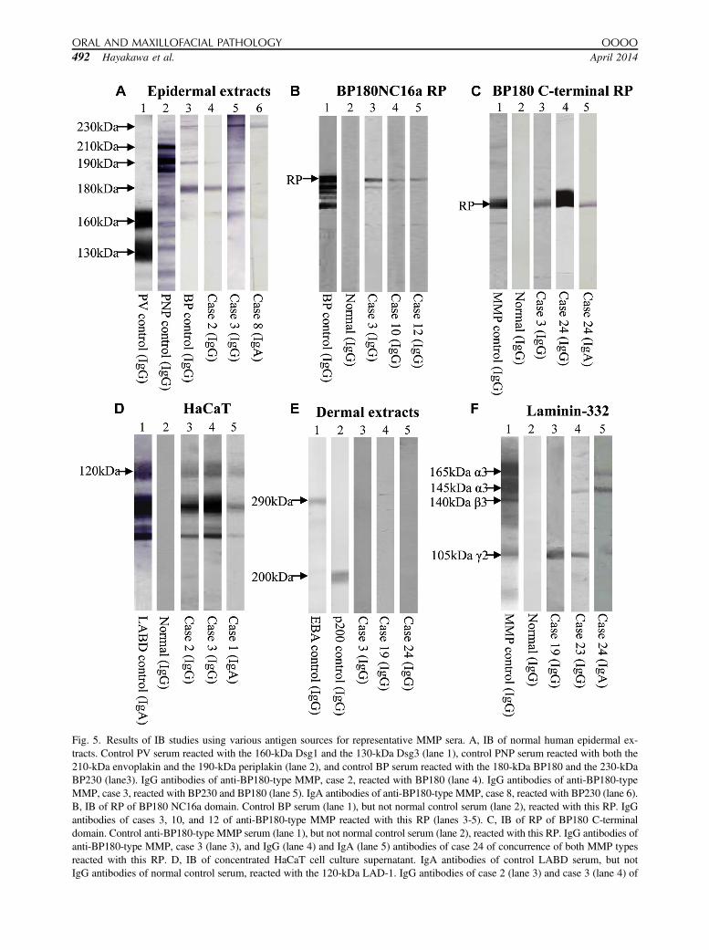
Fig. 5. Results of IB studies using various antigen sources for representative MMP sera. A, IB of normal human epidermal ex-tracts. Control PV serum reacted with the 160-kDa Dsg1 and the 130-kDa Dsg3 (lane 1), control PNP serum reacted with both the210-kDa envoplakin and the 190-kDa periplakin (lane 2), and control BP serum reacted with the 180-kDa BP180 and the 230-kDaBP230 (lane3). IgG antibodies of anti-BP180-type MMP, case 2, reacted with BP180 (lane 4). IgG antibodies of anti-BP180-typeMMP, case 3, reacted with BP230 and BP180 (lane 5). IgA antibodies of anti-BP180-type MMP, case 8, reacted with BP230 (lane 6).B, IB of RP of BP180 NC16a domain. Control BP serum (lane 1), but not normal control serum (lane 2), reacted with this RP. IgGantibodies of cases 3, 10, and 12 of anti-BP180-type MMP reacted with this RP (lanes 3-5). C, IB of RP of BP180 C-terminaldomain. Control anti-BP180-type MMP serum (lane 1), but not normal control serum (lane 2), reacted with this RP. IgG antibodies ofanti-BP180-type MMP, case 3 (lane 3), and IgG (lane 4) and IgA (lane 5) antibodies of case 24 of concurrence of both MMP typesreacted with this RP. D, IB of concentrated HaCaT cell culture supernatant. IgA antibodies of control LABD serum, but notIgG antibodies of normal control serum, reacted with the 120-kDa LAD-1. IgG antibodies of case 2 (lane 3) and case 3 (lane 4) of
ORAL AND MAXILLOFACIAL PATHOLOGY OOOO
492 Hayakawa et al. April 2014

Table III. Relationship between immunoglobulin iso-topes and BP180 domains
IgG IgG þ IgA IgA
NC16a 5 0 0NC16a þ C-terminus 4 1 0C-terminus 2 4 1
BP180, type XVII collagen; Ig, immunoglobulin; NC16a, non-collagenous 16a domain.
OOOO ORIGINAL ARTICLE
Volume 117, Number 4 Hayakawa et al. 493
various BMZ autoantigens. Specifically, 17 sera(56.7%) reacted with epidermal side on ss-IIF or withBP180 C-terminal domain RP and were diagnosed asanti-BP180-type MMP. Nine of 17 anti-BP180-typeMMP patients had IgG/IgA autoantibodies againstBP180 C-terminal domain. In contrast, 7 sera (23.3%)reacted with laminin-332 and were diagnosed as anti-laminin-332-type MMP. This incidence of anti-laminin-332-type MMP was consistent with previous findings.35
Although there are differences between anti-BP180-type MMP and anti-laminin-332-type MMP, the 2 typesare difficult to distinguish by clinical features alone,particularly in patients presenting only with oral symp-toms. Thus, ss-IIF is commonly used to differentiate the2 types. However, in our study, 4 MMP patients werenegative on ss-IIF, and the diagnosis was confirmed byIB results.
One patient (case 24) with concurrence of both typesshowed IgA, but not IgG, antibodies reactive with bothsides of split skin on ss-IIF and with the 165-kDa and145-kDa forms of laminin a3 subunit on IB. This casewould be diagnosed as LABD based on the results ofserologic tests. However, because the patient had onlyoral lesions, we diagnosed this case as IgA anti-lami-nin-332-type MMP.
Two anti-laminin-332-type MMP cases (cases 18and 19) showed reactivity with both epidermal anddermal sides of split skin. However, because the 2 serashowed much weaker reactivity with the epidermal sideand did not react with the BP180 C-terminal domainRP, we ultimately diagnosed them as anti-laminin-332-type MMP, although the possibility that the seracontained low titer antibodies against BP180 not beexcluded.
Sensitivity of ELISA for the BP180 NC16a domainRP in the diagnosis of MMP has not been well docu-mented, although this ELISA is highly sensitive andreliable for the diagnosis of BP.20,50 In our study, 8 of17 anti-BP180-type MMP patients were positive onELISA for the BP180 NC16a domain. These 8 caseswere convincingly diagnosed as anti-BP180-type MMPbased on typical clinical features and findings on IB and
anti-BP180-type MMP and IgA antibodies of case 1 of anti-BP180-tydermal extracts. Control EBA serum reacted with the 290-kDa typpemphigoid serum reacted with the 200-kDa laminin g1 (p200) (laneand case 19 (lane 4) of anti-laminin-332-type MMP, and IgG antibodnot show any reactivity. F, IB of purified human laminin-332. Controand 145-kDa forms of a3 subunit, the 140-kDa b3 subunit, and thecontrol serum did not react with any subunits (lane 2). IgG antibodi105-kDa g2 (lane 4). IgG antibodies of case 23 of concurrence ofantibodies of case 24 of concurrence of both MMP types reacted w(BP, bullous pemphigoid; EBA, epidermolysis bullosa acquisita;LABD, linear IgA dermatosis; LAD-1, linear IgA bullous dermatoparaneoplastic pemphigus; PV, pemphigus vulgaris; RP, recombina
ss-IIF. In addition, all patients with anti-laminin-332type MMP and MMP-unlikely were negative by BP180ELISA. Therefore, although sensitivity was only around50%, positive BP180 ELISA in patients suspected forMMP strongly suggests anti-BP180-type MMP.
In the present study, sera reactive with BP180NC16atended to have only IgG antibodies, whereas serareactive with C-terminus tended to have IgG/IgA anti-bodies (see Table III), which is consistent with previousresults.18,19,51 These findings suggest that distinctimmunoglobulin isotypes target different epitopes onBP180, and this may contribute to different patho-physiology between BP and MMP.
Six patients, who showed no positive reactions onany serologic tests, were diagnosed as MMP-unlikely.However, it is possible that these patients react withother untested antigens. For example, MMP patientswith ocular and oral lesions were reported to react withintegrin b4 and integrin a6, respectively, by IB usingbovine gingival lysate.40 We have also confirmed pos-itive reactivity with integrin b4 in pure ocular MMPsera using novel IB with hemidesmosome-rich fraction(Li et al, manuscript in preparation).
After MMP types were determined, we analyzed theclinical findings in the different subtypes. Anti-BP180-type MMP tended to show diffuse and erythematousstomatitis with shallow ulcers, whereas anti-laminin-332-type MMP tended to show gingivitis and stomatitiswith deep ulceration and necrosis (see clinical featuresin Figure 2). These different clinical presentationsmay be caused by different targets in the epidermalBMZ between BP180 and laminin-332.52,53 In patientswith suspected MMP, clinical findings of deep
pe MMP (lane 5) reacted with LAD-1. E, IB of normal humane VII collagen (lane 1), and control anti-laminin g1 (p200)2). IgG antibodies of case 3 (lane 3) of anti-BP180-type MMPies of case 24 of concurrence of both MMP types (lane 5), didl anti-laminin-332 type MMP serum reacted with the 165-kDa105-kDa g1 subunit of laminin-332 (lane 1), whereas normales of case 19 of anti-laminin-332-type MMP reacted with theboth MMP types reacted with the105-kDa g2 (lane 5). IgAith the 165-kDa and 145-kDa forms of a3 subunit (lane 5).
HaCaT, cell line; IB, immunoblotting; Ig, immunoglobulin;sis antigen-1; MMP, mucous membrane pemphigoid; PNP,nt protein.)
ORAL AND MAXILLOFACIAL PATHOLOGY OOOO
494 Hayakawa et al. April 2014
ulceration on the gingival and buccal mucosa maysuggest a diagnosis of anti-laminin-332-type MMP,even before specific autoantibodies and autoanti-gens are detected. Previous studies indicated that anti-laminin-332-type MMP shows more severe clinicalfeatures than BP180-type MMP, and is characterized bythe presence of lesions involving additional mucousmembranes.54 However, our study did not reflect thistendency, probably because most patients showed onlyoral mucosal lesions.
It has been well documented that anti-laminin-332-type MMP patients frequently show internal malig-nancies, particularly cancers of the digestive tract.54 Inthis context, it is important to distinguish between the 2MMP types. Indeed, our study found that 2 patientswith anti-laminin-332-type MMP had a history ofdigestive malignancies (stomach and liver cancers),although no significant differences in associated malig-nant diseases were observed between anti-BP180-typeMMP and anti-laminin-332-type MMP.
In addition to a frequent association with malignantneoplasms,55 anti-laminin-332-type MMP is known toshow significant complications, including blindness andlaryngeal stenosis.56,57 However, our study did not findclear differences in these clinical features and thera-peutic response between the 2 MMP types.
For histopathologic examination, biopsy specimenstaken from gingival lesions may cause periodontal de-fects. In addition, chronic inflammation in the gingivitismay interfere with histologic evaluation.58 In this study,we were unable to confirm the usefulness of histo-pathologic study, owing to the inadequate quality of ourbiopsy specimens. In addition, we did not performdirect immunofluorescence (DIF) studies. However,DIF using biopsy samples taken from normal-lookingmucosa should be performed in future studies, becausepositive deposition of immunoglobulins and C3 by DIFmay be the only diagnostic finding in cases withoutpositive results from serologic tests.
PV and BP are generally controlled by systemiccorticosteroids in dermatologic practice. However, mostof our patients had only mild and limited oral symp-toms. Therefore, to control the oral symptoms, we usedazulene gargle and topical application of corticosteroidointments. Most cases showed good clinical course withonly topical steroids as both initial and maintenancetreatments, and the few refractory patients were treatedwith intermittent systemic steroids at minimal dose.
In conclusion, when assessing patients with erosiveoral lesions, the possibility of MMP should be consid-ered in the differential diagnosis, and patients should bereferred to a clinician experienced in the treatment ofABDs. To facilitate diagnosis of MMP, ELISA-basedsystems for both BP180 C-terminal domain and lami-nin-332 should be developed. We are now preparing an
ELISA using bacterial RP of BP180 C-terminaldomain. However, our study indicates that the currentlyavailable ELISA against the BP180 NC16a domain RPis able to help render a diagnosis in half of anti-BP180-type MMP patients. Therefore, the current ELISAshould be used in patients with suspected MMPregardless of its subtypes. However, even if a diagnosisof MMP is suggested on the basis of the results fromthe currently available ELISA, further examination isstill necessary to detect other autoantigens to confirmthe MMP subtype.
We gratefully appreciate Ms. Takako Ishikawa for technicalassistance and Ms. Mihoko Ikeda, Ms. Mami Nishida, ShokoNakamura, and Ms. Tomoko Tashima for secretarial work.We thank the patients for their participation.
REFERENCES1. Darling MR, Daley T. Blistering mucocutaneous diseases of the
oral mucosada review, part 1: mucous membrane pemphigoid.J Can Dent Assoc. 2005;71:851-854.
2. Darling MR, Daley T. Blistering mucocutaneous diseases of theoral mucosada review, part 2: pemphigus vulgaris. J Can DentAssoc. 2006;72:63-66.
3. Blöcker IM, Dähnrich C, Probst C, et al. Epitope mapping ofBP230 leading to a novel enzyme-linked immunosorbent assayfor autoantibodies in bullous pemphigoid. Br J Dermatol.2012;166:964-970.
4. Charneux J, Lorin J, Vitry F, et al. Usefulness of BP230 andBP180-NC16a enzyme-linked immunosorbent assays in the initialdiagnosis of bullous pemphigoid: a retrospective study of 138patients. Arch Dermatol. 2011;147:286-291.
5. Roussel A, Benichou J, Randriamanantany ZA, et al. Enzyme-linked immunosorbent assay for the combination of bullouspemphigoid antigens 1 and 2 in the diagnosis of bullous pem-phigoid. Arch Dermatol. 2011;147:293-298.
6. Hashimoto T. Recent advances in the study of the pathophysi-ology of pemphigus. Arch Dermatol Res. 2003;295(suppl 1):S2-S11.
7. Bracke S, Speeckaert R, Van Geel N, De Bacquer D, Lambert J.Evaluation of commercially available ELISA assays as a tool formonitoring and managing pemphigus patients: a prospectivestudy. Eur J Dermatol. 2013;23:33-39.
8. Amagai M, Komai A, Hashimoto T, et al. Usefulness of enzyme-linked immunosorbent assay using recombinant desmogleins 1and 3 for serodiagnosis of pemphigus. Br J Dermatol. 1999;140:351-357.
9. Ishii K, Amagai M, Hall RP, et al. Characterization of autoanti-bodies in pemphigus using antigen-specific enzyme-linkedimmunosorbent assays with baculovirus-expressed recombinantdesmogleins. J Immunol. 1997;159:2010-2017.
10. Beutner EH, Jordon RE. Demonstration of skin antibodies in seraof pemphigus vulgaris patients by indirect immunofluorescentstaining. Proc Soc Exp Biol Med. 1964;117:505-510.
11. Jordon RE, Beutner EH, Witebsky E, Blumental G, Hale WL,Lever WF. Basement zone antibodies in bullous pemphigoid.JAMA. 1967;200:751-756.
12. Amagai M, Klaus-Kovtun V, Stanley JR. Autoantibodies againsta novel epithelial cadherin in pemphigus vulgaris, a disease of celladhesion. Cell. 1991;67:869-877.
13. Diaz LA, Ratrie H, Saunders WS, et al. Isolation of a humanepidermal cDNA corresponding to the 180-kD autoantigen
OOOO ORIGINAL ARTICLE
Volume 117, Number 4 Hayakawa et al. 495
recognized by bullous pemphigoid and herpes gestationis sera:immunolocalization of this protein to the hemidesmosome. J ClinInvest. 1990;86:1088-1094.
14. Stanley JR, Yaar M, Hawley-Nelson P, Katz SI. Pemphigus an-tibodies identify a cell surface glycoprotein synthesized by humanand mouse keratinocytes. J Clin Invest. 1982;70:281-288.
15. Stanley JR, Koulu L, Thivolet C. Distinction between epidermalantigens binding pemphigus vulgaris and pemphigus foliaceusautoantibodies. J Clin Invest. 1984;74:313-320.
16. Hashimoto T, Ogawa MM, Konohana A, Nishikawa T. Detectionof pemphigus vulgaris and pemphigus foliaceus antigens byimmunoblot analysis using different antigen sources. J InvestDermatol. 1990;94:327-331.
17. Giudice GJ, Emery DJ, Diaz LA. Cloning and primary structuralanalysis of the bullous pemphigoid autoantigen BP180. J InvestDermatol. 1992;99:243-250.
18. Giudice GJ, Emery DJ, Zelickson BD, Anhalt GJ, Liu Z,Diaz LA. Bullous pemphigoid and herpes gestationis autoanti-bodies recognize a common non-collagenous site on the BP180ectodomain. J Immunol. 1993;151:5742-5750.
19. Matsumura K, Amagai M, Nishikawa T, Hashimoto T. Themajority of bullous pemphigoid and herpes gestationis serumsamples react with the NC16a domain of the 180-kDabullous pemphigoid antigen. Arch Dermatol Res. 1996;288:507-509.
20. Kobayashi M, Amagai M, Kuroda-Kinoshita K, et al. BP180ELISA using bacterial recombinant NC16a protein as a diagnosticand monitoring tool for bullous pemphigoid. J Dermatol Sci.2002;30:224-232.
21. Yoshida M, Hamada T, Amagai M, et al. Enzyme-linkedimmunosorbent assay using bacterial recombinant proteins ofhuman BP230 as a diagnostic tool for bullous pemphigoid.J Dermatol Sci. 2006;41:21-30.
22. Saleh MA, Ishii K, Kim YJ, et al. Development of NC1 and NC2domains of type VII collagen ELISA for the diagnosis andanalysis of the time course of epidermolysis bullosa acquisitapatients. J Dermatol Sci. 2011;62:169-175.
23. Kim JH, Kim YH, Kim S, et al. Serum levels of anti-type VIIcollagen antibodies detected by enzyme-linked immunosorbentassay in patients with epidermolysis bullosa acquisita are corre-lated with the severity of skin lesions. J Eur Acad DermatolVenereol. 2013;27:e224-e230.
24. Chan LS, Ahmed AR, Anhalt GJ, et al. The first internationalconsensus on mucous membrane pemphigoid: definition, diag-nostic criteria, pathogenic factors, medical treatment, and prog-nostic indicators. Arch Dermatol. 2002;138:370-379.
25. Thorne JE, Anhalt GJ, Jabs DA. Mucous membrane pemphigoidand pseudopemphigoid. Ophthalmology. 2004;111:45-52.
26. Ahmed AR, Kurgis BS, Rogers RS. Cicatricial pemphigoid. J AmAcad Dermatol. 1991;24:987-1001.
27. Zillikens D. Acquired skin disease of hemidesmosomes.J Dermatol Sci. 1999;20:134-154.
28. Schmidt E, Skrobek C, Kromminga A, et al. Cicatricial pem-phigoid: IgA and IgG autoantibodies target epitopes on both intra-and extracellular domains of bullous pemphigoid antigen 180. BrJ Dermatol. 2001;145:778-783.
29. Setterfield J, Shirlaw PJ, Kerr-Muir M, et al. Mucous membranepemphigoid: a dual circulating antibody response with IgG andIgA signifies a more severe and persistent disease. Br J Dermatol.1998;138:602-610.
30. Oyama N, Setterfield JF, Powell AM, et al. Bullous pemphigoidantigen II (BP180) and its soluble extracellular domains are majorautoantigens in mucous membrane pemphigoid: the pathogenicrelevance to HLA class II alleles and disease severity. Br JDermatol. 2006;154:90-98.
31. Balding SD, Prost C, Diaz LA, et al. Cicatricial pemphigoid au-toantibodies react with multiple sites on the BP180 extracellulardomain. J Invest Dermatol. 1996;106:141-146.
32. AumailleyM,Bruckner-TudermanL,CarterWG, et al.A simplifiedlaminin nomenclature. Matrix Biol. 2005;24:326-332.
33. Chan LS, Hammerberg C, Cooper KD. Cicatricial pemphigoid:identification of two distinct sets of epidermal antigens by IgA andIgG class circulating autoantibodies. Arch Dermatol. 1990;126:1466-1468.
34. Hisamatsu Y, Nishiyama T, Amano S, Matsui C, Ghohestani R,Hashimoto T. Usefulness of immunoblotting using purified lam-inin 5 in the diagnosis of anti-laminin 5 cicatricial pemphigoid.J Dermatol Sci. 2003;33:113-119.
35. Domloge-Hultsch N, Gammon WR, Briggaman RA, Gil SG,Carter WG, Yancey KB. Epiligrin, the major human keratinocyteintegrin ligand, is a target in both an acquired autoimmune and aninherited subepidermal blistering skin disease. J Clin Invest.1992;90:1628-1633.
36. Domloge-Hultsch N, Anhalt GJ, Gammon WR, et al. Anti-epiligrin cicatricial pemphigoid: a subepithelial bullous disorder.Arch Dermatol. 1994;130:1521-1529.
37. Leverkus M, Bhol K, Hirako Y, et al. Cicatricial pemphigoid withcirculating autoantibodies to beta4 integrin, bullouspemphigoid 180and bullous pemphigoid 230. Br J Dermatol. 2001;145:998-1004.
38. Chan LS, Soong HK, Foster CS, Hammerberg C, Cooper KD.Ocular cicatricial pemphigoid occurring as a sequela of Stevens-Johnson syndrome. JAMA. 1991;266:1543-1546.
39. Chan LS, Majmudar AA, Tran HH, et al. Laminin-6 and laminin-5are recognized by autoantibodies in a subset of cicatricial pem-phigoid. J Invest Dermatol. 1997;108:848-853.
40. Rashid KA, Gürcan HM, Ahmed AR. Antigen specificity insubsets of mucous membrane pemphigoid. J Invest Dermatol.2006;126:2631-2636.
41. Beutner EH, Jordon RE, Chorzelski TP. The immunopathology ofpemphigus and bullous pemphigoid. J Invest Dermatol. 1968;51:63-80.
42. Woodley D, Sauder D, Talley MJ, Silver M, Grotendorst G,Qwarnstrom E. Localization of basement membrane componentsafter dermal-epidermal junction separation. J Invest Dermatol.1983;81:149-153.
43. Nie Z, Hashimoto T. IgA antibodies of cicatricial pemphigoidsera specifically react with C-terminus of BP180. J Invest Der-matol. 1999;112:254-255.
44. Ishii N, Ohyama B, Yamaguchi Z, Hashimoto T. IgA autoanti-bodies against the NC16a domain of BP180 but not 120-kDaLAD-1 detected in a patient with linear IgA disease. Br J Der-matol. 2008;158:1151-1153.
45. Ishii N, Yoshida M, Hisamatsu Y, et al. Epidermolysis bullosaacquisita sera react with distinct epitopes on the NC1 and NC2domains of type VII collagen: study using immunoblotting ofdomain-specific recombinant proteins and postembeddingimmunoelectron microscopy. Br J Dermatol. 2004;150:843-851.
46. Amano S, Scott IC, Takahara K, et al. Bone morphogeneticprotein 1 is an extracellular processing enzyme of the laminin 5gamma 2 chain. J Biol Chem. 2000;275:22728-22735.
47. Laemmli UK. Cleavage of structural proteins during the assemblyof the head of bacteriophage T4. Nature. 1970;227:680-685.
48. Poot AM, Diercks GF, Kramer D, et al. Laboratory diagnosis ofparaneoplastic pemphigus. Br J Dermatol. 2013;169:1016-1024.
49. Kelly SE, Wojnarowska F. The use of chemically split tissue inthe detection of circulating anti-basement membrane zone anti-bodies in bullous pemphigoid and cicatricial pemphigoid. Br JDermatol. 1988;118:31-40.
50. Zillikens D, Mascaro JM, Rose PA, et al. A highly sensitiveenzyme-linked immunosorbent assay for the detection of
ORAL AND MAXILLOFACIAL PATHOLOGY OOOO
496 Hayakawa et al. April 2014
circulating anti-BP180 autoantibodies in patients with bullouspemphigoid. J Invest Dermatol. 1997;109:679-683.
51. Hashimoto T, Han-Yaku H, Higashiyama M, et al. Four Japanesepatients with cicatricial pemphigoid showing both IgG and IgAantibodies against the 180 kDa bullous pemphigoid antigen. Br JDermatol. 1997;137:305-306.
52. McMillan JR, Akiyama M, Shimizu H. Ultrastructural orientationof laminin 5 in the epidermal basement membrane: an updatedmodel for basement membrane organization. J Histochem Cyto-chem. 2003;51:1299-1306.
53. Powell AM, Sakuma-Oyama Y, Oyama N, Black MM. CollagenXVII/BP180: a collagenous transmembrane protein and compo-nent of the dermoepidermal anchoring complex. Clin Exp Der-matol. 2005;30:682-687.
54. Egan CA, Lazarova Z, Darling TN, Yee C, Coté T, Yancey KB.Anti-epiligrin cicatricial pemphigoid and relative risk for cancer.Lancet. 2001;357:1850-1851.
55. Sadler E, Lazarova Z, Sarasombath P, Yancey KB. A widen-ing perspective regarding the relationship between anti-epiligrin cicatricial pemphigoid and cancer. J Dermatol Sci.2007;47:1-7.
56. Kato K, Moriyama Y, Saito H, Koga H, Hashimoto T. A case ofmucous membrane pemphigoid involving the trachea and bron-chus with autoantibodies to b3 subunit of laminin-332 [publishedonline August 27, 2013]. Acta Derm Venereol.
57. Dainichi T, Takeshita H, Moroi Y, et al. Cicatricial pemphigoidwith autoantibodies against the laminin 5 gamma 2 subunit. Eur JDermatol. 2005;15:189-193.
58. Siegel MA, Anhalt GJ. Direct immunofluorescence of detachedgingival epithelium for diagnosis of cicatricial pemphigoid: reportof five cases. Oral Surg Oral Med Oral Pathol. 1993;75:296-302.
Reprint requests:
Takashi Hashimoto, MDDepartment of DermatologyKurume University School of MedicineKurume University Institute of Cutaneous Cell Biology67 Asahimachi, KurumeFukuoka [email protected]
Related Documents