SPORTS MEDICINE Diagnosis and prognosis of acute hamstring injuries in athletes Gino M. M. J. Kerkhoffs • Nick van Es • Thijs Wieldraaijer • Inger N. Sierevelt • Jan Ekstrand • C. Niek van Dijk Received: 17 December 2011 / Accepted: 10 May 2012 / Published online: 24 May 2012 Ó The Author(s) 2012. This article is published with open access at Springerlink.com Abstract Purpose Identification of the most relevant diagnostic and prognostic factors of physical examination and imag- ing of hamstring injuries in (elite) athletes. Methods A literature search was conducted in MEDLINE and EMBASE for articles between 1950 and April 2011. A survey was distributed among the members of the Euro- pean Society of Sports Traumatology, Knee Surgery and Arthroscopy, which focused on physical examination, prognosis, imaging and laboratory tests of hamstring inju- ries in (elite) athletes. Results Medical history, inspection and palpation of the muscle bellies and imaging are most valuable at the initial assessment according to the literature. Experts considered medical history, posture and gait inspection, inspection and palpation of muscle bellies, range of motion tests, manual muscle testing, referred pain tests and imaging to be most important in the initial assessment of hamstring injuries. Magnetic resonance imaging (MRI) is preferred over ultrasonography and should take place within 3 days post- trauma. Important prognostic factors are injury grade, length of the muscle tear on MR images, MRI-negative injuries and trauma mechanism. Conclusions Posture and gait inspection, inspection and palpation of muscle bellies, range of motion tests, manual muscle testing and referred pain tests within 2 days post- trauma were identified as the most relevant diagnostic factors. Level of evidence Literature review and expert opinion, Level V. Keywords Hamstring strain injury Á Sports injury Á Physical examination Á Imaging Introduction Hamstring injuries are frequent in sports like football [1– 17, 21, 27, 46, 59, 60], Australian rules football [43, 52], sprinting [65], American football [20] and rugby [10]. Considering the explosive character of sprinting, it is not surprising that the incidence of injuries (0.87/1,000 h of exposure) is comparable to the incidence in contact sports (0.92–0.96/1,000 h of exposure) [17, 18, 65]. At the top level, professional football team of 25 players can expect about 7 hamstring injuries per season [16]. These injuries frequently cause a significant loss of time from competition and have a high recurrence rate (12–43 %) [11, 17, 21, 24, 27, 30, 35, 39, 46, 52, 56, 58]. Elite football players sus- taining a hamstring injury cannot participate in competition for a mean of 14 days [17]. The need for a quick and accurate diagnosis and prognosis of hamstring injuries in elite sports is evident and has been given greater emphasis. The number of games in elite sports has increased and the stakes are higher [63]. However, there is little evidence for the diagnostic and prognostic value of several physical tests [50, 61] and discussion continues on the optimal imaging technique [1, 8, 13, 14, 34, 50, 61]. The objective of this study was to identify the most relevant diagnostic and prognostic aspects of physical examination and addi- tional studies of hamstring injuries in (elite) athletes. G. M. M. J. Kerkhoffs (&) Á N. van Es Á T. Wieldraaijer Á I. N. Sierevelt Á C. N. van Dijk ESSKA Sports Committee, Department of Orthopedic Surgery, Academic Medical Center, 1105 AZ Amsterdam, The Netherlands e-mail: [email protected] J. Ekstrand Department of Medical and Health Sciences, Linko ¨ping University, Linko ¨ping, Sweden 123 Knee Surg Sports Traumatol Arthrosc (2013) 21:500–509 DOI 10.1007/s00167-012-2055-x

Welcome message from author
This document is posted to help you gain knowledge. Please leave a comment to let me know what you think about it! Share it to your friends and learn new things together.
Transcript
SPORTS MEDICINE
Diagnosis and prognosis of acute hamstring injuries in athletes
Gino M. M. J. Kerkhoffs • Nick van Es •
Thijs Wieldraaijer • Inger N. Sierevelt •
Jan Ekstrand • C. Niek van Dijk
Received: 17 December 2011 / Accepted: 10 May 2012 / Published online: 24 May 2012
� The Author(s) 2012. This article is published with open access at Springerlink.com
Abstract
Purpose Identification of the most relevant diagnostic
and prognostic factors of physical examination and imag-
ing of hamstring injuries in (elite) athletes.
Methods A literature search was conducted in MEDLINE
and EMBASE for articles between 1950 and April 2011. A
survey was distributed among the members of the Euro-
pean Society of Sports Traumatology, Knee Surgery and
Arthroscopy, which focused on physical examination,
prognosis, imaging and laboratory tests of hamstring inju-
ries in (elite) athletes.
Results Medical history, inspection and palpation of the
muscle bellies and imaging are most valuable at the initial
assessment according to the literature. Experts considered
medical history, posture and gait inspection, inspection and
palpation of muscle bellies, range of motion tests, manual
muscle testing, referred pain tests and imaging to be most
important in the initial assessment of hamstring injuries.
Magnetic resonance imaging (MRI) is preferred over
ultrasonography and should take place within 3 days post-
trauma. Important prognostic factors are injury grade,
length of the muscle tear on MR images, MRI-negative
injuries and trauma mechanism.
Conclusions Posture and gait inspection, inspection and
palpation of muscle bellies, range of motion tests, manual
muscle testing and referred pain tests within 2 days post-
trauma were identified as the most relevant diagnostic factors.
Level of evidence Literature review and expert opinion,
Level V.
Keywords Hamstring strain injury � Sports injury �Physical examination � Imaging
Introduction
Hamstring injuries are frequent in sports like football [1–
17, 21, 27, 46, 59, 60], Australian rules football [43, 52],
sprinting [65], American football [20] and rugby [10].
Considering the explosive character of sprinting, it is not
surprising that the incidence of injuries (0.87/1,000 h of
exposure) is comparable to the incidence in contact sports
(0.92–0.96/1,000 h of exposure) [17, 18, 65]. At the top
level, professional football team of 25 players can expect
about 7 hamstring injuries per season [16]. These injuries
frequently cause a significant loss of time from competition
and have a high recurrence rate (12–43 %) [11, 17, 21, 24,
27, 30, 35, 39, 46, 52, 56, 58]. Elite football players sus-
taining a hamstring injury cannot participate in competition
for a mean of 14 days [17]. The need for a quick and
accurate diagnosis and prognosis of hamstring injuries in
elite sports is evident and has been given greater emphasis.
The number of games in elite sports has increased and the
stakes are higher [63]. However, there is little evidence for
the diagnostic and prognostic value of several physical
tests [50, 61] and discussion continues on the optimal
imaging technique [1, 8, 13, 14, 34, 50, 61]. The objective
of this study was to identify the most relevant diagnostic
and prognostic aspects of physical examination and addi-
tional studies of hamstring injuries in (elite) athletes.
G. M. M. J. Kerkhoffs (&) � N. van Es � T. Wieldraaijer �I. N. Sierevelt � C. N. van Dijk
ESSKA Sports Committee, Department of Orthopedic Surgery,
Academic Medical Center, 1105 AZ Amsterdam,
The Netherlands
e-mail: [email protected]
J. Ekstrand
Department of Medical and Health Sciences,
Linkoping University, Linkoping, Sweden
123
Knee Surg Sports Traumatol Arthrosc (2013) 21:500–509
DOI 10.1007/s00167-012-2055-x

Materials and methods
Literature review
A comprehensive literature study was conducted for arti-
cles between January 1950 and April 2011 on the diagnosis
and prognosis of hamstring injuries. The strategies that
were used consisted of searching online databases (MED-
LINE and EMBASE) and scanning reference lists. The
search terms used in MEDLINE were hamstring* or
thigh[MeSH] combined with the MeSH-terms ‘Sprains and
Strains/diagnosis’, ‘Muscle, Skeletal/injuries’, ‘Magnetic
resonance imaging’ or ‘Ultrasonography’. In EMBASE,
the terms ‘hamstring’/exp/mj’ or ‘thigh/exp/mj’ and
‘injury’/exp combined with prognos*, diagnos*, assess*,
ultrasonograph*, ultrasound, mri, ‘magnetic resonance
imaging’ or imag* were searched for. Articles concerning
medical history, physical examination, prognosis and
imaging of hamstring injuries were selected.
Expert opinion
As part of a project of the Sports Committee of the Euro-
pean Society of Sports Traumatology, Knee Surgery and
Arthroscopy (ESSKA), ESSKA Members (n = 800) were
invited by e-mail to participate in an English web-based
survey in June 2009. The survey focused on the physical
examination, prognosis, imaging and laboratory tests for
hamstring injuries in (elite) athletes. The questions were
formulated by the authors on the basis of a comprehensive
literature review. The survey was a mixture of open
questions, multiple choice questions and Likert-scale
questions. The five options from which the respondents
could choose in the Likert-scale questions were (1) not
important, (2) of little importance, (3) moderately impor-
tant, (4) important and (5) very important. Additional
information was provided on the way in which the physical
tests were carried out. A pilot survey was distributed
among the orthopaedic surgeons of the Academic Medical
Center in Amsterdam in order to identify indistinct, irrel-
evant and missing questions. One week after the first
invitation to take part, a reminder was sent by e-mail to the
ESSKA members in which they were asked to participate
in the survey.
Statistic analysis
Data were analysed using SPSS version 16.0 (Chicago,
USA). Results were mainly presented in a descriptive way as
frequencies with corresponding percentages and averages
with standard deviations. Likert scales were dichotomised by
combining options 1 and 2 and options 3–5 to, respectively,
the categories ‘not important’ and ‘important’.
Results
One hundred and forty ESSKA members (18 % response
rate) from 34 countries with 18 (SD 9.6) years of experi-
ence completed the questionnaire. The selected articles
from the literature search are categorised according to the
type of article and the level of evidence in Tables 1, 2.
Timing of initial physical examination
Traditionally, the clinical assessment of hamstring injuries
is based on a thorough medical history and physical
examination consisting of posture and gait inspection,
inspection and palpation of muscle bellies, ‘range of
motion’ tests (ROM tests) and manual muscle testing. In
the literature, the initial assessment is often carried out
within 12 h to 2 days post-injury [5, 50, 58, 61]. Advan-
tages of an assessment shortly post-injury are the possi-
bility of quick intervention and a more reliable medical
history. However, possible signs of swelling and ecchy-
mosis may arise a few days later and consequently may not
be noticed at the initial examination [4, 12, 31, 62]. 82 %
of the respondents stated that the initial clinical assessment
of an (elite) athlete with a suspected hamstring injury
should take place within 2 days. This is confirmed in recent
work where it is advised to measure active ROM at the end
of the second day [38].
Palpation
Palpation helps identify the site of injury in cranio-caudal
direction, because of possible involvement of the free
proximal tendon, and determines the injured muscles (lat-
eral: m. biceps femoris, medial: m. semitendinosus and/or
m. semimembranosus). Injuries in the m. biceps femoris
[13] and more cranially palpated injuries [5] might corre-
late with a longer rehabilitation interval.
Flexibility
Flexibility in acute and sub-acute phase was addressed.
Flexibility is tested by means of hamstring ROM tests. In
case of a hamstring injury, the range of motion of the hip
and the knee of the injured leg is significantly decreased
compared to the healthy leg [4]. However, the flexibility of
the hip in the acute situation is often influenced by pain as a
consequence of which the test may be less accurate. Active
ROM is decreased in the acute phase of the injury and it is
advised to be measured at the end of the second day [38].
Use of the classic ‘sit-and-reach’ test is discouraged in the
literature as the testing result is influenced by spinal
mobility (i.e. lumbal flexion), leg length, scapular abduc-
tion and stretch on the peripheral nerves by dorsiflexion of
Knee Surg Sports Traumatol Arthrosc (2013) 21:500–509 501
123

Table 1 Overview of the literature review source articles
Article Article design (type) Level of
evidence
Allen et al. [1] Expert opinion/background V
Arnason et al. [2] Epidemiological review
Retrospective cohort study
II
Arnason et al. [3] Original article
Prospective therapeutic study
II
Askling et al. [4] Original article
Prospective prognostic study
II
Askling et al. [5] Original article
Prognostic case series
II
Askling et al. [6] Original article
Prognostic case series
II
Askling et al. [7] Original article
Prognostic case series
IV
Bencardino et al.
[8]
Expert opinion/background V
Blankerbaker et al.
[9]
Expert opinion/background V
Brooks et al. [10] Original article
Cohort study (prevention)
III
Carling et al. [11] Epidemiological review
Prognostic case series
II
Cohen et al. [12] Literature review/background V
Connell et al. [13] Original article
Diagnostic case series
I
Davis [14] Expert opinion/background V
Ekstrand et al. [15] Original article
Prospective cohort study
II
Ekstrand et al. [16] Original article
Prospective two-cohort study
II
Ekstrand et al. [17] Original article
Prospective cohort study
II
Ekstrand et al. [18] Original article
Prospective cohort study
II
Ekstrand et al. [19] Original article
Prospective cohort study
II
Elliott et al. [20] Descriptive epidemiology study
Prospective cohort study
II
Engebretsen et al.
[21]
Original article
Prospective cohort study
II
Fleckenstein et al.
[22]
Original article
Diagnostic case series (descriptive)
III
Fleckenstein et al.
[23]
Expert opinion/background V
Gibbs et al. [24] Original article
Prospective diagnostic study
I
Gielen et al. [25] Expert opinion/background
Descriptive chapter
V
Table 1 continued
Article Article design (type) Level of
evidence
Guerrero et al. [26] Original article
Prognostic case series
III
Hagglund et al.
[27]
Original article
Prospective prognostic study
I
Heiderscheit et al.
[29]
Expert opinion/background V
Heiser et al. [30] Original article
Retrospective cohort study
III
Klingele et al. [31] Original article
Retrospective cohort study
III
Kornberg et al.
[32]
Original article
Therapeutic cohort study
II
Koulouris et al.
[33]
Original article
Retrospective cohort study
III
Koulouris et al.
[34]
Expert opinion/background V
Koulouris et al.
[35]
Original article
Prognostic cohort study
III
Lempainen et al.
[36]
Original article
Retrospective case series
IV
Liemohn et al. [37] Original article
Therapeutic case series
IV
Malliaropoulos
et al. [38]
Original article
Prognostic cohort study
II
Malliaropoulos
et al. [39]
Original article
Prognostic cohort study
I
Martınez Amat
et al. [40]
Original article
Diagnostic cohort study
II
Minarro et al. [41] Original article
Diagnostic cohort study
IV
Nikolaou et al. [42] Biomechanical and histological
evaluation of muscle
IV
Orchard et al. [43] Original article
Retrospective epidemiologic study
III
Orchard et al. [44] Expert opinion/background V
Peetrons [45] Expert opinion/background V
Petersen et al. [46] Original article
Prospective cohort study
II
Puranen et al. [47] Expert opinion/background V
Sallay et al. [48] Original article
Descriptive case series
III
Sarimo et al. [49] Original article
Retrospective case series
IV
Schneider-Kolsky
et al. [50]
Original article
Diagnostic cohort study
I
Schneider-Kolsky
et al. [51]
Author’s reply V
502 Knee Surg Sports Traumatol Arthrosc (2013) 21:500–509
123
the ankle joint [37, 41]. Knee active range of motion deficit
48 h after a unilateral posterior thigh muscle injury is an
objective and accurate measurement, predicting recovery
time in elite athletes [38].
Flexibility tests were pointed out as important by a
majority of the respondents. The ‘sit-and-reach’ test was
considered to be important despite the above-mentioned
negative advice given in the literature. No difference in
importance was found between active and passive ROM
tests (n.s).
Strength
Strength of the hamstring muscles can be tested by means
of knee flexion and hip extension against resistance.
Bilateral comparison is preferred to identify decreased
strength of the injured muscle as a result of pain and/or
fibre disruption [4]. An alternative to measuring the
strength of the hamstring muscles is the ‘take-off-the-shoe’
test (TOST) (or ‘hamstring-drag’ test) in which the patient
is asked to take-off the shoe of the injured leg in a standing
position with the help of the foot of the healthy leg [66].
Although this test is potentially a valuable addition to the
physical examination, the true value should be studied by
comparison with magnetic resonance imaging (MRI) and
recurrence rates [51].
Referred pain
An acute disc prolapse at the L5/S1 level may present with
hamstring and/or calf pain and limitations in hip joint
flexibility, which may mimic a muscle strain. Subtle lum-
bosacral canal impingement of the L5 nerve root however
may in fact also be a common underlying basis for the age-
related predisposition towards hamstring injuries [44]. The
distinction between real hamstring injuries and ‘back-
related’ or ‘neural’ hamstring injuries can be made by the
assessment of referred pain with help of an MRI scan [44,
56]. If the distinction remains difficult, imaging-guided
cortisone injections to the lumbosacral canal region (L5
nerve root) is a relatively painless and complication-free
outpatient procedure with quick recovery that can be used
to distinguish the hamstring-spine dilemma.
Pain felt over the area of the ischial tuberosity and
radiating down the back of the thigh is often labelled as the
‘hamstring syndrome’ [47].
Laboratory tests
Traditional biological markers creatine kinase (CK), lactate
dehydrogenase (LDH), myoglobin (Mb) and uric acid
should not be used for the diagnosis and prognosis of
muscle injuries because of their low sensitivity and speci-
ficity [55]. More research is needed to determine the real
Table 2 Summary of the articles used for this literature review and
level of evidence
Article type Number Level of evidence
I II III IV V
Total 65 6 26 12 6 15
Original article
Epidemiological
review
7 5 2
Prospective 30 6 21 2 1
Retrospective 13 8 5
Literature review 1 1
Expert opinion/
background
13 13
Author’s reply 1 1
Table 1 continued
Article Article design (type) Level of
evidence
Seward et al. [52] Original article
Prospective cohort study
II
Shellock et al. [53] Expert opinion/background V
Slavotinek et al.
[54]
Original article
Prospective RCT
II
Sorichter et al. [55] Original article
Retrospective case–control study
III
Verrall et al. [56] Original article
Prospective prognostic cohort study
II
Verrall et al. [57] Original article
Prospective cohort study
II
Verrall et al. [58] Original article
Prospective cohort study
II
Volpi et al. [59] Epidemiological review
Retrospective cohort study
III
Walden et al. [60] Original article
Prospective cohort study
I
Warren et al. [61] Original article
Prospective observational study
II
Wood et al. [62] Expert opinion/background V
Woods et al. [63] Epidemiological review
Prospective cohort study
II
Woods et al. [64] Epidemiological review
Prospective cohort study
II
Yeung et al. [65] Original article
Prospective cohort study
II
Zeren et al. [66] Original article
Diagnostic cohort study
III
Level of evidence is rendered as ranging from I to V in accordance with
guidelines from the centre for evidence-based medicine, Oxford, UK
Knee Surg Sports Traumatol Arthrosc (2013) 21:500–509 503
123
diagnostic and prognostic value of potential markers, such
as ‘fast myosin heavy chains’ (fast MHC) [26], ‘skeletal-
troponin I’ (sTnI) [55] and ‘alfa-actin’ [40].
Few of the respondents thought that laboratory tests can
be of diagnostic or prognostic importance.
The results of the survey are shown in Table 3.
Imaging
The imaging provides information on the nature and extent
of hamstring injuries. The length of a muscle tear on MR
images or the cross-sectional area of the muscle tear on
ultrasonography (US) is valuable for estimating the con-
valescent period [13, 19, 24, 35, 50, 52]. 88 % of the
respondents in this study use imaging for hamstring injuries
in (elite) athletes.
Imaging technique
MRI and US are the most suitable imaging techniques for
depicting hamstring injuries [8, 34]. Connell et al. [13]
concluded that MRI and US are equally useful in diag-
nosing hamstring injuries at baseline. However, MRI is
more sensitive for identifying minimal injuries, with less
than 5 % of muscle involved: the radiological definition of
a grade-I muscle injury [13, 33, 45]. When imaging is
indicated, MRI is used in 40–77 % of cases, both MRI and
US in 7–40 % and US only in 20–53 % of cases [10, 17,
19, 64]. The most important advantages and disadvantages
of both imaging techniques are presented in Table 4.
Time of imaging
There is currently no consensus in the literature on the ideal
moment of imaging of hamstring injuries. Ekstrand et al.
[19] are in favour of MRI within 24–48 h post-trauma,
whereas Gielen et al. [25] argue that a hamstring injury can
only be correctly graded 48–72 h post-trauma. Signs of
muscle injury on MR images are mainly seen on fat-sup-
pressed T2 images or ‘short-tau inversion recovery’ images
(STIR) and are most evident at 24 h to 5 days post-trauma
[22, 23, 53]. In prospective studies, MRI is often used out
2–5 days post-trauma [13, 35, 58]. Since the amount of
oedema is histologically maximal after 24 h and already
decreases after 48 h [42], imaging 1–2 days post-trauma
seems to be the best moment.
The respondents prefer imaging within 3 days post-
trauma for MRI (66 %) and for US (79 %).
Follow-up imaging
MRI is more sensitive for follow-up imaging than US [13].
Follow-up imaging is useful in the case of complications
and in order to follow the progression of the rehabilitation
and consequently to support the decision for sports
resumption for (elite) athletes [9]. After 6 weeks in
34–94 % of all cases, signs of hamstring injury are still
noticeable on MR images [5]. The ideal moment of follow-
up imaging differs in every single case and is therefore
difficult to generalise.
66 % of the respondents use follow-up imaging in the
case of persisting bad rehabilitation and 61 % to assess the
Table 3 Importance of different physical tests and additional studies
for hamstring injuries in (elite) athletes according to experts
Test Important
(%)
Not
important
(%)
Palpation to identify the site of injury 97 3
Palpation to identify the injured muscle(s) 95 5
Knee flexion against resistance 94 6
Inspection of the posterior thigh 93 7
Posture and gait inspection 86 14
Hip extension against resistance 86 14
Assessing referred pain 86 14
Active straight leg raise 85 15
Sit-and-reach test 83 17
Passive knee extension 81 19
Active knee extension 80 20
Passive straight leg raise 80 20
Take-off-the-shoe test/hamstring-drag test 79 21
Prognostic laboratory tests 13 87
Diagnostic laboratory tests 4 96
Table 4 Advantages and disadvantages of MRI and US as imaging
technique for hamstring injuries
Qualities MRI US
Low costs [13] - ??
Independence of the quality and experience of the
physician [13]
?? -
Ease of use ± ??
Ease of use for prognosis [13] ?? ?
Sensitivity for low-grade injuries [13, 33] ? ±
Diagnosis of avulsion fractures [33] ? ±
Reproducibility ?? ±
Dynamic assessment - ??
Availability ± ??
Evaluation of superficial structures [33] ? ??
Evaluation of deep structures ?? ±
Correct reflection of the extent of the injury [13] ?? ±
Assessment time ± ??
Follow-up imaging [13] ?? ?
?? = much applicable, ? = applicable, ± = less applicable,
- = not applicable
504 Knee Surg Sports Traumatol Arthrosc (2013) 21:500–509
123

progression of the rehabilitation. In total, 91 % of the
respondents use follow-up imaging for hamstring injuries
in (elite) athletes.
Prognostic factors
An accurate prognosis can be obtained on the basis of a
thorough clinical assessment [50].
Different classification systems are provided in the lit-
erature. A clinical classification system resulting from the
treatment for 165 elite track and field athletes with acute,
first-time unilateral hamstring muscle strains was proposed
in 2010. Strains were classified into 4 grades (I, II, III and
IV) based on knee active range of motion deficit at 48 h
[38].
Imaging is a valuable addition. There is the classic
radiological grading system of a hamstring injury with
grade I (minimal muscle damage with \5 % of muscle
length involved) or II (partial rupture with 5–50 % of
muscle length involved) on MRI or US to correspond with
the rehabilitation period [13, 19, 24, 50, 52]. Grade III
hamstring injuries (complete rupture or avulsion fracture)
are serious injuries resulting in a convalescent period of
3 months up to 1.5 years, often requiring surgery [31, 36,
48, 49]. In 2002, an additional grading systems was
introduced, specifically for US, grade 0 (normal US
appearance), grade 1 (subtle ultrasound findings), grade 2
and grade 3 injuries (partial and complete muscle tears)
[45]. In general, the grading should be done by a team
consisting of an orthopaedic surgeon and/or a sports
medicine physician and a radiologist.
A substantial part of supposed hamstring injuries are
negative on MRI (14–45 %) [24, 50, 56, 57]. In these
cases, the symptoms are probably not the result of muscle
fibre disruption, but are caused by referred pain (e.g. from
the lumbar spine) or abnormal neural tension [32, 44, 47]).
It has been described that hamstring injuries that result
from excessive slow-speed stretching require a much
longer convalescent period compared to hamstring injuries
sustained during high-speed running. In the former type of
injury, the m. semimembranosus and the free proximal
tendon are often involved, resulting in a rehabilitation
period of 31–50 weeks. [6, 7].
Athletes sustaining a recurrent hamstring injury have a
longer convalescent period compared to a first-time ham-
string injury [10, 17, 18, 35, 39]. The time to return to
sports after re-injury was—depending on the injury
grade—on average 1.9–11 days longer [10, 17, 35, 39].
Over 50 % of re-injuries occur within 1 month after the
initial injury [10]. This emphasises the risk of an early
return to sport after a hamstring injury.
The factors mentioned in the literature associated with a
longer rehabilitation period are compared with the expert
opinion in Table 5.
Table 5 Prognostic factors
during the injury period
associated with a longer
rehabilitation period for
hamstring injuries in (elite)
athletes
?? = multiple randomised
controlled trials (RCT) (strong
evidence), ? = one RCT
(moderate evidence),
± = contradiction in the
literature, - = no evidence
Factors associated with a longer rehabilitation period Literature Expert
opinion
Complete rupture or avulsion fracture [12, 31, 48, 62] ?? ??
Greater length of muscle tear on MR images or larger cross-sectional
area of muscle tear on ultrasound images [13, 24, 50, 54]
?? ??
MRI-positive hamstring injury [13, 24, 57] ?? ?
Recurrent hamstring injury [10, 17, 18, 35, 39] ? ??
Persisting pain/restriction at ROM tests, strength tests and sport
exercises
? ??
Injury resulting from excessive slow-speed stretching [4] ? ?
Persisting signs of injury on follow-up imaging [5] ? ?
Injury to the m. biceps femoris [13] ± ?
Sports type [5] ± ?
More cranially palpated injury [5] ± ?
Large and deep haematoma - ??
Hamstring injury involving the free proximal tendon [6] ? -
Higher subjective pain score at the time of injury on a Visual
Analogue Scale (VAS) [57]
? -
Being unable to walk pain-free within 24 h of injury [61] ? -
Long period until initial treatment - ?
Low quality of the rehabilitation programme and minimal willingness
of the patient to rehabilitate
- ?
Knee Surg Sports Traumatol Arthrosc (2013) 21:500–509 505
123
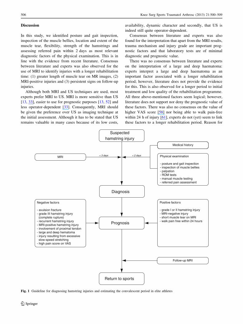
Discussion
In this study, we identified posture and gait inspection,
inspection of the muscle bellies, location and extent of the
muscle tear, flexibility, strength of the hamstrings and
assessing referred pain within 2 days as most relevant
diagnostic factors of the physical examination. This is in
line with the evidence from recent literature. Consensus
between literature and experts was also observed for the
use of MRI to identify injuries with a longer rehabilitation
time: (1) greater length of muscle tear on MR images, (2)
MRI-positive injuries and (3) persistent signs on follow-up
injuries.
Although both MRI and US techniques are used, most
experts prefer MRI to US. MRI is more sensitive than US
[13, 33], easier to use for prognostic purposes [13, 52] and
less operator-dependent [33]. Consequently, MRI should
be given the preference over US as imaging technique at
the initial assessment. Although it has to be stated that US
remains valuable in many cases because of its low costs,
availability, dynamic character and secondly, that US is
indeed still quite operator-dependent.
Consensus between literature and experts was also
found for the interpretation that apart from the MRI results,
trauma mechanism and injury grade are important prog-
nostic factors and that laboratory tests are of minimal
diagnostic and prognostic value.
There was no consensus between literature and experts
on the interpretation of a large and deep haematoma:
experts interpret a large and deep haematoma as an
important factor associated with a longer rehabilitation
period; however, literature does not provide the evidence
for this. This is also observed for a longer period to initial
treatment and low quality of the rehabilitation programme.
All three above-mentioned factors seem logical; however,
literature does not support nor deny the prognostic value of
these factors. There was also no consensus on the value of
higher VAS score [58] nor being able to walk pain-free
within 24 h of injury [61], experts do not (yet) seem to link
these factors to a longer rehabilitation period. Reason for
Medical history
Positive factors
- grade I or II hamstring injury- MRI-negative injury- short muscle tear on MRI- walk pain free within 24 hours
Negative factors
- avulsion fracture- grade III hamstring injury (complete rupture)- recurrent hamstring injury- MRI-positive hamstring injury- involvement of proximal tendon- large and deep hematoma- injury resulting from excessive slow-speed stretching- high pain score on VAS
MRI
Follow-up MRI
Physical examination
- posture and gait inspection- inspection of muscle bellies- palpation- ROM tests- manual muscle testing- referred pain assessment
< 3 days < 2 days
Diagnosis
Suspectedhamstring injury
Prognosis
Return to sports
Fig. 1 Guideline for diagnosing hamstring injuries and estimating the convalescent period in elite athletes
506 Knee Surg Sports Traumatol Arthrosc (2013) 21:500–509
123

this difference between literature and experts could be the
subjective nature of these findings or simply that these
findings are not widely known or accepted by other experts
yet.
There are obvious limitations of this research. Since
many prospective studies evaluated hamstring injuries in
only one type of sports, there is a selection bias [2, 3, 10,
11, 15–17, 20, 21, 27, 43, 46, 52, 59, 60, 64, 65]. The
question rises whether the conclusions of these studies can
be extrapolated to other sports types. In our survey, experts
were not asked to specify for the sports types with which
they deal in their daily practice. Caution is therefore rec-
ommended when adopting the results of this study.
Also, there is the limited value of a questionnaire with
low response rate, again a selection bias [28]; however, we
feel that the research benefits from the information pro-
vided by the selection of ESSKA members with interest in
muscle injuries that answered the questionnaire.
We feel that there is a definite need for further research
in this field. First, all prognostic factors identified in this
review should be validated in a prospective cohort and
even better in different cohorts of active sports participants,
so the difference between the different sport types can also
be monitored. Second, the anatomy of the hamstring
(injuries) can be re-evaluated to see whether we can
identify important prognostic factors on a basic level.
Third, imaging provides numerous keystones to improve
the understanding of the extent of the hamstring injury and
to link this to an accurate prognosis: ideal moment of initial
assessment, use of follow-up imaging in decision-making,
US versus MRI, optimalisation of MRI modalities.
With this combination of best evidence from the liter-
ature and experts from the field, the most relevant and
explicit diagnostic and prognostic factors of physical
examination, imaging and additional studies of hamstring
injuries in (elite) athletes were identified and an assessment
protocol for hamstring injuries in (elite) athletes (Fig. 1)
was proposed. In this way, it was attempted to provide a
guideline for diagnosing hamstring injuries and estimating
the convalescent period in (elite) athletes.
Conclusion
Physical examination of an athlete with suspected acute
hamstring injury should take place within 2 days post-
trauma and consists of posture and gait inspection, location
and extent of the muscle tear, flexibility and strength of the
hamstrings and assessing referred pain.
MRI as imaging technique for acute hamstring injuries in
elite athletes is preferred over ultrasound by both the experts
and recent literature mainly based on its greater sensitivity for
minor injuries and the ease of use for an accurate prognosis.
Important prognostic factors related to a longer reha-
bilitation period are MRI-positive muscle tears, larger
extent of the muscle tear as seen on MRI, recurrent ham-
string injury and injury mechanism.
Acknowledgments We thank all ESSKA colleagues who partici-
pated in the questionnaire and the ESSKA Sports committee col-
leagues for their feedback on the research protocol.
Conflict of interest No competing interests or funding reported.
Open Access This article is distributed under the terms of the
Creative Commons Attribution License which permits any use, dis-
tribution, and reproduction in any medium, provided the original
author(s) and the source are credited.
References
1. Allen GM (2007) Ultrasound in sports medicine—a critical
evaluation. Eur J Radiol 62:79–85
2. Arnason A, Gudmundsson A, Dahl HA, Johannsson E (1996)
Soccer injuries in Iceland. Scand J Med Sci Sports 6:40–45
3. Arnason A, Andersen TE, Holme I, Engebretsen L, Bahr R
(2008) Prevention of hamstring strains in elite football: an
intervention study. Scand J Med Sci Sports 18:40–48
4. Askling C, Saartok T, Thorstensson A (2006) Type of acute
hamstring strain affects flexibility, strength, and time to return to
pre-injury level. Br J Sports Med 40:40–44
5. Askling CM, Tengvar M, Saartok T, Thorstensson A (2007)
Acute first-time hamstring strains during high-speed running: a
longitudinal study including clinical and magnetic resonance
imaging findings. Am J Sports Med 35:197–206
6. Askling CM, Tengvar M, Saartok T, Thorstensson A (2007)
Acute first-time hamstring strains during slow-speed stretching:
clinical, magnetic resonance imaging, and recovery characteris-
tics. Am J Sports Med 35:1716–1724
7. Askling CM, Tengvar M, Saartok T, Thorstensson A (2008)
Proximal hamstring strains of stretching type in different sports:
injury situations, clinical and magnetic resonance imaging char-
acteristics, and return to sport. Am J Sports Med 36:1799–1804
8. Bencardino JT, Mellado JM (2005) Hamstring injuries of the hip.
Magn Reson Imaging Clin N Am 13:677–690, vi
9. Blankenbaker D, De Smet AA (2004) MR-imaging of muscle
injuries. Appl Rad 33:14–17
10. Brooks JH, Fuller CW, Kemp SP, Reddin DB (2006) Incidence,
risk, and prevention of hamstring muscle injuries in professional
rugby union. Am J Sports Med 34:1297–1306
11. Carling C, Le Gall F, Orhant E (2011) A four-season prospective
study of muscle strain reoccurrences in a professional football
club. Res Sports Med 19:92–102
12. Cohen S, Bradley J (2007) Acute proximal hamstring rupture.
J Am Acad Orthop Surg 15:350–355
13. Connell DA, Schneider-Kolsky ME, Hoving JL, Malara F,
Buchbinder R, Koulouris G, Burke F, Bass C (2004) Longitudinal
study comparing sonographic and MRI assessments of acute and
healing hamstring injuries. AJR Am J Roentgenol 183:975–984
14. Davis KW (2008) Imaging of the hamstrings. Semin Muscul-
oskelet Radiol 12:28–41
15. Ekstrand J, Gillquist J (1983) Socces injuries and their mecha-
nisms: a prospective study. Med Sci Sports Exerc 15:267–270
16. Ekstrand J, Timpka T, Hagglund M (2006) Risk of injury in elite
football played on artificial turf versus natural grass: a prospec-
tive two-cohort study. Br J Sports Med 40:975–980
Knee Surg Sports Traumatol Arthrosc (2013) 21:500–509 507
123
17. Ekstrand J, Hagglund M, Walden M (2011) Epidemiology of
muscle injuries in professional football (Soccer). Am J Sports
Med 39:1226–1232
18. Ekstrand J, Hagglund M, Walden M (2011) Injury incidence and
injury patterns in professional football—the UEFA injury study.
Br J Sports Med 45:553–558
19. Ekstrand J, Healy JC, Walden M, Lee JC, English B, Hagglund M
(2012) Hamstring muscle injuries in professional football: the
correlation of MRI findings with return to play. Br J Sports Med
46:112–117
20. Elliott MC, Zarins B, Powell JW, Kenyon CD (2011) Hamstring
muscle strains in professional football players: a 10-year review.
Am J Sports Med 39:843–850
21. Engebretsen AH, Myklebust G, Holme I, Engebretsen L, Bahr R
(2010) Intrinsic risk factors for hamstring injuries among male
soccer players: a prospective cohort study. Am J Sports Med
38:1147–1153
22. Fleckenstein JL, Weatherall PT, Parkey RW, Payne JA, Peshock
RM (1989) Sports-related muscle injuries: evaluation with MR
imaging. Radiology 172:793–798
23. Fleckenstein JL, Shellock FG (1991) Exertional muscle injuries:
magnetic resonance imaging evaluation. Top Magn Reson
Imaging 3:50–70
24. Gibbs NJ, Cross TM, Cameron M, Houang MT (2004) The
accuracy of MRI in predicting recovery and recurrence of acute
grade one hamstring muscle strains within the same season in
Australian Rules football players. J Sci Med Sport 7:248–258
25. Gielen JL, Robinson P, Van Dyck P, Van der Stappen A, Van-
hoenacker FM (2007) Muscle injuries. In: Vanhoenacker FM,
Maas MM, Gielen JL (eds) Imaging of orthopedic sports injuries,
1st edn. Springer, New York, pp 15–39
26. Guerrero M, Guiu-Comadevall M, Cadefau JA, Parra J, Ballius R,
Estruch A, Rodas G, Bedini JL, Cusso R (2008) Fast and slow
myosins as markers of muscle injury. Br J Sports Med
42:581–584
27. Hagglund M, Walden M, Ekstrand J (2006) Previous injury as a
risk factor for injury in elite football: a prospective study over
two consecutive seasons. Br J Sports Med 40:767–772
28. Hasson G, Keeney S, McKenna H (2000) Research guidelines for
the Delphi survey technique. J Adv Nurs 32:1008–1015
29. Heiderscheit BC, Sherry MA, Silder A, Chumanov ES, Thelen
DG (2010) Hamstring strain injuries: recommendations for
diagnosis, rehabilitation, and injury prevention. J Orthop Sports
Phys Ther 40:67–81
30. Heiser TM, Weber J, Sullivan G, Clare P, Jacobs RR (1984)
Prophylaxis and management of hamstring muscle injuries in
intercollegiate football players. Am J Sports Med 12:368–370
31. Klingele KE, Sallay PI (2002) Surgical repair of complete proximal
hamstring tendon rupture. Am J Sports Med 30:742–746
32. Kornberg C, Lew P (1989) The effect of stretching neural
structures on grade one hamstring injuries. J Orthop Sports Phys
Ther 10:481–487
33. Koulouris G, Connell D (2003) Evaluation of the hamstring muscle
complex following acute injury. Skeletal Radiol 32:582–589
34. Koulouris G, Connell D (2005) Hamstring muscle complex: an
imaging review. Radiographics 25:571–586
35. Koulouris G, Connell DA, Brukner P, Schneider-Kolsky M
(2007) Magnetic resonance imaging parameters for assessing risk
of recurrent hamstring injuries in elite athletes. Am J Sports Med
35:1500–1506
36. Lempainen L, Sarimo J, Heikkila J, Mattila K, Orava S (2007)
Distal tears of the hamstring muscles: review of the literature and
our results of surgical treatment. Br J Sports Med 41:80–83
37. Liemohn W, Martin SB, Pariser GL (1997) The effect of ankle
posture on sit-and-reach test performance. J Strength Cond Res
11:239–241
38. Malliaropoulos N, Papacostas E, Kiritsi O, Papalada A, Gou-
goulias N, Maffulli N (2010) Posterior thigh muscle injuries in
elite track and field athletes. Am J Sports Med 38:1813–1819
39. Malliaropoulos N, Isinkaye T, Tsitas K, Maffulli N (2011)
Reinjury after acute posterior thigh muscle injuries in elite track
and field athletes. Am J Sports Med 39:304–310
40. Martınez Amat A, Marchal Corrales JA, Rodrıguez Serrano F,
Boulaiz H, Prados Salazar JC, Hita Contreras F, Caba Perez O,
Carrillo Delgado E, Martiın I, Aranega Jimenez A (2007) Role of
alpha-actin in muscle damage of injured athletes in comparison
with traditional markers. Br J Sports Med 41:442–446
41. Minarro PA, Andujar PS, Garcıa PL, Toro EO (2007) A com-
parison of the spine posture among several sit-and-reach test
protocols. J Sci Med Sport 10:456–462
42. Nikolaou PK, Macdonald BL, Glisson RR, Seaber AV, Garrett
WE Jr (1987) Biomechanical and histological evaluation ofmuscle after controlled strain injury. Am J Sports Med 15:9–14
43. Orchard J, Seward H (2002) Epidemiology of injuries in the
Australian Football League, seasons 1997–2000. Br J Sports Med
36:39–44
44. Orchard JW, Farhart P, Leopold C (2004) Lumbar spine region
pathology and hamstring and calf injuries in athletes: is there a
connection? Br J Sports Med 38:502–504
45. Peetrons P (2002) Ultrasound of muscles. Eur Radiol 12:35–43
46. Petersen J, Thorborg K, Nielsen MB, Holmich P (2010) Acute
hamstring injuries in Danish elite football: a 12-month prospec-
tive registration study among 374 players. Scand J Med Sci
Sports 20:588–592
47. Puranen J, Orava S (1988) The hamstring syndrome. A new
diagnosis of gluteal sciatic pain. Am J Sports Med 16:517–521
48. Sallay PI, Friedman RL, Coogan PG, Garrett WE (1996) Ham-
string muscle injuries among water skiers. Functional outcome
and prevention. Am J Sports Med 24:130–136
49. Sarimo J, Lempainen L, Mattila K, Orava S (2008) Complete
proximal hamstring avulsions: a series of 41 patients with oper-
ative treatment. Am J Sports Med 36:1110–1115
50. Schneider-Kolsky ME, Hoving JL, Warren P, Connell DA (2006)
A comparison between clinical assessment and magnetic reso-
nance imaging of acute hamstring injuries. Am J Sports Med
34:1008–1015
51. Schneider-Kolsky M, Warren P, Connell D (2007) Author’s reply
on comment on: a comparison between clinical assessment and
magnetic resonance imaging of acute hamstring injuries. Am J
Sports Med 35:149–150
52. Seward H, Orchard J, Hazard H, Collinson D (1993) Football
injuries in Australia at the elite level. Med J Aust 159:298–301
53. Shellock FG, Fleckenstein JL (2000) Muscle physiology and
pathophysiology: magnetic resonance imaging evaluation. Semin
Musculoskelet Radiol 4:459–479
54. Slavotinek JP, Verrall GM, Fon GT (2002) Hamstring injury in
athletes: using MR imaging measurements to compare extent of
muscle injury with amount of time lost from competition. AJR
Am J Roentgenol 179:1621–1628
55. Sorichter S, Mair J, Koller A, Gebert W, Rama D, Calzolari C,
Artner-Dworzak E, Puschendorf B (1997) Skeletal troponin I as a
marker of exercise-induced muscle damage. J Appl Physiol
83:1076–1082
56. Verrall GM, Slavotinek JP, Barnes PG, Fon GT, Spriggins AJ
(2001) Clinical risk factors for hamstring muscle strain injury: a
prospective study with correlation of injury by magnetic reso-
nance imaging. Br J Sports Med 35:435–439
57. Verrall GM, Slavotinek JP, Barnes PG, Fon GT (2003) Diag-
nostic and prognostic value of clinical findings in 83 athletes with
posterior thigh injury: comparison of clinical findings with
magnetic resonance imaging documentation of hamstring muscle
strain. Am J Sports Med 31:969–973
508 Knee Surg Sports Traumatol Arthrosc (2013) 21:500–509
123
58. Verrall GM, Slavotinek JP, Barnes PG, Fon GT, Esterman A
(2006) Assessment of physical examination and magnetic reso-
nance imaging findings of hamstring injury as predictors for
recurrent injury. J Orthop Sports Phys Ther 36:215–224
59. Volpi P, Melegati G, Tornese D, Bandi M (2004) Muscle strains
in soccer: a five-year survey of an Italian major league team.
Knee Surg Sports Traumatol Arthrosc 12:482–485
60. Walden M, Hagglund M, Ekstrand J (2005) UEFA champions
League study: a prospective study of injuries in professional foot-
ball during the 2001–2002 season. Br J Sports Med 39:542–546
61. Warren P, Gabbe BJ, Schneider-Kolsky M, Bennell KL (2010)
Clinical predictors of time to return to competition and of
recurrence following hamstring strain in elite Australian foot-
ballers. Br J Sports Med 44:415–419
62. Wood DG, Packham I, Trikha SP, Linklater J (2008) Avulsion of
the proximal hamstring origin. J Bone Jt Surg Am 90:2365–2374
63. Woods C, Hawkins R, Hulse M, Hodson A (2002) The football
association medical research programme: an audit of injuries in
professional football-analysis of preseason injuries. Br J Sports
Med 36:436–441
64. Woods C, Hawkins RD, Maltby S, Hulse M, Thomas A, Hodson
A (2004) The football association medical research programme:
an audit of injuries in professional football–analysis of hamstring
injuries. Br J Sports Med 38:36–41
65. Yeung SS, Suen AM, Yeung EW (2009) A prospective cohort
study of hamstring injuries in competitive sprinters: preseason
muscle imbalance as a possible risk factor. Br J Sports Med
43:589–594
66. Zeren B, Oztekin HH (2006) A new self-diagnostic test for biceps
femoris muscle strains. Clin J Sport Med 16:166–169
Knee Surg Sports Traumatol Arthrosc (2013) 21:500–509 509
123
Related Documents