Diagnosis and management of fetal growth restriction IBRAHIM HAMMAD MATERNAL FETAL MEDICINE FELLOW UNIVERSITY OF UTAH

Welcome message from author
This document is posted to help you gain knowledge. Please leave a comment to let me know what you think about it! Share it to your friends and learn new things together.
Transcript
Diagnosis and management of fetal growth restrictionIBRAHIM HAMMAD
MATERNAL FETAL MEDICINE FELLOW
UNIVERSITY OF UTAH

IntroductionFetal growth restriction (FGR)◦ Fetus that has not reached its growth potential
because of genetic or environmental factors.
Origin may be ◦ Fetal◦ Placental ◦ Maternal◦ Significant overlap

ImportanceIncreased risk of adverse perinatal outcome mainly due to reduced energy reserves:
◦ Premature delivery◦ Perinatal asphyxia, which may be accompanied by meconium aspiration or persistent pulmonary
hypertension◦ Impaired thermoregulation◦ Hypoglycemia◦ Polycythemia and hyperviscosity◦ Impaired immune function◦ Mortality (5-20 times higher)
Antecedent to some cases of hypertension, hyperlipidemia, coronary heart disease, and diabetes mellitus in the adult (Barker hypothesis)
DefinitionWeight below the 10th percentile for gestational age
Other definitions <5th percentile, <3rd percentile
Does not differentiate between◦ Constitutionally small◦ Small because a pathologic process has kept them from achieving their genetic growth potential◦ Not small but a pathologic process has kept them from achieving their genetic growth potential.
Requires an appropriate reference standard. ◦ Based on birth weights across gestation (most baby delivered preterm will be growth restricted)◦ Ultrasound estimated fetal weights across gestation (limited by the inaccuracy and imprecision)◦ customized

Epidemiology 3-10% in developed countries
20-50% in developing contries

NORMAL FETAL GROWTH
Fetus's genetically predetermined
growth potential ( 30-50%)
Modulated fetal health
PlacentaMaternal health
Outside environment

Fetal growth
The first phase
-Cellular hyperplasia
Symmetric
The second phase
-Hyperplasia and hypertrophy
Mixed- asymmetric
The third
-Hypertrophy (rapid accumulation of fat, muscle, connective tissue)
Asymmetric
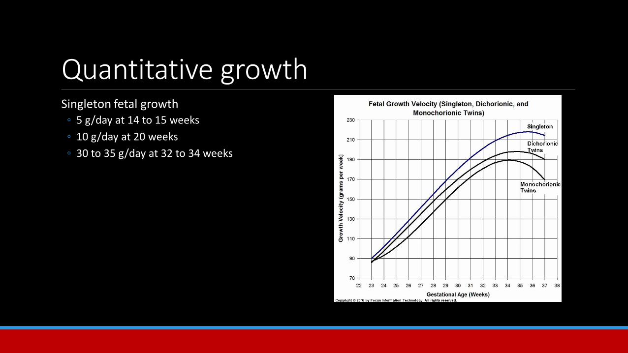
Quantitative growthSingleton fetal growth
◦ 5 g/day at 14 to 15 weeks◦ 10 g/day at 20 weeks ◦ 30 to 35 g/day at 32 to 34 weeks
Symmetric and asymmetric growth restrictionSymmetric FGR
◦ 20-30%◦ Small sized organs due to global impairment of early fetal cellular hyperplasia
Asymmetric FGR ◦ 70-80% ◦ Greater decrease in abdominal size (eg, liver volume and subcutaneous fat tissue)◦ Result from the capacity of the fetus to adapt to a hostile environment by redistributing blood flow

Clinical assessmentReasonable screening tool in low risk pregnancies
No high quality evidence that alternative approaches improve outcome
Base on assessment of past present risk factors
Physical examination
Ultrasound studies

Accurate assessment of gestational ageKnowledge of LMP
Early ultrasound
Serial ultrasound
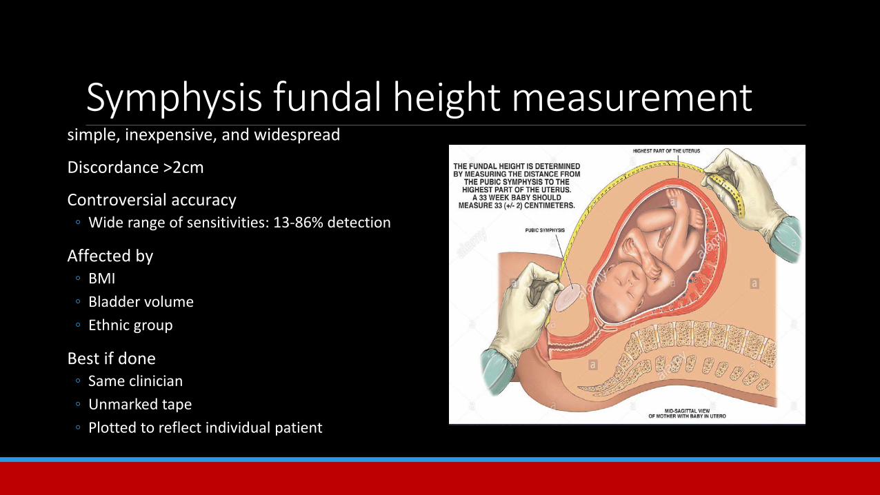
Symphysis fundal height measurementsimple, inexpensive, and widespread
Discordance >2cm
Controversial accuracy ◦ Wide range of sensitivities: 13-86% detection
Affected by ◦ BMI◦ Bladder volume◦ Ethnic group
Best if done◦ Same clinician◦ Unmarked tape◦ Plotted to reflect individual patient
SONOGRAPHIC DIAGNOSISEstimated fetal weight (EFW)
◦ Most common method ◦ Sensitivity, specificity, positive and negative predictive values of EFW for FGR <10 percentile are
approximately 90, 85, 80, and 90 percent, respectively, when performed proximate to delivery◦ Sensitivity is higher in severe FGR

SONOGRAPHIC DIAGNOSISAbdominal circumference (AC)
◦ Sensitivity, specificity, positive and negative predictive values of 61, 95, 86, and 83 percent, respectively
◦ More sensitive in asymmetric FGR◦ More sensitive later in gestation◦ More sensitive when the interval between measurements is
more than two weeks
Small AC also correlates with parameters of morbidity associated with FGR
Biochemical markers of hypoxia and acidemia are more common when the AC is below the 5th percentile for GA

ManagementConfirming the diagnosis
Cause and severity of FGR
Counseling
Monitoring fetal growth and wellbeing
Optimal time for and route of delivery

Confirm the diagnosis<10 percentile weight for gestational age
◦ constitutionally small VS. fetus with growth potential is restricted
Constitutionally small fetus ◦ Modest smallness ◦ Normal growth velocity across gestation◦ Normal physiology (ie, normal amniotic fluid volume and umbilical artery Doppler)◦ Abdominal circumference growth velocity above the lowest decile◦ Appropriate size in terms of maternal characteristics (height, weight, race/ethnicity).

Determine the causeComplete history
Physical examination
Obstetrical imaging and laboratory evaluations

Fetal surveyDetailed fetal anatomic survey
◦ 10 % of FGR have congenital anomalies◦ 20-60% of fetuses with congenital anomalies are FGR
◦ (Detection of trisomy 18 is as high as 100% at 18-22 weeks)
Fetal genetic studiesTrisomies 13, 18, 21 contribute to 5-10% of IUGR cases
karyotype/microarray is indicated:◦ Early (<24 weeks), severe (<5%le), symmetrical FGR◦ Major fetal structural abnormalities◦ Ultrasound markers associated with aneuploidy, (ie, increased nuchal fold, abnormal hand positioning)
Usually not performed after 24 weeks with normal anatomy yield is low
Confined placental mosaicism is detected after delivery in approximately 10% of placentas associated with otherwise idiopathic FGR

Workup for infectionMaternal history, physical examination or fetal ultrasound findings
◦ Cytomegalovirus◦ Toxoplasmosis◦ Rubella◦ Varicella◦ Malaria
Timing of infection is crucial
Amniotic fluid DNA testing can also be performed for specific infections
PREGNANCY MANAGEMENTFGR associated with congenital or chromosomal anomalies depends on the specific abnormality and management focuses on that specific anomaly

PREGNANCY MANAGEMENTOptimal management of FGR related to uteroplacental insufficiency has not been established
Limited evidence from randomized trials
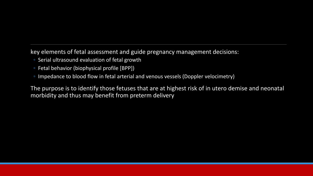
key elements of fetal assessment and guide pregnancy management decisions:◦ Serial ultrasound evaluation of fetal growth◦ Fetal behavior (biophysical profile [BPP])◦ Impedance to blood flow in fetal arterial and venous vessels (Doppler velocimetry)
The purpose is to identify those fetuses that are at highest risk of in utero demise and neonatal morbidity and thus may benefit from preterm delivery

Doppler velocimetryDoppler velocimetry of the umbilical artery is a good tool for fetal assessment in FGR when the etiology is placental dysfunction related to progressive obliteration of the villus vasculature.
Placental vascular changes lead to fetal hemodynamic changes that can be evaluated by umbilical artery Doppler.

General sequence of Doppler and biophysical• Reduction in umbilical venous flow is the initial hemodynamic change.• Venous flow is redistributed away from the fetal liver and towards the heart.
• Umbilical artery Doppler index increases (diminished end diastolic flow) due to increased resistance in the placental vasculature.
• Middle cerebral artery Doppler index decreases (increased end diastolic flow), resulting in preferential perfusion of the brain (brain sparing effect).
• Increasing placental vascular resistance results in absent and then reversed end diastolic flow in the umbilical artery.
• Middle cerebral artery Doppler index normalizes or abnormally increases as diastolic flow falls due to loss of brainsparing hemodynamic changes.
• As cardiac performance deteriorates due to chronic hypoxia and nutritional deprivation, absent or reversed end diastolic flow in the ductus venous and pulsatile umbilical venous flow may develop. These can be preterminal events.

Umbilical artery Doppler (UAD)Numerous randomized trials that UAD significantly reduce perinatal death, as well as unnecessary induction of labor in the preterm growth restricted fetus
A 2013 systematic review of 18 trials ◦ Doppler vs. no Doppler ◦ 29% reduction in perinatal deaths◦ Significantly fewer labor inductions and cesarean deliveries

UADNormal diastolic flow (lowest rate of perinatal morbidity or mortality)
Decreased end diastolic flow (30% villous vasculature ceases to function)
Absent end diastolic flow (60-70% villous vasculature is obliterated)
Reversed diastolic flow is associated with poorer neonatal outcomes than absent diastolic flow

Near the end of this sequence, biophysical changes usually become apparent:
Nonstress test (NST) becomes nonreactive
BPP score falls
Late decelerations accompany contractions

Cardiovascular (Doppler) and behavioral (BPP) can be independent resulting discordant Doppler and BPP findings
This means if delivery needs to be planned both identities need to be taken into consideration
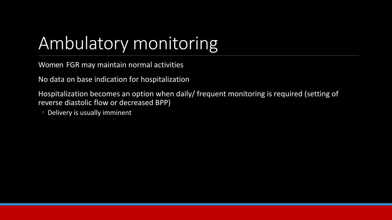
Ambulatory monitoringWomen FGR may maintain normal activities
No data on base indication for hospitalization
Hospitalization becomes an option when daily/ frequent monitoring is required (setting of reverse diastolic flow or decreased BPP)
◦ Delivery is usually imminent
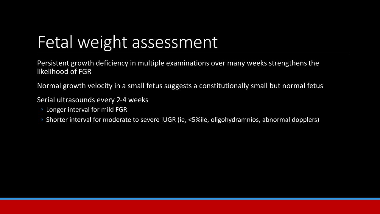
Fetal weight assessmentPersistent growth deficiency in multiple examinations over many weeks strengthens the likelihood of FGR
Normal growth velocity in a small fetus suggests a constitutionally small but normal fetus
Serial ultrasounds every 2-4 weeks◦ Longer interval for mild FGR◦ Shorter interval for moderate to severe IUGR (ie, <5%ile, oligohydramnios, abnormal dopplers)

Nonstress test and biophysical profileNST with amniotic fluid or BPP or combination of both tests is reasonable for monitoring fetal wellbeing
◦ observational studies that reported lower rates of fetal death when comparing tested population vs. historically not tested
◦ Same or lower rates of fetal death in tested pregnancies (primarily high risk) than in a contemporary untested general obstetrical population (primarily low risk)

Biophysical profileBPP evaluates both acute and chronic fetal physiologic parameters
◦ Easy to perform◦ Fetal death within one week of a normal test score is rare

Nonstress test and amniotic fluid assessment Amniotic fluid volume assessment should also be performed weekly
Chronic placental insufficiency results in both FGR and oligohydramnios
Observational studies have reported that pregnancies complicated by FGR and oligohydramnios have a modestly increased risk of perinatal mortality
Normal amniotic fluid volume is infrequently associated with either FGR or fetal demise, unless the cause is a congenital malformation or aneuploidy

How often should you do monitor?Weekly
More frequently (twice) when associated with other anomalies ( ie, oligohydramnios, preeclampsia, decelerating fetal growth, severe growth restriction, increasing umbilical artery Doppler index)
Daily if absent or reversed end diastolic flow

Antenatal steroidsone course of antenatal corticosteroids is given between 24 and 34 weeks of gestation in the week before delivery is expected

Maternal interventionsNo evidence of the benefit of any intervention
◦ Maternal nutritional supplementation◦ Oxygen therapy◦ Plasma volume expansion◦ Low dose aspirin◦ Bed rest◦ Sildenafil

Antihypertensive therapy of hypertensive gravidas does not improve fetal growth
It’s very important to monitor these patient’s blood pressure, low blood pressure could jeopardize fetal blood flow
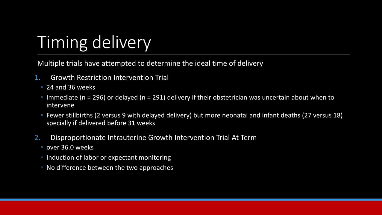
Timing deliveryMultiple trials have attempted to determine the ideal time of delivery
1. Growth Restriction Intervention Trial◦ 24 and 36 weeks◦ Immediate (n = 296) or delayed (n = 291) delivery if their obstetrician was uncertain about when to
intervene◦ Fewer stillbirths (2 versus 9 with delayed delivery) but more neonatal and infant deaths (27 versus 18)
specially if delivered before 31 weeks
2. Disproportionate Intrauterine Growth Intervention Trial At Term◦ over 36.0 weeks◦ Induction of labor or expectant monitoring◦ No difference between the two approaches

Ideally Gestational age
UAD
BPP score (or nonstress test)
Presence or absence of risk factors for, or signs of, uteroplacental insufficiency
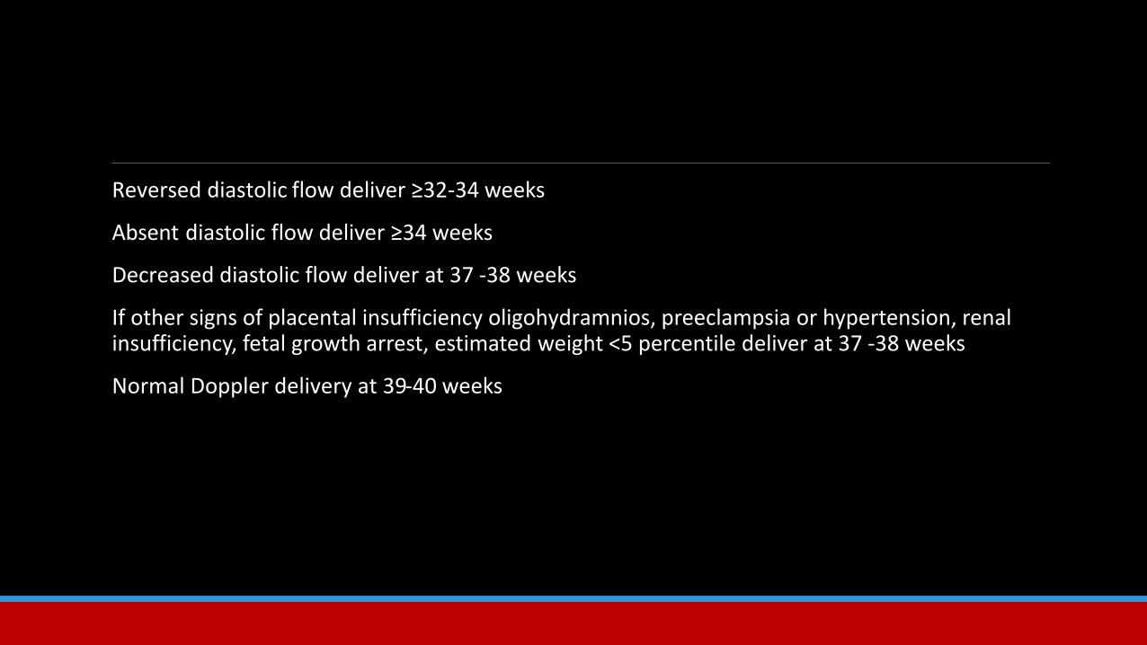
Reversed diastolic flow deliver ≥32-34 weeks
Absent diastolic flow deliver ≥34 weeks
Decreased diastolic flow deliver at 37 -38 weeks
If other signs of placental insufficiency oligohydramnios, preeclampsia or hypertension, renal insufficiency, fetal growth arrest, estimated weight <5 percentile deliver at 37 -38 weeks
Normal Doppler delivery at 39-40 weeks
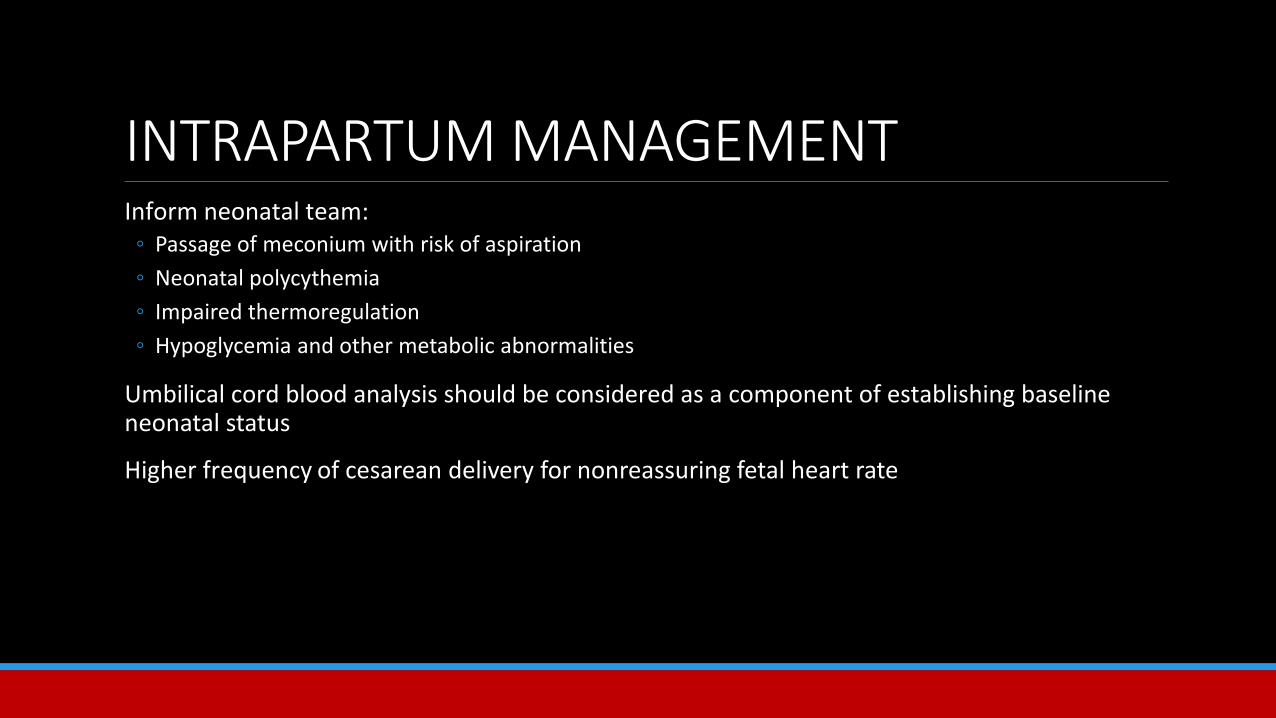
INTRAPARTUM MANAGEMENTInform neonatal team:
◦ Passage of meconium with risk of aspiration◦ Neonatal polycythemia◦ Impaired thermoregulation◦ Hypoglycemia and other metabolic abnormalities
Umbilical cord blood analysis should be considered as a component of establishing baseline neonatal status
Higher frequency of cesarean delivery for nonreassuring fetal heart rate
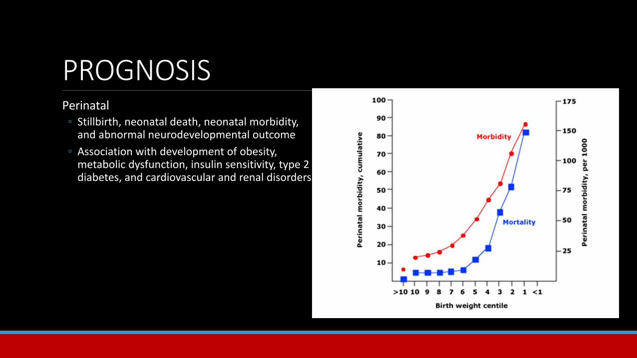
PROGNOSISPerinatal
◦ Stillbirth, neonatal death, neonatal morbidity, and abnormal neurodevelopmental outcome
◦ Association with development of obesity, metabolic dysfunction, insulin sensitivity, type 2 diabetes, and cardiovascular and renal disorders

RECURRENCE RISKRisk of a nonanomalous SGA birth (<5 percentile) in the second pregnancy of women whose first delivery was “SGA” versus “not SGA” was 23 and 3%, respectively

Take home messageAn estimated fetal weight <10 percentile signifies a small for gestational age fetus
Differentiate between FGR and constitutionally small
Detailed fetal anatomic survey
Evaluation of the fetal karyotype/microarray as indicated
Assess for infection is history or ultrasounds indicates it
Serial ultrasound evaluation for fetal growth, fetal behavior and Doppler

Delivery will depend gestational age, Doppler ultrasound of the umbilical artery, biophysical profile score (or nonstress test), and the presence or absence of risk factors for, or signs of, uteroplacental insufficiency

Thank you
Related Documents