Diagnosis and management of cystic lesions of the liver Disclosures All topics are updated as new evidence becomes available and our peer review process is complete. Literature review current through: Jun 2013. | This topic last updated: Apr 24, 2013. INTRODUCTION — Cystic lesions of the liver represent a heterogeneous group of disorders, which differ in etiology, prevalence, and clinical manifestations (table 1 ). Most liver cysts are found incidentally on imaging studies and tend to have a benign course. A minority can cause symptoms, and rarely may be associated with serious morbidity and mortality [1 - 3 ]. Larger cysts are more likely to be symptomatic and cause complications such as spontaneous hemorrhage [4 ], rupture into the peritoneal cavity [3 ] or bile duct [5 ], infection [6 ], and compression of the biliary tree [2,7 ]. Specific types of cysts may have unique complications such as malignant transformation in case of a cystadenoma, or anaphylactic shock due to a hydatid cyst. Some of these complications may occasionally mandate surgical intervention. In some cases, predominantly cystic liver lesions may have solid areas, particularly in the setting malignancy. Conversely, predominantly solid liver lesions may have cystic components, as may be seen with hemangiomas or tumors that have areas that are necrotic. Considerable controversy still exists regarding the definition and classification of cystic lesions of the liver (table 1 ). Furthermore, consensus has not been achieved on the optimal treatment of patients with symptomatic cysts, although a number of therapeutic approaches have been described [8,9 ]. This topic review will provide an overview of the diagnosis and management of cystic lesions in the liver. Detailed discussions on some of the individual causes of cysts are provided on the corresponding topic reviews. SIMPLE CYST — Simple cysts of the liver are cystic formations containing clear fluid that do not communicate with the intrahepatic biliary tree. Although simple cysts are found in approximately 1 percent of autopsied adults, very few become large, and even fewer cause symptoms. Their size ranges from a few millimeters to massive lesions occupying large volumes of the upper abdomen; the largest reported cyst contained 17 liters of fluid [10 ]. Simple cysts tend to occur more commonly in the right lobe, and are more prevalent in women. The female-to-male ratio is approximately 1.5:1 among those with asymptomatic simple cysts while it is 9:1 in those with symptomatic or complicated simple cysts [1 ]. Huge cysts are found almost exclusively in women over 50. Symptomatic patients may present with abdominal discomfort, pain, or nausea. As a general rule, cysts in symptomatic patients are larger than those in asymptomatic ones [9,11 ]. Large cysts can produce atrophy of the adjacent hepatic tissue while huge cysts can cause complete atrophy of a hepatic lobe with compensatory hypertrophy of the other lobe. Complications (such as spontaneous hemorrhage, bacterial infection, torsion of pedunculated cyst, rupture or biliary obstruction) are more common in large cysts [2 - Official reprint from UpToDate ® www.uptodate.com ©2013 UpToDate ® Authors Arie Regev, MD K Rajender Reddy, MD Section Editor Sanjiv Chopra, MD Deputy Editor Anne C Travis, MD, MSc, FACG Page 1 of 16 Diagnosis and management of cystic lesions of the liver 8/4/2013 http://www.uptodate.com/contents/diagnosis-and-management-of-cystic-lesions-of-the-liver...

Diagnosis and Management of Cystic Lesions of the Liver
Nov 30, 2015
Liver cyst
Welcome message from author
This document is posted to help you gain knowledge. Please leave a comment to let me know what you think about it! Share it to your friends and learn new things together.
Transcript

Diagnosis and management of cystic lesions of the liver
Disclosures
All topics are updated as new evidence becomes available and our peer review process is complete. Literature review current through: Jun 2013. | This topic last updated: Apr 24, 2013.
INTRODUCTION — Cystic lesions of the liver represent a heterogeneous group of disorders, which differ
in etiology, prevalence, and clinical manifestations (table 1). Most liver cysts are found incidentally on
imaging studies and tend to have a benign course. A minority can cause symptoms, and rarely may be
associated with serious morbidity and mortality [1-3]. Larger cysts are more likely to be symptomatic and
cause complications such as spontaneous hemorrhage [4], rupture into the peritoneal cavity [3] or bile
duct [5], infection [6], and compression of the biliary tree [2,7]. Specific types of cysts may have unique
complications such as malignant transformation in case of a cystadenoma, or anaphylactic shock due to
a hydatid cyst. Some of these complications may occasionally mandate surgical intervention.
In some cases, predominantly cystic liver lesions may have solid areas, particularly in the setting
malignancy. Conversely, predominantly solid liver lesions may have cystic components, as may be seen
with hemangiomas or tumors that have areas that are necrotic.
Considerable controversy still exists regarding the definition and classification of cystic lesions of the liver
(table 1). Furthermore, consensus has not been achieved on the optimal treatment of patients with
symptomatic cysts, although a number of therapeutic approaches have been described [8,9]. This topic
review will provide an overview of the diagnosis and management of cystic lesions in the liver. Detailed
discussions on some of the individual causes of cysts are provided on the corresponding topic reviews.
SIMPLE CYST — Simple cysts of the liver are cystic formations containing clear fluid that do not
communicate with the intrahepatic biliary tree. Although simple cysts are found in approximately 1
percent of autopsied adults, very few become large, and even fewer cause symptoms. Their size ranges
from a few millimeters to massive lesions occupying large volumes of the upper abdomen; the largest
reported cyst contained 17 liters of fluid [10].
Simple cysts tend to occur more commonly in the right lobe, and are more prevalent in women. The
female-to-male ratio is approximately 1.5:1 among those with asymptomatic simple cysts while it is 9:1 in
those with symptomatic or complicated simple cysts [1]. Huge cysts are found almost exclusively in
women over 50.
Symptomatic patients may present with abdominal discomfort, pain, or nausea. As a general rule, cysts in
symptomatic patients are larger than those in asymptomatic ones [9,11]. Large cysts can produce atrophy
of the adjacent hepatic tissue while huge cysts can cause complete atrophy of a hepatic lobe with
compensatory hypertrophy of the other lobe. Complications (such as spontaneous hemorrhage, bacterial
infection, torsion of pedunculated cyst, rupture or biliary obstruction) are more common in large cysts [2-
Official reprint from UpToDate®
www.uptodate.com ©2013 UpToDate®
Authors Arie Regev, MD K Rajender Reddy, MD
Section Editor Sanjiv Chopra, MD
Deputy Editor Anne C Travis, MD, MSc, FACG
Page 1 of 16Diagnosis and management of cystic lesions of the liver
8/4/2013http://www.uptodate.com/contents/diagnosis-and-management-of-cystic-lesions-of-the-liver...

6].
Diagnosis — The distinction between a simple cyst, cystadenoma, cystadenocarcinoma, echinococcal
cyst and other rare primary or metastatic tumors can be difficult. However, the distinction is extremely
important since these lesions have different clinical significance.
Imaging studies — Ultrasonography is probably the most helpful initial test since it can usually
differentiate a simple cyst from other cystic lesions. It should also be used for follow-up studies. Simple
cysts appear as an anechoic unilocular fluid filled space with imperceptible walls, and with posterior
acoustic enhancement [12,13]. Clinical features combined with the sonographic findings are usually
sufficient to distinguish simple cysts from other lesions that can appear cystic such as a liver abscess,
necrotic malignant tumor, hemangioma, and hamartoma.
On a CT scan a simple cyst is defined as a well-demarcated water attenuation lesion that does not
enhance following the administration of intravenous contrast (image 1). Uncomplicated simple cysts are
virtually never septated. However, hemorrhage into a simple cyst can lead to confusion in the
sonographic differentiation from a cystadenoma or cystadenocarcinoma [4,14]. In one report, hemorrhage
was associated with the appearance of septa in 2 of 57 patients (3.5 percent) with large simple cysts (≥4
cm) [15]. Hemorrhage is much less frequent in smaller cysts.
Magnetic resonance imaging (MRI) demonstrates a well-defined water-attenuation lesion that does not
enhance following the administration of intravenous Gadolinium. On T1-weighted images the cyst shows
a low signal, whereas a very high intensity signal is shown on T2-weighted images (image 1).
Differential diagnosis — The differential diagnosis of a simple cyst includes a variety of hepatic
lesions that can have a cystic appearance, such as cystadenoma, cystadenocarcinoma, hepatic abscess,
a necrotic malignant tumor, hemangioma, and hamartoma (table 2). As mentioned above, the distinction
can usually be made based upon the clinical setting and radiographic findings. A rare exception is a
hepatic metastasis from a neuroendocrine tumor, which can be asymptomatic and can have a sharply-
defined necrotic area [1].
Histology and needle aspiration — Histological examination is seldom needed for establishing the
diagnosis. However, when histology is available, the following criteria can be used for a definitive
diagnosis:
� An outer layer of a thin dense fibrous tissue
� An inner epithelial lining consisting of a single layer of cuboidal or columnar epithelium; this layer is
found in most but not in all simple cysts
� Lack of mesenchymal stroma or cellular atypia
Aspiration is usually not required for diagnosing cysts that have a typical sonographic appearance. When
it is performed, the aspirated fluid is always sterile and cytologically negative. It may vary from clear straw
color to brown. High levels of carcinoembryonic antigen (CEA) in the cyst fluid have been reported in
cystadenocarcinomas but the diagnostic accuracy of this finding has not been established [16].
Treatment — The majority of simple cysts do not require treatment. However, it may be prudent to
monitor large cysts (≥4 cm in diameter) periodically with ultrasonography to assure that they remain
stable. We suggest an initial follow-up study in three months after the diagnosis and then again at 6 to 12
months. Further monitoring is usually unnecessary if the cyst remains unchanged for two to three years.
Page 2 of 16Diagnosis and management of cystic lesions of the liver
8/4/2013http://www.uptodate.com/contents/diagnosis-and-management-of-cystic-lesions-of-the-liver...

The presence of symptoms related to the cyst or increasing size should raise concern that the lesion
could be a cystadenoma, a cystadenocarcinoma, or another rare cystic neoplasm, since simple cysts
tend to remain stable in size. Such patients may require surgical intervention. The causal relationship
between abdominal pain or discomfort and a simple cyst must be admitted with caution, and accepted
only if the cyst is large, and other possible causes of the symptoms have been excluded. These include
cholelithiasis, gastroesophageal reflux disease, gastric dysmotility, peptic ulcer disease, and other causes
of dyspepsia. Percutaneous aspiration has been advocated as a diagnostic test for relief of symptoms
[17]. However, this test is not without risk, and has not been widely accepted.
Several therapeutic approaches have been described for symptomatic, large simple cysts including
needle aspiration with or without injection of sclerosing agents [18-21], internal drainage with
cystojejunostomy [22], wide unroofing [23-26], and varying degrees of liver resection [27]. In most series,
percutaneous needle aspiration was associated with a high failure rate and rapid recurrence [28].
Percutaneous needle aspiration with injection of sclerosing agents is generally safe, effective, and
relatively non-invasive [29-32]; however, it may occasionally have serious complications [33]. Wide
unroofing or cyst resection has been associated with a relatively low incidence of cyst recurrence or
complications [15,23-25,34,35]. The laparoscopic approach has proven to be safe, achieving a wide
unroofing or resection without the need for a debilitating incision [15,25,26,28,35-41].
Several centers have reported recurrence rates ranging from 0 to 14.3 percent and morbidity rates of 0 to
15 percent after laparoscopic unroofing of solitary simple cysts [25,36-38,42]. Potential complications of
laparoscopic unroofing include wound infection, chest infection, bile leak, subphrenic hematoma, and
prolonged postoperative drainage through abdominal drain (>three days) [25,26,38]. Laparoscopic
unroofing may not be possible in patients with a superior or posterior location of the cyst. There are no
prospective studies comparing the different therapeutic approaches. As a result the procedure of choice
should be decided on a case by case basis, taking into consideration the cyst’s location, suspicion of
malignancy, history of abdominal surgery, and local expertise.
CYSTADENOMA — Hepatobiliary cystadenomas is a rare cystic tumor that occurs within the liver
parenchyma or, less frequently, in the extrahepatic bile ducts. The published experience with these
lesions is limited to single case reports and small series [15,43-45]. These reports suggest that
cystadenomas occur in adults, more often in women. The tumors occurred more often in the right lobe
than in the left in one report [43], while two other series reported frequent involvement of the left lobe
[15,44]. The tumors grew to a large size and required surgical intervention in most reports.
Clinical manifestations — The most commonly reported presenting symptoms were a sensation of an
upper abdominal mass, abdominal discomfort or pain, and anorexia. These symptoms had been present
for several years prior to diagnosis in several patients. However, many patients were asymptomatic, and
the lesions were found incidentally on abdominal imaging studies.
Diagnosis — Histologic examination is required for definitive diagnosis, although the lesion may be
suspected on imaging studies. The differential diagnosis includes cystadenocarcinoma, echinococcal
cyst, and a simple cyst. Simple cysts can usually be distinguished because of the absence of septations
and papillary projections and the presence of serous cystic fluid. Echinococcal cysts are frequently
associated with calcifications and patients will have positive serology. (See "Clinical manifestations and
diagnosis of echinococcosis".)
Imaging studies — The appearance of cystadenoma on ultrasonography can usually differentiate it
from a simple cyst (picture 1). On ultrasonography, a cystadenoma typically appears as an hypoechoic
lesion with thickened irregular walls and occasional internal echoes representing debris and wall
Page 3 of 16Diagnosis and management of cystic lesions of the liver
8/4/2013http://www.uptodate.com/contents/diagnosis-and-management-of-cystic-lesions-of-the-liver...

nodularity. These findings are generally indicative of a complicated cyst, which may represent a simple
cyst with previous bleeding, a neoplastic cyst such as a cystadenoma, cystadenocarcinoma, or rarely a
metastasis. On a CT scan a cystadenoma appears as a low attenuated mass, which may be uni- or
multilocular, or may have septations (picture 1). The cyst wall is usually thickened and/or irregular. This is
in contrast to a simple cyst, which is typically devoid of septations and has imperceptible walls.
Histopathology — A cystadenoma is usually a multilocular cystic lesion with a smooth external
surface, and a thin wall with smooth internal lining [43,44]. The cyst frequently contains blood or
chocolate-colored material. Histology is essential for the diagnosis and is usually obtained during or after
resection of a suspicious cyst. Microscopically, cystadenomas are lined by biliary type mucus-secreting
cuboidal or columnar epithelium, supported by dense cellular (mesenchymal) fibrous stroma resembling
ovarian tissue (picture 1). The lining is surrounded by a loose and less cellular layer of collagen. It has
been suggested that hepatobiliary cystadenoma may be composed of two distinct groups that differ in the
presence or absence of a mesenchymal stroma surrounding the epithelial lining of the cyst [44].
Treatment — The preferred treatment for cystadenomas is resection, which should be performed
whenever possible since malignant transformation of the cyst lining has been described in as many as 15
percent of patients [46]. Removal of the cyst can be accomplished by enucleating it from the surrounding
liver. Partial excision is invariably associated with recurrence and with worse prognosis compared to
complete resection [25,28,34]. Aspiration is also associated with rapid recurrence of fluid and symptoms
[15]. A hepatic resection should be considered whenever a cystic lesion is suspected of being a
cystadenocarcinoma since reliable differentiation between cystadenoma and cystadenocarcinoma is not
always possible [47].
CYSTADENOCARCINOMA — Cystadenocarcinomas probably arise from malignant transformation of a
cystadenoma. This tumor is usually found in the elderly, although it has been reported in patients in their
thirties [43]. While the tumors can invade adjacent tissues and metastasize, their prognosis has generally
been better than that associated with cholangiocarcinoma [43]. (See "Treatment of localized
cholangiocarcinoma: Surgical management and adjuvant therapy".)
Diagnosis — The distinction between a cystadenoma and cystadenocarcinoma can be difficult to make
based upon clinical, radiologic, and histological evidence (picture 2) [47,48]. Cystadenocarcinomas are
usually multilocular and resemble cystadenomas. Malignant changes are typically found in the inner
epithelial lining (picture 2).
Macroscopically, cystadenoma, and cystadenocarcinomas have a smooth external surface. However,
internally, cystadenomas have varying degrees of thickness in the wall, although infrequently they may
have a thin wall with a smooth lining. Cystadenocarcinomas generally have a thick wall that may show
large tissue masses protruding from the internal cyst lining [43,44]. Cystadenocarcinomas have
occasionally been identified preoperatively by aspiration and examination of the contents of the cyst, but
this procedure carries a risk of bleeding and peritoneal seeding of the tumor [49]. As noted above,
elevated levels of CEA have been reported in cystic fluid aspirated from a cystadenocarcinoma, but the
diagnostic accuracy of this finding is not clear [16].
Treatment — In contrast to a cystadenoma, if cystadenocarcinoma is suspected, treatment should
consist of a formal liver resection [47,50]. Enucleation is not recommended since it may be associated
with an increased risk of recurrence [47]. The lesion is potentially curable by complete excision. The
effect of nonsurgical therapy (eg, radiation or chemotherapy) is unknown.
ECHINOCOCCAL CYST — Echinococcal (hydatid) cysts of the liver are caused by the larval form of
Echinococcus granulosus, which is usually acquired from infected dogs. These are fluid-filled structures
Page 4 of 16Diagnosis and management of cystic lesions of the liver
8/4/2013http://www.uptodate.com/contents/diagnosis-and-management-of-cystic-lesions-of-the-liver...

limited by a parasite-derived membrane, which contains germinal epithelium (figure 1). Hydatid cysts of
the liver are uncommonly encountered in the United States. (See "Clinical manifestations and diagnosis
of echinococcosis".)
Patients are often asymptomatic. When symptoms do occur, they are usually due to the mass effect of an
enlarging cyst or complications such as intraperitoneal leakage, infection, or biliary obstruction. E.
granulosus cysts can rupture into the biliary tree and produce biliary colic, obstructive jaundice,
cholangitis, or pancreatitis. (See "Endoscopic diagnosis and management of biliary parasitosis".)
Pressure or mass effects on the bile ducts, portal and hepatic veins, or on the inferior vena cava can
result in cholestasis, portal hypertension, venous obstruction, or the Budd-Chiari syndrome.
(See "Etiology of the Budd-Chiari syndrome".) Liver cysts can also rupture into the peritoneum causing
peritonitis, or transdiaphragmatically into the bronchial tree causing pulmonary hydatidosis or a bronchial
fistula. Secondary bacterial infection of the cysts can result in liver abscesses. (See "Pyogenic liver
abscess".)
Detailed discussion on the clinical manifestations, diagnosis, and treatment of echinococcus are
presented separately. (See "Clinical manifestations and diagnosis of echinococcosis" and "Treatment of
echinococcosis".)
OTHER CYSTIC LESIONS OF THE LIVER — A variety of other cystic lesions of the liver have been
described the clinical significance of which are variable.
Ciliated hepatic foregut cyst — A ciliated foregut cyst is a rare, benign solitary cyst consisting of ciliated
pseudostratified columnar epithelium, subepithelial connective tissue, a smooth muscle layer, and an
outer fibrous capsule. Unlike simple solitary cysts, they occur more frequently in men, and are found most
commonly in the left lobe [51]. There are about 60 reported cases of ciliated foregut cyst, the size of
which ranged from 0.4 to 9.0 cm. There are no reported cases of malignant transformation. The clinical
importance of its diagnosis lies in the distinction from other potentially malignant hepatic lesions.
Primary squamous cell carcinoma — There are several reports of primary squamous cell carcinoma
arising in hepatic cysts lined predominantly by stratified squamous epithelium. These lesions appear to
have a poor prognosis, although the information in the literature is sparse [52-54].
Liver metastases — Rarely, certain liver metastases may appear as cystic lesions, usually due to the
occurrence of central necrosis. These include metastases from ovarian carcinoma, pancreas, colon,
kidney, and neuroendocrine tumors.
Polycystic liver disease — Polycystic liver disease most often occurs in patients with polycystic kidney
disease. The incidence of hepatic cysts in polycystic kidney disease increases with age from
approximately 10 percent below the age of 30 to greater than 50 percent over the age of 60. The cysts,
which appear to be derived from biliary epithelium, are more commonly observed in patients with
advanced renal disease. (See "Extrarenal manifestations of autosomal dominant polycystic kidney
disease", section on 'Hepatic cysts'.)
A less common disorder, autosomal dominant polycystic liver disease, is distinct from polycystic kidney
disease, since it is not associated with kidney involvement or cerebral aneurysms. Two mutations have
been found to cause this disorder: a mutation in the PRKCSH gene that encodes a protein called
hepatocystin [55], and a mutation in the SEC63 gene that encodes for a component of the protein
translocation machinery in the endoplasmic reticulum [56].
Biliary cysts — Biliary cysts are cystic dilatations, which may occur singly or in multiples throughout the
bile ducts. They were originally termed choledochal cysts (involving the extrahepatic bile duct) but the
Page 5 of 16Diagnosis and management of cystic lesions of the liver
8/4/2013http://www.uptodate.com/contents/diagnosis-and-management-of-cystic-lesions-of-the-liver...

clinical classification was revised in 1977 to include intrahepatic cysts. Infants with biliary cysts commonly
present with conjugated hyperbilirubinemia (80 percent), failure to thrive, or an abdominal mass (30 to 60
percent). The triad of pain, jaundice, and abdominal mass is found in 11 to 63 percent. In contrast,
chronic and intermittent abdominal pain appears to be the most common presenting symptom (50 to 96
percent) in patients older than two. (See "Biliary cysts".)
SUMMARY AND RECOMMENDATIONS
� Cystic lesions of the liver may pose a diagnostic and therapeutic dilemma. Simple cysts are by far
the most common lesions encountered. Clinical features combined with typical sonographic
findings are usually sufficient to distinguish simple cysts from other cystic lesions (table 1). Needle
aspiration is usually not required for diagnosis. On the other hand, certain clinical and radiologic
features should raise suspicion for an alternative diagnosis such as cystadenoma,
cystadenocarcinoma, or hydatid cyst (table 2).
� Large asymptomatic, non-complicated simple cysts should be monitored by periodic
ultrasonography for the first two to three years following diagnosis. Significant growth, progressive
symptoms, or any suspicion of neoplastic cyst mandate surgical intervention.
� In symptomatic patients, the possibility of coexisting pathology must be excluded. (See "Clinical
manifestations and diagnosis of echinococcosis".) When symptoms are the only indication for
surgery, selection of patients with truly symptomatic cysts is crucial before considering any
intervention. The procedure of choice for symptomatic simple cysts is laparoscopic wide unroofing.
Percutaneous aspiration is ineffective and should be avoided. Prior to surgery, echinococcal cysts
should be ruled out. On opening the cyst roof, close inspection of the interior for neoplastic
components is extremely important. Any suspicion regarding underlying malignancy (eg, solid or
thickened cyst wall, nodules, etc.) mandates a biopsy for frozen section histopathology.
� Laparoscopic unroofing is usually curative for simple cysts. Enucleation may be sufficient for a
cystadenoma, whereas formal hepatic resection is indicated for cystadenocarcinomas.
Use of UpToDate is subject to the Subscription and License Agreement.
REFERENCES
1. Benhamou JP, Menu Y. Non-parasitic cystic diseases of the liver and intrahepatic biliary tree. In: Surgery of the liver and biliary tract, 2nd ed, Blumgart LH (Ed), Churchill Livingstone Inc, New York 1994. p.1197.
2. Gadzijev E, Dragan S, Verica FM, Jana G. Hepatobiliary cystadenoma protruding into the common bile duct, mimicking complicated hydatid cyst of the liver. Report of a case. Hepatogastroenterology 1995; 42:1008.
3. Salemis NS, Georgoulis E, Gourgiotis S, Tsohataridis E. Spontaneous rupture of a giant non parasitic hepatic cyst presenting as an acute surgical abdomen. Ann Hepatol 2007; 6:190.
4. Hanazaki K, Wakabayashi M, Mori H, et al. Hemorrhage into a simple liver cyst: diagnostic implications of a recent case. J Gastroenterol 1997; 32:848.
5. Akriviadis EA, Steindel H, Ralls P, Redeker AG. Spontaneous rupture of nonparasitic cyst of the liver. Gastroenterology 1989; 97:213.
6. Bourgeois N, Kinnaert P, Vereerstraeten P, et al. Infection of hepatic cysts following kidney transplantation in polycystic disease. World J Surg 1983; 7:629.
7. Miyamoto M, Oka M, Izumiya T, et al. Nonparasitic solitary giant hepatic cyst causing obstructive
Page 6 of 16Diagnosis and management of cystic lesions of the liver
8/4/2013http://www.uptodate.com/contents/diagnosis-and-management-of-cystic-lesions-of-the-liver...

jaundice was successfully treated with monoethanolamine oleate. Intern Med 2006; 45:621.
8. Jones RS. Surgical management of non-parasitic liver cysts. In: Surgery of the liver and biliary tract, 2nd ed, Blumgart LH (Ed), Churchill Livingstone, London 1994. p.1211.
9. Taylor BR, Langer B. Current surgical management of hepatic cyst disease. Adv Surg 1997; 31:127.
10. BURCH JC, JONES HE. Large nonparasitic cyst of the liver simulating an ovarian cyst. Am J Obstet Gynecol 1952; 63:441.
11. DOCKERTY MB, GRAY HK, HENSON SW Jr. Benign tumors of the liver. III. Solitary cysts. Surg Gynecol Obstet 1956; 103:607.
12. Nisenbaum HL, Rowling SE. Ultrasound of focal hepatic lesions. Semin Roentgenol 1995; 30:324.
13. Spiegel RM, King DL, Green WM. Ultrasonography of primary cysts of the liver. AJR Am J Roentgenol 1978; 131:235.
14. Hagiwara A, Inoue Y, Shutoh T, et al. Haemorrhagic hepatic cyst: a differential diagnosis of cystic tumour. Br J Radiol 2001; 74:270.
15. Regev A, Reddy KR, Berho M, et al. Large cystic lesions of the liver in adults: a 15-year experience in a tertiary center. J Am Coll Surg 2001; 193:36.
16. Lin CC, Lin SC, Ko WC, et al. Adenocarcinoma and infection in a solitary hepatic cyst: a case report. World J Gastroenterol 2005; 11:1881.
17. Gigot JF, Legrand M, Hubens G, et al. Laparoscopic treatment of nonparasitic liver cysts: adequate selection of patients and surgical technique. World J Surg 1996; 20:556.
18. Andersson R, Jeppsson B, Lunderquist A, Bengmark S. Alcohol sclerotherapy of non-parasitic cysts of the liver. Br J Surg 1989; 76:254.
19. Kairaluoma MI, Leinonen A, Ståhlberg M, et al. Percutaneous aspiration and alcohol sclerotherapy for symptomatic hepatic cysts. An alternative to surgical intervention. Ann Surg 1989; 210:208.
20. Tanaka S, Watanabe M, Akagi S, et al. Laparoscopic fenestration in combination with ethanol sclerotherapy prevents a recurrence of symptomatic giant liver cyst. Surg Laparosc Endosc 1998; 8:453.
21. Blonski WC, Campbell MS, Faust T, Metz DC. Successful aspiration and ethanol sclerosis of a large, symptomatic, simple liver cyst: case presentation and review of the literature. World J Gastroenterol 2006; 12:2949.
22. Wittig JH, Burns R, Longmire WP Jr. Jaundice associated with polycystic liver disease. Am J Surg 1978; 136:383.
23. Henne-Bruns D, Klomp HJ, Kremer B. Non-parasitic liver cysts and polycystic liver disease: results of surgical treatment. Hepatogastroenterology 1993; 40:1.
24. Litwin DE, Taylor BR, Langer B, Greig P. Nonparasitic cysts of the liver. The case for conservative surgical management. Ann Surg 1987; 205:45.
25. Zacherl J, Scheuba C, Imhof M, et al. Long-term results after laparoscopic unroofing of solitary symptomatic congenital liver cysts. Surg Endosc 2000; 14:59.
26. Garcea G, Pattenden CJ, Stephenson J, et al. Nine-year single-center experience with nonparastic liver cysts: diagnosis and management. Dig Dis Sci 2007; 52:185.
27. Fernandez M, Cacioppo JC, Davis RP, Nora PF. Management of solitary nonparasitic liver cyst. Am Surg 1984; 50:205.
28. Koperna T, Vogl S, Satzinger U, Schulz F. Nonparasitic cysts of the liver: results and options of surgical treatment. World J Surg 1997; 21:850.
29. Montorsi M, Torzilli G, Fumagalli U, et al. Percutaneous alcohol sclerotherapy of simple hepatic cysts. Results from a multicentre survey in Italy. HPB Surg 1994; 8:89.
30. Tikkakoski T, Mäkelä JT, Leinonen S, et al. Treatment of symptomatic congenital hepatic cysts with single-session percutaneous drainage and ethanol sclerosis: technique and outcome. J Vasc Interv Radiol 1996; 7:235.
31. Larssen TB, Viste A, Jensen DK, et al. Single-session alcohol sclerotherapy in benign symptomatic
Page 7 of 16Diagnosis and management of cystic lesions of the liver
8/4/2013http://www.uptodate.com/contents/diagnosis-and-management-of-cystic-lesions-of-the-liver...

hepatic cysts. Acta Radiol 1997; 38:993.
32. Okano A, Hajiro K, Takakuwa H, Nishio A. Alcohol sclerotherapy of hepatic cysts: its effect in relation to ethanol concentration. Hepatol Res 2000; 17:179.
33. Wernet A, Sibert A, Paugam-Burtz C, et al. Ethanol-induced coma after therapeutic ethanol injection of a hepatic cyst. Anesthesiology 2008; 108:328.
34. Sanchez H, Gagner M, Rossi RL, et al. Surgical management of nonparasitic cystic liver disease. Am J Surg 1991; 161:113.
35. Mazza OM, Fernandez DL, Pekolj J, et al. Management of nonparasitic hepatic cysts. J Am Coll Surg 2009; 209:733.
36. Diez J, Decoud J, Gutierrez L, et al. Laparoscopic treatment of symptomatic cysts of the liver. Br J Surg 1998; 85:25.
37. Katkhouda N, Hurwitz M, Gugenheim J, et al. Laparoscopic management of benign solid and cystic lesions of the liver. Ann Surg 1999; 229:460.
38. Martin IJ, McKinley AJ, Currie EJ, et al. Tailoring the management of nonparasitic liver cysts. Ann Surg 1998; 228:167.
39. Ooi LL, Cheong LH, Mack PO. Laparoscopic marsupialization of liver cysts. Aust N Z J Surg 1994; 64:262.
40. Watson DI, Jamieson GG. Laparoscopic fenestration of giant posterolateral liver cyst. J Laparoendosc Surg 1995; 5:255.
41. Gamblin TC, Holloway SE, Heckman JT, Geller DA. Laparoscopic resection of benign hepatic cysts: a new standard. J Am Coll Surg 2008; 207:731.
42. Loehe F, Globke B, Marnoto R, et al. Long-term results after surgical treatment of nonparasitic hepatic cysts. Am J Surg 2010; 200:23.
43. Ishak KG, Willis GW, Cummins SD, Bullock AA. Biliary cystadenoma and cystadenocarcinoma: report of 14 cases and review of the literature. Cancer 1977; 39:322.
44. Wheeler DA, Edmondson HA. Cystadenoma with mesenchymal stroma (CMS) in the liver and bile ducts. A clinicopathologic study of 17 cases, 4 with malignant change. Cancer 1985; 56:1434.
45. Devaney K, Goodman ZD, Ishak KG. Hepatobiliary cystadenoma and cystadenocarcinoma. A light microscopic and immunohistochemical study of 70 patients. Am J Surg Pathol 1994; 18:1078.
46. Di Bisceglie AM. Malignant neoplasms of the liver. In: Chiff's Diseases of the Liver, 8th ed, Schiff ER, Sorrell MF, Maddrey WC (Eds), Lippincott-Raven, Philadephia 1999. p.1281.
47. Hai S, Hirohashi K, Uenishi T, et al. Surgical management of cystic hepatic neoplasms. J Gastroenterol 2003; 38:759.
48. Tomimatsu M, Okuda H, Saito A, et al. A case of biliary cystadenocarcinoma with morphologic and histochemical features of hepatocytes. Cancer 1989; 64:1323.
49. Iemoto Y, Kondo Y, Fukamachi S. Biliary cystadenocarcinoma with peritoneal carcinomatosis. Cancer 1981; 48:1664.
50. Devine P, Ucci AA. Biliary cystadenocarcinoma arising in a congenital cyst. Hum Pathol 1985; 16:92.
51. Vick DJ, Goodman ZD, Deavers MT, et al. Ciliated hepatic foregut cyst: a study of six cases and review of the literature. Am J Surg Pathol 1999; 23:671.
52. Nieweg O, Slooff MJ, Grond J. A case of primary squamous cell carcinoma of the liver arising in a solitary cyst. HPB Surg 1992; 5:203.
53. Pliskin A, Cualing H, Stenger RJ. Primary squamous cell carcinoma originating in congenital cysts of the liver. Report of a case and review of the literature. Arch Pathol Lab Med 1992; 116:105.
54. Hsieh CB, Chen CJ, Yu JC, et al. Primary squamous cell carcinoma of the liver arising from a complex liver cyst: report of a case. Surg Today 2005; 35:328.
55. Drenth JP, te Morsche RH, Smink R, et al. Germline mutations in PRKCSH are associated with autosomal dominant polycystic liver disease. Nat Genet 2003; 33:345.
56. Davila S, Furu L, Gharavi AG, et al. Mutations in SEC63 cause autosomal dominant polycystic liver
Page 8 of 16Diagnosis and management of cystic lesions of the liver
8/4/2013http://www.uptodate.com/contents/diagnosis-and-management-of-cystic-lesions-of-the-liver...
disease. Nat Genet 2004; 36:575.
Topic 3588 Version 8.0
Page 9 of 16Diagnosis and management of cystic lesions of the liver
8/4/2013http://www.uptodate.com/contents/diagnosis-and-management-of-cystic-lesions-of-the-liver...

GRAPHICS
Classification of hepatic cysts
* Common
• Uncommon
∆ Rare
Simple (solitary) cyst*
Polycystic disease*
Parasitic
Hydatid (echinococcal)•
Neoplastic
Primary
Cystadenoma•, cystadenocarcinoma∆, squamous cell carcinoma∆
Secondary
Carcinoma of ovary∆, pancreas∆, colon∆, kidney∆, neuroendocrine∆
Duct related
Caroli's disease•
Bile duct duplication•
False cysts
Traumatic intrahepatic hemorrhage∆
Intrahepatic infarction∆
Intrahepatic biloma∆
Ciliated foregut cyst∆
Images of a simple hepatic cyst
Panel A: Abdominal CT scan shows a well-defined, low-attenuation
Page 10 of 16Diagnosis and management of cystic lesions of the liver
8/4/2013http://www.uptodate.com/contents/diagnosis-and-management-of-cystic-lesions-of-the-liver...

lesion in the left hepatic lobe. Panel B: Abdominal ultrasound
demonstrates an unilocular space with internal echoes consistent
with bleeding into the cyst cavity. Panel C: T2-weighted MRI
image, demonstrating a typical high-intensity signal. Panel D:
Surgical view showing wide unroofing of the cyst. Reproduced with permission from: Regev, A, Reddy, KR, Berho, M, et al. Large cystic lesions of the liver in adults: A 15-year experience in a tertiary center. J
Am Coll Surg 2001; 193:36. Copyright © 2001 American College of Surgeons.
Page 11 of 16Diagnosis and management of cystic lesions of the liver
8/4/2013http://www.uptodate.com/contents/diagnosis-and-management-of-cystic-lesions-of-the-liver...

Clinical manifestations and imaging findings suggesting a hepatic
cystic lesion is more than a simple cyst
Manifestation/imaging
findingDifferential diagnosis
Progressive symptoms Cystadenoma, cystadenocarcinoma, metastasis
Abnormal hepatic biochemical tests Cystadenocarcinoma, metastasis
Rapid growth on periodic follow-up Cystadenoma, cystadenocarcinoma, metastasis
Calcifications or daughter cysts Echinococcal cyst
Thick or irregular cyst wall Cystadenoma, cystadenocarcinoma, metastasis,
echinococcal cyst
Nonhomogeneous cyst content Cystadenoma, cystadenocarcinoma, echinococcal
cyst, bleeding into a simple cyst
Sepations or multilocular cyst space Cystadenoma, cystadenocarcinoma, echinococcal
cyst, bleeding into a simple cyst
Hepatobiliary cystadenoma
Panel A: Ultrasound shows a large cystic lesion with septations
and slightly irregular wall. Panel B: CT scan demonstrates the
same cyst occupying most of the left hepatic lobe. Panel C:
Histologic appearance: Low power magnification showing the wall
of the cyst composed of a layer of loose vascular, spindle cell
stroma lined by tall columnar epithelium. Note polypoid
projections (hematoxylin and eosin, original magnification: x 400).
Panel D: Closer view demonstrates the layer of simple columnar
epithelium with focal areas of pseudostratification. No cellular
atypia or pleomorphism is noted (hematoxylin and eosin, original
magnification: x 2000).
Page 12 of 16Diagnosis and management of cystic lesions of the liver
8/4/2013http://www.uptodate.com/contents/diagnosis-and-management-of-cystic-lesions-of-the-liver...

Reproduced with permission from: Regev, A, Reddy, KR, Berho, M, et al. Large
cystic lesions of the liver in adults: A 15-year experience in a tertiary center. J Am Coll Surg 2001; 193:36. Copyright © 2001 American College of Surgeons.
Page 13 of 16Diagnosis and management of cystic lesions of the liver
8/4/2013http://www.uptodate.com/contents/diagnosis-and-management-of-cystic-lesions-of-the-liver...

Hepatic cystadenocarcinoma
Panel A: CT scan, during a percutaneous aspiration. Note irregular
thick cyst wall with nonhomogenous content. Panel B: Gross
pathological examination, revealing a thick wall with multiple
papillary projections. Panel C: Histologic examination: Scanning
magnification showing complex glandular structures embedded in
the stroma of the wall (hematoxylin and eosin, original
magnification: x 400). Panel D: Higher power demonstrates
glands with a cribriform growth pattern and nuclei displaying mild
atypia (hematoxylin and eosin, original magnification: x 2000). Reproduced with permission from: Regev, A, Reddy, KR, Berho, M, et al. Large
cystic lesions of the liver in adults: A 15-year experience in a tertiary center. J
Am Coll Surg 2001; 193:36. Copyright © 2001 American College of Surgeons.
Page 14 of 16Diagnosis and management of cystic lesions of the liver
8/4/2013http://www.uptodate.com/contents/diagnosis-and-management-of-cystic-lesions-of-the-liver...
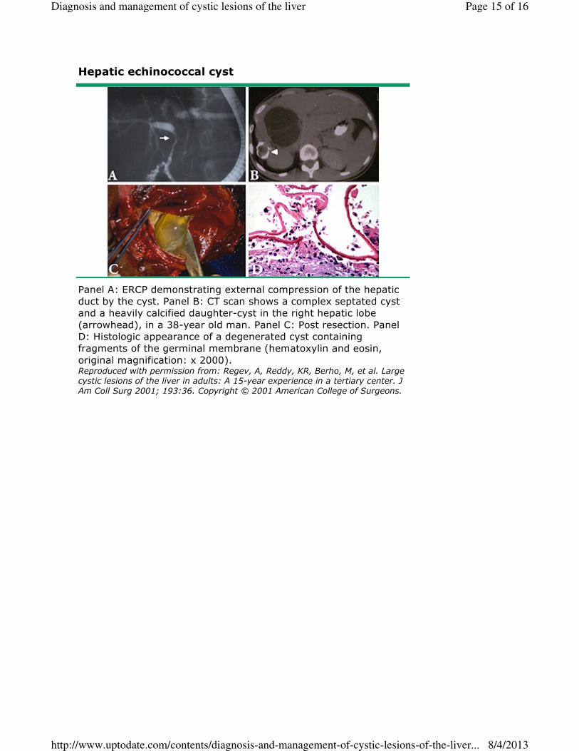
Hepatic echinococcal cyst
Panel A: ERCP demonstrating external compression of the hepatic
duct by the cyst. Panel B: CT scan shows a complex septated cyst
and a heavily calcified daughter-cyst in the right hepatic lobe
(arrowhead), in a 38-year old man. Panel C: Post resection. Panel
D: Histologic appearance of a degenerated cyst containing
fragments of the germinal membrane (hematoxylin and eosin,
original magnification: x 2000). Reproduced with permission from: Regev, A, Reddy, KR, Berho, M, et al. Large cystic lesions of the liver in adults: A 15-year experience in a tertiary center. J
Am Coll Surg 2001; 193:36. Copyright © 2001 American College of Surgeons.
Page 15 of 16Diagnosis and management of cystic lesions of the liver
8/4/2013http://www.uptodate.com/contents/diagnosis-and-management-of-cystic-lesions-of-the-liver...
Page 16 of 16Diagnosis and management of cystic lesions of the liver
8/4/2013http://www.uptodate.com/contents/diagnosis-and-management-of-cystic-lesions-of-the-liver...
Related Documents


![Pancreatic Cytopathology Cystic Lesions Cytol… · Cystic Lesions Cystic Lesions Of The Pancreas [Practical Issues] ... 1-2% of all pancreatic tumors LMP epithelial tumor of uncertain](https://static.cupdf.com/doc/110x72/5f6d9c61a7374f61f46d815c/pancreatic-cytopathology-cystic-lesions-cytol-cystic-lesions-cystic-lesions-of.jpg)