Diagnosing Ventilator-Associated Pneumonia (VAP) In UK NHS ICUs: The Perceived Value And Role of A Novel Optical Technology William Stephen Jones ( [email protected] ) Newcastle upon Tyne Hospitals Foundation Trust https://orcid.org/0000-0001-9352-3916 Jana Suklan Newcastle University Amanda Winter Newcastle upon Tyne Hospitals Foundation Trust Kile Green Newcastle upon Tyne Hospitals Foundation Trust Tom Craven University of Edinburgh Annya Bruce University of Edinburgh Joanne Mair University of Edinburgh Kev Dhaliwal University of Edinburgh Tim Walsh University of Edinburgh John Simpson Newcastle upon Tyne Hospitals Foundation Trust Sara Graziadio Newcastle upon Tyne Hospitals Foundation Trust Joy Allen Newcastle University Research Article Keywords: ventilator-associated pneumonia, VAP, interviews, care pathway analysis, thematic analysis Posted Date: October 25th, 2021 DOI: https://doi.org/10.21203/rs.3.rs-956278/v1

Welcome message from author
This document is posted to help you gain knowledge. Please leave a comment to let me know what you think about it! Share it to your friends and learn new things together.
Transcript

Diagnosing Ventilator-Associated Pneumonia (VAP)In UK NHS ICUs: The Perceived Value And Role of ANovel Optical TechnologyWilliam Stephen Jones ( [email protected] )
Newcastle upon Tyne Hospitals Foundation Trust https://orcid.org/0000-0001-9352-3916Jana Suklan
Newcastle UniversityAmanda Winter
Newcastle upon Tyne Hospitals Foundation TrustKile Green
Newcastle upon Tyne Hospitals Foundation TrustTom Craven
University of EdinburghAnnya Bruce
University of EdinburghJoanne Mair
University of EdinburghKev Dhaliwal
University of EdinburghTim Walsh
University of EdinburghJohn Simpson
Newcastle upon Tyne Hospitals Foundation TrustSara Graziadio
Newcastle upon Tyne Hospitals Foundation TrustJoy Allen
Newcastle University
Research Article
Keywords: ventilator-associated pneumonia, VAP, interviews, care pathway analysis, thematic analysis
Posted Date: October 25th, 2021
DOI: https://doi.org/10.21203/rs.3.rs-956278/v1

License: This work is licensed under a Creative Commons Attribution 4.0 International License. Read Full License
Version of Record: A version of this preprint was published at Diagnostic and Prognostic Research onFebruary 10th, 2022. See the published version at https://doi.org/10.1186/s41512-022-00117-x.

Diagnosing ventilator-associated pneumonia (VAP) in UK 1
NHS ICUs: the perceived value and role of a novel optical 2
technology 3
W. S. Jonesa,b*, J. Suklanb, A. Wintera, K, Greenb, T. Cravenc,d, A. Brucec, J. Mairc, K. Dhaliwalc, T. 4
Walshd, A. J. Simpsona,b, S. Graziadioa,¥, A. J. Allen b,¥ 5
a NIHR Newcastle In Vitro Diagnostics Co-operative, Newcastle upon Tyne Hospitals Foundation Trust, 6
Newcastle upon Tyne, NE1 4LP, United Kingdom 7
8 b NIHR Newcastle In Vitro Diagnostics Co-operative, Translational & Clinical Research Institute, 9
Newcastle University, Newcastle upon Tyne, NE2 4HH, United Kingdom 10
11 C Translational Healthcare Technologies Group, Queen’s Medical Research Institute, University of 12
Edinburgh, Edinburgh EH16 4TJ, United Kingdom 13
14 d Edinburgh Critical Care Research Group, University of Edinburgh, Edinburgh, United Kingdom 15 16 *Corresponding author, email: [email protected] 17 18 ¥ Joint last authors, equal contribution 19
20
21

Abstract 22
Background 23
Diagnosing ventilator-associated pneumonia (VAP) in an intensive care unit (ICU) is a complex process. 24
Our aim was to collect, evaluate and represent the information relating to current clinical practice for 25
the diagnosis of VAP in UK NHS ICUs, and to explore the potential value and role of a novel diagnostic 26
for VAP, which uses optical molecular alveoscopy to visualise the alveolar space. 27
Methods 28
Qualitative study performing semi-structured interviews with clinical experts. Interviews were 29
recorded, transcribed, and thematically analysed. A flow diagram of the VAP patient pathway was 30
elicited and validated with the expert interviewees. 14 clinicians were interviewed from a range of UK 31
NHS hospitals: 12 ICU consultants, 1 professor of respiratory medicine and 1 professor of critical care. 32
Results 33
Five themes were identified, relating to: [1] current practice for the diagnosis of VAP; [2] current 34
clinical need in VAP diagnostics; [3] the potential value and role of the technology; [4] the barriers to 35
adoption; and [5] the evidence requirements for the technology, to help facilitate successful adoption. 36
These themes indicated that diagnosis of VAP is extremely difficult, as is the decision to stop antibiotic 37
treatment. The analysis revealed that there is a clinical need for a diagnostic that provides an accurate 38
and timely diagnosis of the causative pathogen, without the long delays associated with return of 39
culture results, and which is not dangerous to the patient. It was determined that the technology 40
would satisfy important aspects of this clinical need for diagnosing VAP (and pneumonia, more 41
generally), but would require further evidence on safety and efficacy in the patient population to 42
facilitate adoption. 43
Conclusions 44
Care pathway analysis performed in this study was deemed accurate and representative of current 45
practice for diagnosing VAP in a UK ICU as determined by relevant clinical experts, and explored the 46
value and role of a novel diagnostic, which uses optical technology, and could streamline the 47
diagnostic pathway for VAP and other pneumonias. 48
49
Key words: 50
ventilator-associated pneumonia, VAP, interviews, care pathway analysis, thematic analysis. 51
52
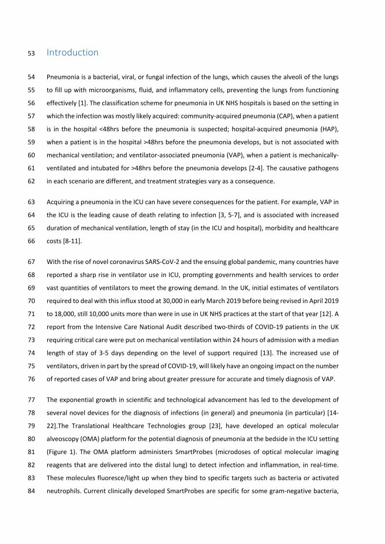
Introduction 53
Pneumonia is a bacterial, viral, or fungal infection of the lungs, which causes the alveoli of the lungs 54
to fill up with microorganisms, fluid, and inflammatory cells, preventing the lungs from functioning 55
effectively [1]. The classification scheme for pneumonia in UK NHS hospitals is based on the setting in 56
which the infection was mostly likely acquired: community-acquired pneumonia (CAP), when a patient 57
is in the hospital <48hrs before the pneumonia is suspected; hospital-acquired pneumonia (HAP), 58
when a patient is in the hospital >48hrs before the pneumonia develops, but is not associated with 59
mechanical ventilation; and ventilator-associated pneumonia (VAP), when a patient is mechanically-60
ventilated and intubated for >48hrs before the pneumonia develops [2-4]. The causative pathogens 61
in each scenario are different, and treatment strategies vary as a consequence. 62
Acquiring a pneumonia in the ICU can have severe consequences for the patient. For example, VAP in 63
the ICU is the leading cause of death relating to infection [3, 5-7], and is associated with increased 64
duration of mechanical ventilation, length of stay (in the ICU and hospital), morbidity and healthcare 65
costs [8-11]. 66
With the rise of novel coronavirus SARS-CoV-2 and the ensuing global pandemic, many countries have 67
reported a sharp rise in ventilator use in ICU, prompting governments and health services to order 68
vast quantities of ventilators to meet the growing demand. In the UK, initial estimates of ventilators 69
required to deal with this influx stood at 30,000 in early March 2019 before being revised in April 2019 70
to 18,000, still 10,000 units more than were in use in UK NHS practices at the start of that year [12]. A 71
report from the Intensive Care National Audit described two-thirds of COVID-19 patients in the UK 72
requiring critical care were put on mechanical ventilation within 24 hours of admission with a median 73
length of stay of 3-5 days depending on the level of support required [13]. The increased use of 74
ventilators, driven in part by the spread of COVID-19, will likely have an ongoing impact on the number 75
of reported cases of VAP and bring about greater pressure for accurate and timely diagnosis of VAP. 76
The exponential growth in scientific and technological advancement has led to the development of 77
several novel devices for the diagnosis of infections (in general) and pneumonia (in particular) [14-78
22].The Translational Healthcare Technologies group [23], have developed an optical molecular 79
alveoscopy (OMA) platform for the potential diagnosis of pneumonia at the bedside in the ICU setting 80
(Figure 1). The OMA platform administers SmartProbes (microdoses of optical molecular imaging 81
reagents that are delivered into the distal lung) to detect infection and inflammation, in real-time. 82
These molecules fluoresce/light up when they bind to specific targets such as bacteria or activated 83
neutrophils. Current clinically developed SmartProbes are specific for some gram-negative bacteria, 84

gram-positive bacteria, and/or neutrophils. These Smartprobes are delivered through a multi-85
functional bundle (Panoptes fibre) that has been extended through serial transbronchial passes into 86
the alveolar sacs, via the working channel of a bronchoscope. Panoptes is a triple lumen optical 87
imaging, delivery and sampling device comprised of two delivery/aspiration capillaries and an imaging 88
fibre. Two imaging systems (Versicolour and Kronoscan) support the real-time visualization of 89
fluorescent bacteria and activated neutrophils within the patient’s alveolar spaces. The OMA platform 90
also has the capacity to perform a mini-lavage by extracting small volumes of liquid instilled in the 91
alveolar space, which could be used for culturing and confirmation of infection. 92
Figure 1: Image showing a bronchoscopy procedure with the imaging fibre and capillary bundle being passed down the 93
working channel of a bronchoscope into the alveolar regions. SmartProbes are delivered via the capillaries and images are 94
sent via the imaging fibre to the imaging system at the patient’s bedside. 95
The accuracy and utility of a diagnostic test is not a fixed property, but is dependent on the clinical 96
setting and patient population in which it is used [24]. To establish the accuracy and clinical utility of 97
a diagnostic device it is first necessary to understand the current diagnostic practice/pathway for the 98
disease of interest. 99
Care pathway analysis, in the context of diagnostics, involves the collection, evaluation and 100
representation of information relating to the diagnostic journey (the pathway) a patient group follows 101
as part of a healthcare system [25-29]. In this study, we performed a set of qualitative, semi-structured 102
interviews with clinical experts in order to conduct a care pathway analysis of the current practice for 103
diagnosing VAP in UK NHS ICUs. 104
Understanding the pathway helps to determine the optimal role, setting and patient population for a 105
new diagnostic, as well as the barriers and facilitators to adoption and future evidence requirements 106
[27]. As part of this pathway analysis we also explored the potential value and role of the OMA 107
platform in the diagnosis of VAP in the ICU. Broadly speaking, there are 4 possible roles for medical 108
tests: (1) Screening, to determine if asymptomatic individuals have a disease, sometimes called 109
Surveillance in ICUs; (2) Diagnostic, to determine if an individual has a disease; (3) Prognostic, to 110
predict the likelihood of an individual developing a disease or deteriorating; and (4) Monitoring, to 111
determine whether a patient’s disease is controlled or is responding to treatment. Depending on how 112
a test will be used it may also be further categorised as a rule-in or a rule-out test [27]. A rule-in test 113
typically requires high specificity (low false positive rate), so that most non-diseased subjects will be 114
diagnosed as non-diseased. Therefore, a positive result makes the presence of disease likely, 115
effectively ruling-in disease. A rule-out test typically requires high sensitivity, so that most diseased 116
subjects will be diagnosed as diseased. Therefore, a negative result essentially rules-out the disease 117

in question. Rule-in tests are important when confirming a diagnosis following other clinical data or 118
when subsequent tests or treatments are dangerous to the patient. Rule-out tests are important when 119
there are severe consequences for missing a disease. 120
Recently, Korevaar et al [24] have recommended that in evaluating a medical test one consider where 121
it would be placed, and how it will affect the current pathway: whether it is a triage test, with the 122
results determining which patients will undergo the existing test(s); an add on test, used before or 123
after an existing test(s) to improve accuracy; a replacement test, replacing the existing test(s), 124
expected to be more accurate, less invasive, less costly or more usable than the test replaced; and a 125
new test, where a completely new test is added to the pathway, were there was not one previously. 126
This consideration will be referred to as its ‘role in the pathway’, to distinguish it from its ‘role as a 127
medical test’, described in the previous paragraph. In this study, we sought to elicit information on 128
the optimal role (in both senses) of the OMA platform for use with suspected VAP patients in UK NHS 129
ICUs. 130
Methods 131
Interview structure 132
Semi-structured interviews were conducted with 14 clinicians from a range of UK NHS hospitals: 12 133
ICU consultants, 1 professor of respiratory medicine and 1 professor of critical care. Interviews lasted 134
between 45-60 minutes in length. Interviews took place in 2019. They were a mixture of face-to-face 135
and telephone interviews and were performed by WSJ and JS. A topic guide was developed prior to 136
the interviews (see Online Material, Appendix 1). Participants were invited to be interviewed via the 137
UK Critical Care Research Group (UKCCRG) mailing list. The sample size used in this study (N=14) was 138
selected based on the concept of data saturation [30] and is consistent with previously published, 139
qualitative research [25]. 140
During the interviews, participants were first asked questions on the current care pathway for the 141
diagnosis of VAP in their ICU. Next, participants watched a short video demonstrating the OMA 142
platform (See Online Material); showing the same video to all participants reduced the potential for 143
bias. Finally, after watching the video, the participants were asked questions about the potential value 144
and role of the OMA platform in the diagnosis of a suspected VAP in the ICU. 145

Patient and public involvement 146
The research question and outcome measures in this study were not informed by patients’ priorities, 147
experience, and/or preferences. Patients were not involved in study design. Patients were not 148
involved in recruitment to, or the conduct of, this study. Patient engagement is part of the 149
dissemination strategy. The research team plans to present the results of this study at various 150
meetings and conferences, where patients will be present. 151
Data analysis 152
Thematic analysis was used to identify the relevant themes from the interviews [31-34]. We used the 153
Gale et al. Framework Analysis approach [35]; see Online Material, Appendix 2 for details of this 154
approach. 155
Results 156
The interview responses from the participants and the results from the thematic and care pathway 157
analysis are synthesised below, divided into 5 key themes. Supporting quotes for each theme are 158
available in the Online Material, Appendix 3. 159
Theme 1: Current practice for the diagnosis of VAP 160
At the beginning of the interviews, participants were asked to describe the current pathway for 161
diagnosing a suspected VAP in their ICU, including information on general ICU functioning. 162
The intensive care unit (ICU) 163
ICUs, sometimes called critical care units or intensive therapy units, are specialist hospital wards that 164
care for severely ill patients who are closely monitored, often with a one to one, or one to two, 165
nurse/patient ratio. 166
Ward rounds occur at least twice daily, and involve a multidisciplinary team of physicians, nurses, 167
microbiologists and other allied healthcare providers. During the ward round the clinical team will 168
review the patient’s clinical characteristics. This review will include the results of the non-specific, 169
routine investigations, which typically include blood, urine and sputum tests. 170

Diagnosing ventilator-associated pneumonia (VAP) 171
Diagnosing a VAP in the ICU is challenging. The initial suspicion of VAP is based on clinical signs 172
associated with the respiratory system, which are not specific to VAP. The typical clinical signs are: 173
high/low temperature, leucocytosis/leukopenia, worsening oxygenation, gas exchange or increasing 174
oxygen requirements, new infiltrates on chest x-ray (CXR) or computerised tomography (CT), 175
suggestive auscultation, general worsening in haemodynamic state, increase in purulent sputum, 176
(colour, thickness and/or frequency), increase in c-reactive protein (CRP), drop in blood pressure, drop 177
in platelet count, and others. Besides pneumonia, these signs can also be indicative of 178
atelectasis/collapsed lung/pneumothorax, sepsis, major trauma (e.g. lung or brain injury), cardiogenic 179
pulmonary edema, pulmonary haemorrhage/embolism/fibrosis, cystic fibrosis, pleural effusion, acute 180
respiratory distress syndrome (ARDS), chronic obstructive pulmonary disease (COPD), mucous 181
impaction and other sources of infection. Several clinical scoring systems exist which aim to provide a 182
semi-objective clinical shortcut to the decision to initiate antibiotics for VAP, the Clinical Pulmonary 183
Infection Score (CPIS) being the most widely used and studied [36]. In general, these scores have 184
unsuitable test characteristics compared to microbiological confirmation and the use of CPIS to guide 185
antibiotic decisions is not recommended by the Infectious Diseases Society of America (IDSA) due to 186
pooled sensitivity and specificity of 65% (95%CI: 61% to 69%) and 64% (95%CI: 60% to 67%) 187
respectively [37, 38]. Retrospectively applied surveillance definitions are widely used because of the 188
use of VAP as a quality indicator and are useful for benchmarking across populations. They have good 189
face validity, making them tempting reference standards, but they may be gamed through 190
interpretation of radiology [39] or timing of microbiology sampling [40], and exhibit low case 191
concordance [41]. Also, the use of these scoring and recording systems is not always feasible in clinical 192
practice, because it is logistically difficult to reliably record this information in the ICU. 193
To improve the accuracy of a diagnosis of suspected VAP requires the performance of an invasive 194
diagnostic procedure, where an upper or lower respiratory specimen is collected and sent for 195
microbiological testing. These procedures are sometimes referred to as special investigations. The 196
correct special investigation to perform is still a matter of debate, with different ICUs employing 197
different procedures [42]. The main procedures are: endotracheal aspirate (ETA), nonbronchoscopic 198
bronchoalveolar lavage (N-BAL), protected specimen brush (PSB), and bronchoalveolar lavage (BAL). 199
The ETA and N-BAL may also be used as non-specific routine investigations. 200

Starting antibiotic treatment for a suspected VAP 201
The treatment for a suspected VAP is antibiotics. The key driver for starting antibiotics is an overall, 202
holistic deterioration in the clinical signs associated with the respiratory system. No single clinical sign 203
or microbiological result is individually sufficient to initiate the decision to treat. Results from special 204
investigations take time to return (up to 72 hours), because of inherent technical limitation in culture-205
based methods. Consequently, antibiotic treatment is typically started on the basis of clinical signs 206
only. In this situation, treatment is with broad spectrum antibiotics. The choice of empirical antibiotic 207
is protocolised in an ICU, in accordance with background resistance rates and influenced by individual 208
patient characteristics. If recent respiratory microbiological isolates are available, they may be used 209
to guide initial treatment, and if microbiology is acquired after initial treatment (i.e. from special 210
investigations), it will be used to tailor a patient’s antibiotics to the causative pathogen, if one is 211
present. The advantages of tailoring antibiotics are several: it leads to more effective killing of bacteria, 212
reduced exposure of patients to unnecessary toxicity (causing Clostridium difficile, etc.)[42], reduced 213
risk of developing antimicrobial resistance (AMR) and reduced costs to the unit and healthcare system. 214
There are no disadvantages to appropriately narrowed antibiotics, but if antibiotics are narrowed 215
incorrectly the bacteria may survive the treatment, with dangerous consequences for the patient. 216
Antibiotics are usually started with a set duration, typically a 5- or 7-day course. This course is strictly 217
completed, unless the antibiotics are narrowed, an alternative diagnosis is confirmed, or if the patient 218
is moving to a palliative mode of care. 219
Stopping antibiotic treatment for a suspected VAP 220
The key driving factor for stopping antibiotics in a suspected VAP patient is an overall, holistic 221
improvement in the patient, based on the clinical signs associated with the respiratory system. 222
Deciding the exact point at which to stop antibiotics was reported to be extremely challenging, partly 223
because there is no good rule-out or monitoring test for pneumonia, but also because there are no 224
standardised and implemented local NHS guidelines to guide clinical decision making; see Online 225
Material, Appendix 4 for a review of guidelines for VAP. 226
Cognitive bias also plays a role in this context. For example, it was reported in the interviews that in 227
the ICU there is a tendency to start with broad-spectrum therapy and maintain this treatment regime, 228
irrespective of whether the patient is getting better or not; If the patient is getting better, then it is 229
assumed that the broad-spectrum antibiotic is working, so they stay broad, and if the patient is getting 230
worse, they may also stay broad and even add on additional antibiotics. It was reported that during 231
handovers between ICU consultants, it is very unlikely that the new consultant will stop antibiotics, 232

even if the plan was to stop them on that day. They are likely to continue for a further 24/48 hours to 233
confirm in their own mind that the patient is clinically well enough to stop. It was also reported that 234
antibiotics are often the only treatment available to help an ICU patient recover from illness, so they 235
are check-mated into continuing antibiotics beyond the conventional course length. This is 236
compounded by the ease and cheapness of prescribing antibiotics. 237
Theme 2: Current clinical need in VAP diagnostics 238
The interviews revealed that there is a clinical need for a diagnostic test that provides a more accurate 239
and timely diagnosis of the causative pathogen (or lack of) and for disease monitoring, without the 240
long delays associated with return of results (i.e. 24-72 hours for the BAL/PSB), and which is not 241
dangerous to the patient (e.g. the transient reduction in oxygenation, associated with bronchoscopic 242
procedures). When a VAP is present, a diagnostic test with these properties is expected to better 243
facilitate the rationalisation and narrowing of antibiotic prescribing for patients with suspected VAP 244
in the ICU, in comparison to current practice. 245
Theme 3: The potential value and role of the OMA platform 246
The potential value of the OMA platform 247
After discussion of current practice and clinical need, we then presented and discussed the OMA 248
platform and its potential value and role in diagnosing a suspected VAP in the ICU. 249
It was indicated that the OMA platform’s provision of the real-time gram information, by the bedside, 250
could provide better rationalisation and narrowing of antibiotics—some antibiotics have more 251
potency against Gram-positive than Gram-negative organisms, and vice versa. Stated differently, if the 252
OMA platform can accurately differentiate between Gram-positive and negative organisms, then the 253
initial empirical, broad spectrum antibiotics could be narrowed to the particular Gram classification. 254
Diagnostic tests targeting bacterial infections often give rise to a conceptual question over whether 255
the bacteria detected represents infection or colonisation in the patient [43]; that is, whether the 256
bacteria detected is causing disease or not, respectively—as the lung is non-sterile tissue and bacteria 257
is expected. In addition to revealing the Gram-positive and/or Gram-negative bacteria in the patient 258
lungs, the OMA platform can also identify activated neutrophils, which are associated with 259
inflammation and infection. This information may help the OMA platform differentiate between 260
colonisation and infection. 261

The OMA platform provides explicit, real-time data through live video-feed to the patient’s bedside. 262
The bacteria and markers of inflammation can be visualised in vivo. It was suggested that this may be 263
more powerful than surrogate markers in influencing decision making. 264
The potential role of the OMA platform 265
The interviews and the care pathway analysis indicated that the optimal role of the OMA platform 266
would be as a replacement or new/add-on special investigation to diagnose VAP in the ICU. In ICUs 267
that currently use BAL or PSB to diagnose VAP, the OMA platform would be a replacement. In ICUs 268
that do not perform special investigations, the OMA platform would be a new test and an add-on to 269
current practice. In both scenarios, the OMA platform would be used as a rule-in diagnostic, to 270
facilitate rapid and Gram-targeted antibiotic treatment. There may be a secondary role for the OMA 271
platform as a rule out diagnostic (less likely) and as a surveillance/monitoring device (less likely). These 272
roles, and their advantages and disadvantages, are discussed below and are visually represented in 273
Figure 2 274
The OMA platform as a rule-in diagnostic 275
The OMA platform, as a rule-in diagnostic, would be performed when there is a clinical suspicion of a 276
VAP, when the patient is eligible for a bronchoscopic and transbronchial procedure (i.e. not physically 277
or logistically contraindicated), and (ideally) prior to antibiotic treatment, to maximise the likelihood 278
of visualising the bacteria and neutrophils, and performing a successful culture through use of the 279
OMA platform’s mini-lavage capabilities. 280
The real-time, Gram and neutrophil information from the OMA platform may allow the clinician to 281
narrow their antibiotic prescription, from a broad spectrum, multi-therapy, empirical antibiotic to a 282
Gram-tailored, (ideally) mono-therapy antibiotic. Assuming the OMA platform is sufficiently accurate, 283
this has the potential to kill bacteria more effectively, reduce exposure of patients to unnecessary 284
toxicity, reduce AMR, and reduce costs, since more effective treatment should reduce usage of ICU 285
beds. 286
The OMA platform as a rule-out diagnostic 287
The OMA platform, as a rule-out diagnostic, would be performed when there is a clinical suspicion of 288
a VAP, and when the patient is eligible for a bronchoscopic and transbronchial procedure. 289
The key advantage to ruling out VAP is that it potentially allows the clinical team to stop antibiotics, 290
pushing them to explore alternative diagnoses for the patient. Also, there is a greater clinical need for 291
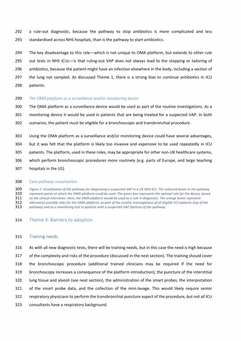
a rule-out diagnostic, because the pathway to stop antibiotics is more complicated and less 292
standardised across NHS hospitals, than is the pathway to start antibiotics. 293
The key disadvantage to this role—which is not unique to OMA platform, but extends to other rule 294
out tests in NHS ICUs—is that ruling-out VAP does not always lead to the stopping or tailoring of 295
antibiotics, because the patient might have an infection elsewhere in the body, including a section of 296
the lung not sampled. As discussed Theme 1, there is a strong bias to continue antibiotics in ICU 297
patients. 298
The OMA platform as a surveillance and/or monitoring device 299
The OMA platform as a surveillance device would be used as part of the routine investigations. As a 300
monitoring device it would be used in patients that are being treated for a suspected VAP. In both 301
scenarios, the patient must be eligible for a bronchoscopic and transbronchial procedure. 302
Using the OMA platform as a surveillance and/or monitoring device could have several advantages, 303
but it was felt that the platform is likely too invasive and expensive to be used repeatedly in ICU 304
patients. The platform, used in these roles, may be appropriate for other non-UK healthcare systems, 305
which perform bronchoscopic procedures more routinely (e.g. parts of Europe, and large teaching 306
hospitals in the US). 307
Care pathway visualisation 308
Figure 2: Visualisation of the pathway for diagnosing a suspected VAP in a UK NHS ICU. The coloured boxes in the pathway 309 represent points at which the OMA platform could be used. The green box represents the optimal role for the device, based 310 on the clinical interviews. Here, the OMA platform would be used as a rule in diagnostic. The orange boxes represent 311 alternative possible roles for the OMA platform, as part of the routine investigations of all eligible ICU patients (top of the 312 pathway) and as a monitoring test in patients with a suspected VAP (bottom of the pathway. 313
Theme 4: Barriers to adoption 314
Training needs 315
As with all new diagnostic tests, there will be training needs, but in this case the need is high because 316
of the complexity and risks of the procedure (discussed in the next section). The training should cover 317
the bronchoscopic procedure (additional trained clinicians may be required if the need for 318
bronchoscopy increases a consequence of the platform introduction), the puncture of the interstitial 319
lung tissue and alveoli (see next section), the administration of the smart probes, the interpretation 320
of the smart probe data, and the collection of the mini-lavage. This would likely require senior 321
respiratory physicians to perform the transbronchial puncture aspect of the procedure, but not all ICU 322
consultants have a respiratory background. 323

Risks of the OMA platform procedure 324
The transbronchial puncture aspect was highlighted as a potential risk to the patients, because of the 325
risk of causing a pneumothorax. It was stated that the risk of causing a pneumothorax is probably 326
quite low, but the risk level is dependent on the patient’s characteristics. The following patients were 327
identified as possessing a heightened risk of developing a pneumothorax: those patients with ARDS, 328
COPD, sepsis, blood clotting abnormalities and patients requiring high ventilator pressure. It was 329
highlighted that if the OMA platform were to cause a pneumothorax in the early stages of adoption in 330
an ICU, it would likely not be used thereafter. The risk of transbronchial puncture is in addition to the 331
transient interference of oxygenation caused by the bronchoscope, and would be increased further if 332
the transbronchial puncture were performed in multiple sites of the lung. The platform’s safety would 333
have to be demonstrated formally and empirically. It is possible that the patients who would get the 334
most out of this platform (e.g. those needing early tailoring of antibiotics) are those that are most at 335
risk of developing complications from the procedure. 336
Increased complexity and effort 337
The OMA platform, as a bronchoscopic technique, adds an extra layer of complexity to diagnosis in 338
comparison to non-bronchoscopic procedures (e.g. ETAs and N-BALS) and to a clinical diagnosis. The 339
OMA platform requires a concerted effort to perform and interpret its results, and carries inherent 340
risks to patients, therefore, there was some concern in the interviews that clinicians may not use this 341
platform, unless strictly required to by guidelines. 342
Cost 343
The potential cost-saving element of the OMA platform, associated with more appropriate antibiotic 344
prescribing (discussed in Theme 3), is likely to be attenuated by the labour-intensive and highly 345
technical nature of the procedure. 346
Theme 5: Evidence requirements to help facilitate successful adoption 347
To feel confident using the OMA platform, the participants indicated that substantial, high quality 348
evidence would be required on the diagnostic accuracy, clinical safety and cost-benefits. 349
It was indicated that the BAL may be a suitable reference standard for evaluating the diagnostic 350
accuracy of the OMA platform. However, it was noted that clinicians who do not use BALs as their 351
standard of care will be less persuaded to adopt the OMA platform if the study does not compare it 352
with the test that they use (e.g. ETAs or N-BALS). Once the accuracy of the platform is adequately 353

demonstrated, a clinical effectiveness/utility trial would be required, where patients would be 354
randomised to the OMA platform or the comparator diagnostic. 355
Cost benefit/effectiveness will be required to help facilitate adoption of the OMA platform. The OMA 356
platform and other bronchoscopic procedures cost more than the non-bronchoscopic procedures. 357
Consequently, it will need to be demonstrated that the OMA platform provides value for money and 358
is affordable, which could stem from better, more tailored patient care and reducing in ICU time. 359
Discussion 360
In this study we performed qualitative, semi-structured interviews with clinical experts. We used the 361
information in these interviews to perform a care pathway analysis of current practice for diagnosing 362
a suspected VAP in the UK NHS ICUs. We also explored the potential value and role of the OMA 363
platform. 364
The care pathway analysis revealed that making a diagnosis of VAP is extremely difficult, primarily 365
because the clinical signs associated with VAP overlap with several other diseases. To improve the 366
accuracy and certainty of a VAP diagnosis, a special investigation may be performed (e.g. ETA, N-BAL, 367
BAL, PSB), where, depending on the method, an upper or lower respiratory specimen is collected and 368
sent for culture. These investigations varying in accuracy, invasiveness and cost. Time delays in 369
receiving these results mean that the decision to treat (predominantly, with antibiotics), is based on 370
clinical signs only. Consequently, antibiotics are typically empirical and broad spectrum. 371
The key driver for starting antibiotics is an overall, holistic deterioration in the clinical signs associated 372
with the respiratory system. Antibiotics will be narrowed where possible, when microbiology becomes 373
available. Antibiotics are usually started with a set duration, typically a 5- or 7-day course and this 374
course is typically completed. 375
The key driving factor for stopping antibiotics is an overall, holistic improvement in the patient, based 376
on the clinical signs associated with the respiratory system, including microbiological information. 377
Deciding the exact point at which to stop antibiotics was reported to be extremely challenging, partly 378
because there is no good rule-out test for pneumonia, and no standardised guidelines. It was also 379
indicated that cognitive biases might affect the decision to stop antibiotics. Biases which have 380
previously been demonstrated to be present in clinical decision making [44, 45], and antibiotic 381
prescribing behaviour [46]. 382

The interviews revealed that there is a clinical need for a diagnostic test that provides a more accurate 383
and timely diagnosis of VAP, which is not dangerous to the patient, and which has properties that 384
better facilitate the rationalisation and narrowing of antibiotic prescribing for patients. 385
Based on the care pathway analysis it was determined the OMA platform could potentially satisfy 386
important aspects of the above clinical need. The OMA platform’s provision of real-time gram 387
information, by the bedside, could provide better rationalisation and narrowing of antibiotics, which 388
may be more powerful than currently-used surrogate markers in influencing clinical decision making. 389
Also, the platforms ability to identify activated neutrophils may also help differentiate between 390
bacterial colonisation and infection. Although the focus of this study was VAP, these value 391
propositions for the OMA platform may be equally applicable to all forms of bacterial pneumonia 392
experienced in the ICU. 393
Conclusions 394
Diagnosing a VAP in the ICU is challenging. The initial suspicion of VAP is based on clinical signs 395
associated with the respiratory system (including bloods and CXRs), which are not specific to VAP, and 396
do not allow for the tailored antibiotic treatment. To improve the accuracy, or to reduce the 397
uncertainty, requires an invasive diagnostic procedure, where an upper or lower respiratory specimen 398
is collected and sent for microbiological testing. These procedures vary in safety, accuracy, and 399
influence on clinical decision making. 400
There is a clinical need for a diagnostic test that provides a more accurate and timely diagnosis of the 401
causative pathogen (or lack of) and for disease monitoring. When a VAP is present, a diagnostic test 402
with these properties would better facilitate the rationalisation and narrowing of antibiotic 403
prescribing in comparison to current practice. The care pathway analysis revealed that the OMA 404
platform would address this aspect of the clinical need, but further evidence would be required on its 405
accuracy, safety and cost-benefit. 406
Future recommendations 407
Further research into the cognitive biases involved in antibiotic decision making in the ICU would be 408
informative for clinicians and developers. 409
410

Declarations 411
Ethics approval and consent of participants 412
Ethical approval was obtained from the HRA to carry out interviews on NHS staff (IRAS number: 413
242651). 414
Consent for publication 415
Not applicable. 416
Availability of data and material 417
Key quotes from the interviews are provided in the Online Appendices. No additional data are 418
available. 419
Competing interests 420
KD, AB, and JM have competing interests. They are involved in the development of the technology 421
under review. Their contributions were restricted to fact-checking of the technology description, and 422
they also helped us with the conceptualisation of the initial study design. This study was performed, 423
analysed and reported by the NIHR Newcastle In Vitro Diagnostics Co-operative (Newcastle MIC), 424
which is an independent group, funded by NIHR, to generate high-quality evidence on diagnostic 425
devices. The Newcastle MIC had full control of the content in the manuscript. 426
Funding 427
This study was funded by a CARB-X grant (4500002330). The NIHR funds the NIHR Newcastle In Vitro 428
Diagnostics Co-operative. 429
Authors’ contributions 430
All authors made substantial contributions to the design of the study. WSJ, JS, AJA and SG performed 431
and analysed the clinical interviews. WSJ drafted the manuscript for this study. All authors critically 432
reviewed iterative drafts of revisions of the manuscript, and approved the final version. 433
Acknowledgements 434
We would like to thank the clinical experts that contributed their time in participating in the 435
interviews. These Individuals will remain anonymous. 436

References 437
1. The National Institute for Health and Care Excellence (NICE), Pneumonia in adults: diagnosis 438
and management. 2019. 439
2. Anand, N. and M.H. Kollef, The alphabet soup of pneumonia: CAP, HAP, HCAP, NHAP, and VAP. 440
Semin Respir Crit Care Med, 2009. 30(1): p. 3-9. 441
3. Koulenti, D., E. Tsigou, and J. Rello, Nosocomial pneumonia in 27 ICUs in Europe: perspectives 442
from the EU-VAP/CAP study. Eur J Clin Microbiol Infect Dis, 2017. 36(11): p. 1999-2006. 443
4. Niederman, M.S., Hospital-acquired pneumonia, health care-associated pneumonia, ventilator-444
associated pneumonia, and ventilator-associated tracheobronchitis: definitions and challenges 445
in trial design. Clin Infect Dis, 2010. 51 Suppl 1: p. S12-7. 446
5. Ferrer, M. and A. Torres, Epidemiology of ICU-acquired pneumonia. Curr Opin Crit Care, 2018. 447
24(5): p. 325-331. 448
6. Spencer, R.C., Epidemiology of infection in ICUs. Intensive Care Med, 1994. 20 Suppl 4: p. S2-6. 449
7. Vallés, J., et al., Excess ICU mortality attributable to ventilator-associated pneumonia: the role 450
of early vs late onset. Intensive Care Med, 2007. 33(8): p. 1363-8. 451
8. Heyland, D.K., et al., The attributable morbidity and mortality of ventilator-associated 452
pneumonia in the critically ill patient. The Canadian Critical Trials Group. Am J Respir Crit Care 453
Med, 1999. 159(4 Pt 1): p. 1249-56. 454
9. Safdar, N., et al., Clinical and economic consequences of ventilator-associated pneumonia: a 455
systematic review. Crit Care Med, 2005. 33(10): p. 2184-93. 456
10. Kollef, M.H., C.W. Hamilton, and F.R. Ernst, Economic impact of ventilator-associated 457
pneumonia in a large matched cohort. Infect Control Hosp Epidemiol, 2012. 33(3): p. 250-6. 458
11. Rello, J., et al., Epidemiology and outcomes of ventilator-associated pneumonia in a large US 459
database. Chest, 2002. 122(6): p. 2115-21. 460
12. Balogun, B., Coronavirus: Ventilator availability in the UK, H.o.C. Library, Editor. 2020. 461
13. Mahase, E., Covid-19: most patients require mechanical ventilation in first 24 hours of critical 462
care. BMJ, 2020. 368: p. m1201. 463
14. Akram, A.R., et al., In situ identification of Gram-negative bacteria in human lungs using a 464
topical fluorescent peptide targeting lipid A. Sci Transl Med, 2018. 10(464). 465
15. Chan, Y.R. and A. Morris, Molecular diagnostic methods in pneumonia. Curr Opin Infect Dis, 466
2007. 20(2): p. 157-64. 467
16. Drabińska, N., et al., From fast identification to resistance testing: Volatile compound profiling 468
as a novel diagnostic tool for detection of antibiotic susceptibility. TrAC Trends in Analytical 469
Chemistry, 2019. 115: p. 1-12. 470
17. Hellyer, T. and J. Simpson, Biomarker-based exclusion of ventilator-associated pneumonia: a 471
multicentre validation study. Critical Care, 2014. 18(Suppl 1): p. P303-P303. 472
18. Hellyer, T.P., et al., Diagnostic accuracy of pulmonary host inflammatory mediators in the 473
exclusion of ventilator-acquired pneumonia. Thorax, 2015. 70(1): p. 41. 474
19. Jung, J.H. and J.E. Lee, Real-time bacterial microcolony counting using on-chip microscopy. 475
Scientific reports, 2016. 6: p. 21473-21473. 476
20. Mills, B., M. Bradley, and K. Dhaliwal, Optical imaging of bacterial infections. Clinical and 477
translational imaging, 2016. 4: p. 163-174. 478
21. Morris, A.C., Management of pneumonia in intensive care. J Emerg Crit Care Med, 2018. 479
22. Slupsky, C.M., Nuclear magnetic resonance-based analysis of urine for the rapid etiological 480
diagnosis of pneumonia. Expert Opinion on Medical Diagnostics, 2011. 5(1): p. 63-73. 481
23. The Proteus Interdisciplinary Research Collaboration is funded by the Engineering and Physical 482
Sciences Research Council (EPSRC). 2017; Available from: https://proteus.ac.uk/about/the-483
consortium/. 484

24. Korevaar, D.A., et al., Targeted test evaluation: a framework for designing diagnostic accuracy 485
studies with clear study hypotheses. Diagnostic and Prognostic Research, 2019. 3(1): p. 22. 486
25. Charman, S., et al., Opportunities and challenges of a novel cardiac output response to stress 487
(CORS) test to enhance diagnosis of heart failure in primary care: qualitative study. BMJ Open, 488
2019. 9(4): p. e028122. 489
26. De Bleser, L., et al., Defining pathways. J Nurs Manag, 2006. 14(7): p. 553-63. 490
27. Graziadio, S., et al., How to Ease the Pain of Taking a Diagnostic Point of Care Test to the Market: 491
A Framework for Evidence Development. Micromachines (Basel), 2020. 11(3). 492
28. Kinsman, L., et al., What is a clinical pathway? Development of a definition to inform the debate. 493
BMC Med, 2010. 8: p. 31. 494
29. Panella, M. and K. Vanhaecht, Is there still need for confusion about pathways? International 495
Journal of Care Pathways, 2010. 14(1): p. 1-3. 496
30. Saunders, B., et al., Saturation in qualitative research: exploring its conceptualization and 497
operationalization. Qual Quant, 2018. 52(4): p. 1893-1907. 498
31. Braun, V. and V. Clarke, Using thematic analysis in psychology. Qualitative Research in 499
Psychology, 2006. 3(2): p. 77-101. 500
32. Castleberry, A. and A. Nolen, Thematic analysis of qualitative research data: Is it as easy as it 501
sounds? Curr Pharm Teach Learn, 2018. 10(6): p. 807-815. 502
33. Maguire, M. and B. Delahunt. Doing a thematic analysis: A practical, step-by-step guide for 503
learning and teaching scholars. 2017. 504
34. Nowell, L.S., et al., Thematic Analysis:Striving to Meet the Trustworthiness Criteria. 505
International Journal of Qualitative Methods, 2017. 16(1): p. 1609406917733847. 506
35. Gale, N.K., et al., Using the framework method for the analysis of qualitative data in multi-507
disciplinary health research. BMC Medical Research Methodology, 2013. 13(1): p. 117. 508
36. Pugin, J., et al., Diagnosis of ventilator-associated pneumonia by bacteriologic analysis of 509
bronchoscopic and nonbronchoscopic "blind" bronchoalveolar lavage fluid. Am Rev Respir Dis, 510
1991. 143(5 Pt 1): p. 1121-9. 511
37. Kalil, A.C., et al., Management of Adults With Hospital-acquired and Ventilator-associated 512
Pneumonia: 2016 Clinical Practice Guidelines by the Infectious Diseases Society of America and 513
the American Thoracic Society. Clinical Infectious Diseases, 2016. 63(5): p. e61-e111. 514
38. Shan, J., H.L. Chen, and J.H. Zhu, Diagnostic accuracy of clinical pulmonary infection score for 515
ventilator-associated pneumonia: a meta-analysis. Respir Care, 2011. 56(8): p. 1087-94. 516
39. Walsh, T.S., A.C. Morris, and A.J. Simpson, V. Ventilator associated pneumonia: can we ensure 517
that a quality indicator does not become a game of chance? BJA: British Journal of Anaesthesia, 518
2013. 111(3): p. 333-337. 519
40. Craven, T.H., et al., Ventilator-associated pneumonia surveillance using two methods. Journal 520
of Hospital Infection, 2020. 104(4): p. 522-528. 521
41. Craven, T.H., et al., Lack of concordance between ECDC and CDC systems for surveillance of 522
ventilator associated pneumonia. Intensive Care Medicine, 2018. 44(2): p. 265-266. 523
42. Arulkumaran, N., et al., Antimicrobial-associated harm in critical care: a narrative review. 524
Intensive Care Med, 2020. 46(2): p. 225-235. 525
43. Dani, A., Colonization and infection. Central European journal of urology, 2014. 67(1): p. 86-87. 526
44. O'Sullivan, E.D. and S.J. Schofield, Cognitive bias in clinical medicine. J R Coll Physicians Edinb, 527
2018. 48(3): p. 225-232. 528
45. Saposnik, G., et al., Cognitive biases associated with medical decisions: a systematic review. 529
BMC medical informatics and decision making, 2016. 16(1): p. 138-138. 530
46. Warreman, E.B., et al., Determinants of in-hospital antibiotic prescription behaviour: a 531
systematic review and formation of a comprehensive framework. Clin Microbiol Infect, 2019. 532
25(5): p. 538-545. 533
534

Figures
Figure 1
Image showing a bronchoscopy procedure with the imaging �bre and capillary bundle being passeddown the working channel of a bronchoscope into the alveolar regions. SmartProbes are delivered via thecapillaries and images are sent via the imaging �bre to the imaging system at the patient’s bedside.
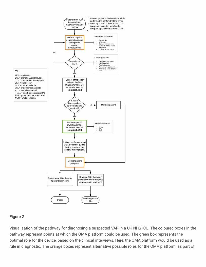
Figure 2
Visualisation of the pathway for diagnosing a suspected VAP in a UK NHS ICU. The coloured boxes in thepathway represent points at which the OMA platform could be used. The green box represents theoptimal role for the device, based on the clinical interviews. Here, the OMA platform would be used as arule in diagnostic. The orange boxes represent alternative possible roles for the OMA platform, as part of

the routine investigations of all eligible ICU patients (top of the pathway) and as a monitoring test inpatients with a suspected VAP (bottom of the pathway.
Supplementary Files
This is a list of supplementary �les associated with this preprint. Click to download.
OMAQualVAPManuscriptAppendicesv02.pdf
Related Documents