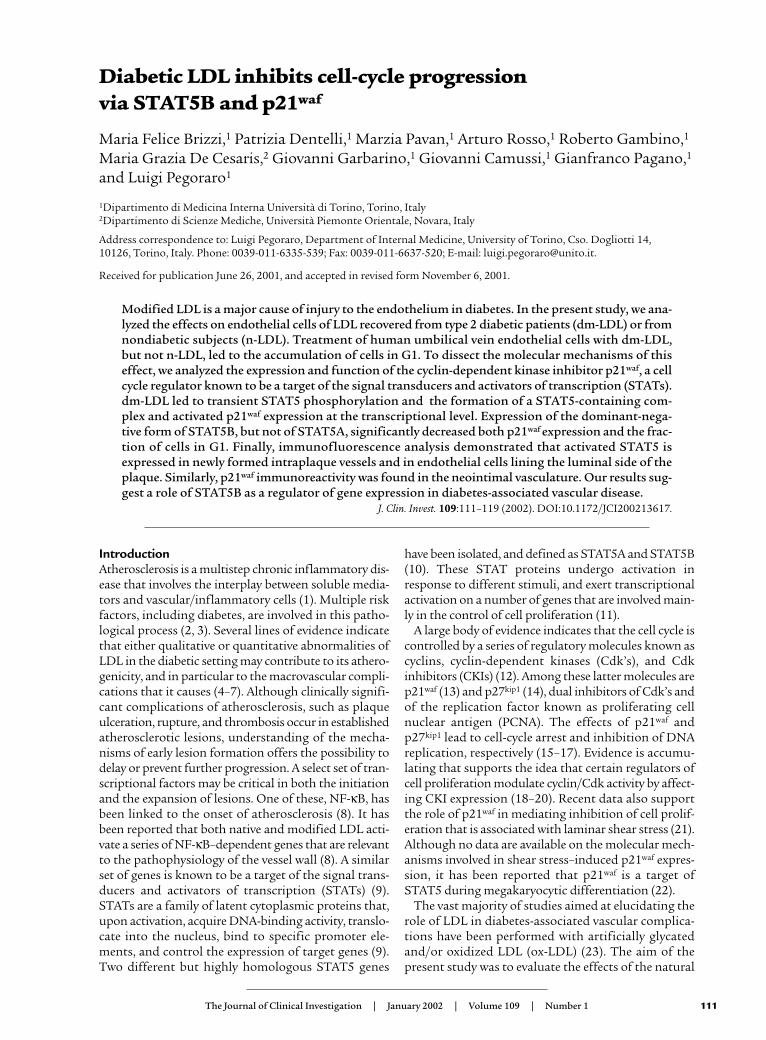
Introduction Atherosclerosis is a multistep chronic inflammatory dis- ease that involves the interplay between soluble media- tors and vascular/inflammatory cells (1). Multiple risk factors, including diabetes, are involved in this patho- logical process (2, 3). Several lines of evidence indicate that either qualitative or quantitative abnormalities of LDL in the diabetic setting may contribute to its athero- genicity, and in particular to the macrovascular compli- cations that it causes (4–7). Although clinically signifi- cant complications of atherosclerosis, such as plaque ulceration, rupture, and thrombosis occur in established atherosclerotic lesions, understanding of the mecha- nisms of early lesion formation offers the possibility to delay or prevent further progression. A select set of tran- scriptional factors may be critical in both the initiation and the expansion of lesions. One of these, NF-κB, has been linked to the onset of atherosclerosis (8). It has been reported that both native and modified LDL acti- vate a series of NF-κB–dependent genes that are relevant to the pathophysiology of the vessel wall (8). A similar set of genes is known to be a target of the signal trans- ducers and activators of transcription (STATs) (9). STATs are a family of latent cytoplasmic proteins that, upon activation, acquire DNA-binding activity, translo- cate into the nucleus, bind to specific promoter ele- ments, and control the expression of target genes (9). Two different but highly homologous STAT5 genes have been isolated, and defined as STAT5A and STAT5B (10). These STAT proteins undergo activation in response to different stimuli, and exert transcriptional activation on a number of genes that are involved main- ly in the control of cell proliferation (11). A large body of evidence indicates that the cell cycle is controlled by a series of regulatory molecules known as cyclins, cyclin-dependent kinases (Cdk’s), and Cdk inhibitors (CKIs) (12). Among these latter molecules are p21 waf (13) and p27 kip1 (14), dual inhibitors of Cdk’s and of the replication factor known as proliferating cell nuclear antigen (PCNA). The effects of p21 waf and p27 kip1 lead to cell-cycle arrest and inhibition of DNA replication, respectively (15–17). Evidence is accumu- lating that supports the idea that certain regulators of cell proliferation modulate cyclin/Cdk activity by affect- ing CKI expression (18–20). Recent data also support the role of p21 waf in mediating inhibition of cell prolif- eration that is associated with laminar shear stress (21). Although no data are available on the molecular mech- anisms involved in shear stress–induced p21 waf expres- sion, it has been reported that p21 waf is a target of STAT5 during megakaryocytic differentiation (22). The vast majority of studies aimed at elucidating the role of LDL in diabetes-associated vascular complica- tions have been performed with artificially glycated and/or oxidized LDL (ox-LDL) (23). The aim of the present study was to evaluate the effects of the natural The Journal of Clinical Investigation | January 2002 | Volume 109 | Number 1 111 Diabetic LDL inhibits cell-cycle progression via STAT5B and p21 waf Maria Felice Brizzi, 1 Patrizia Dentelli, 1 Marzia Pavan, 1 Arturo Rosso, 1 Roberto Gambino, 1 Maria Grazia De Cesaris, 2 Giovanni Garbarino, 1 Giovanni Camussi, 1 Gianfranco Pagano, 1 and Luigi Pegoraro 1 1 Dipartimento di Medicina Interna Università di Torino, Torino, Italy 2 Dipartimento di Scienze Mediche, Università Piemonte Orientale, Novara, Italy Address correspondence to: Luigi Pegoraro, Department of Internal Medicine, University of Torino, Cso. Dogliotti 14, 10126, Torino, Italy. Phone: 0039-011-6335-539; Fax: 0039-011-6637-520; E-mail: [email protected]. Received for publication June 26, 2001, and accepted in revised form November 6, 2001. Modified LDL is a major cause of injury to the endothelium in diabetes. In the present study, we ana- lyzed the effects on endothelial cells of LDL recovered from type 2 diabetic patients (dm-LDL) or from nondiabetic subjects (n-LDL). Treatment of human umbilical vein endothelial cells with dm-LDL, but not n-LDL, led to the accumulation of cells in G1. To dissect the molecular mechanisms of this effect, we analyzed the expression and function of the cyclin-dependent kinase inhibitor p21 waf , a cell cycle regulator known to be a target of the signal transducers and activators of transcription (STATs). dm-LDL led to transient STAT5 phosphorylation and the formation of a STAT5-containing com- plex and activated p21 waf expression at the transcriptional level. Expression of the dominant-nega- tive form of STAT5B, but not of STAT5A, significantly decreased both p21 waf expression and the frac- tion of cells in G1. Finally, immunofluorescence analysis demonstrated that activated STAT5 is expressed in newly formed intraplaque vessels and in endothelial cells lining the luminal side of the plaque. Similarly, p21 waf immunoreactivity was found in the neointimal vasculature. Our results sug- gest a role of STAT5B as a regulator of gene expression in diabetes-associated vascular disease. J. Clin. Invest. 109:111–119 (2002). DOI:10.1172/JCI200213617.

Welcome message from author
This document is posted to help you gain knowledge. Please leave a comment to let me know what you think about it! Share it to your friends and learn new things together.
Transcript
IntroductionAtherosclerosis is a multistep chronic inflammatory dis-ease that involves the interplay between soluble media-tors and vascular/inflammatory cells (1). Multiple riskfactors, including diabetes, are involved in this patho-logical process (2, 3). Several lines of evidence indicatethat either qualitative or quantitative abnormalities ofLDL in the diabetic setting may contribute to its athero-genicity, and in particular to the macrovascular compli-cations that it causes (4–7). Although clinically signifi-cant complications of atherosclerosis, such as plaqueulceration, rupture, and thrombosis occur in establishedatherosclerotic lesions, understanding of the mecha-nisms of early lesion formation offers the possibility todelay or prevent further progression. A select set of tran-scriptional factors may be critical in both the initiationand the expansion of lesions. One of these, NF-κB, hasbeen linked to the onset of atherosclerosis (8). It hasbeen reported that both native and modified LDL acti-vate a series of NF-κB–dependent genes that are relevantto the pathophysiology of the vessel wall (8). A similarset of genes is known to be a target of the signal trans-ducers and activators of transcription (STATs) (9).STATs are a family of latent cytoplasmic proteins that,upon activation, acquire DNA-binding activity, translo-cate into the nucleus, bind to specific promoter ele-ments, and control the expression of target genes (9).Two different but highly homologous STAT5 genes
have been isolated, and defined as STAT5A and STAT5B(10). These STAT proteins undergo activation inresponse to different stimuli, and exert transcriptionalactivation on a number of genes that are involved main-ly in the control of cell proliferation (11).
A large body of evidence indicates that the cell cycle iscontrolled by a series of regulatory molecules known ascyclins, cyclin-dependent kinases (Cdk’s), and Cdkinhibitors (CKIs) (12). Among these latter molecules arep21waf (13) and p27kip1 (14), dual inhibitors of Cdk’s andof the replication factor known as proliferating cellnuclear antigen (PCNA). The effects of p21waf andp27kip1 lead to cell-cycle arrest and inhibition of DNAreplication, respectively (15–17). Evidence is accumu-lating that supports the idea that certain regulators ofcell proliferation modulate cyclin/Cdk activity by affect-ing CKI expression (18–20). Recent data also supportthe role of p21waf in mediating inhibition of cell prolif-eration that is associated with laminar shear stress (21).Although no data are available on the molecular mech-anisms involved in shear stress–induced p21waf expres-sion, it has been reported that p21waf is a target ofSTAT5 during megakaryocytic differentiation (22).
The vast majority of studies aimed at elucidating therole of LDL in diabetes-associated vascular complica-tions have been performed with artificially glycatedand/or oxidized LDL (ox-LDL) (23). The aim of thepresent study was to evaluate the effects of the natural
The Journal of Clinical Investigation | January 2002 | Volume 109 | Number 1 111
Diabetic LDL inhibits cell-cycle progression via STAT5B and p21waf
Maria Felice Brizzi,1 Patrizia Dentelli,1 Marzia Pavan,1 Arturo Rosso,1 Roberto Gambino,1
Maria Grazia De Cesaris,2 Giovanni Garbarino,1 Giovanni Camussi,1 Gianfranco Pagano,1
and Luigi Pegoraro1
1Dipartimento di Medicina Interna Università di Torino, Torino, Italy2Dipartimento di Scienze Mediche, Università Piemonte Orientale, Novara, Italy
Address correspondence to: Luigi Pegoraro, Department of Internal Medicine, University of Torino, Cso. Dogliotti 14, 10126, Torino, Italy. Phone: 0039-011-6335-539; Fax: 0039-011-6637-520; E-mail: [email protected].
Received for publication June 26, 2001, and accepted in revised form November 6, 2001.
Modified LDL is a major cause of injury to the endothelium in diabetes. In the present study, we ana-lyzed the effects on endothelial cells of LDL recovered from type 2 diabetic patients (dm-LDL) or fromnondiabetic subjects (n-LDL). Treatment of human umbilical vein endothelial cells with dm-LDL,but not n-LDL, led to the accumulation of cells in G1. To dissect the molecular mechanisms of thiseffect, we analyzed the expression and function of the cyclin-dependent kinase inhibitor p21waf, a cellcycle regulator known to be a target of the signal transducers and activators of transcription (STATs).dm-LDL led to transient STAT5 phosphorylation and the formation of a STAT5-containing com-plex and activated p21waf expression at the transcriptional level. Expression of the dominant-nega-tive form of STAT5B, but not of STAT5A, significantly decreased both p21waf expression and the frac-tion of cells in G1. Finally, immunofluorescence analysis demonstrated that activated STAT5 isexpressed in newly formed intraplaque vessels and in endothelial cells lining the luminal side of theplaque. Similarly, p21waf immunoreactivity was found in the neointimal vasculature. Our results sug-gest a role of STAT5B as a regulator of gene expression in diabetes-associated vascular disease.
J. Clin. Invest. 109:111–119 (2002). DOI:10.1172/JCI200213617.
plasma constituent recovered from type 2 diabeticpatients (dm-LDL) on endothelial cells. We found thatdm-LDL affects cell-cycle progression via STAT5B-mediated p21waf induction. Moreover, activated STAT5and p21waf immunoreactivity was present in humanintraplaque neovessels.
MethodsPatients and controls. Blood was withdrawn from nineblood donors and from nine type 2 diabetic patients inbad metabolic control (fasting plasma glucose ≥10 mMand hemoglobin A1c ≥ 10%). None of them was underinsulin, and all were treated with sulphonylurea agents.The mean age was 68 ± 4 years in the diabetic group,and 62 ± 3 years in controls. Each group consisted offive men and four women.
Reagents. M199 medium (endotoxin tested), BSA, andprotein A–Sepharose were from Sigma Chemical Co.(St. Louis, Missouri, USA). Bovine calf serum (endotox-in tested) was obtained from HyClone Laboratories Inc.(Logan, Utah, USA). Trypsin was purchased from DifcoLaboratories Inc. (Detroit, Michigan, USA). Nitrocellu-lose filters, horseradish peroxidase–conjugated proteinA, the molecular weight markers [α-32P]dCTP, [γ-32P]ATP, and [α-32P]UTP, the chemiluminescencereagent enhanced, and the Poly(dIdC):poly(dIdC) wereobtained from Amersham Pharmacia Biotech Italia(Milano, Italy). Histone H1 was from Roche Diagnos-tics S.P.A. (Monza, Italy). The presence of endotoxin
contamination of LDL preparations was tested by theLimulus amebocyte assay; the concentration was 0.05ng/ml. Anti-STAT5A (L-20), anti-STAT5B (G-2 and C-17), anti-p21waf, and anti-p27 kip1 antisera wereobtained from Santa Cruz Biotechnology Inc. (Heidel-berg, Germany). Anti–phospho-STAT5, anti-CD105,and anti-CD45 were from New England Biolabs Inc.(Beverly, Massachusetts, USA).
Isolation, characterization, and oxidation of LDL. Bloodwas centrifuged for 15 minutes at 12,000 g at 4°C. LDLwas separated according to the method of Redgraveand Carlson (24). Oxidation of n-LDL was performedin a cell-free system as described (25). Oxidation wasconfirmed by agarose gel electrophoresis. Purity of LDLwas assessed by capillary electrophoresis at 200 nm.
Thiobarbituric acid–reactive substances assay and capillaryelectrophoresis. LDL subfractions were pooled, and per-oxidation was measured using a thiobarbituricacid–reactive substances (TBARS) assay (26). TBARSconcentrations were calculated from a calibration curvethat was prepared using 1,1,3,3-tetramethoxypropaneas a standard. Diene conjugate quantification was per-formed by capillary electrophoresis as described (27).LDL mobility in capillary electrophoresis was calculat-ed as described by Cruzado et al. (28).
Cell cultures and transfections. HUVECs were isolated aspreviously described (29). ECV304 cells were transfect-ed with the dominant-negative STAT5A (∆STAT5A)and STAT5B (∆STAT5B) constructs (11) by the lipo-fectin method. Expression of the ∆STAT5 proteins wasanalyzed by Western blotting with an anti-STAT5 anti-body, and the positive clones were tested by elec-trophoretic mobility shift assay (30).
Flow cytometry. HUVECs or ECV304 cells were stim-ulated in serum-free medium with n-LDL (100 µg/ml)or dm-LDL (100 µg/ml) for 12 hours, and then fixedwith 70% ethanol. After digestion with RNase, DNAwas stained with propidium iodide and analyzed witha flow cytometer.
Western blot analysis and immunoprecipitation studies.HUVEC monolayers were processed as described (30)and incubated with or without 100 µg/ml of either n-LDL or dm-LDL for the indicated time periods. Proteinconcentrations of cell lysates were obtained and sampleswere eluted and processed as previously described (30).
Preparation of nuclear extracts and gel retardation assay.Nuclear extracts from untreated or LDL-treatedHUVECs were prepared as described by Sadowski andGilman (31). The double-stranded p21SIE2 oligonu-cleotide sequence 5′-GATCCTTTCTGAGAAATGG-3′ (22)was used. Gel retardation reactions were performed aspreviously described (30).
Cdk2 kinase assay. Cdk2 immunoprecipitated from dm-LDL–stimulated (100 µg/ml), STAT transfected ECV304clones was divided into two aliquots. Cdk2 immuno-precipitates were resuspended in kinase buffer consist-ing of 20 mM Tris/HCl (pH7.4), 10 mM MgCl2, and 1mM DTT containing 50 µM [γ-32P]ATP and 5 µg/ml ofhistone H1, and incubated for 30 minutes at 30°C.
112 The Journal of Clinical Investigation | January 2002 | Volume 109 | Number 1
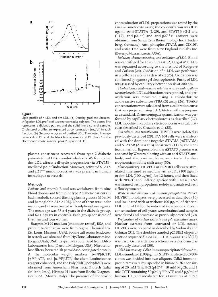
Figure 1Lipid profile of n-LDL and dm-LDL. (a) Density-gradient ultracen-trifugation LDL profile of two representative subjects. The dotted linerepresents a diabetic patient and the solid line a control sample.Cholesterol profiles are expressed as concentration (mg/dl) in eachfraction. (b) Electropherogram of purified LDL. The dotted line rep-resents dm-LDL and the black line represents n-LDL. Peak 1 is theelectroendosmotic marker; peak 2 is purified LDL.
Agarose beads were removed by centrifugation, and thesupernatants were fractionated by 8% SDS-PAGE.
Nuclear run-off transcription. Approximately 2 × 10⁷
HUVECs were stimulated with dm-LDL (100 µg/ml) andlysed in Nonidet P-40 lysis buffer (32). Nuclei were col-lected and stored as previously described (32). Elongationof nascent RNA was performed as previously described(32). Labeled RNA was purified as described by Gariglioet al. (33), and processed as previously described (32).
Detection of reactive oxygen species. 5,6-caroxy-22, 72-dichlorofluorescin-diacetate (DCF-DA) (20 mM finalconcentration) was added to HUVECs in the variousculture conditions at time 0. At the times indicated, thecells were subjected to flow cytometric analysis asdescribed (34). TNF-α was used as positive control (35).
Immunofluorescence microscopy. Carotid specimens fromdiabetic or hypercholesterolemic patients and healthycarotid specimens were fixed and embedded in paraffinand processed as described (36). Sections were stainedwith the anti–phospho-STAT5, the anti-p21waf, and theanti-CD105 antibodies. Indirect immunofluorescenceanalysis was performed as previously described (36).
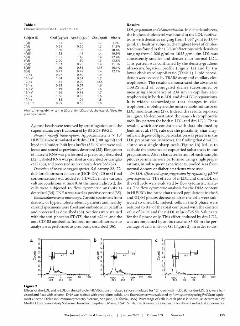
ResultsLDL preparation and characterization. In diabetic subjects,the highest cholesterol was found in the LDL subfrac-tions with densities ranging from 1.037 g/ml to 1.044g/ml. In healthy subjects, the highest level of choles-terol was found in the LDL subfractions with densitiesranging from 1.028 g/ml to 1.035 g/ml. dm-LDL wasconsistently smaller and denser than normal LDL.This pattern was confirmed by the density-gradientultracentrifugation profile (Figure 1a) and by thelower cholesterol/apoB ratio (Table 1). Lipid peroxi-dation was assessed by TBARS assay and capillary elec-trophoresis. The results demonstrated the absence ofTBARS and of conjugated dienes (determined bymeasuring absorbance at 234 nm in capillary elec-trophoresis) in both n-LDL and dm-LDL preparations.It is widely acknowledged that changes in elec-trophoretic mobility are the most reliable indicator ofLDL modifications (27). Indeed, the results reportedin Figure 1b demonstrated the same electrophoreticmobility pattern for both n-LDL and dm-LDL. Theseresults, which are consistent with data obtained byJenkins et al. (37), rule out the possibility that a sig-nificant degree of lipid peroxidation was present in theLDL preparations. Moreover, the finding that dm-LDLeluted as a single sharp peak (Figure 1b) led us toexclude the presence of copurified substances in ourpreparations. After characterization of each sample,pilot experiments were performed using single prepa-rations; in subsequent experiments, pooled sera fromnormal donors or diabetic patients were used.
dm-LDL affects cell-cycle progression by regulating p21waf
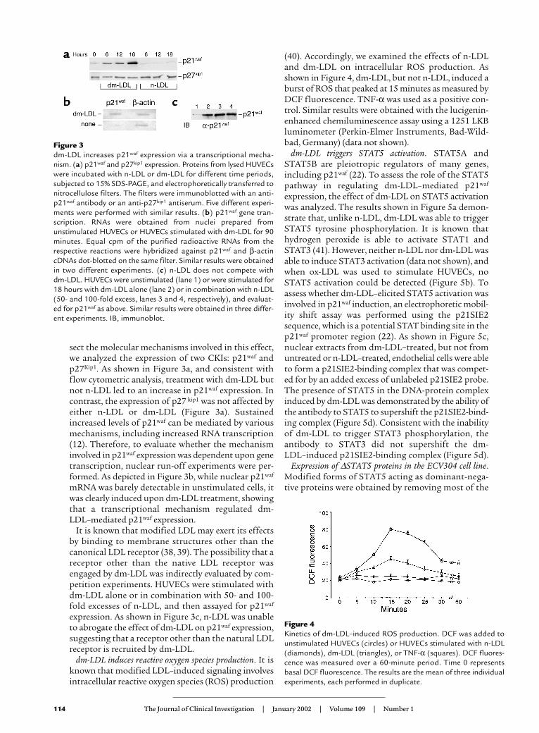
gene expression. The effects of n-LDL and dm-LDL onthe cell cycle were evaluated by flow cytometric analy-sis. The flow cytometric analysis for the DNA contentin HUVECs indicated that the cell populations in the Sand G2/M phases decreased after the cells were sub-jected to dm-LDL. Indeed, cells in the S phase werereduced to 8%, of the total compared with the controlvalue of 24.6% and the n-LDL value of 25.5%. Values arefor the S phase only. This effect, induced by dm-LDL,was accompanied by an increase to 85.8% in the per-centage of cells in G0 or G1 (Figure 2). In order to dis-
The Journal of Clinical Investigation | January 2002 | Volume 109 | Number 1 113
Table 1Characteristics of n-LDL and dm-LDL
Subject ID Chol (µg/µl) ApoB (µg/µl) Chol/apoB HbA1c
1(d) 1.61 1.26 1.3 12%2(d) 0.65 0.50 1.3 11.8%3(d)A 1.50 1.06 1.4 10.8%4(d)A 1.93 1.41 1.4 10.9%5(d) 1.65 1.16 1.4 12.4%6(d) 2.00 1.50 1.3 13.8%7(d)A 1.03 0.75 1.4 11.3%8(d)A 1.10 0.81 1.35 10.1%9(d) 0.71 0.49 1.4 12.1%10(n) 0.97 0.50 1.911(n)A 1.04 0.61 1.712(n) 1.41 0.90 1.5613(n) 0.93 0.57 1.614(n)A 1.19 0.73 1.615(n)A 1.66 0.98 1.716(n) 1.36 0.85 1.617(n) 1.88 1.04 1.818 (n)A 0.89 0.56 1.6
HbA1c, hemoglobin A1c; n, n-LDL; d, dm-LDL; chol, cholesterol. AUsed forpilot experiments.
Figure 2Effects of dm-LDL and n-LDL on the cell cycle. HUVECs, unstimulated (a) or stimulated for 12 hours with n-LDL (b) or dm-LDL (c), were har-vested and fixed with ethanol. DNA was stained with propidium iodide, and fluorescence was evaluated by flow cytometry using FACScan equip-ment (Becton Dickinson Immunocytometry Systems, San Jose, California, USA). Percentage of cells in each phase is shown, as determined byModFit LT software (Verity Software House Inc., Topsham, Maine, USA). Similar results were obtained in three different individual experiments.
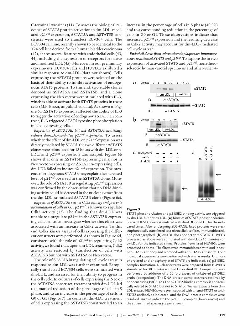
sect the molecular mechanisms involved in this effect,we analyzed the expression of two CKIs: p21waf andp27Kip1. As shown in Figure 3a, and consistent withflow cytometric analysis, treatment with dm-LDL butnot n-LDL led to an increase in p21waf expression. Incontrast, the expression of p27 kip1 was not affected byeither n-LDL or dm-LDL (Figure 3a). Sustainedincreased levels of p21waf can be mediated by variousmechanisms, including increased RNA transcription(12). Therefore, to evaluate whether the mechanisminvolved in p21waf expression was dependent upon genetranscription, nuclear run-off experiments were per-formed. As depicted in Figure 3b, while nuclear p21waf
mRNA was barely detectable in unstimulated cells, itwas clearly induced upon dm-LDL treatment, showingthat a transcriptional mechanism regulated dm-LDL–mediated p21waf expression.
It is known that modified LDL may exert its effectsby binding to membrane structures other than thecanonical LDL receptor (38, 39). The possibility that areceptor other than the native LDL receptor wasengaged by dm-LDL was indirectly evaluated by com-petition experiments. HUVECs were stimulated withdm-LDL alone or in combination with 50- and 100-fold excesses of n-LDL, and then assayed for p21waf
expression. As shown in Figure 3c, n-LDL was unableto abrogate the effect of dm-LDL on p21waf expression,suggesting that a receptor other than the natural LDLreceptor is recruited by dm-LDL.
dm-LDL induces reactive oxygen species production. It isknown that modified LDL–induced signaling involvesintracellular reactive oxygen species (ROS) production
(40). Accordingly, we examined the effects of n-LDLand dm-LDL on intracellular ROS production. Asshown in Figure 4, dm-LDL, but not n-LDL, induced aburst of ROS that peaked at 15 minutes as measured byDCF fluorescence. TNF-α was used as a positive con-trol. Similar results were obtained with the lucigenin-enhanced chemiluminescence assay using a 1251 LKBluminometer (Perkin-Elmer Instruments, Bad-Wild-bad, Germany) (data not shown).
dm-LDL triggers STAT5 activation. STAT5A andSTAT5B are pleiotropic regulators of many genes,including p21waf (22). To assess the role of the STAT5pathway in regulating dm-LDL–mediated p21waf
expression, the effect of dm-LDL on STAT5 activationwas analyzed. The results shown in Figure 5a demon-strate that, unlike n-LDL, dm-LDL was able to triggerSTAT5 tyrosine phosphorylation. It is known thathydrogen peroxide is able to activate STAT1 andSTAT3 (41). However, neither n-LDL nor dm-LDL wasable to induce STAT3 activation (data not shown), andwhen ox-LDL was used to stimulate HUVECs, noSTAT5 activation could be detected (Figure 5b). Toassess whether dm-LDL–elicited STAT5 activation wasinvolved in p21waf induction, an electrophoretic mobil-ity shift assay was performed using the p21SIE2sequence, which is a potential STAT binding site in thep21waf promoter region (22). As shown in Figure 5c,nuclear extracts from dm-LDL–treated, but not fromuntreated or n-LDL–treated, endothelial cells were ableto form a p21SIE2-binding complex that was compet-ed for by an added excess of unlabeled p21SIE2 probe.The presence of STAT5 in the DNA-protein complexinduced by dm-LDL was demonstrated by the ability ofthe antibody to STAT5 to supershift the p21SIE2-bind-ing complex (Figure 5d). Consistent with the inabilityof dm-LDL to trigger STAT3 phosphorylation, theantibody to STAT3 did not supershift the dm-LDL–induced p21SIE2-binding complex (Figure 5d).
Expression of ∆STAT5 proteins in the ECV304 cell line.Modified forms of STAT5 acting as dominant-nega-tive proteins were obtained by removing most of the
114 The Journal of Clinical Investigation | January 2002 | Volume 109 | Number 1
Figure 3 dm-LDL increases p21waf expression via a transcriptional mecha-nism. (a) p21waf and p27kip1 expression. Proteins from lysed HUVECswere incubated with n-LDL or dm-LDL for different time periods,subjected to 15% SDS-PAGE, and electrophoretically transferred tonitrocellulose filters. The filters were immunoblotted with an anti-p21waf antibody or an anti-p27kip1 antiserum. Five different experi-ments were performed with similar results. (b) p21waf gene tran-scription. RNAs were obtained from nuclei prepared fromunstimulated HUVECs or HUVECs stimulated with dm-LDL for 90minutes. Equal cpm of the purified radioactive RNAs from therespective reactions were hybridized against p21waf and β-actincDNAs dot-blotted on the same filter. Similar results were obtainedin two different experiments. (c) n-LDL does not compete withdm-LDL. HUVECs were unstimulated (lane 1) or were stimulated for18 hours with dm-LDL alone (lane 2) or in combination with n-LDL(50- and 100-fold excess, lanes 3 and 4, respectively), and evaluat-ed for p21waf as above. Similar results were obtained in three differ-ent experiments. IB, immunoblot.
Figure 4Kinetics of dm-LDL–induced ROS production. DCF was added tounstimulated HUVECs (circles) or HUVECs stimulated with n-LDL(diamonds), dm-LDL (triangles), or TNF-α (squares). DCF fluores-cence was measured over a 60-minute period. Time 0 representsbasal DCF fluorescence. The results are the mean of three individualexperiments, each performed in duplicate.
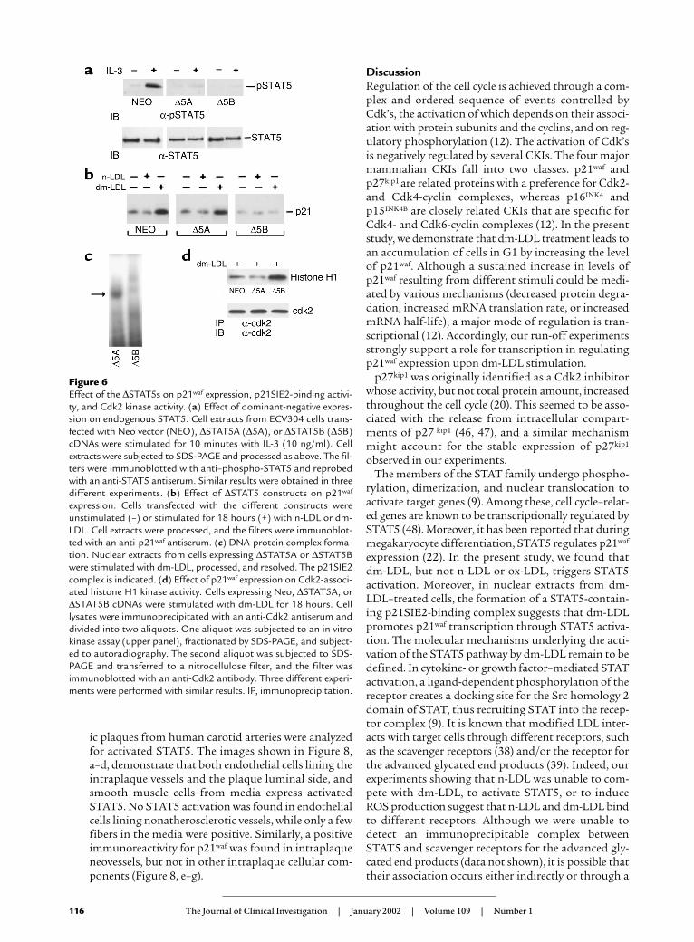
C-terminal tyrosines (11). To assess the biological rel-evance of STAT5 protein activation in dm-LDL–medi-ated p21waf expression, ∆STAT5A and ∆STAT5B con-structs were used to transfect ECV304 cells. TheECV304 cell line, recently shown to be identical to theT24 cell line derived from a human bladder carcinoma(42), shares several features with endothelial cells (43,44), including the expression of receptors for nativeand modified LDL (45). Moreover, in our preliminaryexperiments, ECV304 cells and HUVECs exhibited asimilar response to dm-LDL (data not shown). Cellsexpressing the ∆STAT5 proteins were selected on thebasis of their ability to inhibit activation of endoge-nous STAT5 proteins. To this end, two stable clonesdenoted as ∆STAT5A and ∆STAT5B, and a cloneexpressing the Neo vector were stimulated with IL-3,which is able to activate both STAT5 proteins in thesecells (M.F. Brizzi, unpublished data). As shown in Fig-ure 6a, ∆STAT5 expression affected the ability of IL-3to trigger the activation of endogenous STAT5. In con-trast, IL-3 triggered STAT5 tyrosine phosphorylationin Neo-expressing cells.
Expression of ∆STAT5B, but not ∆STAT5A, drasticallyreduces dm-LDL–mediated p21waf expression. To assesswhether the effect of dm-LDL on p21waf expression wasdirectly mediated by STAT5, the two different ∆STAT5clones were stimulated for 18 hours with dm-LDL or n-LDL, and p21waf expression was assayed. Figure 6bshows that only in ∆STAT5B-expressing cells, not inNeo vector–expressing or ∆STAT5A-expressing cells,dm-LDL failed to induce p21waf expression. The pres-ence of endogenous STAT5B may explain the increasedlevel of p21waf observed in the ∆STAT5A clone. More-over, the role of STAT5B in regulating p21waf expressionwas confirmed by the observation that no DNA-bind-ing activity could be detected in the nuclear extract fromthe dm-LDL–stimulated ∆STAT5B clone (Figure 6c).
Expression of ∆STAT5B rescues Cdk2 activity and preventsaccumulation of cells in G1. p21waf is known to regulateCdk2 activity (12). The finding that dm-LDL wasunable to upregulate p21waf in the ∆STAT5B-express-ing cells led us to investigate whether this effect wasassociated with an increase in Cdk2 activity. To thisend, Cdk2 kinase assays of cells expressing the differ-ent constructs were performed. As shown in Figure 6d,consistent with the role of p21waf in regulating Cdk2activity, we found that, upon dm-LDL treatment, Cdk2activity was restored by transfection of cells with∆STAT5B but not with ∆STAT5A or Neo vector.
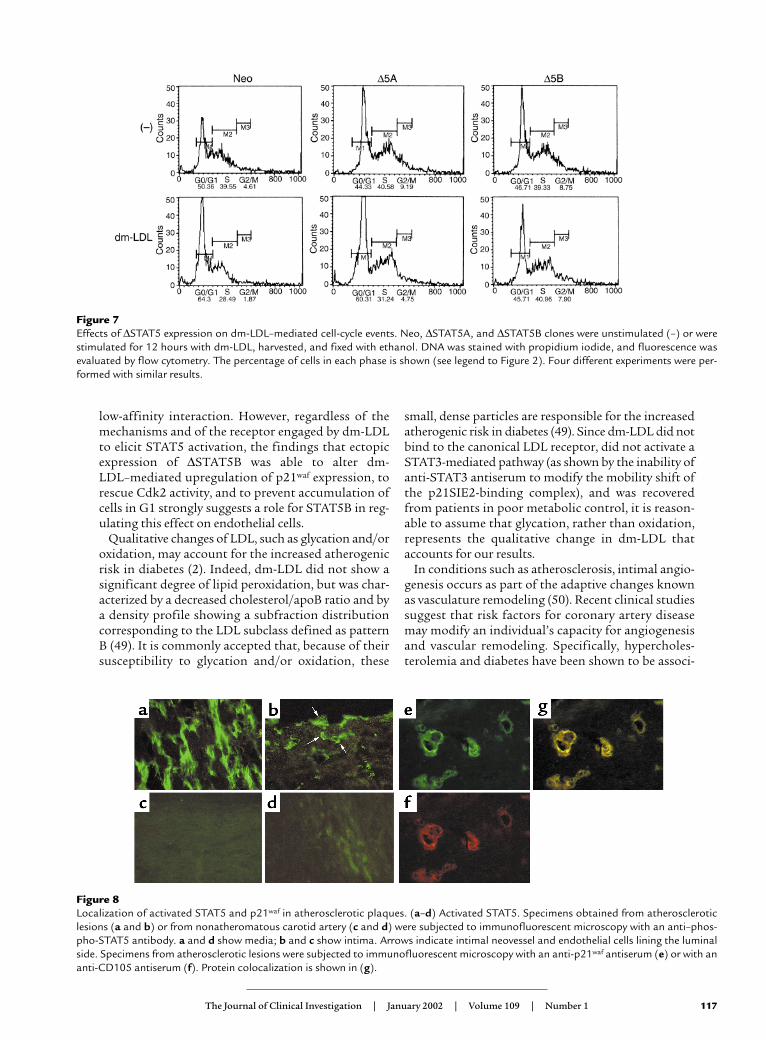
The role of STAT5B in regulating cell-cycle arrest inresponse to dm-LDL was further examined. Ectopi-cally transfected ECV304 cells were stimulated withdm-LDL, and assessed for their ability to progress inthe cell cycle. In cultures of cells expressing the Neo orthe ∆STAT5A construct, treatment with dm-LDL ledto a marked reduction of the percentage of cells in Sphase, and to an increase in the percentage of cells inG0 or G1 (Figure 7). In contrast, dm-LDL treatmentof cells expressing the ∆STAT5B construct led to an
increase in the percentage of cells in S phase (40.9%)and to a corresponding reduction in the percentage ofcells in G0 or G1. These observations indicate thatincreased p21waf expression and the resulting decreasein Cdk2 activity may account for dm-LDL–mediatedcell-cycle arrest.
Endothelial cells from atherosclerotic plaques are immunore-active to activated STAT5 and p21waf. To explore the in vivoexpression of activated STAT5 and p21waf, nonathero-sclerotic human carotid specimens and atherosclerot-
The Journal of Clinical Investigation | January 2002 | Volume 109 | Number 1 115
Figure 5STAT5 phosphorylation and p21SIE2 binding activity are triggeredby dm-LDL but not ox-LDL. (a) Kinetics of STAT5 phosphorylation.Starved HUVECs were stimulated with dm-LDL or n-LDL for the indi-cated times. After undergoing SDS-PAGE, lysed proteins were elec-trophoretically transferred to a nitrocellulose filter, immunoblotted,and photographed. (b) ox-LDL does not activate STAT5. HUVECsprocessed as above were stimulated with dm-LDL (15 minutes) orox-LDL for the indicated times. Proteins from lysed HUVECs wereprocessed as above. The filters were immunoblotted with anti–phos-pho-STAT5 antibody and reprobed with anti-STAT5 antiserum. Fourindividual experiments were performed with similar results. Unphos-phorylated and phosphorylated STAT5 are indicated. (c) p21SIE2complex formation. Nuclear extracts were prepared from HUVECsstimulated for 30 minutes with n-LDL or dm-LDL. Competition wasperformed by addition of a 50-fold excess of unlabeled p21SIE2probe (competitor). The DNA-protein complexes were resolved bynondenaturing PAGE. (d) The p21SIE2-binding complex is antigeni-cally related to STAT5 but not to STAT3. Nuclear extracts from dm-LDL–treated HUVECs were preincubated with an anti-STAT5 or anti-STAT3 antibody as indicated, and the DNA-protein complexes wereresolved. Arrows indicate the p21SIE2 complex (lower arrow) andthe supershifted species (upper arrow).
ic plaques from human carotid arteries were analyzedfor activated STAT5. The images shown in Figure 8,a–d, demonstrate that both endothelial cells lining theintraplaque vessels and the plaque luminal side, andsmooth muscle cells from media express activatedSTAT5. No STAT5 activation was found in endothelialcells lining nonatherosclerotic vessels, while only a fewfibers in the media were positive. Similarly, a positiveimmunoreactivity for p21waf was found in intraplaqueneovessels, but not in other intraplaque cellular com-ponents (Figure 8, e–g).
DiscussionRegulation of the cell cycle is achieved through a com-plex and ordered sequence of events controlled byCdk’s, the activation of which depends on their associ-ation with protein subunits and the cyclins, and on reg-ulatory phosphorylation (12). The activation of Cdk’sis negatively regulated by several CKIs. The four majormammalian CKIs fall into two classes. p21waf andp27kip1 are related proteins with a preference for Cdk2-and Cdk4-cyclin complexes, whereas p16INK4 andp15INK4B are closely related CKIs that are specific forCdk4- and Cdk6-cyclin complexes (12). In the presentstudy, we demonstrate that dm-LDL treatment leads toan accumulation of cells in G1 by increasing the levelof p21waf. Although a sustained increase in levels ofp21waf resulting from different stimuli could be medi-ated by various mechanisms (decreased protein degra-dation, increased mRNA translation rate, or increasedmRNA half-life), a major mode of regulation is tran-scriptional (12). Accordingly, our run-off experimentsstrongly support a role for transcription in regulatingp21waf expression upon dm-LDL stimulation.
p27kip1 was originally identified as a Cdk2 inhibitorwhose activity, but not total protein amount, increasedthroughout the cell cycle (20). This seemed to be asso-ciated with the release from intracellular compart-ments of p27 kip1 (46, 47), and a similar mechanismmight account for the stable expression of p27kip1
observed in our experiments.The members of the STAT family undergo phospho-
rylation, dimerization, and nuclear translocation toactivate target genes (9). Among these, cell cycle–relat-ed genes are known to be transcriptionally regulated bySTAT5 (48). Moreover, it has been reported that duringmegakaryocyte differentiation, STAT5 regulates p21waf
expression (22). In the present study, we found thatdm-LDL, but not n-LDL or ox-LDL, triggers STAT5activation. Moreover, in nuclear extracts from dm-LDL–treated cells, the formation of a STAT5-contain-ing p21SIE2-binding complex suggests that dm-LDLpromotes p21waf transcription through STAT5 activa-tion. The molecular mechanisms underlying the acti-vation of the STAT5 pathway by dm-LDL remain to bedefined. In cytokine- or growth factor–mediated STATactivation, a ligand-dependent phosphorylation of thereceptor creates a docking site for the Src homology 2domain of STAT, thus recruiting STAT into the recep-tor complex (9). It is known that modified LDL inter-acts with target cells through different receptors, suchas the scavenger receptors (38) and/or the receptor forthe advanced glycated end products (39). Indeed, ourexperiments showing that n-LDL was unable to com-pete with dm-LDL, to activate STAT5, or to induceROS production suggest that n-LDL and dm-LDL bindto different receptors. Although we were unable todetect an immunoprecipitable complex betweenSTAT5 and scavenger receptors for the advanced gly-cated end products (data not shown), it is possible thattheir association occurs either indirectly or through a
116 The Journal of Clinical Investigation | January 2002 | Volume 109 | Number 1
Figure 6Effect of the ∆STAT5s on p21waf expression, p21SIE2-binding activi-ty, and Cdk2 kinase activity. (a) Effect of dominant-negative expres-sion on endogenous STAT5. Cell extracts from ECV304 cells trans-fected with Neo vector (NEO), ∆STAT5A (∆5A), or ∆STAT5B (∆5B)cDNAs were stimulated for 10 minutes with IL-3 (10 ng/ml). Cellextracts were subjected to SDS-PAGE and processed as above. The fil-ters were immunoblotted with anti–phospho-STAT5 and reprobedwith an anti-STAT5 antiserum. Similar results were obtained in threedifferent experiments. (b) Effect of ∆STAT5 constructs on p21waf
expression. Cells transfected with the different constructs wereunstimulated (–) or stimulated for 18 hours (+) with n-LDL or dm-LDL. Cell extracts were processed, and the filters were immunoblot-ted with an anti-p21waf antiserum. (c) DNA-protein complex forma-tion. Nuclear extracts from cells expressing ∆STAT5A or ∆STAT5Bwere stimulated with dm-LDL, processed, and resolved. The p21SIE2complex is indicated. (d) Effect of p21waf expression on Cdk2-associ-ated histone H1 kinase activity. Cells expressing Neo, ∆STAT5A, or∆STAT5B cDNAs were stimulated with dm-LDL for 18 hours. Celllysates were immunoprecipitated with an anti-Cdk2 antiserum anddivided into two aliquots. One aliquot was subjected to an in vitrokinase assay (upper panel), fractionated by SDS-PAGE, and subject-ed to autoradiography. The second aliquot was subjected to SDS-PAGE and transferred to a nitrocellulose filter, and the filter wasimmunoblotted with an anti-Cdk2 antibody. Three different experi-ments were performed with similar results. IP, immunoprecipitation.
low-affinity interaction. However, regardless of themechanisms and of the receptor engaged by dm-LDLto elicit STAT5 activation, the findings that ectopicexpression of ∆STAT5B was able to alter dm-LDL–mediated upregulation of p21waf expression, torescue Cdk2 activity, and to prevent accumulation ofcells in G1 strongly suggests a role for STAT5B in reg-ulating this effect on endothelial cells.
Qualitative changes of LDL, such as glycation and/oroxidation, may account for the increased atherogenicrisk in diabetes (2). Indeed, dm-LDL did not show asignificant degree of lipid peroxidation, but was char-acterized by a decreased cholesterol/apoB ratio and bya density profile showing a subfraction distributioncorresponding to the LDL subclass defined as patternB (49). It is commonly accepted that, because of theirsusceptibility to glycation and/or oxidation, these
small, dense particles are responsible for the increasedatherogenic risk in diabetes (49). Since dm-LDL did notbind to the canonical LDL receptor, did not activate aSTAT3-mediated pathway (as shown by the inability ofanti-STAT3 antiserum to modify the mobility shift ofthe p21SIE2-binding complex), and was recoveredfrom patients in poor metabolic control, it is reason-able to assume that glycation, rather than oxidation,represents the qualitative change in dm-LDL thataccounts for our results.
In conditions such as atherosclerosis, intimal angio-genesis occurs as part of the adaptive changes knownas vasculature remodeling (50). Recent clinical studiessuggest that risk factors for coronary artery diseasemay modify an individual’s capacity for angiogenesisand vascular remodeling. Specifically, hypercholes-terolemia and diabetes have been shown to be associ-
The Journal of Clinical Investigation | January 2002 | Volume 109 | Number 1 117
Figure 7Effects of ∆STAT5 expression on dm-LDL–mediated cell-cycle events. Neo, ∆STAT5A, and ∆STAT5B clones were unstimulated (–) or werestimulated for 12 hours with dm-LDL, harvested, and fixed with ethanol. DNA was stained with propidium iodide, and fluorescence wasevaluated by flow cytometry. The percentage of cells in each phase is shown (see legend to Figure 2). Four different experiments were per-formed with similar results.
Figure 8Localization of activated STAT5 and p21waf in atherosclerotic plaques. (a–d) Activated STAT5. Specimens obtained from atheroscleroticlesions (a and b) or from nonatheromatous carotid artery (c and d) were subjected to immunofluorescent microscopy with an anti–phos-pho-STAT5 antibody. a and d show media; b and c show intima. Arrows indicate intimal neovessel and endothelial cells lining the luminalside. Specimens from atherosclerotic lesions were subjected to immunofluorescent microscopy with an anti-p21waf antiserum (e) or with ananti-CD105 antiserum (f). Protein colocalization is shown in (g).
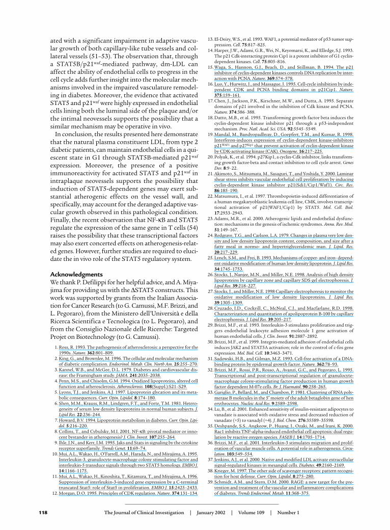
ated with a significant impairment in adaptive vascu-lar growth of both capillary-like tube vessels and col-lateral vessels (51–53). The observation that, througha STAT5B/p21waf-mediated pathway, dm-LDL canaffect the ability of endothelial cells to progress in thecell cycle adds further insight into the molecular mech-anisms involved in the impaired vasculature remodel-ing in diabetes. Moreover, the evidence that activatedSTAT5 and p21waf were highly expressed in endothelialcells lining both the luminal side of the plaque and/orthe intimal neovessels supports the possibility that asimilar mechanism may be operative in vivo.
In conclusion, the results presented here demonstratethat the natural plasma constituent LDL, from type 2diabetic patients, can maintain endothelial cells in a qui-escent state in G1 through STAT5B-mediated p21waf
expression. Moreover, the presence of a positiveimmunoreactivity for activated STAT5 and p21waf inintraplaque neovessels supports the possibility thatinduction of STAT5-dependent genes may exert sub-stantial atherogenic effects on the vessel wall, andspecifically, may account for the deranged adaptive vas-cular growth observed in this pathological condition.Finally, the recent observation that NF-κB and STAT5regulate the expression of the same gene in T cells (54)raises the possibility that these transcriptional factorsmay also exert concerted effects on atherogenesis-relat-ed genes. However, further studies are required to eluci-date the in vivo role of the STAT5 regulatory system.
AcknowledgmentsWe thank P. Defilippi for her helpful advice, and A. Miya-jima for providing us with the ∆STAT5 constructs. Thiswork was supported by grants from the Italian Associa-tion for Cancer Research (to G. Camussi, M.F. Brizzi, andL. Pegoraro), from the Ministero dell’Università e dellaRicerca Scientifica e Tecnologica (to L. Pegoraro), andfrom the Consiglio Nazionale delle Ricerche: TargetedProjet on Biotechnology (to G. Camussi).
1. Ross, R. 1993. The pathogenesis of atherosclerosis: a perspective for the1990s. Nature. 362:801–809.
2. King, G., and Brownlee, M. 1996. The cellular and molecular mechanismof diabetic complication. Endocrinol. Metab. Clin. North Am. 25:255–270.
3. Kannel, W.B., and McGee, D.L. 1979. Diabetes and cardiovascular dis-ease: the Framingham study. JAMA. 241:2035–2038.
4. Penn, M.S., and Chisolm, G.M. 1994. Oxidized lipoproteins, altered cellfunction and atherosclerosis. Atherosclerosis. 108(Suppl.):S21–S29.
5. Lyons, T.J., and Jenkins, A.J. 1997. Lipoprotein glycation and its meta-bolic consequences. Curr. Opin. Lipidol. 8:174–180.
6. Shen, M.M., Krauss, R.M., Lindgren, F.T., and Forte, T.M. 1981. Hetero-geneity of serum low density lipoproteins in normal human subjects. J.Lipid Res. 22:236–244.
7. Howard, B.V. 1994. Lipoprotein metabolism in diabetes. Curr. Opin. Lipi-dol. 5:216–220.
8. Collins, T., and Cybulsky, M.I. 2001. NF-κB: pivotal mediator or inno-cent bystander in atherogenesis? J. Clin. Invest. 107:255–264.
9. Ihle, J.N., and Kerr, I.M. 1995. Jaks and Stats in signaling by the cytokinereceptor superfamily. Trends Genet. 11:69–74.
10. Mui, A.L., Wakao, H., O’Farrell, A.M., Harada, N., and Miyajima, A. 1995.Interleukin-3, granulocyte-macrophage colony stimulating factor andinterleukin-5 transduce signals through two STAT5 homologs. EMBO J.14:1166–1175.
11. Mui, A.L., Wakao, H., Kinoshita, T., Kitamura, T., and Miyajima, A. 1996.Suppression of interleukin-3-induced gene expression by a C-terminaltruncated Stat5: role of Stat5 in proliferation. EMBO J. 15:2425–2433.
12. Morgan, D.O. 1995. Principles of CDK regulation. Nature. 374:131–134.
13. El-Deiry, W.S., et al. 1993. WAF1, a potential mediator of p53 tumor sup-pression. Cell. 75:817–825.
14. Harper, J.W., Adami, G.R., Wei, N., Keyomarsi, K., and Elledge, S.J. 1993.The p21 Cdk-interacting protein Cip1 is a potent inhibitor of G1 cyclin-dependent kinases. Cell. 75:805–816.
15. Waga, S., Hannon, G.J., Beach, D., and Stillman, B. 1994. The p21inhibitor of cyclin-dependent kinases controls DNA replication by inter-action with PCNA. Nature. 369:574–578.
16. Luo, Y., Hurwitz, J., and Massague, J. 1995. Cell-cycle inhibition by inde-pendent CDK and PCNA binding domains in p21Cip1. Nature.375:159–161.
17. Chen, J., Jackson, P.K., Kirschner, M.W., and Dutta, A. 1995. Separatedomains of p21 involved in the inhibition of Cdk kinase and PCNA.Nature. 374:386–388.
18. Datto, M.B., et al. 1995. Transforming growth factor beta induces thecyclin-dependent kinase inhibitor p21 through a p53-independentmechanism. Proc. Natl. Acad. Sci. USA. 92:5545–5549.
19. Mandal, M., Bandyopadhyay, D., Goepfert, T.M., and Kumar, R. 1998.Interferon-induces expression of cyclin-dependent kinase-inhibitorsp21WAF1 and p27Kip1 that prevent activation of cyclin-dependent kinaseby CDK-activating kinase (CAK). Oncogene. 16:217–225.
20. Polyak, K., et al. 1994. p27Kip1, a cyclin-Cdk inhibitor, links transform-ing growth factor-beta and contact inhibition to cell cycle arrest. GenesDev. 8:9–22.
21. Akimoto, S., Mitsumata, M., Sasaguri, T., and Yoshida, Y. 2000. Laminarshear stress inhibits vascular endothelial cell proliferation by inducingcyclin-dependent kinase inhibitor p21(Sdi1/Cip1/Waf1). Circ. Res.86:185–190.
22. Matsumura, I., et al. 1997. Thrombopoietin-induced differentiation ofa human megakaryoblastic leukemia cell line, CMK, involves transcrip-tional activation of p21(WAF1/Cip1) by STAT5. Mol. Cell. Biol.17:2933–2943.
23. Adams, M.R., et al. 2000. Atherogenic lipids and endothelial dysfunc-tion: mechanisms in the genesis of ischemic syndromes. Annu. Rev. Med.51:149–167.
24. Redgrave, T.G., and Carlson, L.A. 1979. Changes in plasma very low den-sity and low density lipoprotein content, composition, and size after afatty meal in normo- and hypertriglyceridemic man. J. Lipid. Res.20:217–229.
25. Lynch, S.M., and Frei, B. 1993. Mechanisms of copper- and iron- depend-ent oxidative modification of human low density lipoprotein. J. Lipid Res.34:1745–1753.
26. Stocks, J., Nanjee, M.N., and Miller, N.E. 1998. Analysis of high densitylipoproteins by capillary zone and capillary SDS gel electrophoresis. J.Lipid Res. 39:218–227.
27. Stocks, J., and Miller, N.E. 1998 Capillary electrophoresis to monitor theoxidative modification of low density lipoproteins. J. Lipid Res.39:1305–1309.
28. Cruzado, I.D., Cockrill, C., McNeal, C.J., and Macfarlane, R.D. 1998.Characterization and quantitation of apolipoprotein B-100 by capillaryelectrophoresis. J. Lipid Res. 39:205–217.
29. Brizzi, M.F., et al. 1993. Interleukin-3 stimulates proliferation and trig-gers endothelial leukocyte adhesion molecule 1 gene activation ofhuman endothelial cells. J. Clin. Invest. 91:2887–2892.
30. Brizzi, M.F., et al. 1999. Integrin-mediated adhesion of endothelial cellsinduces JAK2 and STAT5A activation; role in the control of c-fos geneexpression. Mol. Biol. Cell. 10:3463–3471.
31. Sadowski, H.B., and Gilman, M.Z. 1993. Cell-free activation of a DNA-binding protein by epidermal growth factor. Nature. 362:78–93.
32. Brizzi, M.F., Rossi, P.R., Rosso, A., Avanzi, G.C., and Pegoraro, L. 1995.Transcriptional and post-transcriptional regulation of granulocyte-macrophage colony-stimulating factor production in human growthfactor dependent M-07e cells. Br. J. Haematol. 90:258–265.
33. Gariglio, P., Bellard, M., and Chambon, P. 1981. Clustering of RNA poly-merase B molecules in the 5′ moiety of the adult betaglobin gene of henerythrocytes. Nucleic Acid Res. 9:2589–2598.
34. Lu, B., et al. 2001. Enhanced sensitivity of insulin-resistant adipocytes tovanadate is associated with oxidative stress and decreased reduction ofvanadate (+5) to vanadyl (+4). J. Biol. Chem. 276:35589–35598.
35. Deshpande, S.S., Angkeow, P., Huang, J., Ozaki, M., and Irani, K. 2000.Rac1 inhibits TNF-alpha-induced endothelial cell apoptosis: dual regu-lation by reactive oxygen species. FASEB J. 14:1705–1714.
36. Brizzi, M.F., et al. 2001. Interleukin-3 stimulates migration and prolif-eration of vascular muscle cells. A potential role in atherogenesis. Circu-lation. 103:549–554.
37. Jenkins, A.J., et al. 2000. Native and modified LDL activate extracellularsignal-regulated kinases in mesangial cells. Diabetes. 49:2160–2169.
38. Krieger, M. 1997. The other side of scavenger receptors: pattern recogni-tion for host defense. Curr. Opin. Lipidol. 8:275–280.
39. Schmidt, A.M., and Stern, D.M. 2000. RAGE: a new target for the pre-vention and treatment of the vascular and inflammatory complicationsof diabetes. Trends Endocrinol. Metab. 11:368–375.
118 The Journal of Clinical Investigation | January 2002 | Volume 109 | Number 1
40. Napoli, C., de Nigris, F., and Palinski, W. 2001. Multiple role of reactiveoxygen species in the arterial wall. J. Cell. Biochem. 82:674–682.
41. Maziere, C., et al. 1999. Oxidized LDL activates STAT1 and STAT3 tran-scription factors: possible involvement of reactive oxygen species. FEBSLett. 448:49–52.
42. Bubenik, J., Perlmann, P., Helmstein, K., and Moberger, G. 1970. Cellularand humoral immune responses to human urinary bladder carcinomas.Int. J. Cancer. 5:310–319.
43. Ricard, I., Payet, M.D., and Dupuis, G. 1998. VCAM-1 is internalized by aclathrin-related pathway in human endothelial cells but its alpha 4 beta 1integrin counter-receptor remains associated with the plasma membranein human T lymphocytes. Eur. J. Immunol. 28:1708–1718.
44. Liu, J., et al. 1999. Angiogenesis activators and inhibitors differentially reg-ulate caveolin-1 expression and caveolae formation in vascular endothe-lial cells. Angiogenesis inhibitors block vascular endothelial growth fac-tor-induced down-regulation of caveolin-1. J. Biol. Chem. 274:15781–15785.
45. Escargueil-Blanc, I., et al. 1998. Apoptosis and activation of the sphin-gomyelin-ceramide pathway induced by oxidized low density lipoproteinsare not causally related in ECV-304 endothelial cells. J. Biol. Chem.273:27389–27395.
46. Polyak, K., et al. 1994. Cloning of p27Kip1, a cyclin-dependent kinaseinhibitor and a potential mediator of extracellular antimitogenic signals.
Cell. 78:59–66.47. Toyoshima, H., and Hunter, T. 1994. p27, a novel inhibitor of G1 cyclin-
Cdk protein kinase activity, is related to p21. Cell. 78:67–74.48. Matsumura, I., et al. 1999. Transcriptional regulation of the cyclin D1 pro-
moter by STAT5: its involvement in cytokine-dependent growth ofhematopoietic cells. EMBO J. 18:1367–1377.
49. Austin, M.A., King, M.C., Vranizan, K.M., and Krauss, R.M. 1990. Athero-genic lipoprotein phenotype. A proposed genetic marker for coronaryheart disease risk. Circulation. 82:495–506.
50. Isner, J.M. 1994. Vascular remodeling. Circulation. 89:2937–2941.51. Chen, C.H., et al. 1997. Inhibitory effects of hypercholesterolemia and ox-
LDL on angiogenesis-like endothelial growth in rabbit aortic explants.Essential role of basic fibroblast growth factor. Arterioscler. Thromb. Vasc.Biol. 17:1303–1312.
52. Abaci, A., et al. 1999. Effect of diabetes mellitus on formation of coronarycollateral vessels. Circulation. 99:2239–2242.
53. Chen, C.H., et al. 2000. Oxidized low-density lipoproteins inhibit endothe-lial cell proliferation by suppressing basic fibroblast growth factor expres-sion. Circulation. 101:171–177.
54. Musikacharoen, T., Matsuguchi, T., Kikuchi, T., and Yoshikai, Y. 2001. NF-kappa B and STAT5 play important roles in the regulation of mouse Toll-like receptor 2 gene expression. J. Immunol. 166:4516–4524.
The Journal of Clinical Investigation | January 2002 | Volume 109 | Number 1 119
Related Documents