Diabetic foot ulcers Part II. Management Afsaneh Alavi, MD, FRCPC, a,e R. Gary Sibbald, MD, a,b,e Dieter Mayer, MD, c Laurie Goodman, RN, MScN, d Mariam Botros, DCh, e David G. Armstrong, DPM, MD, PhD, f Kevin Woo, RN, PhD, g Thomas Boeni, MD, h Elizabeth A. Ayello, RN, PhD, i and Robert S. Kirsner, MD, PhD j Toronto, Mississauga, and Kingston, Ontario, Canada; Zurich, Switzerland; Tucson, Arizona; Albany, New York; and Miami, Florida CME INSTRUCTIONS The following is a journal-based CME activity presented by the American Academy of Dermatology and is made up of four phases: 1. Reading of the CME Information (delineated below) 2. Reading of the Source Article 3. Achievement of a 70% or higher on the online Case-based Post Test 4. Completion of the Journal CME Evaluation CME INFORMATION AND DISCLOSURES Statement of Need: The American Academy of Dermatology bases its CME activities on the Academy’s core curriculum, identified professional practice gaps, the educational needs which underlie these gaps, and emerging clinical research findings. Learners should reflect upon clinical and scientific information presented in the article and determine the need for further study. Target Audience: Dermatologists and others involved in the delivery of dermatologic care. Accreditation The American Academy of Dermatology is accredited by the Accreditation Council for Continuing Medical Education to provide continuing medical education for physicians. AMA PRA Credit Designation The American Academy of Dermatology designates this journal-based CME activity for a maximum of 1 AMA PRA Category 1 CreditsÔ. Physicians should claim only the credit commensurate with the extent of their participation in the activity. AAD Recognized Credit This journal-based CME activity is recognized by the American Academy of Dermatology for 1 AAD Credit and may be used toward the American Academy of Dermatology’s Continuing Medical Education Award. Disclaimer: The American Academy of Dermatology is not responsible for statements made by the author(s). Statements or opinions expressed in this activity reflect the views of the author(s) and do not reflect the official policy of the American Academy of Dermatology. The information provided in this CME activity is for continuing education purposes only and is not meant to substitute for the independent medical judgment of a healthcare provider relative to the diagnostic, management and treatment options of a specific patient’s medical condition. Disclosures Editors The editors involved with this CME activity and all content validation/peer reviewers of this journal-based CME activity have reported no relevant financial relationships with commercial interest(s). Authors Dr Robert Kirsner’s conflicts are relevant, but mitigated by unconflicted peer-review and unconflicted editorial review. His relevant financial relationships follow: 3M Pharmaceuticals, Advisory Board, Honoraria; GlaxoSmithKline, Other, Honoraria; Healthpoint, Advisory Board, Honoraria; Healthpoint, Principle Investigator, Grants/ Research Funding Institution; KCI, Consultant, Honoraria; Keraderm, Consultant, Honoraria; Molynecke, Advisory Board, Honoraria; National Healing Corp, Advisory Board, Honoraria; Organogenesis, Inc, Advisory Board, Honoraria; Shire, Consultant, Honoraria; Tissue Repair Company, Principle Investigator, Grants/Research Funding Institution. The other authors of this journal-based CME activity have reported no relevant financial relationships with commercial interest(s). Planners The planners involved with this journal-based CME activity have reported no relevant financial relationships with commercial interest(s). The editorial and education staff involved with this journal-based CME activity have reported no relevant financial relationships with commercial interest(s). Resolution of Conflicts of Interest In accordance with the ACCME Standards for Commercial Support of CME, the American Academy of Dermatology has implemented mechanisms, prior to the planning and implementation of this Journal-based CME activity, to identify and mitigate conflicts of interest for all individuals in a position to control the content of this Journal-based CME activity. Learning Objectives After completing this learning activity, participants should be able to evaluate a person with diabetic foot ulcer with holistic patient assessment (HbA1e) and other metabolic factors; address adequate vascular supply to heal deep and surrounding infection and plantar pressure distribution; identify patient-centered concerns including pain and activities of daily living; assess local wound care; and optimize the use of advanced therapies. Date of release: January 2014 Expiration date: January 2017 Ó 2013 by the American Academy of Dermatology, Inc. http://dx.doi.org/10.1016/j.jaad.2013.07.048 Technical requirements: American Academy of Dermatology: d Supported browsers: FireFox (3 and higher), Google Chrome (5 and higher), Internet Explorer (7 and higher), Safari (5 and higher), Opera (10 and higher). d JavaScript needs to be enabled. Elsevier: Technical Requirements This website can be viewed on a PC or Mac. We recommend a minimum of: d PC: Windows NT, Windows 2000, Windows ME, or Windows XP d Mac: OS X d 128MB RAM d Processor speed of 500MHz or higher d 800x600 color monitor d Video or graphics card d Sound card and speakers Provider Contact Information: American Academy of Dermatology Phone: Toll-free: (866) 503-SKIN (7546); International: (847) 240-1280 Fax: (847) 240-1859 Mail: P.O. Box 4014; Schaumburg, IL 60168 Confidentiality Statement: American Academy of Dermatology: POLICY ON PRIVACY AND CONFIDENTIALITY Privacy Policy - The American Academy of Dermatology (the Academy) is committed to maintaining the privacy of the personal information of visitors to its sites. Our policies are designed to disclose the information collected and how it will be used. This policy applies solely to the information provided while visiting this website. The terms of the privacy policy do not govern personal information furnished through any means other than this website (such as by telephone or mail). E-mail Addresses and Other Personal Information - Personal information such as postal and e-mail address may be used internally for maintaining member records, marketing purposes, and alerting customers or members of additional services available. Phone numbers may also be used by the Academy when questions about products or services ordered arise. The Academy will not reveal any information about an individual user to third parties except to comply with applicable laws or valid legal processes. Cookies - A cookie is a small file stored on the site user’s computer or Web server and is used to aid Web navigation. Session cookies are temporary files created when a user signs in on the website or uses the personalized features (such as keeping track of items in the shopping cart). Session cookies are removed when a user logs off or when the browser is closed. Persistent cookies are permanent files and must be deleted manually. Tracking or other information collected from persistent cookies or any session cookie is used strictly for the user’s efficient navigation of the site. Links - This site may contain links to other sites. The Academy is not responsible for the privacy practices or the content of such websites. Children - This website is not designed or intended to attract children under the age of 13. The Academy does not collect personal information from anyone it knows is under the age of 13. Elsevier: http://www.elsevier.com/wps/find/privacypolicy.cws_home/ privacypolicy 21.e1

Welcome message from author
This document is posted to help you gain knowledge. Please leave a comment to let me know what you think about it! Share it to your friends and learn new things together.
Transcript
Diabetic foot ulcersPart II. Management
Afsaneh Alavi, MD, FRCPC,a,e R. Gary Sibbald, MD,a,b,e Dieter Mayer, MD,c Laurie Goodman, RN, MScN,d
Mariam Botros, DCh,e David G. Armstrong, DPM, MD, PhD,f Kevin Woo, RN, PhD,g Thomas Boeni, MD,h
Elizabeth A. Ayello, RN, PhD,i and Robert S. Kirsner, MD, PhDj
Toronto, Mississauga, and Kingston, Ontario, Canada; Zurich, Switzerland; Tucson, Arizona;
Albany, New York; and Miami, Florida
CME INSTRUCTIONS
The following is a journal-based CME activity presented by the American Academy of
Dermatology and is made up of four phases:
1. Reading of the CME Information (delineated below)
2. Reading of the Source Article
3. Achievement of a 70% or higher on the online Case-based Post Test
4. Completion of the Journal CME Evaluation
CME INFORMATION AND DISCLOSURES
Statement of Need:
The American Academy of Dermatology bases its CME activities on the Academy’s
core curriculum, identified professional practice gaps, the educational needs which
underlie these gaps, and emerging clinical research findings. Learners should reflect
upon clinical and scientific information presented in the article and determine the
need for further study.
Target Audience:
Dermatologists and others involved in the delivery of dermatologic care.
Accreditation
The American Academy of Dermatology is accredited by the Accreditation Council for
ContinuingMedical Education to provide continuingmedical education for physicians.
AMA PRA Credit Designation
The American Academy of Dermatology designates this journal-based CME activity
for a maximum of 1 AMA PRA Category 1 Credits�. Physicians should claim only the
credit commensurate with the extent of their participation in the activity.
AAD Recognized Credit
This journal-based CME activity is recognized by the American Academy of
Dermatology for 1 AAD Credit and may be used toward the American Academy of
Dermatology’s Continuing Medical Education Award.
Disclaimer:
The American Academy of Dermatology is not responsible for statements made by
the author(s). Statements or opinions expressed in this activity reflect the views of the
author(s) and do not reflect the official policy of the American Academy of
Dermatology. The information provided in this CME activity is for continuing
education purposes only and is not meant to substitute for the independent medical
judgment of a healthcare provider relative to the diagnostic, management and
treatment options of a specific patient’s medical condition.
Disclosures
Editors
The editors involved with this CME activity and all content validation/peer reviewers
of this journal-based CME activity have reported no relevant financial relationships
with commercial interest(s).
Authors
Dr Robert Kirsner’s conflicts are relevant, but mitigated by unconflicted peer-review
and unconflicted editorial review. His relevant financial relationships follow: 3M
Pharmaceuticals, Advisory Board, Honoraria; GlaxoSmithKline, Other, Honoraria;
Healthpoint, Advisory Board, Honoraria; Healthpoint, Principle Investigator, Grants/
Research Funding Institution; KCI, Consultant, Honoraria; Keraderm, Consultant,
Honoraria; Molynecke, Advisory Board, Honoraria; National Healing Corp, Advisory
Board, Honoraria; Organogenesis, Inc, Advisory Board, Honoraria; Shire, Consultant,
Honoraria; Tissue Repair Company, Principle Investigator, Grants/Research Funding
Institution. The other authors of this journal-based CME activity have reported no
relevant financial relationships with commercial interest(s).
Planners
The planners involvedwith this journal-based CME activity have reported no relevant
financial relationships with commercial interest(s). The editorial and education staff
involved with this journal-based CME activity have reported no relevant financial
relationships with commercial interest(s).
Resolution of Conflicts of Interest
In accordance with the ACCME Standards for Commercial Support of CME, the
American Academy of Dermatology has implemented mechanisms, prior to the
planning and implementation of this Journal-based CME activity, to identify and
mitigate conflicts of interest for all individuals in a position to control the content of
this Journal-based CME activity.
Learning Objectives
After completing this learning activity, participants should be able to evaluate a
person with diabetic foot ulcer with holistic patient assessment (HbA1e) and other
metabolic factors; address adequate vascular supply to heal deep and surrounding
infection and plantar pressure distribution; identify patient-centered concerns
including pain and activities of daily living; assess local wound care; and optimize the
use of advanced therapies.
Date of release: January 2014
Expiration date: January 2017
� 2013 by the American Academy of Dermatology, Inc.
http://dx.doi.org/10.1016/j.jaad.2013.07.048
Technical requirements:
American Academy of Dermatology:d Supported browsers: FireFox (3 and higher), Google Chrome (5 and higher),
Internet Explorer (7 and higher), Safari (5 and higher), Opera (10 and higher).d JavaScript needs to be enabled.
Elsevier:
Technical Requirements
This website can be viewed on a PC or Mac. We recommend a minimum of:d PC: Windows NT, Windows 2000, Windows ME, or Windows XPd Mac: OS Xd 128MB RAMd Processor speed of 500MHz or higherd 800x600 color monitord Video or graphics cardd Sound card and speakers
Provider Contact Information:
American Academy of Dermatology
Phone: Toll-free: (866) 503-SKIN (7546); International: (847) 240-1280
Fax: (847) 240-1859
Mail: P.O. Box 4014; Schaumburg, IL 60168
Confidentiality Statement:
American Academy of Dermatology: POLICY ON PRIVACY AND
CONFIDENTIALITY
Privacy Policy - The American Academy of Dermatology (the Academy) is
committed to maintaining the privacy of the personal information of visitors to its
sites. Our policies are designed to disclose the information collected and how it will
be used. This policy applies solely to the information provided while visiting this
website. The terms of the privacy policy do not govern personal information
furnished through any means other than this website (such as by telephone or mail).
E-mail Addresses andOther Personal Information - Personal information suchas
postal and e-mail address may be used internally for maintaining member records,
marketingpurposes, andalertingcustomersormembersof additional servicesavailable.
Phone numbers may also be used by the Academy when questions about products or
services ordered arise. TheAcademywill not reveal any information about an individual
user to third parties except to comply with applicable laws or valid legal processes.
Cookies - A cookie is a small file stored on the site user’s computer orWeb server and
is used to aid Web navigation. Session cookies are temporary files created when a
user signs in on the website or uses the personalized features (such as keeping track
of items in the shopping cart). Session cookies are removed when a user logs off or
when the browser is closed. Persistent cookies are permanent files and must be
deleted manually. Tracking or other information collected from persistent cookies or
any session cookie is used strictly for the user’s efficient navigation of the site.
Links - This site may contain links to other sites. The Academy is not responsible for
the privacy practices or the content of such websites.
Children - This website is not designed or intended to attract children under the age
of 13. The Academy does not collect personal information from anyone it knows is
under the age of 13.
Elsevier: http://www.elsevier.com/wps/find/privacypolicy.cws_home/
privacypolicy
21.e1

From
H
ge
M
To
C
Q
O
J AM ACAD DERMATOL
JANUARY 201421.e2 Alavi et al
The management of diabetic foot ulcers can be optimized by using an interdisciplinary team approachaddressing the correctable risk factors (ie, poor vascular supply, infection control and treatment, andplantar pressure redistribution) along with optimizing local wound care. Dermatologists can initiatediabetic foot care. The first step is recognizing that a loss of skin integrity (ie, a callus, blister, or ulcer)considerably increases the risk of preventable amputations. A holistic approach to wound assessment isrequired. Early detection and effective management of these ulcers can reduce complications, includingpreventable amputations and possible mortality. ( J Am Acad Dermatol 2014;70:21.e1-24.)
CAPSULE SUMMARY
d Patients with diabetes mellitus have anincreased risk of developing diabetic footulcers and are at risk for delayed healingthat increases the risk of complications.
d Diabetic foot ulcers are classified asneuropathic, ischemic, or neuroischemic,but the presence of concomitant venousdisease or other conditions that causefoot edema can also delay healing.
d Treatment includes: optimizing vascularsupply, early detection and treatment ofdeep and surrounding tissue infection,and plantar pressure redistribution.
d Comprehensive evaluation of the patientshould be performed in concert withlocal wound care.
d Radiologic assessments should include abaseline foot radiograph along withappropriate follow-up and a magneticresonance imaging scan when indicated.
d Key elements of local wound careassessment and treatment includeadherence with pressure redistribution(off-loading), surgical debridement ofthe callus and ulcer surface, and thetopical antimicrobial treatment of localinfection along with moisture balance.
Patients with diabetesmellitus (DM) are prone tomultiple complications, in-cluding foot ulcers causedby neuropathy and periphe-ral arterial disease (PAD).While a patient with DMmay develop a leg ulcer, thiswould be more likely causedby venous disease, becauseDFUs occur on the feet andvenous leg ulcers most com-monly occur on the leg andonly rarely on the dorsalsurface of the foot (Fig 1).However, should venous dis-ease coexist or edema of anycause be present, addressingthis may facilitate healing.The development of foot ul-cers is part of the causalpathway to amputation,because a foot ulcer pre-cedes nontraumatic lowerlimb amputation in 85% ofpatients with DM. Therefore,prompt recognition and ap-propriate interprofessionaltreatment can prevent un-necessary amputations.1,2
Dermatologists should be fa-miliar with identification ofthe high-risk foot and, if ap-propriate, the necessary re-
ferrals for DFU prevention and treatment.The effective management of at-risk individualsincludes a holistic approach to care, including (1)assessment and optimization of vascular supply
the Departments of Medicine (Dermatology)a and Public
ealth,b University of Toronto; Clinic for Cardiovascular Sur-
ry,c University Hospital of Zurich; Wound-Healing Clinic,d
ississauga; Wound Care Centre,e Women’s College Hospital,
ronto; Department of Surgery,f The University of Arizona
ollege of Medicine/SALSA, Tucson; Faculty of Nursing,g
ueen’s University, Kingston; Department of Prosthetics and
rthotics,h University of Zurich; Excelsior College,i School of
Nursing, Albany; and th
Cutaneous Surgery,j Unive
Funding sources: None.
Reprint requests: Afsaneh
Toronto, Women’s Co
M5S 1B1, Toronto, ON
utoronto.ca.
0190-9622/$36.00
when possible; (2) earlydetection and treatment ofthe infection; and (3)appropriate plantar pressureredistribution. In addition,addressing systemic (meta-bolic) factors that may delayhealing is also helpful.
Among metabolic factors,glucose control likely is ofimportance.3 A greater eleva-tion in blood glucose level isassociated with a higher po-tential for suppressing theinflammatory response anddecreasing the host’s re-sponse to infection. Thebest indicator of glucose con-trol over a period of time isglycated hemoglobin(HbA1c) level. This test mea-sures the average bloodsugar concentration over the90-day life span of the aver-age red blood cell in theperipheral circulation. Thehigher the HbA1C level, themore glycosylation of the he-moglobin in the red bloodcell will occur.4 Blood glu-cose levels [11.1 mmol/L(equivalent to [310 mg/mLor a HbA1c level of [12) isassociated with decreased
neutrophil function, including leukocytechemotaxis.5
In part I, we discussed the epidemiology of DMand pathogenesis and prevention of DFUs. Part II
e Department of Dermatology and
rsity of Miami.
Alavi, MD, FRCPC, University of
llege Hospital, 76 Grenville St,
, Canada. E-mail: afsaneh.alavi@
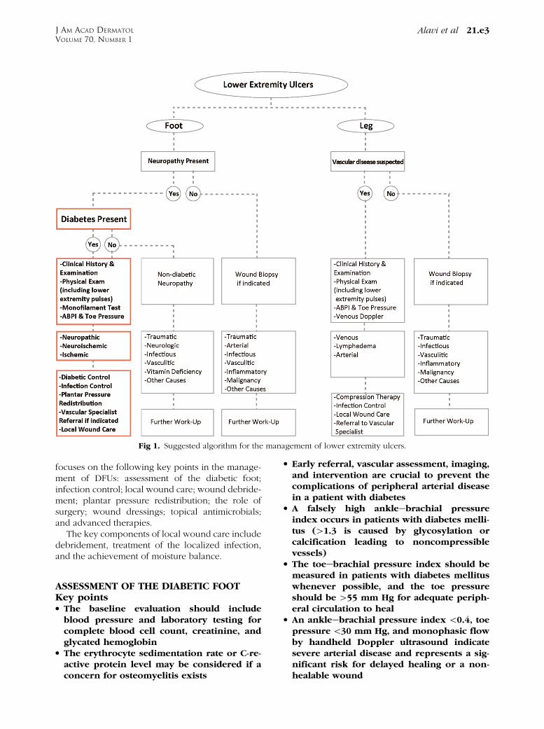
Fig 1. Suggested algorithm for the management of lower extremity ulcers.
J AM ACAD DERMATOL
VOLUME 70, NUMBER 1Alavi et al 21.e3
focuses on the following key points in the manage-ment of DFUs: assessment of the diabetic foot;infection control; local wound care; wound debride-ment; plantar pressure redistribution; the role ofsurgery; wound dressings; topical antimicrobials;and advanced therapies.
The key components of local wound care includedebridement, treatment of the localized infection,and the achievement of moisture balance.
ASSESSMENT OF THE DIABETIC FOOTKey pointsd The baseline evaluation should includeblood pressure and laboratory testing forcomplete blood cell count, creatinine, andglycated hemoglobin
d The erythrocyte sedimentation rate or C-re-active protein level may be considered if aconcern for osteomyelitis exists
d Early referral, vascular assessment, imaging,and intervention are crucial to prevent thecomplications of peripheral arterial diseasein a patient with diabetes
d A falsely high ankleebrachial pressureindex occurs in patients with diabetes melli-tus ([1.3 is caused by glycosylation orcalcification leading to noncompressiblevessels)
d The toeebrachial pressure index should bemeasured in patients with diabetes mellituswhenever possible, and the toe pressureshould be [55 mm Hg for adequate periph-eral circulation to heal
d An ankleebrachial pressure index \0.4, toepressure\30 mm Hg, and monophasic flowby handheld Doppler ultrasound indicatesevere arterial disease and represents a sig-nificant risk for delayed healing or a non-healable wound

J AM ACAD DERMATOL
JANUARY 201421.e4 Alavi et al
d A baseline radiograph of the affected foot isuseful for patients presenting with a newdiabetic foot ulcer
d Magnetic resonance imaging is the most ac-curate available imaging for the diagnosis orexclusion of osteomyelitis
A comprehensive assessment including historyand a physical examination is required for all patientswith DFUs. Other aspects that should be reviewedinclude the presence of neuropathic or nociceptivepain along with a loss of protective sensation.Patient-centered concerns that should be docu-mented include activities of daily living, the presenceof depression, and alcohol consumption andsmoking.
Wound assessmentThe following wound-related items should be
evaluated:d Location on the foot (ie, plantar, forefoot, mid-foot, heel, dorsum, toes, or sides of foot)
d Size (ie, at least longest length with the widestwidth at right angles in cm or mm, ideallyplanimetric or computerized photographymeasurement)
d Depth (cm or mm) measured with a probe, andcheck base for exposed tendon or bone
d Appearance of the wound base (ie, granulation,fibrin, slough, or necrotic tissue [black])
d Amount (ie, none, scant, moderate, or heavy) andtype of exudate (ie, serous, sanguineous, puru-lent, or combinations)
d Periwound skin (ie, edge or wound margin):presence of callus, maceration, or erythema
d Pain level should to be evaluated: timing withdressing change or between dressing changes,severity and type of pain (ie, nociceptive [gnaw-ing, aching, tender, or throbbing] or neuropathic[burning, stinging shooting, or stabbing] or loss ofprotective sensation).
This framework allows clinicians to monitorwound healing or progression. In addition, severalstudies have noted the role of monitored wound andperiwound temperature (which is often not routinelymeasured) through infrared thermometry to identifysurrounding tissue injury. A temperature differenceof[38 of 48when compared to the contralateral limbshould raise a concern of infection when it is inconcordance with the clinical symptoms.6,7
Increased temperature may also indicate deep in-flammation (such as Charcot foot often in theabsence of an ulcer) or unequal vascular supply,with clinical features used to differentiate theseconditions. In addition, during diabetic patient
self-monitoring at risk for development of DFU, anincreased local temperature may indicate a risk ofskin breakdown.8
Although elevated temperature may be suggestiveof infection, documenting additional clinical signsmakes the definitive diagnosis of wound infection.The clinical diagnosis of osteomyelitis in patientswith DMmay be challenging. The probe to bone testby a sterile blunt stainless steel probe is helpful.9
Laboratory studiesWound healing can be delayed in the presence of
coexisting systemic disease or comorbidities (ie,renal or peripheral vascular disease). The baselinelaboratory evaluation in a patient with DM and a footulcer should include a complete blood cell count,creatinine level, erythrocyte sedimentation rate(ESR) or C-reactive protein (CRP) level, and HbA1clevel.10 Several laboratory results may be importantfor management. Anemia may delay healing, andleukocytosis may be associated with infection. Highlevels of creatinine (suggestive of renal dysfunction)may indicate a greater risk for DFU complicationsand alter the choice and the dose of antibiotictherapy. An ESR [40 mm per hour or a CRP [20mm per hour is commonly associated with deeptissue infection or osteomyelitis.11,12 As noted above,HbA1c is a good indicator of diabetes control over 90days. The American Diabetes Association recom-mends a level of HBA1c\0.07 (7%) for adults, but forthe elderly and developing world and patients withbrittle DM, a target of\0.09 (9%) has been suggestedto avoid hypoglycemic episodes.13,14 Nutritional as-sessment, including the level of albumin, should beperformed in persons with chronic wounds.15
Prealbumin has a shorter half-life than albumin andmay better detect improvement in patients withprotein deficiencies.16
Patients with DM, especially those with poorglycemic control, have a relative immunodeficiency.They may not present with the classic overt signs ofinfection.17,18 High levels of HbA1c have been asso-ciated with diabetic comorbidities, such as neurop-athy, coronary artery disease, retinopathy, andnephropathy. Elevated HbA1c has been shown as apredictor of the development of DFUs, but additionalstudies are still required.5
Vascular assessmentA vascular assessment, when indicated, should
include advanced imaging and vascular intervention.Early intervention is important to prevent complica-tions of PAD in persons with DM. Every 1% (0.01)increase in HbA1c correlates with a 25% to 28%increase in the relative risk of PAD.15,19 DM increases

Table I. Arterial measurements related to vascular supply of the leg*
ABPI Toe pressure (mm Hg) TBPI Ankle Doppler wave form Diagnosis
[0.8 [80 [0.6 Normal/triphasic No relevant arterial disease[0.5 [50 [0.4 Biphasic Some arterial disease: modify compression[0.4 [30 [0.2 Biphasic/monophasic Arterial disease predominates\0.4 \30 \0.2 Monophasic High risk for limb ischemia
ABPI, Ankle-brachial pressure; TBPI, toeebrachial pressure index.
*Modified from Sibbald et al.27
J AM ACAD DERMATOL
VOLUME 70, NUMBER 1Alavi et al 21.e5
the risk of PAD 3.5-fold in men and 8.6-fold inwomen.15,20-22
Vascular assessment is required for all neuro-pathic foot ulcers. There is no universal test to assessthe vascular system. Noninvasive vascular assess-ments include the ankleebrachial pressure index(ABPI), toe pressure and/or toeebrachial pressureindex (TBPI), and transcutaneous oxygen pressure(TcPO2) measurement. Additional studies will beindicated based on the results of these tests.23,24 TheABPI Doppler ultrasound measures macrovasculararterial disease and may overestimate the true pres-sure reading in patients with DM because of incom-pressible vessels often related to calcification andadvanced atherosclerosis (Table I).25-27 Up to 80% ofpersons with DM have falsely high ABPIs of [1.2,compared to only 20% of nondiabetic individuals.27
An ABPI\0.5 may not be reliable, and a completesegmental duplex Doppler ultrasound examinationof the lower extremity arteries may be required todetermine the location and extent of the vascularcompromise. As opposed to patients without DMwho have PAD, patients with DM and PAD tend tohave more distal disease.28 The presence of a seg-mental drop in arterial pressure may indicate thepotential for a correctable arterial lesion. If vasculardisease is suspected and intervention planned, arte-rial angiography is required. Computed tomographyor magnetic resonance angiography (unless dilationis planned at the same time as the procedure) may bepart of a comprehensive assessment.
ImagingBaseline radiograph. A baseline radiograph of
the affected foot is useful in patients presenting witha new DFU. Plain radiographs may provideinformation on the presence of a foreign body,soft tissue gas that may be associated with deepinfection, and bony abnormalities associated withosteomyelitis, traumatic fractures, or the multiplefractures of Charcot joint. The classic change sug-gestive of osteomyelitis includes cortical erosion,periosteal reaction, combined lucency, and sclero-sis.16 Plain film imaging has the limitation that someradiographic changes are delayed and may take
up to a month after the clinical presentation. Ifosteomyelitis remains a concern, plain serial radio-graphs may help to monitor and exclude osteomy-elitis. The other limitation of radiography is thepotential for confusion between the subtle changesof osteomyelitis with early Charcot foot or neuro-osteoarthropathy.
Magnetic resonance imaging. A magnetic res-onance imaging (MRI) scan is the most accurateavailable imaging for the diagnosis or exclusion ofosteomyelitis.29 A sensitivity of 90% and specificity of79% has been reported in a recent metaanalysis byDinh et al.9 If MRI is not available, 3-phase bonescans are an alternative for the diagnosis of osteo-myelitis.30 MRI is more cost-effective and offersbetter sensitivity and specificity compared to 3-phase bone scan.31 If imaging is inconclusive, adiagnostic bone biopsy specimen should be ob-tained for histologic assessment, and bacterial cul-ture is the criterion standard for diagnosingosteomyelitis. The bacterial culture may also providean opportunity to identify the causative bacterialagent and any antibiotic sensitivities.
Musculoskeletal assessment. DM-related neu-ropathy can cause changes to the foot that compro-mise normal ambulation through several differentpathways. Numerous pathogenic pathways lead tomuscle atrophy and compensatory biomechanicalalterations in the diabetic foot. These include clawtoes, hammertoes, bunions, hallux rigidis, andCharcot changes. All foot deformities and biome-chanical impairments need evaluation and treatmentto avoid additional damage of the bony structure toprevent foot ulcers and subsequent amputations.Treatment mainly with orthotic shoes and pressureoff-loading devices should prevent excess pressure(calluses), friction, and shear (hemorrhagic blisters)in order to prevent these unwanted complications(Table II).
Biomechanical impairments are central topressure-induced DFUs that are easily identifiableby clinical examination. The foot and the ambulatorygait pattern should be examined in relation to thepatient’s footwear for both effective treatment andprevention.

Table II. Pathogenesis of diabetic foot ulcers
Pathogenesis Consequences Evaluation Therapy
Hyperglycemia/ Glycosylation of collagen [,collagenase resistance [
/ Cross-linking of collagen [/ Stiffness of connective tissue
Limited joint mobility / high plantarpressure / callus / ulcer /infection
Limited skin elasticity / diminishedabsorption of shear and pressure stress/ skin cracks / ulcer / infection
ROM and dorsiflexion of the toes andankles
Inspection
NonoperativePhysiotherapy: stretching; orthotics:outsole modifications: rocker, roller,AFO; skin care, seamless socks
OperativeTendon release
Hyperglycemia/ Abnormal fatty acid metabolism /decreased nutritional delivery
Atrophy of plantar fat pads/ diminishedabsorption of shear and pressure stress/ callus / ulcer / infection anddamage of deep structures (bone andjoint)
Palpation
Hyperglycemia/ Myoinositol uptake Y, glycolsylationof neural proteins [, polyol activity [,and abnormal fatty acid metabolism
/ Nerve ischemia/nerve function YAngiopathy/ Abnormal vasa nervorum / nerveischemia
Motor neuropathy / atrophy ofintrinsic (flexor) foot muscles /diabetes-dependent foot deformities(claw and hammertoe deformity/prominence of metatarsal heads) anddislocation of plantar fat pads /improper weight bearing/diminishedabsorption of shear and pressure stress/ callus / ulcer / infection anddamage of deep structures (bone andjoints)
Inspection NonoperativeDiabetes-adapted insoles, outsolemodifications: rocker, roller, andwidening; custom made orthopedicshoes
OperativeTenotomy, tendon transfer, resectionarthroplasty, and osteotomy
Sensory neuropathy /Loss of protective sensation / improperweight bearing / high plantarpressure / callus / neuropathiculcer / infection
Limited locomotory coordination /micro and macro fractures/ruptures ofbones, ligaments, and tendons
Charcot foot / deformity or instability/ improper weight bearing / highpressure plantar and/or ininappropriate footwear / callus /ulcer / infection
Lack of sensation with 128-Hz tuning forkor Semmes Weinstein monofilament(10 g or 5.07)
Inspection, palpation: red, warm, andswollen foot without ulcer, with (stage1-3) or without (stage 1) deformity;radiograph and magnetic resonanceimaging scans
NonoperativeSeamless socks, TCC, diabetes-adaptedinsoles; outsole modifications: rocker,roller, and widening; custom madeorthopedic shoes; AFO
OperativeOstectomy (bumpectomy), realignmentcorrective arthrodesis, tendon release,resection arthroplasty, and amputation
JAM
ACADD
ERM
ATOL
JANUARY20
1421.e6
Alavi
etal

Autonomic neuropathy / decreasedsympathetic innervation(autosympathectomy)
Decreased perspiration (hypohidrosis,anhidrosis) / dry and atrophic skin/ diminished absorption of shear andpressure stress / ulcer / infection
AV shunting [ / increased boneabsorption / Charcot foot /deformity and or instability /improper weight bearing / highpressure plantar or inappropriatefootwear / callus / ulcer /infection
Inspection, palpation: red, warm, andswollen foot without ulcer, with (stage1-3) or without (stage 1) deformity;radiography and magnetic resonanceimaging scan
Previous or diabetes-independent footdeformities
Hallux valgus, bunion, claw/hammertoes,pes eqinus, pes cavus, club foot, andso on; sensory neuropathy
Improper weight bearing / highpressure plantar or by inappropriatefootwear / callus / ulcer /infection
Inspection NonoperativeDiabetes-adapted insoles, outsolemodifications: rocker, roller, andwidening; custom made orthopedicshoes; AFO
OperativeTenotomy, tendon transfer/release,osteotomy, exostectomy, resectionarthroplasty, realignment arthrodesis,and amputation
Inappropriate footwear and sensoryneuropathy
High dorsal and plantar pressure insidethe shoe / pressure ulcer /infection
Inspection Appropriate footwear
AFO, Ankle foot orthosis; ROM, range of motion; TCC, total contact cast.
JAM
ACADD
ERM
ATOL
VOLU
ME70
,NUM
BER1
Alavi
etal
21.e7
J AM ACAD DERMATOL
JANUARY 201421.e8 Alavi et al
INFECTION CONTROLKey pointsd More than half of individuals with diabeticfoot ulcers develop skin and soft tissueinfection
d Aerobic ‘‘Gram-positive’’ cocci, especiallyStaphylococcus aureus, are the most commoncause of initial diabetic foot infection; how-ever, individuals with chronic foot ulcershave multibacterial organisms, including‘‘Gram-positives,’’ ‘‘Gram-negatives,’’ and an-aerobes (‘‘microbial shift’’ over time)
d Osteomyelitis should be suspected if an ulcerprobes to bone or exposed bone is present.Multiple-phase magnetic resonance imagingis the best noninvasive test for the diagnosisfor osteomyelitis. A bone biopsy specimenwith culture is the definite test for diagnos-ing osteomyelitis. Occasionally, treatmentmay still be prudent if there is a high clinicalindex of suspicion in the presence of aninconclusive (‘‘negative’’) magnetic reso-nance imaging scan
d For mild to moderate infections, initial ther-apy is aimed at aerobic Gram-positive cocci(until microbiology results are known). Em-pirical broad-spectrum therapy, often tai-lored to local antibiotic biograms (in vitrosensitivity), is indicted for severe infections
d The duration of antibiotic therapy for mildinfection should be at least 2 weeks. Formoderate to severe infection, 2 to 4 weeks ofantibiotic therapy is suggested, but clinicalsigns should determine the length of therapy
Diabetic foot infections are one of the mostfrequent and severe complications seen in individ-uals with DM. More than 50% of DFUs developinfection. There is a 10-fold increased chance ofbeing hospitalizedwith a bone or soft tissue infectionin a patient with DM compared to an individualwithout DM.32
All wounds are contaminated and often colo-nized; the relative importance of bacteria may vary.Wound contamination is the presence of nonrep-licating bacteria on wound surface, and woundcolonization is replication of bacteria without tissueinjury or immune response. Critical colonizationinvolves the replication of bacteria in the superficialwound base with tissue damage. As host resistanceweakens, the bacteria invade the wound marginsand base where a critical threshold of 105 bacteriaper gram of tissue is often associated with tissuedamage by the inflammatory cytokines.33 Thiswound perimeter infection often involves a
continuum between the ulcer base and the ulceredges, where the classical signs of infection may beabsent.34 Bacteria react to hostile environments byproducing biofilms that provide bacterial protectionagainst the host immune response. The differentia-tion between a wound in bacterial balance, criticalcolonization, and deep and surrounding tissue in-fection can be challenging because contaminationand colonization would not require antibiotictherapy.
The clinical signs of infection can be dampenedbecause of diminished leukocyte function, PAD, andneuropathy.15 About 50% of patients with DM anddeep foot infection lack a systemic inflammatoryresponse indicators of infection (ie, diminished orlack of elevation of ESR or CRP, with a normal whiteblood cell count and body temperature), leading to adelayed diagnosis.11 Uncontrolled DM can result inimmunopathy with a blunted cellular response tofoot infections. Impairment of macrophage functionas the key player of tissue repair contributes todelayed healing and increased susceptibility toinfection.35
Infection is often a precursor to amputation.Peters et al36 reported that with previous amputation,PAD, and neuropathy were significant risk factors fordiabetic foot infection. Patients with an amputationare more likely to undergo another amputation onthe reminder of their foot, lower leg, or on thecontralateral leg.37,38 In addition, a partial foot am-putation can lead to increased plantar foot pressureon the decreased remaining plantar surface.39
Prevention or early treatment of infectionmight therefore prevent subsequent additionalamputations.
The diabetic foot infection requires a multiprofes-sional approach with attention to local and systemicfactors.29,40 Aerobic Gram-positive cocci, especiallyStaphlyococcus aureus, are the most common causeof diabetic foot infection. Individuals with chronicfoot ulcers and previous antibiotic therapy oftenhave infections with both Gram-positive and -nega-tive organisms. Gangrenous and severely ischemiculcers are commonly coinfected with anaerobicpathogens.29 The diagnosis of infection is clinicalby identifying associated symptoms and signs suchas redness, edema, increased temperature, and pain(Fig 2).
Laboratory examinations have limited use inthe diagnosis of infection except in cases of osteo-myelitis. However, a semiquantitative bacterial swabtest—or preferably a tissue biopsy speicmen—forthe identification of bacterial organisms on cultureand organism sensitivity can be taken before startingantibiotic therapy.40
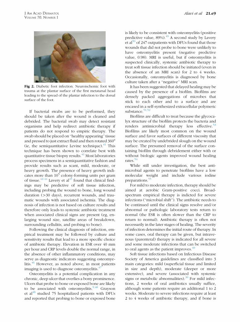
Fig 2. Diabetic foot infection. Neuroischemic foot withtrauma at the plantar surface of the first metatarsal headleading to the spread of the plantar infection to the dorsalsurface of the foot.
J AM ACAD DERMATOL
VOLUME 70, NUMBER 1Alavi et al 21.e9
If bacterial swabs are to be performed, theyshould be taken after the wound is cleaned anddebrided. The bacterial swab may detect resistantorganisms and help redirect antibiotic therapy ifpatients do not respond to empiric therapy. Theswab should be placed on ‘‘healthy appearing’’ tissueand pressed to just extract fluid and then rotated 3608(ie, the semiquantitative Levine technique).41 Thistechnique has been shown to correlate best withquantitative tissue biopsy results.41 Most laboratoriesprocess specimens in a semiquantitative fashion andprovide results such as scant, mild, moderate, orheavy growth. The presence of heavy growth indi-cates more than 105 colony-forming units per gramof tissue.42-44 Lavery et al45 found that clinical find-ings may be predictive of soft tissue infection,including probing the wound to bone, long woundduration ([30 days), recurrent wounds, and trau-matic wounds with associated ischemia. The diag-nosis of infection is not based on culture results andtherefore only leads to systemic antibiotic treatmentwhen associated clinical signs are present (eg, en-larging wound size, satellite areas of breakdown,surrounding cellulitis, and probing to bone).
Following the clinical diagnosis of infection, em-pirical treatment may be followed by culture andsensitivity results that lead to a more specific choiceof antibiotic therapy. Elevation in ESR over 40 mmper hour and CRP levels double the normal range, inthe absence of other inflammatory conditions, mayserve as diagnostic indicators suggesting osteomye-litis.46 However, as noted above, in most patientsimaging is used to diagnose osteomyelitis.47
Osteomyelitis is a potential complication in anychronic, deep ulcer that overlies a bony prominence.Ulcers that probe to bone or exposed bone are likelyto be associated with osteomyelitis.9,48 Graysonet al48 studied 75 hospitalized patients with DFUsand reported that probing to bone or exposed bone
is likely to be consistent with osteomyelitis (positivepredictive value, 89%).49 A second study by Laveryet al50 of 247 outpatients with DFUs found that thosewounds that did not probe to bone were unlikely tohave osteomyelitis present (negative predictivevalue, 0.98). MRI is useful, but if osteomyelitis issuspected clinically, systemic antibiotic therapy totreat soft tissue infection should be initiated (even inthe absence of an MRI scan) for 2 to 4 weeks.Occasionally, osteomyelitis is diagnosed by boneculture taken after a ‘‘negative’’ MRI scan.
It has been suggested that delayed healing may becaused by the presence of a biofilm. Biofilms aredensely packed aggregations of microbes thatstick to each other and to a surface and areencased in a self-synthesized extracellular polymericsubstance.51,52
Biofilms are difficult to treat because the glycoca-lyx structure of the biofilm protects the bacteria andrenders antimicrobial therapy less effective.53
Biofilms are likely most common on the woundsurface and favor surfaces of different viscosity thatmay be created by undebrided slough on the woundsurface. The presumed removal of the surface con-taining biofilm through debridement either with orwithout biologic agents improved wound healingrates.54
While still under investigation, the best anti-microbial agents to penetrate biofilms have a lowmolecular weight and include various iodinepreparations.55
For mild to moderate infection, therapy should beaimed at aerobic Gram-positive cocci. Broad-spectrum empirical therapy is indicted for severeinfections (‘‘microbial shift’’). The antibiotic needs tobe continued until the clinical signs resolve and/orabnormal or pathologic laboratory tests return tonormal (the ESR is often slower than the CRP toreturn to normal). Antibiotic therapy is often notnecessarily in the later stages of healing. The severityof infection determines the initial route of therapy. Insome cases, oral therapy can be given, but intrave-nous (parenteral) therapy is indicated for all severeand some moderate infections that can be switchedto oral agents as the patient improves.40
Soft tissue infections based on Infectious DiseaseSociety of America guidelines are classified into 3main categories: mild (superficial tissue and limitedin size and depth), moderate (deeper or moreextensive), and severe (associated with systemicsigns or metabolic abnormalities).30 For mild infec-tions, 2 weeks of oral antibiotics usually suffice,although some patients require an additional 1 to 2weeks. Moderate to severe infections require at least2 to 4 weeks of antibiotic therapy, and if bone is
J AM ACAD DERMATOL
JANUARY 201421.e10 Alavi et al
involved (ie, osteomyelitis is present), usually 6 to 12weeks minimum treatment is required. If the infectedbone is removed, a shorter treatment period is oftensufficient, but treatment time should be determinedby the resolution of clinical signs.40 In individualswith signs associated with deep and surroundingwound infection, systemic antimicrobial therapies(at least initially) are indicated. Most antibiotic rec-ommendations include coverage for Gram-positive,Gram-negative, and anaerobic organisms. The re-cent emergence of antibiotic resistant bacteria pre-sents a major challenge for the successful treatmentof diabetic foot infections.30,38,56
Local wound careOptimal local wound care includes proper cleans-
ing (using normal saline or sterile water), debride-ment of calluses and wound base especially ifassociated, with devitalized tissue, control of local-ized infection, and abnormal prolonged inflamma-tion and moisture balance. This needs to beaccompanied by systemic treatment for deep andsurrounding infection for successful treatment.Wound cleaning is the process of removal of looseinflammatory materials and debris from the woundsurface.57 The selection of the appropriate woundcleanser is important to minimize chemical andmechanical damage. The standard of the care forwound cleansing is to use agents with low cytotox-icity, including water, isotonic normal saline, andshort contact acetic acid. Soaps and detergents havebeen commonly used for wound hygiene. There isevidence that these surface-active agents raise thesurface pH and interfere with healing through alter-ing the cell viability and tissue function.57 Isotonicnormal saline (0.9% NaCl) is a good cleanser for themajority of wounds, and a Cochrane review foundthat water is a suitable alternative.58 The evidenceregarding commercial wound cleanser safety is verylimited. However; the Cochrane review concludedthat there is no evidence that tap water as a woundcleanser increases the risk of infection.58
WOUND DEBRIDEMENTKey pointsd Active (surgical) debridement (ulcer edgeand base) is an adjunct to plantar pressureredistribution as the standard of care in thetreatment of diabetic foot ulcers
d Additional maintenance removal of callus atevery visit is of importantance to avoid ad-ditional pressure and shear forces
d The main categories of debridement are sur-gical, autolytic, enzymatic, mechanical, andbiologic
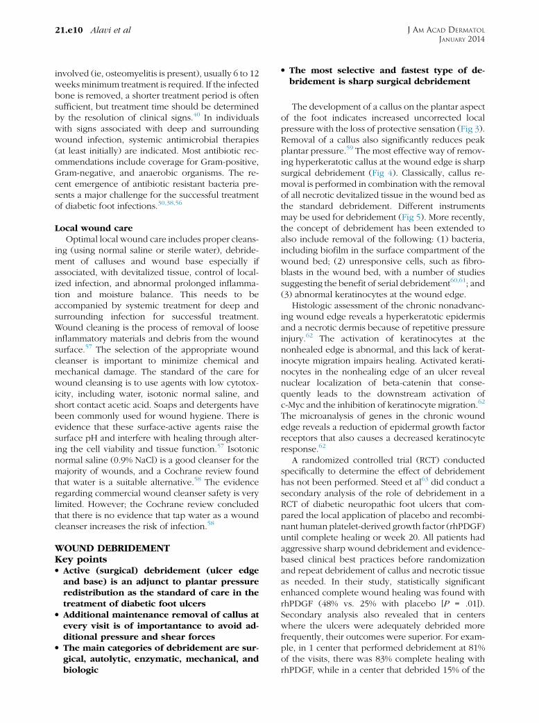
d The most selective and fastest type of de-bridement is sharp surgical debridement
The development of a callus on the plantar aspectof the foot indicates increased uncorrected localpressure with the loss of protective sensation (Fig 3).Removal of a callus also significantly reduces peakplantar pressure.59 The most effective way of remov-ing hyperkeratotic callus at the wound edge is sharpsurgical debridement (Fig 4). Classically, callus re-moval is performed in combination with the removalof all necrotic devitalized tissue in the wound bed asthe standard debridement. Different instrumentsmay be used for debridement (Fig 5). More recently,the concept of debridement has been extended toalso include removal of the following: (1) bacteria,including biofilm in the surface compartment of thewound bed; (2) unresponsive cells, such as fibro-blasts in the wound bed, with a number of studiessuggesting the benefit of serial debridement60,61; and(3) abnormal keratinocytes at the wound edge.
Histologic assessment of the chronic nonadvanc-ing wound edge reveals a hyperkeratotic epidermisand a necrotic dermis because of repetitive pressureinjury.62 The activation of keratinocytes at thenonhealed edge is abnormal, and this lack of kerat-inocyte migration impairs healing. Activated kerati-nocytes in the nonhealing edge of an ulcer revealnuclear localization of beta-catenin that conse-quently leads to the downstream activation ofc-Myc and the inhibition of keratinocyte migration.62
The microanalysis of genes in the chronic woundedge reveals a reduction of epidermal growth factorreceptors that also causes a decreased keratinocyteresponse.62
A randomized controlled trial (RCT) conductedspecifically to determine the effect of debridementhas not been performed. Steed et al63 did conduct asecondary analysis of the role of debridement in aRCT of diabetic neuropathic foot ulcers that com-pared the local application of placebo and recombi-nant human platelet-derived growth factor (rhPDGF)until complete healing or week 20. All patients hadaggressive sharp wound debridement and evidence-based clinical best practices before randomizationand repeat debridement of callus and necrotic tissueas needed. In their study, statistically significantenhanced complete wound healing was found withrhPDGF (48% vs. 25% with placebo [P = .01]).Secondary analysis also revealed that in centerswhere the ulcers were adequately debrided morefrequently, their outcomes were superior. For exam-ple, in 1 center that performed debridement at 81%of the visits, there was 83% complete healing withrhPDGF, while in a center that debrided 15% of the

Fig 3. Stepwise process from callus formation to ulceration and infection.
J AM ACAD DERMATOL
VOLUME 70, NUMBER 1Alavi et al 21.e11
visits, there was 20% complete healing (again withbest practices and rhPDGF).
Falanga et al,64 Steed et al,55 and Cardinal et al60,63
have all proposed the concept of maintenancedebridement after initial excisional debridement.This method of debridement depends on patientcharacteristics, preference, and the practitioner’slevel of expertise. If surgical debridement is per-formed, the goal is often to reach bleeding tissue,implying that adequate arterial flow is needed (Fig 6,A-C ). Warriner et al65 found, in a real-world popu-lation, that more frequent visits and sequentialdebridement had healing benefits, with implicationsof lower costs and higher quality of life for patients.
PLANTAR PRESSURE REDISTRIBUTIONd The total contact cast is the criterion stan-dard off-loading device, but it is contraindi-cated for heel ulcers, deep infection, orperipheral arterial disease
d The removable cast walker is reusable, easyto apply, and less expensive, but the adher-ence to treatment is often lower
d Therapeutic shoes with accommodative in-soles that redistribute high-pressure areasare often used for heel ulcers and required toprevent recurrences posteulcer healing
d Surgery for restoration of limited joint mo-bility (eg, lengthening of the Achilles ten-don) or correction of deformities (eg,exostosectomy, osteotomy, resection arthro-plasty, or corrective arthrodesis) may beconsidered in appropriate patients and maybe more effective than orthopedic devices toprovide plantar pressure redistribution oroffloading
Surgery for the restoration of limited joint mobility(eg, lengthening of the Achilles tendon) or correc-tion of deformities (eg, exostosectomy, osteotomy,resection arthroplasty, or corrective arthrodesis) maybe considered in appropriate patients and may bemore effective than orthopedic devices to provideplantar pressure redistribution or off-loading.66,67
Several studies have concluded that plantar pressureredistribution is the most important component inthe management of neuropathic ulcers. Therefore,inadequate plantar pressure redistribution is associ-ated with delayed healing.68 Wu and Armstrong69
and Lavery et al70 documented that the rate ofhealing with a total contact cast (TCC) was superiorto other off-loading devices, with healing ratesreported as high as 100% for appropriately selecteddiabetic neuropathic foot ulcers and with a meanhealing time of 28 to 38 days.
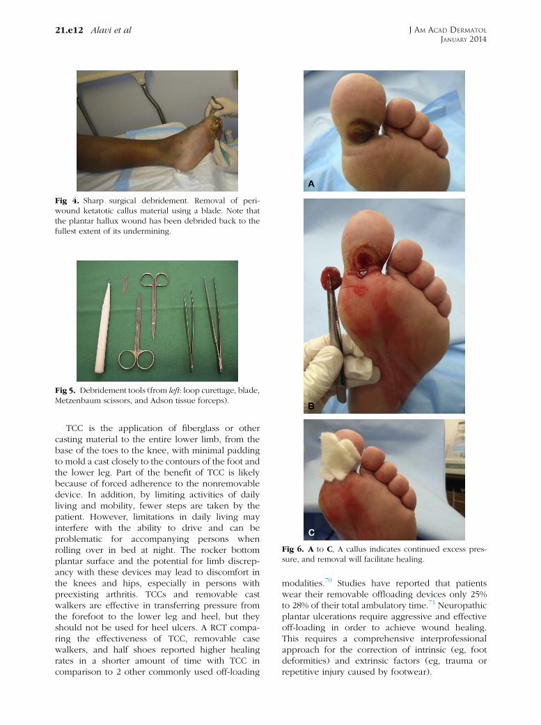
Fig 5. Debridement tools (from left: loop curettage, blade,Metzenbaum scissors, and Adson tissue forceps).
Fig 4. Sharp surgical debridement. Removal of peri-wound ketatotic callus material using a blade. Note thatthe plantar hallux wound has been debrided back to thefullest extent of its undermining.
Fig 6. A to C, A callus indicates continued excess pres-sure, and removal will facilitate healing.
J AM ACAD DERMATOL
JANUARY 201421.e12 Alavi et al
TCC is the application of fiberglass or othercasting material to the entire lower limb, from thebase of the toes to the knee, with minimal paddingto mold a cast closely to the contours of the foot andthe lower leg. Part of the benefit of TCC is likelybecause of forced adherence to the nonremovabledevice. In addition, by limiting activities of dailyliving and mobility, fewer steps are taken by thepatient. However, limitations in daily living mayinterfere with the ability to drive and can beproblematic for accompanying persons whenrolling over in bed at night. The rocker bottomplantar surface and the potential for limb discrep-ancy with these devices may lead to discomfort inthe knees and hips, especially in persons withpreexisting arthritis. TCCs and removable castwalkers are effective in transferring pressure fromthe forefoot to the lower leg and heel, but theyshould not be used for heel ulcers. A RCT compa-ring the effectiveness of TCC, removable casewalkers, and half shoes reported higher healingrates in a shorter amount of time with TCC incomparison to 2 other commonly used off-loading
modalities.70 Studies have reported that patientswear their removable offloading devices only 25%to 28% of their total ambulatory time.71 Neuropathicplantar ulcerations require aggressive and effectiveoff-loading in order to achieve wound healing.This requires a comprehensive interprofessionalapproach for the correction of intrinsic (eg, footdeformities) and extrinsic factors (eg, trauma orrepetitive injury caused by footwear).

J AM ACAD DERMATOL
VOLUME 70, NUMBER 1Alavi et al 21.e13
Various off-loading devices can achieve pressureredistribution, but adherence to using an off-loadingdevice is critical. Patient education regarding altera-tion of gait pattern (eg, swing gait or cane on theopposite hand) may support pressure reduction.72
Plantar pressure measurement within the shoes orfoot scanners known as pedobarography (trans-ducers that detect up to 950 pressure points betweenthe device and the plantar surface either stationary orwith active gait) may detect the area(s) of highpressure. The choice of plantar pressure redistribu-tion strategies is multifactorial, including the patient’sage, balance, strength, activities of daily living, andhome and work environment.73
The selection of an appropriate off-loading deviceis individualized, and activity level or ability to walkwith a rocker bottom heel (poor balance and loss ofproprioception with neuropathy that may result infalls) is a key factor in the individualized decision ofplantar pressure redistribution devices.
Therapeutic shoes with accommodative (but rel-atively stiff) insoles to redistribute high-pressureareas are required for some heel ulcers, when otherdevices cannot be worn. These devices are alsoimportant for maintenance therapy after healing toprevent recurrences. These therapeutic shoe optionsrepresent a relatively less expensive treatment op-tion for long-term use. Therapeutic shoes and visco-elastic plantar inserts function as artificial shockabsorbers to decrease the risk of foot ulceration byproviding an extrinsic accommodative and protec-tive mechanism. The rate of reulceration in the groupwearing therapeutic shoes was still as high as 28% to50%.74 There is a need for proper footwear duringnonworking hours (eg, adequate substitution for orespecially manufactured therapeutic home footwear,supportive slippers, or sandals with side support andno strap between the first and second toe74; TableIII45,68,70,75-81). Uncontrolled edema of the lower legand foot has been shown by Armstrong et al82 todelay the healing of foot ulcers. If the edema iscaused by venous insufficiency and arterial circula-tion is adequate, compression bandages should beused. Compression should be modified for patientswith mild arterial disease and not be used for moresignificant arterial disease. Foot edema can alsoresult from congestive heart failure or low albumin(eg, liver damage, kidney disease, and poor nutrit-ion) where additional alternative corrective action isrequired.
There are many factors that may influence healthbehaviors and adherence. Because of neuropathy,many people with diabetes lack protective footsensation. In addition, what often occurs is that thepatient will wear their device while they are outside
of the home but remove it when they get home.This negates much of the benefit that was obtainedfrom having worn the device outside of the home.Not only will the patient remove the device, butalso many patients report that they will walk instockings alone or barefoot around the house. It isimportant to educate both the patient and theircircle of care about the negative consequences ofthis behavior. The removal of devices around thehome is one of the primary reasons that removabledevices often have much longer healing times andlower healing rates than other options that are notremovable. The concept of a nonremovable devicecreating ‘‘forced compliance’’ is a way to increasepatient adherence to treatment. Patient educationprograms need to emphasize the patient’s respon-sibility for their own health and well-being. In manydiabetes education circles, this is referred to aspatient empowerment. However, patient educationshould not only focus on wearing appropriatefootwear but also on other aspects of puttingthem at risk to foot ulceration. These modifiablebehaviors that inhibit healing include foreign bod-ies within the shoes that can be detected bychecking the shoes, eliminating foot baths, measur-ing water temperature for baths with a thermome-ter, avoiding heating cushions in winter, andexercising caution with personal foot care, espe-cially nail cutting. Regular visits to a foot carespecialist are optimal, especially in high-risk indi-viduals that include those with impaired vision orneuropathy.
ROLE OF SURGERY IN THE MANAGEMENTOF DIABETIC FEET
Diabetic foot surgery has a role in the preventionand management of DFUs. Although the surgicalintervention in these patients is not without risk, theselective correction of chronic deformities and per-sistent foot ulcers can improve outcomes.82,83
Surgery has an important role in the reconstructionof soft tissue defects in these patients.84 Carefulconsideration of the indications for diabetic footsurgery should always include the presence of suf-ficient arterial perfusion for healing.66,67
While the aim of management of the diabetic footfocuses on limb salvage, in select cases amputationmay offer a better functional outcome, although thisoften is not clearly defined. This decision is veryindividualized and multifactorial to match the pa-tient’s lifestyle and their medical, physical, andpsychological comorbidities.85 In general, amputa-tion should be the last resort after all other salvagetechniques have been explored and the patient must
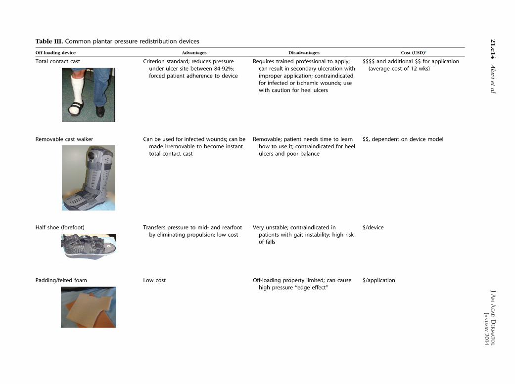
Table III. Common plantar pressure redistribution devices
Off-loading device Advantages Disadvantages Cost (USD)*
Total contact cast Criterion standard; reduces pressureunder ulcer site between 84-92%;forced patient adherence to device
Requires trained professional to apply;can result in secondary ulceration withimproper application; contraindicatedfor infected or ischemic wounds; usewith caution for heel ulcers
$$$$ and additional $$ for application(average cost of 12 wks)
Removable cast walker Can be used for infected wounds; can bemade irremovable to become instanttotal contact cast
Removable; patient needs time to learnhow to use it; contraindicated for heelulcers and poor balance
$$, dependent on device model
Half shoe (forefoot) Transfers pressure to mid- and rearfootby eliminating propulsion; low cost
Very unstable; contraindicated inpatients with gait instability; high riskof falls
$/device
Padding/felted foam Low cost Off-loading property limited; can causehigh pressure ‘‘edge effect’’
$/application
JAM
ACADD
ERM
ATOL
JANUARY20
1421.e14
Alavi
etal

Custom made orthotics Distributes pressure under foot evenly;may be used with over the counterfootweary
Requires trained professional to assessand mold/modify; relatively highercost
$$$
Custom braces, ankle and foot orthoses Immobilize the foot and ankle to managewounds
Inhibits motion of the foot and ankle; canrequire a deeper shoe
$$$$$
*$,\$100; $$, $100-500; $$$, $500-1000; $$$$, $1000-2000; $$$$$,[$2000.yOff the shelf shoes: primarily used for prevention, but required to be used in conjunction with padding or custom orthotics.
JAM
ACADD
ERM
ATOL
VOLU
ME70
,NUM
BER1
Alavi
etal
21.e15
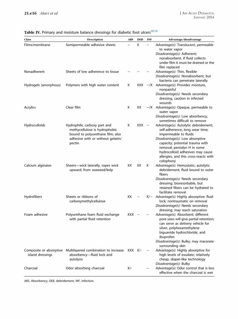
Table IV. Primary and moisture balance dressings for diabetic foot ulcers48,56
Class Description ABS DEB INF Advantage/disadvantage
Films/membrane Semipermeable adhesive sheets e X e Advantage(s): Translucent, permeableto water vapor
Disadvantage(s): Adherent;nonabsorbent, if fluid collectsunder film it must be drained or thefilm replaced
Nonadherent Sheets of low adherence to tissue e e e Advantage(s): Thin, flexibleDisadvantage(s): Nonabsorbent, butbacteria can penetrate laterally
Hydrogels (amorphous) Polymers with high water content X XXX e/X Advantage(s): Provides moisture,nonpainful
Disadvantage(s): Needs secondarydressing, caution in infectedwounds
Acrylics Clear film X XX e/X Advantage(s): Opaque, permeable towater vapor
Disadvantage(s): Low absorbency,sometimes difficult to remove
Hydrocolloids Hydrophilic carboxy part andmethycellulose is hydrophobicbound to polyurethane film; alsoadhesive with or without gelatin/pectin
X XXX e Advantage(s): Autolytic debridement;self-adherence; long wear time;impermeable to fluids
Disadvantage(s): Low absorptivecapacity; potential trauma withremoval; pentalyn H in somehydrocolloid adhesives may causeallergies, and this cross-reacts withcolophony
Calcium alginates Sheets—wick laterally, ropes wickupward; from seaweed/kelp
XX XX X Advantage(s): Hemostatic; autolyticdebridement; fluid bound to outerfibers
Disadvantage(s): Needs secondarydressing; bioresorbable, butretained fibers can be hydrated tofacilitate removal
Hydrofibers Sheets or ribbons ofcarboxymethylcellulose
XX e X/e Advantage(s): Highly absorptive; fluidlock; nontraumatic on removal
Disadvantage(s): Needs secondarydressing; may reach saturation
Foam adhesive Polyurethane foam fluid exchangewith partial fluid retention
XXX e e Advantage(s): Absorbent; differentpore sizes will give partial retention;can serve as delivery vehicle forsilver, polyhexamethylenebiguanide hydrochloride, andibuprofen
Disadvantage(s): Bulky; may maceratesurrounding skin
Composite or absorptiveisland dressings
Multilayered combination to increaseabsorbency—fluid lock andautolysis
XXX X/- e Advantage(s): Highly absorptive forhigh levels of exudate; relativelycheap, diaper-like technology
Disadvantage(s): BulkyCharcoal Odor absorbing charcoal X/- e Advantage(s): Odor control that is less
effective when the charcoal is wet
ABS, Absorbency; DEB, debridement; INF, infection.
J AM ACAD DERMATOL
JANUARY 201421.e16 Alavi et al

Table V. Antimicrobial dressings for diabetic foot ulcers
Antiseptics/antimicrobials Bacterial sensitivity Effect Adverse effect
Iodine-basedPovidone iodine, 10% solution,Cadexomer iodine, and Inadine(available in Europe and Canada, butnot in the United States)
d Broad antibacterial effect (Gram-positives more than Gram-negatives)and MRSA
d Good penetration of biofilms
d Short-term treatment and reassessevery 2-4 wks
d Cadexomer iodine releases the iodineslowly to make it less toxic 1 ca-dexomer sugar for autolytic debride-ment 1 absorbency
d Antibacterial effect (0.005% concentra-tion) without tissue toxicity
d Can be toxic to granulation tissued Antimicrobial action may be neutral-ized by inorganic and organic agents
d Thyroid dysfunctiond Can develop an allergy
Chlorhexidine, polyhexamethylenebiguanide hydrochloride, a derivativeof chlorhexidine—foam and gauze
d Gram-positives more thanGram-negatives, yeast, mold
d 0.02% concentration has been used forwound irrigation
d Promotes wound healing63
d May damage cartilage/ear toxicity64,65
Acetic acid, white vinegar 5% d Gram-negatives, particularlyPseudomonas
d Compresses 5-10 mind Dilute 1:5 (1%) or 1:10 water (0.5%)
d High concentration and long contactd May have some tissue toxicity andinhibits fibroblast growth
Silver compoundsSilver dressings, foams, calcium alginates,hydrofibers, hydrogels, sheets, andpowder; silver sulfadiazine cream;silver nitrate sticks
d Antibacterial, including Escherichia coli,Klebsiella, Staphylococcus aureus, andMRSA
d Antifungald Antiviral
d Must be combined with water to be inthe ionized state—Ag 1, 11, and111
d Silver nanoparticles enhanced contactand bactericidal activity
d Antiinflammatory effect may relate tothe Ag 0 state
d Silver toxicity to reepithelializationprocess
d Toxicity much less with dressings thansilver
d Sulfadiazine cream with much highersilver release
d Sticks plus silver sulfadiazine creammay produce proinflammatory pseu-doeschar/delay healing
Honey (medical grade; often Munukahoney), calcium alginate; hydrogel;hydrocolloid
d Antibacteriald Antifungald Antiviral
d Antiinflammatoryd High osmolar concentration contrib-utes to the antibacterial effect
d Potential risk of botulism with foodproduct honey64
Sodium hypochlorite (bleach) d Broad antibacterial effect (Gram-posi-tives more than Gram-negatives)
d Antibacterial effect (0.005%) withoutlower tissue toxicity
d Irritant with high tissue toxicityd Inhibit fibroblast in 1% concentrationd Best used as disinfectant and not forwound care
Benzalkonium chloride d Gram-positive and -negative, fungi d Compromised bactericidal activitybecause of neutralization with organicmatter in tissue fluids
d Very high tissue toxicity
Hydrogen peroxide d Gram-positive bacteria with 3%concentration
d Limited mechanical debridementd Biofilm reduction
d Bulla formationd Risk of air emboli67 if applied to deepcavities
MRSA, Methicillin-resistant Staphylococcus aureus.
JAM
ACADD
ERM
ATOL
VOLU
ME70
,NUM
BER1
Alavi
etal
21.e17

J AM ACAD DERMATOL
JANUARY 201421.e18 Alavi et al
be in agreement. Although distal arterial bypassesbelow the knee are more frequently undertaken,some patients do not have dilatable or bypassiblelesions. Allowing dry gangrene to demarcate andhaving distal areas (eg, toes) eventually separateand fall off may be a preferred option in someindividuals, especially if they are poor surgicalcandidates.
Wound dressingsKey pointsd The main categories of dressings include,films, hydrogels, acrylics, hydrocolloids, cal-cium alginates, hydrofibers, and foams
d Wounds with high levels of exudate needabsorptive dressings, whereas a dry woundrequires moisture balance dressings that do-nate moisture to the wounds
Wound dressings not only provide a physicalprotective barrier to the wound, but they also helpmaintain an optimal wound environment (homeo-stasis) with moisture balance to enhance healing.Highly exudating wounds require absorptive dress-ings, such as foams, calcium alginates, or hydrofiberdressings. Dry wounds are balanced with dressingsthat donate water (hydrogels) or preserve or bindwater (acrylics, hydrocolloids, and films). Filmseither alone or as the top layer of acrylics andhydrocolloids have a low moisture vapor transmis-sion rate that also assists in keeping the woundadequately moist. The choice of dressing is largelydetermined by the type of the wound and the woundcharacteristics, including exudate, odor, pain, ormicrobiology, including localized critical coloniza-tion. The main categories of the dressings are listedin Table IV.27 For DFUs, the benefit from a specificdressing choice is less important than optimal off-loading. Modern moist interactive dressings do nothave high quality RCTs to show a benefit over gauze.However, they have longer wear time, greater ab-sorbency, may be less painful, and are typically lesstraumatic upon removal. Moreover, in certain pa-tients, they are cost effective because of the lowerfrequency of dressing changes and the savings ofexpensive nursing time.86 Finally, concern whenusing occlusive dressings in patients with DM exists.The latter, suffering from decreased immune re-sponse, are prone to exacerbation of clinicallyunsuspected infection when occlusive dressings(eg, hydrocolloids) are applied. We suggest beingcautious with the use of occlusive moisture retainingdressings next to the wound (ie, films, hydrocolloids,and acrylates), especially if there is any suspicion ofinfection.
Topical antimicrobials: The role of antisepticsand other antimicrobialsKey pointsd The best options for wound cleansers arenormal saline or water
d The topical antimicrobial of choice will bebased on the agent’s antibacterial but alsoantiinflammatory effect and the relativelower toxicity to the host granulation tissue
The best options for wound cleansing are normalsaline or water.87 Potable (drinkable) water is ac-ceptable for most wounds unless there is immuno-suppression or a very deep cavity.58,87 The role oftopical antimicrobials for DFU is not well definedbecause studies have yet to clearly show a benefit.Topical antibiotics may cause contact allergic der-matitis and do not provide moisture balance orautolytic debridement for optimal topical treatmentof chronic wounds. Therefore, topical antiseptics arepreferred for critical colonization because they havean antibacterial effect with lower toxicity to the hosttissue. Currently available antiseptics are listed inTable V,88,89 and they are best administered withsustained release formulations.
Advanced therapiesKey pointsd Advanced therapies should be considered installed but healable wounds with optimalcare and when the wound edge is notmigrating
d Advanced biologic therapies have a role inthe management of stalled wounds, includ-ing growth factors, negative pressure woundtherapy, hyperbaric oxygen, and tissue-engineered (‘‘artificial’’) skin
d The most readily available advanced therapyis autologous skin. In many countries whereother advanced therapies are not available,autologous split skin graft may be the treat-ment of choice and may significantly accel-erate wound healing by various mechanisms
d Reconstructive surgery may offer a more de-finitive solution with adequate coverage ofbony prominences or weight-bearing areas,further supporting the need for interdiscipli-nary management of diabetic foot ulcers
Recent advances in basic science research andrelated techniques have influenced the options forthe treatment of chronic wounds. Sheehan et al90
concluded that if a DFU is not 50% smaller at week 4despite optimal care, it is unlikely to heal by week 12.An earlier decision to use advanced therapies

Table VI. Tissue-engineered skin equivalents (acellular and cellular)81
Name and description Trade names* Types Graft composition
Epidermal Epicel and Epidex Autograft Keratinocyte expanded from skin biopsyBioseed Autologus Keratinocyte in fibrin sealant
Dermal (acellular) Alloderm/Graft Jacket Alloderm Cadaveric decellularized dermisBiobrane Xenograft Porcine type I collagen bonded to a silicone film
membraneE-Z Derm Xenograft Porcine type I collagen bonded to a gauze linerIntegra Xenograft Bovine tendon collagen and shark chondroitinOASIS Xenograft Porcine intestine collagen type I and extracellular matrix
Dermal (cellular) Dermagraft Allograft Allogenic—neonatal foreskin fibroblast in polyglactinsuture
Bilayered Apligraf Allograft Allogenic—engineered neonatal foreskin keratinocytesand fibroblasts plus bovine collagen type I
*Trade names remain property of their respective manufacturers.
J AM ACAD DERMATOL
VOLUME 70, NUMBER 1Alavi et al 21.e19
improves the likelihood that these interventions willbe effective.91 Several studies have shown thatadvanced biologic therapies in combination withstandard care improve the healing of DFUs.92-94
Growth factors. Growth factors are biologicallyactive polypeptides that alter the growth, differenti-ation, and metabolism of target cells and induce acascade of signal transduction pathway.95,96
Beclapermin (REGRANEX GEL; Healthpoint, Dallas,TX), a topical recombinant human rhPDGF, is theonly growth factor approved by the US Food andDrug Administration for the treatment of diabetic footulcers.88,96 PDGF is produced by platelets, macro-phages, vascular endothelium, fibroblasts, and ke-ratinocytes. Multiple RCTs have shown the role ofPDGF in the healing of DFUs.97 In a retrospectivecohort study on 2517 patients with DFUs, the role ofadvanced biologic therapies, including PDGF in thefirst month, has been emphasized.91When indicated,early treatmentwith PDGFhas been associatedwith ashorter healing time of DFUs.98 In a metaanalysis on922 patients with DFUs, Becaplermin gel at a dose of100 mcg per gram administered on a daily basis hasbeen shown to be effective with a healing rate of 83%as an adjunctive to the standard care.54
Negative pressure wound therapy. Negativepressure wound therapy (NPWT) is the delivery ofsubatmospheric pressure to the wound bed. In thissystem, polyurethane or polyvinyl alcohol foam orgauze is cut and placed on the wound surface. Thefoam or gauze then is sealed by a transparent drapeto provide a close airtight system. A vacuum pump isconnected to this space and provides a negativepressure environment. The suction effect causesdeformation of the extracellular matrix and pro-motes cellular proliferation.95 In a multicenter RCTon 162 patients with DFUs and recent amputationsite surgery, NPWT had a healing rate of 56%
compared to 39% in the control group, with similaradverse events in both groups.99 NPWTwas found tobe safe and effective for the postsurgical treatment ofacute diabetic wounds, but it has not shown effec-tiveness in chronic nonhealing wounds.100,101
Hyperbaric oxygen therapy. Hyperbaric oxy-gen therapy (HBOT) is the administration of 100%oxygen to the wound through an airtight vessel at apressure[1 atmosphere absolute.95 Systemic HBOTsessions in the chamber last usually 45 to 120minutes; generally, 1 or 2 sessions daily are carriedout, 4 to 5 times per week, for a total of 20 to 30sessions at a pressure 2 to 3 times greater thanambient atmospheric pressure.95,101-103 In a system-atic review on the role of HBOT on chronic wounds,9 RCTs (471 patients) were identified, 8 of whichenrolled patients with DFUs (455 patients) with aWagner grade from 0 to 4.101 Pooled data of 3 RCTswith 140 patients showed a significant (P = .02)increase in the rate of ulcer healing with HBOT at 6weeks, but this benefit was not evident at longer-term follow-up (1 year). However, there was nostatistically significant difference in the major ampu-tation rate in pooled data of 5 trials including 312patients. To date, there is still controversy about theefficacy of HBOT in the treatment of DFUs. A recentlongitudinal observational cohort study by Margoliset al101 on 6259 individuals with diabetes, adequatelower limb arterial perfusion, and foot ulcer foundthat the use of HBOT neither improved the likeli-hood of healing nor prevented amputation in acohort of patients defined by Centers for Medicareand Medicaid Services eligibility criteria.101 The au-thors concluded that the usefulness of HBOT inpatients with DFUs needs to be reevaluated. Incontrast, L€ohndal104 reviewed 6 individual RCTsand concluded that data evaluating HBOT in DFUtreatment are more robust than evidence in favor of

J AM ACAD DERMATOL
JANUARY 201421.e20 Alavi et al
many other practices in the diabetic foot treatmentarmamentarium, especially in patients with ulcerduration [3 months and no need or possibility ofvascular surgery. Another recent prospective RCTevaluating the effects of HBOT on healing andoxidative stress of ulcer tissue in patients withDFUs suggested that HBOT treatment for 2 weeksinitiates a significant healing response in chronicDFUs, but the observed oxidative stress in local ulcertissue may offset this long-term effect; the authorsconcluded that prolonged HBOT should be avoideduntil additional research has been conducted.105
Chow et al31 calculated that adjunctive HBOT pro-duced an incremental cost per quality-adjusted lifeyear at year 1 of $27,310 US (year 2001 value). Insummary, although there is some evidence thatHBOT might be effective in the treatment of DFUs,larger prospective RCTs are warranted. In any case,candidates for HBOT should be screened for deter-minants of outcome: patients should have transcu-taneous oxygen values above 30 and below 40, andthese levels should double with breathing 100%oxygen with a mask.103
Skin and skin equivalents. Bioengineered skinequivalents have the ability to repair wounds in amultifaceted way, including moisture balance, struc-tural support for tissue regeneration, and the provi-sion of cytokines and growth factors in a physiologicconcentration. Cells from these skin substitutes oftendo not persist for a prolonged time but rather supplygrowth factors and/or extracellular matrices to speedhealing. Skin substitutes are acellular or cellular (ie,epidermal cell, dermal cell, or composites106; TableVI).95,107
The cell-based products have the largest numberof quality RCTs. In an RCT study using a dermal skinequivalent (Dermagraft; Shire RegenerativeMedicine, La Jolla, CA), the rate of complete healingat week 12 was 30% with a faster time to completehealing compared to 18% in the control group.92
Similarly, a bilayered living skin equivalent (Apligraf;Organogenesis, Canton, MA) has been evaluated indiabetic patients, reaching a healing rate of 56% at 12weeks compared with 38% of control patients anddemonstrating a statistically significant reduction inosteomyelitis and amputation in those treated withthe engineered skin construct.108,109 Two additionalstudies also noted an improved healing rate with theuse of skin equivalents.110,111
PSYCHOSOCIAL IMPACT OF DIABETICFOOT ULCERSKey pointd Diabetic foot ulcers are a source of physicaland emotional distress associated with a
subsequent decreased quality of life and ac-tivities of daily living
Persons with DM that develop foot ulcers arefaced with lifestyle changes on an ongoing basisbecause of their DM and other disease-relatedc-morbidities and chronic complications. Balancingtheir DM alone (ie, controlling and monitoring bloodsugar levels through dietary changes, medication,and exercise) along with blood pressure and cho-lesterol control can be an important burden on manyof these patients. The affected patients usuallyrequire frequent wound clinic visits and ongoingrecommendations on the reduction of weight-bear-ingerelated activities. All of these restrictions caninterfere with work, potential income, social life, andmobility.112
Patients with DFUs often experience depression,anger about their condition, frustration, and fear ofamputation. Their frustration can be related to therestrictions in mobility and the subsequent impacton their work, lifestyle, and self-image.113 If de-pression is left untreated, affected patients mayexperience a decreased level of useful activities ofdaily living. Biochemically, diabetes-associated de-pression can be linked to abnormal circulatingcytokines.114,115
Clinicians managing DFUs need to ensure thattheir patient accepts the self-care recommenda-tions in order to increase treatment adherence.Affected patients require ongoing reassurance andempowerment to enable them to take charge oftheir health and become active participants in theircare plan.
CONCLUSIONChronic wounds, including DFUs, are among the
most costly and devastating conditions that derma-tologists encounter. Early detection and effectivemanagement of these ulcers can reduce the severityof complications, including preventable amputa-tions. DFUs are classified as neuropathic, ischemic,or neuroischemic. The high-risk foot can be diag-nosed with a simplified 60-second screening test.Management of the diabetic foot can be optimizedwith an interprofessional team approach that ad-dresses the correctable risk factors (ie, poor vascu-lar supply, infection management, and plantarpressure redistribution [VIP]) along with optimizinglocal wound care. Diabetic foot care can startwith the dermatologist. The first step is the recog-nition of a skin integrity breakdown (eg, callus,blister, or ulcer), which considerably increasesthe risk of preventable amputations. Early referralafter identification of the high-risk foot or

J AM ACAD DERMATOL
VOLUME 70, NUMBER 1Alavi et al 21.e21
recent foot ulcer often saves not only limbs but alsolives.
REFERENCES
1. Wu S, Armstrong DG. Risk assessment of the diabetic foot
and wound. Int Wound J 2005;2:17-24.
2. Lavery LA, Peters EJ, Armstrong DG. What are the most
effective interventions in preventing diabetic foot ulcers? Int
Wound J 2008;5:425-33.
3. Christman AL, Selvin E, Margolis DJ, Lazarus GS, Garza LA.
Hemoglobin A1c predicts healing rate in diabetic wounds.
J Invest Dermatol 2011;131:2121-7.
4. Diabetes.co.uk web site. What is Hb A1-c? Available at: http://
www.diabetes.co.uk/what-is-hba1c.html. Accessed September
30, 2013.
5. McMurry JF Jr. Wound healing with diabetes mellitus. Better
glucose control for better wound healing in diabetes. Surg
Clin North Am 1984;64:769-78.
6. Fierheller M, Sibbald RG. A clinical investigation into the
relationship between increased periwound skin temperature
and local wound infection in patients with chronic leg ulcers.
Adv Skin Wound Care 2010;23:369-79.
7. Armstrong DG, Holtz-Neiderer K, Wendel C, Mohler MJ,
Kimbriel HR, Lavery LA. Skin temperature monitoring reduces
the risk for diabetic foot ulceration in high-risk patients. Am J
Med 2007;120:1042-6.
8. Lavery LA, Higgins KR, Lanctot DR, Constantinides GP,
Zamorano RG, Athanasiou KA, et al. Preventing diabetic
foot ulcer recurrence in high-risk patients: use of tempera-
ture monitoring as a self-assessment tool. Diabetes Care
2007;30:14-20.
9. Dinh MT, Abad CL, Safdar N. Diagnostic accuracy of the
physical examination and imaging tests for osteomyelitis
underlying diabetic foot ulcers: meta-analysis. Clin Infect Dis
2008;47:519-27.
10. Seaman S. Considerations for the global assessment and
treatment of patients with recalcitrant wounds. Ostomy
Wound Manage 2000;46(1A Suppl):10S-29S.
11. Lipsky BA, Berendt AR, Deery HG, Embil JM, Joseph WS,
Karchmer AW, et al. Diagnosis and treatment of diabetic foot
infections. Clin Infect Dis 2004;39:885-910.
12. Lipsky BA, Berendt AR, Embil J, De Lalla F. Diagnosing and
treating diabetic foot infections. Diabetes Metab Res Rev
2004;20(Suppl 1):S56-64.
13. Selvin E, Steffes MW, Zhu H, Matsushita K, Wagenknecht L,
Pankow J, et al. Glycated hemoglobin, diabetes, and cardi-
ovascular risk in nondiabetic adults. N Engl J Med 2010;362:
800-11.
14. American Diabetes Association. Standards of medical care in
diabetes e 2010. Diabetes Care 2010;33(Suppl 1):S11-61.
15. Lepantalo M, Apelqvist J, Setacci C, Ricco JB, de Donato G,
Becker F, et al. Chapter V: diabetic foot. Eur J Vasc Endovasc
Surg 2011;42(Suppl 2):S60-74.
16. Snyder RJ, Kirsner RS, Warriner RA 3rd, Lavery LA, Hanft JR,
Sheehan P. Consensus recommendations on advancing the
standard of care for treating neuropathic foot ulcers in patients
with diabetes. OstomyWoundManage 2010;56(4 Suppl):S1-24.
17. Boyko EJ, Ahroni JH, Cohen V, Nelson KM, Heagerty PJ.
Prediction of diabetic foot ulcer occurrence using commonly
available clinical information: the Seattle Diabetic Foot Study.
Diabetes Care 2006;29:1202-7.
18. Boyko EJ, Ahroni JH, Stensel V, Forsberg RC, Davignon DR,
Smith DG. A prospective study of risk factors for diabetic foot
ulcer. The Seattle Diabetic Foot Study. Diabetes Care 1999;22:
1036-42.
19. Selvin E, Marinopoulos S, Berkenblit G, Rami T, Brancati FL,
Powe NR, et al. Meta-analysis: glycosylated hemoglobin and
cardiovascular disease in diabetes mellitus. Ann Intern Med
2004;141:421-31.
20. Kannel WB, McGee DL. Update on some epidemiologic
features of intermittent claudication: the Framingham Study.
J Am Geriatr Soc 1985;33:13-8.
21. Eskelinen E, Lepantalo M, Hietala EM, Sell H, Kauppila L,
Maenpaa I, et al. Lower limb amputations in Southern
Finland in 2000 and trends up to 2001. Eur J Vasc Endovasc
Surg 2004;27:193-200.
22. Snyder RJ. Controversies regarding vascular disease in the
patient with diabetes: a review of the literature. Ostomy
Wound Manage 2007;53:40-8.
23. Migliacci R, Nasorri R, Ricciarini P, Gresele P. Ankle-brachial
index measured by palpation for the diagnosis of peripheral
arterial disease. Fam Practice 2008;25:228-32.
24. Akhtar B, Siddique S, Khan RA, Zulfiqar S. Detection of
atherosclerosis by ankle brachial index: evaluation of palpa-
tory method versus ultrasound Doppler technique. J Ayub
Med Coll Abbottabad 2009;21:11-6.
25. Armstrong DW, Tobin C, Matangi MF. The accuracy of the
physical examination for the detection of lower extremity
peripheral arterial disease. Can J Cardiol 2010;26:e346-50.
26. American Diabetes Association. Peripheral arterial disease in
people with diabetes. Diabetes Care 2003;26:3333-41.
27. Sibbald RG, Goodman L, Woo KY, Krasner DL, Smart H, Tariq
G, et al. Special considerations in wound bed preparation
2011: an update�. Adv Skin Wound Care 2011;24:415-36.
28. Brownrigg JR, Apelqvist J, Bakker K, Schaper NC, Hinchliffe RJ.
Evidence-based management of PAD & the diabetic foot. Eur
J Vasc Endovasc Surg 2013;45:673-81.
29. Lipsky BA, Berendt AR, Cornia PB, Pile JC, Peters EJ,
Armstrong DG, et al. 2012 Infectious Diseases Society of
America clinical practice guideline for the diagnosis and
treatment of diabetic foot infections. Clin Infect Dis 2012;54:
e132-73.
30. Lipsky BA, Peters EJ, Berendt AR, Senneville E, Bakker K, Embil
JM, et al. Specific guidelines for the treatment of diabetic
foot infections 2011. Diabetes Metab Res Rev 2012;28(Suppl
1):234-5.
31. Chow I, Lemos EV, Einarson TR. Management and prevention
of diabetic foot ulcers and infections: a health economic
review. Pharmacoeconomics 2008;26:1019-35.
32. Fisher TK, Wolcott R, Wolk DM, Bharara M, Kimbriel HR,
Armstrong DG. Diabetic foot infections: a need for innovative
assessments. Int J Low Extrem Wounds 2010;9:31-6.
33. Woo KY, Coutts PM, Sibbald RG. A randomized controlled
trial to evaluate an antimicrobial dressing with silver alginate
powder for the management of chronic wounds exhibiting
signs of critical colonization. Adv Skin Wound Care 2012;25:
503-8.
34. Woo KY, Sibbald RG. A cross-sectional validation study of
using NERDS and STONEES to assess bacterial burden.
Ostomy Wound Manage 2009;55:40-8.
35. Miao M, Niu Y, Xie T, Yuan B, Qing C, Lu S. Diabetes-impaired
wound healing and altered macrophage activation: a possi-
ble pathophysiologic correlation. Wound Repair Regen 2012;
20:203-13.
36. Peters EJ, Lavery LA, Armstrong DG. Diabetic lower extremity
infection: influence of physical, psychological, and social
factors. J Diabetes Complications 2005;19:107-12.
37. Armstrong DG, Lavery LA, Harkless LB, Van Houtum WH.
Amputation and reamputation of the diabetic foot. J Am
Podiatr Med Assoc 1997;87:255-9.
J AM ACAD DERMATOL
JANUARY 201421.e22 Alavi et al
38. Goldner MG, Zarowitz H, Akgun S. Hyperglycemia and
glycosuria due to thiazide derivatives administered in diabe-
tes mellitus. N Engl J Med 1960;262:403-5.
39. Lavery LA, Lavery DC, Quebedeax-Farnham TL. Increased foot
pressures after great toe amputation in diabetes. Diabetes
Care 1995;18:1460-2.
40. Lipsky BA, Berendt AR, Cornia PB, Pile JC, Peters EJ,
Armstrong DG, et al. Executive summary: 2012 Infectious
Diseases Society of America clinical practice guideline for the
diagnosis and treatment of diabetic foot infections. Clin
Infect Dis 2012;54:1679-84.
41. Levine NS, Lindberg RB, Mason AD Jr, Pruitt BA Jr. The
quantitative swab culture and smear: a quick, simple method
for determining the number of viable aerobic bacteria on
open wounds. J Trauma 1976;16:89-94.
42. Bowler PG, Duerden BI, Armstrong DG. Wound microbiology
and associated approaches to wound management. Clin
Microbiol Rev 2001;14:244-69.
43. O’Meara S, Nelson EA, Golder S, Dalton JE, Craig D, Iglesias C,
et al. Systematic review of methods to diagnose infection in
foot ulcers in diabetes. Diabet Med 2006;23:341-7.
44. Ferreira AE, Ponces Freire AM, Voit EO. A quantitative model
of the generation of N(epsilon)-(carboxymethyl)lysine in the
Maillard reaction between collagen and glucose. Biochem J
2003;376:109-21.
45. Lavery LA, Vela SA, Lavery DC, Quebedeaux TL. Reducing
dynamic foot pressures in high-risk diabetic subjects with
foot ulcerations. A comparison of treatments. Diabetes Care
1996;19:818-21.
46. Ertugrul BM, Savk O, Ozturk B, Cobanoglu M, Oncu S, Sakarya
S. The diagnosis of diabetic foot osteomyelitis: examination
findings and laboratory values. Med Sci Monit 2009;15:
CR307-12.
47. Lipsky BA. Medical treatment of diabetic foot infections. Clin
Infect Dis 2004;39(Suppl 2):S104-14.
48. Grayson ML, Gibbons GW, Balogh K, Levin E, Karchmer AW.
Probing to bone in infected pedal ulcers. A clinical sign of
underlying osteomyelitis in diabetic patients. JAMA 1995;
273:721-3.
49. Butalia S, Palda VA, Sargeant RJ, Detsky AS, Mourad O. Does
this patient with diabetes have osteomyelitis of the lower
extremity? JAMA 2008;299:806-13.
50. Lavery LA, Armstrong DG, Peters EJ, Lipsky BA. Probe-to-bone
test for diagnosing diabetic foot osteomyelitis: reliable or
relic? Diabetes Care 2007;30:270-4.
51. Mancl KA, Kirsner RS, Ajdic D. Wound biofilms: lessons learned
from oral biofilms. Wound Repair Regen 2013;21:352-62.
52. Hoiby N, Ciofu O, Johansen HK, Song ZJ, Moser C, Jensen PO,
et al. The clinical impact of bacterial biofilms. Int J Oral Sci
2011;3:55-65.
53. Percival SL, Hill KE, Williams DW, Hooper SJ, Thomas DW,
Costerton JW. A review of the scientific evidence for biofilms
in wounds. Wound Repair Regen 2012;20:647-57.
54. Smiell JM, Wieman TJ, Steed DL, Perry BH, Sampson AR,
Schwab BH. Efficacy and safety of becaplermin (recombinant
human platelet-derived growth factor-BB) in patients with
nonhealing, lower extremity diabetic ulcers: a combined
analysis of four randomized studies. Wound Repair Regen
1999;7:335-46.
55. Schultz GS, Davidson JM, Kirsner RS, Bornstein P, Herman IM.
Dynamic reciprocity in the wound microenvironment.
Wound Repair Regen 2011;19:134-48.
56. Anti-infective Review Panel. Anti-infective guidelines for
community-acquired infections. Toronto: MUMS Guideline
Clearinghouse; 2010.
57. Witkowski JA, Parish LC. Wound cleansers. Clin Dermatol
1996;14:89-93.
58. Fernandez R, Griffiths R. Water for wound cleansing.
Cochrane Database Syst Rev 2012:CD003861.
59. Edwards J, Stapley S. Debridement of diabetic foot ulcers.
Cochrane Database Syst Rev 2010:CD003556.
60. Cardinal M, Eisenbud DE, Armstrong DG, Zelen C, Driver V,
Attinger C, et al. Serial surgical debridement: a retrospective
study on clinical outcomes in chronic lower extremity
wounds. Wound Repair Regen 2009;17:306-11.
61. Steed DL, Donohoe D, Webster MW, Lindsley L. Effect of
extensive debridement and treatment on the healing of
diabetic foot ulcers. Diabetic Ulcer Study Group. J Am Coll
Surg 1996;183:61-4.
62. Lebrun E, Tomic-Canic M, Kirsner RS. The role of surgical
debridement in healing of diabetic foot ulcers. Wound Repair
Regen 2010;18:433-8.
63. Steed DL, Attinger C, Colaizzi T, Crossland M, Franz M,
Harkless L, et al. Guidelines for the treatment of diabetic
ulcers. Wound Repair Regen 2006;14:680-92.
64. Falanga V. Wound healing and its impairment in the diabetic
foot. Lancet 2005;366:1736-43.
65. Warriner RA III, Wilcox JR, Carter MJ, Stewart DG. More
frequent visits to wound care clinics result in faster times to
close diabetic foot and venous leg ulcers. Adv Skin Wound
Care 2012;25:494-501.
66. Kim JY, Hwang S, Lee Y. Selective plantar fascia release for
nonhealing diabetic plantar ulcerations. J Bone Joint Surg
Am 2012;94:1297-302.
67. Mueller MJ, Sinacore DR, Hastings MK, Strube MJ, Johnson JE.
Effect of Achilles tendon lengthening on neuropathic plantar
ulcers. A randomized clinical trial. J Bone Joint Surg Am 2003;
85:1436-45.
68. Armstrong DG, Nguyen HC, Lavery LA, van Schie CH, Boulton
AJ, Harkless LB. Off-loading the diabetic foot wound: a
randomized clinical trial. Diabetes Care 2001;24:1019-22.
69. Wu SC, Armstrong DG. The role of activity, adherence, and
off-loading on the healing of diabetic foot wounds. Plast
Reconstr Surg 2006;117(7 Suppl):248S-53S.
70. Lavery LA, Vela SA, Lavery DC, Quebedeaux TL. Total contact
casts: pressure reduction at ulcer sites and the effect on the
contralateral foot. Arch Phys Med Rehabil 1997;78:1268-71.
71. Bus SA, Valk GD, van Deursen RW, Armstrong DG, Caravaggi
C, Hlavacek P, et al. The effectiveness of footwear and
offloading interventions to prevent and heal foot ulcers and
reduce plantar pressure in diabetes: a systematic review.
Diabetes Metab Res Rev 2008;24(Suppl 1):S162-80.
72. Valk GD, Kriegsman DM, Assendelft WJ. Patient education for
preventing diabetic foot ulceration. Cochrane Database Syst
Rev 2001:CD001488.
73. Lavery LA, Baranoski S, Ayello EA. Options for off-loading the
diabetic foot. Adv Skin Wound Care 2004;17:181-6.
74. Uccioli L, Monticone G, Durola L, Russo F, Mormile F,
Mennuni G, et al. Autonomic neuropathy influences great
toe blood pressure. Diabetes Care 1994;17:284-7.
75. Armstrong DG, Lavery LA, Wu S, Boulton AJ. Evaluation of
removable and irremovable cast walkers in the healing of
diabetic foot wounds: a randomized controlled trial. Diabetes
Care 2005;28:551-4.
76. Brown D, Wertsch JJ, Harris GF, Klein J, Janisse D. Effect of
rocker soles on plantar pressures. Arch Phys Med Rehabil
2004;85:81-6.
77. Fuller E, Schroeder S, Edwards J. Reduction of peak pressure
on the forefoot with a rigid rocker-bottom postoperative
shoe. J Am Podiatr Med Assoc 2001;91:501-7.
J AM ACAD DERMATOL
VOLUME 70, NUMBER 1Alavi et al 21.e23
78. Mueller MJ, Lott DJ, Hastings MK, Commean PK, Smith KE,
Pilgram TK. Efficacy and mechanism of orthotic devices to
unload metatarsal heads in people with diabetes and a
history of plantar ulcers. Phys Ther 2006;86:833-42.
79. Mueller MJ, Strube MJ, Allen BT. Therapeutic footwear
can reduce plantar pressures in patients with diabetes
and transmetatarsal amputation. Diabetes Care 1997;20:
637-41.
80. Mueller MJ, Diamond JE, Sinacore DR, Delitto A, Blair VP 3rd,
Drury DA, et al. Total contact casting in treatment of diabetic
plantar ulcers. Controlled clinical trial. Diabetes Care 1989;12:
384-8.
81. Praet SF, Louwerens JW. The influence of shoe design on
plantar pressures in neuropathic feet. Diabetes Care 2003;26:
441-5.
82. Armstrong DG, Nguyen HC. Improvement in healing with
aggressive edema reduction after debridement of foot
infection in persons with diabetes. Arch Surg 2000;135:
1405-9.
83. Hinchliffe RJ, Valk GD, Apelqvist J, Armstrong DG, Bakker K,
Game FL, et al. A systematic review of the effectiveness of
interventions to enhance the healing of chronic ulcers of the
foot in diabetes. Diabetes Metab Res Rev 2008;24(Suppl 1):
S119-44.
84. Capobianco CM, Stapleton JJ, Zgonis T. Soft tissue recon-
struction pyramid in the diabetic foot. Foot Ankle Spec 2010;
3:241-8.
85. Attinger CE, Brown BJ. Amputation and ambulation in
diabetic patients: function is the goal. Diabetes Metab Res
Rev 2012;28(Suppl 1):93-6.
86. Lo SF, Chang CJ, Hu WY, Hayter M, Chang YT. The effective-
ness of silver-releasing dressings in the management of
non-healing chronic wounds: a meta-analysis. J Clin Nurs
2009;18:716-28.
87. Hodgkinson B, Nay R, Wilson J. A systematic review of topical
skin care in aged care facilities. J Clin Nurs 2007;16:129-36.
88. Harding KG, Jones V, Price P. Topical treatment: which
dressing to choose. Diabetes Metab Res Rev 2000;16(Suppl
1):S47-50.
89. Scimeca CL, Bharara M, Fisher TK, Kimbriel H, Mills JL,
Armstrong DG. An update on pharmacological
interventions for diabetic foot ulcers. Foot Ankle Spec
2010;3:285-302.
90. Sheehan P, Jones P, Caselli A, Giurini JM, Veves A. Percent
change in wound area of diabetic foot ulcers over a 4-week
period is a robust predictor of complete healing in a 12-week
prospective trial. Diabetes Care 2003;26:1879-82.
91. Kirsner RS, Warriner R, Michela M, Stasik L, Freeman K.
Advanced biological therapies for diabetic foot ulcers. Arch
Dermatol 2010;146:857-62.
92. Veves A, Falanga V, Armstrong DG, Sabolinski ML. Graftskin, a
human skin equivalent, is effective in the management of
noninfected neuropathic diabetic foot ulcers: a prospective
randomized multicenter clinical trial. Diabetes Care 2001;24:
290-5.
93. Wieman TJ, Smiell JM, Su Y. Efficacy and safety of a topical
gel formulation of recombinant human platelet-derived
growth factor-BB (becaplermin) in patients with chronic
neuropathic diabetic ulcers. A phase III randomized
placebo-controlled double-blind study. Diabetes Care 1998;
21:822-7.
94. Margolis DJ, Kantor J, Santanna J, Strom BL, Berlin JA.
Effectiveness of platelet releasate for the treatment of
diabetic neuropathic foot ulcers. Diabetes Care 2001;24:
483-8.
95. Woo K, Ayello EA, Sibbald RG. The edge effect: current
therapeutic options to advance the wound edge. Adv Skin
Wound Care 2007;20:99-117.
96. Bennett SP, Griffiths GD, Schor AM, Leese GP, Schor SL.
Growth factors in the treatment of diabetic foot ulcers. Br J
Surg 2003;90:133-46.
97. Mulder G, Tallis AJ, Marshall VT, Mozingo D, Phillips L, Pierce
GF, et al. Treatment of nonhealing diabetic foot ulcers with a
platelet-derived growth factor gene-activated matrix
(GAM501): results of a phase 1/2 trial. Wound Repair Regen
2009;17:772-9.
98. Vivas A, Escandon J, Lebrun E, Choudhary S, Tang J, Kirsner RS.
New therapies for treatment of diabetic foot ulcers: a review
of current clinical trials. Surg Technol Int 2010;20:83-96.
99. Armstrong DG, Lavery LA. Diabetic Foot Study Consortium.
Negative pressure wound therapy after partial diabetic foot
amputation: a multicentre, randomised controlled trial. Lan-
cet 2005;366:1704-10.
100. Armstrong DG, Marston WA, Reyzelman AM, Kirsner RS.
Comparative effectiveness of mechanically and electrically
powered negative pressure wound therapy devices: a mul-
ticenter randomized controlled trial. Wound Repair Regen
2012;20:332-41.
101. Margolis DJ, Gupta J, Hoffstad O, Papdopoulos M, Glick HA,
Thom SR, et al. Lack of effectiveness of hyperbaric oxygen
therapy for the treatment of diabetic foot ulcer and the
prevention of amputation: a cohort study. Diabetes Care
2013;36:1961-6.
102. Hess CL, Howard MA, Attinger CE. A review of mechanical
adjuncts in wound healing: hydrotherapy, ultrasound, neg-
ative pressure therapy, hyperbaric oxygen, and electrostimu-
lation. Ann Plast Surg 2003;51:210-8.
103. Kranke P, Bennett MH, Martyn-St James M, Schnabel A,
Debus SE. Hyperbaric oxygen therapy for chronic wounds.
Cochrane Database Syst Rev 2012:CD004123.
104. L€ondahl M. Hyperbaric oxygen therapy as adjunctive treat-
ment for diabetic foot ulcers. Int J Low Extrem Wounds 2013;
12:152-7.
105. MaL, Li P, Shi Z,HouT, ChenX,Du J. Aprospective, randomized,
controlled study of hyperbaric oxygen therapy: effects on
healing and oxidative stress of ulcer tissue in patients with a
diabetic foot ulcer. Ostomy Wound Manage 2013;59:18-24.
106. Waugh HV, Sherratt JA. Modeling the effects of treating
diabetic wounds with engineered skin substitutes. Wound
Repair Regen 2007;15:556-65.
107. Menaker GM. Dressings. In: Bolognia JL, Rapini R, editors.
Dermatology. 3rd ed. Philadelphia: Mosby Elsevier; 2012:
2365-79.
108. Cavallini M. Autologous fibroblasts to treat deep and com-
plicated leg ulcers in diabetic patients. Wound Repair Regen
2007;15:35-8.
109. Marston WA, Hanft J, Norwood P, Pollak R. The efficacy and
safety of Dermagraft in improving the healing of chronic
diabetic foot ulcers: results of a prospective randomized trial.
Diabetes Care 2003;26:1701-5.
110. Mayer D, Hasse B, Koelliker J, Enzler M, Veith FJ, Rancic Z,
et al. Long-term results of vascular graft and artery preserv-
ing treatment with negative pressure wound therapy in
Szilagyi grade III infections justify a paradigm shift. Ann Surg
2011;254:754-9.
111. Erbersdobler HF, Faist V. Maillard reaction products: uptake,
metabolic transit and selected parameters of biopotency and
safety. Forum Nutr 2003;56:353-5.
112. Firth J, Nelson EA, Briggs M, Gorecki C. A qualitative study to
explore the impact of foot ulceration on health-related

J AM ACAD DERMATOL
JANUARY 201421.e24 Alavi et al
quality of life in patients with rheumatoid arthritis. Int J Nurs
Stud 2011;48:1401-8.
113. Livingstone W, Mortel TF, Taylor B. A path of perpetual
resilience: exploring the experience of a diabetes-related
amputation through grounded theory. Contemp Nurse 2011;
39:20-30.
114. Hood KK, Lawrence JM, Anderson A, Bell R, Dabelea D,
Daniels S, et al. Metabolic and inflammatory links to depres-
sion in youth with diabetes. Diabetes Care 2012;35:2443-6.
115. Alagiakrishnan K, Sclater A. Psychiatric disorders presenting
in the elderly with type 2 diabetes mellitus. Am J Geriatr
Psychiatry 2012;20:645-52.
Related Documents


