Diabetes impairs hippocampal function via glucocorticoid– mediated effects on new and mature neurons Alexis M. Stranahan 1,2 , Thiruma V. Arumugam 2,* , Roy G. Cutler 2 , Kim Lee 2 , Josephine M. Egan 3 , and Mark P. Mattson 2 1 Psychology Department, Princeton University, Princeton, NJ 2 Laboratory of Neurosciences, Cellular and Molecular Neurosciences Section, National Institute on Aging Intramural Research Program, Baltimore, MD 3 Laboratory of Clinical Investigation, Diabetes Section, National Institute on Aging Intramural Research Program, Baltimore, MD. Abstract Multiple organ systems are adversely affected by diabetes, including the brain, which undergoes changes that may increase the risk of cognitive decline. Although diabetes influences the hypothalamic–pituitary–adrenal axis, the role of this neuroendocrine system in diabetes–induced cognitive dysfunction remains unexplored. Here we demonstrate that, in both insulin–deficient rats and insulin–resistant mice, diabetes impairs hippocampus–dependent memory, perforant path synaptic plasticity and adult neurogenesis, and the adrenal steroid corticosterone contributes to these adverse effects. Rats treated with streptozocin have reduced levels of insulin, and exhibit hyperglycemia, increased levels of corticosterone, and impairments in hippocampal neurogenesis, synaptic plasticity and learning. Similar deficits are observed in db/db mice, which are characterized by insulin resistance, elevated corticosterone levels and obesity. Changes in hippocampal plasticity and function in both models are reversed when normal physiological levels of corticosterone are maintained, suggesting that cognitive impairment in diabetes may result from glucocorticoid– mediated deficits in neurogenesis and synaptic plasticity. Keywords glucocorticoid; dentate gyrus; streptozotocin; water maze; object recognition As a result of high calorie diets and sedentary lifestyles, diabetes is rapidly becoming more prevalent in Western societies 1 . In addition to its well–known adverse effects on the cardiovascular and peripheral nervous systems, diabetes also appears to negatively impact the brain, increasing the risk of depression and dementia2 , 3 . Human subjects with either type 1 (caused by insulin deficiency) or type 2 (mediated by insulin resistance) diabetes typically exhibit impaired cognitive function compared to age–matched non–diabetic subjects 3, 4 . Cognitive deficits have also been documented in studies of rodent models of diabetes. For Correspondence: Mark P. Mattson. [email protected]. * Current address: Department of Pharmaceutical Sciences, School of Pharmacy, Texas Tech University Health Sciences Center, Amarillo, TX. Supplementary Information accompanies this paper. Competing interests statement. The authors declare that they have no competing financial interests. Conflict of Interest: The authors declare no conflict of interest. NIH Public Access Author Manuscript Nat Neurosci. Author manuscript; available in PMC 2010 August 25. Published in final edited form as: Nat Neurosci. 2008 March ; 11(3): 309–317. doi:10.1038/nn2055. NIH-PA Author Manuscript NIH-PA Author Manuscript NIH-PA Author Manuscript

Welcome message from author
This document is posted to help you gain knowledge. Please leave a comment to let me know what you think about it! Share it to your friends and learn new things together.
Transcript
Diabetes impairs hippocampal function via glucocorticoid–mediated effects on new and mature neurons
Alexis M. Stranahan1,2, Thiruma V. Arumugam2,*, Roy G. Cutler2, Kim Lee2, Josephine M.Egan3, and Mark P. Mattson21Psychology Department, Princeton University, Princeton, NJ2Laboratory of Neurosciences, Cellular and Molecular Neurosciences Section, National Institute onAging Intramural Research Program, Baltimore, MD3Laboratory of Clinical Investigation, Diabetes Section, National Institute on Aging IntramuralResearch Program, Baltimore, MD.
AbstractMultiple organ systems are adversely affected by diabetes, including the brain, which undergoeschanges that may increase the risk of cognitive decline. Although diabetes influences thehypothalamic–pituitary–adrenal axis, the role of this neuroendocrine system in diabetes–inducedcognitive dysfunction remains unexplored. Here we demonstrate that, in both insulin–deficient ratsand insulin–resistant mice, diabetes impairs hippocampus–dependent memory, perforant pathsynaptic plasticity and adult neurogenesis, and the adrenal steroid corticosterone contributes to theseadverse effects. Rats treated with streptozocin have reduced levels of insulin, and exhibithyperglycemia, increased levels of corticosterone, and impairments in hippocampal neurogenesis,synaptic plasticity and learning. Similar deficits are observed in db/db mice, which are characterizedby insulin resistance, elevated corticosterone levels and obesity. Changes in hippocampal plasticityand function in both models are reversed when normal physiological levels of corticosterone aremaintained, suggesting that cognitive impairment in diabetes may result from glucocorticoid–mediated deficits in neurogenesis and synaptic plasticity.
Keywordsglucocorticoid; dentate gyrus; streptozotocin; water maze; object recognition
As a result of high calorie diets and sedentary lifestyles, diabetes is rapidly becoming moreprevalent in Western societies1. In addition to its well–known adverse effects on thecardiovascular and peripheral nervous systems, diabetes also appears to negatively impact thebrain, increasing the risk of depression and dementia2, 3. Human subjects with either type 1(caused by insulin deficiency) or type 2 (mediated by insulin resistance) diabetes typicallyexhibit impaired cognitive function compared to age–matched non–diabetic subjects3, 4.Cognitive deficits have also been documented in studies of rodent models of diabetes. For
Correspondence: Mark P. Mattson. [email protected].*Current address: Department of Pharmaceutical Sciences, School of Pharmacy, Texas Tech University Health Sciences Center, Amarillo,TX.Supplementary Information accompanies this paper.Competing interests statement. The authors declare that they have no competing financial interests.Conflict of Interest: The authors declare no conflict of interest.
NIH Public AccessAuthor ManuscriptNat Neurosci. Author manuscript; available in PMC 2010 August 25.
Published in final edited form as:Nat Neurosci. 2008 March ; 11(3): 309–317. doi:10.1038/nn2055.
NIH
-PA Author Manuscript
NIH
-PA Author Manuscript
NIH
-PA Author Manuscript
example, rats rendered diabetic by treatment with the pancreatic β–cell toxin streptozocin(STZ; a model of type 1 diabetes) exhibit impaired performance in tests of spatial learningability5, 6. Similar deficits have been reported in the db/db mouse7, a model of Type 2 diabetesin which obesity, hyperglycemia, and elevations in circulating corticosterone levels arise froma mutation that inactivates the leptin receptor8. However, the mechanism(s) responsible forcognitive dysfunction in diabetes has not been established.
Within the hippocampus, changes in the strength of synapses between groups of neurons playa critical role in certain types of learning and memory. At the level of the dentate gyrus,regulation of synaptic connectivity extends beyond changes in the number and strength ofsynapses, to the de novo addition of new neurons in adulthood9. Data from studies of animalmodels suggest impairment of both synaptic plasticity and adult neurogenesis in diabetes.Long–term potentiation (LTP) of synaptic transmission, believed to be a cellular mechanismof learning and memory, is impaired in the dentate gyrus of rats with streptozocin–induceddiabetes10. Diabetic animals also exhibit lower rates of adult neurogenesis11, while exerciseand dietary energy restriction, which have anti–diabetic effects, can enhance synapticplasticity12, 13 and neurogenesis12, 14, 15. Because cognitive ability is impaired in subjects witheither type 1 or type 2 diabetes, and in animal models of both types of diabetes, it is unlikelythat global changes in insulin levels are directly responsible for impaired neuronal plasticity.
Humans with poorly controlled diabetes exhibit hyperactivation of the hypothalamic – pituitary– adrenal (HPA) axis, resulting in elevated circulating levels of cortisol2, 4. Similarly, levelsof adrenal glucocorticoids are elevated in rodents with experimental diabetes16-20. Thespecific mechanism by which diabetes results in hyperactivation of the HPA axis is unknown,but is apparently not the result of the hyperglycemia per se17. Although it is not known whetherglucocorticoids are involved in cognitive dysfunction in diabetes, elevated cortisol levels havebeen associated with poor cognitive ability in humans subjected to psychosocial stress21, duringnormal aging22 and in Alzheimer's disease23. Studies of rodents have provided evidence thatelevated adrenal glucocorticoids mediate deficits in cognitive function caused by chronicstress24, 25. In addition, chronic stress and high levels of corticosterone can impair synapticplasticity26–29. Moreover, stress levels of corticosterone inhibit neurogenesis in thehippocampus of adult rats30 and lifelong levels of corticosterone are correlated with age–relateddeclines in neurogenesis and memory31. It is therefore possible that elevated corticosteronelevels in diabetes may mediate central impairment of neuronal structure and function.
Here we provide direct evidence that elevated glucocorticoid levels contribute to theimpairment of synaptic plasticity and neurogenesis, and associated learning and memorydeficits, in animal models of both insulin resistant and insulin deficient diabetes. These findingssupport a role for HPA axis hyperactivity in diabetes–induced cognitive impairment, andsuggest novel approaches for improving cognitive function in subjects with diabetes.
Lowering corticosterone levels reverses learning deficitsTo evaluate whether elevated corticosterone levels are accompanied by alterations inhippocampus–dependent learning in diabetic animals, we tested cognitive function in diabeticand non–diabetic animals that had been adrenalectomized and administered low-dosecorticosterone replacement30, or sham–operated (Fig. 1a–b). This intervention has previouslybeen used to both lower and normalize corticosterone levels following stress32. First weevaluated performance in the hippocampus–dependent version of the water maze. In both db/db mice and STZ–treated rats, sham–operated diabetic animals had longer escape latencies,and took a less direct route to the hidden platform (Fig. 1c,e; Supplementary Fig. 1a–b). Thesefindings concur with previous reports5-7. Both of these deficits were reversed in diabeticanimals with normal physiological levels of corticosterone (escape latency, db/db mice,F1,32 = 4.91, p = 0.03, STZ–treated rats, F1,46 = 7.19, p = 0.01; path length, db/db mice,
Stranahan et al. Page 2
Nat Neurosci. Author manuscript; available in PMC 2010 August 25.
NIH
-PA Author Manuscript
NIH
-PA Author Manuscript
NIH
-PA Author Manuscript
F1,33 = 4.52, p = 0.04; path length, STZ–treated rats, F1,31 = 13.62, p = 0.001). There was noeffect of adrenalectomy and corticosterone replacement in non–diabetic mice and rats.Additionally, there were no significant differences in swim speed across diabetic and non–diabetic animals with different levels of corticosterone (db/db mice, F1,33 = 1.42, p = 0.24;STZ–treated rats, F1,43 = 0.14, p = 0.71). Although db/db mice that had received adrenalectomyand corticosterone replacement had shorter escape latencies and path lengths on the first dayof training (Fig. 1c, Supplementary Fig. 1a), this was primarily due to improvements duringthe successive trials, as latency and path length during trial 1 were not different from othergroups (data not shown).
In a probe trial conducted 24 hours after the last session of acquisition training, sham–operatedSTZ–diabetic rats spent less time searching in the target quadrant, relative to non–diabetic,sham–operated rats (F1,27 = 5.07, p = 0.03; Supplementary Fig. 1d). This contrasts with theresults of the probe trial in the db/db mice, where we observed no significant effect of diabetesor adrenalectomy on the percentage of time spent searching in the target quadrant (F1,33 = 0.85,p = 0.36; Supplementary Fig. 1c). Performance in the visible platform version of the Morriswater maze, which is not hippocampus–dependent, was similar across conditions in bothmodels (db/db mice, F1,33 = 0.001, p = 0.96; STZ–treated rats, F1,28 = 0.57, p = 0.45; data notshown).
Next we tested recognition memory in the novel object preference test. This task takesadvantage of the natural bias for novelty in rodents, and unlike the water maze, does not dependon aversive motivation. In both models, non–diabetic animals showed robust preference forthe novel object, particularly at the shortest post–training interval. However, sham–operateddiabetic animals showed less of a preference for the novel object (db/db mice, F1,26 = 10.52,p = 0.003; STZ–treated rats, F1,11 = 11.68, p = 0.006, Fig. 1d,f). In contrast, diabetic animalsthat had received adrenalectomy and corticosterone replacement preferred to explore the novelobject, at levels that were similar to sham–operated and adrenalectomized control animals. Wealso recorded the latency to begin exploring, and the total time spent exploring both objectsduring each trial (novel + familiar/duration of behavioral observation; see SupplementaryMethods). In db/db mice, diabetic animals spent more time exploring the objects (F1,28 = 22.78,p = 0.001; Supplementary Fig. 2a), and latency to approach either object was not differentacross groups (Supplementary Fig. 2b). In STZ–treated rats, there were no differences in theamount of time spent exploring the objects (Supplementary Fig. 2c), but sham–operateddiabetic animals waited longer before approaching the objects, and adrenalectomized diabeticanimals waited less (F1,12 = 6.14, p = 0.001; Supplementary Fig. 2d). The parameterssurrounding object exploration are difficult to interpret, because neither total time exploringor the latency to explore was significantly correlated with preference for the novel object (datanot shown). However, together with the water maze results, these data suggest that untreateddiabetes exerts pervasive negative effects on hippocampus–dependent memory, and that theseeffects can be reversed by lowering corticosterone levels.
Normalizing corticosterone levels restores LTPWe further examined the role of corticosterone in the diabetes–induced impairment ofhippocampal learning by measuring synaptic plasticity at perforant path – dentate gyrussynapses in acute slices from an additional group of adrenalectomized or sham–operateddiabetic and non–diabetic rodents (Fig. 2a–b). In agreement with previous studies6, 7, both db/db mice and STZ–diabetic rats show reduced LTP at medial perforant path synapses in thedentate gyrus when recordings are made in the presence of the GABAA receptor antagonistpicrotoxin (100μM; Fig. 2d,f). Adrenalectomy and corticosterone replacement prevented LTPimpairment in both models (db/db mice, F1,36 = 5.15, p = 0.03; STZ–treated rats, F1,35 = 5.90,p = 0.02). Baseline synaptic transmission was not different in db/db mice and controls,
Stranahan et al. Page 3
Nat Neurosci. Author manuscript; available in PMC 2010 August 25.
NIH
-PA Author Manuscript
NIH
-PA Author Manuscript
NIH
-PA Author Manuscript
irrespective of corticosterone manipulation (Supplementary Fig. 3a). However, in rats,adrenalectomy and corticosterone replacement reduced baseline synaptic transmission, in bothdiabetic and non–diabetic animals (F1,39 = 3.65, p = 0.03; Supplementary Fig. 3c). Noalterations in the paired–pulse depression that is characteristic of this pathway were observedin either model (Supplementary Fig. 3b,d). Taken together, these findings suggest that diabetescauses a primarily postsynaptic deficit in dentate gyrus plasticity, which is reversible bylowering levels of corticosterone.
Adult–generated neurons exhibit a number of distinct electrophysiological properties. Amongthese is the transient capacity for GABAergic excitation33, 34. It was recently demonstratedthat changes in adult neurogenesis correlate with changes in medial perforant path LTP in theabsence, but not in the presence of picrotoxin35, 36. We confirmed this in slices from wildtypeanimals that had been infused with the antimitotic drug cytosine arabinoside (AraC), whicheffectively reduces progenitor cell proliferation (BrdU–labeled cells, vehicle = 4440 ± 822.6,AraC = 696.0 ± 230.9; t10 = 5.08, p = 0.0005; Fig. 3a–b). We also counted pyknotic cell profilesto determine whether antimitotic treatment might influence cell death; there was no significantdifference between vehicle– and AraC–treated animals in the number of pyknotic cells in thedentate gyrus (t7 = 1.58, p = 0.16; mean ± s.e.m., vehicle = 216 ± 54; AraC = 450 ± 160).
To evaluate whether AraC treatment might alter synaptic marker expression in the anatomicalregion where medial perforant path synapses are located, we used immunofluorescencelabeling for synaptophysin. There were no differences in the area or intensity of synaptophysinimmunofluorescence in AraC– and vehicle–treated animals, suggesting no loss of synapsesamong the larger population of mature granule neurons (optical intensity index, vehicle = 1.1× 10⁶ ± 3.1 × 10⁵, AraC =1.2 × 10⁶ ± 2.1 × 10⁵; t10 = 0.21, p = 0.84; Fig. 3c–d). In slices fromAraC–treated animals, we observed selective deficits in medial perforant path LTP recordedin plain ACSF (Fig. 4b–c). These deficits were not detected when recordings were made in thepresence of picrotoxin (100μM; Fig. 4b,d). Although there was a significant effect of picrotoxinon the input–output curve, there was no effect of AraC treatment (Fig. 4e). There was also noeffect of AraC infusion on paired–pulse depression, recorded in plain ACSF (Fig. 4f).
To measure LTP in diabetic animals under conditions that would permit the activation of newlygenerated neurons, we induced LTP in the absence of picrotoxin. We found that diabeticanimals show impaired LTP, and maintaining low levels of corticosterone throughadrenalectomy restores LTP to a level similar to controls (db/db mice, F1,33 = 3.10, p = 0.04;STZ–treated rats, F1,39 = 5.24, p = 0.03; Fig. 2c,e). These results suggest that diabetes alterssynaptic plasticity via multiple mechanisms involving both changes in new neurons, andchanges in the mature neuronal population.
Corticosterone–mediated impairment of cell proliferationTo assess what role elevated corticosterone levels might play in the diabetes–inducedsuppression of hippocampal cell proliferation, we administered a single injection of the DNAsynthesis marker bromodeoxyuridine (BrdU; 300mg kg–1 IP) to adrenalectomized and sham–operated animals, with and without diabetes (Fig. 5a–b). In db/db mice, adrenalectomy andcorticosterone replacement prevented the reduction in BrdU labeling in the dentate gyrus thatwe observed in sham–operated db/db mice at two hours post–injection (F1,31 = 5.82, p = 0.02;Fig. 5c, 6a–b). Similarly, in STZ–diabetic rats, adrenalectomy and corticosterone replacementprior to the induction of experimental diabetes prevented the decrease in BrdU–labeled cellnumber observed in sham–operated diabetic animals (F1,41 = 10.76, p = 0.002; Fig. 5e,Supplementary Fig. 4a–b). There was no effect of adrenalectomy and corticosteronereplacement in vehicle–treated rats, or in wildtype mice.
Stranahan et al. Page 4
Nat Neurosci. Author manuscript; available in PMC 2010 August 25.
NIH
-PA Author Manuscript
NIH
-PA Author Manuscript
NIH
-PA Author Manuscript
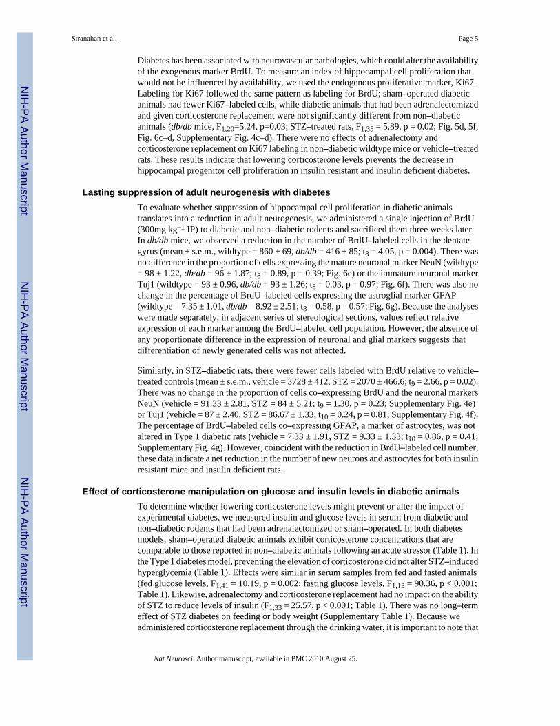
Diabetes has been associated with neurovascular pathologies, which could alter the availabilityof the exogenous marker BrdU. To measure an index of hippocampal cell proliferation thatwould not be influenced by availability, we used the endogenous proliferative marker, Ki67.Labeling for Ki67 followed the same pattern as labeling for BrdU; sham–operated diabeticanimals had fewer Ki67–labeled cells, while diabetic animals that had been adrenalectomizedand given corticosterone replacement were not significantly different from non–diabeticanimals (db/db mice, F1,20=5.24, p=0.03; STZ–treated rats, F1,35 = 5.89, p = 0.02; Fig. 5d, 5f,Fig. 6c–d, Supplementary Fig. 4c–d). There were no effects of adrenalectomy andcorticosterone replacement on Ki67 labeling in non–diabetic wildtype mice or vehicle–treatedrats. These results indicate that lowering corticosterone levels prevents the decrease inhippocampal progenitor cell proliferation in insulin resistant and insulin deficient diabetes.
Lasting suppression of adult neurogenesis with diabetesTo evaluate whether suppression of hippocampal cell proliferation in diabetic animalstranslates into a reduction in adult neurogenesis, we administered a single injection of BrdU(300mg kg–1 IP) to diabetic and non–diabetic rodents and sacrificed them three weeks later.In db/db mice, we observed a reduction in the number of BrdU–labeled cells in the dentategyrus (mean ± s.e.m., wildtype = 860 ± 69, db/db = 416 ± 85; t8 = 4.05, p = 0.004). There wasno difference in the proportion of cells expressing the mature neuronal marker NeuN (wildtype= 98 ± 1.22, db/db = 96 ± 1.87; t8 = 0.89, p = 0.39; Fig. 6e) or the immature neuronal markerTuj1 (wildtype = 93 ± 0.96, db/db = 93 ± 1.26; t8 = 0.03, p = 0.97; Fig. 6f). There was also nochange in the percentage of BrdU–labeled cells expressing the astroglial marker GFAP(wildtype = 7.35 ± 1.01, db/db = 8.92 ± 2.51; t8 = 0.58, p = 0.57; Fig. 6g). Because the analyseswere made separately, in adjacent series of stereological sections, values reflect relativeexpression of each marker among the BrdU–labeled cell population. However, the absence ofany proportionate difference in the expression of neuronal and glial markers suggests thatdifferentiation of newly generated cells was not affected.
Similarly, in STZ–diabetic rats, there were fewer cells labeled with BrdU relative to vehicle–treated controls (mean ± s.e.m., vehicle = 3728 ± 412, STZ = 2070 ± 466.6; t9 = 2.66, p = 0.02).There was no change in the proportion of cells co–expressing BrdU and the neuronal markersNeuN (vehicle = 91.33 ± 2.81, STZ = 84 ± 5.21; t9 = 1.30, p = 0.23; Supplementary Fig. 4e)or Tuj1 (vehicle = 87 ± 2.40, STZ = 86.67 ± 1.33; t10 = 0.24, p = 0.81; Supplementary Fig. 4f).The percentage of BrdU–labeled cells co–expressing GFAP, a marker of astrocytes, was notaltered in Type 1 diabetic rats (vehicle = 7.33 ± 1.91, STZ = 9.33 ± 1.33; t10 = 0.86, p = 0.41;Supplementary Fig. 4g). However, coincident with the reduction in BrdU–labeled cell number,these data indicate a net reduction in the number of new neurons and astrocytes for both insulinresistant mice and insulin deficient rats.
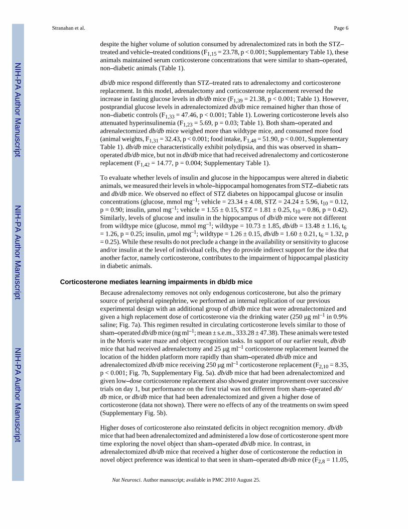
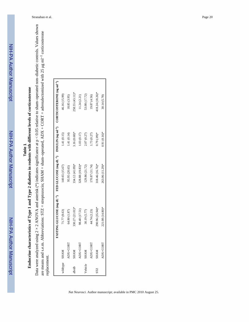
Effect of corticosterone manipulation on glucose and insulin levels in diabetic animalsTo determine whether lowering corticosterone levels might prevent or alter the impact ofexperimental diabetes, we measured insulin and glucose levels in serum from diabetic andnon–diabetic rodents that had been adrenalectomized or sham–operated. In both diabetesmodels, sham–operated diabetic animals exhibit corticosterone concentrations that arecomparable to those reported in non–diabetic animals following an acute stressor (Table 1). Inthe Type 1 diabetes model, preventing the elevation of corticosterone did not alter STZ–inducedhyperglycemia (Table 1). Effects were similar in serum samples from fed and fasted animals(fed glucose levels, F1,41 = 10.19, p = 0.002; fasting glucose levels, F1,13 = 90.36, p < 0.001;Table 1). Likewise, adrenalectomy and corticosterone replacement had no impact on the abilityof STZ to reduce levels of insulin (F1,33 = 25.57, p < 0.001; Table 1). There was no long–termeffect of STZ diabetes on feeding or body weight (Supplementary Table 1). Because weadministered corticosterone replacement through the drinking water, it is important to note that
Stranahan et al. Page 5
Nat Neurosci. Author manuscript; available in PMC 2010 August 25.
NIH
-PA Author Manuscript
NIH
-PA Author Manuscript
NIH
-PA Author Manuscript
despite the higher volume of solution consumed by adrenalectomized rats in both the STZ–treated and vehicle–treated conditions (F1,15 = 23.78, p < 0.001; Supplementary Table 1), theseanimals maintained serum corticosterone concentrations that were similar to sham–operated,non–diabetic animals (Table 1).
db/db mice respond differently than STZ–treated rats to adrenalectomy and corticosteronereplacement. In this model, adrenalectomy and corticosterone replacement reversed theincrease in fasting glucose levels in db/db mice (F1,39 = 21.38, p < 0.001; Table 1). However,postprandial glucose levels in adrenalectomized db/db mice remained higher than those ofnon–diabetic controls (F1,33 = 47.46, p < 0.001; Table 1). Lowering corticosterone levels alsoattenuated hyperinsulinemia (F1,23 = 5.69, p = 0.03; Table 1). Both sham–operated andadrenalectomized db/db mice weighed more than wildtype mice, and consumed more food(animal weights, F1,31 = 32.43, p < 0.001; food intake, F1,48 = 51.90, p < 0.001, SupplementaryTable 1). db/db mice characteristically exhibit polydipsia, and this was observed in sham–operated db/db mice, but not in db/db mice that had received adrenalectomy and corticosteronereplacement (F1,42 = 14.77, p = 0.004; Supplementary Table 1).
To evaluate whether levels of insulin and glucose in the hippocampus were altered in diabeticanimals, we measured their levels in whole–hippocampal homogenates from STZ–diabetic ratsand db/db mice. We observed no effect of STZ diabetes on hippocampal glucose or insulinconcentrations (glucose, mmol mg–1; vehicle = 23.34 ± 4.08, STZ = 24.24 ± 5.96, t10 = 0.12,p = 0.90; insulin, μmol mg–1; vehicle = 1.55 ± 0.15, STZ = 1.81 ± 0.25, t10 = 0.86, p = 0.42).Similarly, levels of glucose and insulin in the hippocampus of db/db mice were not differentfrom wildtype mice (glucose, mmol mg–1; wildtype = 10.73 ± 1.85, db/db = 13.48 ± 1.16, t6= 1.26, p = 0.25; insulin, μmol mg–1; wildtype = 1.26 ± 0.15, db/db = 1.60 ± 0.21, t6 = 1.32, p= 0.25). While these results do not preclude a change in the availability or sensitivity to glucoseand/or insulin at the level of individual cells, they do provide indirect support for the idea thatanother factor, namely corticosterone, contributes to the impairment of hippocampal plasticityin diabetic animals.
Corticosterone mediates learning impairments in db/db miceBecause adrenalectomy removes not only endogenous corticosterone, but also the primarysource of peripheral epinephrine, we performed an internal replication of our previousexperimental design with an additional group of db/db mice that were adrenalectomized andgiven a high replacement dose of corticosterone via the drinking water (250 μg ml–1 in 0.9%saline; Fig. 7a). This regimen resulted in circulating corticosterone levels similar to those ofsham–operated db/db mice (ng ml–1; mean ± s.e.m., 333.28 ± 47.38). These animals were testedin the Morris water maze and object recognition tasks. In support of our earlier result, db/dbmice that had received adrenalectomy and 25 μg ml–1 corticosterone replacement learned thelocation of the hidden platform more rapidly than sham–operated db/db mice andadrenalectomized db/db mice receiving 250 μg ml–1 corticosterone replacement (F2,10 = 8.35,p < 0.001; Fig. 7b, Supplementary Fig. 5a). db/db mice that had been adrenalectomized andgiven low–dose corticosterone replacement also showed greater improvement over successivetrials on day 1, but performance on the first trial was not different from sham–operated db/db mice, or db/db mice that had been adrenalectomized and given a higher dose ofcorticosterone (data not shown). There were no effects of any of the treatments on swim speed(Supplementary Fig. 5b).
Higher doses of corticosterone also reinstated deficits in object recognition memory. db/dbmice that had been adrenalectomized and administered a low dose of corticosterone spent moretime exploring the novel object than sham–operated db/db mice. In contrast, inadrenalectomized db/db mice that received a higher dose of corticosterone the reduction innovel object preference was identical to that seen in sham–operated db/db mice (F2,8 = 11.05,
Stranahan et al. Page 6
Nat Neurosci. Author manuscript; available in PMC 2010 August 25.
NIH
-PA Author Manuscript
NIH
-PA Author Manuscript
NIH
-PA Author Manuscript
p = 0.03; Fig. 7c). Again, db/db mice that had been adrenalectomized and administered a lowdose of corticosterone showed a preference for the novel object that was similar to non–diabeticanimals.
Administration of a high dose of corticosterone had complex effects on the endocrineparameters of adrenalectomized db/db mice. These animals exhibit hyperglycemia, at a levelsimilar to sham–operated db/db mice (mg dL–1; mean ± s.e.m., fasting = 339.30 ± 34.10; fed= 450.59 ± 46.04). Serum insulin levels were also elevated (ng ml–1; mean ± s.e.m., 1.98 ±0.57). db/db mice receiving a high dose of corticosterone show increased water intake, similarto sham–operated db/db mice (ml d–1, mean ± s.e.m., 52.73 ± 7.24). However, their food intakeand body weights were similar to those of wildtype animals (food intake, g d–1; 5.97 ± 0.65;body weight, g; 41.07 ± 4.34). These results suggest that elevated corticosterone levelscontribute centrally to learning deficits, and peripherally to the endocrine characteristics ofdiabetes.
DiscussionDiabetes is associated with multiple adverse effects on the brain, some of which may resultprimarily from direct consequences of chronic hyperglycemia. However, our findingsdemonstrate a pivotal role for the adrenal steroid corticosterone as a mediator of diabetes–induced impairments in hippocampal synaptic plasticity and neurogenesis, and associatedcognitive deficits. Lowering corticosterone levels prevented the diabetes–induced impairmentof learning and memory in insulin deficient rats and insulin resistant mice. Maintaining normalphysiological levels of corticosterone also restored LTP at perforant path–dentate gyrussynapses and prevented the impairment of adult neurogenesis in the dentate gyrus. Therestorative effect of lowering corticosterone levels was observed when recordings were madeunder conditions that either permit or exclude the contribution of newly generated neurons.Enhancement of hippocampal function by normalizing corticosterone levels in diabetic animalswas completely reversed by administration of high levels of corticosterone, demonstrating thatcorticosterone (rather than some other adrenal–derived factor) was responsible for the adverseeffects of diabetes on hippocampal plasticity. These findings strongly support a role forelevated corticosterone levels in impaired hippocampal plasticity and cognition induced bydiabetes.
It is well–established that chronic exposure to high levels of corticosterone is detrimental forlearning and synaptic plasticity in non–diabetic animals24-29. The corticosterone–mediatedadverse effects of diabetes were not determined by changes in insulin production, because theyoccurred in db/db mice with elevated insulin levels, and in insulin–deficient rats. We alsoobserved no change in hippocampal insulin levels under baseline conditions in diabeticanimals. However, this does not rule out the possibility that insulin signaling pathways mightbe impaired in diabetes. The effects of insulin on learning and memory oppose those ofglucocorticoids at multiple levels. Specifically, intrahippocampal insulin37 or activation ofinsulin signaling pathways38 can block the effects of stress on learning and memory. Exposureto elevated corticosterone levels reduces insulin receptor signaling in multiple somatic tissues,including the brain39. Therefore, it is possible that the negative effect of diabetes onhippocampal plasticity may be attributable to an interaction between elevated glucocorticoidsand insulin receptor signaling.
Local cerebral glucose utilization is tightly linked with neural activity and cognition. Incontrast, glucocorticoids inhibit glucose utilization in neurons40. In normal (i.e. non–diabetic)animals, hippocampus–dependent learning is correlated with a decrease in extracellularglucose levels, and intrahippocampal injection of glucose improves performance41. No studiesto date have reported an effect of diabetes on learning–induced changes in hippocampal glucose
Stranahan et al. Page 7
Nat Neurosci. Author manuscript; available in PMC 2010 August 25.
NIH
-PA Author Manuscript
NIH
-PA Author Manuscript
NIH
-PA Author Manuscript

metabolism, but alterations in basal hippocampal glucose transporter expression have beendemonstrated in diabetic animals42. While we observed no difference in glucose levels in wholehippocampal homogenates from insulin resistant mice or insulin deficient rats, our results donot preclude a role for corticosterone in modulating the diabetes–induced alterations inhippocampal glucose metabolism.
Lowering corticosterone levels in diabetes can restore behavioral function on tasks that recruitboth new and mature neurons. While the Morris water maze task is not influenced by antimitotictreatment35, 43, newly generated neurons are activated following this task at a higher rate,relative to mature neurons44. Similar distinctions have been reported with respect to the roleof adult–generated neurons in recognition memory; enhancement of performance on the novelobject preference task following environmental enrichment was reversed by systemic treatmentwith an antimitotic45, but spontaneous alternation in the Y–maze, which also involvesrecognition memory, was unaffected following focal cranial irradiation35. Although it remainsto be determined whether adult–generated granule neurons make a meaningful contribution toperformance on these tasks under baseline conditions, the therapeutically relevant question iswhether new neurons can enhance performance following neurodegeneration or injury.
The fact that lowering corticosterone levels had no effect on postprandial glucose levels inserum from db/db mice is in line with previous studies. Adrenalectomy and corticosteronereplacement failed to normalize fed glucose values in the ob/ob mouse18. Similar results wereobserved following treatment with the glucocorticoid receptor antagonist RU486 in the Zucker(fa/fa) rat, with no effect of antiglucocorticoid treatment on fed glucose levels19. In contrast,treatment with antisense oligonucleotides directed against the glucocorticoid receptor restorednormal fasting glucose levels in Zucker diabetic rats20. Taken together, these results suggestthat inhibiting the actions of corticosterone by various methods will influence fasting, but notfed glucose levels in animal models of type 2 diabetes.
Studies of human subjects have provided evidence that diabetes adversely affects learning andmemory, but also suggest that not all cognitive domains are equally affected. Diabetic humansexhibit accelerated decline on tasks that require episodic memory and rapid informationprocessing, while attention and language abilities are unaffected2. Because episodic memoryplaces a greater demand on temporal lobe structures, and language and attention primarilyrecruit other cortical and prefrontal regions, these data have been interpreted to suggest thatthe hippocampus is particularly susceptible to the negative consequences of diabetes. Otherstudies have begun to explore the role of cortisol in diabetes–induced cognitive deficits inhumans. For example, inhibition of the enzyme 11–β–hydroxysteroid dehydrogenase 1(11βHSD1), which locally modulates the actions of glucocorticoids in the brain by reactivatingcortisol from its inactive form, was shown to ameliorate cognitive deficits in humans with Type2 diabetes46. Overall, the task–specific cognitive impairments induced by diabetes, and thedemonstration of improved cognitive performance in diabetic humans following treatmentsthat alter the availability of cortisol, suggests that elevated cortisol levels in human diabeticsmay also be contributing to deficits in hippocampal function.
Supplementary MaterialRefer to Web version on PubMed Central for supplementary material.
AcknowledgmentsThis research was supported by NIH NRSA Predoctoral fellowship F31AG024690–03 to A.M.S. through PrincetonUniversity, and by the Intramural Research Program of the National Institute on Aging. We thank Dan L. Longo forhelpful suggestions, and Tamikia Lamb, Olga Carlson, Julissa S. Villareal, and Richard Telljohann for technicalassistance. We are also grateful to Elizabeth Gould and Henriette van Praag for comments on the manuscript.
Stranahan et al. Page 8
Nat Neurosci. Author manuscript; available in PMC 2010 August 25.
NIH
-PA Author Manuscript
NIH
-PA Author Manuscript
NIH
-PA Author Manuscript
This work was supported by a NRSA predoctoral fellowship to A.S. and by the National Institute on Aging IntramuralResearch Program.
Appendix
Materials and MethodsAnimals and Surgery
Animal care and experimental procedures followed NIH guidelines and were approved by theNational Institute on Aging Animal Care and Use Committee. Adult male Sprague–Dawleyrats were purchased from Charles River Laboratories and housed individually for a minimumof 2 weeks before the start of experiments. Streptozocin was administered via the femoral veinat a dose of 70 mg kg–1 as described16. In order to be included in the study, STZ–treated ratswere required to exhibit serum glucose levels ≥ 200 mg dL–1. Male leptin receptor mutant(db/db) mice, bred on a C57BL/6 background, were purchased from Jackson Laboratories.Age–matched male C57BL/6 mice were used as controls. To determine whetherglucocorticoids are involved in the neurological consequences of diabetes, rats and mice weresubjected to bilateral adrenalectomy or sham operation. Adrenalectomized animals receivedcorticosterone replacement (25 μg ml–1 or 250 μg ml–1 in 0.9% saline; Sigma) in the drinkingwater to manipulate glucocorticoid levels30. Corticosterone replacement was available to theanimals immediately following surgery. Mice were adrenalectomized at postnatal day 30; ratswere adrenalectomized at postnatal day 90. All rats and mice were administered a singleinjection of the DNA synthetic marker bromodeoxyuridine (300 mg kg–1; n=6–8 animals pergroup). This dose was based on previous studies47 demonstrating maximal labeling at 300 mgkg–1. Animals were euthanized 2 hours or 3 weeks post–BrdU. In a separate experiment,two–month–old wildtype mice were implanted with Alzet minipumps to deliver the antimitoticdrug AraC into the right lateral ventricle (2.2 mg ml–1, 0.25 μl hour–1, pump model 1002;bregma coordinates AP –0.3 mm, ML –1.0mm). These animals were injected once with BrdU(300 mg kg–1; n=6–8 animals per group) and euthanized 24 hours later. All animals had adlibitum access to food and water, and the room was maintained on a 12 hour light–dark schedule(lights on at 06:00). For some experiments, the animals were weighed once weekly, and theirfood and water were weighed on two successive days per week for 4 weeks. Food consumptionwas measured in grams per day, and water bottle weights were converted to volumes. Thetechniques for quantifying levels of glucose, insulin, and corticosterone are described inSupplementary Methods.
Electrophysiology and Behavioral testingThe procedures used for slice preparation and recording are available in SupplementaryMethods. Procedures for water maze training and novel object preference testing are alsoincluded in Supplementary Methods.
Immunohistochemistry and MicroscopyImmunolabeling for BrdU and Ki67 was carried out as described15. Full description of themethods for brightfield and fluorescence tissue labeling are available in SupplementaryMethods. We quantified single- and double-labeled cells using standard protocols15. Detailedcell counting criteria are available in Supplementary Methods. We also quantified the opticalintensity of fluorescence staining for synaptophysin48; full description available inSupplementary Methods.
StatisticsStatistical analyses were made using SPSS version 11.0 (Chicago, Illinois, USA), withsignificance set at (p < 0.05); graphs were generated using Graphpad Prism 4 software (San
Stranahan et al. Page 9
Nat Neurosci. Author manuscript; available in PMC 2010 August 25.
NIH
-PA Author Manuscript
NIH
-PA Author Manuscript
NIH
-PA Author Manuscript
Diego, CA). Cell counts, hormone profiles, feeding, drinking, animal weights, and the amountof LTP were compared using separate 2 × 2 ANOVA designs (diabetes × surgery). Behavioraldata from the Morris water maze and novel object preference task were analyzed using 2 × 2repeated measures ANOVA. The number of BrdU–labeled cells at 3 weeks post–injection wascompared across diabetic and non–diabetic animals using bidirectional, unpaired t–tests.Percentages of cells co–expressing BrdU and a cell type–specific marker were also analyzedusing t–tests. In experiments where we administered a high dose of corticosterone toadrenalectomized db/db mice, behavioral data from the Morris water maze and novel objectpreference tasks were analyzed using one–way repeated measures ANOVA with Tukey's posthoc.
References1. Reaven GM. The insulin resistance syndrome: definition and dietary approaches to treatment. Annu.
Rev. Nutr 2005;25:391–406. [PubMed: 16011472]2. Messier C. Impact of impaired glucose tolerance and type 2 diabetes on cognitive aging. Neurobol
Aging 2005;26(Suppl 1):S26–S30.3. Greenwood CE, Winocur G. High–fat diets, insulin resistance and declining cognitive function.
Neurobiol. Aging 2005;26(Suppl 1):42–45. [PubMed: 16257476]4. Desrocher M, Rovet J. Neurocognitive correlates of type 1 diabetes mellitus in childhood. Child.
Neuropsychol 2004;10:36–52. [PubMed: 14977514]5. Biessels GJ, et al. Place learning and hippocampal synaptic plasticity in streptozotocin–induced
diabetic rats. Diabetes 1996;45:1259–1266. [PubMed: 8772732]6. Biessels GJ, et al. Water maze learning and hippocampal synaptic plasticity in streptozotocin–diabetic
rats: effects of insulin treatment. Brain Res 1998;800:125–135. [PubMed: 9685609]7. Li XL, et al. Impairment of long–term potentiation and spatial memory in leptin receptor–deficient
rodents. Neuroscience 2002;113:607–615. [PubMed: 12150780]8. Hummel KP, Dickie MM, Coleman DL. Diabetes, a new mutation in the mouse. Science
1996;153:1127–1128. [PubMed: 5918576]9. Leuner B, Gould E, Shors TJ. Is there a link between adult neurogenesis and learning? Hippocampus
2006;16:216–224. [PubMed: 16421862]10. Kamal A, Biessels GJ, Urban IJ, Gispen WH. Hippocampal synaptic plasticity in streptozotocin–
diabetic rats: impairment of long–term potentiation and facilitation of long–term depression.Neuroscience 1999;90:737–745. [PubMed: 10218775]
11. Zhang WJ, Tan YF, Yue JT, Vranic M, Wojtowicz JM. Impairment of hippocampal neurogenesis instreptozotocin–treated diabetic rats. Acta Neurol Scand. 2007 [Epub ahead of print].
12. van Praag H, Christie BR, Sejnowski TJ, Gage FH. Running enhances neurogenesis, learning, andlong-term potentiation in mice. Proc. Natl. Acad. Sci. U. S. A 1999;96:13427–13431. [PubMed:10557337]
13. Fontan-Lozano A, et al. Caloric restriction increases learning consolidation and facilitates synapticplasticity through mechanisms dependent on NR2B subunits of the NMDA receptor. J. Neurosci2007;27:10185–10195. [PubMed: 17881524]
14. Lee J, Duan W, Mattson MP. Evidence that brain–derived neurotrophic factor is required for basalneurogenesis and mediates, in part, the enhancement of neurogenesis by dietary restriction in thehippocampus of adult mice. J. Neurochem 2002;82:1367–1375. [PubMed: 12354284]
15. Stranahan AM, Khalil D, Gould E. Social isolation delays the positive effects of running on adultneurogenesis. Nat. Neurosci 2006;9:526–533. [PubMed: 16531997]
16. Magarinos AM, McEwen BS. Experimental diabetes in rats causes hippocampal dendritic andsynaptic reorganization and increased glucocorticoid reactivity to stress. Proc. Natl. Acad. Sci. U. S.A 2000;97:11056–11061. [PubMed: 11005876]
17. Chan O, et al. Hyperglycemia does not increase basal hypothalamo–pituitary–adrenal activity indiabetes but it does impair the HPA response to insulin–induced hypoglycemia. Am J Physiol RegulIntegr Comp Physiol 2005;289:R235–246. [PubMed: 15774766]
Stranahan et al. Page 10
Nat Neurosci. Author manuscript; available in PMC 2010 August 25.
NIH
-PA Author Manuscript
NIH
-PA Author Manuscript
NIH
-PA Author Manuscript
18. Tokuyama K, Himms–Hagen J. Increased sensitivity of the genetically obese mouse to corticosterone.Am J Physiol 1987;252:202–208.
19. Langley SC, York DA. Effects of antiglucocorticoid RU486 on development of obesity in obese fa/fa Zucker rats. Am J Physiol 1990;259:539–544.
20. Watts LM, et al. Reduction of hepatic and adipose tissue glucocorticoid receptor expression withantisense oligonucleotides improves hyperglycemia and hyperlipidemia in diabetic rodents withoutcausing systemic glucocorticoid antagonism. Diabetes 2005;54:1846–1853. [PubMed: 15919808]
21. Oei NY, Everaerd WT, Elzinga BM, van Well S, Bermond B. Psychosocial stress impairs workingmemory at high loads: an association with cortisol levels and memory retrieval. Stress 2006;9:133–141. [PubMed: 17035163]
22. MacLullich AM, et al. Plasma cortisol levels, brain volumes and cognition in healthy elderly men.Psychoneuroendocrinology 2005;30:505–515. [PubMed: 15721061]
23. Elgh E, et al. Cognitive dysfunction, hippocampal atrophy and glucocorticoid feedback in Alzheimer'sdisease. Biol. Psychiatry 2006;59:155–161. [PubMed: 16125145]
24. Oitzl MS, Fluttert M, Sutanto W, de Kloet ER. Continuous blockade of brain glucocorticoid receptorsfacilitates spatial learning and memory in rats. Eur. J. Neurosci 1998;10:3759–3766. [PubMed:9875354]
25. Wright RL, Lightner EN, Harman JS, Meijer OC, Conrad CD. Attenuating corticosterone levels onthe day of memory assessment prevents chronic stress–induced impairments in spatial memory. Eur.J. Neurosci 2006;24:595–605. [PubMed: 16903861]
26. Alfarez DN, Joels M, Krugers HJ. Chronic unpredictable stress impairs long–term potentiation in rathippocampal CA1 area and dentate gyrus in vitro. Eur. J. Neurosci 2003;17:1928–1934. [PubMed:12752792]
27. Kerr DS, Campbell LW, Hao SY, Landfield PW. Corticosteroid modulation of hippocampalpotentials: increased effect with aging. Science 1989;245:1505–1509. [PubMed: 2781293]
28. Korz V, Frey JU. Stress–related modulation of hippocampal long–term potentiation in rats:Involvement of adrenal steroid receptors. J. Neurosci 2003;23:7281–7287. [PubMed: 12917361]
29. Pavlides C, Watanabe Y, McEwen BS. Effects of glucocorticoids on hippocampal long–termpotentiation. Hippocampus 1993;3:183–192. [PubMed: 8353605]
30. Gould E, Cameron HA, Daniels DC, Woolley CS, McEwen BS. Adrenal hormones suppress celldivision in the adult rat dentate gyrus. J. Neurosci 1992;12:3642–3650. [PubMed: 1527603]
31. Montaron MF, et al. Lifelong corticosterone level determines age–related decline in neurogenesisand memory. Neurobiol. Aging 2006;27:645–654. [PubMed: 15953661]
32. Tanapat P, Hastings NB, Rydel TA, Galea LA, Gould E. Exposure to fox odor inhibits cellproliferation in the hippocampus of adult rats via an adrenal hormone-dependent mechanism. J CompNeurol 2001;437:496–504. [PubMed: 11503148]
33. Karten YJ, Jones MA, Jeurling SI, Cameron HA. GABAergic signaling in young granule cells in theadult rat and mouse dentate gyrus. Hippocampus 2006;16:312–320. [PubMed: 16435314]
34. Ge S, et al. GABA regulates synaptic integration of newly generated neurons in the adult brain. Nature2006;439:589–593. [PubMed: 16341203]
35. Saxe MD, et al. Ablation of hippocampal neurogenesis impairs contextual fear conditioning andsynaptic plasticity in the dentate gyrus. Proc. Natl. Acad. Sci. U. S. A 2006;103:17501–17506.[PubMed: 17088541]
36. Snyder JS, Kee N, Wojtowicz JM. Effects of adult neurogenesis on synaptic plasticity in the rat dentategyrus. J. Neurophysiol 2001;85:2423–2431. [PubMed: 11387388]
37. Moosavi M, Naghdi N, Maghsoudi N, Zahedi Asl S. Insulin protects against stress–inducedimpairments in water maze performance. Behav. Brain Res 2007;176:230–236. [PubMed: 17116337]
38. Revest JM, et al. The MAPK pathway and Egr–1 mediate stress–related behavioral effects ofglucocorticoids. Nat. Neurosci 2005;8:664–672. [PubMed: 15834420]
39. Piroli GG, et al. Corticosterone impairs insulin–stimulated translocation of GLUT4 in the rathippocampus. Neuroendocrinology 2007;85:71–80. [PubMed: 17426391]
40. Sapolsky RM. Glucocorticoid toxicity in the hippocampus: reversal by supplementation with brainfuels. J. Neurosci 1986;6:2240–2244. [PubMed: 3746406]
Stranahan et al. Page 11
Nat Neurosci. Author manuscript; available in PMC 2010 August 25.
NIH
-PA Author Manuscript
NIH
-PA Author Manuscript
NIH
-PA Author Manuscript
41. McNay EC, Fries TM, Gold PE. Decreases in rat extracellular hippocampal glucose concentrationassociated with cognitive demand during a spatial task. Proc. Natl. Acad. Sci. U. S. A 2000;97:2881–2885. [PubMed: 10706633]
42. Reagan LP, et al. Localization and regulation of GLUTx1 glucose transporter in the hippocampus ofstreptozotocin diabetic rats. Proc. Natl. Acad. Sci. U. S. A 2001;98:2820–2825. [PubMed: 11226324]
43. Shors TJ, Townsend DA, Zhao M, Kozorovitskiy Y, Gould E. Neurogenesis may relate to some butnot all types of hippocampal–dependent learning. Hippocampus 2002;12:578–584. [PubMed:12440573]
44. Kee N, Teixeira CM, Wang AH, Frankland PW. Preferential incorporation of adult–generated granulecells into spatial memory networks in the dentate gyrus. Nat. Neurosci 2007;10:355–362. [PubMed:17277773]
45. Bruel–Jungerman E, Laroche S, Rampon C. New neurons in the dentate gyrus are involved in theexpression of enhanced long–term memory following environmental enrichment. Eur. J. Neurosci2005;21:513–521. [PubMed: 15673450]
46. Sandeep, et al. 11Beta–hydroxysteroid dehydrogenase inhibition improves cognitive function inhealthy elderly men and type 2 diabetics. Proc Natl Acad Sci U S A 2005;101:6734–9. [PubMed:15071189]
47. Cameron HA, McKay RD. Adult neurogenesis produces a large pool of new granule cells in thedentate gyrus. J. Comp. Neurol 2001;435:406–417. [PubMed: 11406822]
48. Kozorovitskiy Y, et al. Experience induces structural and biochemical changes in the adult primatebrain. Proc. Natl. Acad. Sci. U. S. A 2005;102:17478–17482. [PubMed: 16299105]
Stranahan et al. Page 12
Nat Neurosci. Author manuscript; available in PMC 2010 August 25.
NIH
-PA Author Manuscript
NIH
-PA Author Manuscript
NIH
-PA Author Manuscript
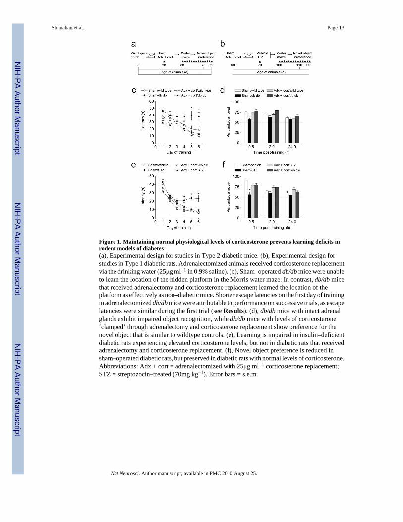
Figure 1. Maintaining normal physiological levels of corticosterone prevents learning deficits inrodent models of diabetes(a), Experimental design for studies in Type 2 diabetic mice. (b), Experimental design forstudies in Type 1 diabetic rats. Adrenalectomized animals received corticosterone replacementvia the drinking water (25μg ml–1 in 0.9% saline). (c), Sham–operated db/db mice were unableto learn the location of the hidden platform in the Morris water maze. In contrast, db/db micethat received adrenalectomy and corticosterone replacement learned the location of theplatform as effectively as non–diabetic mice. Shorter escape latencies on the first day of trainingin adrenalectomized db/db mice were attributable to performance on successive trials, as escapelatencies were similar during the first trial (see Results). (d), db/db mice with intact adrenalglands exhibit impaired object recognition, while db/db mice with levels of corticosterone‘clamped’ through adrenalectomy and corticosterone replacement show preference for thenovel object that is similar to wildtype controls. (e), Learning is impaired in insulin–deficientdiabetic rats experiencing elevated corticosterone levels, but not in diabetic rats that receivedadrenalectomy and corticosterone replacement. (f), Novel object preference is reduced insham–operated diabetic rats, but preserved in diabetic rats with normal levels of corticosterone.Abbreviations: Adx + cort = adrenalectomized with 25μg ml–1 corticosterone replacement;STZ = streptozocin–treated (70mg kg–1). Error bars = s.e.m.
Stranahan et al. Page 13
Nat Neurosci. Author manuscript; available in PMC 2010 August 25.
NIH
-PA Author Manuscript
NIH
-PA Author Manuscript
NIH
-PA Author Manuscript
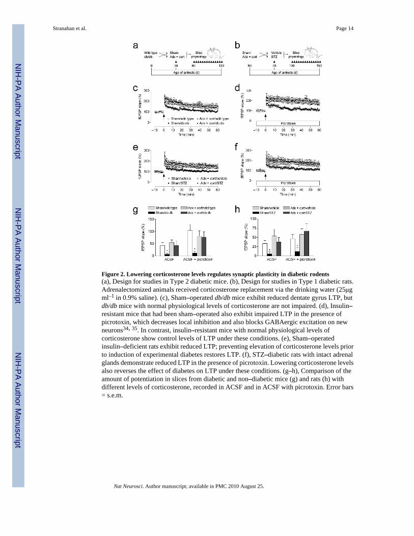
Figure 2. Lowering corticosterone levels regulates synaptic plasticity in diabetic rodents(a), Design for studies in Type 2 diabetic mice. (b), Design for studies in Type 1 diabetic rats.Adrenalectomized animals received corticosterone replacement via the drinking water (25μgml–1 in 0.9% saline). (c), Sham–operated db/db mice exhibit reduced dentate gyrus LTP, butdb/db mice with normal physiological levels of corticosterone are not impaired. (d), Insulin–resistant mice that had been sham–operated also exhibit impaired LTP in the presence ofpicrotoxin, which decreases local inhibition and also blocks GABAergic excitation on newneurons34, 35. In contrast, insulin–resistant mice with normal physiological levels ofcorticosterone show control levels of LTP under these conditions. (e), Sham–operatedinsulin–deficient rats exhibit reduced LTP; preventing elevation of corticosterone levels priorto induction of experimental diabetes restores LTP. (f), STZ–diabetic rats with intact adrenalglands demonstrate reduced LTP in the presence of picrotoxin. Lowering corticosterone levelsalso reverses the effect of diabetes on LTP under these conditions. (g–h), Comparison of theamount of potentiation in slices from diabetic and non–diabetic mice (g) and rats (h) withdifferent levels of corticosterone, recorded in ACSF and in ACSF with picrotoxin. Error bars= s.e.m.
Stranahan et al. Page 14
Nat Neurosci. Author manuscript; available in PMC 2010 August 25.
NIH
-PA Author Manuscript
NIH
-PA Author Manuscript
NIH
-PA Author Manuscript

Figure 3. AraC treatment reduces cell proliferation, without altering synaptic markerimmunoreactivity(a–b), Following a single injection of 300mg kg–1 BrdU with a 24 hour survival period, weobserved a significant reduction in the number of labeled cells in wildtype mice that had beeninfused with the antimitotic drug AraC for ten to fourteen days. The micrograph in (a) showsthe dentate gyrus of a mouse infused with vehicle, and the micrograph in (b) shows the dentategyrus of a mouse infused with AraC. Arrows indicate labeled cells. (c), We also quantifiedsynaptic marker expression in the inner third of the dentate molecular layer, where the medialperforant path synapses are located. This confocal micrograph shows the anatomical region(outlined) where scans were taken for analysis of synaptophysin labeling, and where electrodeswere positioned for electrophysiological recordings in slices. (d), Micrograph taken at theresolution and scale used for analysis of synaptophysin labeling (see Supplementary Methods).We observed no differences in the area or intensity of staining for the synaptic markersynaptophysin, suggesting that AraC treatment does not result in loss of synapses among thelarger population of dentate granule neurons.
Stranahan et al. Page 15
Nat Neurosci. Author manuscript; available in PMC 2010 August 25.
NIH
-PA Author Manuscript
NIH
-PA Author Manuscript
NIH
-PA Author Manuscript

Figure 4. Antimitotic treatment selectively impairs dentate gyrus LTP recorded in the absence ofpicrotoxin(a), Experimental design for studies using minipump delivery of antimitotic drugs. (b),Comparison of the amount of LTP in vehicle– and AraC–infused mice, when recordings weremade in the presence or absence of the GABAA antagonist picrotoxin (100μM). Asterisk (*)indicates significance at p < 0.05 following 2 × 2 ANOVA. (c), LTP at medial perforant pathsynapses in the dentate gyrus is impaired in AraC–infused mice. (d), LTP recordings in thepresence of picrotoxin were not influenced by AraC infusion. (e), The relationship betweenthe slope of the dendritic field potential and the amplitude of the axonal fiber volley isinfluenced by picrotoxin, but not by AraC treatment. (f), Presynaptic paired–pulse depression,measured in plain ACSF, was not altered by treatment with AraC. Error bars = s.e.m.
Stranahan et al. Page 16
Nat Neurosci. Author manuscript; available in PMC 2010 August 25.
NIH
-PA Author Manuscript
NIH
-PA Author Manuscript
NIH
-PA Author Manuscript
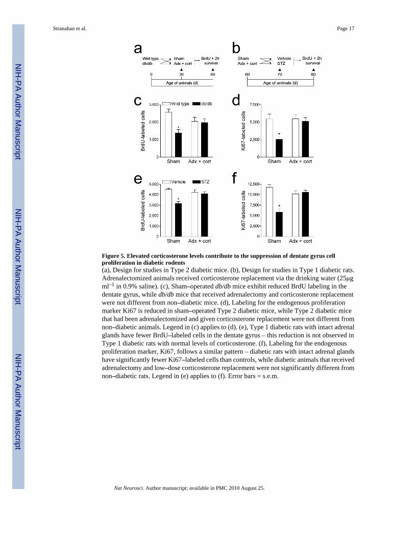
Figure 5. Elevated corticosterone levels contribute to the suppression of dentate gyrus cellproliferation in diabetic rodents(a), Design for studies in Type 2 diabetic mice. (b), Design for studies in Type 1 diabetic rats.Adrenalectomized animals received corticosterone replacement via the drinking water (25μgml–1 in 0.9% saline). (c), Sham–operated db/db mice exhibit reduced BrdU labeling in thedentate gyrus, while db/db mice that received adrenalectomy and corticosterone replacementwere not different from non–diabetic mice. (d), Labeling for the endogenous proliferationmarker Ki67 is reduced in sham–operated Type 2 diabetic mice, while Type 2 diabetic micethat had been adrenalectomized and given corticosterone replacement were not different fromnon–diabetic animals. Legend in (c) applies to (d). (e), Type 1 diabetic rats with intact adrenalglands have fewer BrdU–labeled cells in the dentate gyrus – this reduction is not observed inType 1 diabetic rats with normal levels of corticosterone. (f), Labeling for the endogenousproliferation marker, Ki67, follows a similar pattern – diabetic rats with intact adrenal glandshave significantly fewer Ki67–labeled cells than controls, while diabetic animals that receivedadrenalectomy and low–dose corticosterone replacement were not significantly different fromnon–diabetic rats. Legend in (e) applies to (f). Error bars = s.e.m.
Stranahan et al. Page 17
Nat Neurosci. Author manuscript; available in PMC 2010 August 25.
NIH
-PA Author Manuscript
NIH
-PA Author Manuscript
NIH
-PA Author Manuscript
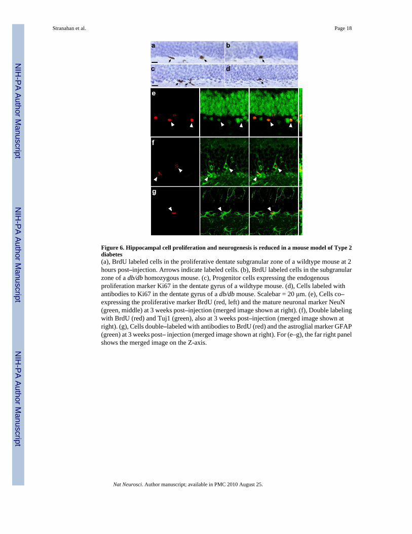
Figure 6. Hippocampal cell proliferation and neurogenesis is reduced in a mouse model of Type 2diabetes(a), BrdU labeled cells in the proliferative dentate subgranular zone of a wildtype mouse at 2hours post–injection. Arrows indicate labeled cells. (b), BrdU labeled cells in the subgranularzone of a db/db homozygous mouse. (c), Progenitor cells expressing the endogenousproliferation marker Ki67 in the dentate gyrus of a wildtype mouse. (d), Cells labeled withantibodies to Ki67 in the dentate gyrus of a db/db mouse. Scalebar = 20 μm. (e), Cells co–expressing the proliferative marker BrdU (red, left) and the mature neuronal marker NeuN(green, middle) at 3 weeks post–injection (merged image shown at right). (f), Double labelingwith BrdU (red) and Tuj1 (green), also at 3 weeks post–injection (merged image shown atright). (g), Cells double–labeled with antibodies to BrdU (red) and the astroglial marker GFAP(green) at 3 weeks post– injection (merged image shown at right). For (e–g), the far right panelshows the merged image on the Z-axis.
Stranahan et al. Page 18
Nat Neurosci. Author manuscript; available in PMC 2010 August 25.
NIH
-PA Author Manuscript
NIH
-PA Author Manuscript
NIH
-PA Author Manuscript
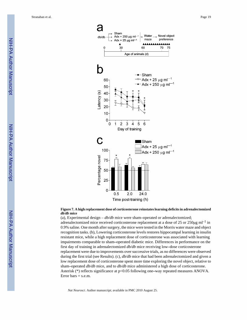
Figure 7. A high replacement dose of corticosterone reinstates learning deficits in adrenalectomizeddb/db mice(a), Experimental design – db/db mice were sham–operated or adrenalectomized;adrenalectomized mice received corticosterone replacement at a dose of 25 or 250μg ml–1 in0.9% saline. One month after surgery, the mice were tested in the Morris water maze and objectrecognition tasks. (b), Lowering corticosterone levels restores hippocampal learning in insulinresistant mice, while a high replacement dose of corticosterone was associated with learningimpairments comparable to sham–operated diabetic mice. Differences in performance on thefirst day of training in adrenalectomized db/db mice receiving low–dose corticosteronereplacement were due to improvements over successive trials, as no differences were observedduring the first trial (see Results). (c), db/db mice that had been adrenalectomized and given alow replacement dose of corticosterone spent more time exploring the novel object, relative tosham–operated db/db mice, and to db/db mice administered a high dose of corticosterone.Asterisk (*) reflects significance at p<0.05 following one–way repeated measures ANOVA.Error bars = s.e.m.
Stranahan et al. Page 19
Nat Neurosci. Author manuscript; available in PMC 2010 August 25.
NIH
-PA Author Manuscript
NIH
-PA Author Manuscript
NIH
-PA Author Manuscript
NIH
-PA Author Manuscript
NIH
-PA Author Manuscript
NIH
-PA Author Manuscript
Stranahan et al. Page 20
Tabl
e 1
End
ocri
ne c
hara
cter
istic
s of T
ype
1 an
d T
ype
2 di
abet
es in
rod
ents
with
diff
eren
t lev
els o
f cor
ticos
tero
ne
Dat
a w
ere
anal
yzed
usi
ng 2
× 2
AN
OV
A a
nd a
ster
isk
(*) i
ndic
ates
sign
ifica
nce
at p
< 0
.05
rela
tive
to sh
am–o
pera
ted
non–
diab
etic
con
trols
. Val
ues s
how
nar
e m
eans
and
s.e.
m. A
bbre
viat
ions
: STZ
= st
rept
ozoc
in; S
HA
M =
sham
–ope
rate
d, A
DX
+ C
OR
T =
adre
nale
ctom
ized
with
25 μg
ml–1
cor
ticos
tero
nere
plac
emen
t.
FAST
ING
GL
UC
OSE
(mg
dL–1
)FE
D G
LU
CO
SE (m
g dL
–1)
INSU
LIN
(ng
ml–1
)C
OR
TIC
OST
ER
ON
E (n
g m
l–1)
wild
type
SHA
M71
.77
(8.6
3)14
0.65
(15.
67)
1.41
(0.1
5)46
.16
(15.
99)
AD
X+C
OR
T64
.80
(1.8
7)95
.95
(20.
65)
1.41
(0.1
4)18
.85
(3.9
5)
db/d
bSH
AM
330.
27 (2
1.01
)*33
4.12
(41.
09)*
3.16
(0.6
8)*
258.
55 (4
3.11
)*
AD
X+C
OR
T98
.40
(17.
51)
328.
88 (1
9.83
)*1.
03 (0
.17)
11.2
4 (2
.21)
Veh
icle
SHA
M30
.19
(5.7
7)12
9.86
(21.
72)
2.07
(0.2
7)53
.08
(17.
72)
AD
X+C
OR
T44
.74
(2.2
3)17
0.87
(11.
74)
1.73
(0.2
7)19
.87
(4.5
6)
STZ
SHA
M29
0.96
(35.
94)*
318.
46 (1
6.76
)*0.
79 (.
09)*
418.
24 (1
8.26
)*
AD
X+C
OR
T22
1.08
(14.
80)*
263.
06 (1
1.29
)*0.
91 (0
.16)
*30
.14
(5.7
8)
Nat Neurosci. Author manuscript; available in PMC 2010 August 25.
Related Documents