A reminder of things we should already remember but have completely forgotten

DH156lecture3oralmucosa
Sep 09, 2014
Welcome message from author
This document is posted to help you gain knowledge. Please leave a comment to let me know what you think about it! Share it to your friends and learn new things together.
Transcript
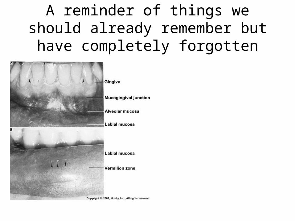
A reminder of things we should already remember but have completely
forgotten

Oral Mucosa-lining of the oral cavity
-mucosal membrane = epithelium + connective tissue-derived from the ectoderm
-epithelium = stratified squamous ( skin equivalent = epidermis)-connective tissue = lamina propria (skin equivalent = dermis)
-lamina propria = thin, vascular layer of connective tissue below the epithelium of an oral mucosa-comprised of collagen fibers-comprised of two layers1. an upper papillary layer – associated with epithelial ridges
-thin, loosely arranged CN fibers-many capillary loops
2. a lower reticular layer – thicker bundles of CN fibers that lie in parallel
-in between these tissues = basement membrane-made by both tissues-epithelial component = basal lamina-connective component = reticular lamina
-below the connective tissue lamina propria = submucosa-for attachment to bone

Oral mucosa classifications• Lining mucosa
– buccal mucosa, labial mucosa, alveolar mucosa, floor of the mouth, ventral tongue, soft palate
– non-keratinized stratified squamous epithelium– soft, moist, ability to stretch and compress
• Masticatory mucosa– attached gingiva, hard palate, dorsal tongue– rubbery, resilient– keratinized or parakeratinized stratified squamous epithelium
• Specialized– dorsal tongue surface– associated with the lingual papillae

Representative Mucosa-the interface between theepithelium and LP interdigitates-upward projections of the LP arecalled connective tissue ridges or papillae(dermal papillae in skin)-downward ridges of the epitheliumare called rete ridges or pegs
underside of epithelium connective tissue ridges
-allows greater attachment enabling dispersion of large forces over a greater areaof connective tissue (greater the forces, the greater the # of rete and connective papillae-also a site of metabolic exchange between the epithelium and LP-below the LP is the submucosa
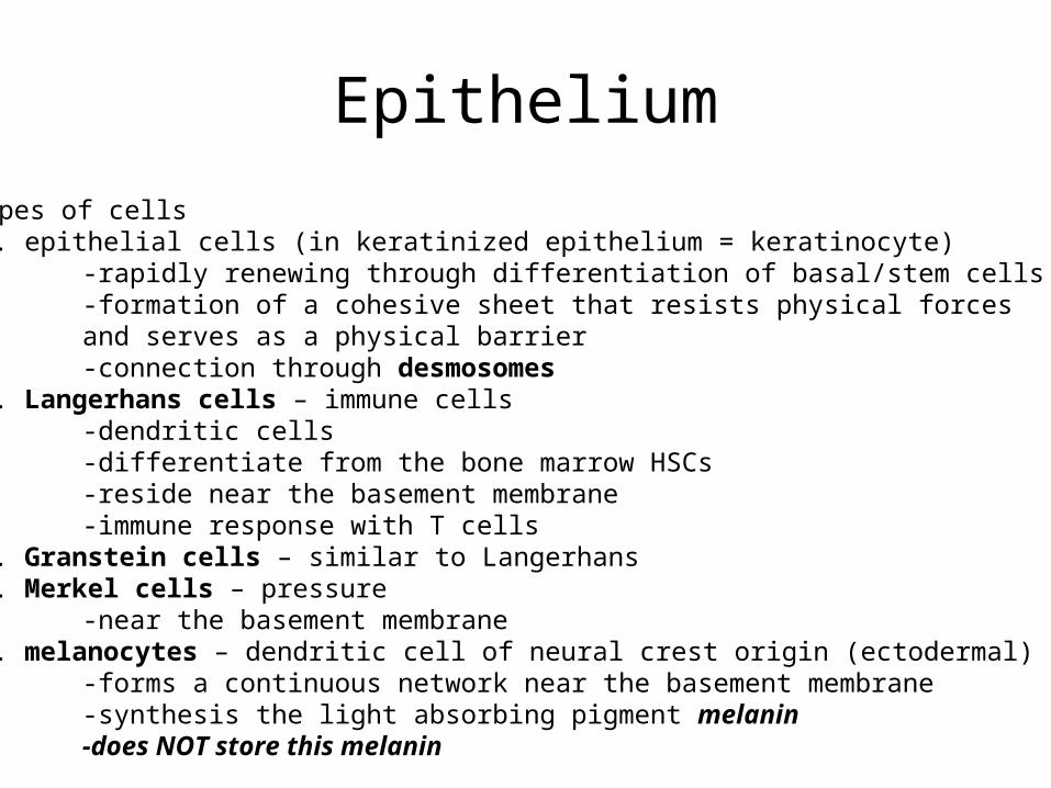
Epithelium
-four types of cells1. epithelial cells (in keratinized epithelium = keratinocyte)
-rapidly renewing through differentiation of basal/stem cells-formation of a cohesive sheet that resists physical forcesand serves as a physical barrier-connection through desmosomes
2. Langerhans cells – immune cells-dendritic cells-differentiate from the bone marrow HSCs-reside near the basement membrane-immune response with T cells
3. Granstein cells – similar to Langerhans4. Merkel cells – pressure
-near the basement membrane 5. melanocytes – dendritic cell of neural crest origin (ectodermal)
-forms a continuous network near the basement membrane-synthesis the light absorbing pigment melanin-does NOT store this melanin
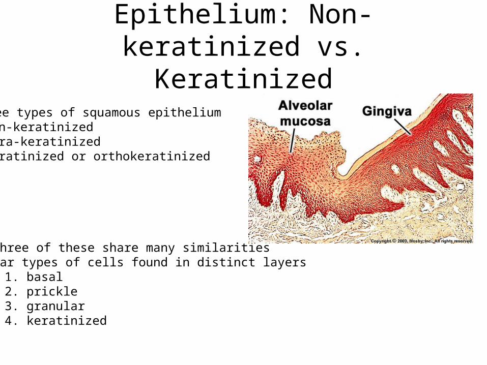
Epithelium: Non-keratinized vs. Keratinized
-three types of squamous epithelium1. non-keratinized2. para-keratinized3. keratinized or orthokeratinized
-all three of these share many similarities-similar types of cells found in distinct layers
1. basal2. prickle3. granular4. keratinized
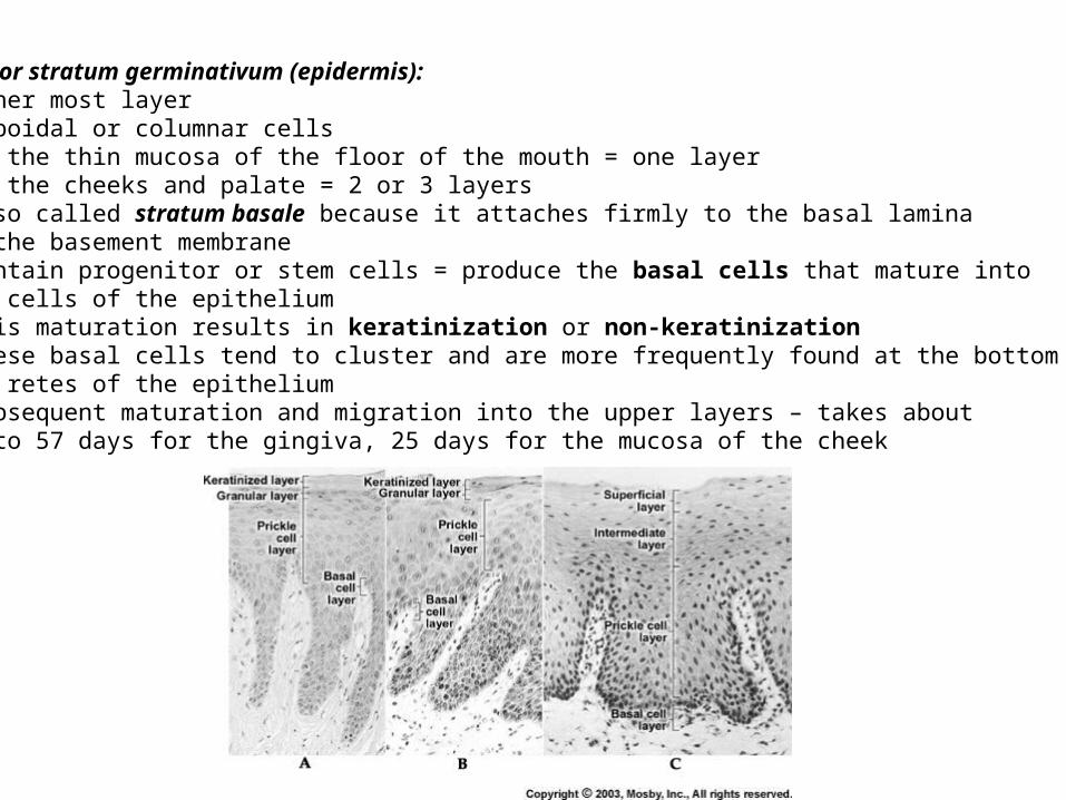
1. basal layer or stratum germinativum (epidermis):-inner most layer-cuboidal or columnar cells-in the thin mucosa of the floor of the mouth = one layer-in the cheeks and palate = 2 or 3 layers-also called stratum basale because it attaches firmly to the basal laminaof the basement membrane -contain progenitor or stem cells = produce the basal cells that mature into all cells of the epithelium-this maturation results in keratinization or non-keratinization -these basal cells tend to cluster and are more frequently found at the bottom of thethe retes of the epithelium-subsequent maturation and migration into the upper layers – takes about41 to 57 days for the gingiva, 25 days for the mucosa of the cheek

2. prickle layer or skin = stratum spinosum:-called the “spiny layer” because of histologicalappearance-tend to shrink from each other and remain connectedby desmosomes-from maturation and migration of basal cells -several layers thick-connected by bundles of intermediate filamentscalled tonofilaments –begin and end at
desmosomes -connectneighbouring epithelial cells-act as cross braces providingstrength
-division of cells within this layer increasesthickness-melanocytes are common-Langerhans cells also found – in the more superficial layers of the prickle layer
-initiate immune responses to pathogensand to cancer
desmosome
gap junction

3. granular or skin = stratum granulosum:-made up of epithelial cells displaced from the prickle-in keratinized epithelium – cell synthesize large quantities of
proteins (including keratin) – cytoplasm appears granular-the granules = keratohyalin granules
-these granules surround the keratin filaments as they develop
-as keratin is made – cells become thinner and flatter- the plasma membrane thickens and becomes less permeable-the cells then die and dehydrate creating bundles of keratin surrounded by keratohyalin sandwiched between two phospholipid membranes

4. keratinized layer/superficial layer (skin = stratum corneum):-in the masticatory mucosa - large amounts of keratin are presentso the outermost layer = keratinized layer (stratum corneum = skin)-BUT in the lining mucosa – no keratin is made – this layer iscalled the superficial layer
– presence of keratin prevents growth of microorganisms and physical damage-covered in secretions from glands to provide some moisture-cells are flattened = squames-keratinized layers – cells do not contain nuclei-pattern of keratinization in these cells = orthokeratinization
-a variation in keratinization is seen in the mucosa of parts of the hard palate and much of the gingiva = parakeratinized mucosa
– only the cells in the surface stain for keratin and the nuclei are retained in many or all of the squames + fewer keratohyaline granules in the underlying granular layer (immature form of orthokeratinization)
-in the skin – parakeratinization is a disease state (e.g. psoriasis)– normal in oral mucosa
-the keratinized mucosa of the oral cavity – 20 cell layers thick-can be thicker than the palms and soles!!!!

Keratinized Epithelium
cuboidal or columnar cellsbundles of tonofibrilssite of cell division
larger ovoid cellslarge tonofibrils (bundles of tonofilaments)
flattened cellslarge keratohyaline granuleswith tonofibrils
extremely flattened cellsdehydratedloss of organelles and nucleicells filled with keratinparakeratinization occurs in sometissues
Basal layer
Prickle layer
Granular layerNK = intermediate
Keratinized layerNK = superficial
Non-Keratinized Epithelium
cuboidal or columnar cellsbundles of tonofibrilssite of cell division
larger ovoid cellssmaller more dispersed tonofilamentbundlesmore numerous filamentsprickles are less numerous
slightly flattened cellsmany dispersed tonofilamentsglycogen granulesnot that different from the prickle layer inappearance = stratum intermedium
slightly flattened cells with dispersedfilaments and glycogenfew organellesnuclei persiststratum superficiale

Lamina Propria
• highly vascularized tissue
• connective tissue with varying amounts of collagen and elastic fibers
• papillae can vary in number, height and width depending on region of mucosa
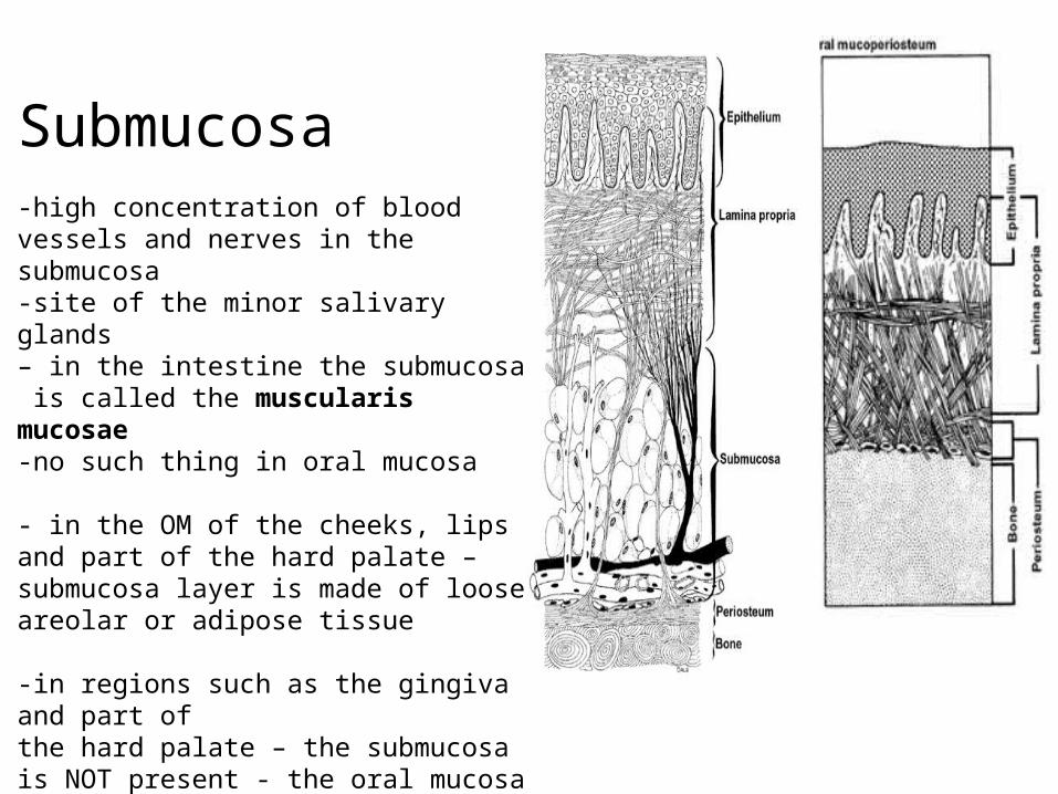
Submucosa-high concentration of blood vessels and nerves in the submucosa -site of the minor salivary glands– in the intestine the submucosa is called the muscularis mucosae-no such thing in oral mucosa
- in the OM of the cheeks, lips and part of the hard palate – submucosa layer is made of loose areolar or adipose tissue
-in regions such as the gingiva and part of the hard palate – the submucosa is NOT present - the oral mucosa attachesdirectly to the periosteum of underlying bone = mucoperiosteum
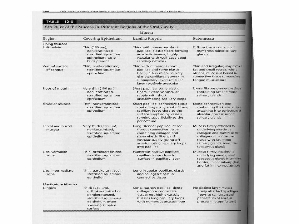
Lining mucosa
• includes the buccal, labial, alveolar mucosa, the mucosa of the floor mouth, ventral surface of tongue, soft palate
• the interface between the epithelium and lamina propria is relatively smooth with fewer and less pronounced rete ridges and connective tissue papillae (comparable to the dermal papillae of skin)
• lamina propria – presence of elastic fibers
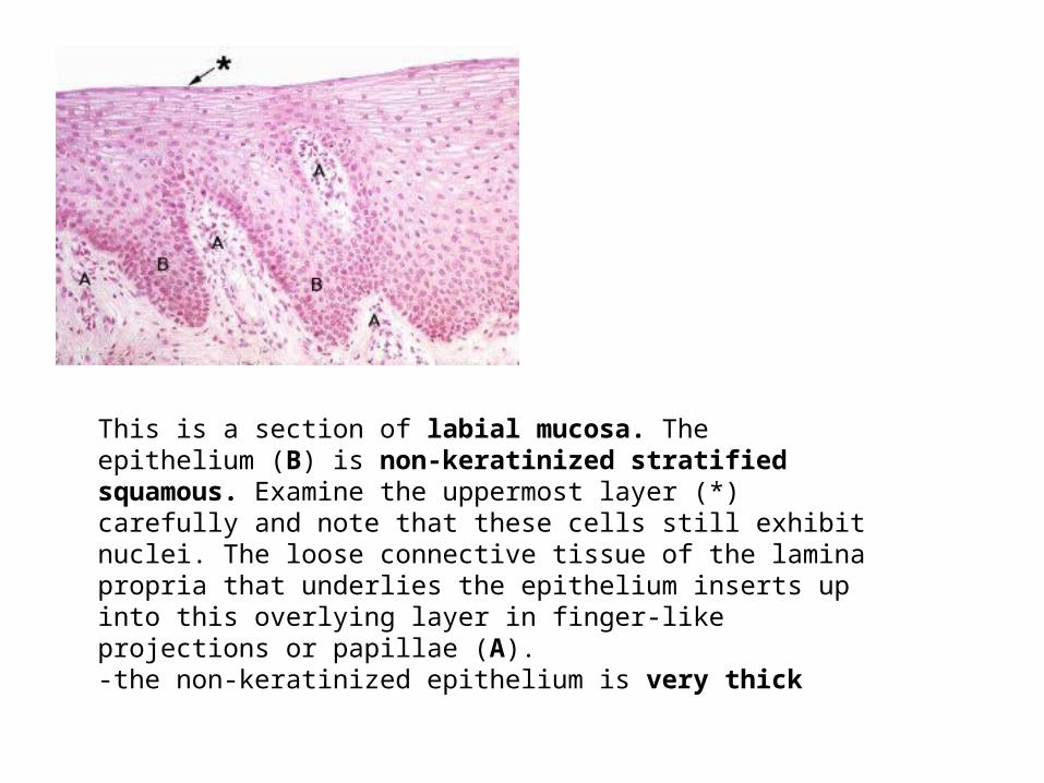
This is a section of labial mucosa. The epithelium (B) is non-keratinized stratified squamous. Examine the uppermost layer (*) carefully and note that these cells still exhibit nuclei. The loose connective tissue of the lamina propria that underlies the epithelium inserts up into this overlying layer in finger-like projections or papillae (A). -the non-keratinized epithelium is very thick
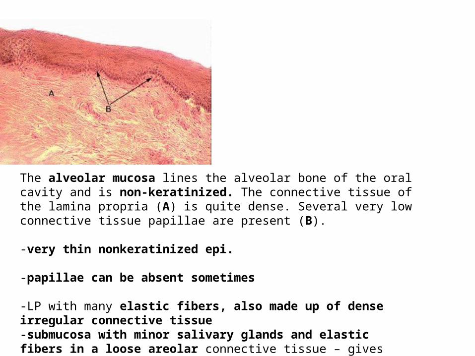
The alveolar mucosa lines the alveolar bone of the oral cavity and is non-keratinized. The connective tissue of the lamina propria (A) is quite dense. Several very low connective tissue papillae are present (B).
-very thin nonkeratinized epi.
-papillae can be absent sometimes
-LP with many elastic fibers, also made up of dense irregular connective tissue-submucosa with minor salivary glands and elastic fibers in a loose areolar connective tissue – gives increased moisture and increase motility

This is the mucosa of the oropharyngeal aspect of the soft palate and is non-keratinized. It resembles the buccal and labial mucosa; - the connective tissue papillae (A) are of moderate height-thick LP with numerous papillae-LP has a distinct elastic layer for increased mobility
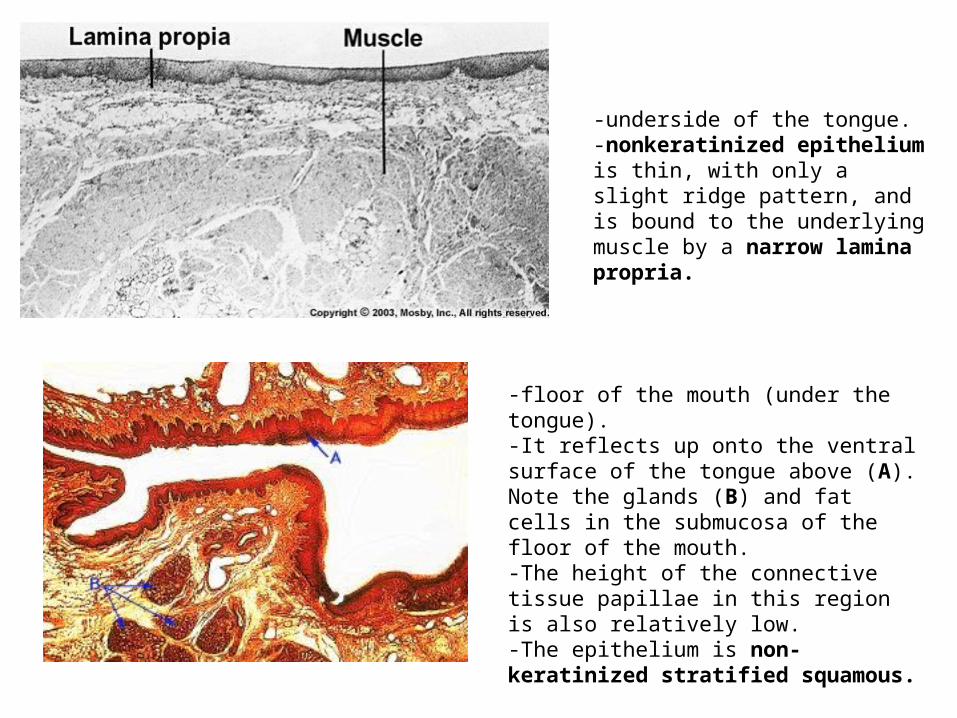
-floor of the mouth (under the tongue). -It reflects up onto the ventral surface of the tongue above (A). Note the glands (B) and fat cells in the submucosa of the floor of the mouth. -The height of the connective tissue papillae in this region is also relatively low. -The epithelium is non-keratinized stratified squamous.
-underside of the tongue. -nonkeratinized epithelium is thin, with only a slight ridge pattern, and is bound to the underlying muscle by a narrow lamina propria.

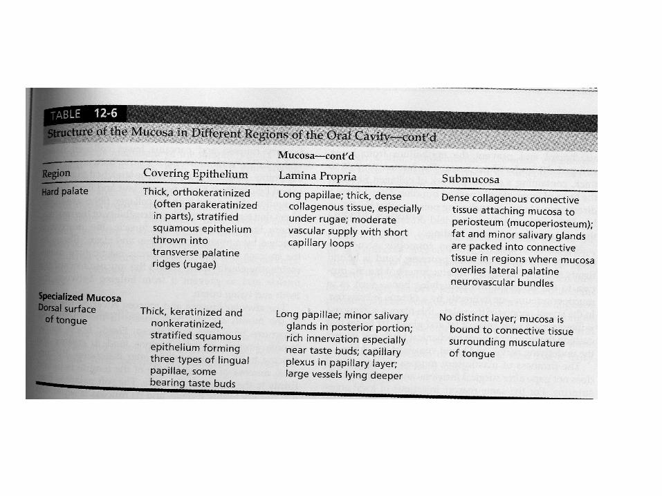
Masticatory Mucosa• hard palate• gingiva• exposed to compressive and shear forces during mastication• dorsum of the tongue has the same functional role but it is
considered as specialized mucosa• epithelium is relatively thick and frequently orthokeratinized• the gingiva and palate also has parakeratinized• very convoluted junction between EP and LP• numerous elongated papillae – good mechanical attachment• thick LP – dense network of CN fibers in the form of large packed
bundles• the LP is bound firmly to underlying bone (mucoperiosteum) or
indirectly by a fibrous (not a loose, fatty) submucosa

Orthokeratinization of stratified squamous epithelium may occur at sites in the oral cavity where the mucosa is subjected to habitual mechanical stress - such as continuous trauma from chewing. Note the stratum corneum (A). No nuclei are visible in contrast with the parakeratinized variety.

The mucosa covering the hard palate exhibits a distinct keratinized layer - High connective tissue papillae are associated with keratinized epithelium. -This form of keratinization in the oral cavity is referred to as orthokeratinized stratified squamous epithelium.

-junction (dashed line) between mucosae covering the hard and the soft palate. -The difference in thickness and the ridge pattern between keratinized epithelium of the hard palate and nonkeratinized epithelium of the soft palate is apparent.
-the hard palate has a thick orthokeratinized epi-overlies a thick LP-submucosa found at the lateral margins of the hard palate
--the firm feeling in the medial portion – due to lack of SM + the LP is directly attachedto bone – therefore the LP serves as a mucoperiosteum
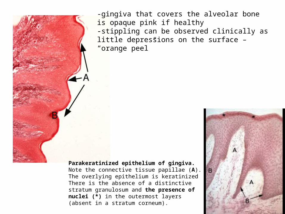
-gingiva that covers the alveolar boneis opaque pink if healthy-stippling can be observed clinically aslittle depressions on the surface – “orange peel”
Parakeratinized epithelium of gingiva.Note the connective tissue papillae (A). The overlying epithelium is keratinizedThere is the absence of a distinctive stratum granulosum and the presence of nuclei (*) in the outermost layers (absent in a stratum corneum).

Alveolar (B)-gingival (A) transition-where masticatory meets lining mucosa-between attached gingiva and alveolar mucosa at a slight indentation calledthe mucogingival groove (bright pink to paler pink)-keratinized or parakeratinized of gingiva (A) to the thicker nonkeratinized of alveolar (B)-the LP of the gingiva has numerous CN bundles attaching to the periosteum = stippling-the LP of the alveolar is loose connective tissue with numerous elastic fibers and athicker submucosa
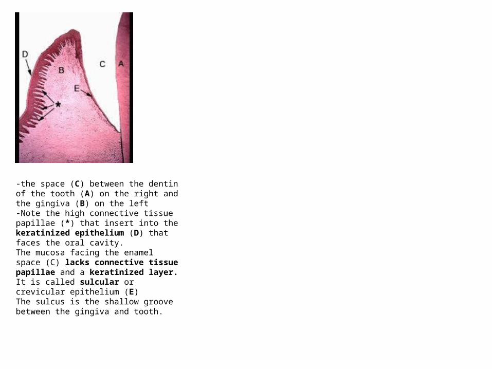
-the space (C) between the dentin of the tooth (A) on the right and the gingiva (B) on the left -Note the high connective tissue papillae (*) that insert into the keratinized epithelium (D) that faces the oral cavity.The mucosa facing the enamel space (C) lacks connective tissue papillae and a keratinized layer. It is called sulcular or crevicular epithelium (E) The sulcus is the shallow groove between the gingiva and tooth.
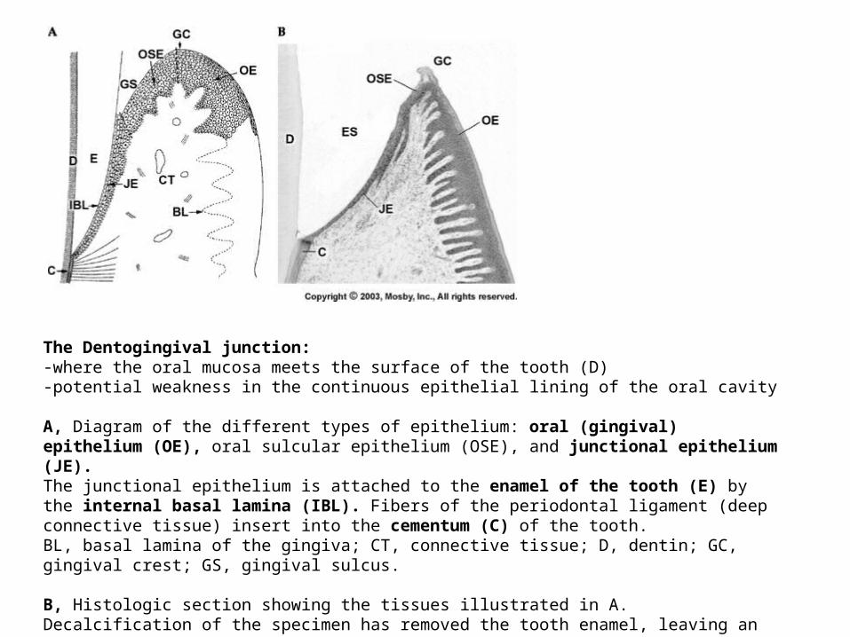
The Dentogingival junction:-where the oral mucosa meets the surface of the tooth (D)-potential weakness in the continuous epithelial lining of the oral cavity
A, Diagram of the different types of epithelium: oral (gingival) epithelium (OE), oral sulcular epithelium (OSE), and junctional epithelium (JE). The junctional epithelium is attached to the enamel of the tooth (E) by the internal basal lamina (IBL). Fibers of the periodontal ligament (deep connective tissue) insert into the cementum (C) of the tooth. BL, basal lamina of the gingiva; CT, connective tissue; D, dentin; GC, gingival crest; GS, gingival sulcus.
B, Histologic section showing the tissues illustrated in A. Decalcification of the specimen has
removed the tooth enamel, leaving an enamel space (ES).

-the walls of the sulcus are lined with an epithelium continuous with the rest of the oralmucosa = oral sucular epithelium (nonkeratinzed)-the floor of the sulcus is lined with junctional epithelium (JE)
-derived from the dental epithelium of the tooth germ-smooth connective interface with the epithelium of the OSE and OE-nonkeratinized -notice lack of connective tissue papillae-cells differ from those of other nonkeratinized oral mucosae
-the free edge of the sulcus is made of keratinized or parakeratinized epi.-called the gingival crest (GC)

The Tongue
• dorsal surface = specialized mucosa• similar to masticatory but is highly extensible• has unique papillae types and structure • mucous membranes are of two parts
– divided by the V-shaped sulcus terminalis• anterior 2/3
– body of the tongue
• posterior 1/3– base
– contains the lingual tonsils

In this cross-section through the tongue note the absence of a submucosa.
The overlying epithelium (B) is of the non-keratinizing stratified squamous type.
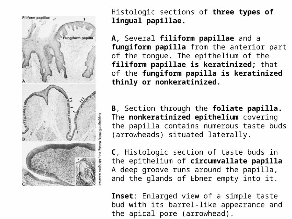
Histologic sections of three types of lingual papillae.
A, Several filiform papillae and a fungiform papilla from the anterior part of the tongue. The epithelium of the filiform papillae is keratinized; that of the fungiform papilla is keratinized thinly or nonkeratinized.
B, Section through the foliate papilla. The nonkeratinized epithelium covering the papilla contains numerous taste buds (arrowheads) situated laterally.
C, Histologic section of taste buds in the epithelium of circumvallate papillaA deep groove runs around the papilla, and the glands of Ebner empty into it.
Inset: Enlarged view of a simple taste bud with its barrel-like appearance and the apical pore (arrowhead).
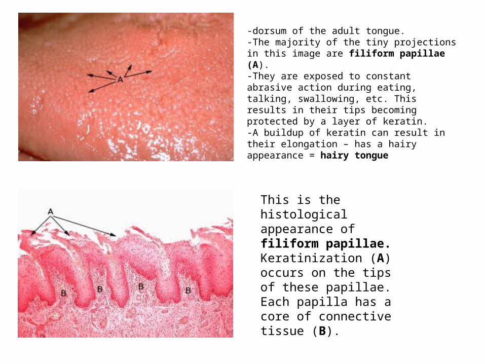
-dorsum of the adult tongue. -The majority of the tiny projections in this image are filiform papillae (A). -They are exposed to constant abrasive action during eating, talking, swallowing, etc. This results in their tips becoming protected by a layer of keratin. -A buildup of keratin can result in their elongation – has a hairy appearance = hairy tongue
This is the histological appearance of filiform papillae. Keratinization (A) occurs on the tips of these papillae. Each papilla has a core of connective tissue (B).
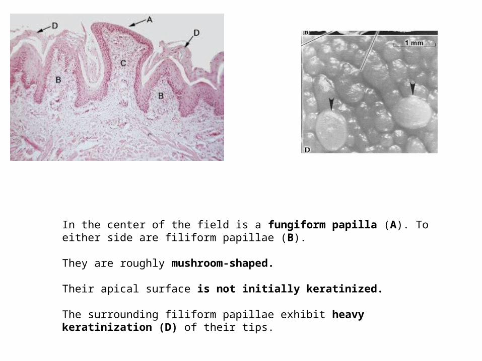
In the center of the field is a fungiform papilla (A). To either side are filiform papillae (B).
They are roughly mushroom-shaped.
Their apical surface is not initially keratinized.
The surrounding filiform papillae exhibit heavy keratinization (D) of their tips.
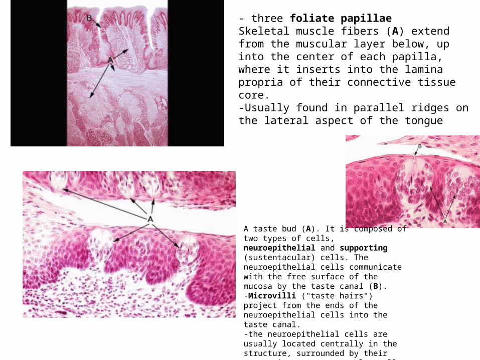
- three foliate papillae Skeletal muscle fibers (A) extend from the muscular layer below, up into the center of each papilla, where it inserts into the lamina propria of their connective tissue core. -Usually found in parallel ridges on the lateral aspect of the tongue
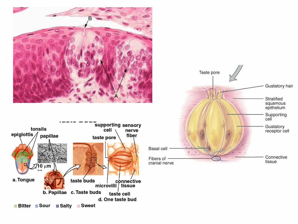
A taste bud (A). It is composed of two types of cells, neuroepithelial and supporting (sustentacular) cells. The neuroepithelial cells communicate with the free surface of the mucosa by the taste canal (B). -Microvilli ("taste hairs") project from the ends of the neuroepithelial cells into the taste canal. -the neuroepithelial cells are usually located centrally in the structure, surrounded by their supporting or sustentacular cells

This is a dorsal view of a sagittally sectioned tongue from a cadaver. Near the center of the field are two relatively large circular areas (B). These are circumvallate (vallate) papillae. -A V-shaped line of circumvallate papillae indicate the approximate site of the boundary between the anterior 2/3's of the tongue (A) and the posterior 1/3 (P). -The surface of the posterior 1/3 is much smoother than the anterior 2/3.

This is a higher magnification of a circumvallate papilla than seen in the preceding image. -A duct (B) from the underlying glands of von Ebner can be seen opening into the moat-like space (C) around the papilla. Directly below the base of this papilla is a collection of nerve tissue (D).

Mucous glands (A) lie between the lamina propria (B) and the underlying musculature of the posterior 1/3 of the dorsum of the tongue. - The epithelium (C) covering the dorsum of the posterior 1/3 of the tongue is not as firmly attached as is that of the anterior 2/3s. The epithelium is classified as non-keratinized stratified squamous.
This section through a lingual tonsil reveals a collection of the structural and functional units called lymphoid nodules (A). -These structures aggregate around a pit or primary crypt (B) that opens onto the free surface of the mucosa. -Typically the surface would be lined by non-keratinizing stratified squamous epithelium that continues down into the crypts.-Mucous glands (C) secrete into the bottom of the crypts. The secretions "flush" the crypts of debris to prevent stagnation and the development of infection. --The posterior aspect of the tonsil is surrounded by a connective tissue capsule (D).

- junction of the mucous membrane of the oral cavity with the skin of the lip (A). Skin is to the right (B) and is composed of the epidermis (epithelial layer) and the dermis (dense connective tissue layer).-Mucosa (C) on the left, is composed of epithelium and lamina propria (dense connective tissue layer). -mucocutaneous junction (A) - the epithelium of the mucosa is continuous with that of the skin and the lamina propria is continuous with the dermis. -Deep to the lamina propria, the submucosa is the counterpart to the hypodermis of the integument.
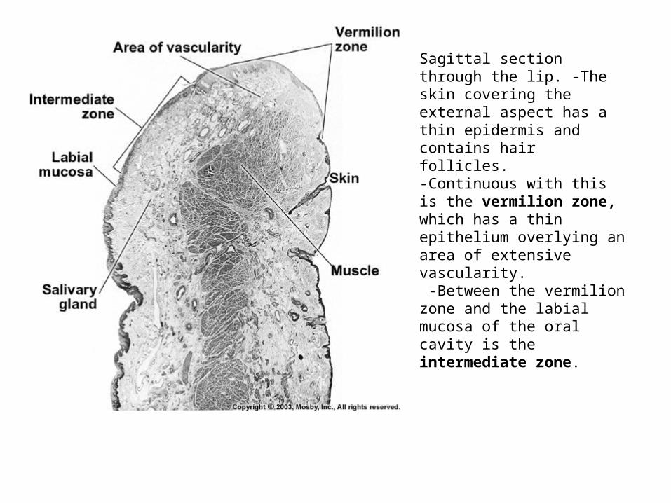
Sagittal section through the lip. -The skin covering the external aspect has a thin epidermis and contains hair follicles.-Continuous with this is the vermilion zone, which has a thin epithelium overlying an area of extensive vascularity. -Between the vermilion zone and the labial mucosa of the oral cavity is the intermediate zone.
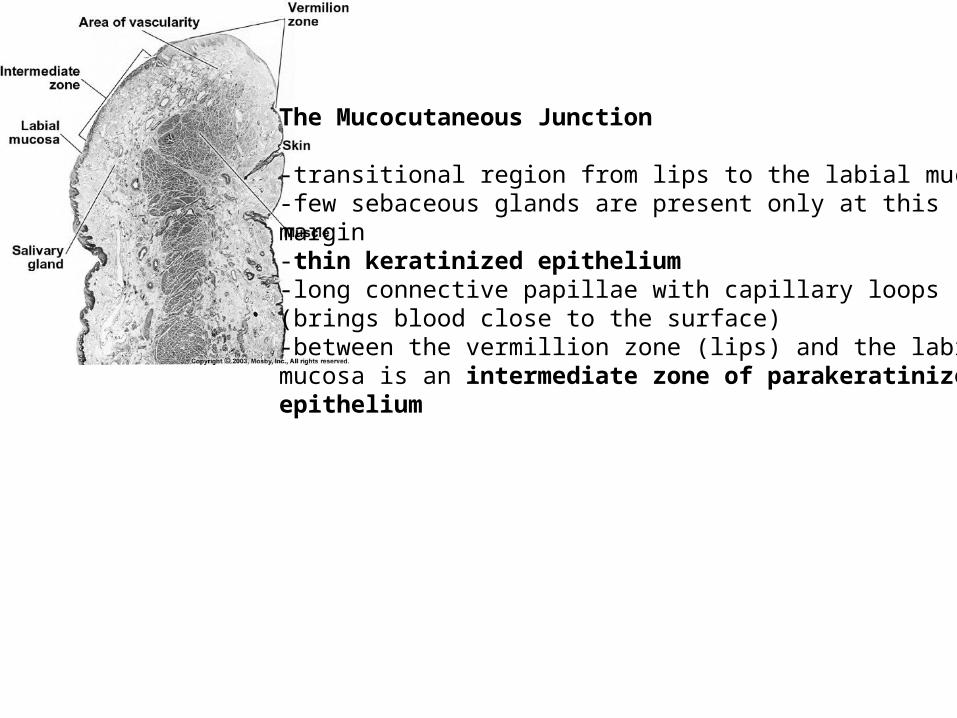
The Mucocutaneous Junction
-transitional region from lips to the labial mucosa-few sebaceous glands are present only at thismargin-thin keratinized epithelium-long connective papillae with capillary loops(brings blood close to the surface)-between the vermillion zone (lips) and the labialmucosa is an intermediate zone of parakeratinizedepithelium