Developmental and Hormonal Regulation of Gibberellin Biosynthesis and Catabolism in Pea Fruit 1[OA] Jocelyn A. Ozga*, Dennis M. Reinecke, Belay T. Ayele, Phuong Ngo, Courtney Nadeau, and Aruna D. Wickramarathna Plant BioSystems, Department of Agricultural, Food, and Nutritional Science, University of Alberta, Edmonton, Alberta, Canada T6G 2P5 In pea (Pisum sativum), normal fruit growth requires the presence of the seeds. The coordination of growth between the seed and ovary tissues involves phytohormones; however, the specific mechanisms remain speculative. This study further explores the roles of the gibberellin (GA) biosynthesis and catabolism genes during pollination and fruit development and in seed and auxin regulation of pericarp growth. Pollination and fertilization events not only increase pericarp PsGA3ox1 message levels (codes for GA 3-oxidase that converts GA 20 to bioactive GA 1 ) but also reduce pericarp PsGA2ox1 mRNA levels (codes for GA 2-oxidase that mainly catabolizes GA 20 to GA 29 ), suggesting a concerted regulation to increase levels of bioactive GA 1 following these events. 4-Chloroindole-3-acetic acid (4-Cl-IAA) was found to mimic the seeds in the stimulation of PsGA3ox1 and the repression of PsGA2ox1 mRNA levels as well as the stimulation of PsGA2ox2 mRNA levels (codes for GA 2-oxidase that mainly catabolizes GA 1 to GA 8 ) in pericarp at 2 to 3 d after anthesis, while the other endogenous pea auxin, IAA, did not. This GA gene expression profile suggests that both seeds and 4-Cl-IAA can stimulate the production, as well as modulate the half-life, of bioactive GA 1 , leading to initial fruit set and subsequent growth and development of the ovary. Consistent with these gene expression profiles, deseeded pericarps converted [ 14 C]GA 12 to [ 14 C]GA 1 only if treated with 4-Cl-IAA. These data further support the hypothesis that 4-Cl-IAA produced in the seeds is transported to the pericarp, where it differentially regulates the expression of pericarp GA biosynthesis and catabolism genes to modulate the level of bioactive GA 1 required for initial fruit set and growth. In pea (Pisum sativum), normal pericarp growth requires the presence of seeds. Removal or destruction of the seeds at 2 to 3 d after anthesis (DAA) results in the slowing of pericarp growth and subsequently abscission (Eeuwens and Schwabe, 1975; Ozga et al., 1992). Signaling molecules originating from the seeds may be responsible for continued fruit development by maintaining hormone levels in the surrounding tissue (Eeuwens and Schwabe, 1975; Sponsel, 1982). Developing pea seeds and pericarps contain gibberel- lins (GAs; Garcia-Martinez et al., 1991; Rodrigo et al., 1997) and auxins (4-chloroindole-3-acetic acid [4-Cl- IAA] and IAA; Marumo et al., 1968; Magnus et al., 1997). During early pericarp growth (2 DAA), appli- cation of the naturally occurring hormones 4-Cl-IAA (Reinecke et al., 1995) and GA (Eeuwens and Schwabe, 1975; Ozga and Reinecke, 1999) to deseeded pericarps can substitute for seeds and stimulate pericarp growth. However, the other naturally occurring auxin in pea fruit, IAA, was ineffective at promoting growth (Reinecke et al., 1995). Studies comparing the growth-promoting proper- ties of 4-, 5-, 6- and 7-Cl-IAAs and the corresponding F-IAA analogs (Reinecke et al., 1995) and the physico- chemical properties of 4-Cl-IAA and 4-substituted analogs (Reinecke et al., 1999) found that the 4-position of the indole ring and the 4-substituents’s size and lipophilicity were required for significant biological activity in pea pericarp growth. Pea pericarps respond in a qualitatively different fashion to two naturally occurring auxins (IAA and 4-Cl-IAA), which, in a variety of other auxin bioassays, exhibited only quan- titative differences in activity (Reinecke, 1999). These data suggest unique ways of auxin action based on alternative molecular recognition mechanisms in this tissue. Pea plants metabolize GAs by the early 13-hydrox- ylation pathway: GA 12 / GA 53 / GA 44 / GA 19 / GA 20 / GA 1 (Sponsel, 1995). Previous studies using the pea split-pericarp assay (test compounds are ap- plied to the inner walls of split and deseeded 2-DAA pericarps) have shown that the presence of seeds or the application of 4-Cl-IAA to deseeded pea pericarps stimulated pericarp GA biosynthesis gene expression, specifically PsGA20ox1 (codes for enzyme that con- verts GA 53 to GA 20 ; van Huizen et al., 1997) and PsGA3ox1 (codes for enzyme that converts GA 20 to GA 1 ; Ozga et al., 2003). IAA was ineffective at stimu- lating pericarp PsGA20ox1 (Ngo et al., 2002) and 1 This work was supported by the Natural Sciences and Engi- neering Research Council of Canada. * Corresponding author; e-mail [email protected]. The author responsible for distribution of materials integral to the findings presented in this article in accordance with the policy described in the Instructions for Authors (www.plantphysiol.org) is: Jocelyn A. Ozga ([email protected]). [OA] Open Access articles can be viewed online without a sub- scription. www.plantphysiol.org/cgi/doi/10.1104/pp.108.132027 448 Plant Physiology, May 2009, Vol. 150, pp. 448–462, www.plantphysiol.org Ó 2009 American Society of Plant Biologists www.plant.org on December 21, 2015 - Published by www.plantphysiol.org Downloaded from Copyright © 2009 American Society of Plant Biologists. All rights reserved.

Welcome message from author
This document is posted to help you gain knowledge. Please leave a comment to let me know what you think about it! Share it to your friends and learn new things together.
Transcript
Developmental and Hormonal Regulation of GibberellinBiosynthesis and Catabolism in Pea Fruit1[OA]
Jocelyn A. Ozga*, Dennis M. Reinecke, Belay T. Ayele, Phuong Ngo,Courtney Nadeau, and Aruna D. Wickramarathna
Plant BioSystems, Department of Agricultural, Food, and Nutritional Science, University of Alberta,Edmonton, Alberta, Canada T6G 2P5
In pea (Pisum sativum), normal fruit growth requires the presence of the seeds. The coordination of growth between the seedand ovary tissues involves phytohormones; however, the specific mechanisms remain speculative. This study further exploresthe roles of the gibberellin (GA) biosynthesis and catabolism genes during pollination and fruit development and in seed andauxin regulation of pericarp growth. Pollination and fertilization events not only increase pericarp PsGA3ox1 message levels(codes for GA 3-oxidase that converts GA20 to bioactive GA1) but also reduce pericarp PsGA2ox1 mRNA levels (codes for GA2-oxidase that mainly catabolizes GA20 to GA29), suggesting a concerted regulation to increase levels of bioactive GA1 followingthese events. 4-Chloroindole-3-acetic acid (4-Cl-IAA) was found to mimic the seeds in the stimulation of PsGA3ox1 and therepression of PsGA2ox1mRNA levels as well as the stimulation of PsGA2ox2mRNA levels (codes for GA 2-oxidase that mainlycatabolizes GA1 to GA8) in pericarp at 2 to 3 d after anthesis, while the other endogenous pea auxin, IAA, did not. This GAgene expression profile suggests that both seeds and 4-Cl-IAA can stimulate the production, as well as modulate the half-life,of bioactive GA1, leading to initial fruit set and subsequent growth and development of the ovary. Consistent with these geneexpression profiles, deseeded pericarps converted [14C]GA12 to [14C]GA1 only if treated with 4-Cl-IAA. These data furthersupport the hypothesis that 4-Cl-IAA produced in the seeds is transported to the pericarp, where it differentially regulates theexpression of pericarp GA biosynthesis and catabolism genes to modulate the level of bioactive GA1 required for initial fruit setand growth.
In pea (Pisum sativum), normal pericarp growthrequires the presence of seeds. Removal or destructionof the seeds at 2 to 3 d after anthesis (DAA) results inthe slowing of pericarp growth and subsequentlyabscission (Eeuwens and Schwabe, 1975; Ozga et al.,1992). Signaling molecules originating from the seedsmay be responsible for continued fruit developmentby maintaining hormone levels in the surroundingtissue (Eeuwens and Schwabe, 1975; Sponsel, 1982).Developing pea seeds and pericarps contain gibberel-lins (GAs; Garcia-Martinez et al., 1991; Rodrigo et al.,1997) and auxins (4-chloroindole-3-acetic acid [4-Cl-IAA] and IAA; Marumo et al., 1968; Magnus et al.,1997). During early pericarp growth (2 DAA), appli-cation of the naturally occurring hormones 4-Cl-IAA(Reinecke et al., 1995) and GA (Eeuwens and Schwabe,1975; Ozga and Reinecke, 1999) to deseeded pericarpscan substitute for seeds and stimulate pericarpgrowth. However, the other naturally occurring auxin
in pea fruit, IAA, was ineffective at promoting growth(Reinecke et al., 1995).
Studies comparing the growth-promoting proper-ties of 4-, 5-, 6- and 7-Cl-IAAs and the correspondingF-IAA analogs (Reinecke et al., 1995) and the physico-chemical properties of 4-Cl-IAA and 4-substitutedanalogs (Reinecke et al., 1999) found that the 4-positionof the indole ring and the 4-substituents’s size andlipophilicity were required for significant biologicalactivity in pea pericarp growth. Pea pericarps respondin a qualitatively different fashion to two naturallyoccurring auxins (IAA and 4-Cl-IAA), which, in avariety of other auxin bioassays, exhibited only quan-titative differences in activity (Reinecke, 1999). Thesedata suggest unique ways of auxin action based onalternative molecular recognition mechanisms in thistissue.
Pea plants metabolize GAs by the early 13-hydrox-ylation pathway: GA12 / GA53 / GA44 / GA19 /GA20 / GA1 (Sponsel, 1995). Previous studies usingthe pea split-pericarp assay (test compounds are ap-plied to the inner walls of split and deseeded 2-DAApericarps) have shown that the presence of seeds orthe application of 4-Cl-IAA to deseeded pea pericarpsstimulated pericarp GA biosynthesis gene expression,specifically PsGA20ox1 (codes for enzyme that con-verts GA53 to GA20; van Huizen et al., 1997) andPsGA3ox1 (codes for enzyme that converts GA20 toGA1; Ozga et al., 2003). IAA was ineffective at stimu-lating pericarp PsGA20ox1 (Ngo et al., 2002) and
1 This work was supported by the Natural Sciences and Engi-neering Research Council of Canada.
* Corresponding author; e-mail [email protected] author responsible for distribution of materials integral to the
findings presented in this article in accordance with the policydescribed in the Instructions for Authors (www.plantphysiol.org) is:Jocelyn A. Ozga ([email protected]).
[OA] Open Access articles can be viewed online without a sub-scription.
www.plantphysiol.org/cgi/doi/10.1104/pp.108.132027
448 Plant Physiology, May 2009, Vol. 150, pp. 448–462, www.plantphysiol.org � 2009 American Society of Plant Biologists www.plant.org on December 21, 2015 - Published by www.plantphysiol.orgDownloaded from
Copyright © 2009 American Society of Plant Biologists. All rights reserved.
PsGA3ox1 expression (Ozga et al., 2003) and pericarpgrowth (Reinecke et al., 1995). Furthermore, elongat-ing pollinated pericarps (3 DAA) were capable ofconverting [14C]GA12 and [14C]GA19 to [14C]GA20; how-ever, conversion to [14C]GA1 was not detected after a24-h incubation period (Ozga et al., 1992; van Huizenet al., 1995). Maki and Brenner (1991) reported metab-olism of [2H]GA53 to [2H]GA1 in pollinated pericarptissue after a 48-h incubation period; however, Rodrigoet al. (1997), using 5-DAA pollinated pericarps and[14C]GA12, obtained results similar to those of Ozgaet al. (1992). Therefore, the ability of young pea peri-carps to metabolize GA20 to GA1 remains unclear.In order to understand further the developmental
and hormonal regulation of the GA biosynthesis andcatabolism pathways during early pea fruit develop-ment, this study explored the developmental-, polli-nation-, seed-, and auxin-specific regulation of PsCPS1(codes for ent-copalyl diphosphate synthase [CPS; Ait-Ali et al., 1997], a key step early in the GA biosynthesispathway [Hedden and Phillips, 2000]), PsGA20ox1 andPsGA20ox2 (code for GA 20-oxidases that convert GA53to GA20; Martin et al., 1996; Ait-Ali et al., 1997),PsGA3ox1 and PsGA3ox2 (code for GA 3-oxidasesthat convert GA20 to bioactive GA1; Lester et al.,1997; Martin et al., 1997; Weston et al., 2008), andPsGA2ox1 and PsGA2ox2 (code for GA 2-oxidases thatcatabolize GA20 to GA29 and GA1 to GA8; Lester et al.,1999) gene expression in pericarps and seeds, and theeffects of seeds and auxin (4-Cl-IAA) on the metabo-lism of GA12 to GA1 in young pea fruits (pericarps). Inaddition, the role PsGA2ox1 in GA metabolism in thepericarp was examined in the slender (sln) pea mutant,which contains a null mutation of PsGA2ox1.
RESULTS AND DISCUSSION
Pericarp and Seed Growth
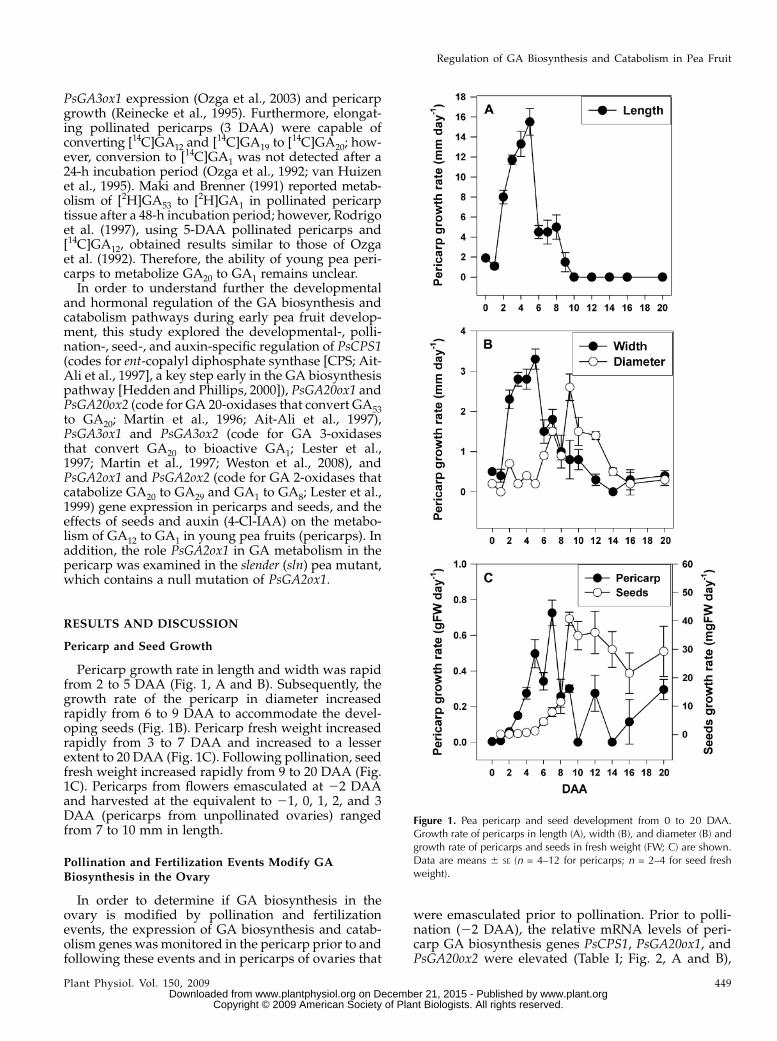
Pericarp growth rate in length and width was rapidfrom 2 to 5 DAA (Fig. 1, A and B). Subsequently, thegrowth rate of the pericarp in diameter increasedrapidly from 6 to 9 DAA to accommodate the devel-oping seeds (Fig. 1B). Pericarp fresh weight increasedrapidly from 3 to 7 DAA and increased to a lesserextent to 20 DAA (Fig. 1C). Following pollination, seedfresh weight increased rapidly from 9 to 20 DAA (Fig.1C). Pericarps from flowers emasculated at 22 DAAand harvested at the equivalent to 21, 0, 1, 2, and 3DAA (pericarps from unpollinated ovaries) rangedfrom 7 to 10 mm in length.
Pollination and Fertilization Events Modify GABiosynthesis in the Ovary
In order to determine if GA biosynthesis in theovary is modified by pollination and fertilizationevents, the expression of GA biosynthesis and catab-olism genes wasmonitored in the pericarp prior to andfollowing these events and in pericarps of ovaries that
were emasculated prior to pollination. Prior to polli-nation (22 DAA), the relative mRNA levels of peri-carp GA biosynthesis genes PsCPS1, PsGA20ox1, andPsGA20ox2 were elevated (Table I; Fig. 2, A and B),
Figure 1. Pea pericarp and seed development from 0 to 20 DAA.Growth rate of pericarps in length (A), width (B), and diameter (B) andgrowth rate of pericarps and seeds in fresh weight (FW; C) are shown.Data are means 6 SE (n = 4–12 for pericarps; n = 2–4 for seed freshweight).
Regulation of GA Biosynthesis and Catabolism in Pea Fruit
Plant Physiol. Vol. 150, 2009 449 www.plant.org on December 21, 2015 - Published by www.plantphysiol.orgDownloaded from
Copyright © 2009 American Society of Plant Biologists. All rights reserved.
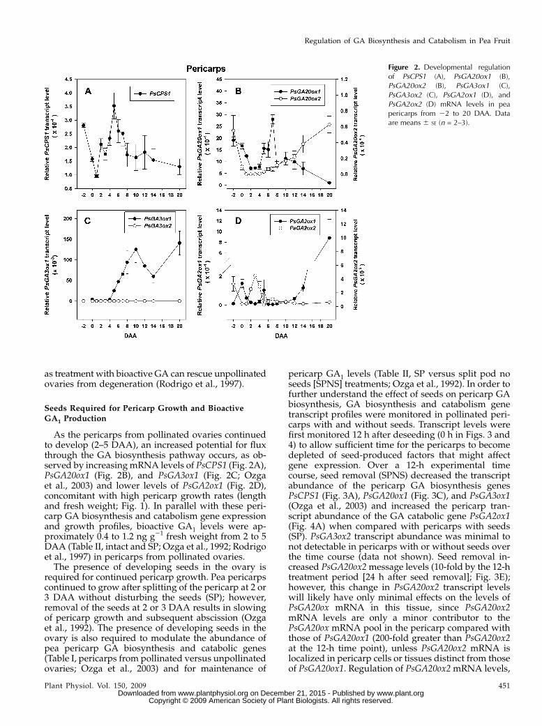
suggesting that unpollinated pericarps are capableof GA biosynthesis from ent-copalyl diphosphatethrough to GA20. However, at 22 DAA, pericarpmRNA levels of PsGA3ox1 and PsGA3ox2 (code forGA 3-oxidases that convert GA20 to biologically activeGA1) were minimal (Table I; Fig. 2C; Ozga et al., 2003)and transcript levels of PsGA2ox2 (codes for the GA2-oxidase that preferentially converts GA1 to biologicallyinactive GA8) were elevated (Table I; Fig. 2D), suggest-ing minimal presence of bioactive GA1 in unpollinatedpericarps. Consistent with these gene expression data,emasculated (unpollinated) ovaries (at 0 DAA) werefound to contain GA20 (approximately 5 ng g21 freshweight; cv Alaska) but minimal to no detectable levelsof GA1 (Garcia-Martinez et al., 1991).
Following pollination and fertilization (complete by0 DAA), the pericarp is the major nutrient sink tissuein developing pea fruit until approximately 8 to 12DAA, when the seeds become the terminal sink (cvAlaska; Johnstone, 2004). The transcription profiles ofthe GA biosynthesis and catabolism genes dramati-cally changed in the pericarp following the pollinationand fertilization events. From 22 (unpollinated) to 0DAA (pollinated), transcript abundance of pericarpPsCPS1, PsGA20ox1, and PsGA20ox2 that code forenzymes that synthesize precursors of bioactive GA1declined (Table I; Fig. 2A and B), while mRNA levels ofPsGA3ox1 that codes for the enzyme that synthesizesGA1 increased (Table I; Ozga et al., 2003) andPsGA2ox2 that codes for an enzyme that catabolizesGA1 decreased (Table I; Fig. 2D). Pollination andfertilization also lessened the increase in transcriptabundance of pericarp PsGA2ox1 (also codes for anenzyme that catabolizes GA1 to GA8) from 50-fold to15-fold (pollinated versus unpollinated pericarps at0 DAA; Table I). In pollinated pericarps from 0 to1 DAA, the abundance of PsGA3ox1 mRNA decreased(Table I; Ozga et al., 2003), along with further declinesin the mRNA abundance of the GA biosynthesis genesPsCPS1, PsGA20ox1, and PsGA20ox2. The GA catabolicgene PsGA2ox1 also decreased from 0 to 1 DAA (Table
I; Fig. 2). PsGA3ox2 mRNA transcripts were notdetected in the pericarp tissue during this develop-mental phase (Table I). This coordination of GA bio-synthesis and catabolism gene transcript levels in thepericarp suggests that regulation at the level of tran-script production or stability is involved in a transitoryincrease in bioactive GA1 in the pericarp followingpollination and fertilization to stimulate initial growthand fruit set.
Although steady-state GA1 levels were reported tobe minimal to undetectable in pollinated and unpolli-nated ovaries at 0 DAA, GA8 levels were two timeshigher in pollinated than unpollinated ovaries at thistime (Garcia-Martinez et al., 1991). Since GA8 is theimmediate biologically inactive product of GA1 (as aresult of 2b-hydroxylation), these data suggest thatGA1 was synthesized to a greater extent in pollinatedthan unpollinated pericarps and/or ovules by 0 DAA.
In the absence of pollination and fertilization, peri-carp PsGA3ox1 mRNA abundance did not peak at 0DAA (as in pollinated pericarps) but peaked at 3 DAA,likely the result of feedback regulation due to minimallevels (or the absence) of bioactive GA1 (Table I; Ozgaet al., 2003). Interestingly, PsGA20ox1 mRNA levelswere higher in unpollinated pericarps than thoseobserved in pollinated pericarps (Table I; Garcia-Martinez et al., 1997; van Huizen et al., 1997); however,the pool of GA20 was reduced by half in the unpolli-nated pericarps by 2 DAA (Garcia-Martinez et al.,1991). This decline in GA20 in unpollinated pericarps islikely a result of catabolism to GA29 by GA 2-oxidase,as PsGA2ox1 mRNA levels increased approximately50-fold from22 to 0 DAA in unpollinated pericarps (asmaller increase of 15-fold was observed in pollinatedpericarps at this time; Table I). Consistent with theelevated mRNA abundance of PsGA2ox1, a 3.3-foldhigher GA29-to-GA20 ratio was found in unpollinatedovaries compared with pericarps from pollinated ova-ries (Santes and Garcia-Martinez, 1995). By 4 DAA, theunpollinated ovaries degenerated (data not shown),likely in part due to minimal GA1 levels in the tissue,
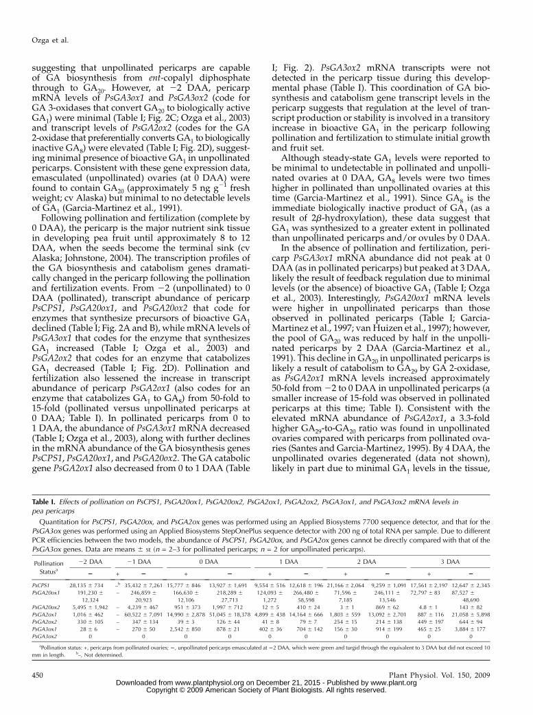
Table I. Effects of pollination on PsCPS1, PsGA20ox1, PsGA20ox2, PsGA2ox1, PsGA2ox2, PsGA3ox1, and PsGA3ox2 mRNA levels inpea pericarps
Quantitation for PsCPS1, PsGA20ox, and PsGA2ox genes was performed using an Applied Biosystems 7700 sequence detector, and that for thePsGA3ox genes was performed using an Applied Biosystems StepOnePlus sequence detector with 200 ng of total RNA per sample. Due to differentPCR efficiencies between the two models, the abundance of PsCPS1, PsGA20ox, and PsGA2ox genes cannot be directly compared with that of thePsGA3ox genes. Data are means 6 SE (n = 2–3 for pollinated pericarps; n = 2 for unpollinated pericarps).
Pollination
Statusa22 DAA 21 DAA 0 DAA 1 DAA 2 DAA 3 DAA
2 + 2 + 2 + 2 + 2 + 2
PsCPS1 28,135 6 734 –b 35,432 6 7,261 15,777 6 846 13,927 6 1,691 9,554 6 516 12,618 6 196 21,1666 2,064 9,259 6 1,091 17,5616 2,197 12,647 6 2,345
PsGA20ox1 191,230 6
12,324
– 246,859 6
20,923
166,630 6
12,106
218,289 6
27,713
124,093 6
1,272
266,480 6
58,598
71,596 6
7,185
246,111 6
13,546
72,7976 83 87,527 6
48,690
PsGA20ox2 5,495 6 1,942 – 4,239 6 467 951 6 373 1,997 6 712 12 6 5 410 6 24 3 6 1 869 6 62 4.8 6 1 143 6 82
PsGA2ox1 1,016 6 462 – 60,522 6 7,091 14,990 6 2,878 51,045 6 18,378 4,899 6 438 14,164 6 666 1,803 6 559 13,092 6 2,701 887 6 116 21,058 6 5,898
PsGA2ox2 330 6 105 – 347 6 134 39 6 3 126 6 44 41 6 8 79 6 7 254 6 15 214 6 138 449 6 197 644 6 94
PsGA3ox1 28 6 6 – 270 6 50 2,542 6 850 878 6 21 402 6 36 704 6 142 156 6 30 914 6 199 465 6 25 3,884 6 177
PsGA3ox2 0 0 0 0 0 0 0 0 0 0
aPollination status: +, pericarps from pollinated ovaries; 2, unpollinated pericarps emasculated at 22 DAA, which were green and turgid through the equivalent to 3 DAA but did not exceed 10
mm in length. b–, Not determined.
Ozga et al.
450 Plant Physiol. Vol. 150, 2009 www.plant.org on December 21, 2015 - Published by www.plantphysiol.orgDownloaded from
Copyright © 2009 American Society of Plant Biologists. All rights reserved.
as treatment with bioactive GA can rescue unpollinatedovaries from degeneration (Rodrigo et al., 1997).
Seeds Required for Pericarp Growth and Bioactive
GA1 Production
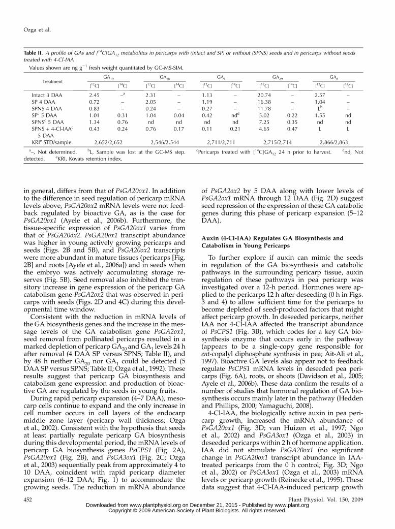
As the pericarps from pollinated ovaries continuedto develop (2–5 DAA), an increased potential for fluxthrough the GA biosynthesis pathway occurs, as ob-served by increasingmRNA levels of PsCPS1 (Fig. 2A),PsGA20ox1 (Fig. 2B), and PsGA3ox1 (Fig. 2C; Ozgaet al., 2003) and lower levels of PsGA2ox1 (Fig. 2D),concomitant with high pericarp growth rates (lengthand fresh weight; Fig. 1). In parallel with these peri-carp GA biosynthesis and catabolism gene expressionand growth profiles, bioactive GA1 levels were ap-proximately 0.4 to 1.2 ng g21 fresh weight from 2 to 5DAA (Table II, intact and SP; Ozga et al., 1992; Rodrigoet al., 1997) in pericarps from pollinated ovaries.The presence of developing seeds in the ovary is
required for continued pericarp growth. Pea pericarpscontinued to grow after splitting of the pericarp at 2 or3 DAA without disturbing the seeds (SP); however,removal of the seeds at 2 or 3 DAA results in slowingof pericarp growth and subsequent abscission (Ozgaet al., 1992). The presence of developing seeds in theovary is also required to modulate the abundance ofpea pericarp GA biosynthesis and catabolic genes(Table I, pericarps from pollinated versus unpollinatedovaries; Ozga et al., 2003) and for maintenance of
pericarp GA1 levels (Table II, SP versus split pod noseeds [SPNS] treatments; Ozga et al., 1992). In order tofurther understand the effect of seeds on pericarp GAbiosynthesis, GA biosynthesis and catabolism genetranscript profiles were monitored in pollinated peri-carps with and without seeds. Transcript levels werefirst monitored 12 h after deseeding (0 h in Figs. 3 and4) to allow sufficient time for the pericarps to becomedepleted of seed-produced factors that might affectgene expression. Over a 12-h experimental timecourse, seed removal (SPNS) decreased the transcriptabundance of the pericarp GA biosynthesis genesPsCPS1 (Fig. 3A), PsGA20ox1 (Fig. 3C), and PsGA3ox1(Ozga et al., 2003) and increased the pericarp tran-script abundance of the GA catabolic gene PsGA2ox1(Fig. 4A) when compared with pericarps with seeds(SP). PsGA3ox2 transcript abundance was minimal tonot detectable in pericarps with or without seeds overthe time course (data not shown). Seed removal in-creased PsGA20ox2message levels (10-fold by the 12-htreatment period [24 h after seed removal]; Fig. 3E);however, this change in PsGA20ox2 transcript levelswill likely have only minimal effects on the levels ofPsGA20ox mRNA in this tissue, since PsGA20ox2mRNA levels are only a minor contributor to thePsGA20ox mRNA pool in the pericarp compared withthose of PsGA20ox1 (200-fold greater than PsGA20ox2at the 12-h time point), unless PsGA20ox2 mRNA islocalized in pericarp cells or tissues distinct from thoseof PsGA20ox1. Regulation of PsGA20ox2mRNA levels,
Figure 2. Developmental regulationof PsCPS1 (A), PsGA20ox1 (B),PsGA20ox2 (B), PsGA3ox1 (C),PsGA3ox2 (C), PsGA2ox1 (D), andPsGA2ox2 (D) mRNA levels in peapericarps from 22 to 20 DAA. Dataare means 6 SE (n = 2–3).
Regulation of GA Biosynthesis and Catabolism in Pea Fruit
Plant Physiol. Vol. 150, 2009 451 www.plant.org on December 21, 2015 - Published by www.plantphysiol.orgDownloaded from
Copyright © 2009 American Society of Plant Biologists. All rights reserved.
in general, differs from that of PsGA20ox1. In additionto the difference in seed regulation of pericarp mRNAlevels above, PsGA20ox2 mRNA levels were not feed-back regulated by bioactive GA, as is the case forPsGA20ox1 (Ayele et al., 2006b). Furthermore, thetissue-specific expression of PsGA20ox1 varies fromthat of PsGA20ox2. PsGA20ox1 transcript abundancewas higher in young actively growing pericarps andseeds (Figs. 2B and 5B), and PsGA20ox2 transcriptswere more abundant in mature tissues (pericarps [Fig.2B] and roots [Ayele et al., 2006a]) and in seeds whenthe embryo was actively accumulating storage re-serves (Fig. 5B). Seed removal also inhibited the tran-sitory increase in gene expression of the pericarp GAcatabolism gene PsGA2ox2 that was observed in peri-carps with seeds (Figs. 2D and 4C) during this devel-opmental time window.
Consistent with the reduction in mRNA levels ofthe GA biosynthesis genes and the increase in the mes-sage levels of the GA catabolism gene PsGA2ox1,seed removal from pollinated pericarps resulted in amarked depletion of pericarp GA20 and GA1 levels 24 hafter removal (4 DAA SP versus SPNS; Table II), andby 48 h neither GA20 nor GA1 could be detected (5DAA SP versus SPNS; Table II; Ozga et al., 1992). Theseresults suggest that pericarp GA biosynthesis andcatabolism gene expression and production of bioac-tive GA are regulated by the seeds in young fruits.
During rapid pericarp expansion (4–7 DAA), meso-carp cells continue to expand and the only increase incell number occurs in cell layers of the endocarpmiddle zone layer (pericarp wall thickness; Ozgaet al., 2002). Consistent with the hypothesis that seedsat least partially regulate pericarp GA biosynthesisduring this developmental period, the mRNA levels ofpericarp GA biosynthesis genes PsCPS1 (Fig. 2A),PsGA20ox1 (Fig. 2B), and PsGA3ox1 (Fig. 2C; Ozgaet al., 2003) sequentially peak from approximately 4 to10 DAA, coincident with rapid pericarp diameterexpansion (6–12 DAA; Fig. 1) to accommodate thegrowing seeds. The reduction in mRNA abundance
of PsGA2ox2 by 5 DAA along with lower levels ofPsGA2ox1 mRNA through 12 DAA (Fig. 2D) suggestseed repression of the expression of these GA catabolicgenes during this phase of pericarp expansion (5–12DAA).
Auxin (4-Cl-IAA) Regulates GA Biosynthesis andCatabolism in Young Pericarps
To further explore if auxin can mimic the seedsin regulation of the GA biosynthesis and catabolicpathways in the surrounding pericarp tissue, auxinregulation of these pathways in pea pericarp wasinvestigated over a 12-h period. Hormones were ap-plied to the pericarps 12 h after deseeding (0 h in Figs.3 and 4) to allow sufficient time for the pericarps tobecome depleted of seed-produced factors that mightaffect pericarp growth. In deseeded pericarps, neitherIAA nor 4-Cl-IAA affected the transcript abundanceof PsCPS1 (Fig. 3B), which codes for a key GA bio-synthesis enzyme that occurs early in the pathway(appears to be a single-copy gene responsible forent-copalyl diphosphate synthesis in pea; Ait-Ali et al.,1997). Bioactive GA levels also appear not to feedbackregulate PsCPS1 mRNA levels in deseeded pea peri-carps (Fig. 6A), roots, or shoots (Davidson et al., 2005;Ayele et al., 2006b). These data confirm the results of anumber of studies that hormonal regulation of GA bio-synthesis occurs mainly later in the pathway (Heddenand Phillips, 2000; Yamaguchi, 2008).
4-Cl-IAA, the biologically active auxin in pea peri-carp growth, increased the mRNA abundance ofPsGA20ox1 (Fig. 3D; van Huizen et al., 1997; Ngoet al., 2002) and PsGA3ox1 (Ozga et al., 2003) indeseeded pericarps within 2 h of hormone application.IAA did not stimulate PsGA20ox1 (no significantchange in PsGA20ox1 transcript abundance in IAA-treated pericarps from the 0 h control; Fig. 3D; Ngoet al., 2002) or PsGA3ox1 (Ozga et al., 2003) mRNAlevels or pericarp growth (Reinecke et al., 1995). Thesedata suggest that 4-Cl-IAA-induced pericarp growth
Table II. A profile of GAs and [14C]GA12 metabolites in pericarps with (intact and SP) or without (SPNS) seeds and in pericarps without seedstreated with 4-Cl-IAA
Values shown are ng g21 fresh weight quantitated by GC-MS-SIM.
TreatmentGA19 GA20 GA1 GA29 GA8
[12C] [14C] [12C] [14C] [12C] [14C] [12C] [14C] [12C] [14C]
Intact 3 DAA 2.45 –a 2.31 – 1.13 – 20.74 – 2.57 –SP 4 DAA 0.72 – 2.05 – 1.19 – 16.38 – 1.04 –SPNS 4 DAA 0.83 – 0.24 – 0.27 – 11.78 – Lb –SPc 5 DAA 1.01 0.31 1.04 0.04 0.42 ndd 5.02 0.22 1.55 ndSPNSc 5 DAA 1.34 0.76 nd nd nd nd 7.25 0.35 nd ndSPNS + 4-Cl-IAAc
5 DAA0.43 0.24 0.76 0.17 0.11 0.21 4.65 0.47 L L
KRIe STD/sample 2,652/2,652 2,546/2,544 2,711/2,711 2,715/2,714 2,866/2,863
a–, Not determined. bL, Sample was lost at the GC-MS step. cPericarps treated with [14C]GA12 24 h prior to harvest. dnd, Notdetected. eKRI, Kovats retention index.
Ozga et al.
452 Plant Physiol. Vol. 150, 2009 www.plant.org on December 21, 2015 - Published by www.plantphysiol.orgDownloaded from
Copyright © 2009 American Society of Plant Biologists. All rights reserved.
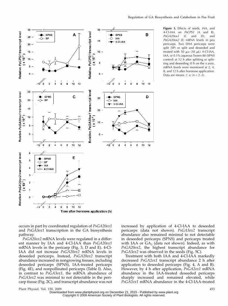
occurs in part by coordinated regulation of PsGA20ox1and PsGA3ox1 transcription in the GA biosynthesispathway.PsGA20ox2 mRNA levels were regulated in a differ-
ent manner by IAA and 4-Cl-IAA than PsGA20ox1mRNA levels in the pericarp (Fig. 3, D and E). 4-Cl-IAA did not increase PsGA20ox2 mRNA levels indeseeded pericarps. Instead, PsGA20ox2 transcriptabundance increased in nongrowing tissues, includingdeseeded pericarps (SPNS), IAA-treated pericarps(Fig. 4E), and nonpollinated pericarps (Table I). Also,in contrast to PsGA3ox1, the mRNA abundance ofPsGA3ox2 was minimal to not detectable in the peri-carp tissue (Fig. 2C), and transcript abundance was not
increased by application of 4-Cl-IAA to deseededpericarps (data not shown). PsGA3ox2 transcriptabundance also remained minimal to not detectablein deseeded pericarps (SPNS) and pericarps treatedwith IAA or GA3 (data not shown). Indeed, as withPsGA20ox2, the highest transcript abundance forPsGA3ox2 was observed in the seeds (Fig. 5C).
Treatment with both IAA and 4-Cl-IAA markedlydecreased PsGA2ox1 transcript abundance 2 h afterapplication to deseeded pericarps (Fig. 4, A and B).However, by 4 h after application, PsGA2ox1 mRNAabundance in the IAA-treated deseeded pericarpssharply increased and remained elevated, whilePsGA2ox1 mRNA abundance in the 4-Cl-IAA-treated
Figure 3. Effects of seeds, IAA, and4-Cl-IAA on PsCPS1 (A and B),PsGA20ox1 (C and D), andPsGA20ox2 (E) mRNA levels in peapericarps. Two DAA pericarps weresplit (SP) or split and deseeded andtreated with 50 mM (30 mL) 4-Cl-IAA,IAA, or 0.1% aqueous Tween 80 (SPNScontrol) at 12 h after splitting or split-ting and deseeding (0 h on the x axis).mRNA levels were monitored at 2, 4,8, and 12 h after hormone application.Data are means 6 SE (n = 2–3).
Regulation of GA Biosynthesis and Catabolism in Pea Fruit
Plant Physiol. Vol. 150, 2009 453 www.plant.org on December 21, 2015 - Published by www.plantphysiol.orgDownloaded from
Copyright © 2009 American Society of Plant Biologists. All rights reserved.
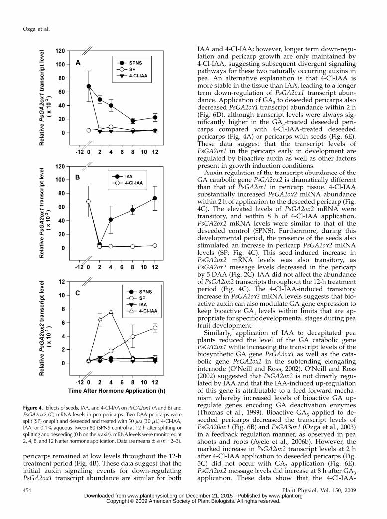
pericarps remained at low levels throughout the 12-htreatment period (Fig. 4B). These data suggest that theinitial auxin signaling events for down-regulatingPsGA2ox1 transcript abundance are similar for both
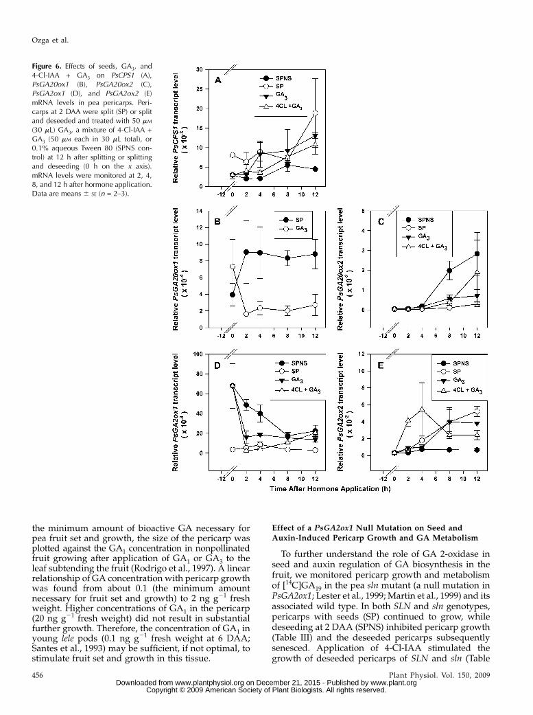
IAA and 4-Cl-IAA; however, longer term down-regu-lation and pericarp growth are only maintained by4-Cl-IAA, suggesting subsequent divergent signalingpathways for these two naturally occurring auxins inpea. An alternative explanation is that 4-Cl-IAA ismore stable in the tissue than IAA, leading to a longerterm down-regulation of PsGA2ox1 transcript abun-dance. Application of GA3 to deseeded pericarps alsodecreased PsGA2ox1 transcript abundance within 2 h(Fig. 6D), although transcript levels were always sig-nificantly higher in the GA3-treated deseeded peri-carps compared with 4-Cl-IAA-treated deseededpericarps (Fig. 4A) or pericarps with seeds (Fig. 6E).These data suggest that the transcript levels ofPsGA2ox1 in the pericarp early in development areregulated by bioactive auxin as well as other factorspresent in growth induction conditions.
Auxin regulation of the transcript abundance of theGA catabolic gene PsGA2ox2 is dramatically differentthan that of PsGA2ox1 in pericarp tissue. 4-Cl-IAAsubstantially increased PsGA2ox2 mRNA abundancewithin 2 h of application to the deseeded pericarp (Fig.4C). The elevated levels of PsGA2ox2 mRNA weretransitory, and within 8 h of 4-Cl-IAA application,PsGA2ox2 mRNA levels were similar to that of thedeseeded control (SPNS). Furthermore, during thisdevelopmental period, the presence of the seeds alsostimulated an increase in pericarp PsGA2ox2 mRNAlevels (SP; Fig. 4C). This seed-induced increase inPsGA2ox2 mRNA levels was also transitory, asPsGA2ox2 message levels decreased in the pericarpby 5 DAA (Fig. 2C). IAA did not affect the abundanceof PsGA2ox2 transcripts throughout the 12-h treatmentperiod (Fig. 4C). The 4-Cl-IAA-induced transitoryincrease in PsGA2ox2 mRNA levels suggests that bio-active auxin can also modulate GA gene expression tokeep bioactive GA1 levels within limits that are ap-propriate for specific developmental stages during peafruit development.
Similarly, application of IAA to decapitated peaplants reduced the level of the GA catabolic genePsGA2ox1 while increasing the transcript levels of thebiosynthetic GA gene PsGA3ox1 as well as the cata-bolic gene PsGA2ox2 in the subtending elongatinginternode (O’Neill and Ross, 2002). O’Neill and Ross(2002) suggested that PsGA2ox2 is not directly regu-lated by IAA and that the IAA-induced up-regulationof this gene is attributable to a feed-forward mecha-nism whereby increased levels of bioactive GA up-regulate genes encoding GA deactivation enzymes(Thomas et al., 1999). Bioactive GA3 applied to de-seeded pericarps decreased the transcript levels ofPsGA20ox1 (Fig. 6B) and PsGA3ox1 (Ozga et al., 2003)in a feedback regulation manner, as observed in peashoots and roots (Ayele et al., 2006b). However, themarked increase in PsGA2ox2 transcript levels at 2 hafter 4-Cl-IAA application to deseeded pericarps (Fig.5C) did not occur with GA3 application (Fig. 6E).PsGA2ox2 message levels did increase at 8 h after GA3application. These data show that the 4-Cl-IAA-
Figure 4. Effects of seeds, IAA, and 4-Cl-IAA on PsGA2ox1 (A and B) andPsGA2ox2 (C) mRNA levels in pea pericarps. Two DAA pericarps weresplit (SP) or split and deseeded and treated with 50 mM (30 mL) 4-Cl-IAA,IAA, or 0.1% aqueous Tween 80 (SPNS control) at 12 h after splitting orsplitting and deseeding (0 h on the x axis). mRNA levels weremonitored at2, 4, 8, and 12 h after hormone application. Data aremeans6 SE (n= 2–3).
Ozga et al.
454 Plant Physiol. Vol. 150, 2009 www.plant.org on December 21, 2015 - Published by www.plantphysiol.orgDownloaded from
Copyright © 2009 American Society of Plant Biologists. All rights reserved.
induced up-regulation of pericarp PsGA2ox2 is notdirectly attributable to a bioactive GA feed-forwardmechanism but is likely a direct effect of this bioactiveauxin on PsGA2ox2message levels to regulate the half-life of GA1 in this tissue. The delayed increase inPsGA2ox2 levels (first observed at 8 h after hormonetreatment) by GA3 may indicate that a feed-forwardmechanism is present, but it is distinct from the early4-Cl-IAA-induced response on PsGA2ox2 gene expres-sion. When GA3 was applied in combination with4-Cl-IAA, the bioactive auxin response of stimulationof PsGA2ox2 expression was again observed within 2 hafter hormone application, followed by the GA3response of a delayed stimulation of PsGA2ox2transcript levels (8–12 h after hormone application;Fig. 6E).Overall, the GA biosynthesis and catabolic tran-
scription profiles suggest that 4-Cl-IAA can stimulatethe production of bioactive GA1 in the pericarp as wellas modulate the half-life of GA1 by regulating themRNA levels of the catabolic gene PsGA2ox2. Inter-estingly, conversion of labeled GAs to GA1 in pericarpshas been difficult to obtain using conventionalmethods of detection (Ozga et al., 1992; van Huizenet al., 1995; Rodrigo et al., 1997). In the current study,when [14C]GA12 was applied to pericarps with seeds,endogenous and 14C-labeled GA19, GA20, and GA29were detected along with endogenous GA1 and GA8,but [14C]GA1 was not detected (SP 5 DAA; Table II). Indeseeded pericarps treated with [14C]GA12, only en-dogenous and 14C-labeled GA19 and GA29 were detected(SPNS 5 DAA; Table II). These results suggest that
seeds are required for GA biosynthesis in the pericarpand are consistent with our previous findings (Ozgaet al., 1992; van Huizen et al., 1995). However, whendeseeded pericarps were treated with 4-Cl-IAA, en-dogenous and 14C-labeled GA1, along with endoge-nous and 14C-labeled GA19, GA20, and GA29, weredetected (SPNS + 4-Cl-IAA 5 DAA; Table II). Theradiolabeling of GA1 is most likely due to the ability of4-Cl-IAA to stimulate the transcript levels of the GAbiosynthesis genes PsGA20ox1 and PsGA3ox1 and de-crease the mRNA levels of the catabolic genePsGA2ox1 in pericarp tissue. In addition, a labeledsubstrate with higher specific radioactivity and onefarther up the pathway ([14C]GA12 used in this studycompared with [14C]GA19 used by van Huizen et al.[1995]) are likely both important to increase sensitivityand avoid increased metabolism of the labeled sub-strate into the inactive GA pool (GA29 and otherinactive GA metabolites).
It also has been proposed that the sensitivity of fruitto bioactive GA may be substantially greater than thatof the stem internodes in pea. Comparison of near-isogenic lines of pea that contain either the wild-typePsGA3ox1 gene (LELE) or a 1-bp point mutation of LEthat greatly increases the Km of the encoded GA3-oxidase (lele) demonstrated that the mutation (lelegenotype) reduced GA1 content and growth of inter-nodes (Ross et al., 1992). The content of GA1 was alsolower (seven to 10 times) in young lele pericarpscompared with those of LELE, but only minor effectson fruit growth were observed (Santes et al., 1993;MacKenzie-Hose et al., 1998). In order to determine
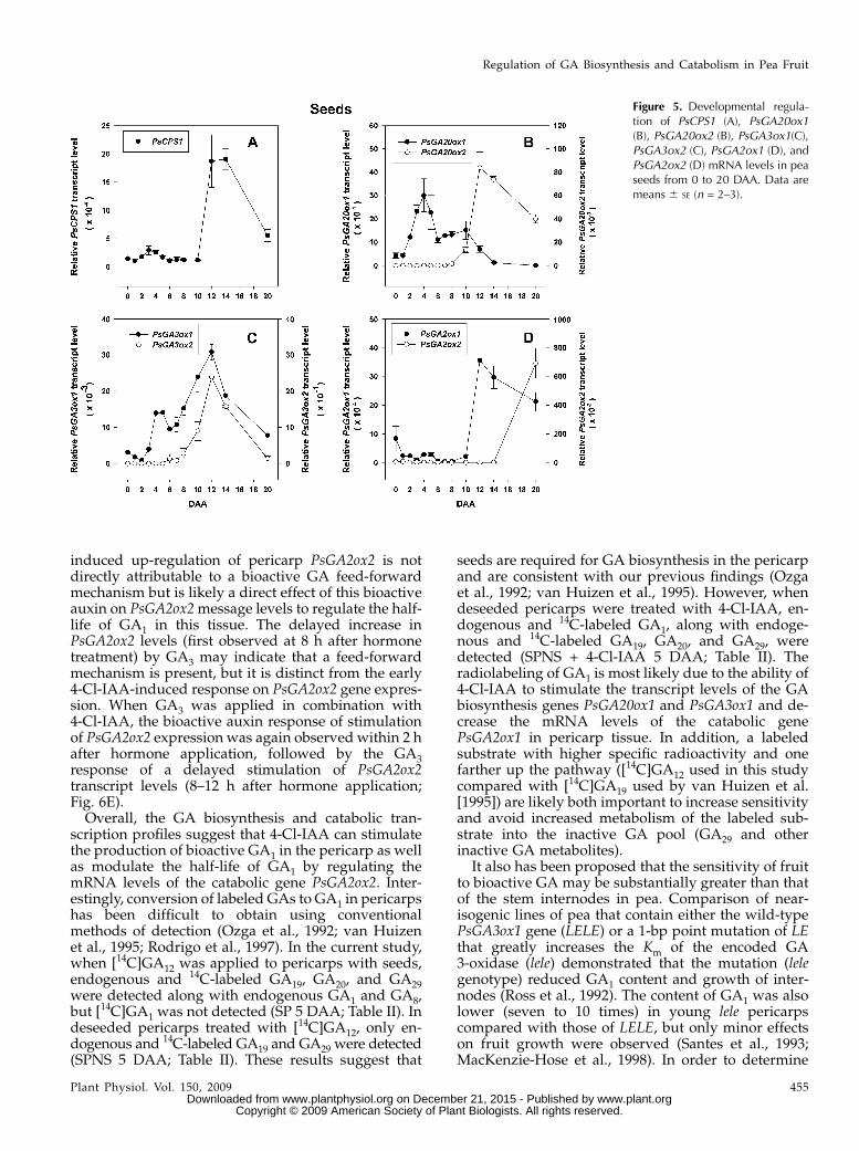
Figure 5. Developmental regula-tion of PsCPS1 (A), PsGA20ox1(B), PsGA20ox2 (B), PsGA3ox1(C),PsGA3ox2 (C), PsGA2ox1 (D), andPsGA2ox2 (D) mRNA levels in peaseeds from 0 to 20 DAA. Data aremeans 6 SE (n = 2–3).
Regulation of GA Biosynthesis and Catabolism in Pea Fruit
Plant Physiol. Vol. 150, 2009 455 www.plant.org on December 21, 2015 - Published by www.plantphysiol.orgDownloaded from
Copyright © 2009 American Society of Plant Biologists. All rights reserved.
the minimum amount of bioactive GA necessary forpea fruit set and growth, the size of the pericarp wasplotted against the GA1 concentration in nonpollinatedfruit growing after application of GA1 or GA3 to theleaf subtending the fruit (Rodrigo et al., 1997). A linearrelationship of GA concentration with pericarp growthwas found from about 0.1 (the minimum amountnecessary for fruit set and growth) to 2 ng g21 freshweight. Higher concentrations of GA1 in the pericarp(20 ng g21 fresh weight) did not result in substantialfurther growth. Therefore, the concentration of GA1 inyoung lele pods (0.1 ng g21 fresh weight at 6 DAA;Santes et al., 1993) may be sufficient, if not optimal, tostimulate fruit set and growth in this tissue.
Effect of a PsGA2ox1 Null Mutation on Seed andAuxin-Induced Pericarp Growth and GA Metabolism
To further understand the role of GA 2-oxidase inseed and auxin regulation of GA biosynthesis in thefruit, we monitored pericarp growth and metabolismof [14C]GA19 in the pea sln mutant (a null mutation inPsGA2ox1; Lester et al., 1999; Martin et al., 1999) and itsassociated wild type. In both SLN and sln genotypes,pericarps with seeds (SP) continued to grow, whiledeseeding at 2 DAA (SPNS) inhibited pericarp growth(Table III) and the deseeded pericarps subsequentlysenesced. Application of 4-Cl-IAA stimulated thegrowth of deseeded pericarps of SLN and sln (Table
Figure 6. Effects of seeds, GA3, and4-Cl-IAA + GA3 on PsCPS1 (A),PsGA20ox1 (B), PsGA20ox2 (C),PsGA2ox1 (D), and PsGA2ox2 (E)mRNA levels in pea pericarps. Peri-carps at 2 DAA were split (SP) or splitand deseeded and treated with 50 mM
(30 mL) GA3, a mixture of 4-Cl-IAA +GA3 (50 mM each in 30 mL total), or0.1% aqueous Tween 80 (SPNS con-trol) at 12 h after splitting or splittingand deseeding (0 h on the x axis).mRNA levels were monitored at 2, 4,8, and 12 h after hormone application.Data are means 6 SE (n = 2–3).
Ozga et al.
456 Plant Physiol. Vol. 150, 2009 www.plant.org on December 21, 2015 - Published by www.plantphysiol.orgDownloaded from
Copyright © 2009 American Society of Plant Biologists. All rights reserved.
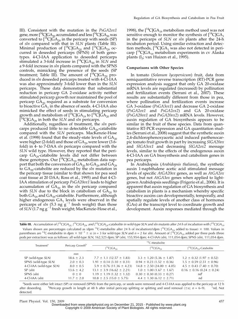
III). Consistent with the mutation in the PsGA2ox1gene,more [14C]GA20 accumulated and less [14C]GA20 wasconverted to [14C]GA29 in the pericarp with seeds (SP)of sln compared with that in SLN plants (Table III).Minimal production of [14C]GA20 and [14C]GA29 oc-curred in deseeded pericarps (SPNS) of both geno-types. 4-Cl-IAA application to deseeded pericarpsstimulated a 3-fold increase in [14C]GA20 in SLN anda 9-fold increase in sln plants compared with the SPNScontrols, mimicking the presence of the seeds (SPtreatment; Table III). The amount of [14C]GA29 pro-duced in sln deseeded pericarps treated with 4-Cl-IAAwas also approximately 3-fold lower than in the SLNpericarps. These data demonstrate that substantialreduction in pericarp GA 2-oxidase activity neitherstimulated pericarp growth nor maintained the pool ofpericarp GA20 required as a substrate for conversionto bioactive GA1 in the absence of seeds. 4-Cl-IAA alsomimicked the effect of seeds in stimulating pericarpgrowth and metabolism of [14C]GA19 to [14C]GA20 and[14C]GA29 in both the SLN and sln pericarps.Additionally, regardless of treatment, the sln peri-
carps produced little to no detectable GA29-catabolitecompared with the SLN pericarps. MacKenzie-Hoseet al. (1998) found that the steady-state levels of GA20were higher (2-fold) and those of GA29 were lower (3.6-fold) in 4- to 7-DAA sln pericarps compared with theSLN wild type. However, they reported that the peri-carp GA29-catabolite levels did not differ betweenthese genotypes. Our [14C]GA19 metabolism data sup-port that both the conversion of GA20 to GA29 and GA29to GA29-catabolite are reduced by the sln mutation inthe pericarp tissue (similar to that shown for pea seedcoat tissue at 20 DAA; Ross et al., 1995) and that 4-Cl-IAA stimulation of pericarp PsGA20ox1 leads to higheraccumulation of GA20 in the sln pericarp comparedwith SLN due to the block in catabolism of GA20 toboth GA29 and GA29-catabolite. Furthermore, althoughhigher endogenous GA1 levels were observed in thepericarps of sln (5.3 ng g21 fresh weight) than thoseof SLN (1.7 ng g21 fresh weight; MacKenzie-Hose et al.,
1998), the [14C]GA19 metabolism method used was notsensitive enough to monitor the synthesis of [14C]GA1in the pericarps of SLN or sln plants after the 24-hincubation period. Using similar extraction and detec-tion methods, [14C]GA1 was also not detected in peri-carp [14C]GA19 metabolism experiments in cv Alaskaplants (I3; van Huizen et al., 1995).
Comparisons with Other Species
In tomato (Solanum lycopersicum) fruit, data fromsemiquantitative reverse transcription (RT)-PCR geneexpression analysis suggest that only GA 20-oxidasemRNA levels are regulated (increased) by pollinationand fertilization events (Serrani et al., 2007). Theseresults are substantially different from those in pea,where pollination and fertilization events increaseGA 3-oxidase (PsGA3ox1) and decrease GA 2-oxidase(PsGA2ox1 and PsGA2ox2) and GA 20-oxidase(PsGA20ox1 and PsGA20ox2) mRNA levels. However,auxin regulation of GA biosynthesis appears to besimilar in the fruit of these species. Data from quan-titative RT-PCR expression and GA quantitation stud-ies (Serrani et al., 2008) suggest that the synthetic auxin2,4-dichlorophenoxyacetic acid induced parthenocar-pic tomato fruit growth in part by increasing SlGA20oxand SlGA3ox1 and decreasing SlGA2ox2 messagelevels, similar to the effects of the endogenous auxin4-Cl-IAA on GA biosynthesis and catabolism genes inpea pericarps.
In Arabidopsis (Arabidopsis thaliana), the syntheticauxin 1-naphthalene acetic acid stimulated messagelevels of specific AtGA20ox genes, as well as AtGA2oxgenes, but not AtGA3ox genes when applied to light-grownArabidopsis seedlings (Frigerio et al., 2006). It isapparent that auxin regulation of GA biosynthesis andcatabolism in plants is a mechanism whereby specificbioactive auxins can developmentally, temporally, andspatially regulate levels of another class of hormones(GAs) at the transcript level to coordinate growth anddevelopment. Auxin responses mediated through the
Table III. Accumulation of [14C]GA20, [14C]GA29, and [14C]GA29-catabolite in wild-type SLN and sln mutants after 24 h of incubation with [14C]GA19
Values shown are percentages calculated as (dpm 14C-metabolite after 24 h of incubation)/(dpm [14C]GA19 added to tissue) 3 100. Values inparentheses are 14C-metabolite in dpm 3 1023 6 SE (n = 3 for wild-type SLN and n = 2 for sln). Amount of [14C]GA19 added per three pods (threepods per extraction) was as follows: all wild-type SLN, 162,525 dpm; SP (sln), 155,954 dpm; 4-Cl-IAA (sln), 111,054 dpm; SPNS (sln), 111,054 dpm.
Treatmenta Pericarp Growthb14C-Metabolite
[14C]GA20 [14C]GA29 [14C]GA29-Catabolite
mm
SP (wild-type SLN) 18.6 6 2.3 7.7 6 1.1 (12.57 6 1.83) 3.3 6 1.20 (5.36 6 1.87) 1.2 6 0.32 (1.97 6 0.52)SPNS (wild-type SLN) 2.0 6 0.3 1.91 6 0.14 (3.10 6 0.31) 0.94 6 0.23 (1.52 6 0.36) 1.5 6 0.59 (2.51 6 0.96)4-Cl-IAA (wild-type SLN) 8.0 6 0.5 5.9 6 0.76 (11.36 6 4.23) 14.8 6 2.50 (24.00 6 4.05) 4.5 6 0.43 (7.40 6 0.70)SP (sln) 13.6 6 4.2 13.1 6 3.9 (16.62 6 2.21) 1.0 6 1.00 (1.67 6 1.67) 0.16 6 0.16 (0.24 6 0.24)SPNS (sln) 0 6 0 1.19 6 1.19 (1.32 6 1.32) 0.30 6 0.30 (0.33 6 0.27) ndc
4-Cl-IAA (sln) 11.7 6 2.0 10.8 6 2.5 (15.0 6 5.75) 4.4 6 1.30 (6.12 6 2.71) nd
aSeeds were either left intact (SP) or removed (SPNS) from the pericarp, or seeds were removed and 4-Cl-IAA was applied to the pericarp at 12 hafter deseeding. bPericarp growth in length at 48 h after initial pericarp splitting or splitting and seed removal (6SE; n = 6–9). cnd, Notdetected.
Regulation of GA Biosynthesis and Catabolism in Pea Fruit
Plant Physiol. Vol. 150, 2009 457 www.plant.org on December 21, 2015 - Published by www.plantphysiol.orgDownloaded from
Copyright © 2009 American Society of Plant Biologists. All rights reserved.
GA biosynthesis pathway and those mediated directlythrough auxin likely involve Aux/IAA and ARF sig-naling elements (Frigerio et al., 2006; Goetz et al., 2006;Serrani et al., 2008).
Fertilization Changes GA Biosynthesis Transcript
Profiles in the Ovule
Following pollination of the ovary and fertilizationof the ovule (0 DAA), a temporary increase inPsGA3ox1 mRNA levels (19-fold) in the ovules oc-curred followed by a substantial decline by 1 DAA(Ozga et al., 2003; Fig. 5C), with no marked changes inthe transcript abundance of PsCPS1, PsGA20ox1, andPsGA20ox2 from 0 to 1 DAA (Fig. 5, A and B). TheGA2ox catabolic genes were elevated in the ovulesfollowing fertilization (PsGA2ox1 at 0 DAA; PsGA2ox2relativemRNA level of 4936 145 at 0 DAA and of 7026252 at 1 DAA; Fig. 5D) and then decreased by 1 DAA(PsGA2ox1; Fig. 5D) or 2 DAA (PsGA2ox2; 104 6 6).This flux in the transcript abundance of the GA bio-synthesis and catabolic genes suggests that an increasein bioactive GA1 is triggered by pollination and fertil-ization, followed by an immediate reduction in GA1levels in the fertilized ovules. Consistent with thishypothesis, minimal to no GA1 or GA8 was detected at2 DAA in the developing seeds (Rodrigo et al., 1997). Itis possible that bioactive GA levels are minimizedduring this developmental window (approximately1–2 DAA) in the fertilized ovule to allow for theformation or development of the proembryo mass(Pharis and King, 1985).
Young Developing Seeds Are Active Sites of
GA Biosynthesis
From 2 to 6 DAA, a sequential increase in mRNAabundance of GA biosynthesis genes occurredthroughout the pathway in the developing seed, in-cluding PsCPS1 (Fig. 5A), PsGA20ox1 (Fig. 5B), andPsGA3ox1 (Fig. 5C; Ozga et al., 2003). The transcriptabundance of the GA2ox catabolic genes, PsGA2ox1and PsGA2ox2, was minimally affected during thisdevelopmental stage (Fig. 5D). Correspondingly, ap-proximately 45 ng g 21 fresh weight GA1 was detectedin the developing seeds by 4 DAA, with a peak in GA1levels in the seeds at 6 DAA (about 90 ng g21 freshweight; Rodrigo et al., 1997). At 4 DAA, 86% of theGA1 observed in the seeds occurred in the testa, withthe remainder in the endosperm (no GA1 was detectedin the embryo; Rodrigo et al., 1997). A subsequentincrease in seed PsGA3ox1 and PsGA3ox2 transcriptabundance was observed from 8 to 12 DAA (Fig. 5C),coincident with rapid seed coat and embryo growth(Fig. 1C) and maximum endosperm volume (reachedat 12 DAA; Eeuwens and Schwabe, 1975). However,GA1 levels decreased to 20 ng g21 fresh weight coin-cident with a peak in GA8 production (approximately70 ng g21 fresh weight) at 8 DAA followed by mini-mally detectable GA1 levels and moderately high
levels of GA8 (approximately 40 ng g21 fresh weightat 12 DAA; Rodrigo et al., 1997).
The marked increase in seed PsGA2ox1 transcriptabundance from 8 to 12 DAA suggests that the GA2-oxidase encoded by this gene is responsible fordecreasing the half-life of GA1 in the seed tissues,leading to lower steady-state levels of this bioactiveGA. It must also be noted that during this develop-mental window, seeds consist of three major distincttissues (seed coat, endosperm, and embryo) that havedifferent functions and developmental patterns.Therefore, it is highly likely that tissue-specific regu-lation of GA biosynthesis occurs for unique develop-mental outcomes within each tissue of the seed that isnot reflected in whole seed analysis. Furthermore,studies that compare GA biosynthesis and catabolismgene expression patterns and GA levels within eachmajor tissue would clarify the tissue-specific nature ofGA biosynthesis within the seed at this developmentalstage.
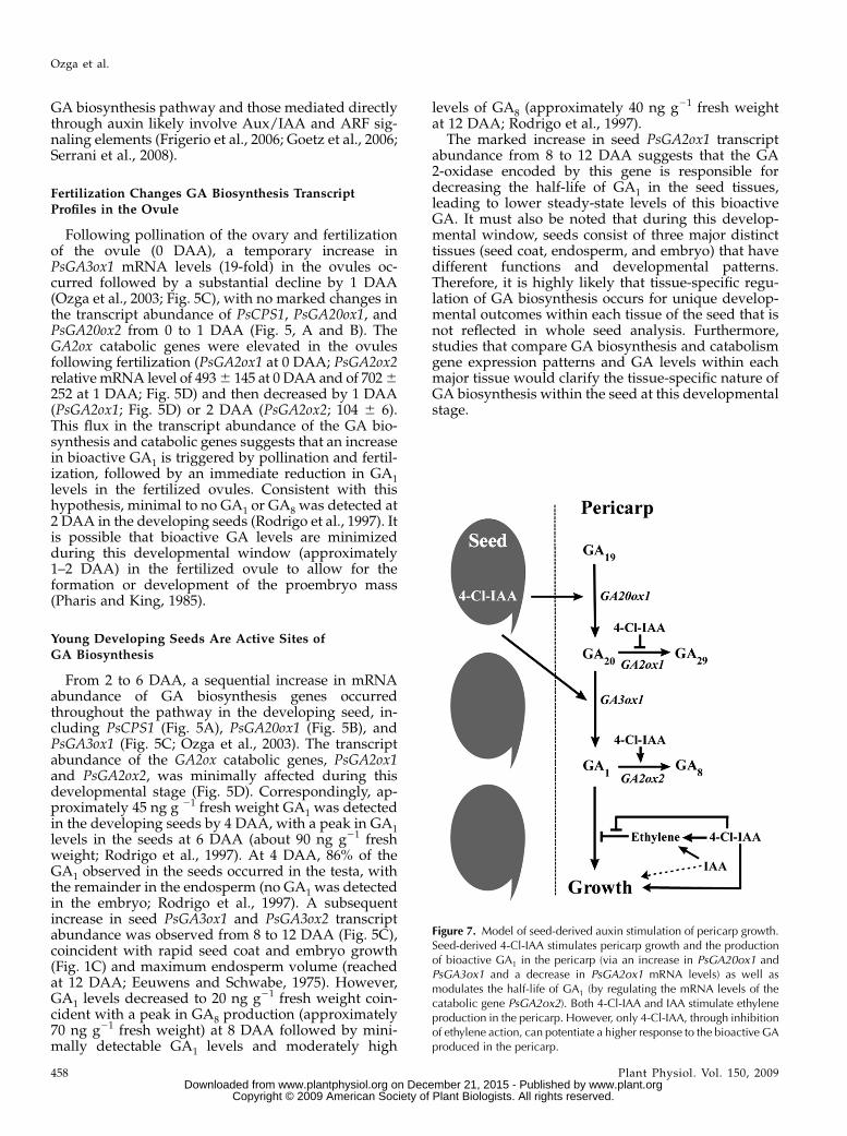
Figure 7. Model of seed-derived auxin stimulation of pericarp growth.Seed-derived 4-Cl-IAA stimulates pericarp growth and the productionof bioactive GA1 in the pericarp (via an increase in PsGA20ox1 andPsGA3ox1 and a decrease in PsGA2ox1 mRNA levels) as well asmodulates the half-life of GA1 (by regulating the mRNA levels of thecatabolic gene PsGA2ox2). Both 4-Cl-IAA and IAA stimulate ethyleneproduction in the pericarp. However, only 4-Cl-IAA, through inhibitionof ethylene action, can potentiate a higher response to the bioactive GAproduced in the pericarp.
Ozga et al.
458 Plant Physiol. Vol. 150, 2009 www.plant.org on December 21, 2015 - Published by www.plantphysiol.orgDownloaded from
Copyright © 2009 American Society of Plant Biologists. All rights reserved.
Seed Maturation Is Accompanied by Increased Seed GA
Catabolic Gene Expression
By 12 DAA, accumulation of cotyledonary storagereserves begins in the rapidly developing pea embryo(Pate and Flinn, 1977). The mRNA profiles of the GAbiosynthesis and catabolism genes in the seeds from 12to 20 DAA are likely primarily reflective of the de-veloping embryo as it grows to fill the seed coat by20 DAA (contact point; Ozga et al., 2003). SeedPsGA20ox1 mRNA abundance decreased after 10DAA (Fig. 5B), and PsGA3ox1 and PsGA3ox2 mRNAlevels decreased after 12 DAA and continued to de-crease until 20 DAA (Fig. 5C). However, from 10 to 12DAA, the transcript abundance of PsCPS1, PsGA20ox2,and PsGA2ox1 dramatically increased and remainedelevated through 20 DAA (Fig. 5, A, B, and D). Thehigh levels of PsCPS1 and PsGA20ox2 transcripts arelikely mainly localized in the embryo of the seed, ashigh levels of PsCPS1 and PsGA20ox2 were found inthe embryos compared with the testa of 20-DAA peaseeds (cv Torsdag; Ait-Ali et al., 1997). In contrast, highlevels of PsGA2ox1 mRNA were found in the testacompared with the cotyledons of 26-DAA seeds of pea(cv Progress No. 9; Martin et al., 1999). This change inthe GA biosynthesis and catabolism gene mRNAabundance profile from 12 to 20 DAA reflects thechange in the GA profile in the developing seed at thecontact point (20 DAA), with high levels of GA20 (446ng g21 fresh weight) and GA29 (189 ng g21 freshweight) and no detectable levels of GA1 and GA8 inthe seeds (cv Alaska [I3]; Ayele et al., 2006a).
SUMMARY
We propose the following working model for hor-monally directed fruit set and seed and pericarpcoordinated development. Pollination and fertilizationevents stimulate a pulse of bioactive GA1 synthesis inthe pericarp and the ovules (via an increase inPsGA3ox1 and a decrease in PsGA2ox1 and PsGA2ox2mRNA levels in the pericarp and an increase inPsGA3ox1 in the ovules) to promote initial seed andfruit set. Subsequently, seeds maintain pericarpgrowth (both in length [2–8 DAA] and in width [6–12DAA] to accommodate the developing seeds) at leastin part by stimulating pericarp GA biosynthesis (in-creasing PsGA20ox1 and PsGA3ox1 and decreasingPsGA2ox1 message levels), thereby maintaining a crit-ical level of GA1 for pericarp growth. Furthermore, wehypothesize that auxin (4-Cl-IAA in pea) is one of theseed-derived signals that is involved in stimulation ofGA biosynthesis in the pericarp at an early develop-mental stage (2–5 DAA) to promote growth (Fig. 7).4-Cl-IAA is present in both pea seed and pericarptissues at levels that suggest that transport from theseed to the pericarp is possible (Magnus et al., 1997).4-Cl-IAA can stimulate deseeded pericarp growth andthe production of bioactive GA1 in the pericarp (via an
increase in PsGA20ox1 and PsGA3ox1 and a decrease inPsGA2ox1 mRNA levels) as well as modulate the half-life of GA1 (by regulating the mRNA levels of thecatabolic gene PsGA2ox2). Additionally, 4-Cl-IAA (butnot IAA) can potentiate a higher response to thebioactive GA produced in the pericarp through inhi-bition of ethylene action (Johnstone et al., 2005). 4-Cl-IAA can also affect fruit growth directly throughauxin-mediated responses (van Huizen et al., 1996).
In developing seeds, bioactive GA1 synthesis istriggered by pollination and fertilization events, fol-lowed by an immediate reduction in GA1 levels in thefertilized ovules, possibly to allow the formation ordevelopment of the proembryo mass. From 2 to 6DAA, a sequential increase in mRNA abundance ofGA biosynthesis genes occurs to promote the produc-tion of GA1 to drive seed growth (mainly testa tissue).From 8 to 12 DAA, a transition in the seed GAbiosynthesis and catabolism pathways occurs to pro-duce sufficient bioactive GA for continued seed tissuegrowth and development, with a shift to the produc-tion of GA20 and minimal bioactive GA in the embryoas the seed enters into its maturation phase.
MATERIALS AND METHODS
Labeled GAs
[14C]GA12 was biosynthesized from R,S-[4,5-14C]mevalonic acid (110 mCi
mmol21) using a cell-free system of pumpkin (Cucurbita maxima) endosperm as
described by Birnberg et al. (1986) and modified by Ozga et al. (1992). The
specific radioactivity of [14C]GA12 was determined from its mass spectra to be
180.5 mCi mmol21 using the method described by Bowen et al. (1972). Protio-
and deutero-GA19, GA20, GA1, GA8, and [14C]GA19 were purchased from or
provided by Dr. L.N. Mander. [13C-3H]GA29 was a gift from Dr. B.O. Phinney.
Plant Material and Treatments
Plants of pea (Pisum sativum ‘Alaska’ [I3]) were grown under a 16-/8-h
light/dark photoperiod (19�C/17�C) with an average photon flux density of
402 mEm22 s21 (van Huizen et al., 1995). Pericarps (22, 0, 1, 2, 3, 4, 5, 6, 7, 8, 10,
12, 14, and 20 DAA) and ovules (22 DAA) or seeds (0, 1, 2, 3, 4, 5, 6, 7, 8, 10, 12,
14, and 20 DAA) were collected from the first to the fifth flowering nodes for
experiments monitoring GA gene expression during fruit development. For
nonpollinated pericarps, flowers (first to fifth flowering nodes) were emas-
culated at 22 DAA, and the pericarps were harvested from the plant at
equivalent to 21, 0, 1, 2, and 3 DAA (ovules removed at harvest). For
hormone-treated pericarps, one fruit between the third and fifth flowering
nodes was used per plant; subsequent flowers and lateral buds were removed
as they developed. Terminal apical meristems of plants were intact, and
pericarps remained attached to the plant throughout the experiments.
Pericarps were treated with hormones using a split-pod technique (Ozga
et al., 1992). Fruits at 2 DAAmeasuring 15 to 20 mm in length were split down
the dorsal suture 1 h prior to the 8-h dark period, and seeds were either left
intact (SP) or removed (SPNS). Surgical manipulation of the pea fruit was
completed 12 h prior to all hormone applications. Deseeded pericarps were
treated with IAA, 4-Cl-IAA, or GA3 at 50 mM in 0.1% (v/v) aqueous Tween 80
(30 mL total) or 4-Cl-IAA plus GA3 (50 mM each in 0.1% [v/v] aqueous Tween
80; 30 mL total). All solutions were applied directly to the inside surface of the
pericarp wall (endocarp). The SP and SPNS controls were treated with 30 mL
of 0.1% (v/v) aqueous Tween 80. Treated pericarps were covered with plastic
bags to maintain high humidity. Pericarps were harvested at 0, 2, 4, 8, and 12 h
after the hormone treatment. Seeds, if present, were removed from the
pericarp at harvest. All tissues were harvested into liquid N2 and subse-
quently stored at 280�C until extraction.
Regulation of GA Biosynthesis and Catabolism in Pea Fruit
Plant Physiol. Vol. 150, 2009 459 www.plant.org on December 21, 2015 - Published by www.plantphysiol.orgDownloaded from
Copyright © 2009 American Society of Plant Biologists. All rights reserved.
For the [14C]GA12 metabolism experiments, pea plants (cv Alaska [I3]) were
grown as described byMaki and Brenner (1991). Pericarps at 3 DAAwere split
(SP) or split and deseeded (SPNS) and treated with a 10-s dip of either 4-Cl-
IAA (50 mM) in 0.1% Tween 80 or 0.1% Tween 80 (control) immediately and 20
h after surgical treatment. Splitting of the pericarp and seed removal were
completed 24 h prior to [14C]GA12 application to the inside surface (endocarp)
of the pericarp. Pericarps were harvested onto dry ice 24 h after [14C]GA12
application and stored at280�C until extraction. Intact pericarps (3 DAA) and
4-DAA SP and SPNS pericarps (harvested 24 h after surgical treatment at 3
DAA) were also harvested for endogenous GA determination. For all treat-
ments, the seeds were removed from the pericarps prior to harvest of tissue if
present, and the pericarps remained attached to the plant throughout the
experiment.
For the [14C]GA19 metabolism experiments, plants of genotype SLN (cv
Torsdag wild type; JI992) and sln (JI3011) from the John Innes Pisum collection
were grown as described above. SLN and sln pericarps were deseeded and
treated with 4-Cl-IAA as described above (30 mL, 50 mM in 0.1% [v/v] aqueous
Tween 80), and SP and SPNS controls were treated with 0.1% (v/v) aqueous
Tween 80. 17-[14C]GA19 (specific radioactivity of 54 mCi mmol21) was applied
(60,000 dpm per pod in 5 mL of 50% [v/v] aqueous methanol) at 12 h after
hormone application (24 h after deseeding) to the inside surface of the
pericarp wall (endocarp). Treated pericarps (seeds were removed if present)
were harvested onto dry ice at 24 h after [14C]GA19 application and stored at
280�C until extraction.
RNA Isolation
Two whole or half pericarps and seeds were ground to a fine powder in
liquid N2, and 100 to 500 mg fresh weight of pericarp or 10 to 500 mg fresh
weight of seed tissue subsamples was used for RNA extraction. Total RNA
was extracted using a modified TRIzol (Invitrogen) procedure. In brief, after
initial extraction with the TRIzol reagent and centrifugation, the supernatant
was cleaned by chloroform partitioning (0.2 mL mL21 TRIzol reagent).
Subsequently, for further purification, the following steps were carried out
in order: high-salt precipitation (1.2 M sodium citrate, 0.8 M NaCl) to remove
polysaccharides and proteoglycans, 4 M LiCl precipitation, and precipitation
with a 1:20 (v/v) solution of 3 M sodium acetate (pH 5):100% ethanol. The total
RNA samples were then treated with DNase (Ambion DNA-free kit) and
stored at 280�C prior to real-time RT-PCR analysis.
Gene Expression Analysis
Primers and Probes
The target gene quantifying amplicons CPS1-92 (used for PsCPS1 quan-
tification), GA20ox1-104 (used for PsGA20ox1 quantification), GA20ox2-88
(used for PsGA20ox2 quantification), GA2ox1-73 (used for PsGA2ox1 quanti-
fication), and GA2ox2-83 (used for PsGA2ox2 quantification) were designed by
Ayele et al. (2006b). Primers and probes used to quantify PsGA3ox1
(PsGA3ox1-87) and the reference gene amplicon 18S-62 (used for pea 18S
rRNA quantification) were designed by Ozga et al. (2003). For PsGA3ox2
quantitation, the following primers and probe were used to produce a 104-bp
amplicon (PsGA3ox2-104) that spans nucleotides 476 to 579 of DQ864759:
forward primer, 5#-ATCATGGGGTCACCGTCTAA-3#; reverse primer,
5#-GCTAGTGTCTTCATTTGCTTTTGA-3#; probe, 5#-CCTAATGACTACGAA-
TATT-3#. The primers for PsGA3ox2 produced a single product of correct
length, and sequencing of the product confirmed the specificity (data not
shown). TaqMan probes were used for all gene expression studies. All probes
for the GA genes were labeled at the 5# end with the fluorescent reporter dye
6-carboxyfluorescein and at the 3# end with a minor-groove-binding nonfluo-
rescent quencher (Applied Biosystems). The 18S-62 reference gene probe was
labeled at the 5# end with the VIC fluorescent reporter dye and at the 3# endwith the TAMRA quencher (Applied Biosystems).
Real-Time RT-PCR Assay
Real-time RT-PCR assays were performed on an Applied Biosystems
StepOnePlus sequence detector (PsGA3ox1 and PsGA3ox2) or an Applied
Biosystems model 7700 sequence detector (all other genes) using the TaqMan
One-Step RT-PCR Master Mix Reagent Kit (Applied Biosystems). For each 25-
mL reaction, 200 ng of total RNA was mixed with 13 Master Mix (final
concentration; containing AmpliTaq Gold DNA polymerase), 2.53 Multi-
Scribe (final concentration; reverse transcriptase and RNase inhibitor mix),
300 nM (final concentration) each forward and reverse primer, 100 nM probe
(final concentration), and diethyl pyrocarbonate-treated water (to bring the
reaction volume to 25 mL). Samples were subjected to thermal cycling
conditions as described previously (Ozga et al., 2003), and the average of
the two subsamples was used to calculate the sample transcript abundance.
Total RNA from one sample was run on each plate and used as a control to
correct for plate-to-plate amplification differences (Ayele et al., 2006a). The
relative transcript abundance of the target genes in the individual plant
samples was determined by the 22DCt method (Livak and Schmittgen, 2001),
where DCt is the difference between the target sample Ct and the average Ct of
the reference sample. For PsCPS1, PsGA20ox1, PsGA20ox2, PsGA2ox1, and
PsGA2ox2, transcript levels were compared across genes, developmental
stages, and tissues using the lowest sample average Ct value (Ct = 37.975)
obtained in the study for these genes as the reference sample. For PsGA3ox1
and PsGA3ox2, transcript levels were compared across genes, developmental
stages, and tissues using the lowest sample average Ct value of 40 obtained for
these genes as the reference sample. At least two, and often three, replicate
plant samples were assayed.
The pea 18S small subunit nuclear rRNA gene was used as a loading
control to estimate the variation in total RNA input of the samples. For
PsCPS1, PsGA20ox1, PsGA20ox2, PsGA2ox1, and PsGA2ox2 genes, 10 pg of
DNase-treated total RNAwas used for 18S rRNA quantitation using the same
reaction and thermocycling conditions described above on the Applied
Biosystems model 7700 sequence detector. The coefficient of variation of the
18S rRNA amplicon Ct values among the samples was between 1% and 1.3%;
therefore, the target amplicon mRNA values were not normalized to the 18S
signal (Livak and Schmittgen, 2001).
For PsGA3ox1 and PsGA3ox2 genes, a competitive primer approach was
used to quantify 18S rRNA transcript levels. 18S rRNAwas quantified on 3 ng
of DNase-treated total RNA generated from a single dilution of the original 25
ng mL21 stocks (final concentration of 120 pg mL21) using the same reaction
and thermocycling conditions described above on the Applied Biosystems
StepOnePlus sequence detector. The addition of competitive primers [primers
with the same sequence as the 18S primers but lacking the 3# hydroxyl groupnecessary for DNA polymerase elongation, in this case (CH2)6NH2] along with
primers containing a 3# hydroxyl group to the PCR mixture allows a larger
amount of template RNA to be used while still maintaining an acceptable
reaction profile, decreasing the variation inherent in serial dilutions of
RNA samples. An optimal 1:9 ratio of primers containing 3# hydroxyl and
3# (CH2)6NH2 chain terminators was used as determined through experiments
varying input RNA and the ratio of 3# hydroxyl to 3# (CH2)6NH2 primers. The
coefficient of variation of the 18S rRNA amplicon Ct values among these
samples was again low (2.4%–2.9%); therefore, the target amplicon mRNA
values were not normalized to the 18S signal (Livak and Schmittgen, 2001).
[14C]GA12 Metabolism Experiments
Pericarps (39–41; 4–25 g fresh weight) were homogenized in cold 80%
aqueous methanol (4 mL g21 tissue) containing 10 mg L21 butylated
hydroxytoluene using a Polytron homogenizer. To each of these extracts,
[2H]GA19, [2H]GA20, [2H]GA1, [2H]GA8, and [13C-3H]GA29 were added at
homogenization as internal standards for gas chromatography-mass spec-
trometry-selected ionmonitoring (GC-MS-SIM). The tissue homogenates were
extracted for 12 h by shaking and gently stirring in darkness at 4�C. The tissuewas allowed to settle, the supernatant was decanted, and the tissue was
reextracted with the same volume of solvent for an additional 12 h as
described above. After decanting the supernatant, the tissue pellet was
washed three times with 2 mL of extraction solvent, and both supernatants
and the solvent wash were pooled and evaporated to dryness under vacuum
in silylated glass vials.
The extracts were resuspended in 20 mM imidazole buffer (pH 7.0) and
applied to a conditioned DEAE-Sephacel (Pharmacia) column (DEAE-Sepha-
cel [1.2 mL g21 fresh weight tissue] conditioned with the following solvents
sequentially [ratio of solvent to bed volume]: hexane [23], acetonitrile [23],
water [23], 0.2 M imidazole buffer [pH 7.0; 23], and water [103 to remove
excess buffer]). The columns were washed sequentially with the following
solvents (volume of solvent 23 bed volume): hexane, ethyl acetate, acetoni-
trile, methanol, and methanol with 2% acetic acid. The methanol fraction
containing the GAs was collected, dried under vacuum, and then brought up
in 1 mL of methanol:ethyl acetate (1:1) followed by two drops of water. This
Ozga et al.
460 Plant Physiol. Vol. 150, 2009 www.plant.org on December 21, 2015 - Published by www.plantphysiol.orgDownloaded from
Copyright © 2009 American Society of Plant Biologists. All rights reserved.
extract was applied to a hydrated silica column (1 g of hydrated SiO2 at 22%
water [w/w] per 1 g fresh weight of tissue) packed in 95:5 hexane:ethyl acetate
(formate saturated). The hydrated silica column was then washed with 10
times its bed volume with 5:95 hexane:ethyl acetate (v/v; formate saturated),
and this fraction, which contained the free-GAs, was dried under vacuum
prior to further purification using HPLC.
HPLC
The partially purified extracts were resuspended in 400 mL of 20% aqueous
methanol and passed through 0.45-mm nylon filters prior to injection onto a
4.5- 3 250-mm Spherisorb C18 column (5 mm; Beckman). The samples were
eluted at a flow rate of 1.0 mL min21 using a linear gradient of 0.01% aqueous
trifluoroacetic acid (solvent A) and 100% methanol (solvent B). Conditions of
the linear gradient were 20% solvent B for 1 min, gradient to 100% solvent B in
45 min, and isocratic 100% solvent B for 5 min. Radioactivity in the effluent
was monitored using a flow-through radiochemical detector (Beckman 171).
Radioactive fractions eluting near standard retention times of GA8 (9.2 min),
GA29 (12.6 min), GA1 (17.8 min), GA20 (26.4 min), and GA19 (29.2 min) were
collected and dried down.
[14C]GAs were converted to their methyl esters using diazomethane. The
[14C]GA methyl esters were subsequently converted to trimethylsilyl ether
derivatives (Gaskin and MacMillan, 1991) for identification and quantitation
by GC-MS-SIM. Mass spectral analyses of derivatized samples were per-
formed using a Hewlett-Packard model 5890 Series II Gas Chromatograph
interfaced to a Hewlett-Packard model 5972A Mass Selective Detector
equipped with a HP-5 MS column (30 m 3 0.25 mm 3 0.25 m film thickness).
Helium was used as the carrier gas at a flow rate of 1 mL min21. The samples
were injected on-column with the initial column temperature at 50�C for 2
min, followed by temperature programming at 10�C min21 to 150�C and then
at 3�C min21 to 300�C. Selective ion monitoring of three prominent ions for
each GA of interest and Kovats retention index data were used for confirma-
tion of [14C]GA identity. For quantitation, the protio- and deutero-ions
monitored were corrected for donation of natural isotopes to the peak area.
A separate calibration curve of peak area ratio versus molar ratio of [2H]GA/
protio-GA was constructed for GA19, GA20, GA1, and GA8 (Gaskin and
MacMillan, 1991). Using the corrected peak area and the calibration curve,
the total amounts of the protio-GAs were calculated. For GA29 and the14C-labeled GAs (protio-ion + 16 atomic mass units; 8 14C-molecules per GA
molecule), the total amounts were calculated by reference to the stable isotope-
labeled internal standard using equations for isotope dilution analysis
(Bandurski and Schulze, 1977). For calculation of endogenous GAs, the
most prominent ion measured (usually M+) was used for quantitation, and
the calculated value was checked for reproducibility using the second most
prominent ion.
[14C]GA19 Metabolism Experiments
Using a Polytron homogenizer, radiolabeled pericarps (three per sample)
were homogenized in silylated 30-mL Corex tubes with 10 mL of cold 80%
(v/v) methanol containing 10 mg L21 butylated hydroxytoluene. An external
standard, 10,000 dpm of 17-[14C]GA7, was added at the time of homogeniza-
tion for determination of radioactive metabolite recovery. After homogeniza-
tion, samples were gently shaken in darkness at 4�C for 12 to 16 h and then
centrifuged at 10,000g for 30 min. The supernatant was removed, and the
residue was resuspended in 10 mL of the homogenization solvent and gently
shaken at 4�C in darkness for at least 4 h. The residue extracts were
centrifuged at 10,000g for 30 min, and the combined supernatants were
reduced to the aqueous phase using a vacuum concentrator (Savant). The pH
of the aqueous extracts was adjusted to 8.0 with NH4OH (0.1 N) and the
extracts partitioned against n-hexane (5 mL) four times in silylated 20-mL
glass scintillation vials. The aqueous fraction was then adjusted to pH 3.0 with
0.1 N HCl and partitioned against ethyl acetate (5 mL) five times. The
combined ethyl acetate extracts were reduced to approximately 3 mL using a
vacuum concentrator and partitioned against 5% (w/v) aqueous NaHCO3 (5
mL) four times. The combined NaHCO3 extracts were transferred to 30-mL
silylated Pyrex tubes, the pHwas adjusted to 3.0 with concentrated HCl on ice,
and the extracts partitioned against ethyl acetate (5 mL) five times. The ethyl
acetate extracts were combined and evaporated to near dryness, transferred to
7-mL silylated scintillation vials, and dried down under vacuum.
The ethyl acetate extracts were subject to the same HPLC procedure as
described above. Radioactive fractions eluting near standard retention times
of GA8 (9.2 min), GA29 (12.6 min), GA3 (16.3 min), GA1 (17.8 min), GA5 (25.0
min), GA20 (26.4 min), GA19 (29.2 min), and GA7 (31.0 min) were collected and
dried down. Collected [14C]GAs were methylated using diazomethane and
rechromatographed as their methyl esters by C18-HPLC using the same
solvent system.
Sequence data from this article can be found in the GenBank/EMBL data
libraries under accession numbers AF001219, AF056935, AF100954, DQ864759,
U58830, U63652, and U70471.
Received November 4, 2008; accepted March 9, 2009; published March 18,
2009.
LITERATURE CITED
Ait-Ali T, Swain SM, Reid JB, Sun TP, Kamiya Y (1997) The LS locus of pea
encodes the gibberellin biosynthesis enzyme ent-kaurene synthase A.
Plant J 11: 443–454
Ayele BT, Ozga JA, Kurepin LV, Reinecke DM (2006a) Developmental and
embryo axis regulation of gibberellin biosynthesis during germination
and young seedling growth of pea. Plant Physiol 142: 1267–1281
Ayele BT, Ozga JA, Reinecke DM (2006b) Regulation of GA biosynthesis
genes during germination and young seedling growth of pea (Pisum
sativum L.). J Plant Growth Regul 25: 219–232
Bandurski RS, Schulze A (1977) Concentration of indole-3-acetic acid and
its derivatives in plants. Plant Physiol 60: 211–213
Birnberg PR, Maki SL, Brenner ML, Davis GC, Carnes MG (1986) An
improved enzymatic synthesis of labeled gibberellin A12-aldehyde and
gibberellin A12. Anal Biochem 153: 1–8
Bowen DH, MacMillan J, Graebe JE (1972) Determination of specific
radioactivity of [14C]-compounds by mass spectroscopy. Phytochemis-
try 11: 2253–2257
Davidson SE, Swain SM, Reid JB (2005) Regulation of the early GA
biosynthesis pathway in pea. Planta 222: 1010–1019
Eeuwens CJ, Schwabe WW (1975) Seed and pod wall development in
Pisum sativum L. in relation to extracted and applied hormones. J Exp
Bot 26: 1–14
Frigerio M, Alabadi D, Perez-Gomez J, Garcia-Carcel L, Phillips AL,
Hedden P, BlazquezMA (2006) Transcriptional regulation of gibberellin
metabolism genes by auxin signaling in Arabidopsis. Plant Physiol 142:
553–563
Garcia-Martinez JL, Lopez-Diaz I, Sanchez-Beltran MJ, Phillips AL,
Ward DA, Gaskin P, Hedden P (1997) Isolation and transcript analysis
of gibberellin 20-oxidase genes in pea and bean in relation to fruit
development. Plant Mol Biol 33: 1073–1084
Garcia-Martinez JL, Santes C, Croker SJ, Hedden P (1991) Identification,
quantitation and distribution of gibberellins in fruits of Pisum sativum L.
cv. Alaska during pod development. Planta 184: 53–60
Gaskin P, MacMillan J (1991) GC-MS of the Gibberellins and Related
Compounds: Methodology and a Library of Spectra. University of
Bristol (Cantock’s Enterprises), Bristol, UK
Goetz M, Vivian-Smith A, Johnson SD, Koltunow AM (2006) AUXIN
RESPONSE FACTOR8 is a negative regulator of fruit initiation in
Arabidopsis. Plant Cell 18: 1873–1886
Hedden P, Phillips AL (2000) Gibberellin metabolism: new insights
revealed by the genes. Trends Plant Sci 5: 523–530
Johnstone MMG (2004) Phytohormones in early pea fruit growth. MSc
thesis. University of Alberta, Edmonton, Alberta, Canada
Johnstone MMG, Reinecke DM, Ozga JA (2005) The auxins IAA and 4-Cl-
IAA differentially modify gibberellin action via ethylene response in
developing pea fruit. J Plant Growth Regul 24: 214–225
Lester DR, Ross JJ, Davies PJ, Reid JB (1997) Mendel’s stem length gene
(Le) encodes a gibberellin 3b-hydroxylase. Plant Cell 9: 1435–1443
Lester DR, Ross JJ, Smith JJ, Elliott RC, Reid JB (1999) Gibberellin
2-oxidation and the SLN gene of Pisum sativum. Plant J 19: 65–73
Livak KJ, Schmittgen TD (2001) Analysis of relative gene expression data
using real-time quantitative PCR and the 22DDCt method. Methods 25:
402–408
MacKenzie-Hose AK, Ross JJ, Davies NW, Swain SM (1998) Expression of
gibberellin mutations in fruits of Pisum sativum L. Planta 204: 397–403
Magnus V, Ozga JA, Reinecke DM, Pierson GL, Larue TA, Cohen JD,
Regulation of GA Biosynthesis and Catabolism in Pea Fruit
Plant Physiol. Vol. 150, 2009 461 www.plant.org on December 21, 2015 - Published by www.plantphysiol.orgDownloaded from
Copyright © 2009 American Society of Plant Biologists. All rights reserved.
Brenner ML (1997) 4-Chloroindole-3-acetic acid and indole-3-acetic
acid in Pisum sativum. Phytochemistry 46: 675–681
Maki SL, Brenner ML (1991) [14C]GA12-aldehyde, [14C]GA12, and [2H]- and
[14C]GA53 metabolism by elongating pea pericarp. Plant Physiol 97:
1359–1366
Martin DN, Proebsting WM, Hedden P (1997) Mendel’s dwarfing gene:
cDNAs from the Le alleles and function of the expressed proteins. Proc
Natl Acad Sci USA 94: 8907–8911
Martin DN, Proebsting WM, Hedden P (1999) The SLENDER gene of pea
encodes a gibberellin 2-oxidase. Plant Physiol 121: 775–781
Martin DN, Proebsting WM, Parks TD, Dougherty WG, Lange T, Lewis
MJ, Gaskin P, Hedden P (1996) Feed-back regulation of gibberellin
biosynthesis and gene expression in Pisum sativum L. Planta 200:
159–166
Marumo S, Hattori H, Abe H, Munakata K (1968) Isolation of 4-chloro-
indolyl-3-acetic acid from immature seeds of Pisum sativum. Nature 219:
959–960
Ngo P, Ozga JA, Reinecke DM (2002) Specificity of auxin regulation of
gibberellin 20-oxidase gene expression in pea pericarp. Plant Mol Biol
49: 439–448
O’Neill DP, Ross JJ (2002) Auxin regulation of the gibberellin pathway in
pea. Plant Physiol 130: 1974–1982
Ozga JA, Brenner ML, Reinecke DM (1992) Seed effects on gibberellin
metabolism in pea pericarp. Plant Physiol 100: 88–94
Ozga JA, Reinecke DM (1999) Interaction of 4-chloroindole-3-acetic acid
and gibberellins in early pea fruit development. Plant Growth Regul 27:
33–38
Ozga JA, van Huizen R, Reinecke DM (2002) Hormone and seed-specific
regulation of pea fruit growth. Plant Physiol 128: 1379–1389
Ozga JA, Yu J, Reinecke DM (2003) Pollination-, development-, and auxin-
specific regulation of gibberellin 3b-hydroxylase gene expression in pea
fruit and seeds. Plant Physiol 131: 1137–1146
Pate JS, FlinnAM (1977) Fruit and seed development. In JF Sutcliffe, JS Pate, eds,
The Physiology of the Garden Pea. Academic Press, New York, pp 431–468
Pharis RP, King RW (1985) Gibberellins and reproductive development in
seed plants. Annu Rev Plant Physiol 36: 517–568
Reinecke DM (1999) 4-Chloroindole-3 acetic acid and plant growth. Plant
Growth Regul 27: 3–13
Reinecke DM, Ozga JA, Ilic N, Magnus V, Kojic-Prodic B (1999) Molec-
ular properties of 4-substituted indole-3-acetic acids affecting pea
pericarp elongation. Plant Growth Regul 27: 39–48
Reinecke DM, Ozga JA, Magnus V (1995) Effect of halogen substitution of
indole-3-acetic acid on biological activity in pea fruit. Phytochemistry
40: 1361–1366
Rodrigo MJ, Garcia-Martinez JL, Santes CM, Gaskin P, Hedden P (1997)
The role of gibberellins A1 and A3 in fruit growth of Pisum sativum L. and
the identification of gibberellins A4 and A7 in young seeds. Planta 201:
446–455
Ross JJ, Reid JB, Dungey HS (1992) Ontogenetic variation in levels of
gibberellin A1 in Pisum: implications for the control of stem elongation.
Planta 186: 166–171
Ross JJ, Reid JB, Swain SM, Hasan O, Poole AT, Hedden P, Willis CL
(1995) Genetic regulation of gibberellin deactivation in Pisum. Plant J 7:
513–523
Santes CM, Garcia-Martinez JL (1995) Effect of the growth retardant 3,5-
dioxo-4-butyryl-cyclohexane carboxylic acid ethyl ester, an acylcyclo-
hexanedione compound, on fruit growth and gibberellin content of
pollinated and unpollinated ovaries in pea. Plant Physiol 108: 517–523
Santes CM, Hedden P, Sponsel VM, Reid JM, Garcia-Martinez JL (1993)
Expression of the le mutation in young ovaries of Pisum sativum and its
effect on fruit development. Plant Physiol 101: 759–764
Serrani JC, Ruiz-Rivero O, Fos M, Garcia-Martinez JL (2008) Auxin-
induced fruit-set in tomato is mediated in part by gibberellins. Plant J
56: 922–934
Serrani JC, Sanjuan R, Ruiz-Rivero O, Fos M, Garcia-Martinez JL (2007)
Gibberellin regulation of fruit set and growth in tomato. Plant Physiol
145: 246–257
Sponsel VM (1982) Effects of applied gibberellins and naphthylacetic acid
on pod development in fruits of Pisum sativum L. cv. Progress No. 9.
J Plant Growth Regul 1: 147–152
Sponsel VM (1995) The biosynthesis and metabolism of gibberellins in
higher plants. In PJ Davies, ed, Plant Hormones: Physiology, Biochem-
istry and Molecular Biology, Ed 2. Kluwer Academic Publishers,
Dordrecht, The Netherlands, pp 66–97
Thomas SG, Phillips AL, Hedden P (1999) Molecular cloning and func-
tional expression of gibberellin 2-oxidases, multifunctional enzymes
involved in gibberellin deactivation. Proc Natl Acad Sci USA 96:
4698–4703
van Huizen R, Ozga JA, Reinecke DM (1996) Influence of auxin and
gibberellin on in vivo protein synthesis during early pea fruit growth.
Plant Physiol 112: 53–59
van Huizen R, Ozga JA, Reinecke DM (1997) Seed and hormonal regu-
lation of gibberellin 20-oxidase expression in pea pericarp. Plant Physiol
115: 123–128
van Huizen R, Ozga JA, Reinecke DM, Twitchin B, Mander LN (1995)
Seed and 4-chloroindole-3-acetic acid regulation of gibberellin metab-
olism in pea pericarp. Plant Physiol 109: 1213–1217
Weston DE, Elliott RC, Lester DR, Rameau C, Reid JB, Murfet IC, Ross JJ
(2008) The pea DELLA proteins LA and CRYare important regulators of
gibberellin synthesis and root growth. Plant Physiol 147: 199–205
Yamaguchi S (2008) Gibberellin metabolism and its regulation. Annu Rev
Plant Biol 59: 225–251
Ozga et al.
462 Plant Physiol. Vol. 150, 2009 www.plant.org on December 21, 2015 - Published by www.plantphysiol.orgDownloaded from
Copyright © 2009 American Society of Plant Biologists. All rights reserved.
Related Documents