Development of Useful Recombinant Promoter and Its Expression Analysis in Different Plant Cells Using Confocal Laser Scanning Microscopy Deepak Kumar 1 , Sunita Patro 1. , Rajiv Ranjan 1. , Dipak K. Sahoo 1,2 , Indu B. Maiti 2 , Nrisingha Dey 1 * 1 Department of Gene Function and Regulation, Institute of Life Sciences, Department of Biotechnology, Government of India, Nalco Square, Chandrasekherpur, Bhubaneswar, Orissa, India, 2 Kentucky Tobacco Research and Development Center (KTRDC), College of Agriculture, University of Kentucky, Lexington, Kentucky, United States of America Abstract Background: Designing functionally efficient recombinant promoters having reduced sequence homology and enhanced promoter activity will be an important step toward successful stacking or pyramiding of genes in a plant cell for developing transgenic plants expressing desired traits(s). Also basic knowledge regarding plant cell specific expression of a transgene under control of a promoter is crucial to assess the promoter’s efficacy. Methodology/Principal Findings: We have constructed a set of 10 recombinant promoters incorporating different up- stream activation sequences (UAS) of Mirabilis mosaic virus sub-genomic transcript (MS8, -306 to +27) and TATA containing core domains of Figwort mosaic virus sub-genomic transcript promoter (FS3, 2271 to +31). Efficacies of recombinant promoters coupled to GUS and GFP reporter genes were tested in tobacco protoplasts. Among these, a 369-bp long hybrid sub-genomic transcript promoter (MSgt-FSgt) showed the highest activity in both transient and transgenic systems. In a transient system, MSgt-FSgt was 10.31, 2.86 and 2.18 times more active compared to the CaMV35S, MS8 and FS3 promoters, respectively. In transgenic tobacco (Nicotiana tabaccum, var. Samsun NN) and Arabidopsis plants, the MSgt-FSgt hybrid promoter showed 14.22 and 7.16 times stronger activity compared to CaMV35S promoter respectively. The correlation between GUS activity and uidA-mRNA levels in transgenic tobacco plants were identified by qRT-PCR. Both CaMV35S and MSgt-FSgt promoters caused gene silencing but the degree of silencing are less in the case of the MSgt-FSgt promoter compared to CaMV35S. Quantification of GUS activity in individual plant cells driven by the MSgt-FSgt and the CaMV35S promoter were estimated using confocal laser scanning microscopy and compared. Conclusion and Significance: We propose strong recombinant promoter MSgt-FSgt, developed in this study, could be very useful for high-level constitutive expression of transgenes in a wide variety of plant cells. Citation: Kumar D, Patro S, Ranjan R, Sahoo DK, Maiti IB, et al. (2011) Development of Useful Recombinant Promoter and Its Expression Analysis in Different Plant Cells Using Confocal Laser Scanning Microscopy. PLoS ONE 6(9): e24627. doi:10.1371/journal.pone.0024627 Editor: Haibing Yang, Purdue University, United States of America Received January 13, 2011; Accepted August 16, 2011; Published September 9, 2011 Copyright: ß 2011 Kumar et al. This is an open-access article distributed under the terms of the Creative Commons Attribution License, which permits unrestricted use, distribution, and reproduction in any medium, provided the original author and source are credited. Funding: This work was supported by the Department of Biotechnology, Govt. of India [Project No. BT/PR6805/PBD/16/622/2005 to ND] and the Council for Scientific and Industrial Research, Govt. of India [Project No. 38(1147)/07/EMR-II to ND]. The funders had no role in the study design, data collection and analysis, decision to publish or preparation of manuscript. Competing Interests: The authors have declared that no competing interests exist. * E-mail: [email protected] . These authors contributed equally to this work. Introduction The overall strength of a promoter as well as its tissue-specific expression pattern depends on the combination of the spatial orientation of cis-elements and interaction with nuclear protein factors [1,2]. Such interactions suggest that the cis-element- containing upstream activation sequence (UAS) plays an important role in determining promoter function [3]. Moving the DNA element (cis-factor) that binds a specific trans-factor from one promoter into a different promoter can result in a novel transcription model [4,5], that can modulate the transcriptional activity and cell specific expression pattern of the promoter [6]. Several recombi- nant/synthetic promoters have been created earlier by (a) ligating the upstream activation sequence (UAS) of one promoter with the TATA box containing domain of another promoter; like tacI/tacII hybrid promoters [7], E4/E8 hybrid promoters [8] and chimeric plant promoter (Mac promoter) [9]; (b) placing desired cis-elements in conjunction with heterologous promoters [10]; (c) bringing together cis-elements from different promoters [11–13]. The strength and tissue specificity of chimeric promoters derived by incorporating sub-domains of the mannopine synthase (mas2) and octopine synthase (ocs) promoters were investigated earlier [14]. The Mac promoter, incorporating the mas region from +65 to 2301 and the CaMV35S enhancer region from 290 to 2941, has been reported to enhance the level of GUS expression by several folds as compared to the CaMV35S promoter [9]. Similarly, the strength and tissue specificity of Mac- and super-promoters were analyzed [14–16]. Novel plant transformation vectors incorporating the super-promoter were tested in transgenic tobacco and maize plants, and also transiently in maize protoplasts [16]. PLoS ONE | www.plosone.org 1 September 2011 | Volume 6 | Issue 9 | e24627

Welcome message from author
This document is posted to help you gain knowledge. Please leave a comment to let me know what you think about it! Share it to your friends and learn new things together.
Transcript
Development of Useful Recombinant Promoter and ItsExpression Analysis in Different Plant Cells UsingConfocal Laser Scanning MicroscopyDeepak Kumar1, Sunita Patro1., Rajiv Ranjan1., Dipak K. Sahoo1,2, Indu B. Maiti2, Nrisingha Dey1*
1 Department of Gene Function and Regulation, Institute of Life Sciences, Department of Biotechnology, Government of India, Nalco Square, Chandrasekherpur,
Bhubaneswar, Orissa, India, 2 Kentucky Tobacco Research and Development Center (KTRDC), College of Agriculture, University of Kentucky, Lexington, Kentucky, United
States of America
Abstract
Background: Designing functionally efficient recombinant promoters having reduced sequence homology and enhancedpromoter activity will be an important step toward successful stacking or pyramiding of genes in a plant cell for developingtransgenic plants expressing desired traits(s). Also basic knowledge regarding plant cell specific expression of a transgeneunder control of a promoter is crucial to assess the promoter’s efficacy.
Methodology/Principal Findings: We have constructed a set of 10 recombinant promoters incorporating different up-stream activation sequences (UAS) of Mirabilis mosaic virus sub-genomic transcript (MS8, -306 to +27) and TATA containingcore domains of Figwort mosaic virus sub-genomic transcript promoter (FS3, 2271 to +31). Efficacies of recombinantpromoters coupled to GUS and GFP reporter genes were tested in tobacco protoplasts. Among these, a 369-bp long hybridsub-genomic transcript promoter (MSgt-FSgt) showed the highest activity in both transient and transgenic systems. In atransient system, MSgt-FSgt was 10.31, 2.86 and 2.18 times more active compared to the CaMV35S, MS8 and FS3 promoters,respectively. In transgenic tobacco (Nicotiana tabaccum, var. Samsun NN) and Arabidopsis plants, the MSgt-FSgt hybridpromoter showed 14.22 and 7.16 times stronger activity compared to CaMV35S promoter respectively. The correlationbetween GUS activity and uidA-mRNA levels in transgenic tobacco plants were identified by qRT-PCR. Both CaMV35S andMSgt-FSgt promoters caused gene silencing but the degree of silencing are less in the case of the MSgt-FSgt promotercompared to CaMV35S. Quantification of GUS activity in individual plant cells driven by the MSgt-FSgt and the CaMV35Spromoter were estimated using confocal laser scanning microscopy and compared.
Conclusion and Significance: We propose strong recombinant promoter MSgt-FSgt, developed in this study, could be veryuseful for high-level constitutive expression of transgenes in a wide variety of plant cells.
Citation: Kumar D, Patro S, Ranjan R, Sahoo DK, Maiti IB, et al. (2011) Development of Useful Recombinant Promoter and Its Expression Analysis in Different PlantCells Using Confocal Laser Scanning Microscopy. PLoS ONE 6(9): e24627. doi:10.1371/journal.pone.0024627
Editor: Haibing Yang, Purdue University, United States of America
Received January 13, 2011; Accepted August 16, 2011; Published September 9, 2011
Copyright: � 2011 Kumar et al. This is an open-access article distributed under the terms of the Creative Commons Attribution License, which permitsunrestricted use, distribution, and reproduction in any medium, provided the original author and source are credited.
Funding: This work was supported by the Department of Biotechnology, Govt. of India [Project No. BT/PR6805/PBD/16/622/2005 to ND] and the Council forScientific and Industrial Research, Govt. of India [Project No. 38(1147)/07/EMR-II to ND]. The funders had no role in the study design, data collection and analysis,decision to publish or preparation of manuscript.
Competing Interests: The authors have declared that no competing interests exist.
* E-mail: [email protected]
. These authors contributed equally to this work.
Introduction
The overall strength of a promoter as well as its tissue-specific
expression pattern depends on the combination of the spatial
orientation of cis-elements and interaction with nuclear protein
factors [1,2]. Such interactions suggest that the cis-element-
containing upstream activation sequence (UAS) plays an important
role in determining promoter function [3]. Moving the DNA
element (cis-factor) that binds a specific trans-factor from one
promoter into a different promoter can result in a novel transcription
model [4,5], that can modulate the transcriptional activity and cell
specific expression pattern of the promoter [6]. Several recombi-
nant/synthetic promoters have been created earlier by (a) ligating
the upstream activation sequence (UAS) of one promoter with the
TATA box containing domain of another promoter; like tacI/tacII
hybrid promoters [7], E4/E8 hybrid promoters [8] and chimeric
plant promoter (Mac promoter) [9]; (b) placing desired cis-elements
in conjunction with heterologous promoters [10]; (c) bringing
together cis-elements from different promoters [11–13]. The
strength and tissue specificity of chimeric promoters derived by
incorporating sub-domains of the mannopine synthase (mas2) and
octopine synthase (ocs) promoters were investigated earlier [14]. The
Mac promoter, incorporating the mas region from +65 to 2301 and
the CaMV35S enhancer region from 290 to 2941, has been
reported to enhance the level of GUS expression by several folds as
compared to the CaMV35S promoter [9]. Similarly, the strength
and tissue specificity of Mac- and super-promoters were analyzed
[14–16]. Novel plant transformation vectors incorporating the
super-promoter were tested in transgenic tobacco and maize plants,
and also transiently in maize protoplasts [16].
PLoS ONE | www.plosone.org 1 September 2011 | Volume 6 | Issue 9 | e24627
Risk of successive transformation by single promoter is that it
might lead to silencing of transgenes in successive generations by
homologous recombination [13,17–20]. Use of different heterologous
promoters is necessary during pyramiding (stacking) of genes in a
single plant cell for developing a particular trait in plants. As in the
case of developing b carotene (Provitamin A) enriched rice multiple
genes coupled to different promoters were inserted in rice endosperm
for engineering the concerned metabolic pathway/s [21].
Unique clustering of different plant-specific elements like ASF-1
(TGACG), ARR1AT (NGATT), Dof-1 (AAAG), WRKY (TGAC)
AINTEGUMENTA (ANT), ATHB-9 and the GATA Box
(GATA) among the Mirabilis mosaic virus sub-genomic transcript
promoter (2356 to 2125 of MS8) [22] and the TATA containing
sub-domain of the Figwort mosaic virus sub-genomic transcript
promoter (2151 to +31 of FS3) [23] sequences were identified
using PLACE and PlantPAN databases [24,25]. This finding
prompted us to develop useful recombinant promoters by fusing
different up-stream sub-domains of the MS8 promoter with
different core promoter elements of the FS3 promoter.
In the present study, we have characterized a set of 10 unique
pararetrovirus-based recombinant sub-genomic transcript promot-
ers, and their expression activities were compared to that of the
CaMV35S, MS8 and FS3 promoters in transient expression
systems. An in-depth expression analysis of one of these hybrid
promoters (MSgt-FSgt), which conferred the highest level of
activity, was further studied in transgenic tobacco and Arabidopsis
plants. Correlation between the MSgt-FSgt promoter-driven GUS
activity and the uidA-mRNA level in transgenic plants was
assayed. The interaction between the MSgt-FSgt promoter
sequence and tobacco nuclear proteins was also analyzed by
Electrophoretic Mobility Shift Assay (EMSA) and DNA Foot-
printing. The cell specificity of MSgt-FSgt was compared to that of
the CaMV35S promoter in different plant cells of the stem, leaf,
and root using Confocal Laser Scanning Microscopy (CLSM). The
recombinant MSgt-FSgt promoter reported here will be a valuable
addition to the tools available for plant gene expression studies.
Materials and Methods
MaterialsRestriction and modifying enzymes were purchased from
Promega (Madison, WI USA), and were used according to the
manufacturer’s instructions. The Nytran membrane was obtained
from Schleicher & Schuell (Keene, NH, USA). All fine chemicals,
including MUG, X-gal, X-gluc, DEPC were purchased from
Sigma-Aldrich (St. Louis, USA). Platinum high fidelity Taq DNA
polymerase was obtained from Gibco-BRL (Maryland, USA).
DNA methyltransferase M.SssI and E.coli K12 ER2925 were
purchased from New England Biolabs, Beverly, MA, USA.
Construction of expression vectors containingrecombinant promoters for transient expression assay
Pararetrovirus-based recombinant sub-genomic transcript pro-
moters (10) were constructed by combining five different upstream
activation sequence (UAS): (MS-UAS1, MS-UAS2, MS-UAS3,
MS-UAS4 and MS-UAS5) of the Mirabilis mosaic virus sub-genomic
transcript promoter (MMVSgt) [22] with two different TATA
box-containing promoter fragments (FS-1 and FS-2) of the Figwort
mosaic virus sub-genomic transcript promoter (FS3) [23] (Figure 1).
The boundaries of respective UAS (of MS8 promoter) and TATA-
box-containing fragments (of FS3 promoter) present in each
hybrid promoter were listed in Table 1. Different UASs of the
MMV-Sgt promoter were PCR amplified individually using
specific pairs of synthetic oligonucleotides (Table 2) containing
the appropriate sequence to generate EcoRI and HincII sites at the
59-end and SmaI and HindIII sites at the 39-end. PCR-amplified
fragments were digested with EcoRI and HindIII, gel-purified and
cloned into the corresponding sites of the pUC119 vector. The
resulting plasmids were designated as pUMS-UAS1, pUMS-
UAS2, pUMS-UAS3, pUMS-UAS4 and pUMS-UAS5. Similarly,
the FS-1 and FS-2 promoters were PCR-amplified using the FS3
promoter DNA as a template and synthetic primers (Table 2)
containing the appropriate restriction sites to generate EcoRI and
HincII overhangs at the 59-end and SmaI and HindIII overhangs at
the 39-end. These fragments (59-EcoRI – HincII- fragment – SmaI –
HindIII-39) were cloned into the corresponding sites of pUC119.
These resulting plasmids were designated as pUFS-1 and pUFS-2.
The 182-bp (FS-1) and 165-bp (FS-2) Figwort mosaic virus sub-
genomic transcript promoter fragments were isolated as HincII –
HindIII fragments from the pUFS-1 and pUFS-2 plasmids and
then inserted individually into the pUMS-UAS1, pUMS-UAS2,
pUMS-UAS3, pUMS-UAS4 and pUMS-UAS5 plasmids at the
SmaI and HindIII sites. The resulting plasmids were designated as
pUMS-UAS1-FS1, pUMS-UAS2-FS1, pUMS-UAS3-FS1, pUM-
S-UAS4-FS1, pUMS-UAS5-FS1, pUMS-UAS1-FS2, pUMS-
UAS2-FS2, pUMS-UAS3-FS2, pUMS-UAS4-FS2 and pUMS-
UAS5-FS2. All promoter inserts were subjected to nucleotide
sequencing and the UAS portion of the MS8 promoter was linked
to the TATA-containing promoter from FS3 via a SmaI site
(cccggg) in all of the hybrid promoters. The hybrid promoter
fragments, MS-UAS1-FS1 (419 bp), MS-UAS2-FS1 (369 bp),
MS-UAS3-FS1 (319 bp), MS-UAS4-FS1 (269 bp), MS-UAS5-
FS1 (219 bp), MS-UAS1-FS2 (402 bp), MS-UAS2-FS2 (352 bp),
MS-UAS3-FS2 (302 bp), MS-UAS4-FS2 (252 bp) and MS-UAS5-
FS2 (202 bp) were isolated by EcoRI and HindIII restriction
digestion and sub-cloned into a plant protoplast expression vector
containing the GUS reporter gene (pUCPMAGUS) by replacing
the CaMV35S promoter [26]. The resulting plasmids were
designated as pUPMS-UAS1-FS1GUS, pUPMS-UAS2-FS1GUS,
pUPMS-UAS3-FS1GUS, pUPMS-UAS4-FS1GUS, pUPMS-UA-
S5-FS1GUS, pUPMS-UAS1-FS2GUS, pUPMS-UAS2-FS2GUS,
pUPMS-UAS3-FS2GUS, pUPMS-UAS4-FS2GUS and pUPMS-
UAS5-FS2GUS respectively.
Construction of MSgt-FSgt (MS-UAS2-FS1), FS3, MS8, MS-UAS2, FS-1 and CaMV35S promoter-GUS vectors fortransient assay
The hybrid promoter MS-UAS2-FS1 and the respective clone
(constructed as above) are hereinafter referred to as the MSgt-FSgt
hybrid promoter and pUPMSgt-FSgt, respectively. The boundar-
ies of the MS8 and FS3 promoter fragments present in the MSgt-
FSgt promoter (MS-UAS2-FS1) are shown in Table 1.
The upstream activation sequence (-306 to -125) of the MMV-Sgt
promoter (MS-UAS2) and the TATA-containing sequence (2151
to +31) of the FMV-Sgt promoter (FS-1) were cloned into EcoRI and
HindIII sites of pUCPMAGUS vector [26]. The resulting plasmids
were designated as pUPMS-UAS2GUS and pUPFS-1GUS.
Promoter-GFP constructs for transient assayThe GFP cDNA gene was PCR-amplified using a synthetic
primer pair (Table 2) to insert the XhoI site at the 59-end and the
SstI site at the 39-end. The amplified GFP gene was digested with
XhoI and SstI and subsequently cloned into the XhoI and SstI sites
of pUCPMAGUS, pFS3GUS, pMS8GUS and pUPMSgt-
FSgtGUS (set-a) replacing the GUS gene to generate plasmids
pUCPMAGFP, pFS3GFP, pMS8GFP and pUPMSgt-FSgtGFP
(set-b) respectively.
Recombinant Promoters
PLoS ONE | www.plosone.org 2 September 2011 | Volume 6 | Issue 9 | e24627
Construction of plant expression vectorsThe MSgt-FSgt promoter fragment 369-bp from pUPMSgt-FSgt
was gel purified (as EcoRI – HindIII fragment) and inserted into
pKYLXGUS, a plant expression vector [26,27] using EcoRI and
HindIII sites that flank the promoter, thereby replacing the CaMV35S
promoter. The resulting plant expression vector was designated as
pKMSgt-FSgtGUS.
Protoplast isolation, electroporation and transient assayof recombinant promoters
Protoplasts from a tobacco cell suspension culture (Nicotiana
tabacum cv. Xanthi Brad) were purified on a 20% sucrose gradient
after digestion by cellulase (Sigma, USA) and pectinase (Sigma,
USA) and electroporated following a standard protocol as
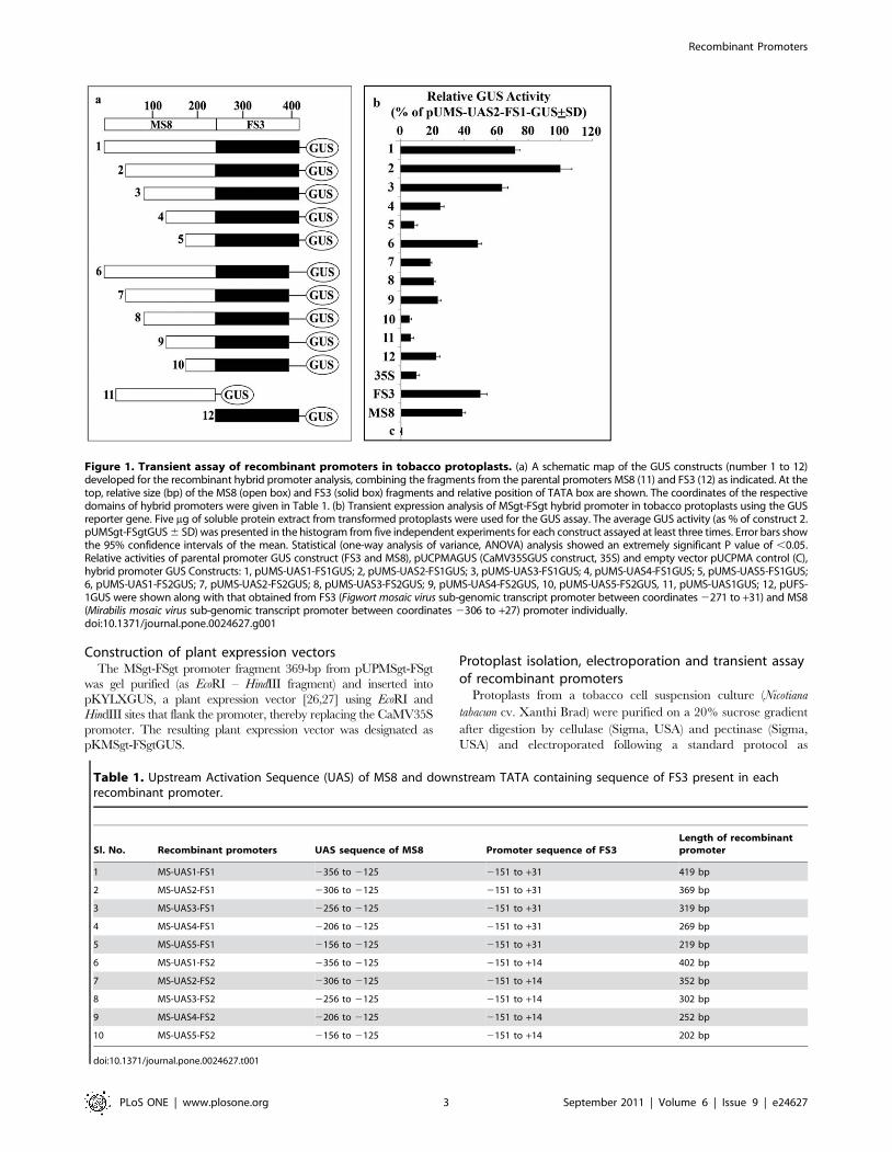
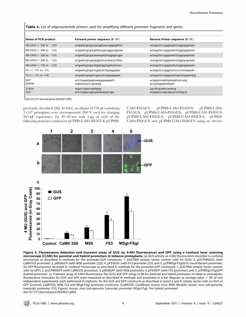
Table 1. Upstream Activation Sequence (UAS) of MS8 and downstream TATA containing sequence of FS3 present in eachrecombinant promoter.
Sl. No. Recombinant promoters UAS sequence of MS8 Promoter sequence of FS3Length of recombinantpromoter
1 MS-UAS1-FS1 2356 to 2125 2151 to +31 419 bp
2 MS-UAS2-FS1 2306 to 2125 2151 to +31 369 bp
3 MS-UAS3-FS1 2256 to 2125 2151 to +31 319 bp
4 MS-UAS4-FS1 2206 to 2125 2151 to +31 269 bp
5 MS-UAS5-FS1 2156 to 2125 2151 to +31 219 bp
6 MS-UAS1-FS2 2356 to 2125 2151 to +14 402 bp
7 MS-UAS2-FS2 2306 to 2125 2151 to +14 352 bp
8 MS-UAS3-FS2 2256 to 2125 2151 to +14 302 bp
9 MS-UAS4-FS2 2206 to 2125 2151 to +14 252 bp
10 MS-UAS5-FS2 2156 to 2125 2151 to +14 202 bp
doi:10.1371/journal.pone.0024627.t001
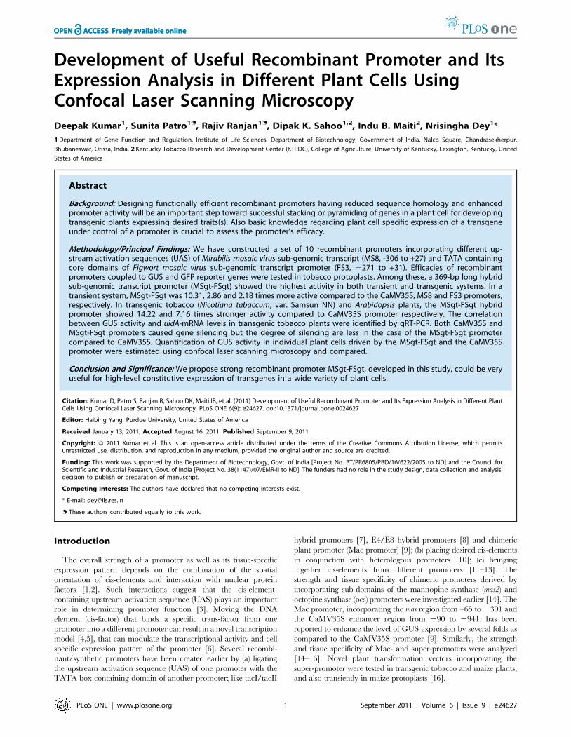
Figure 1. Transient assay of recombinant promoters in tobacco protoplasts. (a) A schematic map of the GUS constructs (number 1 to 12)developed for the recombinant hybrid promoter analysis, combining the fragments from the parental promoters MS8 (11) and FS3 (12) as indicated. At thetop, relative size (bp) of the MS8 (open box) and FS3 (solid box) fragments and relative position of TATA box are shown. The coordinates of the respectivedomains of hybrid promoters were given in Table 1. (b) Transient expression analysis of MSgt-FSgt hybrid promoter in tobacco protoplasts using the GUSreporter gene. Five mg of soluble protein extract from transformed protoplasts were used for the GUS assay. The average GUS activity (as % of construct 2.pUMSgt-FSgtGUS 6 SD) was presented in the histogram from five independent experiments for each construct assayed at least three times. Error bars showthe 95% confidence intervals of the mean. Statistical (one-way analysis of variance, ANOVA) analysis showed an extremely significant P value of ,0.05.Relative activities of parental promoter GUS construct (FS3 and MS8), pUCPMAGUS (CaMV35SGUS construct, 35S) and empty vector pUCPMA control (C),hybrid promoter GUS Constructs: 1, pUMS-UAS1-FS1GUS; 2, pUMS-UAS2-FS1GUS; 3, pUMS-UAS3-FS1GUS; 4, pUMS-UAS4-FS1GUS; 5, pUMS-UAS5-FS1GUS;6, pUMS-UAS1-FS2GUS; 7, pUMS-UAS2-FS2GUS; 8, pUMS-UAS3-FS2GUS; 9, pUMS-UAS4-FS2GUS, 10, pUMS-UAS5-FS2GUS, 11, pUMS-UAS1GUS; 12, pUFS-1GUS were shown along with that obtained from FS3 (Figwort mosaic virus sub-genomic transcript promoter between coordinates 2271 to +31) and MS8(Mirabilis mosaic virus sub-genomic transcript promoter between coordinates 2306 to +27) promoter individually.doi:10.1371/journal.pone.0024627.g001
Recombinant Promoters
PLoS ONE | www.plosone.org 3 September 2011 | Volume 6 | Issue 9 | e24627
previously described [26]. In brief, an aliquot of 750 ml containing
26106 protoplasts were electroporated (200 V used for charging
965-mF capacitance for 40–50 ms) with 5 mg of each of the
following promoter constructs: pUPMS-UAS1-FS1GUS, pUPMS-
UAS2-FS1GUS, pUPMS-UAS3-FS1GUS, pUPMS-UAS4-
FS1GUS, pUPMS-UAS5-FS1GUS, pUPMS-UAS1-FS2GUS,
pUPMS-UAS2-FS2GUS, pUPMS-UAS3-FS2GUS, pUPMS-
UAS4-FS2GUS and pUPMS-UAS5-FS2GUS using an electro-
Table 2. List of oligonucleotide primers used for amplifying different promoter fragments and genes.
Name of PCR product Forward primer sequence (59–39) Reverse Primer sequence (59–39)
MS-UAS1 (2356 to 2125) actgaattcgtcgacagcggtaaaacaggtgattact actaagcttcccgggtaattctctggtgagataatc
MS-UAS2 (2306 to 2125) actgaattcgtcgacgttttacagtcaggacagataat actaagcttcccgggtaattctctggtgagataatc
MS-UAS3 (2256 to 2125) actgaattcgtcgacaaaaagattactggtgacagta actaagcttcccgggtaattctctggtgagataatc
MS-UAS4 (2206 to 2125) actgaattcgtcgacgtggttttcacattacacctttaa actaagcttcccgggtaattctctggtgagataatc
MS-UAS5 (2156 to 2125) actgaattcgtcgacatgtgctggctgattatctcacc actaagcttcccgggtaattctctggtgagataatc
FS-1 (2151 to +31) actgaattcgtcgactcgaacatcttgaaggtgtac actaagcttcccgggcactccccctctctaaaaatt
FS-2 (2151 to +14) actgaattcgtcgactcgaacatcttgaaggtgtac actaagcttcccgggaaattttgtttttagaatttgtg
GFPGAPDH
actctcgagatgagtaaaggagaagaacttcagtaaacgacccgtaaatg
actgagctcttatttgtatagttcatccatggccagttggtgttaatgttt
b-ActinGUS
atgactcagatcatgtttgagactctcgagacaggcgattaaagagctgat
agccttcgcaatccacatctgactgagctccatgcatacaccactttgcta
doi:10.1371/journal.pone.0024627.t002
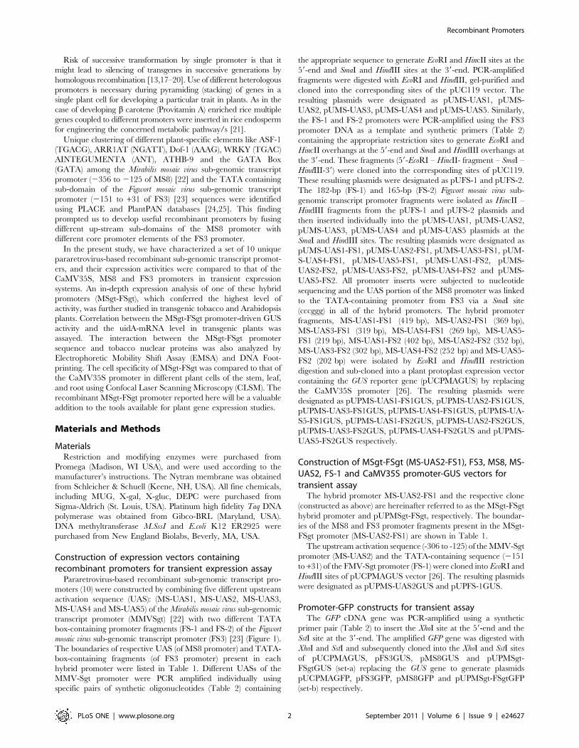
Figure 2. Fluorescence detection and transient assay of GUS (as 4-MU fluorescence) and GFP using a confocal laser scanningmicroscope (CLSM) for parental and hybrid promoters in tobacco protoplasts. (a) GUS activity as 4-MU fluorescence recorded in confocalmicroscope as described in methods for the promoter-GUS constructs: 1, pUCPMA (empty vector control, with no GUS); 2, pUCPMAGUS (withCaMV35S promoter) 3, pMS8GUS (with MS8 promoter [22]); 4, pFS3GUS (with FS3 promoter [23]) and 5, pUPMSgt-FSgtGUS (recombinant promoter).(b) GFP fluorescence recorded in confocal microscope as described in methods for the promoter-GFP constructs: 1, pUCPMA (empty vector control,with no GFP); 2, pUCPMAGFP (with CaMV35S promoter); 3, pMS8GFP (with MS8 promoter); 4, pFS3GFP (with FS3 promoter); and 5, pUPMSgt-FSgtGFP(hybrid promoter). (c) Transient assay of 4-MU fluorescence (for GUS) and GFP using CLSM for parental and hybrid promoters in tobacco protoplasts,fluorescence intensities for GUS and GFP were measured as described in methods and presented in a bar diagram as average value 6 SD of twoindependent experiments, each performed in triplicate, for the GUS and GFP constructs as described in panel a and b: empty vector with no GUS orGFP (Control), CaMV35S, MS8, FS3 and MSgt-FSgt promoter constructs. (CaMV35S: Cauliflower mosaic virus; MS8: Mirabilis mosaic virus sub-genomictranscript promoter; FS3: Figwort mosaic virus sub-genomic transcript promoter; MSgt-FSgt: the hybrid promoter).doi:10.1371/journal.pone.0024627.g002
Recombinant Promoters
PLoS ONE | www.plosone.org 4 September 2011 | Volume 6 | Issue 9 | e24627
poration cuvette (0.4-cm electrode gap). The plasmid pUCPMA-
GUS containing the CaMV35S promoter [26] was used as a
control to compare the activities of the promoters.
GUS activities in transformed protoplasts were measured after
20 h of incubation at 28uC [28]. The average activities of these
promoter constructs were expressed as the mean of five successive
independent experiments.
Promoter expression analysis using CLSMTobacco protoplasts were electroporated with promoter con-
structs carrying GUS reporter (set a) and GFP reporter (set b)
constructs for assaying their transient activities as described earlier.
Protoplasts electroporated with promoter constructs carrying the
GUS reporter (set-a) were incubated in a 1mM MUG (4-methyl-
umbelliferyl-beta-d-glucuronide) at 37uC for 30 in to generate 4-
MU (7-hydroxy-4-methylcoumarin) for the detection of GUS
localization (as blue fluorescence) in protoplasts using CLSM (TCS
SP5, Leica Microsystems CMS GmbH, D-68165 Mannheim,
Germany) with similar specifications as described earlier [29].
Similarly, to excite the expressed GFP [30,31] in protoplasts, the
argon laser (40%) with AOTF of 488 nm (at 30%) was used. The
fluorescence emissions were collected between 501 and 598 nm as
described earlier [29]. Following image acquisition, 4-MU (for
GUS) and GFP fluorescence intensities were quantified using the
LAS AF Software as per the instructions of Leica Microsystems.
The GFP and GUS (4-MU) fluorescence intensities from 200
individual protoplasts were assayed, and the mean data were
presented with respective 6 SD.
Tobacco plant transformation and analysis of transgenicplants
The following constructs, pKYLX (vector) [27], pKYLXGUS
(containing the CaMV35S promoter), pKMS8GUS [22],
pKFS3GUS [23], and pKMSgt-FSgtGUS were introduced into
the Agrobacterium tumefaciens strain C58C1:pGV3850 by the freeze
thaw method [32]. Tobacco plants (Nicotiana tabacum cv Samsun
NN) were transformed with the engineered Agrobacterium as
previously described [33]. Ten to twelve (10–12) independent
plant lines were generated for each construct and maintained
under green house conditions (photoperiod: 16/8 hour at
220 mmole m22 s21, Temperature: 28u63uC, Humidity: 70–
75%). Kanamycin-resistant plants (T1 generation) were used for
further analysis. GUS activity in seedling, root, leaf and stem were
measured according to the protocol described earlier [28,34].
Transgenic seedlings obtained from each construct were subjected
to histochemical GUS staining using 1% X-gluc solution.
Transformation of Arabidopsis thaliana plants andanalysis of transgenic plants
Arabidopsis thaliana (ecotype Columbia) plants were transformed
by pKYLXGUS and pKMSgt-FSgtGUS by floral dip method
[35]. The Arabidopsis flower buds were dipped in an Agrobacter-
ium cell suspension containing freshly prepared 5% sucrose (wt/
vol) and 0.02–0.05% Silwet L-77 (vol/vol) for 30–45 sec. After
that these plants were grown at 22uC under long day condition
(16 hours light and 8 hours dark cycle) till setting of seeds. Seeds
were collected after maturation and dried. After surface steriliza-
tion, seeds were suspended in sterile 0.05% agarose and spreaded
on MS selection plate (4.3 g Murashige & Skoog salts, 10 g
sucrose, 0.5 g MES, 8 g agar per liter; pH 5.7, Kanamycin
100 mg/l and Cefotaxime 100 mg/l) and allowed to germinate.
Only true transformants produced green healthy leaves (non-
transformants became dried and bleached). A total number of 75
independent transgenic plants were raised. GUS activities in whole
seedlings (21 days old) were measured according to the protocol
described earlier [28]. Histochemical GUS staining was performed
as described earlier.
Molecular analysis of transgenic plants: RNA isolation,northern blot analysis
Total RNA was obtained from transgenic seedlings expressing
pKYLX, pKYLXGUS, pKMS8GUS, pKFS3GUS and pKMSgt-
FSgtGUS using the RNeasy plant mini Kit (Qiagen, Tokyo,
Japan) according to manufacturer’s protocol. The GUS and b-Actin
genes were PCR-amplified from pKYLXGUS plasmid (for GUS)
and tobacco genomic DNA [36] respectively. The PCR fragments
were gel-purified using the QIAquick gel extraction kit (Qiagen,
Tokyo, Japan) and was labeled with [a- P32] dCTP using the
Random Primer labeling Kit (NEBlot Kit, New England Biolabs
Inc., MA, USA). Total RNA (5 mg) isolated from the transgenic
seedlings was subjected to electrophoresis in a 1.2% agarose gel
containing 0.66 M formaldehyde and then transferred overnight
to an IMMOBILON-NY+ membrane (Millipore, Billerica, MA,
USA) by capillary action in 10X SSC (1.5M NaCl, 150 mM
sodium citrate). Hybridization with the 32P labeled GUS-probe was
carried out at 65C overnight and northern blot analysis was
carried out using standard protocol [37]. The washed membrane
with the bound probe was scanned with the FLA-5000 Imaging
System (Fuji FILM Life Science, USA). The membrane was
subsequently washed at 80uC in a solution containing 1X SSC,
0.1% SDS, and 0.05% Na–pyrophosphate to remove the GUS
probe. The stripped membrane was then re-probed with the b-
Actin to ensure equal loading.
Real-time PCR analysis of GUS transcripts driven by MS8,FS3, CaMV35S and MSgt-FSgt promoter
Total RNA was isolated from transgenic seedlings expressing
the pKMS8GUS, pKFS3GUS, pKYLXGUS, and pKMSgt-
FSgtGUS promoter constructs and subsequently mRNA was
isolated from total RNA using the PolyATract mRNA isolation
system (Promega, Madison, WI, USA). About 2.0 mg of mRNA
was used to synthesize the first strand cDNA using AMV reverse
transcriptase and oligo (dT) primers at 42uC for 1.5 hours. Real
time PCR reactions were performed using SYBR Premix Ex
TaqTM II (Perfect Real Time, Takara Bio Inc.) according to
manufacturer’s instruction on an Opticon-2 qRT-PCR instrument
(MJ Research, Bio-Rad; Model CFD-3220). GAPDH cDNA was
used as an internal control for normalization of GUS mRNA levels
in the real time PCR. The PCR cycling program was conducted as
follows; 94uC for 30 sec followed by 37 cycles of 94uC for 5 sec,
57uC for 30 sec, 72uC for 30 sec and finally 72uC for 5 minutes.
The GUS mRNA levels in MS8, FS3 and MSgt-FSgt and
CaMV35S promoters were calculated using the 22DDCT method
[38,39] and data presented here are relative to CaMV35S
promoter line.
Quantitative reverse transcription-polymerase chainreaction (qRT-PCR)
First strand cDNA was synthesized using 2.0 mg mRNA purified
from total RNA extracted from transgenic tobacco seedlings
transformed with pKYLX, pKYLXGUS, pKMS8GUS,
pKFS3GUS and pKMSgt-FSgtGUS and transgenic Arabidopsis
seedlings transformed with pKYLX, pKYLXGUS and pKMSgt-
FSgtGUS as described above. PCR amplifications of GUS and
GAPDH were performed using 1 ml of first strand reaction products
in the presence of gene specific primers (Table 2). Twenty-six
Recombinant Promoters
PLoS ONE | www.plosone.org 5 September 2011 | Volume 6 | Issue 9 | e24627
cycles of PCR was conducted with denaturation at 92uC for 1
minute, annealing at 60uC for 30 seconds and extension at 72uCfor 1 minute. Eight ml samples of each PCR reaction were
analyzed on 1% agarose gel.
Preparation of transgenic plant samples for CLSManalysis
Transgenic tobacco plants were generated using the MSgt-FSgt
and CaMV35S promoters fused to the GUS reporter gene as
described earlier. The transgenic plant parts were initially kept
under vacuum infiltration for 10 min and then incubated at 37uCovernight in the presence of 1 mM MUG to produce 4-MU. The
transverse sections (approximately 80 micron thick) of leaf blade,
leaf midrib, stem and root were obtained by sectioning the treated
plant tissue using a microtome (Cryostat, Leica CM 1850).
Fluorescence images of thin transverse sections of these tissues
were captured using a CLSM. To quantify GUS activity, the
treated tissue sections were excited with the 405 diode laser (Argon
364 nm UV laser may be more appropriate), and fluorescence
emissions were collected between 440 and 455 nm with the
detector (PMT) gain set at 1150V. Intracellular GUS localizations
were detected by blue fluorescence of 4-MU using CLSM and
intensities of blue-fluorescence were measured from different cells
according to the protocol described earlier [29].
Nuclear protein binding assay for the hybrid promoterTobacco nuclear extracts were prepared from greenhouse-
grown leaves using standard protocol [40] with slight modifica-
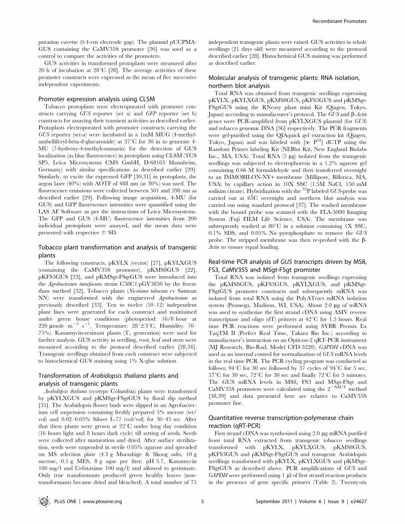
Figure 3. Expression analysis of parental and recombinant hybrid promoters in transgenic tobacco plants. (a) Promoter activity wasmonitored in 21-day old tobacco (N. tabacum cv Samsun NN) seedlings (R1 progeny, 2nd generation, KanR) grown aseptically on an MS-agar mediumin presence of kanamycin (200 mg/ml) and 1% sucrose. Soluble protein extracts (5 mg) from whole seedlings were used for the GUS assay. The datapresent average 6SD of four independent experiments for each construct: pKYLX (empty vector control), pKYLXGUS (with the CaMV35S promoter),pKMS8GUS (with MS8 promoter), pKFS3GUS (with FS3 promoter), pKMSgt-FSgtGUS (hybrid promoter). (b) Histochemical localization of GUS activity(blue coloration) in transgenic tobacco seedlings (magnification x 10.0) obtained from control (1), CaMV35S (2), MS8 (3) FS3 (4), and MSgt-FSgt (5)promoter constructs represents the relative strength of the promoter constructs. (c) Display of electrophoresis of total RNA, obtained from 21 daysold tobacco seedlings developed with control (1), CaMV35S (2), MS8(3), FS3 (4), and MSgt-FSgt (5) promoter constructs used as loading control. (d)Northern blot detection of the GUS reporter gene in transgenic tobacco developed with control (1), CaMV35S (2), MS8 (3), FS3 (4) and MSgt-FSgt (5)promoter constructs. (e) The same membrane was re-probed with 32P-labelled b-Actin gene to confirm the equal loading of RNA samples. (f)Electrophoresis of RT-dependent PCR amplifications of GUS transcripts from total RNA obtained from transgenic plant generated for empty vectorcontrol (1), CaMV35S (2), MS8 (3), FS3 (4), and MSgt-FSgt (5) promoter constructs as described in (a). (g) Electrophoresis of RT-independent (-RT) PCRamplifications of GUS transcripts from total RNA obtained from transgenic plant developed for empty vector control (1), CaMV35S (2), MS8 (3) FS3 (4),and MSgt-FSgt (5) promoter constructs. Electrophoresis was performed to check the presence of DNA in RNA preparation. (h) Electrophoresis of RT-dependent PCR amplifications of GAPDH transcripts from total RNA from transgenic plant developed for empty vector control (1), CaMV35S (2), MS8(3), FS3 (4), and MSgt-FSgt (5) promoter constructs. (i) The relative abundance of GUS transcript (data present average fold differences of GUStranscript 6 SD of two independent experiments) assayed by real-time quantitative reverse transcription PCR (RT-qPCR). Total RNA was isolated fromtransgenic seedlings (2nd generation, 21-day old) developed for pKYLX (empty vector control), pKYLXGUS (with CaMV35S promoter), pKMS8GUS(with MS8 promoter), pKFS3GUS (with FS3 promoter), pKMSgt-FSgtGUS (hybrid promoter).doi:10.1371/journal.pone.0024627.g003
Recombinant Promoters
PLoS ONE | www.plosone.org 6 September 2011 | Volume 6 | Issue 9 | e24627
tions. The MSgt-FSgt promoter DNA fragment (369 bp) was
labeled using Prime-a-gene labeling system (Promega, USA) in
the presence of c-P32 dATP at 37uC for 1 hr. The EMSA
binding reaction was carried out in a 30-ml volume containing32P-labeled MSgt-FSgt probe in binding buffer (100 mM Tris-
HCl pH 7.5, 0.5 mM DTT, 1 mM EDTA, 7% v/v glycerol,
1 mM PMSF, 5 mg/ml BSA, 0.1–0.2 mg/ml poly dI-dC, 40–
50 mM NaCl) in the presence of nuclear protein extract (5–
6 mg). Reactions were incubated at room temperature for
30 min. The DNA-protein complexes were resolved in non-
denaturing 5% polyacrylamide gel [40]. Competitive EMSA
reactions were carried out in the presence of the 32P-labeled
MSgt-FSgt DNA (as probe) and 20, 50 and 100 fold excesses of
non-labeled DNA (MSgt-FSgt).
DNaseI foot-printing experiments were carried using standard
protocol [41]. The probe (end-labeled MSgt-FSgt promoter) was
incubated in the presence of 100 mM Tris-HCl pH 7.5, 0.5 mM
DTT, 1 mM EDTA, 7% v/v glycerol, 1 mM PMSF, 5 mg/ml
BSA and 0.1–0.2 mg/ml polydI-dC for 30 min at room temper-
ature. DNaseI (0.025 U/ml) was added and incubated for 45 sec
at room temperature with 5 mg and 10 mg of nuclear protein
separately. The reaction products were then denatured and
loaded onto a 6% denaturing polyacrylamide sequencing gel
along with sequencing reactions for the MSgt-FSgt DNA
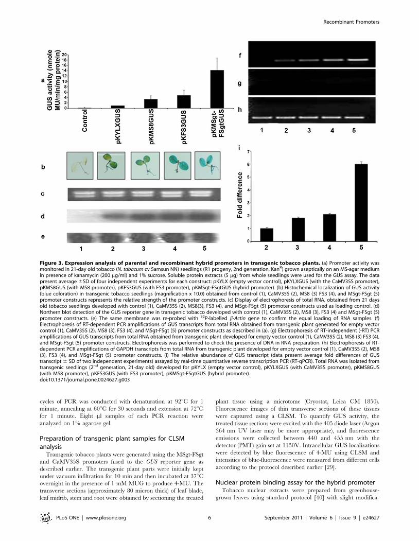
Figure 4. Comparative expression analysis of CaMV35S promoter and recombinant hybrid promoter in transgenic Arabidopsisplants. (a) Histochemical localization of GUS activity (blue coloration) in 21 days old transgenic Arabidopsis seedlings (magnification x 10.0)developed for the constructs: pKYLX (empty vector, Control), pKYLXGUS (with the CaMV35S promoter, 35S), and pKMSgt-FSgtGUS (hybrid promoter,MSgt-FSgt). (b) Promoter activity was assayed in 21 days old Arabidopsis seedlings (R1 progeny, 2nd generation, KanR) grown aseptically on an MS-agar medium in presence of kanamycin (100 mg/ml) and 1% sucrose. Soluble protein extracts (5 mg) from whole seedlings were used for the GUSassay. The data present average 6 SD of four independent experiments for each construct: pKYLX (empty vector control), pKYLXGUS (with theCaMV35S promoter), and pKMSgt-FSgtGUS (hybrid promoter); plasmids construction strategy described in the methods section. (c) Electrophoresis ofRT-dependent PCR amplifications of GUS transcripts from total RNA isolated from transgenic Arabidopsis plant developed for pKYLX (empty vectorcontrol), pKYLXGUS (with CaMV35S promoter), and pKMSgt-FSgtGUS (hybrid promoter); plasmids construction strategy described in the methodssection, the arrow indicating expected band. (d) Electrophoresis of RT-dependent PCR amplifications of GAPDH transcripts from total RNA obtainedfrom transgenic plant developed for empty vector (1), CaMV35S (2), and MSgt-FSgt (3) promoter constructs, arrow indicating expected band. (e)Electrophoresis of RT-independent (-RT) PCR amplifications of GUS transcripts from total RNA obtained from transgenic plant developed for emptyvector (1), CaMV35S (2), and MSgt-FSgt (3) promoter constructs showing no amplification (to check the presence of genomic DNA in the RNApreparation).doi:10.1371/journal.pone.0024627.g004
Recombinant Promoters
PLoS ONE | www.plosone.org 7 September 2011 | Volume 6 | Issue 9 | e24627
fragment (Thermo Sequenase Cycle Sequencing Kit; USB
Corporation, Cleveland, OH, USA).
In vitro methylation assayThe putative CG methylation sensitive sites present in MSgt-FSgt
and the CaMV35S promoters were determined in-silico using
Support Vector Machine (http://bio.dfci.harvard.edu/Methylator)
[42]. The effect of in vitro methylation on the transient expression of
MSgt-FSgt and CaMV35S promoters coupled to GUS reporter
gene were investigated according to Pradhan et al. [43] with some
modifications. Plasmids pUPMSgt-FSgtGUS and pUCPMAGUS
individually were grown in a dam2 and dcm2 E.coli K12 strain
ER2925 and purified. DNA methyltransferase M.SssI was used to
methylate each plasmid construct at 37uC for 1.5 hours in presence
of 0.5 mM S-adenosyl methionine. The methylated plasmids were
purified and quantified using a spectrophotometer (CECIL
BioQuest CE 2501 CECIL Instruments Ltd, England). Approxi-
mately 5 mg each methylated and unmethylated plasmids from both
constructs were used for transient assay in a protoplast system
(Nicotiana tabaccum cv. Xanthi brad) following protocol as described
earlier.
Statistical AnalysisStatistical analyses were performed by using Graph Pad Prism
(version 5.01). Unpaired students t test was used for analyzing the
activity of methylated-MSgt-FSgt and methylated-CaMV35S
promoters. A P value of ,0.05 was considered to reveal a
significant difference.
Results
Comparison of activity of recombinant promoters withMS-UAS2, FS-1, FS3, MS8 and CaMV35S promoters intransient protoplast assay
Recombinant promoters (Figure 1a, Table 1) fused to the GUS
reporter gene were tested individually for their transcriptional
activities using a transient tobacco protoplast assay (Nicotiana
tabacum cv. Xanthi Brad). Transformed protoplast with empty
vector (pUCPMA) was used as a control. As shown in Figure 1b,
the relative GUS activities of all hybrid promoters were expressed
considering the activity of MSgt-FSgt promoter as 100%. The
GUS activities obtained from the hybrid constructs MS-UAS1-
FS1, MSgt-FSgt, MS-UAS3-FS1, MS-UAS4-FS1, MS-UAS1-FS2
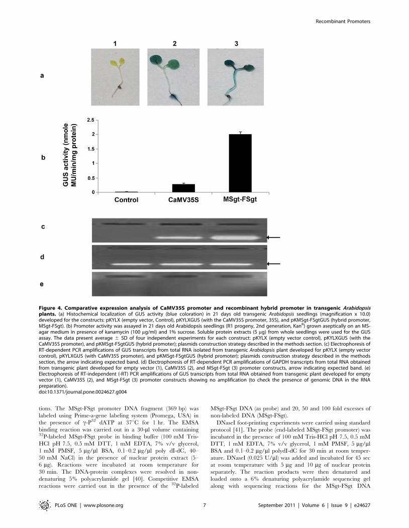
Figure 5. Expression analysis of CaMV35S and MSgt-FSgt promoters in transgenic Arabidopsis plants. Seventy five independenttransgenic Arabidopsis lines were generated with the constructs pKYLXGUS and pKMSgt-FSgtGUS as described in methods. The average GUS activity6 SD from two independent experiments of 75 independent lines (21 days old) developed for each construct pKYLXGUS (35S-GUS), pKMSgt-FSgtGUS(MSgt-FSgtGUS) and empty vector pKYLX (control) are presented in the panel (a) line number 1 to 25, panel (b) line number 26 to 50 and panel (c)line number 51 to 75. Silenced (very low/basal level expression) lines were shown as different symbols for the CaMV35S and MSgt-FSgt promoters.doi:10.1371/journal.pone.0024627.g005
Recombinant Promoters
PLoS ONE | www.plosone.org 8 September 2011 | Volume 6 | Issue 9 | e24627
and MS-UAS4-FS2 were found to be 7.32, 10.31, 6.49, 2.54, 4.93
and 2.37 times stronger, respectively, than the CaMV35S
promoter (Figure 1b). The recombinant promoter, MSgt-FSgt,
showed maximum GUS activity, approximately10 fold higher
than that of CaMV35S promoter. The activity of MSgt-FSgt was
found to be 5.22, 13.99, 2.18 and 2.9 times stronger than the FS-1,
MS-UAS-2, FS3 [23] and MS8 [22] promoters respectively
(Figure 1b).
Comparison of promoter activities of MS8, FS3, CaMV35Sand MSgt-FSgt fused to GUS and GFP reporters usingCLSM
Tobacco protoplasts were transformed with two different sets ofpromoter constructs as set-a and set-b separately as described inmethods. In both cases the pUCPMA vector was used as a control.Blue fluorescence of transformed protoplasts obtained from using
set-a (with GUS reporter gene; Figure 2a) were captured by CLSM
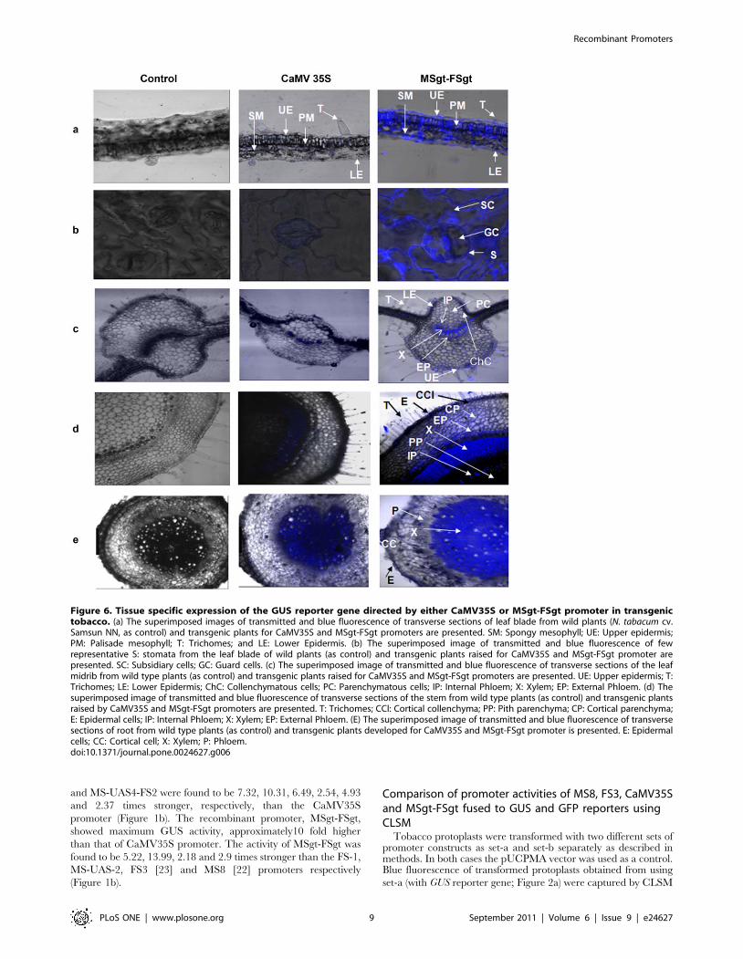
Figure 6. Tissue specific expression of the GUS reporter gene directed by either CaMV35S or MSgt-FSgt promoter in transgenictobacco. (a) The superimposed images of transmitted and blue fluorescence of transverse sections of leaf blade from wild plants (N. tabacum cv.Samsun NN, as control) and transgenic plants for CaMV35S and MSgt-FSgt promoters are presented. SM: Spongy mesophyll; UE: Upper epidermis;PM: Palisade mesophyll; T: Trichomes; and LE: Lower Epidermis. (b) The superimposed image of transmitted and blue fluorescence of fewrepresentative S: stomata from the leaf blade of wild plants (as control) and transgenic plants raised for CaMV35S and MSgt-FSgt promoter arepresented. SC: Subsidiary cells; GC: Guard cells. (c) The superimposed image of transmitted and blue fluorescence of transverse sections of the leafmidrib from wild type plants (as control) and transgenic plants raised for CaMV35S and MSgt-FSgt promoters are presented. UE: Upper epidermis; T:Trichomes; LE: Lower Epidermis; ChC: Collenchymatous cells; PC: Parenchymatous cells; IP: Internal Phloem; X: Xylem; EP: External Phloem. (d) Thesuperimposed image of transmitted and blue fluorescence of transverse sections of the stem from wild type plants (as control) and transgenic plantsraised by CaMV35S and MSgt-FSgt promoters are presented. T: Trichomes; CCl: Cortical collenchyma; PP: Pith parenchyma; CP: Cortical parenchyma;E: Epidermal cells; IP: Internal Phloem; X: Xylem; EP: External Phloem. (E) The superimposed image of transmitted and blue fluorescence of transversesections of root from wild type plants (as control) and transgenic plants developed for CaMV35S and MSgt-FSgt promoter is presented. E: Epidermalcells; CC: Cortical cell; X: Xylem; P: Phloem.doi:10.1371/journal.pone.0024627.g006
Recombinant Promoters
PLoS ONE | www.plosone.org 9 September 2011 | Volume 6 | Issue 9 | e24627
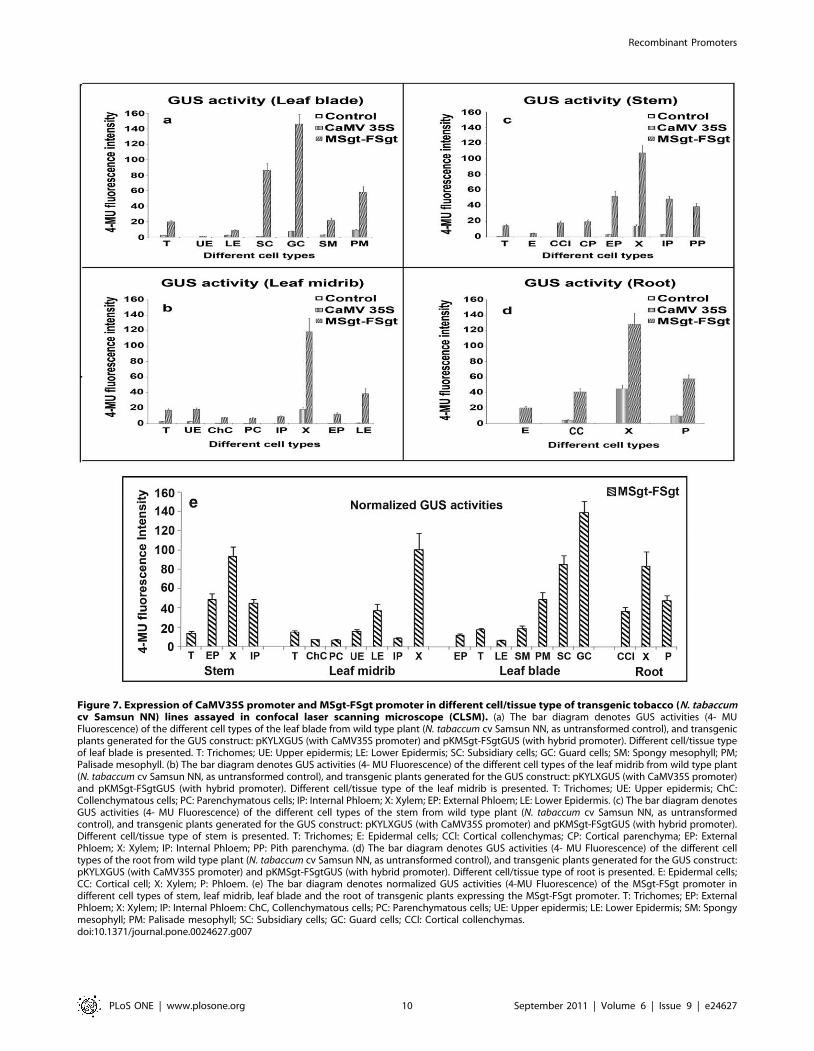
Figure 7. Expression of CaMV35S promoter and MSgt-FSgt promoter in different cell/tissue type of transgenic tobacco (N. tabaccumcv Samsun NN) lines assayed in confocal laser scanning microscope (CLSM). (a) The bar diagram denotes GUS activities (4- MUFluorescence) of the different cell types of the leaf blade from wild type plant (N. tabaccum cv Samsun NN, as untransformed control), and transgenicplants generated for the GUS construct: pKYLXGUS (with CaMV35S promoter) and pKMSgt-FSgtGUS (with hybrid promoter). Different cell/tissue typeof leaf blade is presented. T: Trichomes; UE: Upper epidermis; LE: Lower Epidermis; SC: Subsidiary cells; GC: Guard cells; SM: Spongy mesophyll; PM;Palisade mesophyll. (b) The bar diagram denotes GUS activities (4- MU Fluorescence) of the different cell types of the leaf midrib from wild type plant(N. tabaccum cv Samsun NN, as untransformed control), and transgenic plants generated for the GUS construct: pKYLXGUS (with CaMV35S promoter)and pKMSgt-FSgtGUS (with hybrid promoter). Different cell/tissue type of the leaf midrib is presented. T: Trichomes; UE: Upper epidermis; ChC:Collenchymatous cells; PC: Parenchymatous cells; IP: Internal Phloem; X: Xylem; EP: External Phloem; LE: Lower Epidermis. (c) The bar diagram denotesGUS activities (4- MU Fluorescence) of the different cell types of the stem from wild type plant (N. tabaccum cv Samsun NN, as untransformedcontrol), and transgenic plants generated for the GUS construct: pKYLXGUS (with CaMV35S promoter) and pKMSgt-FSgtGUS (with hybrid promoter).Different cell/tissue type of stem is presented. T: Trichomes; E: Epidermal cells; CCl: Cortical collenchymas; CP: Cortical parenchyma; EP: ExternalPhloem; X: Xylem; IP: Internal Phloem; PP: Pith parenchyma. (d) The bar diagram denotes GUS activities (4- MU Fluorescence) of the different celltypes of the root from wild type plant (N. tabaccum cv Samsun NN, as untransformed control), and transgenic plants generated for the GUS construct:pKYLXGUS (with CaMV35S promoter) and pKMSgt-FSgtGUS (with hybrid promoter). Different cell/tissue type of root is presented. E: Epidermal cells;CC: Cortical cell; X: Xylem; P: Phloem. (e) The bar diagram denotes normalized GUS activities (4-MU Fluorescence) of the MSgt-FSgt promoter indifferent cell types of stem, leaf midrib, leaf blade and the root of transgenic plants expressing the MSgt-FSgt promoter. T: Trichomes; EP: ExternalPhloem; X: Xylem; IP: Internal Phloem: ChC, Collenchymatous cells; PC: Parenchymatous cells; UE: Upper epidermis; LE: Lower Epidermis; SM: Spongymesophyll; PM: Palisade mesophyll; SC: Subsidiary cells; GC: Guard cells; CCl: Cortical collenchymas.doi:10.1371/journal.pone.0024627.g007
Recombinant Promoters
PLoS ONE | www.plosone.org 10 September 2011 | Volume 6 | Issue 9 | e24627
as described in the Method section, and were presented. Similarly,
the GFP fluorescence images of protoplasts transformed with set-b
constructs were presented in Figure 2b. The intensity of the blue
fluorescence due to 4-MU (for GUS) and green fluorescence (for
GFP) from the individual promoter construct was quantified from
200 independent transformed protoplasts using the LAS-AF
software. CLSM-based analysis of the GUS reporter (blue
fluorescence) demonstrated that the activity of the MSgt-FSgt
promoter was 1.79, 3.73 and 6.96 times stronger than that of the
FS3, MS8 and CaMV35S promoters, respectively (Figure 2c). The
expression level of the GFP reporter gene under the control of
these promoters as measured by CLSM revealed that the MSgt-
FSgt promoter was 1.51, 3.43 and 6.81 times stronger than the
FS3, MS8 and CaMV35S promoters (Figure 2c).
Analysis of transgenic tobacco plants developed withMS8, FS3, CaMV35S and MSgt-FSgt promoter constructs
Transgenic tobacco plants were generated using the promoter
constructs pKYLX (empty vector), pKYLXGUS, pKMS8GUS,
pKFS3GUS, pKMSgt-FSgtGUS. No phenotypic changes were
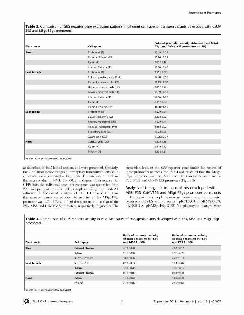
Table 3. Comparison of GUS reporter gene expression patterns in different cell types of transgenic plants developed with CaMV35S and MSgt-FSgt promoters.
Plant parts Cell typesRatio of promoter activity obtained from MSgt-FSgt and CaMV 35S promoters (± SD)
Stem Trichomes (T) 25.6962.35
External Phloem (EP) 15.8663.19
Xylem (X) 7.6661.17
Internal Phloem (IP) 15.0862.48
Leaf Midrib Trichomes (T) 7.5561.02
Collenchymatous cells (ChC) 17.2062.50
Parenchymatous cells (PC) 19.7562.68
Upper epidermal cells (UE) 7.8561.72
Lower epidermal cells (LE) 53.3964.82
Internal Phloem (IP) 57.1668.96
Xylem (X) 6.3560.89
External Phloem (EP) 41.9866.45
Leaf Blade Trichomes (T) 8.5760.93
Lower epidermis (LE) 3.2960.50
Spongy mesophyll (SM) 7.5761.41
Palisade mesophyll (PM) 6.4860.92
Subsidiary cells (SC) 90.269.95
Guard cells (GC) 20.0662.77
Root Cortical cells (CC) 9.5761.36
Xylem (X) 2.8160.32
Phloem (P) 6.2961.31
doi:10.1371/journal.pone.0024627.t003
Table 4. Comparison of GUS reporter activity in vascular tissues of transgenic plants developed with FS3, MS8 and MSgt-FSgtpromoters.
Plant parts Cell types
Ratio of promoter activityobtained from MSgt-FSgtand MS8 (± SD)
Ratio of promoter activityobtained from MSgt-FSgtand FS3 (± SD)
Stem External Phloem 4.1960.42 4.6060.12
Xylem 2.5660.32 2.1660.78
Internal Phloem 3.8860.32 4.7261.11
Leaf Midrib Internal Phloem 0.9260.17 1.0460.05
Xylem 3.2260.43 3.0960.19
External Phloem 0.7260.03 0.8460.43
Root Xylem 1.7660.63 1.6860.42
Phloem 2.3760.87 2.4560.61
doi:10.1371/journal.pone.0024627.t004
Recombinant Promoters
PLoS ONE | www.plosone.org 11 September 2011 | Volume 6 | Issue 9 | e24627
marked between control and transgenic tobacco plants expressing
these promoter constructs. Total proteins isolated from T1
seedlings were used to assess GUS activity [28]. A comparison
of the GUS activities of the various constructs with the CaMV35S
(considered as 1) was presented in Figure 3a. In transgenic plants,
the MSgt-FSgt promoter was found to exhibit 14.22, 4.26 and 2.94
times stronger activity, compared to the CaMV35S, MS8, and
FS3 promoters, respectively (Figure 3a).
Histochemical staining of transgenic tobacco seedlings (T1
generation, 21 days old) generated with the CaMV35S, MS8, FS3
and MSgt-FSgt promoter constructs were presented in Figure 3b.
When GUS activities were measured from soluble proteins
extracted from the roots, leaves and stems of transgenic seedlings,
the relative GUS activities in the tissues were in the following
order: roots. leaves. stems (data not shown).
Northern blot analysis was carried out using total RNA isolated
from transgenic seedlings generated with pKYLX, pKYLXGUS,
pKMS8GUS, pKFS3GUS, pKMSgt-FSgtGUS. Equal loading of
RNA for the northern blot analysis was confirmed (Figure 3c) and
the results were shown in Figure 3d. The highest intensities of
GUS transcript accumulation were observed for the MSgt-FSgt
promoter, followed by the FS3, MS8 and CaMV35S promoters.
The membrane was washed and reprobed using the b-Actin probe
as discussed in the Methods for further confirmation of equal
loading as shown in Figure 3e.
Accumulation of GUS-specific mRNAs driven by each promoter
in transgenic tobacco plants (T1 generation) were determined by
semi-quantitative RT-PCR and real time PCR. The RT-
dependent PCR amplifications of GUS transcript driven by the
CaMV35S, MS8, FS3 and MSgt-FSgt promoters in transgenic
plants were shown in Figure 3f. Accumulation of GUS transcripts
was found to be the highest in transgenic plants carrying the MSgt-
FSgt promoter followed by the FS3, MS8 and CaMV35S
promoters. The fold differences in the uidA-mRNA expression
levels for CaMV35S, MS8, FS3 and MSgt-FSgt promoter
constructs were measured by real-time PCR. Results were
presented as the mean of three independent experiments with
respective standard deviation (6 SD) in Figure 3i.
Comparison of MSgt-FSgt and CaMV35S promoteractivity in transgenic Arabidopsis plant
We have generated approximately 75 independent transgenic
Arabidopsis plants expressing following promoter constructs
pKYLX (empty vector), pKYLXGUS, pKMSgt-FSgtGUS indi-
vidually as described in Methods. Furthermore, transgenic
Arabidopsis plants expressing MSgt-FSgt promoter were found
to be phonotypically identical with wild type plants.
Histochemical staining of transgenic Arabidopsis seedlings express-
ing MSgt-FSgtGUS and CaMV35SGUS constructs were shown in
Figure 4a; and the corresponding GUS activities in Figure 4b. The
GUS activity of MSgt-FSgt was 7.16 times stronger than the
CaMV35S promoter activity (Figure 4b). In the transgenic
Arabidopsis plant, the RT-dependent PCR (semi q-RT PCR)
amplification of GUS transcripts driven by the CaMV35S and
MSgt-FSgt promoters were determined and it was observed that
accumulation of GUS transcripts was more in transgenic Arabi-
dopsis plants harboring the MSgt-FSgt promoter compared to that
in transgenic plants with the CaMV35S promoter (Figure 4c).
The average GUS activity of 75 independent lines (21 days old)
from each construct was evaluated from two independent
experiments and the results were presented in Figure 5. Approx-
imately 10 out of 75 (13.3%) independent transgenic Arabidopsis
lines expressing CaMV35S showed the basal or minimum level of
GUS expression while in case of MSgt-FSgt promoter 5 lines out
of 75 (6.7%) transgenic Arabidopsis lines showed the basal or
minimum level of GUS expression (Figure 5a, 5b and 5c).
Cell/Tissue specificity of MSgt-FSgt and CaMV35Spromoter
The GUS gene expression levels in different cells/tissue types in
the leaf, stem and root of tobacco plants transformed with the
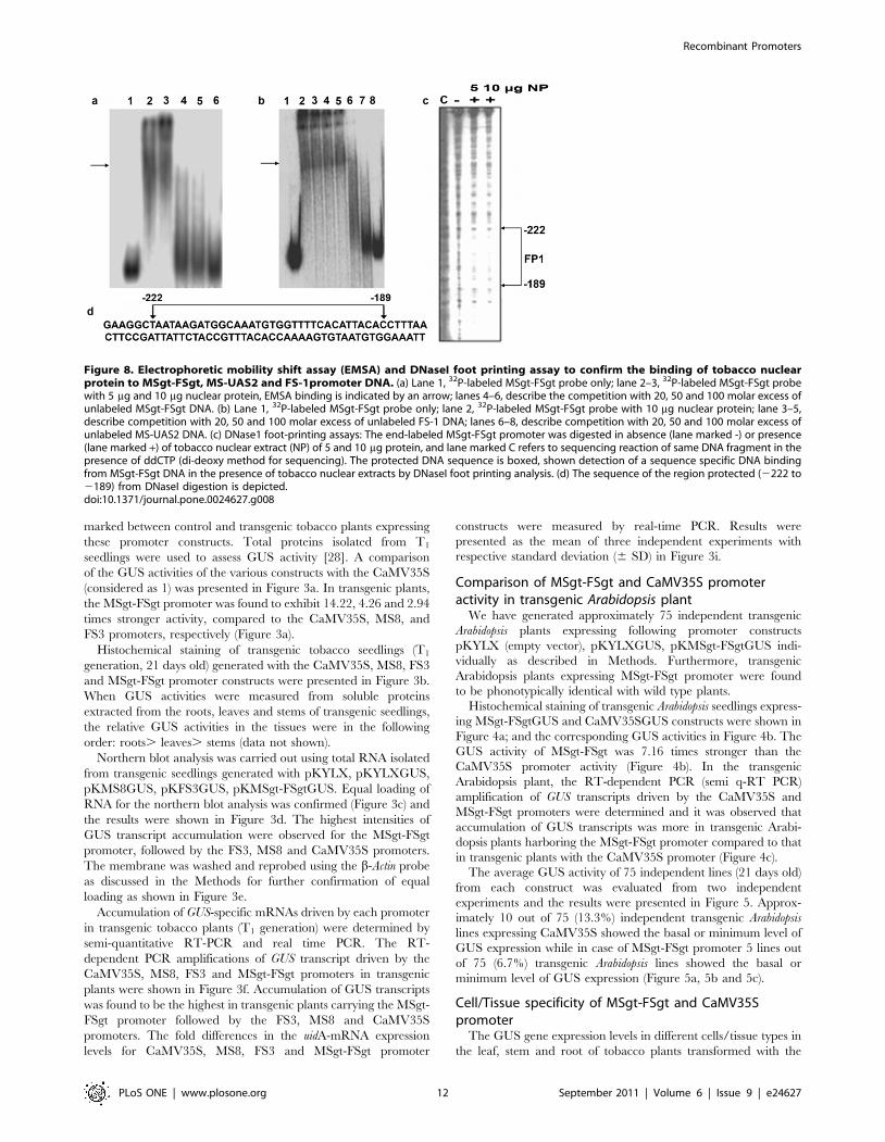
Figure 8. Electrophoretic mobility shift assay (EMSA) and DNaseI foot printing assay to confirm the binding of tobacco nuclearprotein to MSgt-FSgt, MS-UAS2 and FS-1promoter DNA. (a) Lane 1, 32P-labeled MSgt-FSgt probe only; lane 2–3, 32P-labeled MSgt-FSgt probewith 5 mg and 10 mg nuclear protein, EMSA binding is indicated by an arrow; lanes 4–6, describe the competition with 20, 50 and 100 molar excess ofunlabeled MSgt-FSgt DNA. (b) Lane 1, 32P-labeled MSgt-FSgt probe only; lane 2, 32P-labeled MSgt-FSgt probe with 10 mg nuclear protein; lane 3–5,describe competition with 20, 50 and 100 molar excess of unlabeled FS-1 DNA; lanes 6–8, describe competition with 20, 50 and 100 molar excess ofunlabeled MS-UAS2 DNA. (c) DNase1 foot-printing assays: The end-labeled MSgt-FSgt promoter was digested in absence (lane marked -) or presence(lane marked +) of tobacco nuclear extract (NP) of 5 and 10 mg protein, and lane marked C refers to sequencing reaction of same DNA fragment in thepresence of ddCTP (di-deoxy method for sequencing). The protected DNA sequence is boxed, shown detection of a sequence specific DNA bindingfrom MSgt-FSgt DNA in the presence of tobacco nuclear extracts by DNaseI foot printing analysis. (d) The sequence of the region protected (2222 to2189) from DNaseI digestion is depicted.doi:10.1371/journal.pone.0024627.g008
Recombinant Promoters
PLoS ONE | www.plosone.org 12 September 2011 | Volume 6 | Issue 9 | e24627
CaMV35S and MSgt-FSgt promoters were assayed using CLSM
as described in the Method section. In the leaf blade, the MSgt-
FSgt promoter exhibited 8.5760.93, 3.2960.50, 7.5761.41,
6.4860.92, 90.269.95 and 20.0662.77 fold higher activity than
the 35S promoter in the trichome, lower epidermis, spongy
mesophyll, palisade mesophyll, subsidiary and guard cells,
respectively (Figure 6a, 6b and 7a and Table 3). The expression
acts of the MSgt-FSgt and CaMV35S promoters were also
compared in the transgenic leaf midribs using CLSM-based
techniques (Figure 6c and 7b). When compared, GUS gene
expression under control of the hybrid promoter was found to be
7.55 61.02, 17.2062.5, 19.7562.68, 7.8561.72, 53.3964.82,
57.1668.96, 6.3560.89, and 41.9866.45 times stronger in
trichomes, collenchymatous cells, parenchymatous cells, upper
epidermal cells, lower epidermal cells, internal phloem, xylem and
external phloem of leaf midrib, compared to the CaMV35S
promoter (Table 3). In the stem, the GUS gene expression levels of
the MSgt-FSgt promoter were 25.6962.35, 15.8663.19,
7.6661.17 and 15.0862.48 fold more in trichomes, external
phloem, xylem and internal phloem respectively, than the
CaMV35S promoter (Table 3, Figure 6d and 7c). In the case of
root, the activity of the CaMV35S promoter was undetectable in
root epidermal cells, moderate in root cortical cells and in phloem
tissue, but exhibited the highest activity in the root xylem tissue. In
contrast, the MSgt-FSgt promoter has the capacity to drive the
reporter gene expression at a much higher level in all root cell
types (Figure 6e and 7d). In summary, the MSgt-FSgt promoter
activity was 9.5761.36, 2.8160.32 and 6.2961.31 times stronger
in the root cortical cell, xylem and phloem tissues, respectively,
compared to the 35S promoter (Table 3). After normalizing the
activity of the CaMV35S promoter across the tissues, we observed
high level of the MSgt-FSgt promoter expression in xylem (X),
external phloem (EP), and internal phloem (IP) of stem; xylem (X)
and lower epidermis (LE) of the leaf midrib; guard cell, subsidiary
cells (SC) and palisade mesophyll (PM) of the leaf blade and xylem
(X), phloem (P) and cortical cell (CC) of root (Figure 7e).
Comparison of promoter activity of MSgt-FSgt, FS3 andMS8 in vascular tissue
The GUS gene expression patterns in different vascular tissues
and cell types of leaves, stems and roots of transgenic tobacco
plants carrying the FS3, MS8 and MSgt-FSgt promoters were
studied by CLSM. We observed that the MSgt-FSgt promoter
was stronger than either of the parent promoters FS3 and MS8
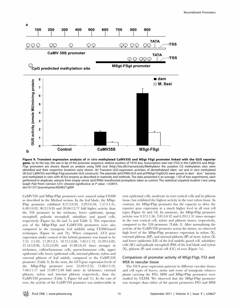
Figure 9. Transient expression analysis of in vitro methylated CaMV35S and MSgt FSgt promoter linked with the GUS reportergene. (a) At the top, the size in bp of the promoter sequence, relative position of TATA box, transcription start site (TSS) in the CaMV35S and MSgt-FSgt promoters are shown. Based on analysis using SVM tool (http://bio.dfci.harvard.edu/Methylator) the putative CG methylation sites wereidentified and their respective locations were shown. (b) Transient GUS-expression activities of demethylated (dam -ve) and in vitro methylated(M.SssI) CaMV35S and MSgt-FSgt promoter GUS constructs. The plasmids (pUCPMA-GUS and pUPMSgt-FSgtGUS) were grown in dam2 dcm2 bacteriaand methylated in vitro with M.SssI enzyme as described in materials and methods. The data presented is an average 6SD of two experiments, eachperformed in duplicate, extracts from empty vector (pUCPMA) transformed protoplasts taken as control. The statistical unpaired student t test usingGraph Pad Prism (version 5.01) showed significance at P value = 0.0073.doi:10.1371/journal.pone.0024627.g009
Recombinant Promoters
PLoS ONE | www.plosone.org 13 September 2011 | Volume 6 | Issue 9 | e24627
in directing the expression of the transgene (GUS) in all plant
vascular tissues and cells. The MSgt-FSgt promoter exhibited
3.0960.19 and 3.2260.43 times (Table 4) higher activity than
the FS3 and MS8 promoters in xylem tissue of the leaf midrib.
Expression of MSgt-FSgt was found to be 4.6060.12,
2.1660.78, and 4.7261.11 times stronger than the FS3
promoter in external phloem, xylem and internal phloem tissues
of the plant stem, respectively (Table 4). The MSgt-FSgt
promoter was also found to be 4.1960.42, 2.5660.32, and
3.88 60.32 times stronger than the MS8 promoter in external
phloem, xylem and internal phloem tissues (Table 4). Similarly,
the MSgt-FSgt promoter was 1.7660.63 and 2.3760.87 times
stronger than the MS8 promoter in xylem and phloem tissues of
the root, respectively. MSgt-FSgt also showed 1.6860.42 and
2.4560.61 fold stronger activity than the FS3 promoter in root
xylem and phloem tissue (Table 4).
Nuclear protein binding assay of the MSgt-FSgt hybridpromoter
The interaction between tobacco nuclear proteins and the MSgt-
FSgt promoter DNA was carried out as described in Methods. As
shown in Figure 8a, specific DNA-protein binding (marked by an
arrow) was observed in the presence of 5 mg and 10 mg of nuclear
protein extract. The binding was completely abolished by
competition with a 50 fold molar excess of unlabeled MSgt-FSgt
DNA indicating that the interaction is specific (Figure 8a). This
specific binding was not eliminated by competition with a 100 fold
molar excess of FS3 but it was completely abolished by a 100 fold
molar excess of the MS8 fragment (Figure 8b).
The promoter sequence where nuclear protein binds was identified
by DNaseI foot printing analysis. It was a 33 bp long sequence that
stretched between positions 2222 to 2189 (Figure 8c). A PLACE
(http://www.dna.affrc.go.jp/PLACE) search of the identified se-
quence, 59AATAAGATGGCAAA TGTGGTTTTCACATTA-
CAC 39, (Figure 8d) revealed the presence of an important plant
cis-sequence (CANNTG) that represent Myc consensus.
Analysis of in vitro methylation assay of MSgt-FSgt andCaMV35S promoters
In silico analysis of MSgt-FSgt and CaMV35S promoter
sequences using software (Support Vector Machine) identified a
total number of 5 and 14 CG islands in MSgt-FSgt and CaMV35S
promoter respectively (Figure 9a). The transient activities of
methylated MSgt-FSgt and CaMV35S promoter in the tobacco
protoplast system (Nicotiana tabaccum Xanthi brad) were
determined as described in Methods. It was observed that
methylated-MSgt-FSgt and CaMV35S promoter showed 64%
and 79% less GUS activity compared to unmethylated MSgt-FSgt
and CaMV35S promoter respectively (Fig. 9b). Statistical analysis
revealed that the decrease in activity in the case of the methylated-
MSgt-FSgt promoter (64%) was significant over decrease in the
activity of the methylated-CaMV35S promoter (79%). The P
value obtained was 0.0073 indicating a high level of significance.
Discussion
Several recombinant promoters were constructed by ligating
different up-stream activation sequences of the MS8 promoter to
the TATA-containing promoter sequence of the FS3 promoter.
One recombinant promoter, MSgt-FSgt, developed in this study
showed enhanced activity compared to wild-type promoters and
the CaMV35S promoter [44–48] in tobacco protoplasts and
transgenic tobacco and Arabidopsis plants. The observed increase in
the relative activities of recombinant promoters may be due to the
cooperative combinatorial interactions between the cis-elements
present in the up-stream activation sequence (2306 to 2125) of
MS8 and the TATA element of the FS3 promoter (FS-1). DNaseI
foot-printing analysis revealed that a 33 bp long (between the
coordinates 2222 to 2189) nuclear protein binding site
(59AATAAGATGGCAAATGTGG TTTTCACATTACAC39)
may be important for MSgt-FSgt promoter function, because
deletion of this portion causes severe loss of MSgt-FSgt promoter
activity as evidenced from the differences in activity between the
MS-UAS3-FS1 promoter (construct No. 3) and the MS-UAS5-
FS1 promoter (construct No. 5) (Figure 1).
In the present study, the MSgt-FSgt promoter showed 10.31
and 14.22 times stronger activity than CaMV35S promoter in the
transient protoplast and transgenic tobacco systems respectively.
In earlier reports as well as in the present study, it was
demonstrated that the parental promoters MS8 [22] and FS3
[23] were 2 to 4 folds stronger than CaMV35S. However, Mirabilis
mosaic virus full-length transcript promoter (MMV-FLt12) was
reported to be 14 times stronger than CaMV35S in tobacco
protoplast transient system [29] and about 25 times stronger than
the CaMV35S in transgenic tobacco [26]. Based on these
observations we may conclude that the MMVFLt12 promoter
[26] is stronger than the MSgt-FSgt promoter. However, the
recombinant promoter MSgt-FSgt is important because (i) it
contains heterogeneous sequences compared to CaMV35S and
MMVFLt12 (ii) it directed higher levels of constitutive gene
expression in different plant cell types compared to the starting
promoters (MS8 and FS3) and CaMV35S (iii) in a separate study
by Kumar et al. (unpublished data), it was observed that the MSgt-
FSgt promoter showed higher transgene activity under abiotic
stress conditions (salt, salicylic/abscisic acid) compared to
CaMV35S promoter.
Detailed analysis of GUS-specific transcripts isolated from
transgenic plants containing CaMV35S, MS8, FS3 and MSgt-
FSgt promoter constructs using Northern blot analysis, real-time
PCR and qRT-PCR showed a fair correlation between the steady-
state level of mRNA (transcript level) and GUS activity (protein
level). Based on the above experimental evidence, it was confirmed
that the activity of the MSgt-FSgt promoter was considerably
stronger than that obtained from the CaMV35S, MS8 and FS3
promoters. Also it was noted that the MSgt-FSgt promoter
directed reporter gene expression more consistently compared to
the CaMV35S promoter in two different independent plant
systems studied.
The intensities of blue fluorescence obtained from 4-MU (lex
363 nm and lem 460 nm), the hydrolyzed product of MUG in the
presence of the GUS enzyme, could be considered to be an
indirect measure of relative promoter strength. 4-MU-intensity
obtained from a particular cell/tissue type of a transgenic plant
harboring the GUS-reporter gene (treated with 1 mM MUG
solution) may reflect the cell/tissue specificity of a given promoter.
The cell-specific expression of MSgt-FSgt was measured precisely
using the analytical software ‘LAS-AF’ supplied with the confocal
microscopy system (Leica, Germany), and was compared to that
obtained from the MS8, FS3 and CaMV35S promoters. The
normalized activity of MSgt-FSgt promoter clearly indicated that
it is a near constitutive promoter and stronger than CaMV35S.
The value obtained for the control sample (background) may be
due to the presence of several candidates for blue fluorescence
emissions; those are phenolic substances such as chlorogenic acid,
caffeic acid, coumarines, ferulic acid, sinapic acid etc. All of these
compounds emit blue fluorescence when excited at 405 nm [49].
The elevated vascular tissue-specific expression of the MSgt-
FSgt promoter compared to the two wild-type promoters (FS3 and
Recombinant Promoters
PLoS ONE | www.plosone.org 14 September 2011 | Volume 6 | Issue 9 | e24627
MS8) and the CaMV35S may be due to the presence of increased
numbers of major plant cis-elements like Dof-1 and ARR1 in the
MSgt-FSgt promoter in comparison to the wild-type promoters
and CaMV35S. Dof-1 plays various roles in plant growth and
development [50,51]. Recent studies showed involvement of Dof-1
in the vascular development of higher plants [52]. ARR1 and
ARR2 factors usually act as transcriptional activators that promote
the expression of a gene in plant cells through their own target
sequences [53]. Besides the Dof-1 and ARR1 elements, the specific
distribution pattern of the other cis-elements like Asf-1, G-Box etc.
with different intervening spacer sequences in MSgt-FSgt
promoter may also contribute to the vascular tissue specific and
other functional role of the MSgt-FSgt promoter. Moreover,
analyzing the MS8, FS3 and MSgt-FSgt promoter sequence by
PlantPAN database [25] other cis-elements like AINTEGU-
MENTA (ANT), ATHB-9 were also found to be increased in
MSgt-FSgt promoter when compared to the two wild-type
promoters (MS8 and FS3). Among these, ANT acts as a
transcriptional activator playing a critical role not only in
regulating ovule and female gametophyte development but also
for gene expression in vegetative tissues [54]. The highest activity
of MSgt-FSgt hybrid promoter in comparison to FS3 or MS8 in
the transgenic system might be due to the increased presence of
these cis-regulatory elements.
DNA methylation plays a crucial role in gene silencing [55].
Several studies have provided evidence that the level of gene
expression from in vitro methylated DNA is strongly reduced
when assayed in animal cells [56–59] and in plant cells [60–63],
and this phenomenon could result in transgene silencing. We
observed 64% decrease for MSgt-FSgt promoter is significant over
79% decrease for the CaMV35S promoter (P = 0.0073). In stably
transformed independent transgenic Arabidopsis lines (T1 genera-
tion; 21 days old), it was also observed that the MSgt-FSgt and
CaMV35S promoters resulted 6.7% and 13.3% of transgenic
plants with minimum (basal) level of GUS activity respectively.
This implies that both promoters are prone to be silenced but the
degree of silencing could be higher in the case of the CaMV35S
promoter compared to the MSgt-FSgt promoter.
There may be few possible constrains associated with using this
promoter in plant genetic engineering. First, this recombinant
promoter, being constitutive in nature, lacks tissue specificity.
Second, in some situations, we may also need to restrict the
expression level of a transgene, otherwise, it may cause problems
to the plant itself, being a strong promoter, MSgt-FSgt, may not be
ideal for use in such situations.
The observations of the present study revealed a promising
future of MSgt-FSgt promoter as a potential candidate promoter
for ectopic gene expression in plants. This study also highlighted
the usefulness of CLSM in studying quantitative and qualitative
gene expression in individual plant cells.
Acknowledgments
We are thankful to Director, Institute of Life Sciences, Bhubaneswar for his
help and encouragement. We also thank Dr. David Zaitlin and Dr. S.
Pattanaik, University of Kentucky, Lexington, USA for critical reading and
necessary corrections of the manuscript. We also thank Mr. Abhimanyu
Das for technical supports.
Author Contributions
Conceived and designed the experiments: ND IBM. Performed the
experiments: DK SP RR DKS ND. Analyzed the data: DK ND.
Contributed reagents/materials/analysis tools: ND IBM. Wrote the paper:
ND DK.
References
1. Atchison M (1988) Enhancers: Mechanism of action and cell specificity. Ann
Rev Cell Biol 4: 127–153.
2. Dynan WS (1989) Modularity in promoters and enhancers. Cell 58: 1–4.
3. Guarente L (1984) Yeast promoters: Positive and negative elements. Cell 36:
799–800.
4. Guarente L, Yocum R, Gifford P (1982) A GAL10-CYC1 hybrid yeast promoter
identifies the GAL4 regulatory region as an upstream site. Proc Natl Acad Sci
USA 79: 7410–7414.
5. Struhl K (1985) Naturally occurring poly (dA-dT) sequences are upstream
promoter elements for constitutive transcription in yeast. Proc Natl Acad Sci
USA 82: 8419–8423.
6. Bienz M, Pelham HRB (1986) Heat shock regulatory elements functions as an
inducible enhancer in the Xenopus HSP70 gene and when linked to a
heterologous promoter. Cell 45: 753–760.
7. de Boer HA, Comstock LJ, Vasser M (1983) The tac promoter: a functional
hybrid derived from the trp and lac promoters. Proc Natl Acad Sci U S A 80(1):
21–25.
8. Bestwick RK, Kellogg JA (2000) Synthetic Hybrid Tomato E4/E8 Plant
Promoter - Patent 6118049.
9. Comai L, Moran P, Maslyar D (1990) Novel and useful properties of a chimeric
plant promoter combining CaMV35S and MAS elements. Plant Mol Biol 15:
373–381.
10. Rushton PJ, Reinstadler A, Lipka V, Lippok B, Somssich IE (2002) Synthetic
plant promoters containing defined regulatory elements provide novel insights
into pathogen- and wound-induced signaling. Plant Cell 14: 749–762.
11. Last DI, Brettell RIS, Chamberlain DA, Chaudhary AM, Larkin PJ, et al. (1991)
pEmu: an improved promoter for gene expression in cereal cells. Theor Appl
Genet 81: 581–588.
12. Sawant S, Singh PK, Madanala R, Tuli R (2001) Designing of an artificial
expression cassette for the high-level expression of transgenes in plants. Theor
Appl Genet 102: 635–644.
13. Bhullar S, Chakravarthy S, Advani S, Datta S, Pental D, et al. (2003) Strategies
for development of functionally equivalent promoters with minimum sequence
homology for transgene expression in plants: cis-elements in a novel DNA
context versus domain swapping. Plant Physiol 132: 988–998.
14. Ni M, Cui D, Einstein J, Narasimhulu S, Vergara CE, et al. (1995) Strength and
tissue specificity of chimeric promoters derived from the octopine and
mannopine synthase genes. The Plant J 7: 661–676.
15. Leisner SM, Gelvin SB (1988) Structure of the octopine synthase upstreamactivator sequence. Proc Natl Acad Sci USA 85: 2553–2557.
16. Lee LY, Kononov ME, Bassuner B, Frame BR, Wang K, et al. (2007) NovelPlant Transformation Vectors Containing the Superpromoter. Plant Physiol
145: 1294–1300.
17. Mol JNM, Stuitje AR, van der Krol A (1989) Genetic manipulation of floral
pigmentation genes. Plant Mol Biol 13: 287–294.
18. Meyer P, Saedler H (1996) Homology dependent gene silencing in plants. Annu
Rev 47: 23–46.
19. Vaucheret H, Fagrad M (2001) Transcriptional gene silencing in plants: targets,inducers and regulators. Trends Genet 17: 29–35.
20. Chaturvedi CP, Sawant SV, Kiran K, Mehrotra R, Lodhi N, et al. (2006)Analysis of polarity in the expression from a multifactorial bidirectional
promoter designed for high-level expression of transgenes in plants.J Biotechnol 123: 1–12.
21. Ye X, Al-Babili S, Kolti A, Zhang J, Lucca P, et al. (2000) Engineering the pro-vitamin A (b-carotene) biosynthetic pathways in (carotenoid-free) rice endo-
sperm. Science 287: 303–305.
22. Dey N, Maiti IB (2003) Promoter deletion and comparative expression analysis
of mirabilis mosaic caulimovirus (MMV) sub-genomic transcript (Sgt-) promoterin transgenic plants. Transgenics 4: 35–53.
23. Bhattacharyya S, Dey N, Maiti IB (2002) Analysis of cis-sequence of subgenomic
transcript promoter from the Figwort mosaic virus and comparison of promoter
activity with the Cauliflower mosaic virus promoters in monocot and dicot cells.Virus Res 90: 47–62.
24. Higo K, Ugawa Y, Iwamoto M, Korenaga T (1999) Plant cis-acting regulatory
DNA elements (PLACE) database. Nucleic Acids Res 27: 297–300.
25. Chang WC, Lee TY, Huang HD, Huang HY, Pan RL (2008) PlantPAN: Plant
promoter analysis navigator, for identifying combinatorial cis-regulatoryelements with distance constraint in plant gene groups. BMC Genomics 9: 561.
26. Dey N, Maiti IB (1999a) Structure and promoter/leader deletion analysis ofmirabilis mosaic virus (MMV) full length transcript promoter in transgenic
plants. Plant Mol Biol 40: 771–782.
27. Schardl CL, Byrd AD, Benzion G, Altschuler MA, Hildebrand DF, et al. (1987)
Design and construction of a versatile system for the expression of foreign genesin plants. Gene 61: 1–11.
28. Jefferson RA, Kavanagh TA, Bevan MW (1987) GUS fusions: beta-
glucuronidase as a sensitive and versatile gene fusion marker in higher plants.
EMBO J 6: 3901–3907.
Recombinant Promoters
PLoS ONE | www.plosone.org 15 September 2011 | Volume 6 | Issue 9 | e24627
29. Sahoo DK, Ranjan R, Kumar D, Kumar A, Sahoo BS, et al. (2009) An
alternative method of promoter assessment by confocal laser scanning
microscopy. J Virol Methods 161: 114–121.
30. Bongaerts RJ, Hautefort I, Sidebotham JM, Hinton JC (2002) Green fluorescent
protein as a marker for conditional gene expression in bacterial cells. Method
Enzymol 358: 43–66.
31. Zimmer M (2002) Green Fluorescent Protein: Applications, Structure, and
Related photophysical behavior. Chemical Rev 102: 759–781.
32. Hofgen R, Willmitzer L (1988) Storage of competent cells for Agrobacterium
transformation. Nucleic Acids Research, 16: 9877.
33. Maiti IB, Murphy JF, Shaw JW, Hunt AG (1993) Plants that express a potyvirus
proteinase genes are resistant to virus infection. Proc Natl Acad Sci USA 90:
6110–6114.
34. Bradford MM (1976) A rapid and sensitive method for quantitation of
microgram quantities of protein utilizing the principle of protein-dye binding.
Anal Biochem 72: 248–254.
35. Zhang X, Henriques R, Lin SS, Niu QW, Chua NH (2006) Agrobacterium-
mediated transformation of Arabidopsis thaliana using the floral dip method.
Nature Protocol 1(2): 641–6.
36. Allen GC, Flores-Vergara MA, Krasnyanski S, Kumar S, Thompson WF (2006)
A modified protocol for rapid DNA isolation from plant tissues using cetyl
trimethyl ammoniumbromide. Nature Protocol 1(5): 2320–2325.
37. Wu L, Fan J, Jiang L, Wang H, Song R, et al. (2003) A specific cis-hairpin
ribozyme facilitates infection of a TMV-based DNA vector in tobacco
protoplasts. J Virol Methods 111: 101–109.
38. Livak KJ, Schmittgen TD (2001) Analysis of relative gene expression data using
real-time quantitative PCR and the 2-DDCt method. Methods 25: 402–408.
39. Pfaffl MW (2001) A new mathematical model for relative quantification in real-
time RT-PCR. Nucleic Acids Res 29: 2002–2007.
40. Escobar C, Aristizabal F, Navas A, Del Campo FF, Fenoll C (2001) Isolation of
active DNA-binding nuclear proteins from tomato galls induced by root-knot
nematodes. Plant Mol Biol Reporter 19: 375a–375h.
41. Lam E, Benfey PN, Gilmartin PM, Fang R-X, Chua NH (1989) Site specific
mutations alter in vitro factor binding and change promoter expression pattern
in transgenic plants. Proc Natl Acad Sci USA 86: 7890–7894.
42. Bhasin M, Zhang H, Ellis L Reinherz, Pedro A Reche (2005) Prediction of
methylated CpGs in DNA sequences using a support vector machine. FEBS
Letters 579: 4302–4308.
43. Pradhan S, Nigel A R Urwin, Gareth I Jnkins, Roger L P Adams (1999) Effects
of CWG methylation on expression of plant genes. Biochemical Journal 341:
473–476.
44. Benfey PN, Chua N (1990) The cauliflower mosaic virus 35S promoter:
combinatorial regulation of transcription in plants. Science 250: 959–966.
45. Benfey PN, Ren L, Chua NH (1989) The CaMV35S enhancer contains at least
two domains which can confer different developmental and tissue-specific
expression patterns. EMBO J 8: 2195–2202.
46. Benfey PN, Ren L, Chua NH (1990a) Combinatorial and synergistic properties
of CaMV35S enhancer subdomains. EMBO J 9: 1685–1696.47. Fang RX, Nagy F, Sivasubramaniam S, Chua NH (1989) Multiple cis regulatory
elements for maximal expression of the cauliflower mosaic virus 35S promoter in
transgenic plant. Plant Cell 1: 141–150.48. Odell JT, Nagy F, Chua NH (1985) Identification of DNA sequences required
for activity of the cauliflower mosaic virus 35S promoter. Nature 313: 810–812.49. Lang M, Stober F, Lichtenthaler HK (1991) Fluorescence emission spectrum of
plant leaves and plant constituents. Radiat Environ Biophys 30: 333–347.
50. Yanagisawa S (2002) The Dof family of plant transcription factor. Trends PlantSci 7: 555–560.
51. Yanagisawa S (2004) Dof domain proteins: plant-specific transcription factorsassociated with diverse phenomena unique to plants. Plant Cell Physiol 45:
386–391.52. Konoshi M, Yanagisawa S (2007) Sequential activation of two Dof transcription
factor gene promoters during vascular development in Arabidopsis thaliana.
Plant Physiol Biochem 45: 623–629.53. Sakai H, Aoyama T, Oka A (2000) Arabidopsis ARR1 and ARR2 response
regulators operate as transcriptional activators. Plant J 24(6): 703–711.54. Klucher KM, Chow H, Reiser L, Fischer RL (1996) The AINTEGUMENTA
gene of arabidopsis required for ovule and female gametophyte development is
related to the floral homeotic gene APETALA2. THE PLANT CELL 8(2):137–153.
55. Ingelbrecht Ivan, Van Houdt H, Van Montagu M, Depicker A (1994)Posttranscriptional silencing of reporter transgenes in tobacco correlates with
DNA methylation. Proc Natl Acad Sci USA 91: 10502–10506.56. Keshet I, Yisraely J, Cedar H (1985) Effect of regional DNA methylation on
gene expression. Proc Natl Acad Sci USA 82: 2560–2564.
57. Gotz F, Schulze-Forster H, Wagner H, Kroger H, Simon D (1990)Transcriptional inhibition of SV40 by in vitro DNA methylation. Biochim
Biophys Acta 1087: 323–329.58. Levine A, Cantoni GL, Razin A (1991) Inhibition of promoter activity by
methylation possible involvement of protein mediators. Proc Natl Acad Sci USA
88: 6515–6518.59. Hsieh C (1994) Dependence of transcriptional repression on CpG methylation
density. Mol Cell Biol 14: 5487–5494.60. Hershkovitz M, Gruenbaum Y, Renbaum P, Razin A, Loyter A (1990) Effect of
CpG methylation on gene expression in transfected plant protoplasts. Gene 94:189–193.
61. Weber H, Ziechmann C, Graessmann A (1990) In vitro DNA methylation
inhibits gene expression in transgenic tobacco. EMBO J 9: 4409–4415.62. Hohn T, Corsten S, Rieke S, MuE ller M, Rothnie H (1996) Methylation of
coding region alone inhibits gene expression in plant protoplasts. Proc Natl AcadSci USA 93: 8334–8339.
63. Dieguez MJ, Bellotto M, Afsar K, Mittelsten Scheid O, Paszkowski J (1997)
Methylation of cytosines in nonconventional methylation acceptor sites cancontribute to reduced gene expression. Mol Gen Genet 253: 581–588.
Recombinant Promoters
PLoS ONE | www.plosone.org 16 September 2011 | Volume 6 | Issue 9 | e24627
Related Documents