Development of Monoclonal Antibodies to Human Microsomal Epoxide Hydrolase and Analysis of “Preneoplastic Antigen”-Like Molecules Hongying Duan a,1 , Kazunori Yoshimura b,2 , Nobuharu Kobayashi a , Kazuo Sugiyama a,3 , Jun-ichi Sawada c,4 , Yoshiro Saito c , Christophe Morisseau d , Bruce D. Hammock d , and Toshitaka Akatsuka a a Department of Microbiology, Faculty of Medicine, Saitama Medical University, Moroyama-cho, Iruma-gun, Saitama 350-0495, Japan b Department of Physiology, Faculty of Medicine, Saitama Medical University, Moroyama-cho, Iruma-gun, Saitama 350-0495, Japan c Division of Biochemistry and Immunochemistry, National Institute of Health Sciences, Kamiyoga 1-18-1, Setagaya-ku, Tokyo 158-8501, Japan d Department of Entomology and Cancer Center, University of California, Davis, One Shields Avenue, Davis, CA 95616-8584, USA Abstract Microsomal epoxide hydrolase (mEH) is a drug metabolizing enzyme which resides on the endoplasmic reticulum (ER) membrane and catalyzes the hydration of reactive epoxide intermediates that are formed by cytochrome P450s. mEH is also thought to have a role in bile acid transport on the plasma membrane of hepatocytes. It is speculated that efficient execution of such multiple functions is secured by its orientation and association with cytochrome P450 enzymes on the ER membrane and formation of a multiple transport system on the plasma membrane. In certain disease status, mEH loses its association with membrane and can be detected as distinct antigens in the cytosol of preneoplastic foci of liver (preneoplastic antigen), in the serum in association with hepatitis C virus infection (AN antigen), or in some brain tumors. To analyze the antigenic structures of mEH in physiological and pathological conditions, we developed monoclonal antibodies against different portions of mEH. Five different kinds of antibodies were obtained: three, anti-N-terminal portions; one, anti-C-terminal; one, anti- conformational epitope. By combining these antibodies, we developed antigen detection methods which are specific to either the membrane-bound form or the linearized form of mEH. These methods detected mEH in the culture medium released from a hepatocellular carcinoma cell line and a glioblastoma cell line, which was found to be a multimolecular complex with a unique © 2012 Elsevier Inc. All rights reserved. Corresponding author: Dr. Toshitaka Akatsuka, Department of Microbiology, Faculty of Medicine, Saitama Medical University, Moroyama-cho, Iruma-gun, Saitama 350-0495, Japan. Phone: 81-49-276-1165. Fax: 81-49-295-9107. [email protected]. 1 Laboratory of Hepatitis Viruses, Division of Viral Products, Center for Biologics Evaluation, Food and Drug Administration, Bethesda, MD 20892, USA; 2 Department of Rehabilitation, Nihon Institute of Medical Science, 1276 Simokawahara, Moroyama-cho, Saitama 350-0435, Japan; 3 Center for Integrated Medical Research, Keio University, Shinano-machi 35, Shinjuku-ku, Tokyo 160-8582, Japan; 4 Pharmaceuticals and Medical Devices Agency, 3-3-2 Kasumigaseki, Chiyoda-ku, Tokyo 100-0013, Japan. Publisher's Disclaimer: This is a PDF file of an unedited manuscript that has been accepted for publication. As a service to our customers we are providing this early version of the manuscript. The manuscript will undergo copyediting, typesetting, and review of the resulting proof before it is published in its final citable form. Please note that during the production process errors may be discovered which could affect the content, and all legal disclaimers that apply to the journal pertain. NIH Public Access Author Manuscript Toxicol Appl Pharmacol. Author manuscript; available in PMC 2013 April 1. Published in final edited form as: Toxicol Appl Pharmacol. 2012 April 1; 260(1): 17–26. doi:10.1016/j.taap.2012.01.023. NIH-PA Author Manuscript NIH-PA Author Manuscript NIH-PA Author Manuscript

Welcome message from author
This document is posted to help you gain knowledge. Please leave a comment to let me know what you think about it! Share it to your friends and learn new things together.
Transcript
Development of Monoclonal Antibodies to Human MicrosomalEpoxide Hydrolase and Analysis of “Preneoplastic Antigen”-LikeMolecules
Hongying Duana,1, Kazunori Yoshimurab,2, Nobuharu Kobayashia, Kazuo Sugiyamaa,3,Jun-ichi Sawadac,4, Yoshiro Saitoc, Christophe Morisseaud, Bruce D. Hammockd, andToshitaka Akatsukaa
aDepartment of Microbiology, Faculty of Medicine, Saitama Medical University, Moroyama-cho,Iruma-gun, Saitama 350-0495, JapanbDepartment of Physiology, Faculty of Medicine, Saitama Medical University, Moroyama-cho,Iruma-gun, Saitama 350-0495, JapancDivision of Biochemistry and Immunochemistry, National Institute of Health Sciences, Kamiyoga1-18-1, Setagaya-ku, Tokyo 158-8501, JapandDepartment of Entomology and Cancer Center, University of California, Davis, One ShieldsAvenue, Davis, CA 95616-8584, USA
AbstractMicrosomal epoxide hydrolase (mEH) is a drug metabolizing enzyme which resides on theendoplasmic reticulum (ER) membrane and catalyzes the hydration of reactive epoxideintermediates that are formed by cytochrome P450s. mEH is also thought to have a role in bileacid transport on the plasma membrane of hepatocytes. It is speculated that efficient execution ofsuch multiple functions is secured by its orientation and association with cytochrome P450enzymes on the ER membrane and formation of a multiple transport system on the plasmamembrane. In certain disease status, mEH loses its association with membrane and can be detectedas distinct antigens in the cytosol of preneoplastic foci of liver (preneoplastic antigen), in theserum in association with hepatitis C virus infection (AN antigen), or in some brain tumors. Toanalyze the antigenic structures of mEH in physiological and pathological conditions, wedeveloped monoclonal antibodies against different portions of mEH. Five different kinds ofantibodies were obtained: three, anti-N-terminal portions; one, anti-C-terminal; one, anti-conformational epitope. By combining these antibodies, we developed antigen detection methodswhich are specific to either the membrane-bound form or the linearized form of mEH. Thesemethods detected mEH in the culture medium released from a hepatocellular carcinoma cell lineand a glioblastoma cell line, which was found to be a multimolecular complex with a unique
© 2012 Elsevier Inc. All rights reserved.Corresponding author: Dr. Toshitaka Akatsuka, Department of Microbiology, Faculty of Medicine, Saitama Medical University,Moroyama-cho, Iruma-gun, Saitama 350-0495, Japan. Phone: 81-49-276-1165. Fax: 81-49-295-9107. [email protected] of Hepatitis Viruses, Division of Viral Products, Center for Biologics Evaluation, Food and Drug Administration,Bethesda, MD 20892, USA;2Department of Rehabilitation, Nihon Institute of Medical Science, 1276 Simokawahara, Moroyama-cho, Saitama 350-0435, Japan;3Center for Integrated Medical Research, Keio University, Shinano-machi 35, Shinjuku-ku, Tokyo 160-8582, Japan;4Pharmaceuticals and Medical Devices Agency, 3-3-2 Kasumigaseki, Chiyoda-ku, Tokyo 100-0013, Japan.Publisher's Disclaimer: This is a PDF file of an unedited manuscript that has been accepted for publication. As a service to ourcustomers we are providing this early version of the manuscript. The manuscript will undergo copyediting, typesetting, and review ofthe resulting proof before it is published in its final citable form. Please note that during the production process errors may bediscovered which could affect the content, and all legal disclaimers that apply to the journal pertain.
NIH Public AccessAuthor ManuscriptToxicol Appl Pharmacol. Author manuscript; available in PMC 2013 April 1.
Published in final edited form as:Toxicol Appl Pharmacol. 2012 April 1; 260(1): 17–26. doi:10.1016/j.taap.2012.01.023.
NIH
-PA Author Manuscript
NIH
-PA Author Manuscript
NIH
-PA Author Manuscript
antigenic structure different from that of the membrane-bound form of mEH. These antibodies andantigen detection methods may be useful to study pathological changes of mEH in various humandiseases.
Keywordsmicrosomal epoxide hydrolase; drug-metabolism; monoclonal antibodies; immunoassay; tumor-associated antigens
IntroductionThe microsomal epoxide hydrolase (mEH) is a drug-metabolizing enzyme that convertsepoxides to diols and plays an important role in the metabolism of some mutagenic andcarcinogenic epoxides (Newman et al., 2005). It is mainly expressed on the ER membrane inthe liver (Newman et al., 2005) and constitutes about 2% of microsomal proteins (Gill et al.,1982). It was reported that mEH associates with various cytochrome P450s (Holder et al.,1974; Ishii et al., 2005), and functional cooperation between those enzymes is suggested(Taura et al., 2002). It is also expressed on the surface of hepatocytes (Zhu et al., 1999) andmay act as a sodium-dependent bile acid transporter (von Dippe et al., 2003). In humans,mEH is the product of single locus (EPHX1) on chromosome 1. Several single nucleotidepolymorphism sequences were found in association with the onset of several diseases andcancers (Smith et al., 1997; Park et al., 2005; McGlynn et al., 1995; Sonzogni et al., 2002).Association of the mEH with cancers and diseases has been further suggested by thefollowing observations. The mEH was identified as the brain tumor antigen in someglioblastoma cell lines (Kessler et al., 2000). Although mEH is tightly associated withmembranes in normal cells, the mEH is sometimes detected in the cytosol of neoplastichuman livers and released into the blood (Gill et al., 1983). The appearance of mEH in theblood is also associated with other types of liver disease (Hammock et al., 1984). Inhepatitis C infection, we have shown that the hepatitis C-associated antigen (AN antigen)appears in the early phase of the viral infection (Akatsuka et al., 1986b), which is followedby the development of the antibody in the acute phase of hepatitis (Akatsuka et al., 1986a).In a recent study, we have shown that the AN antigen is mainly composed of mEH(Akatsuka et al., 2007). These lines of evidence suggest that mEH loses the association withmembranes in some disease processes which are accompanied by the changes of itsstructure. To enable us to analyze the changes of mEH status in situ, we developedmonoclonal antibodies against different parts of mEH molecule and antigen detectionmethods which can quantitate differentially the membrane-bound form and soluble form ofmEH.
Materials and methodsCell Lines
Sf9 was a gift from Dr. Stephen M. Feinstone (CBER, FDA) and cultured in Sf900II SFM(Invitrogen, Carlsbad, CA) at 27°C. The hepatocellular carcinoma (HCC) cell lines, Huh-1(Huh et al., 1982) and Huh-7 (Nakabayashi et al., 1982) were obtained from the JapaneseCancer Research Resources Bank. Huh-1 was cultured in RPMI1640 with 10% FCS; Huh-7was cultured in RPMI1640 with 2% FCS and 30 nmol/L Na22SeO3. THLE-5b, a normalliver cell line which was immortalized by transfecting with the plasmid containing SV40 Tantigen (Lechner et al., 1991), was a gift from Dr. Curt C. Harris (NCI, NIH) and cultured inRPMI1640 with 5% FCS. Human fibroblast cell line M1 (Royer-Pokora et al., 1984) was agift from Dr. William E. Biddison (NIAID, NIH). Glioblastoma cell lines U87MG (Ishii et
Duan et al. Page 2
Toxicol Appl Pharmacol. Author manuscript; available in PMC 2013 April 1.
NIH
-PA Author Manuscript
NIH
-PA Author Manuscript
NIH
-PA Author Manuscript
al., 1999) and LN-Z308 (Albertoni et al., 1998) were gifts from Dr. Ryo Nishikawa(Saitama Medical Univ., Japan) and LN-71 (Ishii et al., 1999) was a gift from Dr. Erwin G.Van Meir (Emory Univ.). These four cell lines were cultured in DMEM with 10% FCS.Myeloma cell line NS-1 was a gift from Dr. Mineo Arita (National Institute of InfectiousDiseases, Japan) and cultured in RPMI1640 with 15% FCS.
Purified mEH antigensExpression of human mEH in a recombinant baculovirus system (Morisseau et al., 2001)and the purification procedure of the solubilized form of mEH (Akatsuka et al., 2007) havebeen described. Briefly, the infected cells were solubilized with Triton X-100 and subjectedto Q-Sepharose column chromatography. Antigen-positive fractions were pooled and storedin a buffer containing 0.05% Triton X-100. A part of this preparation (the solubilized formof mEH, S-mEH) was used for ELISA screening of hybridoma cultures, and the rest wassubmitted for preparative SDS-PAGE to obtain the linearized form of mEH (L-mEH) whichwas used for immunization of mice, competitive antibody binding assays, and as thestandard for antigen detection assays. The preparative SDS-PAGE was performed byapplying 1.0 mg of the S-mEH to Model 491 PrepCell (Bio-Rad, Hercules, CA) consistingof a 40 ml polyacrylamide (10%) gel and a 6 ml stacking gel. During electrophoresis at 12W, proteins were eluted with running buffer with a flow rate of 0.5 ml/min, and 3.5 mlfractions were collected. Each fraction was tested by dot blot assay on an Immobilon-Pmembrane (Millipore, Bedford, MA) using rabbit anti-mEH antibody (Akatsuka et al.,2007), and then, aliquots of antigen-positive fractions were subjected to a 10% minigel(Mini-Protean; Bio-Rad, Hercules, CA) for silver staining. Factions with a homogenous 47kDa band were pooled, applied to a 0.4 ml Extracti-Gel column (Pierce, Rockford, IL), andafter elution with PBS containing 0.05% Triton-X100, concentrated with Centriplus YM-10(Millipore, Bedford, MA). Purification of the membrane-bound form of mEH (M-mEH) hasbeen described (Akatsuka et al., 2007). Briefly, a homogenate of recombinant baculovirus-infected Sf9 cells was first passed through a gel-filtration column (Sephacryl S-300,Amersham, Uppsala, Sweden). The void volume fraction was then subjected toultracentrifugation in a sucrose-gradient followed by a second ultracentrifugation in a CsClgradient. Each fraction was tested by an antibody sandwich ELISA using horseradishperoxidase (HRP)-labeled anti-AN antigen monoclonal antibody 1F12, and positivefractions were pooled and concentrated with Centriplus YM-100 (Millipore, Bedford, MA).Protein concentration was measured by micro BCA assay (Pierce, Rockford, IL).
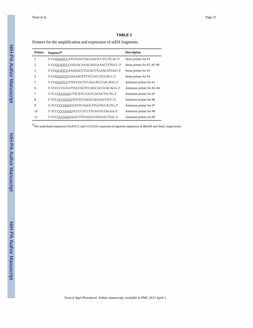
GST-mEHsA full-length and eight truncated human mEH cDNAs (Fig. 1) were amplified using themEH cDNA (Akatsuka et al., 2007) by PCR with KOD-Plus polymerase (Toyobo, Tokyo,Japan) and primers shown in Table 1. The PCR products were digested with BamHI andSmaI, and ligated into the same cloning sites of pGEX-2T (GE Healthcare, Uppsala,Sweden). Competent BL21 cells of E. coli were transformed with the recombinants, andafter IPTG induction (0.1 mM, 5 hr.), 1 ml culture of the cells was extracted with 300 μl ofSDS sample buffer and used as the ELISA antigen.
SDS-PAGE and Western BlottingFor Coomassie blue staining and immunoblotting of GST-mEH produced by E. coli, 13 and5 μl of cell suspensions were extracted with SDS sample buffer and applied onto a 10%minigel (Mini-Protean; Bio-Rad), respectively. After separation and transfer to anImmobilon-P membrane (Millipore, Bedford, MA), the antigen was detected with goat anti-GST (Amersham, Buckinghamshire, UK) (1:1,000) followed by HRP-labeled rabbit anti-goat IgG (KPL, Gaithersburg, MA) and TMB substrate (KPL), or with mouse anti-mEHobtained in this study (1:500) followed by alkaline phosphatase-labeled goat anti-mouse IgG
Duan et al. Page 3
Toxicol Appl Pharmacol. Author manuscript; available in PMC 2013 April 1.
NIH
-PA Author Manuscript
NIH
-PA Author Manuscript
NIH
-PA Author Manuscript
(KPL) and BCIP/NBT substrate (KPL), respectively. For the immunoblotting of mEH-expressing cell lines, 1×10⁵ cells were extracted in 25 μl of SDS sample buffer and appliedonto a 10% minigel. The transferred antigen was detected with the culture supernatant ofanti-mEH hybridoma culture (1:10) followed by alkaline phosphatase-labeled goat anti-mouse IgG (KPL) and BCIP/NBT substrate (KPL).
ELISA for Antibody DetectionEach well of 96-well ELISA plates (Costar, Acton, MA) received 50 μl of one of thefollowing antigen solutions diluted in PBS: M-mEH, S-mEH, and L-mEH, 0.4 μg/ml; GST-mEH, (1:250) dilution; and the mEH peptide aa 54–71, 0.5 μg/ml. After incubation at 4°Covernight, the well was washed with PBS containing 0.05% Tween 20 (PBS-T) and blockedwith 200 μl of PBS containing 5% BSA (blocking solution) for 1 h at 37°C. After washing,50 μl of culture supernatants, ascitic fluids, or sera diluted in PBS containing 1% BSA(dilution buffer) was added and incubated at 37°C for 1 h. Each well was washed three timesand received 50 μl of HRP-conjugated anti-mouse IgG or anti-rabbit IgG (H- and L-chainspecific) (KPL, Gaithersburg, MA) which was diluted 1:1,000 (0.5 μg/ml) with PBScontaining 10% FCS. After incubation at 37°C for 1 h and washing, the color was developedby adding 200 μl of o-phenylenediamine dihydrochloride substrate (SIGMA, St. Louis,MO), and the reaction was stopped by adding 50 μl of 6N H2SO4. The plates were measuredat an optical density of 492 nm. We previously described the aa 54–71 mEH peptide, therabbit antibody against this peptide (Maekawa et al., 2003), and the rabbit anti-mEHantibody (Akatsuka, et al., 2007).
Monoclonal AntibodiesTwo female BALB/c mice (age, 6 weeks) were purchased from Tokyo Laboratory AnimalScience Co. Ltd. (Tokyo, Japan). They were injected s.c. with 2 μg of L-mEH four times(first with Freund’s complete adjuvant, second with incomplete adjuvant, third and fourthimmunizations without adjuvant) at one- to two-month intervals. Two months thereafter,they were boosted i.p. with 40 μg of L-mEH, and after another 3 days, their spleen cellswere harvested. Hybridization of spleen cells with myeloma cells NS-1 was performed asdescribed (Akatsuka et al., 1986b), and culture supernatants were tested for anti-mEHactivity by ELISA using S-mEH as the antigen. Antibody-secreting cells were cloned threetimes or more by limiting dilution with the culture medium containing 10% BM-Conditioned H1 (Roche, Mannheim, Germany). Ascitic fluids were obtained by injectinghybridomas i.p. into pristane (SIGMA)-primed BALB/c nude mice (Clea Japan, Tokyo,Japan) (1–5 × 10⁶ cells/mouse). Antibody isotype was determined with mouse monoclonalantibody isotyping kit (GE Healthcare) following the manufacturer’s instructions
Competitive Antibody Binding AssayCompetitive antibody binding assay (CBA) was performed following the proceduredescribed elsewhere (Stone et al., 1980). A flat-bottomed eight well strip plate (Costar) wasused as the solid phase and 50 μl of L-mEH diluted (0.4 μg/ml) in PBS was added to eachwell and incubated overnight at 4°C. For the assays between the type V and the anti-ANantibody, M-mEH was coated instead of L-mEH to each well. After blocking with 200 μl ofblocking solution for 1 hr at 37°C and washing, 50 μl of unlabeled IgG (5-fold dilutions indilution buffer starting from 5 μg/ml) was incubated in each well at 37°C for 1 h. Then, 2 ×10⁵ cpm of 125I-labeled IgG in 50 μl of dilution buffer was added and incubated at 37°C for1 h. Each well was separated after washing, and the bound radioactivity was counted by agammacounter. IgG was purified from ascitic fluid using Protein G Sepharose (GEHealthcare) following the manufacturer’s instructions, and labeling with 125I was performedby the chloramine-T method (Mcconahey et al., 1966).
Duan et al. Page 4
Toxicol Appl Pharmacol. Author manuscript; available in PMC 2013 April 1.
NIH
-PA Author Manuscript
NIH
-PA Author Manuscript
NIH
-PA Author Manuscript
ELISA and RIA for Ag DetectionIgG was labeled with 125I as described above or with HRP using Peroxidase Labeling Kit(Dojindo, Kumamoto, Japan). Antibody sandwich RIA and ELISA were performed usingflat-bottomed eight well strip plates (Costar) and 96-well ELISA plates (Costar),respectively. Each well adsorbed 50 μl of unlabeled IgG (5 μg/ml in PBS; 1 h at 37°C), andafter washing with PBS-T and blocking, received 50 μl of antigen solution in dilution buffer.After incubation and washing, 1 × 10⁵ cpm of 125I-labeled IgG or HRP-labeled IgG in 50 μlof dilution buffer was added, and after another incubation and washing, the boundradioactivity was counted by a gammacounter or the color was developed by adding o-phenylenediamine dihydrochloride substrate as described above for CBA and Ag bindingELISA. Culture supernatants used for mEH antigen detection were prepared as follows: Thecells were seeded in 10-cm dishes, and after 4 days incubation (~80% confluency), thesupernatant was collected, and after centrifugation at 30,000 × g for 10 min, filtrated througha 0.45 μM filter and stored at −80°C until use. Aliquots of the culture supernatant wereconcentrated about 10-fold by ultrafiltration through a 10 kDa cut-off membrane (CentriplusYM-10, Millipore) and half of each concentrate was further subjected to ultrafiltrationthrough a100 kDa cut-off membrane (Centricon YM-100, Millipore) and the flow-throughfractions were collected.
Analysis of mEH activity in culture media and inhibitory activity of monoclonal antibodiesConcentrated culture media were dissolved 10 folds in Tris/HCl buffer (0.1 M pH 9.0)containing 0.1 mg/ml BSA and the activities were measured with [3H]-cis-stilbene oxideafter incubation for 15 min at 30°C (Morisseau et al., 2001). Purified human mEH (S-mEH)diluted 100-folds was used as positive control, and protein content was measured with BCAassay (Pierce, Rockford, IL) using BSA as standard. Detection limit in this condition wasaround 0.3 nmol.min−1.ml−1. Monoclonal antibodies were diluted 107 fold in the samebuffer as above and incubated with S-mEH. mEH inhibitor, 2-nonylthio-propiamide (NTPA)was used at 100 μM as the positive control.
ResultsPreparation of mEH antigens for immunization and antibody detection
We developed monoclonal antibodies which recognize different portions of human mEH.The solubilized form of mEH (S-mEH) was purified as described previously (Akatsuka etal., 2007) and used for the screening of hybridomas. A part of S-mEH was submitted forpreparative SDS-PAGE to obtain the linearized form of mEH (L-mEH) and used forimmunization of mice, because we wished to obtain antibodies against the linear epitopes ofmEH (see Discussion). For epitope mapping of monoclonal antibodies, we expressed ninemEH fragments (F-mEH) with truncations at the N-terminus or the C-terminus as GST-fusion proteins in E. coli (Fig. 1A). SDS-PAGE followed by Coomasie Blue staining (datanot shown) or immunoblotting with an anti-GST antibody (Fig. 1B) and anti-mEH antibody(Fig. 1C) revealed that each mEH fragment with the predicted size was successfullyexpressed in E. coli. ELISA testing using the F-mEH 1 to 4 as the antigens showed that theimmunized mice developed antibodies against F-mEH 1 and 2 but not 3 or 4 after the firstand the second immunizations. Antibodies which reacted with all the four antigens appearedonly after the third immunization, which suggested that the N-terminus of mEH had higherimmunogenicity than the C-terminus (data not shown).
Development of monoclonal antibodies against mEHAfter we confirmed that the two immunized mice developed antibodies to F-mEH 1 to 4, weestablished hybridomas. In two separate fusions, sixty-five colonies were found to produce
Duan et al. Page 5
Toxicol Appl Pharmacol. Author manuscript; available in PMC 2013 April 1.
NIH
-PA Author Manuscript
NIH
-PA Author Manuscript
NIH
-PA Author Manuscript
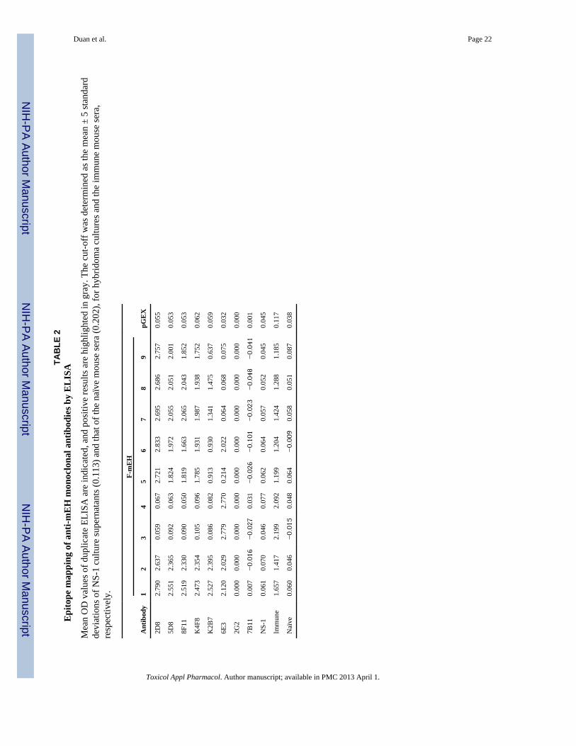
antibodies against the S-mEH, among which 23 antibodies reacted only with F-mEH 1 and2, 16 reacted with F-mEH 1 to 4, and 26 reacted only with the S-mEH. Eight hybridomaswere subjected to limiting dilution three times or more, and tested by ELISA using all of theGST-mEH antigens (F-mEH 1 to 9). The results shown in Table 2 suggest that the fiveantibodies (2D8, 5D8, 8F11, K4F8, and K2B7) recognize the N-terminus (aa 21–143, F-mEH 9), and 6E3 recognizes the C-terminus (aa 327–353, F-mEH 4). Antibodies 2G2 and7B11 reacted with the S-mEH but not with any of the F-mEH 1 to 9, therefore, they seemedto recognize the conformational epitope which was lost during the preparation of the F-mEHs by SDS treatment. This speculation was substantiated by the ELISA in which theantibodies were tested against the three antigens: the S-mEH, the L-mEH, and themembrane-bound form of mEH (M-mEH). The antibodies 2G2, 7B11, and the anti-ANantigen monoclonal antibodies 1H9 and 1F12, which recognize the three-dimensionalstructure of mEH (Akatsuka et al., 2007), reacted with M-mEH and S-mEH but not with L-mEH (data of 2G2 are shown in Fig. 2).
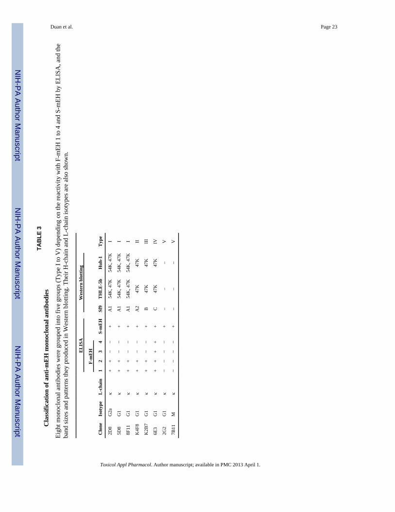
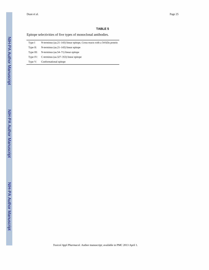
The six antibodies which reacted with one or more of the F-mEH 1 to 9 seemed to recognizelinear structures of mEH, and were tested for their reactivity in Western blotting using threekinds of mEH-expressing cell lines: Sf9 cells infected with a recombinant baculovirus (Fig.3A), THLE-5b, a normal human liver cell line which was immortalized by transfecting withthe plasmid containing SV40 T-antigen (Lechner et al., 1991) (data not shown), and ahepatocellular carcinoma (HCC) cell line, Huh-1 (Huh et al.,1982) (Fig. 3B). When extractsof each of the three cell lines were blotted, all of the six antibodies showed 47 kDa bands,the size of which corresponded to the whole mEH molecule. In the blotting of mEH-expressing Sf9 cells (Fig. 3A), additional smaller bands, which may be degradation productsof mEH, could be seen, and the pattern of the bands could be divided into three types: typeA for 2D8, 5D8, 8F11 and K4F8; type B for K2B7; and type C for 6E3. Based on thesepatterns of bands, the five antibodies which recognize the N-terminus (aa 21–143) could beseparated into the two groups: type A group (2D8, 5D8, 8F11 and K4F8) and type B group(K2B7) (Table 3). In the blotting of the hepatocyte-derived cell lines THLE-5b and Huh-1, ahigher band of 54 kDa could be seen with the three out of four antibodies in the type Agroup (2D8, 5D8 and 8F11), but not with K4F8 in the same type A group or other fourantibodies (Fig. 3B). Therefore, the four antibodies in the type A group could be furtherdivided into the two groups: 2D8, 5D8 and 8F11 (type A1), which cross-react with amolecule of 54 kDa protein, and K4F8 (type A2), which only recognizes a 47 kDa band(Table 3). Therefore, the eight antibodies could be divided into the five types (type I to V)depending on their epitope specificities (Table 3).
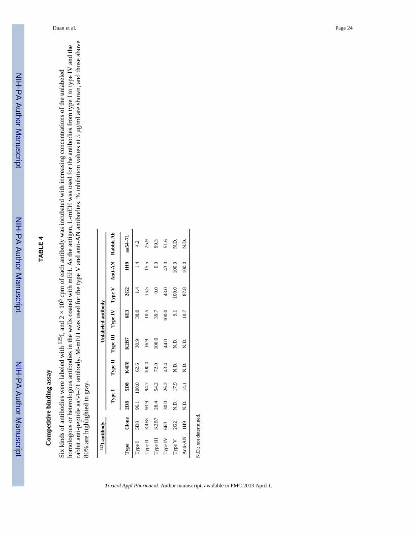
The epitope specificity of the antibodies was further tested by competitive antibody bindingassay (CBA) in which a constant amount of 125I-labelled antibody was allowed to competewith increasing concentrations of the unlabeled homologous or heterologous antibodies forbinding to L-mEH. As an unlabeled antibody, rabbit antiserum which was raised against apeptide (aa 54–71) (Maekawa et al., 2003) was also included. As shown in Table 4, bindingof labeled 5D8 was inhibited by 2D8 as well as by homologous unlabeled 5D8, but not byother types of antibodies, which indicates the two kinds of type I antibodies (2D8 and 5D8)recognize the same or closely adjacent epitopes. Binding of K4F8 (type II) was greatlyinhibited by the two type I antibodies as well as homologous K4F8. Although inhibition ofbinding of labeled 5D8 by unlabeled K4F8 was relatively low, type I and type II antibodiesseemed to recognize distinct but adjacent epitopes on mEH. Binding of labeled K2B7 wasalmost completely inhibited by the rabbit anti-peptide antibody as well as by homologousunlabeled K2B7, which indicated that the epitope of type III overlaps with the region of aa54–71. Binding of labeled 6E3 was inhibited only by homologous unlabeled 6E3. CBA wasalso performed for the antibodies which recognize conformational epitopes using M-mEH asthe antigen. Binding of type V antibody 2G2 was found to be inhibited not only by
Duan et al. Page 6
Toxicol Appl Pharmacol. Author manuscript; available in PMC 2013 April 1.
NIH
-PA Author Manuscript
NIH
-PA Author Manuscript
NIH
-PA Author Manuscript
homologous 2G2 but also by 1H9 which had been developed against the AN antigenpurified from the liver of a patient with hepatitis C (Akatsuka et al., 1986b) and later provedto be highly selective for a conformational epitope on the membrane-bound form of mEH(Akatsuka et al., 2007). To confirm the possibility that the epitope of K2B7 (type III)overlaps with aa 54–71, we tested the antibodies by ELISA with the peptide aa 54–71 coatedto the plates, and found that only K2B7 and the rabbit antiserum revealed significantreactivity with the peptide (Fig. 4). Taken together, the epitope selectivity of the five typesof the antibody can be summarized as shown in Table 5. These five types of antibodies weretested if they inhibit mEH catalytic activity, but any of the antibodies did not revealsignificant inhibition against purified recombinant human mEH (S-mEH) (less than 13.3%,data not shown)
Development of mEH antigen detection systemsPreviously, we have detected a hepatitis C-related antigen (AN-antigen) in sera frompatients with hepatitis C (Akatsuka et al., 1986b) and chimpanzees experimentally infectedwith hepatitis C virus (HCV) (Akatsuka et al., 1986a) by radioimmunoassay (RIA) usinganti-AN-antigen monoclonal antibody, 1F12. Recently, we have shown that most of theantigenicity of AN-antigen consists of mEH and anti-AN-antigen monoclonal antibodiesrecognize conformational epitopes of mEH (Akatsuka et al., 2007). The antigen wassomewhat similar to preneoplastic antigen (PNA) which had been described as an antigen inpreneoplastic foci in livers that is released into the blood (Okita et al., 1975), and later foundto be immunologically identical to mEH (Levin et al., 1978). The mEH has been alsoreported to reveal a somewhat anomalous antigenicity in some glioblastoma cell lines(Kessler et al., 2000). To assess the similarity between PNA, AN-antigen, and the braintumor antigen, and their differences from the normal mEH bound to the membrane, wedeveloped sensitive mEH antigen detection assay systems using the monoclonal antibodies.Among the five types of antibodies, type I antibodies which cross-react with a 54kDaprotein were excluded, and K4F8 (type II), K2B7 (type III), 6E3 (type IV), and 2G2 (typeV) were labeled with 125I or horseradish peroxidase (HRP). Two kinds of the purified mEHobtained from recombinant baculovirus-infected Sf9 cells were used as the antigenstandards. The membrane-bound form of mEH was purified by density gradientcentrifugation without using detergents (Akatsuka et al., 2007) (M-mEH); the linearizedform of mEH (L-mEH) was described above. We tried to detect the antigens by antibodysandwich RIA and ELISA methods using several combinations of unlabeled and labeledantibodies. As shown in Table 6 (titration curves of three kinds of ELISA are shown in Fig.5), M-mEH was successfully detected by the combinations between type V and homologousor heterologous antibodies; the homologous combination (V-*V) was the most sensitive(Fig. 5A) followed by the combination of unlabeled type IV and labeled type V (IV-*V)(Fig. 5B). L-mEH could be detected by the combination of unlabeled type IV and labeledtype II antibodies (IV-*II) (Fig. 5C). Interestingly, M-mEH could not be detected withsatisfactory sensitivity by IV-*II (Fig. 5C) or by any other combinations of antibodiesexcluding type V (Table 6). Importantly, the sandwich assay method based on thecombination of IV-*II enables us to detect the mEH antigen in multiple forms only if it isdenatured with detergents such as SDS before incubation for the assay.
Analyses of antigenic structure of PNA-like mEH antigensWe applied three kinds of RIAs with the antibody combinations, IV-*II, IV-*V, and V-*Vto the detection of mEH in culture supernatants of seven cell lines (Table 7). Two lines(THLE-5b and Huh-1) were also used for Western blotting in this study (Table 3). THLE-5b(Lechner et al., 1991) was derived from normal human hepatocytes which wereimmortalized by transfection of SV40 T antigen gene, and maintain many functions ofnormal hepatocytes including phase II drug-metabolizing enzymes. Huh-7 (Nakabayashi et
Duan et al. Page 7
Toxicol Appl Pharmacol. Author manuscript; available in PMC 2013 April 1.
NIH
-PA Author Manuscript
NIH
-PA Author Manuscript
NIH
-PA Author Manuscript
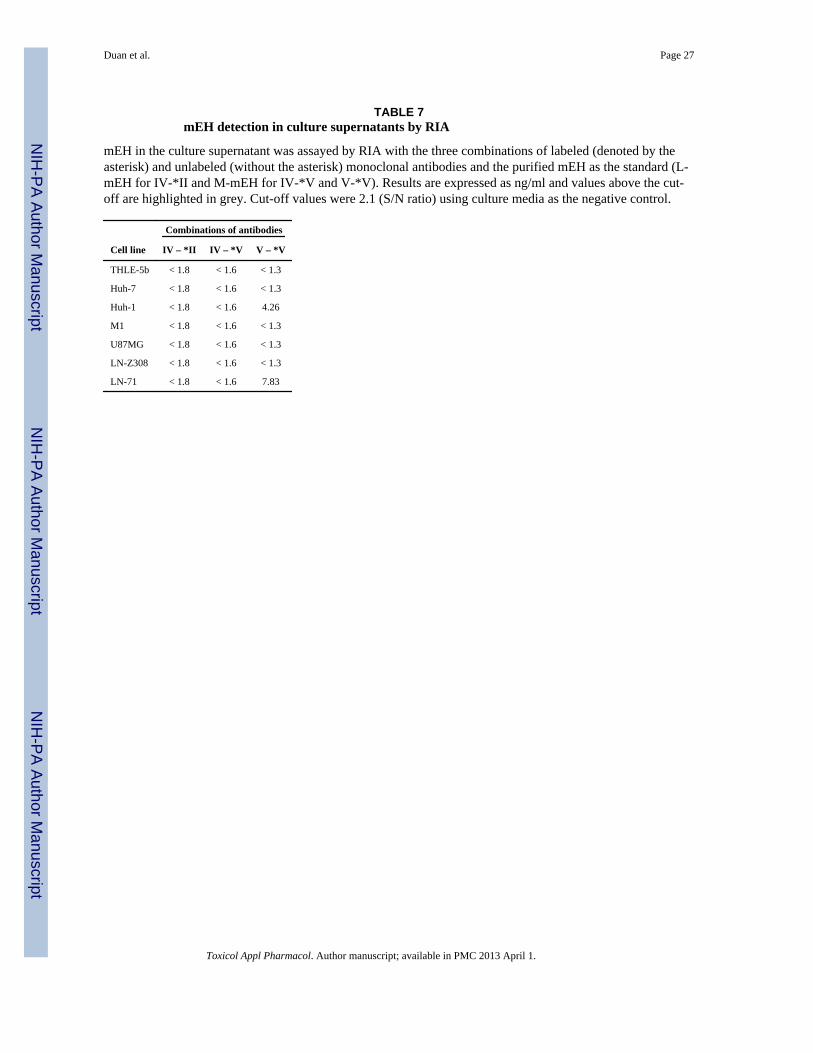
al., 1982) is a well-differentiated HCC cell line and secretes a variety of major plasmaproteins; e.g., albumin, transferrin alpha-fetoprotein and the acute phase proteins. Huh-1(Huh et al., 1982) is a HBs antigen-producing undifferentiated HCC line and producestumors in nude mice. M1 is a cell line derived from human fibroblasts (Royer-Pokora et al.,1984). LN-71 (Ishii et al., 1999), LN-Z308 (Albertoni et al., 1998), and U87MG (Ishii et al.,1999) are human glioblastoma cell lines; LN-71 was described as brain tumor antigen BF7/GF2 positive whereas LN-Z308 as negative (Kessler et al., 2000). All the cell lines werefound to express mEH with a 47 kDa band which was detected by the type II antibody K4F8in Western blotting (data not shown). Significant signals were obtained from thesupernatants of Huh-1 and LN-71 by the V-*V method. Those antigens could not bedetected by the IV-*V method even though the amounts exhibited by V-*V method (4.26and 7.83 ng/ml) were higher than the detection limit of IV-*V (1.6 ng/ml). They could notbe detected by the IV-*II method either. Since we did not add detergent before this assay,the mEH in these supernatant may have been folded in a shape which is undetectable by theIV-*II assay. We speculated that this folding is somehow different from that of M-mEH inthat the type IV epitope is hindered inside the mEH molecule in the supernatant andundetectable by the IV-*V method.
For further analysis, we concentrated the supernatants of THLE-5b, Huh-1 and LN-71cultures about 10-fold by ultrafiltration through 10 kDa cut-off membranes. Then aliquots ofthese concentrates as well as the standard antigens, M-mEH and L-mEH, were subjected toultrafiltration through 100 kDa cut-off membranes, and the flow-through fractions wereobtained. When we measured these samples by the three antigen detection ELISA methods,the media of THLE-5b did not show positive signals. On the other hand, the mEHs in thesupernatants of Huh-1 and LN-71 were well detected by V-*V, and we found that thedetected antigens did not pass through the 100 kDa cut-off membranes indicating that theywere in the shape of multimolecular complex larger than 100 kDa (Fig. 6A). However, incontrast to M-mEH, the mEHs in the supernatants of Huh-1 and LN-71 could scarcely bedetected by IV-*V (Fig. 6B) as was observed for the unconcentrated supernatants (Table 7).When we tested these samples by IV-*II without adding SDS, only L-mEH could bedetected (data not shown). After addition of SDS, the antigens in the supernatants of the twocell lines, M-mEH as well as L-mEH could be detected, and we found that part of theantigens in the supernatants and most of L-mEH passed through the 100 kDa cut-offmembranes (Fig. 6C). We could also see positive signals from the supernatant of THLE-5bespecially after ultrafiltration through the100 kDa cut-off membrane. In addition, all of thethree culture media showed significant mEH activity (0.025 – 0.046 nmol.min−1.mg−1)These results suggested that all of the three cell lines secrete a small amount of mEH withthe size smaller than 100 kDa probably existing as single molecules folded in particularshapes which could not be detected by either of IV-*II (without detergent treatment), V-*V,or IV-*V method. The majority of the mEH in the supernatants of Huh-1 and LN-71were inthe shape of multimolecular complex which was different from that of M-mEH and couldnot be detected by IV-*V.
DiscussionIn this study, we demonstrate that mEH in some cancerous cells takes a different shape fromthat of the membrane-bound form of mEH and is released to the culture medium. To analyzethe antigenic structure of mEH, we developed monoclonal antibodies that recognizedifferent parts of the mEH molecule. When we tested sera from the immunized mice byELISA using mEH fragments as the antigens, we found that the antibody against the C-terminal half (aa 244–455) was produced only after the third immunization, while theantibody against the N-terminus (aa 1–243) appeared after the first immunization, indicatingthat the C-terminal half is less immunogenic than the N-terminus. We developed the
Duan et al. Page 8
Toxicol Appl Pharmacol. Author manuscript; available in PMC 2013 April 1.
NIH
-PA Author Manuscript
NIH
-PA Author Manuscript
NIH
-PA Author Manuscript
monoclonal antibody 6E3 against the epitope in the C-terminal half (aa 327–353) (type IVepitope) by hyperimmunizing mice with the purified mEH. In addition, we could obtain atleast four kinds of monoclonal antibodies which recognize the N-terminus or theconformational epitopes (Table 3, 5).
To obtain antibodies against the linear epitopes of mEH, we used L-mEH for immunization.L-mEH was prepared by preparative SDS-PAGE and does not react with the antibodiesagainst conformational epitopes as shown by ELISA (Fig. 2). However, the antibodies from26 out of 64 hybridoma colonies reacted only with the S-mEH but not with any of F-mEH 1to 4. From these 26 hybridomas, 2G2 and 7B11 were obtained after limiting dilution,grouped as type V and shown to recognize conformational epitopes (Fig. 2). Therefore, itseems that L-mEH regained conformational epitopes after being injected into mice. The S-mEH, which was purified after solubilization with Triton X-100 and used for screening ofhybridomas, did not react with either of the type V antibodies nor anti-AN antibodies justafter purification, but during storage at 4°C, it gained the reactivity with these antibodies(data not shown). These findings are further supported by the fact that the purified mEHexists as high molecular weight aggregates (~600 kDa) in the absence of SDS (Lu et al.,1975; DuBois et al., 1979). In addition, all of the 12 monoclonal antibodies raised againstAN antigen (Akatsuka et al., 1986b) were found to recognize conformational epitopes ofmEH (Akatsuka et al., 1986a), and in CBA against the AN antigen, all antibodies showed100% inhibition each other (unpublished). One of the anti-AN antibodies (1H9) and the typeV antibody 2G2 compete each other well in the assay against the M-mEH (Table 4). Takentogether, it seems that mEH is prone to form a multimolecular complex with a commonconformational epitope. Any of the monoclonal antibodies did not show significantinhibition of mEH catalytic activity. It is rare to find an inhibitory antibody probablybecause the active site of mEH is deep inside the enzyme which forms a peculiarconformation and complex.
In Western blotting of extracts from THLE-5b and Huh-1, the three antibodies (2D8, 5D8,and 8F11) reacted with a 54 kDa band in addition to the 47 kDa band, which correspondedto an entire mEH molecule (Fig. 3B), and were grouped as type I (Table 3). Since this54kDa band was not observed on the blots of Sf9 cells expressing mEH by infection with arecombinant baculovirus (Fig. 3A) or BHK-21 cells expressing mEH by transfection with anexpression plasmid (Akatsuka et al., 2007) (data not shown), we think this is a proteindistinct from mEH and was detected by a cross-reaction. A similar cross-reaction has beenreported for anti-rat mEH monoclonal antibody, 25A-3 (Ananthanarayanan et al., 1988).25A-3 recognized a 54 kDa protein which was thought to be the Na+-independent bile acidcarrier protein, but the antigen has not been molecularly identified (D. Levy, personalcommunication). Because mEH is thought to be a part of a multi-protein transport system onthe membranes (Ananthanarayanan et al., 1988), it is worth identifying the 54 kDa protein inhuman cells recognized by type I antibodies to explore its possible relationship to mEH.
When we tested several combinations of the antibody in the development of sandwich assaymethods for mEH antigen detection, M-mEH could be detected by the combination betweenthe type V antibody and one of the type II to V antibodies (Table 6). This indicates that M-mEH expresses all of type II to V epitopes on its surface, however, it could not be detectedby the combinations between the two of the type II to IV antibodies. One possibleexplanation is that the linear epitopes, type II to IV, are clustered in a very small areasurrounded by the complex of mEH molecules with multiple type V epitopes, and once anantibody molecule binds to one of the linear epitopes, the other linear epitopes are hinderedsterically from the access by another antibody molecule. On the other hand, L-mEH couldbe detected well by IV-*II, but the opposite combination II-*IV did not work well. In theepitope mapping study by ELISA using the antigens F-mEH 1 to 9, the type IV antibody
Duan et al. Page 9
Toxicol Appl Pharmacol. Author manuscript; available in PMC 2013 April 1.
NIH
-PA Author Manuscript
NIH
-PA Author Manuscript
NIH
-PA Author Manuscript

6E3 showed much lower reactivity to F-mEH 5 than to F-mEH 6 although F-mEH 5contained the entire sequence of F-mEH 6 and an additional sequence on its C-terminal side(Table 2). This finding implies that the type IV epitope has the potential to be masked by theadjacent sequence located on its C-terminal side. Therefore, once the linearized mEH (L-mEH) is bound to the type II or type III antibody or to the plastic plate, it may change itsconformation on the C-terminal side and type IV epitope may lose the accessibility to theantibody. This may also explain why the mEH released from Huh-1 and LN-71 to theculture medium could not be detected by the IV-*V method. The mEH in those cells mayhave changed its orientation on the membrane and formed a multimolecular complex withincreased solubility and with the type IV epitope being masked by the adjacent sequence.
In this study, we found that mEH is released from Huh-1 and LN-71 to the culture mediumand both antigens showed similar results in our analyses which demonstrated that they havedifferent structure from the membrane-bound form of mEH. This antigen seems to be relatedto the preneoplastic antigen (PNA). (Okita et al., 1975) demonstrated that there is an antigenin preneoplastic foci in rat livers that is released into the blood, which was in later studiesshown to be similar if not identical to the mEH (Levin et al., 1978). In later studies, a rapidradiochemical assay for the PNA was developed for human blood and shown to beassociated with liver cancer, but it was also found to be associated with several other typesof liver damage (Hammock et al., 1984). The AN antigen was purified from liver of apatient infected with HCV (Tohmatsu et al., 1985). The antigen is released into the blood inthe early phase of HCV infection and the antibody appears in the acute phase of hepatitis(Akatsuka et al., 2007; Akatsuka et al., 1986a; Akatsuka et al., 1986b). The AN antigen iscomposed of particles with molecular weight of more than 1,500 kDa (Tohmatsu et al.,1985) and was found to be mainly composed of mEH (Akatsuka et al., 2007). However, theAN antigen was shown to be somehow different from the membrane-bound form of mEH inthat the former reacted only with the antibody in sera from HCV-infected patients whereasthe latter reacted with both the antibody from patients with HCV infection and that frompatients with hepatitis A virus infection (Akatsuka et al., 2007). In this study, we did notanalyze the AN antigen or the antigens in sera from patients with HCV infection, but suchstudies will provide important information about the influence of virus infection on theexpression and structure of mEH and its role in development of hepatitis.
Because mEH coordinates with other enzymes on ER membrane and plays multiplefunctions, disintegration of the enzymes may lead to pathological consequences such astumorigenesis. We observed that mEH changes its location in the cells during certain virusinfection in vitro, and it accompanies with the changes of mEH activities (manuscript inpreparation). Further analysis of PNA-like antigens may clarify the association of mEH withthe pathogenesis of some human diseases and cancer.
In summary, we have obtained monoclonal antibodies to at least five different epitopes ofhuman mEH and developed the methods which selectively detect either the membrane-bound form or the linearized form of mEH. These methods enable us to discriminate thenative form of mEH from the variant soluble form produced in some cancerous cells. Themethod which selectively detects the linearized form of mEH enables us to measure themEH in any form of structure if only it has an entire sequence and linearized by SDStreatment. These tools may be valuable for elucidation of the role of mEH in various diseaseprocesses.
AcknowledgmentsThe authors thank Hiroe Akatsuka and Akira Takagi for technical assistance. We also thank Dr. W.E. Biddison, Dr.R. Nishikawa, and Dr. E.G. Van Meir for providing cell lines. This work was supported by a Saitama MedicalUniversity Internal Grant; a grant from Eisai Co., Ltd.; and partly supported by the National Institutes of Health
Duan et al. Page 10
Toxicol Appl Pharmacol. Author manuscript; available in PMC 2013 April 1.
NIH
-PA Author Manuscript
NIH
-PA Author Manuscript
NIH
-PA Author Manuscript
National Institute of Environmental Health Sciences [R01 ES002710]. BDH is a George and Judy Marcus SeniorFellow of the American Asthma Foundation.
Abbreviations
mEH microsomal epoxide hydrolase
ER endoplasmic reticulum
ELISA enzyme-linked immunosorbent assay
PAGE polyacrylamide gel electrophoresis
CsCl cesium chloride
PCR polymerase chain reaction
IPTG isopropyl β-d-thiogalactoside
GST glutathione S-transferase
TMB 3, 37, 5, 57-tetramethylbenzidone
BCIP 5-bromo-4-chloro-3-indoyl-phosphate
NBT nitroblue tetrazolium
PBS phosphate-buffered saline
BSA bovine serum albumin
HRP horseradish peroxidase
FCS fetal calf serum
CBA competitive antibody binding assay
S-mEH solubilized form of mEH
M-mEH membrane-bound form of mEH
L-mEH linearized form of mEH
RIA radioimmunoassay
PNA preneoplastic antigen
HCC hepatocellular carcinoma
HCV hepatitis C virus
OD optical density
Reference List1. Akatsuka T, Tohmatsu J, Abe K, Shikata T, Ishikawa T, Nakajima K, Yoshihara N, Odaka T. Non-
A, Non-B Hepatitis Related AN6520 Ag Is a Normal Cellular Protein Mainly Expressed in Liver. II.J Med Virol. 1986a; 20:43–56. [PubMed: 2428929]
2. Akatsuka T, Tohmatsu J, Yoshihara N, Katsuhara N, Okamoto T, Shikata T, Odaka T. Detection ofan Antigen (AN6520) Possibly Related to Non-A, Non-B Hepatitis, by Monoclonal Antibodies. I. JMed Virol. 1986b; 20:33–42. [PubMed: 2428928]
3. Akatsuka T, Kobayashi N, Ishikawa T, Saito T, Shindo M, Yamauchi M, Kurokohchi K, MiyazawaH, Duan H, Matsunaga T, Komoda T, Morisseau C, Hammock BD. Autoantibody response tomicrosomal epoxide hydrolase in hepatitis C and A. J Autoimmun. 2007; 28:7–18. [PubMed:17296285]
Duan et al. Page 11
Toxicol Appl Pharmacol. Author manuscript; available in PMC 2013 April 1.
NIH
-PA Author Manuscript
NIH
-PA Author Manuscript
NIH
-PA Author Manuscript
4. Albertoni M, Daub DM, Arden KC, Viars CS, Powell C, Van Meir EG. Genetic instability leads toloss of both p53 alleles in a human glioblastoma. Oncogene. 1998; 16:321–326. [PubMed:9467957]
5. Ananthanarayanan M, von Dippe P, Levy D. Identification of the hepatocyte Na+-dependent bileacid transport protein using monoclonal antibodies. J Biol Chem. 1988; 263:8338–8343. [PubMed:3372528]
6. DuBois GC, Appella E, Armstrong R, Levin W, Lu AY, Jerina DM. Hepatic microsomal epoxidehydrase. Chemical evidence for a single polypeptide chain. J Biol Chem. 1979; 254:6240–6243.[PubMed: 447710]
7. Gill SS, Ota K, Ruebner B, Hammock BD. Microsomal and cytosolic epoxide hydrolase in rhesusmonkey liver, and in normal and neoplastic human liver. Life Sci. 1983; 32:2693–2700. [PubMed:6855465]
8. Gill SS, Wie SI, Guenthner TM, Oesch F, Hammock BD. Rapid and sensitive enzyme-linkediimunosorbent assay for the microsomal epoxide hydrolase. Carcinogenesis. 1982; 3:1307–1310.[PubMed: 6817936]
9. Hammock BD, Loury DN, Moody DE, Ruebner B, Baselt R, Milam KM, Volberding P, KettermanA, Talcott R. A methodology for the analysis of the preneoplastic antigen. Carcinogenesis. 1984;5:1467–1473. [PubMed: 6488472]
10. Holder J, Yagi H, Dansette P, Jerina DM, Levin W, Lu AYH, Conney AH. Effects of Inducers andEpoxide Hydrolase on the Metabolism of Benzo[a]pyrene by Liver Microsomes and aReconstituted System: Analysis by High Pressure Liquid Chromatography. Proc Natl Acad SciUSA. 1974; 71:4356–4360. [PubMed: 4530987]
11. Huh N, Nemoto N, Utakoji T. Metabolic activation of benzo[a]pyrene, aflatoxin B1, anddimethylnitrosamine by a human hepatoma cell line. Mutat Res. 1982; 94:339–348. [PubMed:6287253]
12. Ishii N, Maier D, Merlo A, Tada M, Sawamura Y, Diserens AC, Van Meir EG. Frequent co-alterations of TP53, p16/CDKN2A, p14ARF, PTEN tumor suppressor genes in human glioma celllines. Brain Pathol. 1999; 9:469–479. [PubMed: 10416987]
13. Ishii Y, Takeda S, Yamada H, Oguri K. Functional protein-protein interaction of drug metabolizingenzymes. Front Biosci. 2005; 10:887–895. [PubMed: 15569627]
14. Kessler R, Hamou MF, Albertoni M, de Tribolet N, Arand M, Van Meir EG. Identification of thePutative Brain Tumor Antigen BF7/GE2 as the (De)Toxifying Enzyme Microsomal EpoxideHydrolase. Cancer Res. 2000; 60:1403–1409. [PubMed: 10728706]
15. Lechner, JF.; Smoot, DT.; Pfeifer, AM.; Tokiwa, T.; Harris, CC. A non-tumorigenic human liverepithelial cell culture model for chemical and biological carcinogenesis investigations. In: Rhim,JS.; Drifschlo, A., editors. Neoplastic Transformation in Human Cell Systems. Humana Press;New York: 1991. p. 307-321.
16. Levin W, Lu AYH, Thomas PE, Ryan D, Kizer DE, Griffin MJ. Identification of epoxidehydrolase as the preneoplastic antigen in rat liver hyperplastic nodules. Proc Natl Acad Sci USA.1978; 75:3240–3243. [PubMed: 210455]
17. Lu AYH, Ryan D, Jerina DM, Daly JW, Levin W. Liver Microsomal Epoxide Hydrase. J BiolChem. 1975; 250:8283–8288. [PubMed: 240858]
18. Maekawa K, Itoda M, Hanioka N, Saito Y, Murayama N, Nakajima O, Soyama A, Ishida S, OzawaS, Ando M, Sawada J. Non-synonymous single nucleotide alterations in the microsomal epoxidehydrolase gene and their functional effects. Xenobiotica. 2003; 33:277–287. [PubMed: 12637245]
19. Mcconahey PJ, Dixon FJ. A Method of Trace Iodination of Proteins for Immunologic Studies. IntArch Allergy Appl Immunol. 1966; 29:185–189. [PubMed: 4160044]
20. McGlynn KA, Rosvold EA, Lustbader ED, Hu Y, Clapper ML, Zhou T, Wild CP, Xia XL, Baffoe-Bonnie A, Ofori-Adjei D, Chen GC, London WT, Shen FM, Buetow KH. Susceptibility tohepatocellular carcinoma is associated with genetic variation in the enzyme detoxication ofaflatoxin B1. Proc Natl Acad Sci USA. 1995; 92:2384–2387. [PubMed: 7892276]
21. Morisseau C, Newman JW, Dowdy DL, Goodrow MH, Hammock BD. Inhibition of microsomalepoxide hydrolases by ureas, amides, and amines. Chem Res Toxicol. 2001; 14:409–415.[PubMed: 11304129]
Duan et al. Page 12
Toxicol Appl Pharmacol. Author manuscript; available in PMC 2013 April 1.
NIH
-PA Author Manuscript
NIH
-PA Author Manuscript
NIH
-PA Author Manuscript
22. Nakabayashi H, Taketa K, Miyano K, Yamane T, Sato J. Growth of human hepatoma cells lineswith differentiated functions in chemically defined medium. Cancer Res. 1982; 42:3858–3863.[PubMed: 6286115]
23. Newman JW, Morisseau C, Hammock BD. Epoxide hydrolases: their roles and interactions withlipid metabolism. Prog Lipid Res. 2005; 44:1–51. [PubMed: 15748653]
24. Okita K, Farber E. An antigen common to preneoplastic hepatocyte populations and to liver cancerinduced by N-2-fluorenyl-acetamide, ethionine, or other hepatocarcinogens. Gann Monograph onCancer Res. 1975; 17:283–299.
25. Park JY, Chen L, Wadhwa N, Tockman MS. Polymorphisms for microsomal epoxide hydrolaseand genetic susceptibility to COPD. Int J Mol Med. 2005; 15:443–448. [PubMed: 15702235]
26. Royer-Pokora B, Peterson WD, Haseltine WA. Biological and biochemical characterization of anSV40-transformed xeroderma pigmentosum cell line. Exp Cell Res. 1984; 151:408–420.[PubMed: 6323201]
27. Smith CA, Harrison DJ. Association between polymorphism in gene for microsomal epoxidehydrolase and susceptibility to emphysema. Lancet. 1997; 350:630–633. [PubMed: 9288046]
28. Sonzogni L, Silvestri L, De Silvestri A, Gritti C, Foti L, Zavaglia C, Bottelli R, Mondelli MU,Civardi E, Silini EM. Polymorphisms of Microsomal Epoxide Hydrolase Gene and Severity ofHCV-Related Liver Disease. Hepatology. 2002; 36:195–201. [PubMed: 12085365]
29. Stone MR, Nowinski RC. Topological Mapping of Murine Leukemia Virus Proteins byCompetition-Binding Assays with Monoclonal Antibodies. Virology. 1980; 100:370–381.[PubMed: 6153246]
30. Taura K, Yamada H, Naito E, Ariyoshi N, Mori Ma, Oguri K. Activation of microsomal epoxidehydrolase by interaction with cytochromes P450: kinetic analysis of the association and substrate-specific activation of epoxide hydrolase function. Arch Biochem Biophys. 2002; 402:275–280.[PubMed: 12051674]
31. Tohmatsu J, Morimoto T, Katsuhara N, Abe K, Shikata T. AN6520 Ag: An Antigen Purified FromLiver With Non-A, Non-B Hepatitis. J Med Virol. 1985; 15:357–371. [PubMed: 2580055]
32. von Dippe P, Zhu QS, Levy D. Cell surface expression and bile acid transport function of onetopological form of m-epoxide hydrolase. Biochem Biophys Res Commun. 2003; 309:804–809.[PubMed: 13679044]
33. Zhu QS, von Dippe P, Xing W, Levy D. Membrane Topology and Cell Surface Targeting ofMicrosomal Epoxide Hydrolase. J Biol Chem. 1999; 274:27898–27904. [PubMed: 10488137]
Duan et al. Page 13
Toxicol Appl Pharmacol. Author manuscript; available in PMC 2013 April 1.
NIH
-PA Author Manuscript
NIH
-PA Author Manuscript
NIH
-PA Author Manuscript
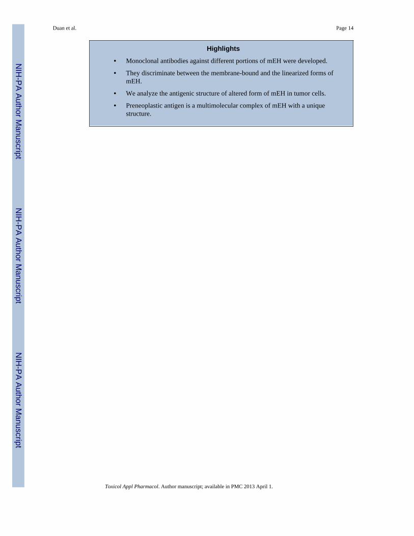
Highlights
• Monoclonal antibodies against different portions of mEH were developed.
• They discriminate between the membrane-bound and the linearized forms ofmEH.
• We analyze the antigenic structure of altered form of mEH in tumor cells.
• Preneoplastic antigen is a multimolecular complex of mEH with a uniquestructure.
Duan et al. Page 14
Toxicol Appl Pharmacol. Author manuscript; available in PMC 2013 April 1.
NIH
-PA Author Manuscript
NIH
-PA Author Manuscript
NIH
-PA Author Manuscript
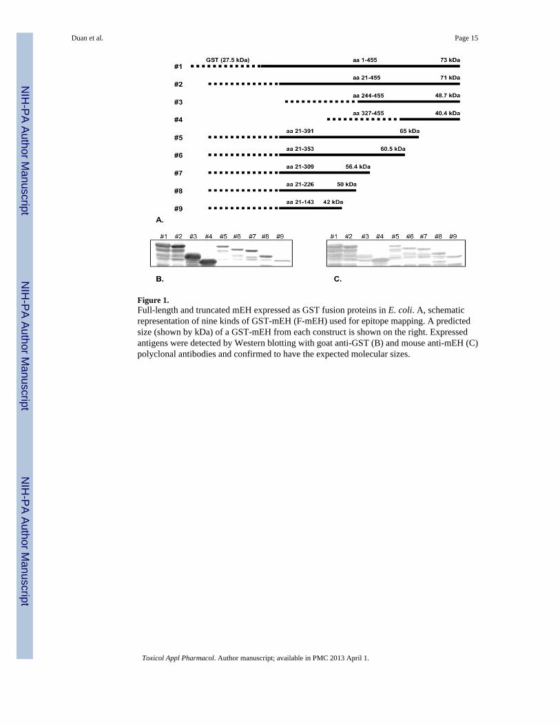
Figure 1.Full-length and truncated mEH expressed as GST fusion proteins in E. coli. A, schematicrepresentation of nine kinds of GST-mEH (F-mEH) used for epitope mapping. A predictedsize (shown by kDa) of a GST-mEH from each construct is shown on the right. Expressedantigens were detected by Western blotting with goat anti-GST (B) and mouse anti-mEH (C)polyclonal antibodies and confirmed to have the expected molecular sizes.
Duan et al. Page 15
Toxicol Appl Pharmacol. Author manuscript; available in PMC 2013 April 1.
NIH
-PA Author Manuscript
NIH
-PA Author Manuscript
NIH
-PA Author Manuscript
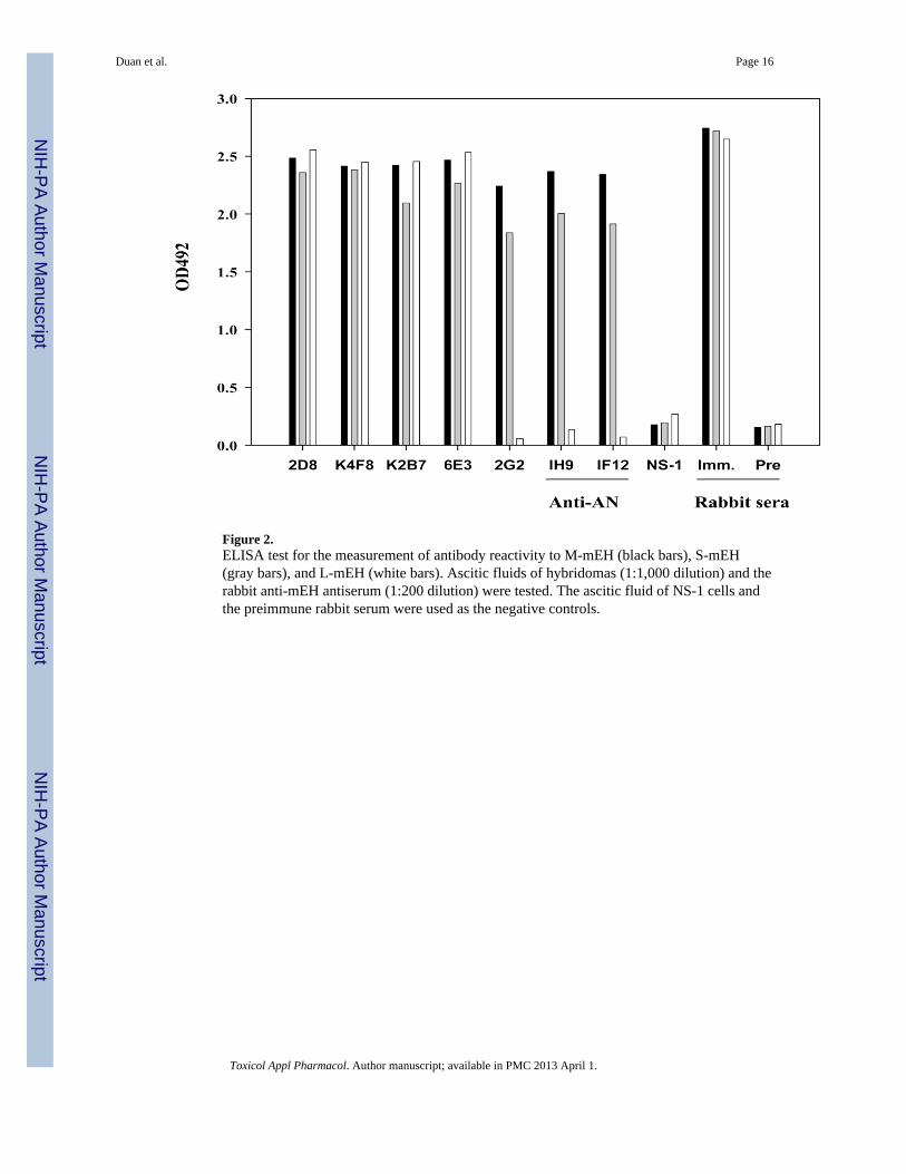
Figure 2.ELISA test for the measurement of antibody reactivity to M-mEH (black bars), S-mEH(gray bars), and L-mEH (white bars). Ascitic fluids of hybridomas (1:1,000 dilution) and therabbit anti-mEH antiserum (1:200 dilution) were tested. The ascitic fluid of NS-1 cells andthe preimmune rabbit serum were used as the negative controls.
Duan et al. Page 16
Toxicol Appl Pharmacol. Author manuscript; available in PMC 2013 April 1.
NIH
-PA Author Manuscript
NIH
-PA Author Manuscript
NIH
-PA Author Manuscript
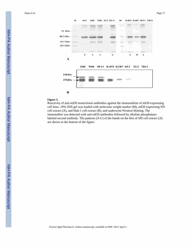
Figure 3.Reactivity of anti-mEH monoclonal antibodies against the immunoblots of mEH-expressingcell lines. 10% SDS gel was loaded with molecular weight marker (M), mEH-expressing Sf9cell extract (A), and Huh-1 cell extract (B), and underwent Western blotting. Theimmunoblot was detected with anti-mEH antibodies followed by alkaline phosphatase-labeled second antibody. The patterns (A-C) of the bands on the blot of Sf9 cell extract (A)are shown at the bottom of the figure.
Duan et al. Page 17
Toxicol Appl Pharmacol. Author manuscript; available in PMC 2013 April 1.
NIH
-PA Author Manuscript
NIH
-PA Author Manuscript
NIH
-PA Author Manuscript
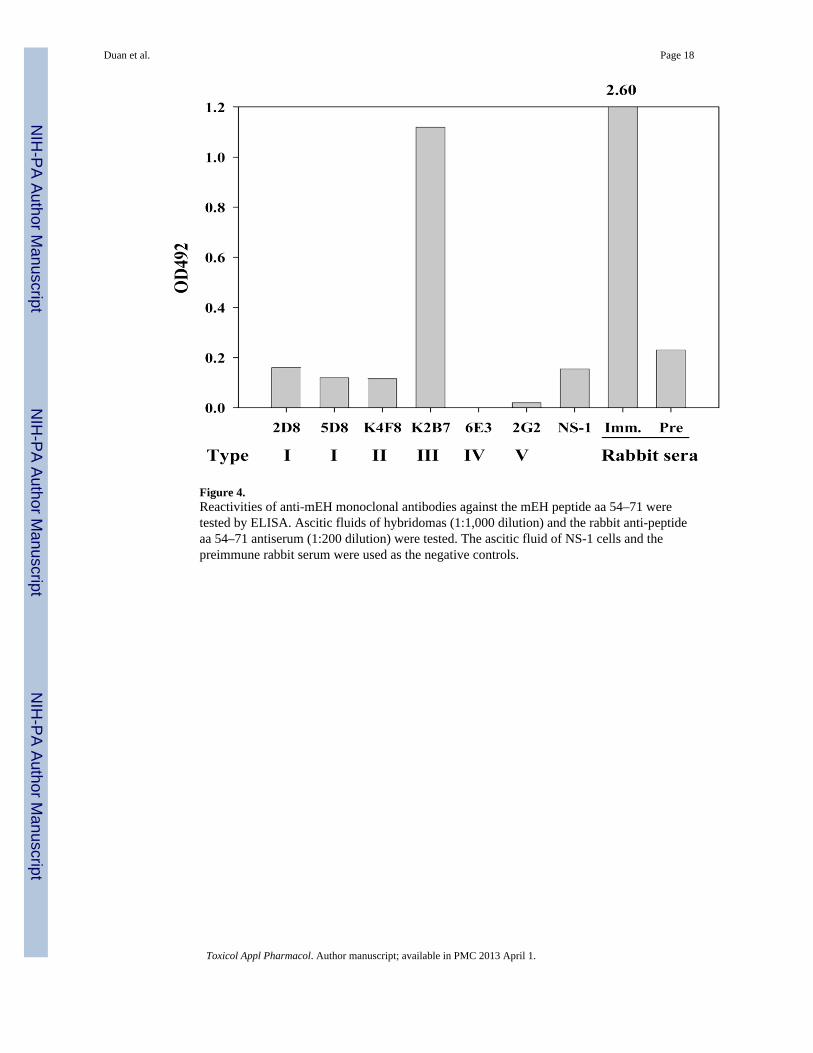
Figure 4.Reactivities of anti-mEH monoclonal antibodies against the mEH peptide aa 54–71 weretested by ELISA. Ascitic fluids of hybridomas (1:1,000 dilution) and the rabbit anti-peptideaa 54–71 antiserum (1:200 dilution) were tested. The ascitic fluid of NS-1 cells and thepreimmune rabbit serum were used as the negative controls.
Duan et al. Page 18
Toxicol Appl Pharmacol. Author manuscript; available in PMC 2013 April 1.
NIH
-PA Author Manuscript
NIH
-PA Author Manuscript
NIH
-PA Author Manuscript
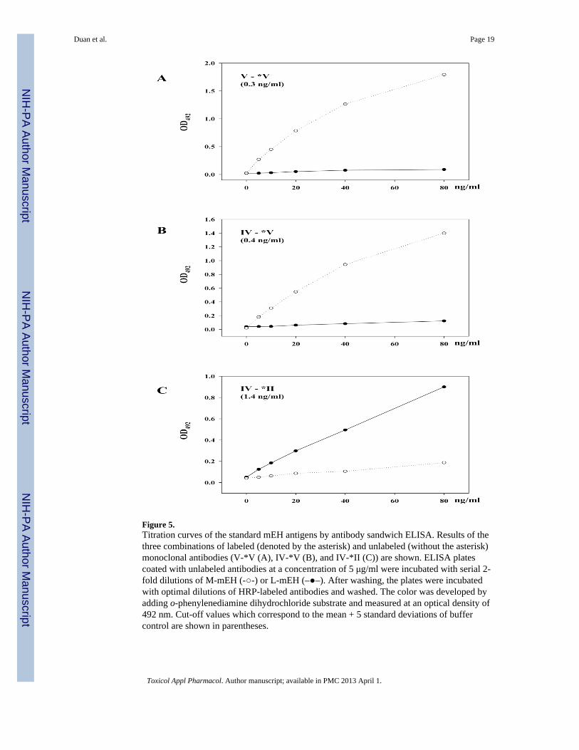
Figure 5.Titration curves of the standard mEH antigens by antibody sandwich ELISA. Results of thethree combinations of labeled (denoted by the asterisk) and unlabeled (without the asterisk)monoclonal antibodies (V-*V (A), IV-*V (B), and IV-*II (C)) are shown. ELISA platescoated with unlabeled antibodies at a concentration of 5 μg/ml were incubated with serial 2-fold dilutions of M-mEH (-○-) or L-mEH (–●–). After washing, the plates were incubatedwith optimal dilutions of HRP-labeled antibodies and washed. The color was developed byadding o-phenylenediamine dihydrochloride substrate and measured at an optical density of492 nm. Cut-off values which correspond to the mean + 5 standard deviations of buffercontrol are shown in parentheses.
Duan et al. Page 19
Toxicol Appl Pharmacol. Author manuscript; available in PMC 2013 April 1.
NIH
-PA Author Manuscript
NIH
-PA Author Manuscript
NIH
-PA Author Manuscript
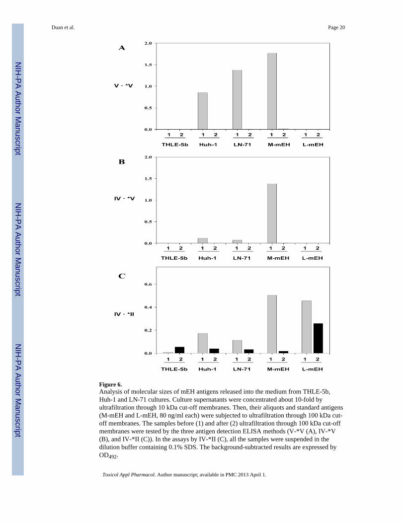
Figure 6.Analysis of molecular sizes of mEH antigens released into the medium from THLE-5b,Huh-1 and LN-71 cultures. Culture supernatants were concentrated about 10-fold byultrafiltration through 10 kDa cut-off membranes. Then, their aliquots and standard antigens(M-mEH and L-mEH, 80 ng/ml each) were subjected to ultrafiltration through 100 kDa cut-off membranes. The samples before (1) and after (2) ultrafiltration through 100 kDa cut-offmembranes were tested by the three antigen detection ELISA methods (V-*V (A), IV-*V(B), and IV-*II (C)). In the assays by IV-*II (C), all the samples were suspended in thedilution buffer containing 0.1% SDS. The background-subtracted results are expressed byOD492.
Duan et al. Page 20
Toxicol Appl Pharmacol. Author manuscript; available in PMC 2013 April 1.
NIH
-PA Author Manuscript
NIH
-PA Author Manuscript
NIH
-PA Author Manuscript
NIH
-PA Author Manuscript
NIH
-PA Author Manuscript
NIH
-PA Author Manuscript
Duan et al. Page 21
TABLE 1
Primers for the amplification and expression of mEH fragments.
Primer Sequencea Description
1 5′-CGGGATCCATGTGGCTAGAAATCCTCCTCAC-3′ Sense primer for #1
2 5′-CGGGATCCCGGGACAAAGAGGAAACTTTGCC-3′ Sense primer for #2, #5–#9
3 5′-CGGGATCCAAAGGCCTGCACTTGAACATGGC-3′ Sense primer for #3
4 5′-CGGGATCCGAGAAGTTTTCCACCTGGACC-3′ Sense primer for #4
5 5′-CGGGATCCTTGCCGCTCCAGCACCGACAGG-3′ Antisense primer for #1
6 5′-TCCCCCGGGTTGCCGCTCCAGCACCGACAGG-3′ Antisense primer for #2–#4
7 5′-TCCCCCGGGCTTCATCCGCTCACGCTTCTG-3′ Antisense primer for #5
8 5′-TCCCCCGGGGTCGTCCAGGGAGAACTTCC-3′ Antisense primer for #6
9 5′-TCCCCCGGGGGTGTCAGGCTTGGTGCACTG-3′ Antisense primer for #7
10 5′-TCCCCCGGGGTCCCCTCCTTGAATGTAGAA-3′ Antisense primer for #8
11 5′-TCCCCCGGGGGGCTTCGGGGTATGGCCTGC-3′ Antisense primer for #9
aThe underlined sequences GGATCC and CCCGGG represent recognition sequences of BamHI and SmaI, respectively.
Toxicol Appl Pharmacol. Author manuscript; available in PMC 2013 April 1.
NIH
-PA Author Manuscript
NIH
-PA Author Manuscript
NIH
-PA Author Manuscript
Duan et al. Page 22
TAB
LE 2
Epi
tope
map
ping
of a
nti-m
EH
mon
oclo
nal a
ntib
odie
s by
EL
ISA
Mea
n O
D v
alue
s of d
uplic
ate
ELIS
A a
re in
dica
ted,
and
pos
itive
resu
lts a
re h
ighl
ight
ed in
gra
y. T
he c
ut-o
ff w
as d
eter
min
ed a
s the
mea
n ±
5 st
anda
rdde
viat
ions
of N
S-1
cultu
re su
pern
atan
ts (0
.113
) and
that
of t
he n
aïve
mou
se se
ra (0
.202
), fo
r hyb
ridom
a cu
lture
s and
the
imm
une
mou
se se
ra,
resp
ectiv
ely.
Ant
ibod
y
F-m
EH
pGE
X1
23
45
67
89
2D8
2.79
02.
637
0.05
90.
067
2.72
12.
833
2.69
52.
686
2.75
70.
055
5D8
2.55
12.
365
0.09
20.
063
1.82
41.
972
2.05
52.
051
2.00
10.
053
8F11
2.51
92.
330
0.09
00.
050
1.81
91.
663
2.06
52.
043
1.85
20.
053
K4F
82.
473
2.35
40.
105
0.09
61.
785
1.93
11.
987
1.93
81.
752
0.06
2
K2B
72.
527
2.39
50.
086
0.08
20.
913
0.93
01.
341
1.47
50.
637
0.05
9
6E3
2.12
02.
029
2.77
92.
770
0.21
42.
022
0.06
40.
068
0.07
50.
032
2G2
0.00
00.
000
0.00
00.
000
0.00
00.
000
0.00
00.
000
0.00
00.
000
7B11
0.00
7−0.016
−0.027
0.03
1−0.026
−0.101
−0.023
−0.048
−0.041
0.00
1
NS-
10.
061
0.07
00.
046
0.07
70.
062
0.06
40.
057
0.05
20.
045
0.04
5
Imm
une
1.65
71.
417
2.19
92.
092
1.19
91.
204
1.42
41.
288
1.18
50.
117
Naï
ve0.
060
0.04
6−0.015
0.04
80.
064
−0.009
0.05
80.
051
0.08
70.
038
Toxicol Appl Pharmacol. Author manuscript; available in PMC 2013 April 1.
NIH
-PA Author Manuscript
NIH
-PA Author Manuscript
NIH
-PA Author Manuscript
Duan et al. Page 23
TAB
LE 3
Cla
ssifi
catio
n of
ant
i-mE
H m
onoc
lona
l ant
ibod
ies
Eigh
t mon
oclo
nal a
ntib
odie
s wer
e gr
oupe
d in
to fi
ve g
roup
s (Ty
pe I
to V
) dep
endi
ng o
n th
e re
activ
ity w
ith F
-mEH
1 to
4 a
nd S
-mEH
by
ELIS
A, a
nd th
eba
nd si
zes a
nd p
atte
rns t
hey
prod
uced
in W
este
rn b
lotti
ng. T
heir
H-c
hain
and
L-c
hain
isot
ypes
are
als
o sh
own.
Clo
neIs
otyp
eL
-cha
in
EL
ISA
Wes
tern
blo
tting
Typ
e
F-m
EH
S-m
EH
Sf9
TH
LE
-5b
Huh
-11
23
4
2D8
G2a
κ+
+−
−+
A1
54K
, 47K
54K
, 47K
I
5D8
G1
κ+
+−
−+
A1
54K
, 47K
54K
, 47K
I
8F11
G1
κ+
+−
−+
A1
54K
, 47K
54K
, 47K
I
K4F
8G
1κ
++
−−
+A
247
K47
KII
K2B
7G
1κ
++
−−
+B
47K
47K
III
6E3
G1
κ+
++
++
C47
K47
KIV
2G2
G1
κ−
−−
−+
––
–V
7B11
Mκ
−−
−−
+–
––
V
Toxicol Appl Pharmacol. Author manuscript; available in PMC 2013 April 1.
NIH
-PA Author Manuscript
NIH
-PA Author Manuscript
NIH
-PA Author Manuscript
Duan et al. Page 24
TAB
LE 4
Com
petit
ive
bind
ing
assa
y
Six
kind
s of a
ntib
odie
s wer
e la
bele
d w
ith 12
5 I, a
nd 2
× 1
05 cpm
of e
ach
antib
ody
was
incu
bate
d w
ith in
crea
sing
con
cent
ratio
ns o
f the
unl
abel
edho
mol
ogou
s or h
eter
olog
ous a
ntib
odie
s in
the
wel
ls c
oate
d w
ith m
EH. A
s the
ant
igen
, L-m
EH w
as u
sed
for t
he a
ntib
odie
s fro
m ty
pe I
to ty
pe IV
and
the
rabb
it an
ti-pe
ptid
e aa
54–7
1 an
tibod
y. M
-mEH
was
use
d fo
r the
type
V a
nd a
nti-A
N a
ntib
odie
s. %
inhi
bitio
n va
lues
at 5
μg/
ml a
re sh
own,
and
thos
e ab
ove
80%
are
hig
hlig
hted
in g
ray.
125 I
-ant
ibod
yU
nlab
eled
ant
ibod
y
Typ
eC
lone
Typ
e I
Typ
e II
Typ
e II
IT
ype
IVT
ype
VA
nti-A
NR
abbi
t Ab
2D8
5D8
K4F
8K
2B7
6E3
2G2
1H9
aa54
–71
Type
I5D
896
.110
0.0
62.6
30.9
38.0
1.4
1.4
4.2
Type
IIK
4F8
93.9
94.7
100.
016
.910
.515
.515
.525
.9
Type
III
K2B
728
.454
.272
.010
0.0
39.7
0.0
0.0
99.3
Type
IV6E
330
.026
.243
.444
.010
0.0
43.0
43.0
51.6
Type
V2G
2N
.D.
17.9
N.D
.N
.D.
9.1
100.
010
0.0
N.D
.
Ant
i-AN
1H9
N.D
.14
.1N
.D.
N.D
.10
.787
.810
0.0
N.D
.
N.D
.: no
t det
erm
ined
.
Toxicol Appl Pharmacol. Author manuscript; available in PMC 2013 April 1.
NIH
-PA Author Manuscript
NIH
-PA Author Manuscript
NIH
-PA Author Manuscript
Duan et al. Page 25
TABLE 5
Epitope selectivities of five types of monoclonal antibodies.
Type I N-terminus (aa.21–143) linear epitope, Cross-reacts with a 54-kDa protein
Type II: N-terminus (aa.21–143) linear epitope
Type III: N-terminus (aa.54–71) linear epitope
Type IV: C-terminus (aa.327–353) linear epitope
Type V: Conformational epitope
Toxicol Appl Pharmacol. Author manuscript; available in PMC 2013 April 1.
NIH
-PA Author Manuscript
NIH
-PA Author Manuscript
NIH
-PA Author Manuscript
Duan et al. Page 26
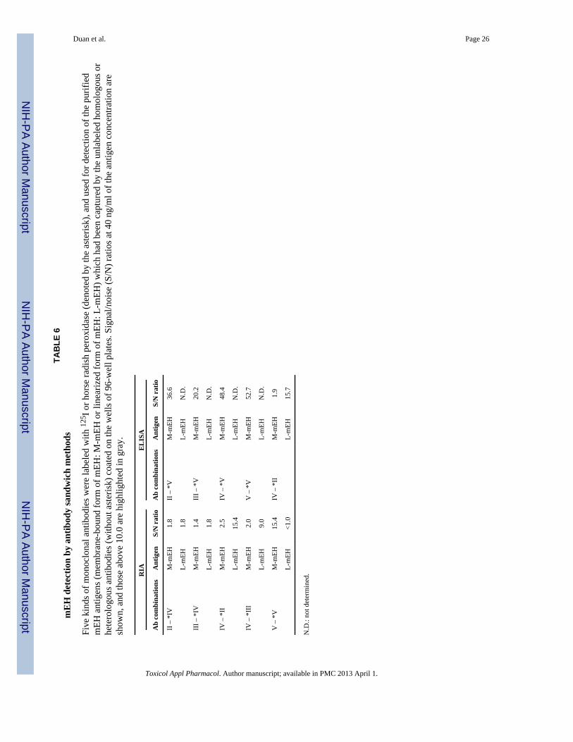
TAB
LE 6
mE
H d
etec
tion
by a
ntib
ody
sand
wic
h m
etho
ds
Five
kin
ds o
f mon
oclo
nal a
ntib
odie
s wer
e la
bele
d w
ith 12
5 I o
r hor
se ra
dish
per
oxid
ase
(den
oted
by
the
aste
risk)
, and
use
d fo
r det
ectio
n of
the
purif
ied
mEH
ant
igen
s (m
embr
ane-
boun
d fo
rm o
f mEH
: M-m
EH o
r lin
eariz
ed fo
rm o
f mEH
: L-m
EH) w
hich
had
bee
n ca
ptur
ed b
y th
e un
labe
led
hom
olog
ous o
rhe
tero
logo
us a
ntib
odie
s (w
ithou
t ast
eris
k) c
oate
d on
the
wel
ls o
f 96-
wel
l pla
tes.
Sign
al/n
oise
(S/N
) rat
ios a
t 40
ng/m
l of t
he a
ntig
en c
once
ntra
tion
are
show
n, a
nd th
ose
abov
e 10
.0 a
re h
ighl
ight
ed in
gra
y.
RIA
EL
ISA
Ab
com
bina
tions
Ant
igen
S/N
rat
ioA
b co
mbi
natio
nsA
ntig
enS/
N r
atio
II –
*IV
M-m
EH1.
8II
– *
VM
-mEH
36.6
L-m
EH1.
8L-
mEH
N.D
.
III –
*IV
M-m
EH1.
4II
I – *
VM
-mEH
20.2
L-m
EH1.
8L-
mEH
N.D
.
IV –
*II
M-m
EH2.
5IV
– *
VM
-mEH
48.4
L-m
EH15
.4L-
mEH
N.D
.
IV –
*II
IM
-mEH
2.0
V –
*V
M-m
EH52
.7
L-m
EH9.
0L-
mEH
N.D
.
V –
*V
M-m
EH15
.4IV
– *
IIM
-mEH
1.9
L-m
EH<1
.0L-
mEH
15.7
N.D
.: no
t det
erm
ined
.
Toxicol Appl Pharmacol. Author manuscript; available in PMC 2013 April 1.
NIH
-PA Author Manuscript
NIH
-PA Author Manuscript
NIH
-PA Author Manuscript
Duan et al. Page 27
TABLE 7mEH detection in culture supernatants by RIA
mEH in the culture supernatant was assayed by RIA with the three combinations of labeled (denoted by theasterisk) and unlabeled (without the asterisk) monoclonal antibodies and the purified mEH as the standard (L-mEH for IV-*II and M-mEH for IV-*V and V-*V). Results are expressed as ng/ml and values above the cut-off are highlighted in grey. Cut-off values were 2.1 (S/N ratio) using culture media as the negative control.
Cell line
Combinations of antibodies
IV – *II IV – *V V – *V
THLE-5b < 1.8 < 1.6 < 1.3
Huh-7 < 1.8 < 1.6 < 1.3
Huh-1 < 1.8 < 1.6 4.26
M1 < 1.8 < 1.6 < 1.3
U87MG < 1.8 < 1.6 < 1.3
LN-Z308 < 1.8 < 1.6 < 1.3
LN-71 < 1.8 < 1.6 7.83
Toxicol Appl Pharmacol. Author manuscript; available in PMC 2013 April 1.
Related Documents