Development and Validation of a Fluorescence Polarization-Based Competitive Peptide-Binding Assay for HLA-A*0201sA New Tool for Epitope Discovery Rico Buchli,* ,‡,§ Rodney S. VanGundy, ‡ Heather D. Hickman-Miller, § Christopher F. Giberson, ‡ Wilfried Bardet, § and William H. Hildebrand ‡,§ Pure Protein L.L.C., Oklahoma City, Oklahoma 73104, and Department of Microbiology and Immunology, Health Sciences Center, UniVersity of Oklahoma, Oklahoma City, Oklahoma 73104 ReceiVed February 10, 2005; ReVised Manuscript ReceiVed July 15, 2005 ABSTRACT: Various approaches are currently proposed to successfully develop therapies for the prevention and treatment of infectious diseases and cancer. One of the most promising approaches is the development of vaccines that elicit cytotoxic T lymphocyte (CTL) responses. Consequently, identification and exact definition of molecular parameters involved in peptide-MHC class-I interactions of putative CTL epitopes are of prime importance for the development of immunomodulating compounds. To better facilitate epitope discovery, we developed and validated a novel state-of-the-art biochemical HLA-A*0201 assay, which is comprised of technologically advanced cutting edge reagents. The technique is based on competition and uses a FITC-labeled reference peptide and highly purified soluble HLA-A*0201 molecules to quantitatively measure the binding capacity of nonlabeled peptide candidates. Detection by fluorescence polarization allows real-time measurement of binding ratios without separation steps. During standardization, the problem of assay parameter variation is discussed, showing the dramatic influence of HLA and reference peptide concentrations as well as the choice of the reference peptide itself on IC 50 determinations. For validation, a panel of 15 well-defined HLA-A*0201 ligands from various sources covering a broad range of binding affinities was tested. Binding data were used to compare against pre-existing quantitative assay systems. The results obtained demonstrated significant correlation among assay procedures, suggesting that the application of fluorescence polarization in combination with recombinant sHLA molecules is highly advantageous for the accurate assessment of peptide binding. Furthermore, the assay also features high- throughput screening capacity, providing uniquely efficient means of identifying and evaluating immune target molecules. The ability of the immune system to recognize virus- infected or cancerous cells has opened the door to the development of vaccines to treat or prevent various types of infectious diseases or cancers. An increased understanding of the mechanisms by which T cells recognize virus and tumor-specific antigens has stimulated much interest in the use of specific T cells as adoptive immunotherapy for infectious and malignant diseases. Particularly, CD8 + T lymphocytes (CTLs) 1 play a critical role in controlling and/ or eliminating these infected and neoplastic cells. They are implicated in immunity to not only pathogens such as viruses, which utilize the host cell’s biosynthetic machinery to produce viral proteins (1-11), but also to parasitic and microbial invaders (12, 13). CTL responses are likewise extended to include stimulation by aberrant proteins such as those associated with malignancies (14-23). In fact, class-I molecules have the potential of binding and presenting to CTLs any protein introduced into the endogenous processing pathway by either natural or artificial means (24-29). This knowledge serves as a motivating factor behind the develop- ment of peptide-based vaccines intended to elicit protective CTL responses to pathogens and other abnormalities, which otherwise remain cytoplasmically concealed from detection. Peptide-based virus and tumor vaccination has been proven efficient in protecting against virus infection and tumor growth in many mouse experimental models and more recently in humans (8, 14, 15, 17, 19, 20, 22, 30-38). This type of vaccination strategy offers many attractive features such as ease of manufacturing and characterization, as well as an excellent safety profile in past clinical studies by using defined epitopes, which are isolated from the context of the antigen of origin. The design of peptide-based vaccines has several potential advantages including not only a more potent response than that obtained by the use of whole antigens (39, 40) but also allows the control over qualitative aspects of the immune response. Broad responses simultaneously targeting multiple dominant and subdominant epitopes can be induced (36, 41). Accessing subdominant specificities to overcome T cell tolerance might be of particular value in * To whom correspondence should be addressed: Pure Protein L.L.C., 800 Research Parkway, Suite 340, Oklahoma City, OK 73104- 3698. Telephone: 405-271-3838. Fax: 405-271-3848. E-mail: [email protected]. ‡ Pure Protein L.L.C. § University of Oklahoma. 1 Abbreviations: CTL, cytotoxic T lymphocyte; MHC, major his- tocompatibility complex; 2m, -2 microglobulin; HLA, human leukocyte antigen; FP, fluorescence polarization; sHLA, soluble HLA; FITC, fluorescein isothiocyanate; DMF, dimethylformamide; BGG, bovine γ globulin; pFITC, FITC-labeled peptide; cps, counts per second; IC 50, inhibitory concentration; Kd, equilibrium dissociation constant; mP, millipolarization. 12491 Biochemistry 2005, 44, 12491-12507 10.1021/bi050255v CCC: $30.25 © 2005 American Chemical Society Published on Web 08/26/2005

Welcome message from author
This document is posted to help you gain knowledge. Please leave a comment to let me know what you think about it! Share it to your friends and learn new things together.
Transcript

Development and Validation of a Fluorescence Polarization-Based CompetitivePeptide-Binding Assay for HLA-A*0201sA New Tool for Epitope Discovery
Rico Buchli,*,‡,§ Rodney S. VanGundy,‡ Heather D. Hickman-Miller,§ Christopher F. Giberson,‡
Wilfried Bardet,§ and William H. Hildebrand‡,§
Pure Protein L.L.C., Oklahoma City, Oklahoma 73104, and Department of Microbiology and Immunology,Health Sciences Center, UniVersity of Oklahoma, Oklahoma City, Oklahoma 73104
ReceiVed February 10, 2005; ReVised Manuscript ReceiVed July 15, 2005
ABSTRACT: Various approaches are currently proposed to successfully develop therapies for the preventionand treatment of infectious diseases and cancer. One of the most promising approaches is the developmentof vaccines that elicit cytotoxic T lymphocyte (CTL) responses. Consequently, identification and exactdefinition of molecular parameters involved in peptide-MHC class-I interactions of putative CTL epitopesare of prime importance for the development of immunomodulating compounds. To better facilitate epitopediscovery, we developed and validated a novel state-of-the-art biochemical HLA-A*0201 assay, which iscomprised of technologically advanced cutting edge reagents. The technique is based on competition anduses a FITC-labeled reference peptide and highly purified soluble HLA-A*0201 molecules to quantitativelymeasure the binding capacity of nonlabeled peptide candidates. Detection by fluorescence polarizationallows real-time measurement of binding ratios without separation steps. During standardization, the problemof assay parameter variation is discussed, showing the dramatic influence of HLA and reference peptideconcentrations as well as the choice of the reference peptide itself on IC50 determinations. For validation,a panel of 15 well-defined HLA-A*0201 ligands from various sources covering a broad range of bindingaffinities was tested. Binding data were used to compare against pre-existing quantitative assay systems.The results obtained demonstrated significant correlation among assay procedures, suggesting that theapplication of fluorescence polarization in combination with recombinant sHLA molecules is highlyadvantageous for the accurate assessment of peptide binding. Furthermore, the assay also features high-throughput screening capacity, providing uniquely efficient means of identifying and evaluating immunetarget molecules.
The ability of the immune system to recognize virus-infected or cancerous cells has opened the door to thedevelopment of vaccines to treat or prevent various types ofinfectious diseases or cancers. An increased understandingof the mechanisms by which T cells recognize virus andtumor-specific antigens has stimulated much interest in theuse of specific T cells as adoptive immunotherapy forinfectious and malignant diseases. Particularly, CD8+ Tlymphocytes (CTLs)1 play a critical role in controlling and/or eliminating these infected and neoplastic cells. They areimplicated in immunity to not only pathogens such as viruses,which utilize the host cell’s biosynthetic machinery toproduce viral proteins (1-11), but also to parasitic andmicrobial invaders (12, 13). CTL responses are likewise
extended to include stimulation by aberrant proteins such asthose associated with malignancies (14-23). In fact, class-Imolecules have the potential of binding and presenting toCTLs any protein introduced into the endogenous processingpathway by either natural or artificial means (24-29). Thisknowledge serves as a motivating factor behind the develop-ment of peptide-based vaccines intended to elicit protectiveCTL responses to pathogens and other abnormalities, whichotherwise remain cytoplasmically concealed from detection.
Peptide-based virus and tumor vaccination has been provenefficient in protecting against virus infection and tumorgrowth in many mouse experimental models and morerecently in humans (8, 14, 15, 17, 19, 20, 22, 30-38). Thistype of vaccination strategy offers many attractive featuressuch as ease of manufacturing and characterization, as wellas an excellent safety profile in past clinical studies by usingdefined epitopes, which are isolated from the context of theantigen of origin. The design of peptide-based vaccines hasseveral potential advantages including not only a more potentresponse than that obtained by the use of whole antigens(39, 40) but also allows the control over qualitative aspectsof the immune response. Broad responses simultaneouslytargeting multiple dominant and subdominant epitopes canbe induced (36, 41). Accessing subdominant specificities toovercome T cell tolerance might be of particular value in
* To whom correspondence should be addressed: Pure ProteinL.L.C., 800 Research Parkway, Suite 340, Oklahoma City, OK 73104-3698. Telephone: 405-271-3838. Fax: 405-271-3848. E-mail:[email protected].
‡ Pure Protein L.L.C.§ University of Oklahoma.1 Abbreviations: CTL, cytotoxic T lymphocyte; MHC, major his-
tocompatibility complex; â2m, â-2 microglobulin; HLA, humanleukocyte antigen; FP, fluorescence polarization; sHLA, soluble HLA;FITC, fluorescein isothiocyanate; DMF, dimethylformamide; BGG,bovineγ globulin; pFITC, FITC-labeled peptide; cps, counts per second;IC50, inhibitory concentration;Kd, equilibrium dissociation constant;mP, millipolarization.
12491Biochemistry2005,44, 12491-12507
10.1021/bi050255v CCC: $30.25 © 2005 American Chemical SocietyPublished on Web 08/26/2005

the case of tumor antigens, thus facilitating the developmentof cancer vaccines even further (14, 42).
CTLs recognize peptide fragments of cellular or viralproteins in the form of short peptides comprising 8-11amino acids presented in association with major histocom-patibility complex (MHC) class-I molecules on the surfaceof infected or neoplastic cells (43-47). These peptides areusually derived from intracellular protein pools and associ-ated in the lumen of the endoplasmic reticulum with MHCclass-I heavy-chain andâ2-microglobulin (â2m) molecules,after which the MHC-peptide complex is transported to thecell surface. Considering the myriad of possible peptidesequences, coupled with the genetic variability of MHCmolecules among individuals, the challenge of selecting the“right” target antigen or epitope presents a major obstaclein the development of such cell-mediated vaccines. Conse-quently, one of the major reasons to study peptide bindingto class-I molecules is to be able to determine which peptidesare likely to be antigenic, starting from the primary sequenceof viral or cancer-related proteins. Although many factorsinfluencing MHC class-I-restricted CTL responses to peptideshave been elucidated, a major parameter determining cell-surface presentation of a given peptide is the affinity of thepeptide for MHC class-I molecules (48, 49). Recent reportsshow that peptides possessing good binding characteristicsare a prerequisite for successful candidate CTL epitopeswhere remarkable association between the strength of bindingand the immunogenicity of individual peptides in patientsand HLA transgenic mice could be demonstrated. In thismatter, several lines of evidence, both at the biological andfunctional level, emphasize the biologic relevance of peptide-binding assays in the identification and evaluation of potentialpeptide candidates (48, 50-56).
Many different peptide-MHC-binding assays have beensuggested over the years. The assays are performed using anumber of different protocols with the common theme ofassessing the relative abilities of synthetically definedpeptides to associate with specific class-I heavy chains andâ2m in Vitro (56-82). However, most of them have beenconducted under poorly controlled conditions, and only afew quantitative biochemical binding assays have beenestablished (56, 57, 66, 79, 80, 82, 83). The most traditionalbiochemical binding assays utilize detergent-solubilized HLAclass-I complex molecules in combination with iodinatedpeptides to measure peptide binding (56, 78, 79, 81, 83). Inthese conventional radioactive competition assays, radioactiveiodine [125I] is measured after chromatographic separationof bound from free peptide ligand (56, 77, 79). Unfortunately,this assay method involves multiple washes, liquid transfers,and incubation times that make it very labor-intensive andtime-consuming. In addition to generating large volumes ofradioactive waste, these steps cannot be completely auto-mated. An additional disadvantage is that the complex isimmobilized on an affinity matrix possibly interfering withthe kinetics of the assay. A further limitation in most of theseheterogeneous binding assay applications has been the lackof availability of high-quality class-I HLA proteins. In recentyears, alternative competitive assay approaches have beendeveloped using fluorescence labeled peptides in combinationwith cell-bound HLA class-I molecules from which naturallybound class-I peptides were eluted (49, 66, 82). In thesecellular-based fluorescence procedures, binding events were
detected by flow cytometry. However, these assays stillconsist of multiple steps and demand highly trained operatorsand expensive reagents, making them inherently slow anddifficult to automate. Therefore, the improvement of reliableand quantitative assays will considerably aid in dissectingthe quantitative aspects of class-I peptide interactions.
To overcome the difficulties of current assay proceduresand further facilitate epitope discovery for MHC class-Imolecules, we have developed and optimized a peptidecompetition assay based on the interaction between recom-binant, soluble HLA molecule A*0201 and a fluorescence-labeled peptide reference using the technique of fluorescencepolarization (FP). FP is unique among methods used toanalyze molecular-binding events because it gives a direct,nearly instantaneous measurement of a ligand’s bound/freeratio. The technique is based on the principle that smallmolecules rotate faster than large molecules. During binding,the small fluorescent peptide, which has free rotationalmobility, is converted to a larger fluorescent peptide/HLAcomplex with restricted rotational mobility (84, 85), resultingin an increase in polarization. Within the FP field, anisotropyvalues are sometimes preferred because it is easier todeconvolute anisotropy values into their component valuesthan it is with polarization values. Polarization and anisotropyare both derived from the measured vertical and horizontalintensities. The values are mathematically related and easilyinterconverted [A ) 2P/(3 - P)]. Both values represent aweighted average of the bound versus unbound states of thefluorescent molecule. However, it should be noted that, inthe majority of applications, anisotropy does not give anymore information than polarization and polarization valuescan be manipulated as if they were anisotropy values.
Our FP competition assay approach differs from othertypes of binding studies in one important regard: it requiresno steps to separate the free from the bound tracer and istherefore fast, simple, and accurate. In this assay, anunlabeled peptide competes with a fluorescent-labeled pep-tide used as a tracer, whereas the usage of individual sHLAclass-I molecules of defined specificity and purity offers thecapacity for efficient standardization. This homogeneousassay is not only conceptually simple but also extremelysensitive and provides a viable option for easy automation,random sample accessibility, and simple adaption to existinginstrumentation.
MATERIALS AND METHODS
Synthetic Peptides. FITC-labeled peptides were com-mercially synthesized by Synpep (Dublin, CA) or AmericanPeptides (Sunnyvale, CA) using solid-phase strategies andpurified with reverse-phase HPLC. The sequence of thereference peptides used for HLA-A*0201 are shown in Table1, wherein a FITC-labeled lysine was introduced into theprimary sequence of the native molecule as described earlier
Table 1: Sequences of FITC-Labeled Peptide References Useda
name sequence
A*0201/P1 F L P S D K F P S VA*0201/P2 S L Y N K V A T LA*0201/P4 L V F G K E V V E VA*0201/P5 A L M D K V L K V
a The amino acid lysine carrying the FITC-label is in bold font.
12492 Biochemistry, Vol. 44, No. 37, 2005 Buchli et al.

(86). Unmodified unlabeled peptides were synthesized andpurified by the Molecular Biology Resource Facility of theWarren Medical Research Institute at OUHSC (OklahomaCity, OK). Purity of all synthetic peptides was greater than95%, and their composition was ascertained by massspectrometric analysis. Peptides were stored dry in pre-aliquoted amounts of 0.2 and 0.4µmol for labeled andunlabeled peptides, respectively. All lyophilized aliquots usedin binding assays were originally dissolved in 100% DMSOor DMF at a concentration of 10 mM. Subsequent dilutionswere done in 1× bovine γ globulin in phosphate-bufferedsaline (BGG/PBS; 0.5 mg/mL; 0.05%; Sigma, St. Louis,MO). Each pFITC preparation was standardized using amolar fluorescence intensity curve generated by a premea-sured FITC standard (Pierce) reconstituted in 1× BGG (0.5mg/mL). Aliquoted working dilutions for FITC-labeledpeptides (200µM) were kept at-20 °C and reused for aperiod of up to 4 months without a measurable decline inthe signal.
Large-Scale Production and Purification of RecombinantsHLA Molecules.Production and purification of recombinantsHLA molecules was performed as described previously (86,87). Briefly, stable sHLA-transfected EBV-transformed B-lymphoblastoid cells (721.221) were expanded in RPMI-1640/20% FCS under 1.5 mg/mL G418 selection beforeseeding two hollow-fiber bioreactor units of the CP-2500Cell Pharm System (Biovest International, Minneapolis,MN). After inoculation, a typical production run lastingbetween 4 and 6 weeks produced approximately 30-40 Lof secreted sHLA product, which was collected as a crudeharvest. Upon completion of a bioreactor run, sHLAcomplexes were affinity-purified from the harvest obtainedusing a Sepharose 4B-W6/32 matrix. Purified molecules werebuffer-exchanged with PBS at pH 7.2 and concentrated using10-kDa cutoff Macrosep centrifugal concentrators (PallFiltron, Northborough, MA). The final product was filter-sterilized and stored at 4°C until further use. The concentra-tion of the purified molecules was determined using theMicro BCA protein assay kit (Pierce, Rockford, IL) usingBGG as a standard. For subunit size confirmation and purityevaluation, SDS-PAGE was applied as quality control. Themolecular weight of sHLA-A*0201 was determined to be47.2 kDa, and the molecular weight ofâ2m was determinedto be 11.7 kDa.
FP-Based Peptide Competition Assay.FP measurementswere performed on an Analyst AD Assay Detection System(Molecular Devices; Sunnyvale, CA) using a continuoushigh-intensity, xenon-arc lamp as light source with filtersettings suitable for FITC excitation (485 nm) and emission(530 nm). As a standard reading configuration, the excitationpolarization filter was set in S (parallel) position (static),whereas the emission polarization filter was dynamicallypolarizing the light in either S or P (perpendicular) orienta-tion. A fluorescein dichroic mirror (505 nm) was used todirect the polarized light into the assay well. Emittedpolarized light was detected by the fluorescence photomul-tiplier tube with the SmartRead, sensitivity 2 setup optionin counts per second (cps). Two intensity measurements werecollected for each well, one when the dynamic polarizer wasin S and one when the polarizer was in the P position.
For a standardized assay setup, each individual well of ablack 96-well LJL HE PS microplate (Molecular Devices)
was loaded with 5µL of a 16× â2m solution (198.7µg/mL; 16 936 nM) (Fitzgerald Industries International, Con-cord, MA), 10µL of 4× competitor at various dilutions, and5 µL of an 8× pFITC preparation (16 nM). For allpreparations, 1× BGG/PBS was used as a buffer. To startpeptide exchange, a 2× sHLA mix (100 µg/mL; 2117 nM)was activated by incubating at 53°C for 15 min beforeadding 20µL to the previously loaded wells reaching a finalvolume of 40µL. After addition of all fluids, a final ratio[â2m]/[heavy chain] of 3:1 was achieved, where 2 parts ofâ2m are derived from the addedâ2m solution and 1 partfrom the sHLA complex. To remove potential air bubbles,the plate was spun down for 1 min at 2500 rpm. Note thatthe labeled and unlabeled ligand was presented simulta-neously to the activated sHLA. The plate was incubated atroom temperature and read periodically until no furtherincrease in polarization was observed, indicating that equi-librium was reached (24-48 h). After each reading, the platewas covered with a lid and sealed with Parafilm to protectfrom light and to prevent evaporation of the constituents.
Specific control groups included (a) protein only, (b) traceronly, and (c) buffer only, which are used for backgroundcorrection and calculation of theG factor as described earlier(86). The G factor{G factor ) S/P(1- (27/1000))/(1+(27/1000))} is a scaling (correction) factor, taking relativepolarization measurements and making them appear absolute.For this study, the theoretical value for free fluorescein (27mP) was used for G-factor determination with a value of1.00( 0.01. This value slightly dropped to 0.98( 0.02 forreadings beyond 72 h, probably caused by minor evaporationof the reactant solution. Baseline polarization values deter-mined for all pFITCs in their free state (average 30.3( 4.3)were not significantly different from the theoretical valueused resulting in close to identical G-factor values. Oncethe G factor was determined, FP values given as mP(millipolarization) were calculated by the equation: polariza-tion (mP) ) 1000(S- GP)/(S+ GP), where S and P arebackground-subtracted intensities of the fluorescence mea-sured in the S and P directions.
For assay stability testing, plates containing bindingexperiments were measured at an extended time period of3-7 days. No change in maximal polarization levels couldbe observed, indicating good stability of all constituents. Asadditional check on the system, the plates were also read inthe fluorescence intensity mode to control fluorescent peptideconcentrations according to the established standard curvefor single peptides.
Competition Assay Data Analysis.Competition experi-ments were analyzed by plotting FPmax (maximal polariza-tion) values as a function of the logarithms of competitorconcentrations. The binding affinity of each competitorpeptide was expressed as the concentration that inhibits 50%binding of the FITC-labeled reference peptide. Observedinhibitory concentrations (IC50) were determined by nonlinearcurve fitting to a dose-response model with a variable slopeusing the specific software Prism (Graph Pad Software, Inc.,San Diego, CA). The appropriate values for FPmax wereextracted from curve fittings of single-association reactionsof FITC-labeled peptides to sHLA molecules in the presenceof the competitor using a monoexponential association modelas described earlier (86).
Competitive Peptide-Binding Assay for sHLA-A*0201 Biochemistry, Vol. 44, No. 37, 200512493

Epitope Screening.Experimental epitope screening at athreshold concentration of 80µM was performed as describedfor the competition assay above with only minor modifica-tions. Briefly, 10µL of 4× competitor peptides (320µM)(Table 2) were added to individual wells of a black 96-wellmicroplate. Additional components such as 5µL of an 16×â2m solution and 5µL of an 8× pFITC preparation wereadded before starting peptide exchange with 20µL ofactivated 2× sHLA solution, reaching a final volume of 40µL. After equilibrium was reached, FP values were collectedon the Analyst AD (Molecular Devices) and transformed intopercent inhibition values by choosing the reaction withoutthe inhibitor as 0% inhibition.
RESULTS
Competition binding assay methodologies have becomeexceedingly popular for assessing the ability of syntheticallydefined peptide epitopes to associate with specific HLAclass-I complexes. This has been accomplished by comparingthe relative affinities of multiple peptide ligands for the sameHLA receptor. Experimentally, IC50 values, representativeof the affinity of the ligand-recognition site, have beendetermined by incubating HLA molecules from varioussources with a labeled peptide in the presence of differentconcentrations of the competitor. However, results presentedfrom various assay systems have shown a high variabilityrate, and thus, difficulties have been encountered in obtainingreproducible data. To further enhance the capabilities of suchassays and better facilitate epitope discovery for MHC class-Imolecules, we have developed a novel peptide competitionassay based on the interaction between recombinant, sHLAmolecules A*0201 and a fluorescence-labeled peptide refer-ence using the technique of fluorescence polarization. Todemonstrate feasibility but also increase the sensitivity andreproducibility of the assay, we initially conducted a detailed
analysis of all parameters critical for the standardization ofthe assay. This analysis was also intended to elucidate criticalfactors for interassay data comparisons.
Influence of Different Reference Peptides on IC50 Values.A primary requirement for the development of a moreadvanced competition assay is the careful selection of areference peptide. Recently, we demonstrated the bindingcharacteristics of several pFITC ligands to sHLA-A*0201through kinetics experiments, which seem to be well-suitedto act as a reference peptide (86). The labeled peptidecandidates were derived from peptides originally identifiedas naturally presented class-I ligands, CTL epitopes, orconstructed from the A*0201 consensus sequence (86). Alldesigns were made by substituting at various positions atnonanchor residues for a FITC-conjugated lysine (Table 1).
To visualize the effect of pFITC molecules with differentequilibrium dissociation constants (Kd) on IC50 determina-tions, we performed FP-based competition assays using aconstant concentration of activated sHLA (40µg/mL; 847nM), excessâ2m (1694 nM), and labeled peptide (10 nM)in the presence of different concentrations of the unlabeledligand KLGEFYNQMM as a test competitor (Figure 1). Theselected A2-related competitor peptide was derived from theinfluenza B virus (NP; 85-94) chosen because of itssuccessful use in several HLA studies (49, 73, 88, 89). Foreach of the assays shown in Figure 1, bound fluorescentpeptide was displaced from the sHLA molecule. The top ofeach curve represents a plateau at a value equal to bindingin the absence of the competing unlabeled peptide that wasset as 100% binding. Because a log axis cannot accommodatea concentration of zero [log(0) is undefined], a value for avery low competitor concentration was entered (-2). Thebottom of the curve is a plateau equal to the values for freelabeled peptide, which indicates almost complete competitionat higher concentrations of the competitor (set as 0%
Table 2: Summary of Peptide Competition Data Obtained in Association with HLA-A*0201 Including Specific Information on the PeptidesOrigin and Position
sequencea peptide originb positioncsequence
IDd
peptidelength(aa)
StD compassaye
(FP) IC50(nM)
cell-freesystemsf
(125I) IC50(nM)
referencesg(cell-freesystems)
cellularsystemsh
(fluorescence)IC50 (nM)
referencesi(cellularsystems)
YLLPAIVHI human; probableRNA-dependent helicase p72
146-154 Q92841 9 509.8 18 131
YMDDVVLGA hepatitis B virus (HBV); Pol 538-546 P03156 9 823.1 200 40, 95, 97FLPSDFFPSV hepatitis B virus (HBV); core 18-27 P03146 10 870 0.57; 1.2; 2.5;
2.8; 3.0; 3.3j40, 48, 56,
94, 96, 97400; 500j 49, 82, 118
YLVSFGVWI hepatitis B virus (HBV); core 118-126 P03146 9 1742 1.9 48GILGFVFTL influenza A virus;
matrix protein M158-66 P21429 9 2247 6; 12.4j 56, 132 4000 100
GLYSSTVPV hepatitis B virus (HBV); Pol 61-69 P03156 9 2353 20; 33j 48, 99 4500 49KLGEFYNQMM influenza B virus; NP 85-94 P13885 10 2820 5500 49LLSSNLSWL hepatitis B virus (HBV); Pol 407-415 P03156 9 3828 455 99 19 500 49GLSRYVARL hepatitis B virus (HBV); Pol 442-450 P03156 9 5113 71; 76; 79j 40, 97, 99SLYNTVATL human immunodeficiency
virus type 1 (HIV-1); Gag p1777-85 P05888 9 6068 50 133 1500 49
ILKEPVHGV human immunodeficiencyvirus type 1 (HIV-1); Pol
476-484 P03368 9 7082 192; 242j 40, 56 8000 49, 100
NLQSLTNLL hepatitis B virus (HBV); Pol 400-408 P03156 9 9389 1000; 2000j 48, 99 22 000 49NLVPMVATV cytomegalovirus (CMV); pp65 495-503 P06725 9 19 220 12 500 100HLESLFTAV hepatitis B virus (HBV); Pol 551-559 P03156 9 33 150 5000; 10 000j 81, 99DLVHFASPL hepatitis B virus (HBV); Pol 817-825 CAA46352 9 365 600 16 667 81
a Nonlabeled parental peptide sequence synthesized for IC50 evaluation.b Protein source including species and category.c Peptide position withinthe original protein.d Swiss Prot reference link.e Data represent IC50 values obtained by fluorescence polarization using the standardized assayconditions described within the text.f Data represent IC50 values published for cell-free (HLA lysate) competition systems using125I as peptidetracer.g Peptide references for cell-free systems.h Data represent IC50 values published for cellular systems (cell-bound HLA) using a fluorescence-labeled peptide reference.i Peptide references for cellular systems.j Multiple values from different publications listed.
12494 Biochemistry, Vol. 44, No. 37, 2005 Buchli et al.

binding). Because of the usage of the G factor, which correctsfor the contribution of the measurement pathway to theobserved total polarization, the value for nonspecific binding(0%) is equal to the assumed theoretical value of 27 mP.High ratios between the maximum and minimum signalswere observed for all competition experiments, with maximalFP values from 183.3 to 224.9 mP depending on the pFITCused (data not shown).
The concentration of the unlabeled competitor that pro-duced 50% inhibition of fluorescent-labeled peptide binding(halfway between the upper and lower plateaus of theobtained curve) and defined as the inhibitory concentration(IC50) was calculated by nonlinear regression analysis usingthe software program Prism. The obtained IC50 values forthe labeled pFITC ligands ranged from 2480 to 10 500 nM,demonstrating the specific nature of the interaction. Nocompetition was observed testing a nonrelated peptide overthe same concentration range used (data not shown). Whenthe IC50 values were plotted as a function of the variousKd
values of the pFITC tracers, a linear relationship was found(inset of Figure 1), confirming that the competitive displace-ment of a reference peptide from its binding site dependson the affinity of the labeled pFITC ligand (90, 91).Conclusively, it takes more unlabeled competitor peptide(high IC50) to compete for a tightly bound tracer peptide (lowKd) than for a loosely bound tracer peptide (highKd), whichmakes it mandatory to assign a single peptide to be used asthe reference peptide in a standardized assay environment.
Influence of pFITC Concentrations on IC50 Values.Tofully assess the influence of the pFITC concentration on IC50
values in FP-based competition assays, we tested our modelcompetitor KLGEFYNQMM at several nanomolar concen-trations of pFITC P5 utilizing a constant concentration of40 µg/mL (847 nM) activated sHLA and excessâ2m (1694nM) (Figure 2). Final analysis showed declining IC50 valueswith reducing pFITC concentrations confirming a linearrelationship between the fluorescent-labeled tracer peptideconcentration and IC50 determinations (90-92) (inset ofFigure 2). As seen, choosing a higher concentration of tracerwill take a larger concentration of unlabeled peptide tocompete for half the binding sites. Therefore, to achievehigher accuracy and consistency during the assay perfor-mance, working concentrations of pFITC reference peptideshave to be standardized and constantly controlled.
Influence of sHLA Concentrations on IC50 Values.Theeffect of the sHLA concentration on binding competitioncurves for the unlabeled peptide KLGEFYNQMM is shownin Figure 3A. It is evident that increasing the concentrationof sHLA caused a significant shift of the competition curveto the right, considerably increasing the concentration ofunlabeled competitor required to inhibit 50% of the bindingof the FITC-labeled reference peptide P5. Regression analysisshowed that the IC50 values obtained from the competitionbinding curves vary as a linear function of the receptorconcentration (inset of Figure 3). Doubling the sHLAconcentration caused nearly a 2.5 times increase in IC50
values for peptide KLGEFYNQMM. Results were furtherelaborated by testing the peptide FLPSDFFPSV under equalconditions (Figure 3B). This competitor peptide was derivedfrom an epitope located between residues 18-27 of thehepatitis B nucleocapsid core protein and known to bindA*0201 with high affinity (34, 56, 81, 93). Indeed, resultsdiffer in that the regression lines do not concur to each otherand thex variable for peptide KLGEFYNQMM is twice of
FIGURE 1: Soluble HLA-A*0201 competition assays testing theinfluence of various pFITC reference peptides on the IC50 valuefor the unlabeled competitor KLGEFYNQMM. Serial dilutions ofthe competitor were prepared in 96-well plates and incubated witha constant concentration of activated sHLA-A*0201, excessâ2m,and FlTC-labeled reference peptides P1, P2, P4, and P5. Afterequilibrium was reached at room temperature, the FP of each wellwas read on an Analyst AD using a filter set appropriate forfluorescein. The IC50 for the competitor was calculated fromnonlinear regression analysis using the software program Prism.The x axis plots the logarithm of the concentration of the testpeptide, where they axis plots the response expressed in percentbinding of the pFITC to sHLA. To better compare data, maximumand minimum responses were normalized expressing FPmax as 100%and FPmin (27 mP) as 0% binding. The IC50 concentrations of theunlabeled competitor that produces pFITC binding halfway betweenthe upper and lower plateaus are indicated. A linear relationship(inset) is shown plotting IC50 values as a function of the variousKd values of the pFITC reference peptides.
FIGURE 2: Soluble HLA-A*0201 competition assays testing theinfluence of different pFITC concentrations on the IC50 value ofthe unlabeled competitor peptide KLGEFYNQMM. Serial dilutionsof the competitor were incubated together with a constant concen-tration of activated sHLA-A*0201, excessâ2m, and the FlTC-labeled reference peptide P5 at concentrations of 1.0, 5.0, and 10nM. After equilibrium was reached at room temperature, polariza-tion values were read and IC50 values for the competitor werecalculated. A linear relationship (inset) was found by plotting IC50values as a function of the pFITC concentration.
Competitive Peptide-Binding Assay for sHLA-A*0201 Biochemistry, Vol. 44, No. 37, 200512495

that for peptide FLPSDFFPSV, indicating that the FLPSD-FFPSV affinity is 2-fold higher than KLGEFYNQMM forsHLA-A*0201. A direct comparison of IC50 values atidentical sHLA concentrations confirmed these findings. Ourresults clearly demonstrate the effect of the HLA componenton the assay outcome and show the necessity of utilizinghigh-quality HLA molecules, which have to be applied understandardized conditions to guarantee reproducibility.
DMSO/DMF Tolerance and Assay Stability.HLA-A2peptides are quit-biased toward hydrophobicity, given thehydrophobic peptide-binding pocket of the HLA-A2 mol-ecule and the preference for hydrophobic residues throughout.This bias inevitably leads to differences in solubility and,therefore, bioavailability in the assay. Therefore, dissolvingHLA peptide candidates is a critical first step in a successfulscreening application because the wrong choice of solventcan lead to inaccurate or complete lack of performance. Oneof the most powerful solvents currently used, which is ableto minimize these effects, is DMSO, widely accepted in manyend-use applications for peptides. However, under certain
circumstances, DMSO can be a weak oxidizing agent, andits use may result in gradual oxidation of C-containingpeptides to dimers or polymers through disulfide bridgeformation during long-term storage. Peptides containing Mor W in their primary sequences may also be affected. Forthese particular cases, DMF replaced DMSO helping to avoidsuch oxidation effects. Accordingly, all peptides used forIC50 determinations were diluted following these guidelines.
Because screening platforms must be capable of with-standing DMSO/DMF concentrations of 1-5%, sensitivityof the FP signal to DMSO and DMF was quantified (Figure4). The FP assay for the interaction of pFITC peptide P5was chosen as a model system to test the effect of thesesolvents using concentrations between 0 and 12.5%. Resultsshowed that concentrations up to 5% of DMSO or DMF hadno significant effect on maximum polarization and are well-tolerated by comparing maximum signals with controlreactions free of solvent. At higher concentrations, the FPsignal was nearly insensitive to DMSO up to 12.5%, whereasDMF detectably influenced the binding interaction of sHLAwith the fluorescein-labeled peptide, resulting in a decreasein the FP signal to 40% binding. Nonetheless, both solventstested for our approach were found to be highly compatiblewith the assay. For overall quality assurance, dilutionschemes for competitors were chosen so that the highestconcentration of a titration did not exceed a final assayconcentration of 4% DMSO/DMF.
During assay development, it was also of interest todetermine complex stability under applied assay conditions,a critical aspect in approaching high-throughput applications.Therefore, a variety of competition reactions were chosento test for the stability of the maximal FP signal over time(data not graphed). The plates were incubated at roomtemperature, and FP was measured over a period of 7 days.
FIGURE 3: Effect of the sHLA concentration on competition bindingdata. Competition binding curves were obtained when the FPreadout adjusted to percent binding was plotted as a function ofthe competitor concentration. Experiments were performed usingvariable sHLA-A*0201 concentrations with excessâ2m, 1 nMfluorescent peptide P5, and increasing amounts of either (A)competitor peptide KLGEFYNQMM or (B) competitor peptideFLPSDFFPSV. Data were collected, and IC50 values were calculatedby the nonlinear curve-fitting program in Prism. Insets show thelinear relationship between sHLA and IC50 determinations.
FIGURE 4: Effects of (A) DMSO and (B) DMF on the signal outputfor the sHLA-A*0201 and pFITC P5 interaction. Detergent (0-12.5%) was incubated with FP assay solution containing 1 nMfluorescein-labeled peptide P5, 50µg/mL (1059 nM) sHLA, andexcessâ2m (2117 nM). The FP signal was read after reactionsreached equilibrium. As seen, the assay can withstand detergentconcentrations of up to 5% without any perceptible effect on pFITCbinding.
12496 Biochemistry, Vol. 44, No. 37, 2005 Buchli et al.
Typically, the assay reactions reached equilibrium within thefirst 24-48 h, and no further change in polarization signalor calculated IC50 value was detected. For plates that werenot carefully sealed with Parafilm, a slight increase of signalcould be observed because of evaporation of the reactionsolution. However, this evaporation artifact had no visualeffect on the IC50 value itself. Ultimately, the results showedthat the assay is extremely robust, because IC50 valuesremained unchanged during the investigation period of 7days.
Determination of IC50 Values on Established Allele-Specific Test Peptides.At this point, a standardized procedurewas applied and optimized according to the results obtainedfrom our titration experiments, which clearly demonstratedthat IC50 values depend on (1) the affinity, (2) the concentra-tion of the tracer peptide, (3) the amount of binding sitesavailable (sHLA concentration), as well as (4) the affinityof the competitor itself. To achieve maximal performancein sensitivity and accuracy, the FITC-labeled referencepeptide P5 ALMDKVL-K(FITC)-V was selected, whichshowed the largest dynamic range between the bound andthe free fluorescent-labeled peptide, thus allowing thesensitive detection of small decreases in polarization whenadding a competitor peptide. For the procedure, a concentra-tion of 1 nM of FITC-labeled peptide was adopted, whichlies within the dynamic range of the FP detector and showedminimal signal fluctuations. Finally, to obtain consistentaffinity values, a fixed amount of 50µg/mL (1059 nM)sHLA-A*0201 was selected for all validation experiments.The addition of a 2-fold excess ofâ2m (2117 nM) oversHLA resulting in a â2m/heavy-chain ratio of 3:1 wasestablished recently as the most optimal combination formaximum signal output (86) and was automatically includedin all experiments throughout this study. Competitor peptideswere typically tested at concentrations ranging from 100 nMto 80µM. Data were visualized by plotting the logarithm ofthe concentration of the competitor on thex axis, where they axis plotted the response, which were changes in fluores-cence polarization (mP) monitoring the binding of thereference peptide to sHLA-A*0201. All IC50 values werecalculated applying nonlinear regression analysis by fittingthe inhibition data to a dose-response model as describedearlier in the text.
Because FP has no actual zero value because the minimumpolarization depends on the fluorescent ligand used (27 mPfor fluorescein), signal-to-noise ratios were calculated byinterpreting noise as the variability in measured FP andinterpreting signal as the maximum change in FP. Typically,a ∼8 mP standard deviation in replicate assay wellscompared to a maximal signal of 220-230 mP was achieved.This low noise compared to the maximal signal allows forsensitive detection of the effects of competitors and thusaccurate determination of potencies. Experiments performedunder suboptimal conditions, as seen in Figures 1-3, showedsomewhat higher deviation values of∼12 mP. They areexpressed in percentages of maximal binding.
For our proposed validation approach, we selected a panelof 15 peptides to represent binders that reflect several ordersof magnitude of A*0201 binding affinity (Table 2). Thepeptides were derived from various sources, including HBVnucleocapsid- and polymerase-derived peptides, two variantsof a CTL epitope from HIV-1, the influenza matrix protein
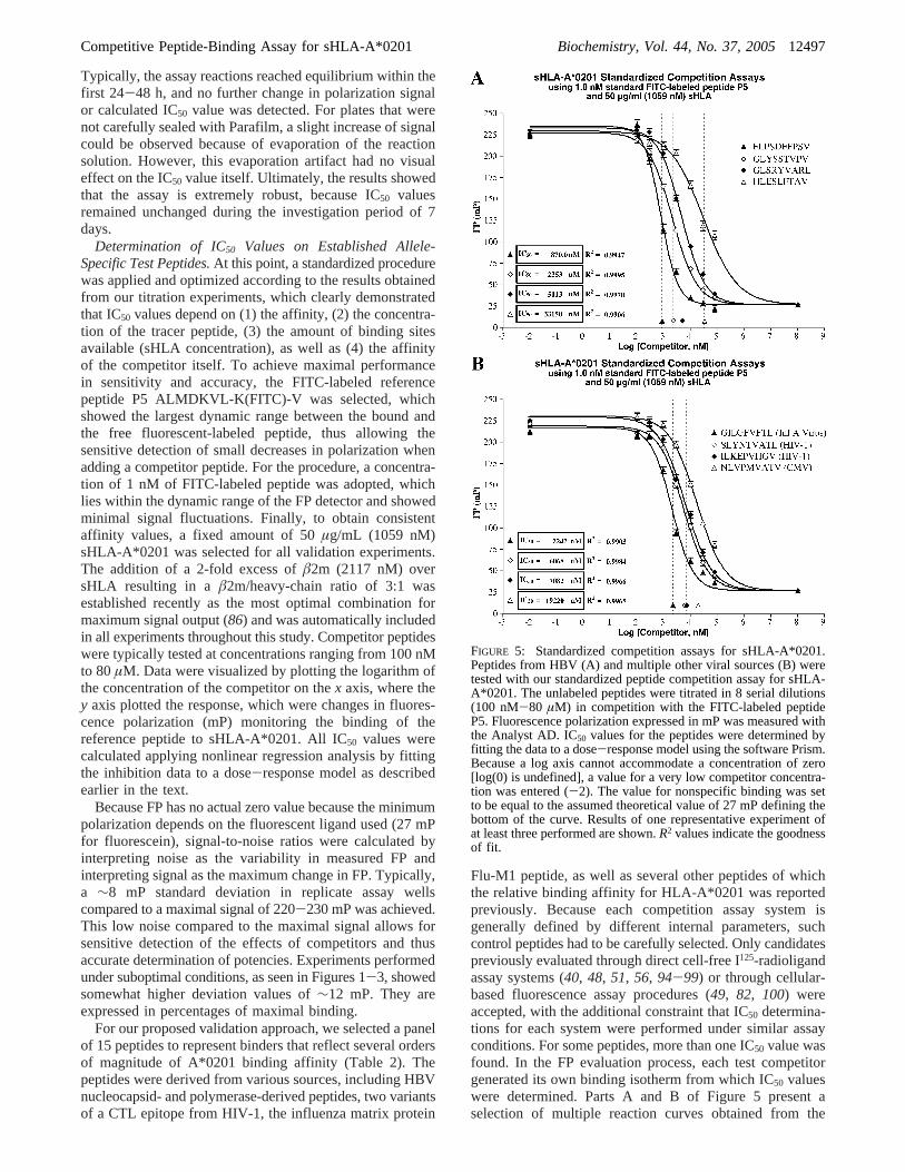
Flu-M1 peptide, as well as several other peptides of whichthe relative binding affinity for HLA-A*0201 was reportedpreviously. Because each competition assay system isgenerally defined by different internal parameters, suchcontrol peptides had to be carefully selected. Only candidatespreviously evaluated through direct cell-free I125-radioligandassay systems (40, 48, 51, 56, 94-99) or through cellular-based fluorescence assay procedures (49, 82, 100) wereaccepted, with the additional constraint that IC50 determina-tions for each system were performed under similar assayconditions. For some peptides, more than one IC50 value wasfound. In the FP evaluation process, each test competitorgenerated its own binding isotherm from which IC50 valueswere determined. Parts A and B of Figure 5 present aselection of multiple reaction curves obtained from the
FIGURE 5: Standardized competition assays for sHLA-A*0201.Peptides from HBV (A) and multiple other viral sources (B) weretested with our standardized peptide competition assay for sHLA-A*0201. The unlabeled peptides were titrated in 8 serial dilutions(100 nM-80 µM) in competition with the FITC-labeled peptideP5. Fluorescence polarization expressed in mP was measured withthe Analyst AD. IC50 values for the peptides were determined byfitting the data to a dose-response model using the software Prism.Because a log axis cannot accommodate a concentration of zero[log(0) is undefined], a value for a very low competitor concentra-tion was entered (-2). The value for nonspecific binding was setto be equal to the assumed theoretical value of 27 mP defining thebottom of the curve. Results of one representative experiment ofat least three performed are shown.R2 values indicate the goodnessof fit.
Competitive Peptide-Binding Assay for sHLA-A*0201 Biochemistry, Vol. 44, No. 37, 200512497

competition experiments, whereas Table 2 summarizesassessed IC50 values for the peptides along with their exactamino acid sequences. All test peptides were able to inhibitat least 50% binding of the FITC-labeled reference peptidecovering a spectrum of HLA A*0201 binding affinitiesspanning over 4 orders of magnitude, with IC50 valuesranging from 500 to 365 000 nM. To mention is that only apartial competition curve was observed for the weak binderDLVHFASPL over the concentration range used, andtherefore, the IC50 value is given as an approximation. Nocompetition was detected in the case of using irrelevantunlabeled competitor peptides (data not shown).
To be in line with previously published results, we usedthe cell-free I125-radioligand assay systems and the cellular-based fluorescence assay procedures as guidelines to definean FP-based classification system (Table 3), where peptideswith an FP-based IC50 value of 5000 nM and lower wereconsidered high-affinity binding, 5000-50 000 nM IC50
values were considered medium-affinity binding, 50 000-1 000 000 nM IC50 values were judged low-affinity binding,and IC50 values above 1 mM were regarded as no binder.Additionally, low-affinity binders were further subdividedinto a low- (50 000-350 000 nM) and very low-affinitycategory (350 000-1 000 000 nM) because quantitativeanalysis of very low binders was usually less accurate andgenerally based on only a few data points at the upper endof the dose-response curve. According to our FP-basedclassification system, eight test peptides belonged to the high-affinity category; six to the medium, and one was identifiedas a very low binder.
Epitope Screening.Recent reports show that peptidespossessing good binding characteristics are a prerequisite forsuccessful candidate CTL epitopes (51, 52). In this matter,the availability of a high-throughput peptide screeningmethod would be practical to discover novel MHC-restrictedepitopes by screening large panels of candidate peptides. Totest the feasibility of such a high-throughput approach, weconverted our standard competition assay procedure into arapid screening process with the ultimate goal of identifyingpeptides not capable of binding to sHLA-A*0201. At aselected high threshold concentration of 80µM, various
peptides were used for this screen test including the sameA2-specific peptides utilized earlier in addition to the B7-specific peptides LVMAPRTVL (HLA-B*0702 signal se-quence) (68, 101-104) and IPSYKKLIM (prostatic acidphosphatase precursor) (105). Experimentally, each peptidecandidate (80µM) was incubated with activated sHLA-A*0201 (50µg/mL; 1059 nM) in the presence of 1 nM FlTC-labeled reference peptides P5 and excessâ2m (2117 nM)and peptide/MHC interactions were monitored over time.Final equilibrium polarization levels were transformed inpercent inhibition to indicate the extent of binding to sHLA-A*0201. Representative screening data are shown in Figure6. As expected, all A2-specific peptides showed positivescreening results, whereas both B7-specific peptides werenot able to efficiently compete against the FITC-labeledreference peptide. Statistically, no significant differenceamong screening values for high-affinity peptides wasdetected, showing an average inhibition level of 97.2( 1.9%.This observation can be explained by the fact that high-affinity peptides reach complete inhibition below the 80µMthreshold concentration causing saturation of the system. Assuch, a systematic ranking of these candidate epitopes withinthis affinity category is not possible. However, a correct orderwithin this group can be obtained by either screening at alower threshold concentration or by performing a completeIC50 analysis as shown in Figure 5. In contrast, the rank orderfor medium- to low-affinity peptides was consistent with ourprevious results, allowing a ranking procedure without furtheranalysis. Most important, the screening procedure clearlyshowed only minor to no inhibition capacity for the B7-specific peptides LVMAPRTVL and IPSYKKLIM, respec-tively, which provides the key for future screening applica-tions, allowing the immediate identification of negativebinders and their elimination from subsequent functionalstudies.
System-System Affinity Comparison.To determine whetherthe peptide-binding data that we have obtained is in generalagreement with other studies in addition to be able to betterjudge the quantitative differences of IC50 values of our setof congeners, FP-derived binding values were compared tothe peptide-binding data extracted from the literature. To
Table 3: Definition of IC50 Categories for Different Assay Systems
category
assay systems high affinity medium affinity low affinity very low affinity no affinity references
StD FP comp assay
< 5000 50 000 350 000 1 000 000IC50 (nM)
5000 50 000 350 000 1 000 000 >
< 3.7 4.7 5.5 6.0log(IC50; nM)
3.7 4.7 5.5 6.0 >
cell-free I125 radioassaysa
< 50 500 50 000IC50 (nM)
34, 48, 81, 9950 500 50 000 >
< 1.7 2.7 4.7log(IC50; nM)
1.7 2.7 4.7 >
cellular fluorescence assaysa
< 5000 15 000 1 000 000IC50 (nM)
49, 66, 100, 1185000 15 000 1 000 000 >
< 3.7 4.2 6.0 log(IC50; nM)3.7 4.2 6.0 >
a Border values are defined according to published categories; the very low-affinity category was undefined.
12498 Biochemistry, Vol. 44, No. 37, 2005 Buchli et al.

simplify the problem of direct comparison of absolute IC50
values, all affinity determinations were transformed loga-rithmically and plotted against each other in a log-logformat. Results displayed strong linear correlations through-out the measurable range between the FP format and thecell-free I125-radioligand assay system (Figure 7A) as wellas between the cellular-based fluorescence assay procedure(Figure 7B). Regression analysis delivered a regression linefor the cell-free I125-radioligand assay system with a slopeand intercept of 0.37 and 2.91, respectively. A similar linearfirst-order relationship was found for the cellular-basedfluorescence assays with a slope of 0.55 and an intercept of1.45, respectively. Specific outcomes were subdivided intotwo groups: very well-correlating peptides with an overallcorrelation factor 0.86 and 0.82, respectively, and poorlycorrelating peptides, which contributed to a much lowercorrelation factor of only 0.71 and 0.62. The occurrence ofoutliers was, however, not surprising, considering the varietyof peptide-binding values collected from independent ex-perimental procedures. Overall, the difference in performancebetween these particular systems seems to be only minor,considering the highly selective choice of comparative data.Conclusively, such an attribute suggests that accurate as-sessment of peptide binding can be obtained and the FP-based assay can be used for successful validation of unknownpeptide candidates for their potential to bind sHLA-A*0201molecules.
DISCUSSION
The identification of HLA-restricted CTL epitopes hasimportant implications for preventive and/or therapeuticapplications in infectious diseases or neoplasias. As such, abody of knowledge enhancing the ability to identify anddesign peptides that bind across many HLA types hasemerged over the past decade, and many different method-ologies have been reported with the common theme of
assessing the ability of synthetically defined peptide epitopesto associate with specific HLA class-I complexes (Figure8). A common approach for the identification of tumorantigen/virus-derived sequences recognized by CTLs consistsof screening cDNA-based expression libraries (106, 107) orvarious forms of combinatorial synthetic peptide libraries ina cytotoxicity assay format (108-110). In parallel, proteomicapproaches use high-performance liquid chromatographyfractionation and mass spectrometry sequencing for identi-fication of MHC peptides eluted from normal cells of specifictissues or presented by particular MHC alleles (68, 87, 101,111, 112). This nondifferential method can also be used foranalysis of peptides recovered from primary tumor cells andfrom cells involved with pathologies other than cancer, suchas autoimmune diseases and viral infections, with the aimof identifying peptides of significance for treating thesediseases. More recently, mapping studies were performedwith the primary intent of characterizing differences betweenpeptide elution maps, such as between pathogenicallyinfected versus uninfected cell lines (113). Differentialproteomics approaches can also be used for comparisons ofMHC peptide patterns induced by mutations, as a result ofcancer induction or metastatic progression, induced bychanges in cell-growth conditions or stress. Also verycommon is the utilization of computer-based algorithms,which became quit common to select immunological targetsfor T-cell recognition. This predictive approach basicallylooks for nonameric or decameric peptide sequences withina target protein, which may potentially bind to MHC class-Imolecules. The matrixes are usually based on a previouslydetermined motif and/or individual ligand sequences (89,114, 115). Overall, these procedures are often lengthy, labor-intensive, and not free from pitfalls. Therefore, the identifica-tion of tumor- and virus-specific CTL epitopes has proceededat a much lower rate than that of the corresponding encodinggenes or proteins, and CTL epitopes derived from tumor
FIGURE 6: Epitope screening at a selected high threshold concentration of 80µM. Our panel of A2-specific peptides including two negativecontrols related to HLA allele B7 was screened at a single threshold concentration applying our peptide competition assay for sHLA-A*0201. The unlabeled peptides (80µM) were incubated with a constant concentration of activated sHLA-A*0201 (50µg/mL; 1059 nM),excessâ2m (2117 nM), and 1 nM FlTC-labeled reference peptides P5. After equilibrium was reached, fluorescence polarization was measuredwith the Analyst AD and transformed in percent inhibition relative to the control reaction without the competitor. Results demonstrate thefeasibility of a high-throughput approach for epitope discovery enabling the screening of a large number of peptides with the goal toidentify high-affinity binding peptides.
Competitive Peptide-Binding Assay for sHLA-A*0201 Biochemistry, Vol. 44, No. 37, 200512499

antigens identified several years ago remain undefined.Consequently, there exists a need for novel approaches infinding validated, well-characterized MHC peptides for aprotein of interest. Our biochemical approach, using FP-basedpeptide competition, offers a new tool set of assays, allowingepitope discovery and validation (Figure 8). Generally,competition assays measure the binding of a labeled ligand(also called reference or tracer peptide) in the presence ofvarious concentrations of an unlabeled ligand (also calledthe competitor or inhibitor) to HLA. As each competitorgenerates its own binding isotherm, an affinity dependentIC50 value, the concentration of competitor necessary todisplace 50% of the labeled ligand, can be obtained anddirectly compared to other values derived from the samesystem. The advantage of this approach is that, because onlya reference ligand is labeled, an adverse effect on affinitypotentially caused by the labeling process does not affectthe comparison of the unlabeled ligands. This type of assayis not only conceptually simple but also a sensitive and mostviable alternative for high-throughput applications.
A high-throughput epitope discovery process normallybegins with a rapid screening for large numbers of peptides,tested at a selected high threshold concentration with the goal
of eliminating peptides, which are not capable to bind tosHLA. Failing fast and cheap is highly desirable in the drugdiscovery world, meaning that less time and expense iswasted on compounds that would not have passed the next-stage hurdles because of the lack of binding. As seen inFigure 6, our newly developed epitope screening approachis highly flexible to identify peptides from any protein ofinterest, making this approach a key factor in early epitopediscovery. The measurements provide data of very highprecision and reproducibility.
High-affinity binding is thought to be a critical factorcontrolling immunogenicity of peptides (98, 116). Withinthe epitope discovery process, characterization of peptide andMHC interactions can be achieved using the same plate-based assay format with slightly different assay conditions.This validation approach enables the determination of theinhibitory concentration IC50 on positively identified peptidecandidates (Figure 5), with the goal to optimize data qualityby higher resolution analysis and also to deliver compre-hensive information for assay development stages (Figures1-3). A common approach is the ranking of identifiedpeptides according to their immunogenic potential with theassumption that high-affinity binding peptides are preferredover low-affinity candidates. However, definitions of targetidentification vary. In general, identification falls along aspectrum ranging from simply cataloging a molecule’sexistence to ascertaining a therapeutically relevant functionin cell and animal models. Identification always comes backto unbiased searches, those that do not skew results towarda certain subset of targets. The quality of anyone data set issuch that you can never choose the top one or two and beconfident that the selection criteria have actually led you tothe one or two best candidates. Therefore, any one of thesecandidates has generally the potential to be successful asvaccines. In addition, the IC50 determination approach is alsoused to evaluate binding results achieved after introductionof sequence alterations thought to improve the potency ofthe peptide.
The FP-based technology provides a range of additionalapproaches to further analyze and validate epitope targetssuch as the characterization of the fluorescent-labeled versionof the peptide of interest (86), allowing real-time kineticmeasurements and determination of equilibrium dissociationconstants (Kd). Furthermore, with the availability of morethan 50 of the most common sHLA alleles, direct experi-mental detection of overlapping peptide-binding capacitiesamong this large set of alleles became feasible, reflectingthe ability of MHC class-I alleles with genetically more orless distinct peptide-binding sites to share the binding ofidentical peptides (86). Classification of allele overlappingpeptide-binding specificities may become an important issuein vaccine design, with direct implications concerningpopulation coverage. Dependent upon individual alleliccomposition, it is import to identify the binding capacity ofsingle peptides to determine their usefulness for treating alarger subset of patients who express the MHC alleles thatare capable of binding that specific peptide.
Overall, several lines of evidence, both at the biologicaland functional level, emphasize the biologic relevance ofpeptide-binding assays in the identification and evaluationof potential peptide candidates (50-53). Our binding assayscomprise a number of significant advantages compared to
FIGURE 7: System-system affinity comparison using a variety ofestablished peptide epitopes. The relationship between peptide-binding affinity determinations for the HLA class-I moleculeA*0201 assessed through the FP system and values from (A) directcell-free I125-radioligand assay systems or (B) cellular-basedfluorescence assay procedures was evaluated. Logarithmic IC50values were graphed against each other, and a correlation factorwas determined using linear regression analysis. Affinity valuesare distributed over several orders of magnitude. The classificationof the peptide-binding affinity into high (H), medium (M), low (L),and very low (VL) are comparable to the classifications publishedby other investigators using the same reference peptides. In addition,specific outcomes were subdivided into two groups: very well-correlating peptides ([) and poorly correlating peptides (*),contributing to much lower correlation factors.
12500 Biochemistry, Vol. 44, No. 37, 2005 Buchli et al.

existing other HLA class-I binding assays. The mostimportant features are the excellent reproducibility and highsensitivity. Good reproducibility is a key parameter inscreening extensive sets of peptides for their affinity to HLA,and crucial information is derived from comparisons ofvarious peptides. Furthermore, FP is unique among methodsused to analyze molecular binding because it gives a direct,nearly instantaneous measurement of a peptide tracer’sbound/free ratio in solution. A truly homogeneous technique,which does not require the separation of bound and freespecies, makes the assay a very simple one-step procedure,which is particularly appropriate for high-throughput screen-ing where all reagents remain in solution. Such assays areeasily automated and have the ability to rapidly screen wholelibraries for peptide candidates. Methods that depend onseparation are not only more time-consuming, but they alsodisturb the reaction equilibrium and therefore prevent ac-curate quantification of binding. In addition, it is worthwhileto note that the assay is nonradioactive and can thus beperformed in virtually any laboratory without the risk ofradioactive contamination.
A common practical problem encountered experimentallyin a variety of competitive binding studies is the lack ofstandardization, seldomly allowing direct comparison of IC50
values between different assay approaches. As visualized inTables 2 and 3, differences for IC50 determinations betweenchosen systems range from 2- to 900-fold when comparingFP data to the I125-radioligand assay systems and 0.2- to5-fold when compared to the cellular-based fluorescenceassay systems. As such, it seems apparent that the task ofIC50 determinations by competitive ligand-binding studiesrequires a more standardized approach than has been initiallyconsidered adequate, justifying the further development ofsuch studies, particularly in view of identifying new potentialepitopes for vaccine development. Therefore, as a first steptoward a more robust approach, we attempted to better assess
the factors influencing competition binding curves andconsequently IC50 values. One of the most critical factorsfound to affect IC50 values is the HLA molecule itself, whichwas identified as generally the most variable assay compo-nent. The usage of HLA derived from homozygous cell lines,as lysate or intact cells, is common practice in competitionassay studies (56, 66, 79, 82, 83). However, the problem ofestablishing suitable models and reliable data for MHC class-I/peptide interactions is vastly complicated when a hetero-geneity of alleles is present. In some cases, antibodies againstan individual allele are available to achieve more specificitybut required specialized purification procedures, which arecumbersome, time-consuming, and usually do not deliversufficient quantities for experiments in a large scale. Anotherfactor that influences the efficiency and reproducibility instudies utilizing HLA-expressing cell lines is the level ofclass-I expression that generally varies from cell line to cellline. Because the functional response to a ligand is relatedto receptor density, this variability can lead to a rather widerange of variation, which is not suitable for reliable datacomparison. Therefore, the most important advantage ofutilizing recombinant sHLA molecules produced in cell linesthat do not spontaneously express HLA (“null” cell lines)resides in the capacity to standardize the amount of sHLAemploying sHLA preparations of defined specificity andpurity, as well as exhibit excellent stability on storage.
When we focused on standardization issues, a strikingexample of the effect of sHLA concentration on the IC50
values of unlabeled peptides is shown in Figure 3. It isevident that increasing the concentration of sHLA markedlyincreased the concentration of unlabeled competitor requiredto inhibit 50% of the binding of FITC-labeled referencepeptide resulting in an underestimation of the potency of thedisplacing ligand. Therefore, to achieve experimental ac-curacy and reproducibility, it is necessary to work with astandardized quantity of sHLA molecules. The importance
FIGURE 8: Epitope discovery and validation tools. The upper part of the scheme summarizes various methods currently applied in CTL-epitope discovery. The lower part shows approaches feasible for epitope validation using FP-based technologies, which are subdivided intosystems using nonlabeled versus labeled peptides as validation objects. Methodologies, which use standardized FP-based peptide-bindingassays for analysis, are circled.
Competitive Peptide-Binding Assay for sHLA-A*0201 Biochemistry, Vol. 44, No. 37, 200512501

of receptor concentration in determining relative potenciesof competitive inhibitors has been described earlier (91).Furthermore, it was demonstrated that direct binding studiesof ligand-receptor interactions of high affinity are subjectto artifactual distortions because of the need to utilize highconcentrations of the receptor. Therefore, it would beadvantageous to design competition experiments such thatthe [sHLA]/Kd ratio is much lower than 0.1. However, suchconditions do not seem experimentally approachable for FP-based peptide-binding experiments, because relatively highconcentrations of sHLA receptors are needed to yield asignificant change in polarization. Consequently, moreinhibitor is required to see a 50% drop in the amount oftracer ligand during competition. The result is that theobserved IC50 values are an overestimation of the truedissociation constant for the unlabeled ligand (Ki). Overall,the FP system is affected by severe competitor depletion (90),explaining why IC50 values are several orders of magnitudehigher than theirKi values, an effect also seen in other assaysystems (49, 66, 82). Because of this depletion effect, thehistorically used method of Cheng and Prusoff (117),transforming IC50 values intoKi values, is not applicablebecause the necessary substitution of total concentrations forfree concentrations cannot be made without introducingsignificant errors in the calculation ofKi. In addition, thepresence of unknown amounts of endogenous, class-I-associated peptides that could potentially compete with thetracer and test peptide during the course of the assay wouldeven further complicate such mathematical transformations.Nevertheless, results derived from dose-response curvesremain extremely useful in comparing relative affinities ofligands to the HLA receptor and in determining whether theligand of interest in a series of compounds can cause thesame maximal response (indicative of a closely similarinteraction with the recognition site) as other ligands.
In addition to the effect of the HLA concentration,variations in the choice of the tracer peptide and theinconsistent usage of tracer concentrations are also contribut-ing factors for inconsistent data output. The impact ofdifferent reference peptides on IC50 values shown in Figure1 clearly indicates that the development of a standardizedassay necessitates the consistent usage of the same referencepeptide to deliver reproducible results. As such, a criticalinitial step toward development of any sHLA assay is thechoice of a single FITC-labeled peptide candidate able tospecifically bind to the allele of interest. Because of thespecific nature of competition assays, the selection processfor a suitable tracer involves considerations relating moreto the chemical rather than the biological nature of thecandidate. Because pFITC-labeled reference peptides are notunder investigation in these assays, maintaining the nativereactivity of the candidate reference peptides was notnecessary. Therefore, to achieve most optimal assay perfor-mance qualities, the highest priorities are normally given tofeatures such as stability and affinity of the design, as wellas dynamic range of the polarization signal allowing sensitivedetection of a small decrease in polarization upon the additionof a competitor peptide. Extensive analysis showed that thepFITC ligand ALMDKVL-K(FITC)-V (P5), an artificiallydesigned peptide whose primary sequence was derived fromsequence pools of naturally processed peptides, best met therequirements for an optimal A*0201 tracer candidate (86)
by showing a very high stability and a unique dynamic rangeof over 215 mP.
Furthermore, an optimal pFITC reference concentrationhad to be selected, to ascertain sensitivity but also guarantee-ing maximal dynamic range. Unlike classical binding curveprofiles, FP signals are greater for low ligand concentrationbecause both the bound and free fluorescent ligand contributeto the final signal. Consequently, to achieve maximal changein polarization, it is necessary to use low fluorescent ligandconcentrations because increasing pFITC concentrationswould require increasing “free” sHLA to ensure that the samefraction of ligand is bound. Therefore, a low concentrationof 1 nM FITC-labeled peptide was chosen, which enables ahigh ratio between maximum and minimum FP signals andalso displays low standard deviation values. To control theaccuracy of all fluorescent peptide concentrations used withinthis study, concentration values were normalized by compar-ing the molar fluorescence intensity of each fluorophore-labeled peptide with the FITC fluorophore molecule itself.The importance of this task should not be underestimated,because inaccurate assignment of concentrations can causehigh IC50 fluctuations. As shown in Figure 2, applying higherpFITC concentrations took larger concentrations of unlabeledpeptide to compete for half of the binding sites and thereforeincreased the IC50 values. According to the steepness of theregression line, it became obvious that a(1 nM variance inconcentration will cause a(627 nM variance in the IC50
value.Another important step of our study besides standardiza-
tion was to further explore the binding specificity of the HLAA*0201-restricted FP-based competition assay. For thisreason, a panel of well-defined HLA class-I ligands fromvarious sources covering a broad range of binding affinitieswas tested (Table 2). These peptides were almost all of viralorigin except for a single peptide derived from an RNA-dependent helicase p72. Results showed that, in all cases,the previously known A*0201 specificity was confirmed.More importantly, the assay was capable of measuring IC50
values over a range of 4 orders of magnitude (from 500 to365 000 nM), quantitatively characterizing the bindingstrength of various existing sequences. Because dose-response curves are unable to confirm or disprove any modeof interaction of peptides with sHLA, they are only usefulin comparing relative affinities of ligands with the HLAreceptor as well as determining whether the ligand of interestin a series of compounds can cause the same maximalresponse (indicative of a closely similar interaction with therecognition site) as other peptides. Therefore, a commonapproach for peptide evaluation is the quantitative rankingof identified peptides according to arbitrarily defined cat-egories along a spectrum of high-, medium-, and low-affinitybinding to ascertain a therapeutically relevant function incell and animal models. Built upon existing models earlierdefined by Sette et al. (34, 48, 56, 81, 94, 99) and van derBurg et al. (49, 66, 82, 100, 118), an FP-based IC50 valueclassification scheme was created (Table 3). When wefocused on the correlation between peptide-binding affinityand immunogenicity, all control peptides selected withknown CTL activity showed, as expected, a high to medium-high affinity to sHLA-A*0201. Among our viral controls,the HBV-derived epitope FLPSDFFPSV was found todisplay very high-affinity values. As one of the most
12502 Biochemistry, Vol. 44, No. 37, 2005 Buchli et al.
referenced peptides found in the literature, FLPSDFFPSVis known for high-affinity binding to A*0201 (34, 56, 81,93) as well as the ability to induce potent and specific CTLresponses (119). Additional members of the high-affinitycategory were the peptides SLYNTVATL, another well-studied HIV-derived CTL epitope (120-122), and theinfluenza matrix peptide GILGFVFTL, found to be a majortarget of influenza-specific CTL, both in humans (123-129)and in HLA-A2 transgenic mice (130).
As final step in validating our FP system, IC50 determina-tions were compared to the two quantitative assay systemsmost commonly referred to in the literature, the direct cell-free I125-radioligand assay (40, 48, 51, 56, 94-99) and thecellular-based fluorescence assay (49, 82, 100). A significantcorrelation between the logarithm of the IC50 values fromour FP-based system and values from the two referencesystems was found by linear regression analysis (Figure 7).During analysis, some outlier values were noted for bothsystems, a discrepancy certainly expected considering theselection of affinity data from independent assay sources.As such, single differences found in affinity might rather bedue to the lack of normalization in obtaining binding valuesas discussed above, which resembles a current problem inanalysis of competition data between individual assaysystems. In addition, the solubility of the peptide as well asthe challenge of accurate weighing of solid peptides mayfurther contribute to the inaccurate determination of bindingvalues. Particularly, dissolving peptides is a critical stepbecause the wrong choice of solvents and methodology canlead to inaccurate or complete lack of performance. Asdescribed earlier within the text, the usage of DMSO orDMF, which are widely accepted in many end-use applica-tions for peptides, seems most appropriate for biochemicalpeptide-binding assay procedures. Nevertheless, results ob-tained clearly suggest that accurate assessment of peptidebinding can be obtained by using our novel FP-based assayprocedure.
In summary, our standardized FP-based peptide-bindingassay provides uniquely efficient means of identifying andevaluating immune target molecules that are recognized bycytotoxic T cells. The wealth of newly available genomicsequence information provides a superb source for theidentification of novel targets to use as the basis of the designof new specific and selective therapeutic agents and diag-nostics. The concept of our assays can be adapted forbasically every HLA class-I allele of interest. Although thedata presented here were generated using sHLA-A*0201,additional studies indicate that this approach works equallywell with other sHLA alleles. Numerous significant MHC-restricted epitopes have already been defined for B*0702(113), and more studies are on the way. Therefore, the presentreport can also be read as an instruction for the developmentof class-I binding assays that are still lacking. This assay’scapacity for high-throughput evaluation of peptide/HLAinteractions, coupled with the ability of the immune systemto recognize virus-infected or cancerous cells, will open newpossibilities to identify an ever-burgeoning number ofimmunologically active peptide epitopes for the developmentof vaccines to treat or prevent various types of infectiousdiseases or cancers. Furthermore, the FP assay has greatpotential to provide new database information and help toincrease missing knowledge, which can be utilized for the
development of more sophisticated algorithms to predict andquantify the binding affinity to HLA class-I molecules.
ACKNOWLEDGMENT
We thank Mrs. Beatrice Buchli for assistance with thepreparation of the manuscript.
REFERENCES
1. Byrne, J. A., and Oldstone, M. B. (1984) Biology of clonedcytotoxic T lymphocytes specific for lymphocytic choriomeningitisvirus: Clearance of virusin ViVo, J. Virol. 51, 682-686.
2. Harty, J. T., and Bevan, M. J. (1992) CD8+ T cells specific for asingle nonamer epitope of Listeria monocytogenes are protectivein ViVo, J. Exp. Med. 175, 1531-1538.
3. Heslop, H. E., Ng, C. Y., Li, C., Smith, C. A., Loftin, S. K.,Krance, R. A., Brenner, M. K., and Rooney, C. M. (1996) Long-term restoration of immunity against Epstein-Barr virus infectionby adoptive transfer of gene-modified virus-specific T lympho-cytes,Nat. Med. 2, 551-555.
4. Jamieson, B. D., Butler, L. D., and Ahmed, R. (1987) Effectiveclearance of a persistent viral infection requires cooperationbetween virus-specific Lyt2+ T cells and nonspecific bonemarrow-derived cells,J. Virol. 61, 3930-3937.
5. Kulkarni, A. B., Collins, P. L., Bacik, I., Yewdell, J. W., Bennink,J. R., Crowe, J. E., Jr., and Murphy, B. R. (1995) Cytotoxic Tcells specific for a single peptide on the M2 protein of respiratorysyncytial virus are the sole mediators of resistance induced byimmunization with M2 encoded by a recombinant vaccinia virus,J. Virol. 69, 1261-1264.
6. Lin, Y. L., and Askonas, B. A. (1981) Biological properties of aninfluenza A virus-specific killer T cell clone. Inhibition of virusreplicationin ViVo and induction of delayed-type hypersensitivityreactions,J. Exp. Med. 154, 225-234.
7. McMichael, A. J., Gotch, F. M., Noble, G. R., and Beare, P. A.(1983) Cytotoxic T-cell immunity to influenza,N. Engl. J. Med.309, 13-17.
8. Nayersina, R., Fowler, P., Guilhot, S., Missale, G., Cerny, A.,Schlicht, H. J., Vitiello, A., Chesnut, R., Person, J. L., Redeker,A. G. et al. (1993) HLA A2 restricted cytotoxic T lymphocyteresponses to multiple hepatitis B surface antigen epitopes duringhepatitis B virus infection,J. Immunol. 150, 4659-4671.
9. Riddell, S. R., Watanabe, K. S., Goodrich, J. M., Li, C. R., Agha,M. E., and Greenberg, P. D. (1992) Restoration of viral immunityin immunodeficient humans by the adoptive transfer of T cellclones,Science 257, 238-241.
10. Schmitz, J. E., Kuroda, M. J., Santra, S., Sasseville, V. G., Simon,M. A., Lifton, M. A., Racz, P., Tenner-Racz, K., Dalesandro, M.,Scallon, B. J., Ghrayeb, J., Forman, M. A., Montefiori, D. C.,Rieber, E. P., Letvin, N. L., and Reimann, K. A. (1999) Controlof viremia in simian immunodeficiency virus infection by CD8+
lymphocytes,Science 283, 857-860.11. Yap, K. L., Ada, G. L., and McKenzie, I. F. (1978) Transfer of
specific cytotoxic T lymphocytes protects mice inoculated withinfluenza virus,Nature 273, 238-239.
12. Hill, A. V., Elvin, J., Willis, A. C., Aidoo, M., Allsopp, C. E.,Gotch, F. M., Gao, X. M., Takiguchi, M., Greenwood, B. M.,Townsend, A. R. et al. (1992) Molecular analysis of the associationof HLA-B53 and resistance to severe malaria,Nature 360, 434-439.
13. Le, T. P., Church, L. W., Corradin, G., Hunter, R. L., Charoenvit,Y., Wang, R., de la Vega, P., Sacci, J., Ballou, W. R., Kolodny,N., Kitov, S., Glenn, G. M., Richards, R. L., Alving, C. R., andHoffman, S. L. (1998) Immunogenicity of Plasmodium falciparumcircumsporozoite protein multiple antigen peptide vaccine for-mulated with different adjuvants,Vaccine 16, 305-312.
14. Feltkamp, M. C., Vreugdenhil, G. R., Vierboom, M. P., Ras, E.,van der Burg, S. H., ter Schegget, J., Melief, C. J., and Kast, W.M. (1995) Cytotoxic T lymphocytes raised against a subdominantepitope offered as a synthetic peptide eradicate human papillo-mavirus type 16-induced tumors,Eur. J. Immunol. 25, 2638-2642.
15. Iwasaki, A., and Barber, B. H. (1998) Induction by DNAimmunization of a protective antitumor cytotoxic T lymphocyteresponse against a minimal-epitope-expressing tumor,CancerImmunol. Immunother. 45, 273-279.
Competitive Peptide-Binding Assay for sHLA-A*0201 Biochemistry, Vol. 44, No. 37, 200512503

16. Kittlesen, D. J., Thompson, L. W., Gulden, P. H., Skipper, J. C.,Colella, T. A., Shabanowitz, J., Hunt, D. F., Engelhard, V. H.,Slingluff, C. L., Jr., and Shabanowitz, J. A. (1998) Humanmelanoma patients recognize an HLA-A1-restricted CTL epitopefrom tyrosinase containing two cysteine residues: Implicationsfor tumor vaccine development,J. Immunol. 160, 2099-2106.
17. Mayordomo, J. I., Loftus, D. J., Sakamoto, H., De Cesare, C. M.,Appasamy, P. M., Lotze, M. T., Storkus, W. J., Appella, E., andDeLeo, A. B. (1996) Therapy of murine tumors with p53 wild-type and mutant sequence peptide-based vaccines,J. Exp. Med.183, 1357-1365.
18. Melief, C. J. (1992) Tumor eradication by adoptive transfer ofcytotoxic T lymphocytes,AdV. Cancer Res. 58, 143-175.
19. Melief, C. J., Offringa, R., Toes, R. E., and Kast, W. M. (1996)Peptide-based cancer vaccines,Curr. Opin. Immunol. 8, 651-657.
20. Morgan, D. J., Kreuwel, H. T., Fleck, S., Levitsky, H. I., Pardoll,D. M., and Sherman, L. A. (1998) Activation of low avidity CTLspecific for a self-epitope results in tumor rejection but notautoimmunity,J. Immunol. 160, 643-651.
21. Muul, L. M., Spiess, P. J., Director, E. P., and Rosenberg, S. A.(1987) Identification of specific cytolytic immune responsesagainst autologous tumor in humans bearing malignant melanoma,J. Immunol. 138, 989-995.
22. Vierboom, M. P., Nijman, H. W., Offringa, R., van der Voort, E.I., van Hall, T., van den Broek, L., Fleuren, G. J., Kenemans, P.,Kast, W. M., and Melief, C. J. (1997) Tumor eradication by wild-type p53-specific cytotoxic T lymphocytes,J. Exp. Med. 186,695-704.
23. Vose, B. M., and Bonnard, G. D. (1982) Human tumour antigensdefined by cytotoxicity and proliferative responses of culturedlymphoid cells,Nature 296, 359-361.
24. Donnelly, J. J., Ulmer, J. B., Hawe, L. A., Friedman, A., Shi, X.P., Leander, K. R., Shiver, J. W., Oliff, A. I., Martinez, D.,Montgomery, D. et al. (1993) Targeted delivery of peptide epitopesto class I major histocompatibility molecules by a modifiedPseudomonasexotoxin,Proc. Natl. Acad. Sci. U.S.A. 90, 3530-3534.
25. Goletz, T. J., Klimpel, K. R., Leppla, S. H., Keith, J. M., andBerzofsky, J. A. (1997) Delivery of antigens to the MHC class Ipathway using bacterial toxins,Hum. Immunol. 54, 129-136.
26. Gooding, L. R., and O’Connell, K. A. (1983) Recognition bycytotoxic T lymphocytes of cells expressing fragments of the SV40tumor antigen,J. Immunol. 131, 2580-2586.
27. Kim, D. T., Mitchell, D. J., Brockstedt, D. G., Fong, L., Nolan,G. P., Fathman, C. G., Engleman, E. G., and Rothbard, J. B. (1997)Introduction of soluble proteins into the MHC class I pathway byconjugation to an HIV tat peptide,J. Immunol. 159, 1666-1668.
28. Moore, M. W., Carbone, F. R., and Bevan, M. J. (1988)Introduction of soluble protein into the class I pathway of antigenprocessing and presentation,Cell 54, 777-785.
29. Yewdell, J. W., and Bennink, J. R. (1990) The binary logic ofantigen processing and presentation to T cells,Cell 62, 203-206.
30. Cease, K. B., and Berzofsky, J. A. (1994) Toward a vaccine forAIDS: The emergence of immunobiology-based vaccine develop-ment,Annu. ReV. Immunol. 12, 923-989.
31. Celis, E., Tsai, V., Crimi, C., DeMars, R., Wentworth, P. A.,Chesnut, R. W., Grey, H. M., Sette, A., and Serra, H. M. (1994)Induction of anti-tumor cytotoxic T lymphocytes in normalhumans using primary cultures and synthetic peptide epitopes,Proc. Natl. Acad. Sci. U.S.A. 91, 2105-2109.
32. Cox, A. L., Skipper, J., Chen, Y., Henderson, R. A., Darrow, T.L., Shabanowitz, J., Engelhard, V. H., Hunt, D. F., and Slingluff,C. L., Jr. (1994) Identification of a peptide recognized by fivemelanoma-specific human cytotoxic T cell lines,Science 264,716-719.
33. Kast, W. M., Roux, L., Curren, J., Blom, H. J., Voordouw, A. C.,Meloen, R. H., Kolakofsky, D., and Melief, C. J. (1991) Protectionagainst lethal Sendai virus infection byin ViVo priming of virus-specific cytotoxic T lymphocytes with a free synthetic peptide,Proc. Natl. Acad. Sci. U.S.A. 88, 2283-2287.
34. Kast, W. M., Brandt, R. M., Sidney, J., Drijfhout, J. W., Kubo,R. T., Grey, H. M., Melief, C. J., and Sette, A. (1994) Role ofHLA-A motifs in identification of potential CTL epitopes inhuman papillomavirus type 16 E6 and E7 proteins,J. Immunol.152, 3904-3912.
35. Marchand, M., van Baren, N., Weynants, P., Brichard, V., Dreno,B., Tessier, M. H., Rankin, E., Parmiani, G., Arienti, F., Humblet,
Y., Bourlond, A., Vanwijck, R., Lienard, D., Beauduin, M.,Dietrich, P. Y., Russo, V., Kerger, J., Masucci, G., Jager, E., DeGreve, J., Atzpodien, J., Brasseur, F., Coulie, P. G., van derBruggen, P., and Boon, T. (1999) Tumor regressions observed inpatients with metastatic melanoma treated with an antigenicpeptide encoded by gene MAGE-3 and presented by HLA-A1,Int. J. Cancer 80, 219-230.
36. Oukka, M., Manuguerra, J. C., Livaditis, N., Tourdot, S., Riche,N., Vergnon, I., Cordopatis, P., and Kosmatopoulos, K. (1996)Protection against lethal viral infection by vaccination withnonimmunodominant peptides,J. Immunol. 157, 3039-3045.
37. Rosenberg, S. A., Yang, J. C., Schwartzentruber, D. J., Hwu, P.,Marincola, F. M., Topalian, S. L., Restifo, N. P., Dudley, M. E.,Schwarz, S. L., Spiess, P. J., Wunderlich, J. R., Parkhurst, M. R.,Kawakami, Y., Seipp, C. A., Einhorn, J. H., and White, D. E.(1998) Immunologic and therapeutic evaluation of a syntheticpeptide vaccine for the treatment of patients with metastaticmelanoma,Nat. Med. 4, 321-327.
38. Schulz, M., Zinkernagel, R. M., and Hengartner, H. (1991) Peptide-induced antiviral protection by cytotoxic T cells,Proc. Natl. Acad.Sci. U.S.A. 88, 991-993.
39. An, L. L., and Whitton, J. L. (1997) A multivalent minigenevaccine, containing B-cell, cytotoxic T-lymphocyte, and Thepitopes from several microbes, induces appropriate responsesinViVo and confers protection against more than one pathogen,J.Virol. 71, 2292-2302.
40. Ishioka, G. Y., Fikes, J., Hermanson, G., Livingston, B., Crimi,C., Qin, M., del Guercio, M. F., Oseroff, C., Dahlberg, C.,Alexander, J., Chesnut, R. W., and Sette, A. (1999) Utilizationof MHC class I transgenic mice for development of minigeneDNA vaccines encoding multiple HLA-restricted CTL epitopes,J. Immunol. 162, 3915-3925.
41. Tourdot, S., Oukka, M., Manuguerra, J. C., Magafa, V., Vergnon,I., Riche, N., Bruley-Rosset, M., Cordopatis, P., and Kosmato-poulos, K. (1997) Chimeric peptides: A new approach toenhancing the immunogenicity of peptides with low MHC classI affinity: Application in antiviral vaccination,J. Immunol. 159,2391-2398.
42. Disis, M. L., Gralow, J. R., Bernhard, H., Hand, S. L., Rubin, W.D., and Cheever, M. A. (1996) Peptide-based, but not wholeprotein, vaccines elicit immunity to HER-2/neu, oncogenic self-protein,J. Immunol. 156, 3151-3158.
43. Bjorkman, P. J., Saper, M. A., Samraoui, B., Bennett, W. S.,Strominger, J. L., and Wiley, D. C. (1987) Structure of the humanclass I histocompatibility antigen, HLA-A2,Nature 329, 506-512.
44. Bjorkman, P. J., Saper, M. A., Samraoui, B., Bennett, W. S.,Strominger, J. L., and Wiley, D. C. (1987) The foreign antigenbinding site and T cell recognition regions of class I histocom-patibility antigens,Nature 329, 512-518.
45. Rotzschke, O., Falk, K., Deres, K., Schild, H., Norda, M., Metzger,J., Jung, G., and Rammensee, H. G. (1990) Isolation and analysisof naturally processed viral peptides as recognized by cytotoxicT cells,Nature 348, 252-254.
46. Townsend, A. R., Rothbard, J., Gotch, F. M., Bahadur, G., Wraith,D., and McMichael, A. J. (1986) The epitopes of influenzanucleoprotein recognized by cytotoxic T lymphocytes can bedefined with short synthetic peptides,Cell 44, 959-968.
47. van Bleek, G. M., and Nathenson, S. G. (1990) Isolation of anendogenously processed immunodominant viral peptide from theclass I H-2Kb molecule,Nature 348, 213-216.
48. Sette, A., Vitiello, A., Reherman, B., Fowler, P., Nayersina, R.,Kast, W. M., Melief, C. J., Oseroff, C., Yuan, L., Ruppert, J. etal. (1994) The relationship between class I binding affinity andimmunogenicity of potential cytotoxic T cell epitopes,J. Immunol.153, 5586-5592.
49. van der Burg, S. H., Visseren, M. J., Brandt, R. M., Kast, W. M.,and Melief, C. J. (1996) Immunogenicity of peptides bound toMHC class I molecules depends on the MHC-peptide complexstability, J. Immunol. 156, 3308-3314.
50. Baker, B. M., Gagnon, S. J., Biddison, W. E., and Wiley, D. C.(2000) Conversion of a T cell antagonist into an agonist byrepairing a defect in the TCR/peptide/MHC interface: Implicationsfor TCR signaling,Immunity 13, 475-484.
51. Chen, W., Khilko, S., Fecondo, J., Margulies, D. H., andMcCluskey, J. (1994) Determinant selection of major histocom-patibility complex class I-restricted antigenic peptides is explainedby class I-peptide affinity and is strongly influenced by nondomi-nant anchor residues,J. Exp. Med. 180, 1471-1483.
12504 Biochemistry, Vol. 44, No. 37, 2005 Buchli et al.

52. Feltkamp, M. C., Vierboom, M. P., Kast, W. M., and Melief, C.J. (1994) Efficient MHC class I-peptide binding is required butdoes not ensure MHC class I-restricted immunogenicity,Mol.Immunol. 31, 1391-1401.
53. Kersh, G. J., Kersh, E. N., Fremont, D. H., and Allen, P. M. (1998)High- and low-potency ligands with similar affinities for theTCR: The importance of kinetics in TCR signaling,Immunity 9,817-826.
54. Rammensee, H. G., Falk, K., and Rotzschke, O. (1993) Peptidesnaturally presented by MHC class I molecules,Annu. ReV.Immunol. 11, 213-244.
55. Ressing, M. E., Sette, A., Brandt, R. M., Ruppert, J., Wentworth,P. A., Hartman, M., Oseroff, C., Grey, H. M., Melief, C. J., andKast, W. M. (1995) Human CTL epitopes encoded by humanpapillomavirus type 16 E6 and E7 identified throughin ViVo andin Vitro immunogenicity studies of HLA-A*0201-binding peptides,J. Immunol. 154, 5934-5943.
56. Sette, A., Sidney, J., del Guercio, M. F., Southwood, S., Ruppert,J., Dahlberg, C., Grey, H. M., and Kubo, R. T. (1994) Peptidebinding to the most frequent HLA-A class I alleles measured byquantitative molecular binding assays,Mol. Immunol. 31, 813-822.
57. Babbitt, B. P., Matsueda, G., Haber, E., Unanue, E. R., and Allen,P. M. (1986) Antigenic competition at the level of peptide-Iabinding,Proc. Natl. Acad. Sci. U.S.A. 83, 4509-4513.
58. Bouillot, M., Choppin, J., Cornille, F., Martinon, F., Papo, T.,Gomard, E., Fournie-Zaluski, M. C., and Levy, J. P. (1989)Physical association between MHC class I molecules and im-munogenic peptides,Nature 339, 473-475.
59. Werdelin, O. (1982) Chemically related antigens compete forpresentation by accessory cells to T cells,J. Immunol. 129, 1883-1891.
60. Schumacher, T. N., Heemels, M. T., Neefjes, J. J., Kast, W. M.,Melief, C. J., and Ploegh, H. L. (1990) Direct binding of peptideto empty MHC class I molecules on intact cells andin Vitro, Cell62, 563-567.
61. Hosken, N. A., and Bevan, M. J. (1990) Defective presentationof endogenous antigen by a cell line expressing class I molecules,Science 248, 367-370.
62. Townsend, A., Elliott, T., Cerundolo, V., Foster, L., Barber, B.,and Tse, A. (1990) Assembly of MHC class I molecules analyzedin Vitro, Cell 62, 285-295.
63. Luescher, I. F., Loez, J. A., Malissen, B., and Cerottini, J. C. (1992)Interaction of antigenic peptides with MHC class I molecules onliving cells studied by photoaffinity labeling,J. Immunol. 148,1003-1011.
64. Cerundolo, V., Alexander, J., Anderson, K., Lamb, C., Cresswell,P., McMichael, A., Gotch, F., and Townsend, A. (1990) Presenta-tion of viral antigen controlled by a gene in the major histocom-patibility complex,Nature 345, 449-452.
65. Storkus, W. J., Zeh, H. J., III, Salter, R. D., and Lotze, M. T.(1993) Identification of T-cell epitopes: Rapid isolation of classI-presented peptides from viable cells by mild acid elution,J.Immunother. 14, 94-103.
66. van der Burg, S. H., Ras, E., Drijfhout, J. W., Benckhuijsen, W.E., Bremers, A. J., Melief, C. J., and Kast, W. M. (1995) An HLAclass I peptide-binding assay based on competition for binding toclass I molecules on intact human B cells. Identification ofconserved HIV-1 polymerase peptides binding to HLA-A*0301,Hum. Immunol. 44, 189-198.
67. Townsend, A., Ohlen, C., Bastin, J., Ljunggren, H. G., Foster, L.,and Karre, K. (1989) Association of class I major histocompat-ibility heavy and light chains induced by viral peptides,Nature340, 443-448.
68. Huczko, E. L., Bodnar, W. M., Benjamin, D., Sakaguchi, K., Zhu,N. Z., Shabanowitz, J., Henderson, R. A., Appella, E., Hunt, D.F., and Engelhard, V. H. (1993) Characteristics of endogenouspeptides eluted from the class I MHC molecule HLA-B7determined by mass spectrometry and computer modeling,J.Immunol. 151, 2572-2587.
69. Elvin, J., Potter, C., Elliott, T., Cerundolo, V., and Townsend, A.(1993) A method to quantify binding of unlabeled peptides toclass I MHC molecules and detect their allele specificity,J.Immunol. Methods 158, 161-171.
70. Fahnestock, M. L., Tamir, I., Narhi, L., and Bjorkman, P. J. (1992)Thermal stability comparison of purified empty and peptide-filledforms of a class I MHC molecule,Science 258, 1658-1662.
71. Gnjatic, S., Bressac-de Paillerets, B., Guillet, J. G., and Choppin,J. (1995) Mapping and ranking of potential cytotoxic T epitopes
in the p53 protein: Effect of mutations and polymorphism onpeptide binding to purified and refolded HLA molecules,Eur. J.Immunol. 25, 1638-1642.
72. Bijlmakers, M. J., Neefjes, J. J., Wojcik-Jacobs, E. H., and Ploegh,H. L. (1993) The assembly of H2-Kb class I molecules translatedin Vitro requires oxidized glutathione and peptide,Eur. J. Immunol.23, 1305-1313.
73. Parker, K. C., DiBrino, M., Hull, L., and Coligan, J. E. (1992)Theâ2-microglobulin dissociation rate is an accurate measure ofthe stability of MHC class I heterotrimers and depends on whichpeptide is bound,J. Immunol. 149, 1896-1904.
74. Ottenhoff, T. H., Geluk, A., Toebes, M., Benckhuijsen, W. E.,van Meijgaarden, K. E., and Drijfhout, J. W. (1997) A sensitivefluorometric assay for quantitatively measuring specific peptidebinding to HLA class I and class II molecules,J. Immunol.Methods 200, 89-97.
75. Jensen, P. E., Moore, J. C., and Lukacher, A. E. (1998) A europiumfluoroimmunoassay for measuring peptide binding to MHC classI molecules,J. Immunol. Methods 215, 71-80.
76. Sigal, L. J., Berens, S., and Wylie, D. (1994) A lactate dehydro-genase (LDH)-based immunoassay for detection of cell surfaceantigens and its application to the study of MHC class I-bindingpeptides,J. Immunol. Methods 177, 261-268.
77. Buus, S., Stryhn, A., Winther, K., Kirkby, N., and Pedersen, L.O. (1995) Receptor-ligand interactions measured by an improvedspun column chromatography technique. A high efficiency andhigh throughput size separation method,Biochim. Biophys. Acta1243, 453-460.
78. Chen, B. P., and Parham, P. (1989) Direct binding of influenzapeptides to class I HLA molecules,Nature 337, 743-745.
79. Olsen, A. C., Pedersen, L. O., Hansen, A. S., Nissen, M. H., Olsen,M., Hansen, P. R., Holm, A., and Buus, S. (1994) A quantitativeassay to measure the interaction between immunogenic peptidesand purified class I major histocompatibility complex molecules,Eur. J. Immunol. 24, 385-392.
80. Khilko, S. N., Corr, M., Boyd, L. F., Lees, A., Inman, J. K., andMargulies, D. H. (1993) Direct detection of major histocompat-ibility complex class I binding to antigenic peptides using surfaceplasmon resonance. Peptide immobilization and characterizationof binding specificity,J. Biol. Chem. 268, 15425-15434.
81. Ruppert, J., Sidney, J., Celis, E., Kubo, R. T., Grey, H. M., andSette, A. (1993) Prominent role of secondary anchor residues inpeptide binding to HLA-A2.1 molecules,Cell 74, 929-937.
82. Kessler, J. H., Mommaas, B., Mutis, T., Huijbers, I., Vissers, D.,Benckhuijsen, W. E., Schreuder, G. M., Offringa, R., Goulmy,E., Melief, C. J., van der Burg, S. H., and Drijfhout, J. W. (2003)Competition-based cellular peptide binding assays for 13 prevalentHLA class I alleles using fluorescein-labeled synthetic peptides,Hum. Immunol. 64, 245-255.
83. Buus, S., Sette, A., Colon, S. M., Jenis, D. M., and Grey, H. M.(1986) Isolation and characterization of antigen-Ia complexesinvolved in T cell recognition,Cell 47, 1071-1077.
84. Checovich, W. J., Bolger, R. E., and Burke, T. (1995) FluorescencepolarizationsA new tool for cell and molecular biology,Nature375, 254-256.
85. Jameson, D. M., and Seifried, S. E. (1999) Quantification ofprotein-protein interactions using fluorescence polarization,Methods 19, 222-233.
86. Buchli, R., VanGundy, R. S., Hickman-Miller, H. D., Giberson,C. F., Bardet, W., and Hildebrand, W. H. (2004) Real-timemeasurement ofin Vitro peptide binding to soluble HLA-A*0201by fluorescence polarization,Biochemistry 43, 14852-14863.
87. Prilliman, K., Lindsey, M., Zuo, Y., Jackson, K. W., Zhang, Y.,and Hildebrand, W. (1997) Large-scale production of class I boundpeptides: Assigning a signature to HLA-B*1501,Immunogenetics45, 379-385.
88. Robbins, P. A., Garboczi, D. N., and Strominger, J. L. (1995)HLA-A*0201 complexes with two 10-Mer peptides differing atthe P2 anchor residue have distinct refolding kinetics,J. Immunol.154, 703-709.
89. Parker, K. C., Bednarek, M. A., and Coligan, J. E. (1994) Schemefor ranking potential HLA-A2 binding peptides based on inde-pendent binding of individual peptide side-chains,J. Immunol.152, 163-175.
90. Jacobs, S., Chang, K. J., and Cuatrecasas, P. (1975) Estimationof hormone receptor affinity by competitive displacement oflabeled ligand: Effect of concentration of receptor and of labeledligand,Biochem. Biophys. Res. Commun. 66, 687-692.
Competitive Peptide-Binding Assay for sHLA-A*0201 Biochemistry, Vol. 44, No. 37, 200512505

91. Rodbard, D. (1973) Mathematics of hormone-receptor interaction.I. Basic principles,AdV. Exp. Med. Biol. 36, 289-326.
92. Cheng, Y. C., and Prusoff, W. H. (1974) A new rapid assay formeasuring deoxycytidylate- and deoxythymidylate-kinase activi-ties,Anal. Biochem. 60, 545-550.
93. Kubo, R. T., Sette, A., Grey, H. M., Appella, E., Sakaguchi, K.,Zhu, N. Z., Arnott, D., Sherman, N., Shabanowitz, J., Michel, H.et al. (1994) Definition of specific peptide motifs for four majorHLA-A alleles, J. Immunol. 152, 3913-3924.
94. del Guercio, M. F., Sidney, J., Hermanson, G., Perez, C., Grey,H. M., Kubo, R. T., and Sette, A. (1995) Binding of a peptideantigen to multiple HLA alleles allows definition of an A2-likesupertype,J. Immunol. 154, 685-693.
95. Wentworth, P. A., Vitiello, A., Sidney, J., Keogh, E., Chesnut, R.W., Grey, H., and Sette, A. (1996) Differences and similarities inthe A2.1-restricted cytotoxic T cell repertoire in humans andhuman leukocyte antigen-transgenic mice,Eur. J. Immunol. 26,97-101.
96. Bertoletti, A., Costanzo, A., Chisari, F. V., Levrero, M., Artini,M., Sette, A., Penna, A., Giuberti, T., Fiaccadori, F., and Ferrari,C. (1994) Cytotoxic T lymphocyte response to a wild type hepatitisB virus epitope in patients chronically infected by variant virusescarrying substitutions within the epitope,J. Exp. Med. 180, 933-943.
97. Bertoni, R., Sidney, J., Fowler, P., Chesnut, R. W., Chisari, F.V., and Sette, A. (1997) Human histocompatibility leukocyteantigen-binding supermotifs predict broadly cross-reactive cyto-toxic T lymphocyte responses in patients with acute hepatitis,J.Clin. InVest. 100, 503-513.
98. Keogh, E., Fikes, J., Southwood, S., Celis, E., Chesnut, R., andSette, A. (2001) Identification of new epitopes from four differenttumor-associated antigens: Recognition of naturally processedepitopes correlates with HLA-A*0201-binding affinity,J. Immu-nol. 167, 787-796.
99. Vitiello, A., Sette, A., Yuan, L., Farness, P., Southwood, S., Sidney,J., Chesnut, R. W., Grey, H. M., and Livingston, B. (1997)Comparison of cytotoxic T lymphocyte responses induced bypeptide or DNA immunization: Implications on immunogenicityand immunodominance,Eur. J. Immunol. 27, 671-678.
100. Solache, A., Morgan, C. L., Dodi, A. I., Morte, C., Scott, I.,Baboonian, C., Zal, B., Goldman, J., Grundy, J. E., and Madrigal,J. A. (1999) Identification of three HLA-A*0201-restrictedcytotoxic T cell epitopes in the cytomegalovirus protein pp65 thatare conserved between eight strains of the virus,J. Immunol. 163,5512-5518.
101. Barnea, E., Beer, I., Patoka, R., Ziv, T., Kessler, O., Tzehoval,E., Eisenbach, L., Zavazava, N., and Admon, A. (2002) Analysisof endogenous peptides bound by soluble MHC class I mol-ecules: A novel approach for identifying tumor-specific antigens,Eur. J. Immunol. 32, 213-222.
102. Sidney, J., del Guercio, M. F., Southwood, S., Engelhard, V. H.,Appella, E., Rammensee, H. G., Falk, K., Rotzschke, O., Takigu-chi, M., Kubo, R. T. et al. (1995) Several HLA alleles shareoverlapping peptide specificities,J. Immunol. 154, 247-259.
103. Smith, K. D., and Lutz, C. T. (1996) Peptide-dependent expressionof HLA-B7 on antigen processing-deficient T2 cells,J. Immunol.156, 3755-3764.
104. Maier, R., Falk, K., Rotzschke, O., Maier, B., Gnau, V., Ste-vanovic, S., Jung, G., Rammensee, H. G., and Meyerhans, A.(1994) Peptide motifs of HLA-A3, -A24, and -B7 molecules asdetermined by pool sequencing,Immunogenetics 40, 306-308.
105. Sidney, J., Southwood, S., del Guercio, M. F., Grey, H. M.,Chesnut, R. W., Kubo, R. T., and Sette, A. (1996) Specificityand degeneracy in peptide binding to HLA-B7-like class Imolecules,J. Immunol. 157, 3480-3490.
106. Aarnoudse, C. A., Kruse, M., Konopitzky, R., Brouwenstijn, N.,and Schrier, P. I. (2002) TCR reconstitution in Jurkat reportercells facilitates the identification of novel tumor antigens by cDNAexpression cloning,Int. J. Cancer 99, 7-13.
107. Monji, M., Nakatsura, T., Senju, S., Yoshitake, Y., Sawatsubashi,M., Shinohara, M., Kageshita, T., Ono, T., Inokuchi, A., andNishimura, Y. (2004) Identification of a novel human cancer/testisantigen, KM-HN-1, recognized by cellular and humoral immuneresponses,Clin. Cancer Res. 10, 6047-6057.
108. Rubio-Godoy, V., Dutoit, V., Zhao, Y., Simon, R., Guillaume,P., Houghten, R., Romero, P., Cerottini, J. C., Pinilla, C., andValmori, D. (2002) Positional scanning-synthetic peptide library-based analysis of self- and pathogen-derived peptide cross-
reactivity with tumor-reactive Melan-A-specific CTL,J. Immunol.169, 5696-5707.
109. Rubio-Godoy, V., Pinilla, C., Dutoit, V., Borras, E., Simon, R.,Zhao, Y., Cerottini, J. C., Romero, P., Houghten, R., and Valmori,D. (2002) Toward synthetic combinatorial peptide libraries inpositional scanning format (PS-SCL)-based identification ofCD8+ tumor-reactive T-cell ligands: A comparative analysis ofPS-SCL recognition by a single tumor-reactive CD8+ cytolyticT-lymphocyte clone,Cancer Res. 62, 2058-2063.
110. Pinilla, C., Rubio-Godoy, V., Dutoit, V., Guillaume, P., Simon,R., Zhao, Y., Houghten, R. A., Cerottini, J. C., Romero, P., andValmori, D. (2001) Combinatorial peptide libraries as an alterna-tive approach to the identification of ligands for tumor-reactivecytolytic T lymphocytes,Cancer Res. 61, 5153-5160.
111. Prilliman, K. R., Lindsey, M., Jackson, K. W., Cole, J., Bonner,R., and Hildebrand, W. H. (1998) Complexity among constituentsof the HLA-B*1501 peptide motif,Immunogenetics 48, 89-97.
112. Sudo, T., Kamikawaji, N., Kimura, A., Date, Y., Savoie, C. J.,Nakashima, H., Furuichi, E., Kuhara, S., and Sasazuki, T. (1995)Differences in MHC class I self-peptide repertoires among HLA-A2 subtypes,J. Immunol. 155, 4749-4756.
113. Hickman, H. D., Luis, A. D., Bardet, W., Buchli, R., Battson, C.L., Shearer, M. H., Jackson, K. W., Kennedy, R. C., andHildebrand, W. H. (2003) Cutting edge: Class I presentation ofhost peptides following HIV infection,J. Immunol. 171, 22-26.
114. Rammensee, H., Bachmann, J., Emmerich, N. P., Bachor, O. A.,and Stevanovic, S. (1999) SYFPEITHI: Database for MHCligands and peptide motifs,Immunogenetics 50, 213-219.
115. Reche, P. A., Glutting, J. P., and Reinherz, E. L. (2002) Predictionof MHC class I binding peptides using profile motifs,Hum.Immunol. 63, 701-709.
116. Barber, B. (1997) Introduction: Emerging vaccine strategies,Semin. Immunol. 9, 269-270.
117. Cheng, Y., and Prusoff, W. H. (1973) Relationship between theinhibition constant (K1) and the concentration of inhibitor whichcauses 50% inhibition (I50) of an enzymatic reaction,Biochem.Pharmacol. 22, 3099-3108.
118. van Elsas, A., van der Burg, S. H., van der Minne, C. E., Borghi,M., Mourer, J. S., Melief, C. J., and Schrier, P. I. (1996) Peptide-pulsed dendritic cells induce tumoricidal cytotoxic T lymphocytesfrom healthy donors against stably HLA-A*0201-binding peptidesfrom the Melan-A/MART-1 self-antigen,Eur. J. Immunol. 26,1683-1689.
119. Vitiello, A., Ishioka, G., Grey, H. M., Rose, R., Farness, P.,LaFond, R., Yuan, L., Chisari, F. V., Furze, J., Bartholomeuz, R.et al. (1995) Development of a lipopeptide-based therapeuticvaccine to treat chronic HBV infection. I. Induction of a primarycytotoxic T lymphocyte response in humans,J. Clin. InVest. 95,341-349.
120. Goulder, P. J., Sewell, A. K., Lalloo, D. G., Price, D. A., Whelan,J. A., Evans, J., Taylor, G. P., Luzzi, G., Giangrande, P., Phillips,R. E., and McMichael, A. J. (1997) Patterns of immunodominancein HIV-1-specific cytotoxic T lymphocyte responses in two humanhistocompatibility leukocyte antigens (HLA)-identical siblingswith HLA-A*0201 are influenced by epitope mutation,J. Exp.Med. 185, 1423-1433.
121. Goulder, P. J., Altfeld, M. A., Rosenberg, E. S., Nguyen, T., Tang,Y., Eldridge, R. L., Addo, M. M., He, S., Mukherjee, J. S., Phillips,M. N., Bunce, M., Kalams, S. A., Sekaly, R. P., Walker, B. D.,and Brander, C. (2001) Substantial differences in specificity ofHIV-specific cytotoxic T cells in acute and chronic HIV infection,J. Exp. Med. 193, 181-194.
122. Ogg, G. S., Jin, X., Bonhoeffer, S., Dunbar, P. R., Nowak, M. A.,Monard, S., Segal, J. P., Cao, Y., Rowland-Jones, S. L., Cerundolo,V., Hurley, A., Markowitz, M., Ho, D. D., Nixon, D. F., andMcMichael, A. J. (1998) Quantitation of HIV-1-specific cytotoxicT lymphocytes and plasma load of viral RNA,Science 279, 2103-2106.
123. Gotch, F., Rothbard, J., Howland, K., Townsend, A., and Mc-Michael, A. (1987) Cytotoxic T lymphocytes recognize a fragmentof influenza virus matrix protein in association with HLA-A2,Nature 326, 881-882.
124. Gotch, F., McMichael, A., Rothbard, J., McMichael, A. J., Gotch,F. M., Santos-Aguado, J., Strominger, J. L., Howland, K., andTownsend, A. (1988) Recognition of influenza A matrix proteinby HLA-A2-restricted cytotoxic T lymphocytes. Use of analoguesto orientate the matrix peptide in the HLA-A2 binding site,J.Exp. Med. 168, 2045-2057.
12506 Biochemistry, Vol. 44, No. 37, 2005 Buchli et al.
125. McMichael, A. J., Gotch, F. M., Santos-Aguado, J., and Strominger,J. L. (1988) Effect of mutations and variations of HLA-A2 onrecognition of a virus peptide epitope by cytotoxic T lymphocytes,Proc. Natl. Acad. Sci. U.S.A. 85, 9194-9198.
126. Chen, B. P., Rothbard, J., and Parham, P. (1990) Apparent lackof MHC restriction in binding of class I HLA molecules to solid-phase peptides,J. Exp. Med. 172, 931-936.
127. Robbins, P. A., Lettice, L. A., Rota, P., Santos-Aguado, J.,Rothbard, J., McMichael, A. J., Strominger, J. L., Burakoff, S.J., Vega, M. A., and Gotch, F. M. (1989) Comparison betweentwo peptide epitopes presented to cytotoxic T lymphocytes byHLA-A2. Evidence for discrete locations within HLA-A2,J.Immunol. 143, 4098-4103.
128. Shimojo, N., Cowan, E. P., Engelhard, V. H., Maloy, W. L.,Coligan, J. E., and Biddison, W. E. (1989) A single amino acidsubstitution in HLA-A2 can alter the selection of the cytotoxic Tlymphocyte repertoire that responds to influenza virus matrixpeptide 55-73, J. Immunol. 143, 558-564.
129. Gammon, M. C., Bednarek, M. A., Biddison, W. E., Bondy, S.S., Hermes, J. D., Mark, G. E., Williamson, A. R., and Zweerink,H. J. (1992) Endogenous loading of HLA-A2 molecules with ananalog of the influenza virus matrix protein-derived peptide andits inhibition by an exogenous peptide antagonist,J. Immunol.148, 7-12.
130. Vitiello, A., Marchesini, D., Furze, J., Sherman, L. A., and Chesnut,R. W. (1991) Analysis of the HLA-restricted influenza-specificcytotoxic T lymphocyte response in transgenic mice carrying achimeric human-mouse class I major histocompatibility complex,J. Exp. Med. 173, 1007-1015.
131. Chen, Y., Sidney, J., Southwood, S., Cox, A. L., Sakaguchi, K.,Henderson, R. A., Appella, E., Hunt, D. F., Sette, A., andEngelhard, V. H. (1994) Naturally processed peptides longer thannine amino acid residues bind to the class I MHC molecule HLA-A2.1 with high affinity and in different conformations,J. Immunol.152, 2874-2881.
132. Man, S., Newberg, M. H., Crotzer, V. L., Luckey, C. J., Williams,N. S., Chen, Y., Huczko, E. L., Ridge, J. P., and Engelhard, V.H. (1995) Definition of a human T cell epitope from influenza Anon-structural protein 1 using HLA-A2.1 transgenic mice,Int.Immunol. 7, 597-605.
133. Brander, C., Hartman, K. E., Trocha, A. K., Jones, N. G., Johnson,R. P., Korber, B., Wentworth, P., Buchbinder, S. P., Wolinsky,S., Walker, B. D., and Kalams, S. A. (1998) Lack of strongimmune selection pressure by the immunodominant, HLA-A*0201-restricted cytotoxic T lymphocyte response in chronichuman immunodeficiency virus-1 infection,J. Clin. InVest. 101,2559-2566.
BI050255V
Competitive Peptide-Binding Assay for sHLA-A*0201 Biochemistry, Vol. 44, No. 37, 200512507
Related Documents


