Development and preclinical evaluation of an alphavirus replicon vaccine for influenza Bolyn Hubby 1 , Todd Talarico, Maureen Maughan, Elizabeth A. Reap, Peter Berglund, Kurt I. Kamrud, Laura Copp, Whitney Lewis, Chad Cecil, Pamela Norberg, Jordan Wagner, Aubrey Watson, Sarah Negri, Bruce K. Burnett, Andrew Graham, Jonathan F. Smith, and Jeffrey D. Chulay * AlphaVax, Inc., 2 Triangle Drive, Research Triangle Park, NC 27709, USA Abstract We used a propagation-defective, single-cycle, alphavirus replicon vector system to produce virus- like replicon particles (VRP) expressing the hemagglutinin (HA) and neuraminidase (NA) proteins from influenza A/Wyoming/03/2003 (H3N2). Efficient production methods were scaled to produce pilot lots of HA VRP and NA VRP and clinical lots of HA VRP. HA VRP-induced high-titered antibody responses in mice, rabbits and rhesus macaques, as measured by ELISA or hemagglutination inhibition (HI) assays, and robust cellular immune responses in mice and rhesus macaques, as measured by IFN-γ ELISPOT. NA VRP also induced cellular immune responses in mice. A toxicology study with HA VRP and NA VRP in rabbits showed no adverse effects in any parameter. These studies support clinical testing of alphavirus replicon vaccines for influenza. Keywords Influenza vaccine; Alphavirus replicon vaccine; Cellular immunity 1. Introduction Influenza is an acute, usually self-limited febrile illness characterized by systemic and respiratory signs and symptoms and caused by infection with influenza virus type A or B. Continuous evolution of the hemagglutinin (HA) and neuraminidase (NA) genes encoding major virion surface antigens leads to minor antigenic differences from year-to-year (antigenic drift) and major antigenic differences that lead to global pandemics (antigenic shift). During inter-pandemic periods, influenza transmission occurs every year and antigenic drift contributes to epidemics of influenza A that occur every few years and are associated with excess mortality related to influenza-associated pneumonia in the very young and the elderly [1]. Existing influenza vaccines provide substantial protection when administered prior to © 2007 Elsevier Ltd. All rights reserved. *Corresponding author. Present address: Applied Genetic Technologies Corporation, Alachua, FL 32615, United States. Tel.: +1 386 462 2204; fax: +1 386 462 7396. E-mail address: E-mail: [email protected] (J.D. Chulay). 1 Present address: Liquidia Technologies, Inc., P.O. Box 110085, Research Triangle Park, NC 27709, United States. This article was published in an Elsevier journal. The attached copy is furnished to the author for non-commercial research and education use, including for instruction at the author’s institution, sharing with colleagues and providing to institution administration. Other uses, including reproduction and distribution, or selling or licensing copies, or posting to personal, institutional or third party websites are prohibited. In most cases authors are permitted to post their version of the article (e.g. in Word or Tex form) to their personal website or institutional repository. Authors requiring further information regarding Elsevier’s archiving and manuscript policies are encouraged to visit: http://www.elsevier.com/copyright NIH Public Access Author Manuscript Vaccine. Author manuscript; available in PMC 2009 July 7. Published in final edited form as: Vaccine. 2007 November 23; 25(48): 8180–8189. doi:10.1016/j.vaccine.2007.09.038. NIH-PA Author Manuscript NIH-PA Author Manuscript NIH-PA Author Manuscript

Welcome message from author
This document is posted to help you gain knowledge. Please leave a comment to let me know what you think about it! Share it to your friends and learn new things together.
Transcript
Development and preclinical evaluation of an alphavirus repliconvaccine for influenza
Bolyn Hubby1, Todd Talarico, Maureen Maughan, Elizabeth A. Reap, Peter Berglund, Kurt I.Kamrud, Laura Copp, Whitney Lewis, Chad Cecil, Pamela Norberg, Jordan Wagner, AubreyWatson, Sarah Negri, Bruce K. Burnett, Andrew Graham, Jonathan F. Smith, and Jeffrey D.Chulay*AlphaVax, Inc., 2 Triangle Drive, Research Triangle Park, NC 27709, USA
AbstractWe used a propagation-defective, single-cycle, alphavirus replicon vector system to produce virus-like replicon particles (VRP) expressing the hemagglutinin (HA) and neuraminidase (NA) proteinsfrom influenza A/Wyoming/03/2003 (H3N2). Efficient production methods were scaled to producepilot lots of HA VRP and NA VRP and clinical lots of HA VRP. HA VRP-induced high-titeredantibody responses in mice, rabbits and rhesus macaques, as measured by ELISA or hemagglutinationinhibition (HI) assays, and robust cellular immune responses in mice and rhesus macaques, asmeasured by IFN-γ ELISPOT. NA VRP also induced cellular immune responses in mice. Atoxicology study with HA VRP and NA VRP in rabbits showed no adverse effects in any parameter.These studies support clinical testing of alphavirus replicon vaccines for influenza.
KeywordsInfluenza vaccine; Alphavirus replicon vaccine; Cellular immunity
1. IntroductionInfluenza is an acute, usually self-limited febrile illness characterized by systemic andrespiratory signs and symptoms and caused by infection with influenza virus type A or B.Continuous evolution of the hemagglutinin (HA) and neuraminidase (NA) genes encodingmajor virion surface antigens leads to minor antigenic differences from year-to-year (antigenicdrift) and major antigenic differences that lead to global pandemics (antigenic shift). Duringinter-pandemic periods, influenza transmission occurs every year and antigenic driftcontributes to epidemics of influenza A that occur every few years and are associated withexcess mortality related to influenza-associated pneumonia in the very young and the elderly[1]. Existing influenza vaccines provide substantial protection when administered prior to
© 2007 Elsevier Ltd. All rights reserved.*Corresponding author. Present address: Applied Genetic Technologies Corporation, Alachua, FL 32615, United States. Tel.: +1 386462 2204; fax: +1 386 462 7396. E-mail address: E-mail: [email protected] (J.D. Chulay).1Present address: Liquidia Technologies, Inc., P.O. Box 110085, Research Triangle Park, NC 27709, United States.This article was published in an Elsevier journal. The attached copy is furnished to the author for non-commercial research and educationuse, including for instruction at the author’s institution, sharing with colleagues and providing to institution administration.Other uses, including reproduction and distribution, or selling or licensing copies, or posting to personal, institutional or third partywebsites are prohibited.In most cases authors are permitted to post their version of the article (e.g. in Word or Tex form) to their personal website or institutionalrepository. Authors requiring further information regarding Elsevier’s archiving and manuscript policies are encouraged to visit:http://www.elsevier.com/copyright
NIH Public AccessAuthor ManuscriptVaccine. Author manuscript; available in PMC 2009 July 7.
Published in final edited form as:Vaccine. 2007 November 23; 25(48): 8180–8189. doi:10.1016/j.vaccine.2007.09.038.
NIH
-PA Author Manuscript
NIH
-PA Author Manuscript
NIH
-PA Author Manuscript
exposure, and protective immunity is generally considered to be primarily dependent onneutralizing antibodies directed against HA [2,3], although there is evidence that antibodiesagainst NA can also confer protection [4,5]. The only vaccines available for widespread useare inactivated vaccines prepared from influenza viruses grown in embryonated eggs, but theirsupply is limited, in large part by a paucity of specific pathogen-free eggs, and the need fornew approaches to influenza vaccines is well recognized [6].
A large number of alternatives to inactivated influenza vaccines have been studied, includingrecombinant proteins [7,8], poxvirus vectors [9], DNA vaccines [10], immunostimulatorycomplexes [11], proteosome vaccines [12] and live attenuated vaccines [13,14]. In the presentstudy, we describe the construction, process development and preclinical evaluation of apropagation-defective, single-cycle, alphavirus replicon vaccine for influenza that is suitablefor clinical testing.
2. Materials and methods2.1. Plasmid construction
Alphavirus replicon plasmids containing the HA or NA gene from the A/Wyoming/3/2003(H3N2) strain of influenza virus (CDC# 2003714420), under control of an EV71 IRES, wereconstructed as described by Kamrud et al. [15]. The HA and NA genes were amplified byreverse transcriptase-polymerase chain reaction (RT-PCR) from purified viral RNA usinggene-specific primers which incorporated XbaI restriction sites. The PCR products wereseparately subcloned into the pCDNA3.3/MS transfer plasmid. A region spanning the IRESand the HA gene was digested from the transfer plasmid using AscI enzyme and cloned intopERK spacer-replicon vectors containing spacers of different sizes. For both HA and NA, areplicon vector containing a spacer of 383 nucleotides in length upstream of an EV71 IRESwas selected based on protein expression and replicon packaging titers. DNA sequencingshowed no differences from the published sequences for HA and NA.
2.2. VRP production and characterizationVRP were produced using a modification of previously described methods [16,17]. PurifiedDNA plasmids were linearized by NotI endonuclease digestion and used as templates for invitro RNA transcription using RNA Express T7 kits (Promega, Madison, WI). RNA was treatedwith DNase, purified by anion exchange chromatography followed by desalting, and stored at−80 °C until use. A Vero working cell bank, cryopreserved at passage 142, was thawed andcultured in Eagle’s minimum essential medium (EMEM) with 5% fetal bovine serum (FBS)in 175 cm2 flasks at 37 °C, 5% CO2. Culture medium was changed after 24 h and 72 h latercells were washed with phosphate buffered saline (PBS), detached by treatment with 0.05%trypsin (HyClone, Logan, UT) and transferred to 850 cm2 roller bottles. After 72 h cells wereharvested, washed and re-suspended in PBS to a concentration of 1.5–2.0 × 10⁸ cells/mL, mixedwith RNA (30 µg each of replicon, capsid helper and glycoprotein helper), transferred to 0.4cm gap cuvettes and electroporated using a Gene Pulser Xcell electroporation unit (BioRadLaboratories, Hercules, CA). Electroporated cells were resuspended in 100 mL OptiPRO SFM(Invitrogen, Carlsbad, CA) with 4 mM glutamine and cultured at 37 °C, 5% CO2 in 850 cm2
roller bottles. After 16–24 h the medium and cells were pooled and drawn into a Sartoporecapsule filter (Sartorius, Edgewood, NY). Cells collected on the filter were washed with PBSand VRP recovered by washing with a high salt buffer. A portion of the salt wash material (atotal of 3 × 10⁸ infectious units) was tested in a cytopathic effect (CPE) assay to confirm theabsence of detectable replication-competent virus as previously described [15]. In brief, VRPeluted by salt wash were added to Vero cell culture monolayers in T75 tissue culture flasks ata controlled multiplicity of infection (MOI) of <0.5 and incubated at 37 °C in a 5% CO2atmosphere for 1 h. The inoculum was removed and the cells were incubated for 24 h. The cell
Hubby et al. Page 2
Vaccine. Author manuscript; available in PMC 2009 July 7.
NIH
-PA Author Manuscript
NIH
-PA Author Manuscript
NIH
-PA Author Manuscript
culture supernatant from each Passage 1 flask was transferred to a fresh flask of Vero cells andincubated for 1 h, the inoculum removed and fresh culture medium added. CPE was assessedafter incubation for 72 h.
The salt wash material was concentrated on a Hydrosart 100,000 molecular weight cutoffregenerated cellulose flat-sheet tangential flow filtration (TFF) membrane (Sartorius) anddiafiltered against 2 M NaCl and then against PBS with 3 mM MgCl2. After treatment withBenzonase to degrade contaminating Vero DNA, VRP were diafiltered against 2 M NaCl andthen 300 mM NaCl in 10 mM phosphate. The TFF pool was filtered through a 0.2 µm filterand loaded on a Cellufine Sulfate column that was sequentially washed with 250 mM NaCland 500 mM NaCl in 10 mM phosphate. VRP were eluted with a step gradient to 800 mMNaCl in 10 mM phosphate. Purified VRP were sampled for quality control analysis andformulated as bulk vaccine in an excipient mix that included either human serum albumin(HSA), rabbit serum albumin (RSA) or normal mouse serum (NMS) to stabilize the VRP duringstorage at −80 °C.
VRP concentration, expressed as infectious units (IU) per mL, was determined by animmunofluorescence assay (IFA) in which serial dilutions of VRP were added to Vero cellmonolayers in 48-well plates, cultured overnight, and reacted with goat antibody specific forHA or NA followed by fluorescein isothiocyanate-labeled anti-goat antibody to detect cellsexpressing the HA or NA protein. In some experiments, VRP were added to Vero cellmonolayers in 48-well plates at a MOI of 10 IU/cell, cultured overnight, washed three timeswith cold PBS, lysed in 150 µL of extraction buffer (50 mM Tris–HCl, pH 7.5, 100 mM NaCl,1 mM EDTA, 0.5% NP-40, 0.5% SDS, 1 × Complete® Protease Inhibitor (Roche, Indianapolis,IN)) and 1 µg of total protein per lane was resolved on 4–12% gradient SDS-PAGE(Invitrogen). HA and NA proteins were visualized by Western blot analysis after transfer ontoPVDF membranes (BioRad), reaction with goat polyclonal antibodies specific for HA or NAfollowed by alkaline phosphatase (AP) conjugated anti-goat antibody, and color developmentusing an AP conjugate substrate kit (BioRad).
2.3. Quality control testing of VRPVarious process pools were tested for residual protein, DNA and Benzonase concentrations,sodium dodecyl sulfate polyacrilamide gel electrophoresis (SDS-PAGE) and Western blotcharacterization, Southern blot estimation of residual Vero DNA size and quantitativepolymerase chain reaction (qPCR) to determine genome equivalent concentration. Protein wasmeasured by the bicinchoninic acid (BCA) method using a commercially available kit (PierceBiotechnology, Rockford, IL) and bovine serum albumin (BSA) as the reference standard.DNA was measured by the picogreen method using a commercially available kit (Invitrogen).Benzonase was measured by enzyme-linked immunosorbent assay (ELISA) using acommercially available kit (EMD Chemicals, Gibbstown, NJ). SDS-PAGE was performed onNuPAGE 4–12% gradient Bis–tris gels (Invitrogen) under reducing conditions. Western blotanalysis was performed as above using mouse antibodies against VEE virus envelopeglycoproteins and capsid protein. For Southern blot analysis, samples were treated withproteinase K and DNA extracted on Minelute spin columns (Qiagen), separated by agarose gelelectrophoresis, blotted to a nylon membrane and cross-linked. The DNA was hybridized todenatured, psoralen-biotin-labeled, AluI-digested Vero DNA, washed, reacted withstreptavidin-labeled alkaline phosphatase (Ambion) and a chemiluminescent reagent, and theimage captured on X-ray film. qPCR was performed on an Applied Biosystem 7500 Fast Real-Time PCR system. Ampliset primers and probes were constructed based on the nsP2 regionof the replicon, and RNA was quantified during 40 cycles from a standard curve generatedusing standards containing from 1 × 10³ to 1 × 10⁶ copies of control RNA per 5 µL.
Hubby et al. Page 3
Vaccine. Author manuscript; available in PMC 2009 July 7.
NIH
-PA Author Manuscript
NIH
-PA Author Manuscript
NIH
-PA Author Manuscript
2.4. Immunization of miceGroups of 6- to 7-week-old female BALB/c mice (n=6) housed at Integrated LaboratorySystems (ILS, Research Triangle Park, NC) were immunized twice with 5 × 10⁵ IU of HAVRP or NA VRP, formulated with NMS, on study day (SD) 1 and 22, by bilateral footpadinjection of 20 µL. Serum was collected on SD-1, 21 and 29 and splenic lymphocytes werecollected on SD 29.
2.5. Immunization of rhesus macaquesGroups of rhesus macaques of Chinese origin were housed at Southern Research Institute (SRI,Fredrick, MD) and immunized three times, on SD 1, 29 and 162, by intramuscular (IM)injection of 0.5 mL containing 1 × 10⁹ IU of HA VRP (n = 3) or an irrelevant VRP (n = 4)formulated with HSA. Blood for separation of serum and peripheral blood mononuclear cells(PBMC) was collected on SD 1, 15, 29, 43, 57, 71, 113, 162, 169, 176, 190 and 204.
2.6. ToxicologyA toxicology study conducted in compliance with Good Laboratory Practices (GLP) wasconducted using five groups of 16 New Zealand white rabbits each (eight males and eightfemales) that were immunized on four occasions at 2-week intervals, on SD 1, 15, 29 and 43.Group 1 animals were treated with PBS via subcutaneous (SC) injection in the forelimb andIM injection in the hind limb. Groups 2 and 3 were treated with a mixture of HA VRP and NAVRP formulated with RSA via SC or IM injection, respectively. Groups 4 and 5 were treatedwith a mixture of HA VRP and NA VRP formulated with HSA via SC or IM injection,respectively. The target dosage level was a total of 1 × 10⁹ IU per injection with equal numbersof HA VRP and NA VRP. On the day of each injection, aliquots of vaccine and placebo wereshipped via overnight courier and tested to confirm that shipping and storage conditions didnot result in any change in the potency or other characteristics of the products. Toxicity wasevaluated by recording mortality/morbidity, body temperature, body weight, food consumptionand ophthalmic examinations. Blood samples for clinical pathology (hematology, chemistryand coagulation parameters) were obtained before the first dose, on SD 3 and at termination.Serum for measurement of antibodies to influenza virus and HSA was obtained before eachdose and at termination. Local reactogenicity was evaluated by examining the injection sitesdaily for 7 days after each injection and at termination, using a dermal Draize scoring system(none = 0, minimal = 1, mild = 2, moderate = 3, severe = 4).
Half of the animals were sacrificed 2 or 3 days after the last injection (SD 45/46) and the otherhalf 2 weeks after the last injection (SD 57/58). A gross necropsy, which included examinationof the external surface of the body, the injection/treatment sites, all orifices, the cranial,thoracic, and abdominal cavities and their contents, was conducted as soon as possiblefollowing euthanasia. The following organs (sex appropriate) were weighed as soon as possibleafter dissection: adrenal glands, brain, epididymides, heart, kidneys, liver, lungs (withmainstem bronchi), ovaries, spleen, testes, thymus and uterus (with cervix). All tissuesrecommended in the World Health Organization guidance document on non-clinical testing ofvaccines [18] were collected and preserved in 10% neutral buffered formalin (NBF) with theexception of the eyes, testes, epididymides and optic nerves, which were fixed in ModifiedDavidson’s fixative. For all animals, the following tissues (sex appropriate) were histologicallyevaluated: injection site (skin and underlying muscle), draining lymph nodes (axillary lymphnodes for subcutaneous route and iliac lymph nodes for intramuscular route), mandibular andmesenteric lymph nodes, brain, heart, kidneys, liver, lung, ovaries, spleen, testes, thymus andgross lesions. For animals in Groups 1, 4 and 5, all other collected tissues were alsohistologically evaluated.
Hubby et al. Page 4
Vaccine. Author manuscript; available in PMC 2009 July 7.
NIH
-PA Author Manuscript
NIH
-PA Author Manuscript
NIH
-PA Author Manuscript
2.7. ELISAELISA was used to measure antibodies to HA. Briefly, 96-well plates were coated overnightwith 50 ng of recombinant HA protein (A/Wyoming/03/2003, Protein Sciences, Meridien, CT)per well and blocked with 3% BSA in PBS. Serial two-fold dilutions of serum in PBScontaining 1% BSA and 0.5% Tween 20 were added to wells and incubated at 30 °C for 1 h.Wells were washed with PBS, alkaline phosphatase-labeled goat anti-mouse, anti-rabbit oranti-human IgG was added and the plates were incubated at 30 °C for 1 h. Wells were washedand alkaline phosphatase substrate (p-nitrophenyl phosphate) added. Absorbance wasmeasured in a VERSA max plate reader approximately every 5 min until the OD405 was ≥ theexpected value in positive control wells while remaining <0.2 in negative control wells. Serumendpoint titer was defined as the reciprocal of the maximal dilution at which OD405 was ≥0.2and the OD405 for the next highest dilution was <0.2.
2.8. HI assayAntibodies to influenza virus were measured by hemagglutination inhibition (HI) assay.Briefly, sera were treated with receptor-destroying enzyme from Vibrio cholerae (DenkaSeiken, Campbell, CA) overnight at 37 °C, heat inactivated for 60–90 min at 56 °C, adsorbedwith 2% turkey erythrocytes (CBT Farms, Chestertown, MD) for 1 h at 2–8 °C and centrifugedat 1000 × g for 5 min. Supernatants were then serially diluted in V-shaped well microtiter platesin a final volume of 25 µL. An equal volume containing 4 agglutinating units of MDCK cell-grown influenza A/Wyoming/3/2003 virus (seed virus obtained from CDC, Atlanta, GA) wasadded, plates were incubated at room temperature for 45 min before adding 50 µL of 0.5%turkey erythrocytes in PBS, and HI titers were read after 30–60 min. Titer was defined as thereciprocal of the maximal dilution at which hemagglutination was inhibited.
2.9. Ouchterlony assayBecause data from previous toxicology studies indicated that VRP formulated in HSAsometimes induced an Arthus-type reaction, antibodies to HSA were measured by Ouchterlonyassay. Five mL of 1% agarose (Fisher, Fairlawn, NJ) in PBS was added to 60 mm Petri dishesand allowed to solidify for 2 h at room temperature. Using a punch rig and vacuum tip punch,one center well and six surrounding wells were cut in the agarose. HSA (Buminate 25%, Baxter,West-lake, CA) was diluted 1:1000 with PBS and 20 µL added to the center well. Twenty µLof undiluted positive control antibody (rabbit anti-HSA IgG, Accurate Chemical and Scientific,Westbury CT) was added to one well and 20 µL of undiluted test serum was added to theremaining five wells on each dish. Dishes were incubated at 37 °C in a humidified incubatorwith 5% CO2 for 41 h and inspected for the presence of lines of precipitation between wells.
2.10. IFN-γ ELISPOT assayT cell responses to HA and NA were measured by gamma interferon enzyme-linkedimmunospot (IFN-γ ELISPOT) assay using previously described methods [19]. In brief, spleniclymphocytes from mice or PBMC from rhesus macaques, isolated by density gradientcentrifugation, were stimulated with pools of HA or NA peptides (15-mers overlapping by 11amino acids) at a final concentration of 1 µg/mL for each peptide, or with no peptide, anirrelevant control peptide, or a mitogen. Cells were added to ELISPOT assay plates coated withanti-mouse or anti-primate (Mab Tech clone G2.4) IFN-γ antibody and left undisturbed at 37°C in a humidified atmosphere with 5% CO2 for 16–20 h. Wells were washed with PBScontaining 0.05% Tween 20, treated with a biotinylated anti-mouse or anti-primate (Mab Techclone 7-B6-1) IFN-γ monoclonal antibody, washed with PBS, and incubated with Avidin-Peroxidase Complex for 1 h at room temperature. Wells were then washed, incubated withsubstrate (3-amino-9-ethylcarbazole) for 4 min at room temperature and spot developmentstopped by distilled water rinse. After drying overnight, plates were shipped to Zellnet
Hubby et al. Page 5
Vaccine. Author manuscript; available in PMC 2009 July 7.
NIH
-PA Author Manuscript
NIH
-PA Author Manuscript
NIH
-PA Author Manuscript
Consulting (New York, NY) for spot enumeration by automated analysis with a Zeiss KSELISPOT system. The mean number of spot-forming cells (SFC) from duplicate wells, aftersubtraction of counts from cells cultured with no peptide, was determined for each animal. Aresponse was considered positive if this value was greater than 20 SFC per 106 spleniclymphocytes or PBMC.
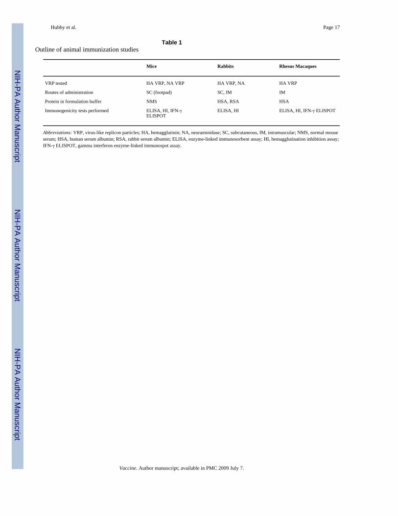
An outline of the animal immunization studies is provided in Table 1.
2.11. Statistical analysisIn the toxicology study, body weights, body weight changes, food consumption, bodytemperature, organ weight, and clinical pathology parameters were analyzed using theKolmogorov-Smirnov test for normality, the Levene Median test for equal variance, and byone-way analysis of variance (ANOVA). If the ANOVA indicated statistical significanceamong experimental groups then the Dunnett’s t-test was used to delineate which groups (ifany) differed from the control. Statistical analysis was conducted using Sigma-Stat™Statistical Software, Version 1 (Jandel Scientific, San Rafael, CA), with a two-tailedprobability value of <0.05 as the critical level of significance for all tests.
3. Results3.1. Characterization of HA and NA replicon constructs
Different concentrations of replicon, capsid and glycoprotein RNA were tested prior to pilotlot and clinical lot manufacture to determine the optimal ratio for VRP production. An RNAconcentration of 30 µg for replicon, capsid helper and glycoprotein helper was found to beoptimal for production of both HA VRP and NA VRP. Western blot analysis of proteinsextracted from Vero cells infected with VRP for 18–22 h showed expression of proteins of theexpected molecular weights that were reactive with antibodies specific for HA or NA (Fig. 1).
3.2. Production of pilot lots and GMP lots of VRPPilot lots of HA VRP and NA VRP were produced from a total of 20 cuvette electroporationsper lot and used to provide material for reference standards, stability studies and toxicologystudies. Clinical lots of influenza HA VRP were produced from a total of 60 cuvetteelectroporations per lot using the same process as the pilot lots.
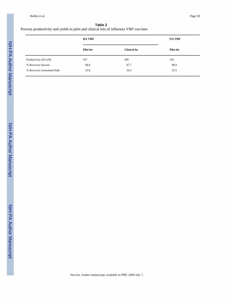
The production process was robust and reproducible for HA VRP during scale-up from pilotlot to clinical manufacturing. The overall process productivity, defined as infectious unitsproduced per cell, and percent yields during purification of HA VRP and NA VRP are shownin Table 2. The yields for the harvest by high salt elution (first step in the purification process)were calculated based upon a mass balance of VRP in the high salt wash pool and VRP in themedium and low salt wash pools. The yield in the formulated bulk is the overall yield after allprocess sampling and a single freeze thaw on the bulk material. No significant change in titerwas observed after the freeze thaw.
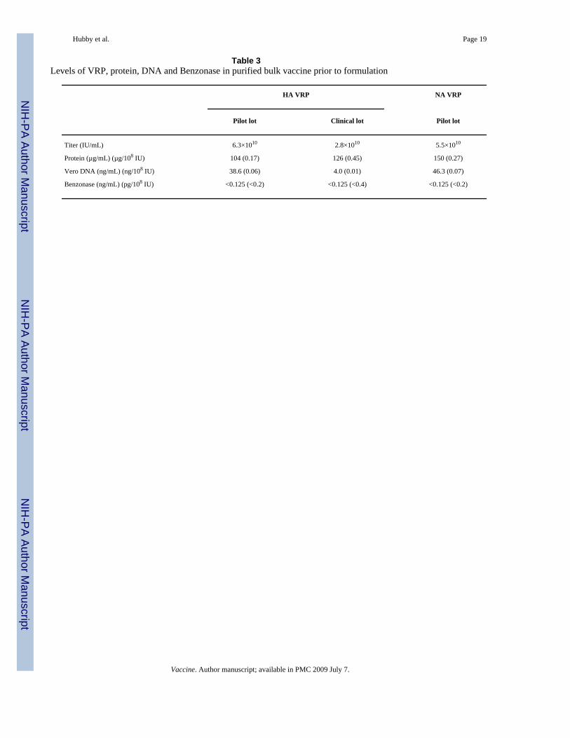
The purity of VRP with respect to protein and DNA in the product eluted from the CellufineSulfate column was assessed prior to addition of excipients to produce the formulated bulk andwas generally consistent in process intermediate pools from pilot to clinical scale productionoperations, although the DNA concentration was lower in the clinical lot (Table 3). SDS-PAGEanalysis of purified HA VRP (Fig. 2) and NA VRP (data not shown) demonstrated prominentprotein bands that were confirmed by Western blot analysis to be VEE envelope and capsidproteins. Silver-stained bands representing the alphavirus envelope and capsid proteins couldbe visualized amongst the many protein species on the gel after purification by TFF but weremore clearly seen when material was purified and concentrated during elution from the
Hubby et al. Page 6
Vaccine. Author manuscript; available in PMC 2009 July 7.
NIH
-PA Author Manuscript
NIH
-PA Author Manuscript
NIH
-PA Author Manuscript
Cellufine Sulfate chromatography resin. As visualized by Southern blot analysis, the size ofVero DNA detected ranged from >10,000 base pairs in upstream process samples to very smallfragments <300 base pairs at the end of TFF, and DNA was not detectable in VRP eluted fromthe Cellufine Sulfate chromatography resin (Fig. 3). The absence of DNA in the CellufineSulfate elution pool was not due to interfering substances, as 1 ng of digested Vero DNA spikedinto process samples was clearly visible in the control lane.
By qPCR analysis of formulated bulk vaccine, the genome equivalent to IU ratio was 18.3 and15.7 for the pilot lot and clinical lot of HA VRP and 25.5 for the pilot lot of NA VRP.
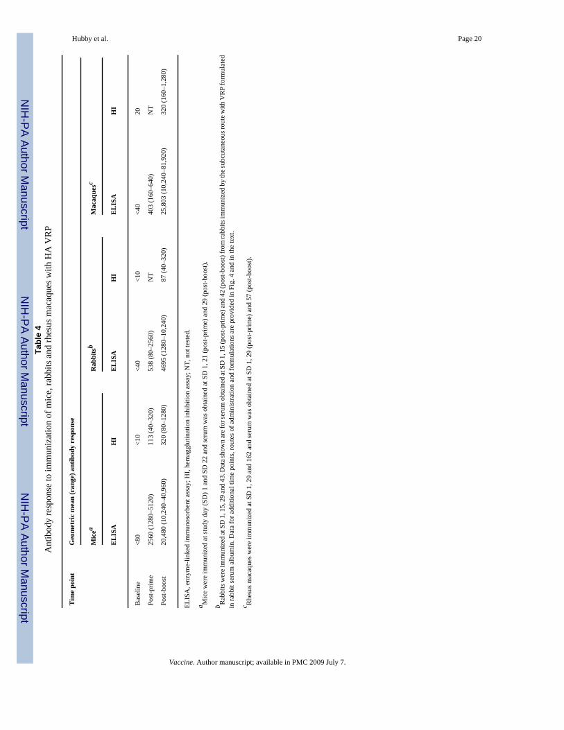
3.3. Humoral immune responses in mice, rabbits and rhesus macaquesMice, rabbits and rhesus macaques immunized with HA VRP developed anti-HA antibodiesas measured by ELISA and HI assays (Table 4).
In the rabbit toxicology study, anti-HA antibodies developed in all rabbits immunized withVRP and none of the animals immunized with PBS. Antibody responses increased withrepeated immunizations and were higher in animals immunized by the SC route than in animalsimmunized by the IM route (Fig. 4). The geometric mean (range) HI antibody titer at SD 42was <10 in placebo recipients, 55 (20–80) and 55 (20–80) in animals immunized by the IMroute with VRP formulated with RSA or HSA, respectively, and 87 (40–320) and 83 (40–640)in animals immunized by the SC route with VRP formulated with RSA or HSA, respectively.Anti-HSA antibodies, as measured by Ouchterlony assay, developed in 31 of 32 animalsimmunized with VRP formulated in HSA and in none of the animals immunized with PBS orwith VRP formulated in RSA.
3.4. Cellular immune responses in mice and rhesus macaquesSpleen cells from mice immunized with HA VRP or NA VRP developed robust T cell responsesas measured by IFN-γ ELISPOT assay. After immunization with 5 × 10⁵ IU of VRP at Days1 and 22, mean SFC per 106 splenic lymphocytes at Day 29 were 324 (range 186–592) for HAand 2183 (range 1558–2791) for NA.
PBMC from rhesus macaques immunized with HA VRP developed robust T cell responses asmeasured by IFN-γ ELISPOT assay (Fig. 5). After immunization with 1 × 10⁹ IU of HA VRPat Days 1, 29 and 162, modest T cell responses were detected in two of three animals after thefirst dose of vaccine (24 and 51 SFC per 106 PBMC at Day 15) and strong T cell responseswere detected in three of three animals after the second dose (698, 1155 and 1167 SFC per106 PBMC at Day 43). T cell responses persisted at elevated but lower levels (81–388 SFCper 106 PBMC) at most later time points during the study, and these responses did not appearto be boosted after the third dose of vaccine at Day 162. Among four animals immunized with1 × 109 IU of an irrelevant VRP, one had 25 SFC per 106 PBMC at SD 169 and one had 54and 75 SFC per 106 PBMC at SD 57 and 169, respectively. The mean (±SD) T cell responsefor the other 41 post-immunization values in these four animals was 2.1 ± 3.9 (range 0–15).
3.5. Results from toxicology testingPotency testing of samples obtained on the day of each injection demonstrated that animals inthe active vaccine groups received an average of 2.4 × 10⁸ IU of HA VRP and 4.1 × 10⁸ IU ofNA VRP per dose. Multiple inoculations had no adverse effects on clinical and cagesideobservations, body weights and body weight changes, food consumption, body temperature,or absolute and relative organ weights. Treatment with HA VRP and NA VRP had little effecton dermal Draize observations. Most animals were normal throughout the treatment period.Minimal edema or erythema was noted in all treated groups, affecting briefly one to threeanimals/sex/group. Mild edema or erythema was even more sporadic, affecting briefly one
Hubby et al. Page 7
Vaccine. Author manuscript; available in PMC 2009 July 7.
NIH
-PA Author Manuscript
NIH
-PA Author Manuscript
NIH
-PA Author Manuscript
Group 4 male, one Group 3 female, and three Group 4 females. The mean maximum dermalDraize score at any time point in any group did not exceed 0.4 for edema and did not exceed0.88 for erythema. In the animals that did develop edema or erythema, the injection sites weregenerally normal in appearance within 2–4 days after each immunization.
Clinical pathology evaluation revealed statistically significant increases in total protein andglobulin concentration and decreased albumin to globulin ratio values in animals treated withVRP compared to the PBS control. These were considered to be most likely due to polyclonalimmunoglobulin synthesis induced by test material administration. Statistically significantdifferences in coagulation parameters (activated partial thromboplastin time, fibrinogenconcentration, and prothrombin time) were also considered to be a reflection of a hostinflammatory response. None of these differences were considered to be biologically ortoxicologically adverse.
Multiple inoculations with HA VRP and NA VRP had no adverse effect on gross pathologyand no effect on organ weights, organ-to-body weight ratio or organ-to-brain weight ratio.Treatment-related gross pathology findings were limited to red discoloration, sometimes withgelatinous material, at the injection sites in a few animals treated with VRP formulated withHSA and necropsied on SD 45, and occasional enlargement or discoloration of draining lymphnodes in several animals in each of the five treatment groups.
Microscopic examination of tissues from SD 45/46 revealed test-article-related lesions at theintramuscular and subcutaneous injections sites, consistent with host inflammatory andimmunologic reactions to injection of the test material. Lesions in the iliac and axillary draininglymph nodes and fascia adjacent to the sciatic nerves were consistent with drainage of the sitesof inflammation. Lesions associated with VRP formulated with HSA were greater in severitygrade and incidence than those associated with VRP formulated with RSA. Vasculitis wasobserved at the injection site only in animals that received VRP formulated with HSA. Theincidence and severity of lesions had decreased at the recovery autopsies on SD 57/58.
4. DiscussionThere is a compelling need for new approaches to influenza immunization. The limited durationof protective antibody responses after immunization with inactivated vaccines, and thecontinuous evolution of influenza viruses to produce variants that are not inhibited byantibodies to previous strains, contribute to the need for annual remanufacture andadministration of influenza vaccines, and global production capacity is currently inadequate.The alphavirus replicon vector system offers a new approach to addressing these problems.
The VRP vaccines described in this paper had high-level expression of HA and NA thatappeared to have authentic conformational structure. Both proteins were glycosylated, HA0was processed to the expected HA1 and HA2 fragments, and immunization with HA VRPinduced antibodies in mice, rabbits and macaques that were active in hemagglutinationinhibition assays. In addition, preliminary experiments indicated that cells transfected withreplicons expressing HA or NA had hemagglutinating or neuraminidase activity, respectively(data not shown), indicating that HA and NA expressed from replicons are able to formauthentic, biologically active multimers.
The platform process used to scale up production of HA VRP and NA VRP was robust andgave products of yield and purity similar to that of VRP expressing other genes [17], includingan HIV Gag VRP vaccine that demonstrated excellent safety and dose-dependentimmunogenicity in a Phase 1 clinical trial [20]. The yields during product purification wereexcellent (33–50%) and the final product had low levels of protein and DNA contaminants.
Hubby et al. Page 8
Vaccine. Author manuscript; available in PMC 2009 July 7.
NIH
-PA Author Manuscript
NIH
-PA Author Manuscript
NIH
-PA Author Manuscript
When these studies were initiated, it was envisioned that an alphavirus replicon vaccine forinfluenza would include both HA VRP and NA VRP. After the GLP toxicology study wasperformed, analysis of efficacy data in ferrets immunized with different VRP vaccinesindicated that a VRP vaccine expressing only the HA protein provided protective efficacy asgood as that provided by a mixture of VRP vaccines expressing HA and NA proteins [21].Therefore it was decided to proceed to clinical trials with a vaccine containing HA VRP only,and this product was also evaluated in nonhuman primates.
Protective immunity to influenza has been correlated with anti-influenza antibodies measuredin the HI assay [2], and immunization with HA VRP induced high titers of HI antibodies inmice, rabbits and rhesus macaques. The role of cellular immune responses in protection againstinfluenza is less clear. Although mice lacking functional class I major histocompatibilitycomplex (MHC) glycoproteins and class I MHC-restricted, CD8+ effector T cells are able toclear influenza virus infections [22], CD8+ cyto-toxic T lymphocytes (CTL) can protect againstinfluenza in the absence of antibodies to influenza [23,24], and CTL responses to influenzahave been correlated with protection against influenza in humans [25]. Mice and rhesusmacaques immunized with HA VRP developed T cell responses as measured by IFN-γELISPOT assay, and CD4+ and CD8+ T cells secreting IFN-γ and TNF-α have been identifiedby cytokine flow cytometry in PBMC from rhesus macaques immunized with HA VRP (E.A.Reap, unpublished data). Because the antigenic protein is produced within the VRP-infectedcell, the antigen is proteolytically processed and presented by both Class I and Class II MHCproteins to stimulate both CD4+ and CD8+ T cell responses. In contrast, conventional influenzavaccines contain preformed proteins that are taken up by antigen-presenting cells and processedfor presentation by Class II MHC proteins, inducing only CD4+ T cell responses. The abilityof VRP to induce potentially protective CD8+ T cell responses may provide an additionalbenefit for VRP vaccines compared to conventional inactivated influenza vaccines.
VRP, like the alphaviruses from which that are derived, are enveloped particles, and purifiedpreparations require the presence of protein in the formulation buffer to achieve stability. Thestabilizing protein used for the vaccine to be tested in humans is HSA, but because mouse andrabbit albumin are sufficiently different from that in humans, repeated injections of VRPformulated in HSA can cause an immune response in these species, and VRP for studies inthese species were formulated in NMS or RSA. However, GLP toxicology testing requires thatthe exact product to be used clinically must be the product tested in a toxicology study. Becauseprevious toxicology studies in rabbits with other VRP vaccines formulated with HSA hadsometimes shown increasing local reactogenicity with repeated dosing accompanied byhistological evidence of inflammatory reactions, including necrotizing vasculitis, consistentwith an Arthus-type reaction (J.D. Chulay, unpublished data), a GLP toxicology study wasperformed with HA VRP and NA VRP formulated with either HSA or RSA. Results of thisstudy showed only mild local reactogenicity with no systemic toxicity. By histologicalexamination, inflammatory responses were greater and evidence of vasculitis was seen onlyin animals immunized with VRP formulated with HSA. The Arthus-type reactions induced byVRP formulated with HSA may be related in part to the ability of VRP to provide adjuvantactivity when co-administered with a foreign protein [26].
In summary, results of these studies, which describe efficient methods for production of VRPvaccines expressing influenza proteins that induce potent humoral and cellular immuneresponses in mice, rabbits and rhesus macaques with no adverse effects in GLP toxicologytesting, support clinical testing of alphavirus replicon vaccines for influenza. Phase 1 testingof an HA VRP vaccine was initiated in April, 2007
Hubby et al. Page 9
Vaccine. Author manuscript; available in PMC 2009 July 7.
NIH
-PA Author Manuscript
NIH
-PA Author Manuscript
NIH
-PA Author Manuscript
AcknowledgementsWe thank Randy Lamm, Kevin Williams, Tim Wagner, Deepa Patel, Renee Doggett and Holly Stone for performinganalytical assays, ILS for immunizations and sample collection in mice, SRI for immunizations and sample collectionin rhesus macaques, and Bridge GPS (formerly Gene Logic) for performing the GLP toxicology study in rabbits.Studies involving the use of animals complied with all relevant federal guidelines and institutional policies and wereapproved by an Institutional Animal Care and Use Committee. This research was supported in part by NIH grantsUC1-AI62632 and UC1-AI062582.
References1. Treanor, JJ. Influenza virus. In: Mandell, GL.; Bennett, JE.; Dolin, R., editors. Principles and practice
of infectious diseases. Vol. fifth ed.. Philadelphia: Churchill Livingstone; 2000. p. 1823-1849.2. Hobson D, Curry RL, Beare AS, Ward-Gardner A. The role of serum haemagglutination-inhibiting
antibody in protection against challenge infection with influenza A2 and B viruses. J Hyg (Lond)1972;70(Dec 4):767–777. [PubMed: 4509641]
3. Thomas, DB.; Patera, AC.; Graham, CM.; Smith, CA. Antibody-mediated immunity. In: Nicholson,KG.; Webster, RG.; Hay, AJ., editors. Textbook of Influenza. Oxford: Blackwell Science Ltd; 1998.p. 267-277.
4. Beutner KR, Chow T, Rubi E, Strussenberg J, Clement J, Ogra PL. Evaluation of a neuraminidase-specific influenza A virus vaccine in children: antibody responses and effects on two successiveoutbreaks of natural infection. J Infect Dis 1979;140(Dec 6):844–850. [PubMed: 396336]
5. Sandbulte MR, Jimenez GS, Boon AC, Smith LR, Treanor JJ, Webby RJ. Cross-ReactiveNeuraminidase Antibodies Afford Partial Protection against H5N1 in Mice and Are Present inUnexposed Humans. PLoS Med 2007;4(Feb 2):e59. [PubMed: 17298168]
6. Webby RJ, Webster RG. Are we ready for pandemic influenza? Science 2003;302(Nov 5650):1519–1522. [PubMed: 14645836]
7. Lakey DL, Treanor JJ, Betts RF, Smith GE, Thompson J, Sannella E, et al. Recombinant baculovirusinfluenza A hemagglutinin vaccines are well tolerated and immunogenic in healthy adults. J Infect Dis1996;174(Oct 4):838–841. [PubMed: 8843225]
8. Treanor JJ, Betts RF, Smith GE, Anderson EL, Hackett CS, Wilkinson BE, et al. Evaluation of arecombinant hemagglutinin expressed in insect cells as an influenza vaccine in young and elderlyadults. J Infect Dis 1996;173(June 6):1467–1470. [PubMed: 8648221]
9. Bender BS, Rowe CA, Taylor SF, Wyatt LS, Moss B, Small PA Jr. Oral immunization with areplication-deficient recombinant vaccinia virus protects mice against influenza. J Virol 1996;70(Sep9):6418–6424. [PubMed: 8709274]
10. Ulmer JB, Donnelly JJ, Parker SE, Rhodes GH, Felgner PL, Dwarki VJ, et al. Heterologous protectionagainst influenza by injection of DNA encoding a viral protein. Science 1993;259(March 5102):1745–1749. [PubMed: 8456302]
11. Coulter A, Harris R, Davis R, Drane D, Cox J, Ryan D, et al. Intranasal vaccination withISCOMATRIX adjuvanted influenza vaccine. Vaccine 2003;21(Feb 9–10):946–949. [PubMed:12547607]
12. Plante M, Jones T, Allard F, Torossian K, Gauthier J, St-Felix N, et al. Nasal immunization withsubunit proteosome influenza vaccines induces serum HAI, mucosal IgA and protection againstinfluenza challenge. Vaccine 2001;20(Oct 1–2):218–225. [PubMed: 11567767]
13. Treanor JJ, Kotloff K, Betts RF, Belshe R, Newman F, Iacuzio D, et al. Evaluation of trivalent, live,cold-adapted (CAIV-T) and inactivated (TIV) influenza vaccines in prevention of virus infection andillness following challenge of adults with wild-type influenza A (H1N1), A (H3N2), and B viruses.Vaccine 2000;18(9–10):899–906. [PubMed: 10580204]
14. Kroger AT, Atkinson WL, Marcuse EK, Pickering LK. General recommendations on immunization:recommendations of the Advisory Committee on Immunization Practices (ACIP). MMWR RecommRep 2006;55(Dec RR15):1–48. [PubMed: 17136024]
15. Kamrud KI, Custer M, Dudek JM, Owens G, Alterson KD, Lee JS, et al. Alphavirus replicon approachto promoterless analysis of IRES elements. Virology 2007;360(April 2):376–387. [PubMed:17156813]
Hubby et al. Page 10
Vaccine. Author manuscript; available in PMC 2009 July 7.
NIH
-PA Author Manuscript
NIH
-PA Author Manuscript
NIH
-PA Author Manuscript
16. Pushko P, Parker M, Ludwig GV, Davis NL, Johnston RE, Smith JF. Replicon-helper systems fromattenuated Venezuelan equine encephalitis virus: expression of heterologous genes in vitro andimmunization against heterologous pathogens in vivo. Virology 1997;239(2):389–401. [PubMed:9434729]
17. Talarico T, Maughan M, Pancorbo B, Ruiz J, Graham A. Development and manufacture ofalphavaccines. Bioprocessing 2006 Fall;:8–14.
18. Griffiths, E.; Gruber, M.; Masset, D.; Verdier, F.; Wood, D.; Knezevic, I. WHO guidelines onnonclinical evaluation of vaccines.http://wwwwhoint/biologicals/publications/nonclinical_evaluation_vaccines_nov_2003pdf
19. Reap EA, Dryga SA, Morris J, Rivers B, Norberg PK, Olmsted RA, et al. Cellular and humoralimmune responses to alphavirus replicon vaccines expressing cytomegalovirus pp65, IE1 and gBproteins. Clin Vaccine Immunol 2007;14(6):748–755. [PubMed: 17442845]
20. Chulay, J.; Burke, D.; Karim, SSA.; Russell, N.; Wecker, M.; Allen, M., et al. AIDS Vaccine 06; 29Aug–1 Sep 2006. Amsterdam: Safety and immunogenicity of an alphavirus replicon HIV Gag vaccine(AVX101) in healthy HIV-uninfected adults. p. 11-09
21. Hubby, B.; Boyers, A.; Ellis, W.; Copp, L.; Mann, A.; Lambkin, R., et al. Immunogenicity and efficacyin ferrets of alphavirus replicon vaccines for influenza. Second European Influenza Conference;2005; 11–14 Sep 2005; Malta. 2005.
22. Eichelberger M, Allan W, Zijlstra M, Jaenisch R, Doherty PC. Clearance of influenza virus respiratoryinfection in mice lacking class I major histocompatibility complex-restricted CD8+ T cells. J ExpMed 1991 Oct 1;174(Oct 4):875–880. [PubMed: 1919440]
23. Yap KL, Ada GL, McKenzie IF. Transfer of specific cytotoxic T lymphocytes protects miceinoculated with influenza virus. Nature 1978;273(May 5659):238–239. [PubMed: 306072]
24. Taylor PM, Askonas BA. Influenza nucleoprotein-specific cytotoxic T-cell clones are protective invivo. Immunology 1986;58(July 3):417–420. [PubMed: 2426185]
25. McMichael AJ, Gotch FM, Noble GR, Beare PA. Cytotoxic T-cell immunity to influenza. N Engl JMed 1983;309(July 1):13–17. [PubMed: 6602294]
26. Thompson JM, Whitmore AC, Konopka JL, Collier ML, Richmond EM, Davis NL, et al. Mucosaland systemic adjuvant activity of alphavirus replicon particles. Proc Natl Acad Sci USA 2006;103(March 10):3722–3727. [PubMed: 16505353]
Hubby et al. Page 11
Vaccine. Author manuscript; available in PMC 2009 July 7.
NIH
-PA Author Manuscript
NIH
-PA Author Manuscript
NIH
-PA Author Manuscript
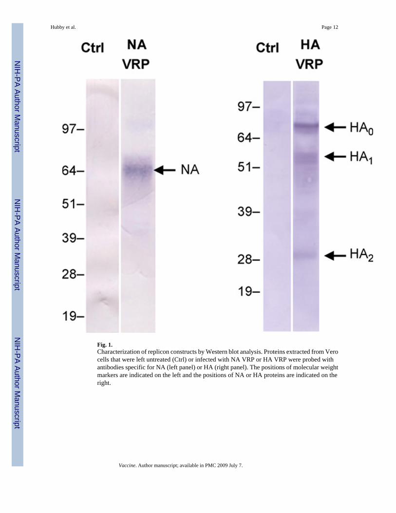
Fig. 1.Characterization of replicon constructs by Western blot analysis. Proteins extracted from Verocells that were left untreated (Ctrl) or infected with NA VRP or HA VRP were probed withantibodies specific for NA (left panel) or HA (right panel). The positions of molecular weightmarkers are indicated on the left and the positions of NA or HA proteins are indicated on theright.
Hubby et al. Page 12
Vaccine. Author manuscript; available in PMC 2009 July 7.
NIH
-PA Author Manuscript
NIH
-PA Author Manuscript
NIH
-PA Author Manuscript
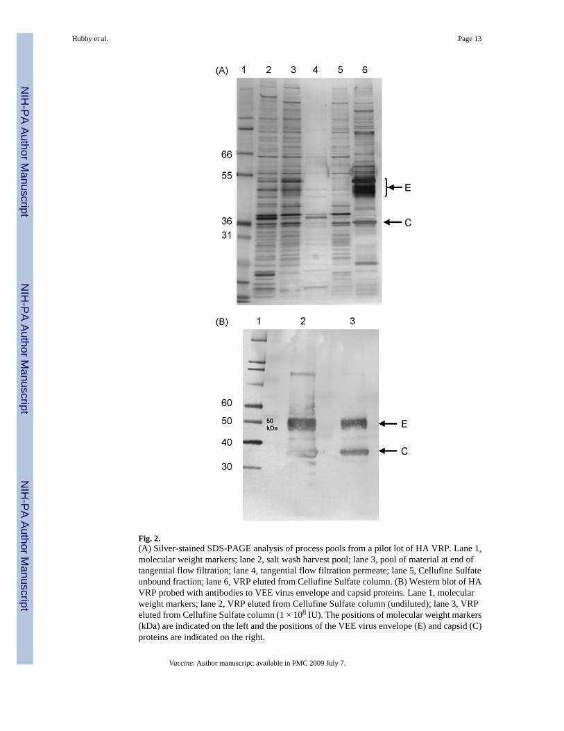
Fig. 2.(A) Silver-stained SDS-PAGE analysis of process pools from a pilot lot of HA VRP. Lane 1,molecular weight markers; lane 2, salt wash harvest pool; lane 3, pool of material at end oftangential flow filtration; lane 4, tangential flow filtration permeate; lane 5, Cellufine Sulfateunbound fraction; lane 6, VRP eluted from Cellufine Sulfate column. (B) Western blot of HAVRP probed with antibodies to VEE virus envelope and capsid proteins. Lane 1, molecularweight markers; lane 2, VRP eluted from Cellufine Sulfate column (undiluted); lane 3, VRPeluted from Cellufine Sulfate column (1 × 10⁸ IU). The positions of molecular weight markers(kDa) are indicated on the left and the positions of the VEE virus envelope (E) and capsid (C)proteins are indicated on the right.
Hubby et al. Page 13
Vaccine. Author manuscript; available in PMC 2009 July 7.
NIH
-PA Author Manuscript
NIH
-PA Author Manuscript
NIH
-PA Author Manuscript
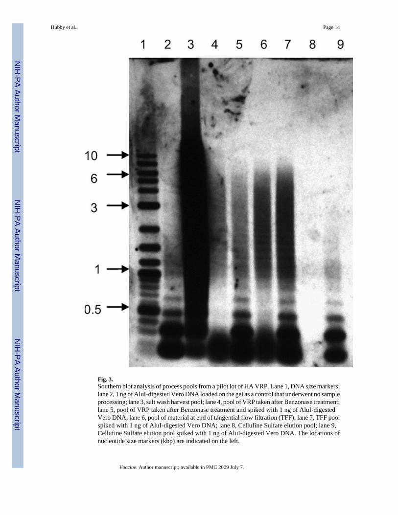
Fig. 3.Southern blot analysis of process pools from a pilot lot of HA VRP. Lane 1, DNA size markers;lane 2, 1 ng of AluI-digested Vero DNA loaded on the gel as a control that underwent no sampleprocessing; lane 3, salt wash harvest pool; lane 4, pool of VRP taken after Benzonase treatment;lane 5, pool of VRP taken after Benzonase treatment and spiked with 1 ng of AluI-digestedVero DNA; lane 6, pool of material at end of tangential flow filtration (TFF); lane 7, TFF poolspiked with 1 ng of AluI-digested Vero DNA; lane 8, Cellufine Sulfate elution pool; lane 9,Cellufine Sulfate elution pool spiked with 1 ng of AluI-digested Vero DNA. The locations ofnucleotide size markers (kbp) are indicated on the left.
Hubby et al. Page 14
Vaccine. Author manuscript; available in PMC 2009 July 7.
NIH
-PA Author Manuscript
NIH
-PA Author Manuscript
NIH
-PA Author Manuscript
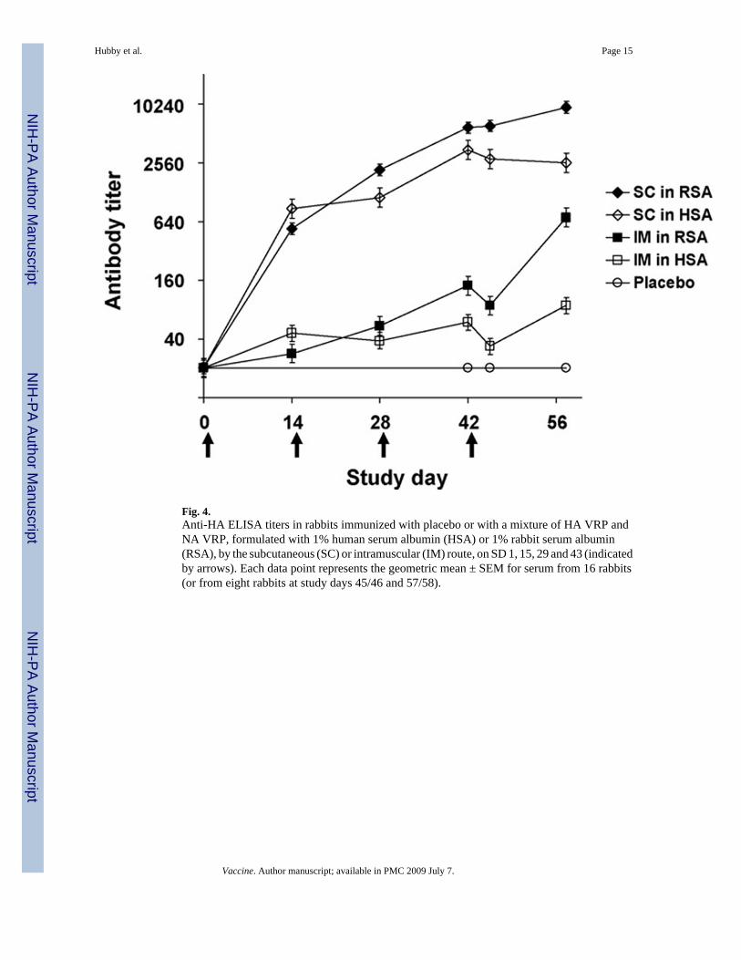
Fig. 4.Anti-HA ELISA titers in rabbits immunized with placebo or with a mixture of HA VRP andNA VRP, formulated with 1% human serum albumin (HSA) or 1% rabbit serum albumin(RSA), by the subcutaneous (SC) or intramuscular (IM) route, on SD 1, 15, 29 and 43 (indicatedby arrows). Each data point represents the geometric mean ± SEM for serum from 16 rabbits(or from eight rabbits at study days 45/46 and 57/58).
Hubby et al. Page 15
Vaccine. Author manuscript; available in PMC 2009 July 7.
NIH
-PA Author Manuscript
NIH
-PA Author Manuscript
NIH
-PA Author Manuscript
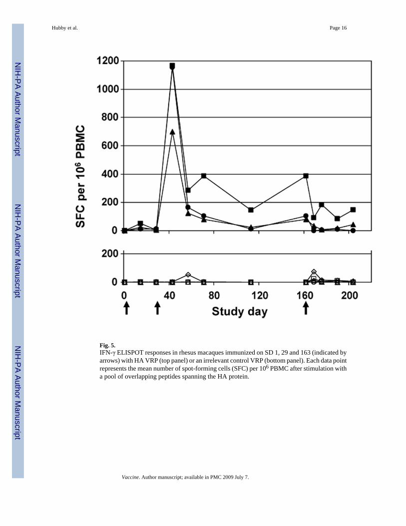
Fig. 5.IFN-γ ELISPOT responses in rhesus macaques immunized on SD 1, 29 and 163 (indicated byarrows) with HA VRP (top panel) or an irrelevant control VRP (bottom panel). Each data pointrepresents the mean number of spot-forming cells (SFC) per 106 PBMC after stimulation witha pool of overlapping peptides spanning the HA protein.
Hubby et al. Page 16
Vaccine. Author manuscript; available in PMC 2009 July 7.
NIH
-PA Author Manuscript
NIH
-PA Author Manuscript
NIH
-PA Author Manuscript
NIH
-PA Author Manuscript
NIH
-PA Author Manuscript
NIH
-PA Author Manuscript
Hubby et al. Page 17
Table 1Outline of animal immunization studies
Mice Rabbits Rhesus Macaques
VRP tested HA VRP, NA VRP HA VRP, NA HA VRP
Routes of administration SC (footpad) SC, IM IM
Protein in formulation buffer NMS HSA, RSA HSA
Immunogenicity tests performed ELISA, HI, IFN-γELISPOT
ELISA, HI ELISA, HI, IFN-γ ELISPOT
Abbreviations: VRP, virus-like replicon particles; HA, hemagglutinin; NA, neuraminidase; SC, subcutaneous, IM, intramuscular; NMS, normal mouseserum; HSA, human serum albumin; RSA, rabbit serum albumin; ELISA, enzyme-linked immunosorbent assay; HI, hemagglutination inhibition assay;IFN-γ ELISPOT, gamma interferon enzyme-linked immunospot assay.
Vaccine. Author manuscript; available in PMC 2009 July 7.
NIH
-PA Author Manuscript
NIH
-PA Author Manuscript
NIH
-PA Author Manuscript
Hubby et al. Page 18
Table 2Process productivity and yields in pilot and clinical lots of influenza VRP vaccines
HA VRP NA VRP
Pilot lot Clinical lot Pilot lot
Productivity (IU/cell) 357 300 335
% Recovery harvest 98.8 97.7 98.9
% Recovery formulated bulk 33.8 50.2 33.5
Vaccine. Author manuscript; available in PMC 2009 July 7.
NIH
-PA Author Manuscript
NIH
-PA Author Manuscript
NIH
-PA Author Manuscript
Hubby et al. Page 19
Table 3Levels of VRP, protein, DNA and Benzonase in purified bulk vaccine prior to formulation
HA VRP NA VRP
Pilot lot Clinical lot Pilot lot
Titer (IU/mL) 6.3×10¹⁰ 2.8×10¹⁰ 5.5×10¹⁰
Protein (µg/mL) (µg/108 IU) 104 (0.17) 126 (0.45) 150 (0.27)
Vero DNA (ng/mL) (ng/108 IU) 38.6 (0.06) 4.0 (0.01) 46.3 (0.07)
Benzonase (ng/mL) (pg/108 IU) <0.125 (<0.2) <0.125 (<0.4) <0.125 (<0.2)
Vaccine. Author manuscript; available in PMC 2009 July 7.
NIH
-PA Author Manuscript
NIH
-PA Author Manuscript
NIH
-PA Author Manuscript
Hubby et al. Page 20Ta
ble
4A
ntib
ody
resp
onse
to im
mun
izat
ion
of m
ice,
rabb
its a
nd rh
esus
mac
aque
s with
HA
VR
P
Tim
e po
int
Geo
met
ric
mea
n (r
ange
) ant
ibod
y re
spon
se
Mic
eaR
abbi
tsb
Mac
aque
sc
EL
ISA
HI
EL
ISA
HI
EL
ISA
HI
Bas
elin
e<8
0<1
0<4
0<1
0<4
020
Post
-prim
e25
60 (1
280–
5120
)11
3 (4
0–32
0)53
8 (8
0–25
60)
NT
403
(160
–640
)N
T
Post
-boo
st20
,480
(10,
240–
40,9
60)
320
(80–
1280
)46
95 (1
280–
10,2
40)
87 (4
0–32
0)25
,803
(10,
240–
81,9
20)
320
(160
–1,2
80)
ELIS
A, e
nzym
e-lin
ked
imm
unos
orbe
nt a
ssay
; HI,
hem
aggl
utin
atio
n in
hibi
tion
assa
y; N
T, n
ot te
sted
.
a Mic
e w
ere
imm
uniz
ed a
t stu
dy d
ay (S
D) 1
and
SD
22
and
seru
m w
as o
btai
ned
at S
D 1
, 21
(pos
t-prim
e) a
nd 2
9 (p
ost-b
oost
).
b Rab
bits
wer
e im
mun
ized
at S
D 1
, 15,
29
and
43. D
ata s
how
n ar
e for
seru
m o
btai
ned
at S
D 1
, 15
(pos
t-prim
e) an
d 42
(pos
t-boo
st) f
rom
rabb
its im
mun
ized
by
the s
ubcu
tane
ous r
oute
with
VR
P fo
rmul
ated
in ra
bbit
seru
m a
lbum
in. D
ata
for a
dditi
onal
tim
e po
ints
, rou
tes o
f adm
inis
tratio
n an
d fo
rmul
atio
ns a
re p
rovi
ded
in F
ig. 4
and
in th
e te
xt.
c Rhe
sus m
acaq
ues w
ere
imm
uniz
ed a
t SD
1, 2
9 an
d 16
2 an
d se
rum
was
obt
aine
d at
SD
1, 2
9 (p
ost-p
rime)
and
57
(pos
t-boo
st).
Vaccine. Author manuscript; available in PMC 2009 July 7.
Related Documents