RESEARCH ARTICLE Development and External Validation of a Prognostic Nomogram for Metastatic Uveal Melanoma Sara Valpione 1,2 *, Justin C. Moser 3 , Raffaele Parrozzani 4 , Marco Bazzi 5 , Aaron S. Mansfield 6 , Simone Mocellin 2 , Jacopo Pigozzo 1 , Edoardo Midena 5,7 , Svetomir N. Markovic 6 , Camillo Aliberti 8 , Luca G. Campana 9 , Vanna Chiarion-Sileni 1 1 Melanoma Oncology Unit, Veneto Region Oncology Research Institute (IOV-IRCCS), Padova, Italy, 2 Department of Surgery, Oncology and Gastroenterology, Padova, Italy, 3 Department of Internal Medicine, Mayo Clinic, Rochester, Minnesota, US, 4 GB Bietti Foundation-IRCCS, Rome, Italy, 5 Department of Statistical Sciences, University of Padova, Padova, Italy, 6 Division of Medical Oncology, Mayo Clinic, Rochester, Minnesota, United States of America, 7 Department of Ophthalmology, University of Padova, Padova, Italy, 8 Interventional Radiology, Veneto Region Oncology Research Institute (IOV-IRCCS) Padova, Italy, 9 Sarcoma and Melanoma Unit, Veneto Region Oncology Research Institute (IOV-IRCCS) Padova, Italy * [email protected] Abstract Background Approximately 50% of patients with uveal melanoma (UM) will develop metastatic disease, usually involving the liver. The outcome of metastatic UM (mUM) is generally poor and no standard therapy has been established. Additionally, clinicians lack a validated prognostic tool to evaluate these patients. The aim of this work was to develop a reliable prognostic no- mogram for clinicians. Patients and Methods Two cohorts of mUM patients, from Veneto Oncology Institute (IOV) (N=152) and Mayo Clinic (MC) (N=102), were analyzed to develop and externally validate, a prognostic nomogram. Results The median survival of mUM was 17.2 months in the IOV cohort and 19.7 in the MC cohort. Percentage of liver involvement (HR 1.6), elevated levels of serum LDH (HR 1.6), and a WHO performance status=1 (HR 1.5) or 2–3 (HR 4.6) were associated with worse progno- sis. Longer disease-free interval from diagnosis of UM to that of mUM conferred a survival advantage (HR 0.9). The nomogram had a concordance probability of 0.75 (SE .006) in the development dataset (IOV), and 0.80 (SE .009) in the external validation (MC). Nomogram predictions were well calibrated. PLOS ONE | DOI:10.1371/journal.pone.0120181 March 17, 2015 1 / 12 a11111 OPEN ACCESS Citation: Valpione S, Moser JC, Parrozzani R, Bazzi M, Mansfield AS, Mocellin S, et al. (2015) Development and External Validation of a Prognostic Nomogram for Metastatic Uveal Melanoma. PLoS ONE 10(3): e0120181. doi:10.1371/journal. pone.0120181 Academic Editor: Andrzej T Slominski, University of Tennessee, UNITED STATES Received: July 17, 2014 Accepted: February 3, 2015 Published: March 17, 2015 Copyright: © 2015 Valpione et al. This is an open access article distributed under the terms of the Creative Commons Attribution License, which permits unrestricted use, distribution, and reproduction in any medium, provided the original author and source are credited. Data Availability Statement: The authors confirm that, for approved reasons, some access restrictions apply to the data underlying the findings. Due to an ethical restriction, raw data are unsuitable for public deposition as they contain patient medical records. Anonymized data are available upon request to Dr.Sara Valpione [email protected]. Funding: The authors have no support or funding to report. Competing Interests: The authors have declared that no competing interests exist.

Welcome message from author
This document is posted to help you gain knowledge. Please leave a comment to let me know what you think about it! Share it to your friends and learn new things together.
Transcript

RESEARCH ARTICLE
Development and External Validation of aPrognostic Nomogram for Metastatic UvealMelanomaSara Valpione1,2*, Justin C. Moser3, Raffaele Parrozzani4, Marco Bazzi5, AaronS. Mansfield6, Simone Mocellin2, Jacopo Pigozzo1, Edoardo Midena5,7, SvetomirN. Markovic6, Camillo Aliberti8, Luca G. Campana9, Vanna Chiarion-Sileni1
1 Melanoma Oncology Unit, Veneto Region Oncology Research Institute (IOV-IRCCS), Padova, Italy,2 Department of Surgery, Oncology and Gastroenterology, Padova, Italy, 3 Department of Internal Medicine,Mayo Clinic, Rochester, Minnesota, US, 4 GB Bietti Foundation-IRCCS, Rome, Italy, 5 Department ofStatistical Sciences, University of Padova, Padova, Italy, 6 Division of Medical Oncology, Mayo Clinic,Rochester, Minnesota, United States of America, 7 Department of Ophthalmology, University of Padova,Padova, Italy, 8 Interventional Radiology, Veneto Region Oncology Research Institute (IOV-IRCCS)Padova, Italy, 9 Sarcoma and Melanoma Unit, Veneto Region Oncology Research Institute (IOV-IRCCS)Padova, Italy
Abstract
Background
Approximately 50% of patients with uveal melanoma (UM) will develop metastatic disease,
usually involving the liver. The outcome of metastatic UM (mUM) is generally poor and no
standard therapy has been established. Additionally, clinicians lack a validated prognostic
tool to evaluate these patients. The aim of this work was to develop a reliable prognostic no-
mogram for clinicians.
Patients and Methods
Two cohorts of mUM patients, from Veneto Oncology Institute (IOV) (N=152) and Mayo Clinic
(MC) (N=102), were analyzed to develop and externally validate, a prognostic nomogram.
Results
The median survival of mUM was 17.2 months in the IOV cohort and 19.7 in the MC cohort.
Percentage of liver involvement (HR 1.6), elevated levels of serum LDH (HR 1.6), and a
WHO performance status=1 (HR 1.5) or 2–3 (HR 4.6) were associated with worse progno-
sis. Longer disease-free interval from diagnosis of UM to that of mUM conferred a survival
advantage (HR 0.9). The nomogram had a concordance probability of 0.75 (SE .006) in the
development dataset (IOV), and 0.80 (SE .009) in the external validation (MC). Nomogram
predictions were well calibrated.
PLOS ONE | DOI:10.1371/journal.pone.0120181 March 17, 2015 1 / 12
a11111
OPEN ACCESS
Citation: Valpione S, Moser JC, Parrozzani R, BazziM, Mansfield AS, Mocellin S, et al. (2015)Development and External Validation of a PrognosticNomogram for Metastatic Uveal Melanoma. PLoSONE 10(3): e0120181. doi:10.1371/journal.pone.0120181
Academic Editor: Andrzej T Slominski, University ofTennessee, UNITED STATES
Received: July 17, 2014
Accepted: February 3, 2015
Published: March 17, 2015
Copyright: © 2015 Valpione et al. This is an openaccess article distributed under the terms of theCreative Commons Attribution License, which permitsunrestricted use, distribution, and reproduction in anymedium, provided the original author and source arecredited.
Data Availability Statement: The authors confirmthat, for approved reasons, some access restrictionsapply to the data underlying the findings. Due to anethical restriction, raw data are unsuitable for publicdeposition as they contain patient medical records.Anonymized data are available upon request toDr.Sara Valpione [email protected].
Funding: The authors have no support or funding toreport.
Competing Interests: The authors have declaredthat no competing interests exist.

Conclusions
The nomogram, which includes percentage of liver involvement, LDH levels, WHO perfor-
mance status and disease free-interval accurately predicts the prognosis of mUM and could
be useful for decision-making and risk stratification for clinical trials.
IntroductionUveal melanoma (UM) is the most common primary intraocular malignancy in the adult, rep-resenting 5–6% of all melanomas (annual incidence in Europe approximately 5:1000000), andis associated with age and, light skin and blue pigmented eyes[1,2]. Although local control isachieved in most cases, approximately 50% of patients will develop systemic disease [1]. Al-though the liver is the most common site of metastatic disease, UM can metastasize to any in-cluding the lungs, bones, soft tissues, gastrointestinal tract, ovaries, kidneys and centralnervous system (CNS)[2, 3]. The reported median life expectancy of patients affected by meta-static UM (mUM) ranges from 3.6 to 15 months [4]. Site; number and diameter of metastases;percentage of liver substitution, presence of symptoms; alteration of liver function tests, espe-cially alkaline phosphatase (ALP) and lactic dehydrogenase (LDH); older age; male sex; and ashorter metastasis-free interval have been associated with a poorer prognosis [2, 5–10]. Due toseveral known and unknown factors, as for mucosal, acral and skin melanomas, UM is a poorresponder to antiblastic chemotherapy and radiotherapy[11]. Moreover, UM cannot benefitfrom target therapy tailored for cutaneous melanoma, as BRAF inhibitors, because of the ab-sence of the target mutation[12] and, for reasons yet to be explored, response to new immuno-therapies is poorer than for cutaneous melanoma[13]. Treatments for mUM can be dividedinto liver directed treatments; such as surgical resection [9], ablation [14], radiation [14], he-patic arterial chemoinfusion [15, 16], immunoembolization [17], transarterial chemoemboliza-tion [18], radioembolization [19], isolated or percutaneous hepatic perfusion [20, 21]; and intosystemic treatments; such as chemotherapy (antineoplastic drugs used alone or in combina-tions [22]), immunotherapy (interferon [23], interleukin-2 [24], and, more recently, ipilimu-mab[13]), anti-angiogenetic drugs [25, 26], and targeted agents such as MEK inhibitors [27,28]. Despite the efforts to improve mUM outcomes, prognosis remains poor and clinicianslack a standard prognostic tool. The study objective was to identify the independent prognosticfactors for mUM in order to formulate a reproducible prognostic algorithm, that could be easi-ly enough to be integrated into clinical practice.
Patients and Methods
Patients and TherapiesThe prospective melanoma databases at the Melanoma Oncology Unit of the Veneto OncologyInstitute (IOV) and at Mayo Clinic, Rochester (MC) were queried under institutional reviewboard approval for mUM. IOV patients (N = 152) were diagnosed and treated between Sep-tember 1990 to October 2013, MC patients (N = 102) were diagnosed and treated between Jan-uary 2000 and August 2013. The majority of patients from IOV (72.4%) and MC (84.3%) weretreated with Iodine-125-brachitherapy for their primary melanoma; the remaining patients re-ceived enucleation, with the exception of those in whom treatment of their primary melanomawas futile as they presented with stage IV disease. Material for fluorescent in-situ hybridizationanalysis (FISH) of the primary tumor was obtained with a 25-gauge trans-scleral fine needle
Prognostic Model for Metastatic Uveal Melanoma
PLOSONE | DOI:10.1371/journal.pone.0120181 March 17, 2015 2 / 12

aspiration biopsy. FISH was used to evaluate the cell karyotype and alterations of chromosomes1, 3, 6, 8 and 10. Procedure and testing was performed using previously published methods[29–31]. Metastases were discovered at initial staging (6 IOV patients and 3 MC patients) orduring follow-up via ultrasound tomography or computed tomography (CT). Diagnosis wasconfirmed via core biopsy or fine needle aspiration cytology. Staging was completed with CTor magnetic resonance (MR) when not previously performed. Therapy was chosen accordingto the localization of metastases and the availability of clinical trials.
Gender, age; date, size and characteristics of UM; date and site of metastases; date, type andoutcome of therapies; date of last follow up or death and cause of death were collected from pa-tient records. Date and cause of death were collected from local registry offices, and telephoneinterviewing of family or from general practitioners for patients lost to follow up. Levels ofLDH, alkaline phosphatase (ALP), U-glutamyltranspeptidase (UGT) and transaminases wererecorded at diagnosis of metastatic disease and were computed as proportion of the respectiveupper normal value. The site and extent of metastases at baseline were quantified with CT(IOV cohort and most MC patients) or MR (a minority of MC patients). Liver metastasis vol-umes were calculated with three-dimensional reconstruction by helical CT or MR of the liverand registered as the percentage of liver substitution; we retrospectively reviewed CT images ofthe IOV cohort with Syngo CT Oncology software (version 2009E, Siemens, Germany) for con-firmation and to assess the maximum diameter and the number of liver metastases. It was de-cided not to include this information as the percentage of liver substitution was considered thebest indicator of effective volume of hepatic disease. Moreover, some patients had many smallmetastases, making analysis of diameter and number very complex.
The study was approved by the Institutional Review Boards of Veneto Region Oncology Re-search Institute, Padova, and Mayo Clinic, Rochester. All clinical investigations have been con-ducted according to the principles expressed in the Declaration of Helsinki. All patients gavewritten informed consent to the use of their records for research purposes.
Statistical AnalysisPatient features and clinical characteristics were analyzed using the Mann-Whitney two-tailedU test (continuous variables) and χ2 test (categorical variables). Disease-free interval (DFI) wasdefined as the time from initial UM diagnosis to first noted metastasis. Overall survival (OS)was defined as the time from diagnosis of first metastasis to the date of death or last follow-up.OS was estimated using the Kaplan-Meier method and survival between cohorts was analyzedusing the log-rank test. Cox proportional hazards regression was used on the IOV dataset to ex-amine the association between potential prognostic variables and survival. Age at diagnosis ofUM was not tested because it collimated with the other temporal covariates. Schoenfeld residu-al-based methodology was used to verify the proportional hazard assumption of the Coxmodel. The Wald test was used to assess the significance of each variable included within thefull model. Only variables with p values�.05 were maintained in the final model after fast-backward variable selection. The performance of the model was measured in terms of calibra-tion (with the area under the receiver operating characteristic curve) and discrimination (Har-rell’s C-index). Shrinkage slope after 100 bootstrap replications was calculated as a measure ofoverfitting. The prognostic model was externally validated using the MC dataset. The prognos-tic nomogram was tailored using the final regression model with the total number of points de-rived by specifying values used to calculate the expected survival probabilities at 6, 12 and 24months. Missing values were estimated with multiple imputation using additive regression,bootstrapping, and predictive matching. The estimation procedure was corrected using 20 mul-tiple imputations. Patients whose death was unrelated to UM progression were censored at last
Prognostic Model for Metastatic Uveal Melanoma
PLOSONE | DOI:10.1371/journal.pone.0120181 March 17, 2015 3 / 12

follow-up (2 IOV and 0 MC patients). P values were calculated using two-tailored testing, andconfidence intervals (CI) are reported at the 95% level. Statistical analysis was performed usingR 2.15.2 (survival, Hmisc and rms libraries).
Results
Patient CharacteristicsDescriptive statistics are summarized in Table 1.
Significant differences in the clinical characteristics between the two cohorts were noted inthe percentage of liver substitution (MC patients had a lower frequency of hepatic metastases,and a lower percentage of liver substitution) performance status (PS) (MC cohort had lowerfrequency of patients with worse PS) and median values of LDH (IOV cohort had a lower me-dian value).
Out of the 152 patients in IOV series, 131 received at least 1 line of therapy within 2 monthsfrom diagnosis of stage IV disease while 21 patients did not receive any treatment due to poorPS (4 had World Health Organization PS 3) or patient preference. All patients fromMC re-ceived treatment for mUM with a higher percentage receiving systemic therapies over locore-gional or combined approaches (locoregional plus systemic).
After a median follow-up of 11.4 months (0.4–89.9), 33 (22%) out of 152 patients from IOVwere alive and 11 (7.3%) were lost to follow-up. Among the 108 patients from IOV who weredeceased at the time of the analysis, 107 (98.0%) died of liver failure due to disease progression,and one patient treated by IHP with melphalan died of acute liver failure 2 days after the proce-dure. The median follow-up of the 70 (68.6%) MC patients alive or lost to follow-up at the timeof the analysis was 14.9 months (1.0–55.8), all deaths were due to disease progression.
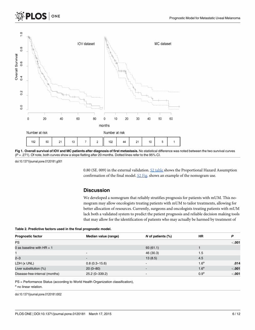
Survival and Prognostic NomogramThe estimated median OS was 17.2 months (range 1.2–86.4) and 19.7 months (range12.4–23.4) in the IOV and MC series, respectively. Twelve and 24-months survival was 63.4%and 34.5% for IOV and 61.7% and 35.6% for MC patients, respectively (Fig. 1). The survivalanalysis demonstrated that the difference between the two cohorts was not significant(P = .271).The following covariates were tested in a multivariate Cox regression model: sex,size and characteristics of primary UM, age at diagnosis of mUM, DFI, hepatic enzyme levelsat diagnosis of mUM (LDH, ALP, UGT, transaminases), site and number of metastases, per-centage of liver replacement and first line treatment. We observed that locoregional therapywas associated with a trend for longer survival [32, 33], although this was not statistically sig-nificant in the multivariate model. Clinical and histopathological characteristics of primarymelanoma did not correlate with survival. A trend for shorter DFI in patients with worse pri-mary melanoma characteristics, i.e. stage (P = .060), ciliary body involvement (P = .058) andepitheloid (P = .069) or mixed histology (P = 0.57) was noted, however not statistically signifi-cant. We observed enrichment for strongly pigmented and genomically aberrant cases. S1 tableshows the correlation with survival for primary melanoma characteristics and their influenceon OS at multivariate analysis. The significant covariates of the final model are showed inTable 2 while the nomogram for the predictive model is reported in Fig. 2. Increasing liver sub-stitution (HR 1.6), serum LDH (HR 1.6) and PS (HR of 1.5 and 4.6 for PS 1 and 2–3, respec-tively) were associated with worse OS. Longer DFI was associated with better prognosis (HR0.9). The effect of each predictor on survival is represented in S1 Fig.
Although number of organs involved by metastatic disease and type of therapy received(locoregional, systemic or combined) showed some association with prognosis, this was notsignificant. No association with survival was noted for sex, liver function tests other than LDH
Prognostic Model for Metastatic Uveal Melanoma
PLOSONE | DOI:10.1371/journal.pone.0120181 March 17, 2015 4 / 12

and age of metastasis. Remodeling the nomogram to include molecular alterations did not im-prove its performance. The calibration accuracy was confirmed by the receiver operating char-acteristic curves shown in Fig. 3. The absence of systematic bias is confirmed by the closenessof the receiver operating characteristic curves; rough shrinkage was 0.9. The nomogram wasvalidated with the external dataset of MC patients by assessing the reliability, as reported inTable 3. The concordance probability was 0.75 (SE. 006) in the development dataset, and
Table 1. IOV and MC patient characteristic at diagnosis of stage IV disease.
Patients characteristics IOV (N = 152) MC(N = 102)N (%) Median (range) N (%) Median (range) P
Sex .271
Female 73 (48.1) 41 (40.2)
Male 79 (51.9) 61 (59.8)
Age of primary UM (years) 60.9 (25.3–82.5) 59.0 (28.0–92.0) .352
Age of 1st metastasis (years) 63.6 (34.3–89.4) 61.5 (30.0–92.0) .262
DFI (months) 25.2 (0–339.2) 24.1 (0.1–140.8) .703
Number of organs involved .803
1 108 (71.1) 72 (70.6)
2 32 (21.0) 19 (18.6)
3� 12 (7.9) 11 (10.8)
Liver metastasis <.001
No 9 (5.9) 14 (13.7)
Yes 143 (94.0) 88 (86.3)
<20% 63 (41.4) 68 (66.7)
20�50% 48 (31.5) 16 (15.7)
50%� 19 (12.5) 4 (3.9)
Missing data 14 (9.2) 0
LDH (x UNL) 0.8 (0.3–15.6) 0.9 (0.5–10.4) .005
UGT (x UNL) 1.1 (0.1–27.9) 0.9 (0.4–25.3) .761
AST (x UNL) 0.7 (0.2–11.8) 0.6 (0.3–10.4) .901
ALT (x UNL) 0.6 (0.2–7.6) 0.9 (0.3–3.1) .109
PS .008
0 93 (61.1) 61 (59.8)
1 46 (30.3) 9 (8.8)
2 9 (5.9) 2 (1.9)
3 4 (2.6) 1 (0.9)
Missing data 0 29 (28.4)
1st line treatment of mUM <.001
No treatment 21 (13.8) 0
Any treatment 131 (86.2) 102 (100)
Systemic therapy alone 56 (54.9) 74 (72.5)
Locoregional therapy alone 17 (16.7) 17 (16.7)
Locoregional + systemic therapy 68 (44.7) 5 (4.9)
Missing data 0 9 (8.8)
IOV = Veneto Oncology Research Institute,
MC = Mayo Clinic,
PS = Performance Status (according to World Health Organization classification),
(x UNL) = multiples of the upper normal limit.
doi:10.1371/journal.pone.0120181.t001
Prognostic Model for Metastatic Uveal Melanoma
PLOSONE | DOI:10.1371/journal.pone.0120181 March 17, 2015 5 / 12
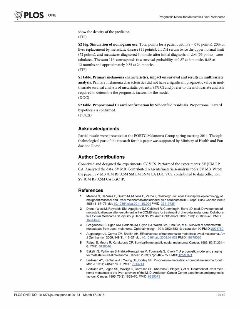
0.80 (SE. 009) in the external validation. S2 table shows the Proportional Hazard Assumptionconfirmation of the final model. S2 Fig. shows an example of the nomogram use.
DiscussionWe developed a nomogram that reliably stratifies prognosis for patients with mUM. This no-mogram may allow oncologists treating patients with mUM to tailor treatments, allowing forbetter allocation of resources. Currently, surgeons and oncologists treating patients with mUMlack both a validated system to predict the patient prognosis and reliable decision making toolsthat may allow for the identification of patients who may actually be harmed by treatment of
Fig 1. Overall survival of IOV and MC patients after diagnosis of first metastasis. No statistical difference was noted between the two survival curves(P = .271). Of note, both curves show a slope flatting after 20 months. Dotted lines refer to the 95% CI.
doi:10.1371/journal.pone.0120181.g001
Table 2. Predictive factors used in the final prognostic model.
Prognostic factor Median value (range) N of patients (%) HR P
PS <.001
0 as baseline with HR = 1 93 (61.1) 1
1 - 46 (30.3) 1.5
2–3 - 13 (8.5) 4.5
LDH (x UNL) 0.8 (0.3–15.6) - 1.6a .014
Liver substitution (%) 20 (0–80) - 1.6a <.001
Disease-free-interval (months) 25.2 (0–339.2) - 0.9a <.001
PS = Performance Status (according to World Health Organization classification),a no linear relation.
doi:10.1371/journal.pone.0120181.t002
Prognostic Model for Metastatic Uveal Melanoma
PLOSONE | DOI:10.1371/journal.pone.0120181 March 17, 2015 6 / 12

their metastatic disease. Although this is not as large of a concern in cancers that carry a rela-tive good prognosis and have multiple treatment options with proven clinical benefit, it is acrucial determinant of clinical care for very rare tumors with no established standard treatmentand potentially toxic therapies [28].
The two largest previously published series of patients with mUM did evaluate for prognos-tic factors [3, 34]. Other studies have tried to identify prognostic factors for patients withmUM, however these analyses were based on small series. Moreover, these works took into ac-count only a limited number of putative prognostic factors [3, 5–10, 35, 36]. Of these studies,Eskelin et. al [6] constructed a prognostic model with PS, dimensions of liver metastasis, ALPlevels (as substitute of LDH) and time on treatment using a multivariate analysis in 54 patients.Kodjikian et al. found that ciliary body involvement and more than 10 metastases conferred aworse prognosis by analyzing primary melanoma characteristics, age and the number of livermetastasis before surgery in 63 patients using a multivariate Cox regression. Finally, Rietschelet al. [10], showed that lung/soft tissue metastases, long DFI, locoregional treatments, femalesex and younger age all conferred better prognosis, using a multivariate analysis on 119 pa-tients. Unfortunately, none of these studies considered all of the previously identified prognos-tic factors and compared them in a multivariate analysis, nor did they adopt any calibration orvalidation strategy. These previous experiences testify to the difficulty faced when studying theprognostic factors for UM; the rarity of this disease makes the collection of a large, comprehen-sive series of all prognostic factors complex, with wide variations in diagnostic and treatmentmodalities over the time of observation. However, with the availability of new regional thera-pies and targeted drugs, a simple and validated model for patient risk stratification is needed.A reliable tool to evaluate the prognosis could aid clinicians in selecting the candidates for inva-sive or potentially toxic treatments, which should be reserved for patients with longer life
Fig 2. Nomogram of the final prognostic model. The sum of the prognostic factor points corresponds to the survival probability at 6, 12 and 24 months.
doi:10.1371/journal.pone.0120181.g002
Prognostic Model for Metastatic Uveal Melanoma
PLOSONE | DOI:10.1371/journal.pone.0120181 March 17, 2015 7 / 12

expectancy. With the collaboration of two independent groups, enough data were collected toperform a reliable validation of a prognostic model, with modalities and sample sizes compara-ble to currently accepted nomograms for rare tumors[36].
Both clinico-pathological and modern molecular prognostic factors have been identified forprimary uveal melanoma and are predictors of metastases. However, their usefulness in themetastatic setting has yet to be evaluated. Additionally, the modern molecular procedures, thathave been demonstrated to be superior to clinic-pathological characteristics[37], are currentlylimited to experimental studies. Primary melanoma features, included ciliary body involvement,did not impact on survival in our series. However, we observed a trend for shortest DFI associ-ated to worse primary melanoma characteristics, and this is consistent with the hypothesis thatDFI is influenced by the biological aggressiveness of the tumor (reflected by primary melanomacharacteristics), but also the result of the interaction of several concurrent variables (such as bi-ological determinants of tumor and immunological equilibrium). We analyzed whether the ad-dition of molecular alterations could improve the performance of the nomogram, however no
Fig 3. Receiver operating characteristic curves of the calibration. Estimate of calibration accuracy wasperformed using adaptive spline regression. The line adjacent the ideal line corresponds to the apparentpredictive accuracy. The blue line corresponds to corrected estimates.
doi:10.1371/journal.pone.0120181.g003
Table 3. External validation of the nomogram.
Nomogam External validation
C Index Dxy SE C Index Dxy SE
0.75 0.50 .006 0.80 0.60 .009
SE = Standard Error.
doi:10.1371/journal.pone.0120181.t003
Prognostic Model for Metastatic Uveal Melanoma
PLOSONE | DOI:10.1371/journal.pone.0120181 March 17, 2015 8 / 12

statistically significant trends with these alterations were noted. One reason for this could bethat these alterations have been associated with the development of metastatic disease for pa-tients with mUM, while this study looked at the prognosis of patients who have already devel-oped metastatic disease. The genomic (mainly chromosome 3, 6 and 8 aberrations [38, 39]) andgenetic (for example the two gene-expression profiles identified by Onken et al. [40]) abnormal-ities are predictors of distant recurrence after UM primary diagnosis, then other biological fac-tors, yet to be studied, may determine the aggressiveness of metastases when the disease hasspread. We encourage future studies to explore the potential genomic and genetic alterationsthat may influence the prognosis of patients with mUM. Our experience suggests that large, col-laborative studies are needed to obtain an adequate sample size to study potential molecularpredictors of survival or new therapeutic options, given the rarity of the disease. Melanin is de-scribed as one of the potential causes for melanoma refractoriness to treatments[41–44]. Thereare no extensive data in the literature on the pattern of pigmentation and its prognostic impor-tance in this rare subtype of melanoma. In our series all cases presented strong pigmentation.These data, although insufficient to assert conclusions, are hypothesis generating for furtherstudies focused on mUM, also in the prospective to find the reasons of mUM poorer responseto immunotherapies compared to its skin counterpart[13].
In both of our independent series, the survival is longer than other studies; however, similarsurvival times have been reported by Rietschel et al. [10] and Kodjikian et al. [45]. It is oftennot possible to extrapolate the time from the onset of stage IV disease and to that of the initia-tion of therapy from clinical trials. Additionally, most of the variables we studied were onlypartially included in other works, making it difficult to compare the OS and clinical predictorsof our study with those that have been previously reported. Possible explanations for the differ-ences in survival could include referral bias as both centers included in this study are large re-ferral centers and lead time bias, as we noted shorter DFIs (25.2 and 24.1 months for the IOVand MC dataset, respectively) than those previously reported (30 months in asymptomatic and79 in symptomatic patients [10]). These differences were unlikely a consequence of the regularliver surveillance, as patients were referred from centers with different follow-up practices,varying from regular liver function tests and liver ultrasound every 6 months to no surveillance.Regardless, the analysis performed was not influenced by the duration of survival itself, butrather by the influence different factors had on survival. To confirm the reproducibility of ourresults, the nomogram was validated with very good performance despite the significant differ-ences observed for some prognostic predictors between IOV’s and Mayo’s patients. We ob-tained two bi-phasic survival curves, with a steep slope flatting out after about 20 months(Fig. 1), similar to the curve reported by Rietschel et al. [10], suggesting as a confirmation ofthe heterogeneity of this disease. Although the liver was the most prevalent metastatic site, weidentified a number of patients who had metastases in other organs. However, the majority ofthe patients with mUM died as a result of liver progression despite first developing metastaticdisease at another site. Therefore, we cannot advise for routine screening of extra-hepatic sites.
ConclusionsIn summary, we developed and externally validated a nomogram that predicts survival in pa-tients with mUM. This nomogram may be useful in stratifying patients in future clinical trialsand help providers prognosticate.
Supporting InformationS1 Fig. The effect of individual predictors on log survival time. A value of zero was used asthe reference value for all predicted 95% confidence intervals are shown. “Rug plots” on curves
Prognostic Model for Metastatic Uveal Melanoma
PLOSONE | DOI:10.1371/journal.pone.0120181 March 17, 2015 9 / 12
show the density of the predictor.(TIF)
S2 Fig. Simulation of nomogram use. Total points for a patient with PS = 0 (0 points), 20% ofliver replacement by metastatic disease (11 points), a LDH serum twice the upper normal limit(72 points), and metastases diagnosed 6 months after initial diagnosis of UM (33 points) weretabulated. The sum 116, corresponds to a survival probability of 0.87 at 6 months, 0.68 at12 months and approximately 0.35 at 24 months.(TIF)
S1 table. Primary melanoma characteristics, impact on survival and results in multivariateanalysis. Primary melanoma characteristics did not have a significant prognostic value in mul-tivariate survival analysis of metastatic patients. 95% CI and p refer to the multivariate analysisrequired to determine the prognostic factors for the model.(DOC)
S2 table. Proportional Hazard confirmation by Schoenfeld residuals. Proportional Hazardhypothesis is confirmed.(DOCX)
AcknowledgmentsPartial results were presented at the EORTCMelanoma Group spring meeting 2014. The oph-thalmological part of the research for this paper was supported by Ministry of Health and Fon-dazione Roma.
Author ContributionsConceived and designed the experiments: SV VCS. Performed the experiments: SV JCM RPCA. Analyzed the data: SV MB. Contributed reagents/materials/analysis tools: SV MB. Wrotethe paper: SV MB JCM RP ASM SM EM SNM CA LGC VCS. contributed to data collection:SV JCM RP ASM CA LGC JP.
References1. Mallone S, De Vries E, Guzzo M, Midena E, Verne J, Coebergh JW, et al. Descriptive epidemiology of
malignant mucosal and uveal melanomas and adnexal skin carcinomas in Europe. Eur J Cancer. 2012;48(8):1167–75. doi: 10.1016/j.ejca.2011.10.004 PMID: 22119735
2. Diener-West M, Reynolds SM, Agugliaro DJ, Caldwell R, Cumming K, Earle JD, et al. Development ofmetastatic disease after enrollment in the COMS trials for treatment of choroidal melanoma: Collabora-tive Ocular Melanoma Study Group Report No. 26. Arch Ophthalmol. 2005; 123(12):1639–43. PMID:16344433
3. Gragoudas ES, Egan KM, Seddon JM, Glynn RJ, Walsh SM, Finn SM, et al. Survival of patients withmetastases from uveal melanoma. Ophthalmology. 1991; 98(3):383–9; discussion 90 PMID: 2023760
4. Augsburger JJ, Correa ZM, Shaikh AH. Effectiveness of treatments for metastatic uveal melanoma. AmJ Ophthalmol. 2009; 148(1):119–27. doi: 10.1016/j.ajo.2009.01.023 PMID: 19375060
5. Rajpal S, Moore R, Karakousis CP. Survival in metastatic ocular melanoma. Cancer. 1983; 52(2):334–-6. PMID: 6190546
6. Eskelin S, Pyrhonen S, Hahka-Kemppinen M, Tuomaala S, Kivela T. A prognostic model and stagingfor metastatic uveal melanoma. Cancer. 2003; 97(2):465–75. PMID: 12518371
7. Bedikian AY, Kantarjian H, Young SE, Bodey GP. Prognosis in metastatic choroidal melanoma. SouthMed J. 1981; 74(5):574–7. PMID: 7244714
8. Bedikian AY, Legha SS, Mavligit G, Carrasco CH, Khorana S, Plager C, et al. Treatment of uveal mela-noma metastatic to the liver: a review of the M. D. Anderson Cancer Center experience and prognosticfactors. Cancer. 1995; 76(9):1665–70. PMID: 8635073
Prognostic Model for Metastatic Uveal Melanoma
PLOSONE | DOI:10.1371/journal.pone.0120181 March 17, 2015 10 / 12

9. Hsueh EC, Essner R, Foshag LJ, Ye X, Wang HJ, Morton DL. Prolonged survival after complete resec-tion of metastases from intraocular melanoma. Cancer. 2004; 100(1):122–9. PMID: 14692032
10. Rietschel P, Panageas KS, Hanlon C, Patel A, Abramson DH, Chapman PB. Variates of survival in met-astatic uveal melanoma. J Clin Oncol. 2005; 23(31):8076–80. PMID: 16258106
11. Slominski AT, Carlson JA. Melanoma resistance: a bright future for academicians and a challenge forpatient advocates. Mayo Clin Proc. 2014; 89(4):429–33. doi: 10.1016/j.mayocp.2014.02.009 PMID:24684870
12. Cruz F 3rd, Rubin BP, Wilson D, Town A, Schroeder A, Haley A, et al. Absence of BRAF and NRASmu-tations in uveal melanoma. Cancer Res. 2003; 63(18):5761–6. PMID: 14522897
13. Maio M, Danielli R, Chiarion-Sileni V, Pigozzo J, Parmiani G, Ridolfi R, et al. Efficacy and safety of ipili-mumab in patients with pre-treated, uveal melanoma. Ann Oncol. 2013; 24(11):2911–5. doi: 10.1093/annonc/mdt376 PMID: 24067719
14. Mariani P, Servois V, Piperno-Neumann S. Therapeutic options in metastatic uveal melanoma. DevOphthalmol. 2012; 49:166–81. doi: 10.1159/000328333 PMID: 22042020
15. Peters S, Voelter V, Zografos L, Pampallona S, Popescu R, Gillet M, et al. Intra-arterial hepatic fote-mustine for the treatment of liver metastases from uveal melanoma: experience in 101 patients. AnnOncol. 2006; 17(4):578–83. PMID: 16469752
16. Leyvraz S, Piperno-Neumann S, Suciu S, Baurain JF, Zdzienicki M, Testori A, et al. Hepatic intra-arterial versus intravenous fotemustine in patients with liver metastases from uveal melanoma (EORTC18021): a multicentric randomized trial. Ann Oncol. 2014; 25(3):742–6. doi: 10.1093/annonc/mdt585PMID: 24510314
17. Sato T, Eschelman DJ, Gonsalves CF, Terai M, Chervoneva I, McCue PA, et al. Immunoembolizationof malignant liver tumors, including uveal melanoma, using granulocyte-macrophage colony-stimulating factor. J Clin Oncol. 2008; 26(33):5436–42. doi: 10.1200/JCO.2008.16.0705 PMID:18838710
18. Fiorentini G, Aliberti C, Del Conte A, Tilli M, Rossi S, Ballardini P, et al. Intra-arterial hepatic chemoem-bolization (TACE) of liver metastases from ocular melanoma with slow-release irinotecan-elutingbeads. Early results of a phase II clinical study. In Vivo. 2009; 23(1):131–7. PMID: 19368137
19. Gonsalves CF, Eschelman DJ, Sullivan KL, Anne PR, Doyle L, Sato T. Radioembolization as salvagetherapy for hepatic metastasis of uveal melanoma: a single-institution experience. AJR Am J Roent-genol. 2011; 196(2):468–73. doi: 10.2214/AJR.10.4881 PMID: 21257902
20. Feldman ED, Pingpank JF, Alexander HR Jr. Regional treatment options for patients with ocular mela-noma metastatic to the liver. Ann Surg Oncol. 2004; 11(3):290–7. PMID: 14993024
21. Olofsson R, Cahlin C, All-Ericsson C, Hashimi F, Mattsson J, Rizell M, et al. Isolated Hepatic Perfusionfor Ocular MelanomaMetastasis: Registry Data Suggests a Survival Benefit. Ann Surg Oncol. 2013.
22. Leyvraz S, Keilholz U. Ocular melanoma: what's new? Curr Opin Oncol. 2012; 24(2):162–9. doi: 10.1097/CCO.0b013e32834ff069 PMID: 22234256
23. Kivela T, Suciu S, Hansson J, Kruit WH, Vuoristo MS, Kloke O, et al. Bleomycin, vincristine, lomustineand dacarbazine (BOLD) in combination with recombinant interferon alpha-2b for metastatic uveal mel-anoma. Eur J Cancer. 2003; 39(8):1115–20. PMID: 12736111
24. Dorval T, FridmanWH, Mathiot C, Pouillart P. Interleukin-2 therapy for metastatic uveal melanoma. EurJ Cancer. 1992; 28A(12):2087. PMID: 1419309
25. Valsecchi ME, Sato T. The potential role of sunitinib targeting melanomas. Expert Opin Investig Drugs.2013; 22(11):1473–83. doi: 10.1517/13543784.2013.837449 PMID: 24050392
26. Tarhini AA, Frankel P, Margolin KA, Christensen S, Ruel C, Shipe-Spotloe J, et al. Aflibercept (VEGFTrap) in inoperable stage III or stage iv melanoma of cutaneous or uveal origin. Clin Cancer Res. 2011;17(20):6574–81. doi: 10.1158/1078-0432.CCR-11-1463 PMID: 21880788
27. Falchook GS, Lewis KD, Infante JR, Gordon MS, Vogelzang NJ, DeMarini DJ, et al. Activity of the oralMEK inhibitor trametinib in patients with advancedmelanoma: a phase 1 dose-escalation trial. LancetOncol. 2012; 13(8):782–9. doi: 10.1016/S1470-2045(12)70269-3 PMID: 22805292
28. Carvajal RD, Sosman JA, Quevedo JF, MilhemMM, Joshua AM, Kudchadkar RR, et al. Effect of selu-metinib vs chemotherapy on progression-free survival in uveal melanoma: a randomized clinical trial.JAMA. 2014; 311(23):2397–405. doi: 10.1001/jama.2014.6096 PMID: 24938562
29. Midena E, Bonaldi L, Parrozzani R, Tebaldi E, Boccassini B, Vujosevic S. In vivo detection of monoso-my 3 in eyes with medium-sized uveal melanoma using transscleral fine needle aspiration biopsy. EurJ Ophthalmol. 2006; 16(3):422–5. PMID: 16761244
30. Midena E, Bonaldi L, Parrozzani R, Radin PP, Boccassini B, Vujosevic S. In vivo monosomy 3 detec-tion of posterior uveal melanoma: 3-year follow-up. Graefes Arch Clin Exp Ophthalmol. 2008; 246-(4):609–14. PMID: 17934749
Prognostic Model for Metastatic Uveal Melanoma
PLOSONE | DOI:10.1371/journal.pone.0120181 March 17, 2015 11 / 12

31. Bonaldi L, Midena E, Filippi B, Tebaldi E, Marcato R, Parrozzani R, et al. FISH analysis of chromo-somes 3 and 6 on fine needle aspiration biopsy samples identifies distinct subgroups of uveal melano-mas. J Cancer Res Clin Oncol. 2008; 134(10):1123–7. doi: 10.1007/s00432-008-0382-6 PMID:18386059
32. Valpione S, Aliberti C, Parrozzani R, Bazzi M, Pigozzo J, Midena E, et al. A retrospective analysis of141 patients with liver metastases from uveal melanoma: a two-cohort study comparing transarterialchemoembolization with CPT-11 charged microbeads and historical treatments. Melanoma Res. 2015.
33. Moser JC, Pulido JS, Dronca RS, McWilliams RR, Markovic SN, Mansfield AS. The Mayo Clinic experi-ence with the use of kinase inhibitors, ipilimumab, bevacizumab, and local therapies in the treatment ofmetastatic uveal melanoma. Melanoma Res. 2015; 25(1):59–63. doi: 10.1097/CMR.0000000000000125 PMID: 25396683
34. Diener-West M, Reynolds SM, Agugliaro DJ, Caldwell R, Cumming K, Earle JD, et al. Screening for me-tastasis from choroidal melanoma: the Collaborative Ocular Melanoma Study Group Report 23. J ClinOncol. 2004; 22(12):2438–44. PMID: 15197206
35. Kath R, Hayungs J, Bornfeld N, Sauerwein W, Hoffken K, Seeber S. Prognosis and treatment of dis-seminated uveal melanoma. Cancer. 1993; 72(7):2219–23. PMID: 7848381
36. Gold JS, Gonen M, Gutierrez A, Broto JM, Garcia-del-Muro X, Smyrk TC, et al. Development and vali-dation of a prognostic nomogram for recurrence-free survival after complete surgical resection oflocalised primary gastrointestinal stromal tumour: a retrospective analysis. Lancet Oncol. 2009; 10-(11):1045–52. doi: 10.1016/S1470-2045(09)70242-6 PMID: 19793678
37. Augsburger JJ, Correa ZM, Trichopoulos N. An alternative hypothesis for observed mortality rates dueto metastasis after treatment of choroidal melanomas of different sizes. Trans AmOphthalmol Soc.2007; 105:54–9; discussion 9–60 PMID: 18427594
38. Ewens KG, Kanetsky PA, Richards-Yutz J, Al-Dahmash S, De Luca MC, Bianciotto CG, et al. Genomicprofile of 320 uveal melanoma cases: chromosome 8p-loss and metastatic outcome. Invest OphthalmolVis Sci. 2013; 54(8):5721–9. doi: 10.1167/iovs.13-12195 PMID: 23821189
39. Thomas S, Putter C, Weber S, Bornfeld N, Lohmann DR, Zeschnigk M. Prognostic significance of chro-mosome 3 alterations determined by microsatellite analysis in uveal melanoma: a long-term follow-upstudy. Br J Cancer. 2012; 106(6):1171–6. doi: 10.1038/bjc.2012.54 PMID: 22353812
40. Onken MD, Worley LA, Ehlers JP, Harbour JW. Gene expression profiling in uveal melanoma revealstwo molecular classes and predicts metastatic death. Cancer Res. 2004; 64(20):7205–9. PMID:15492234
41. Brozyna AA, VanMiddlesworth L, Slominski AT. Inhibition of melanogenesis as a radiation sensitizerfor melanoma therapy. Int J Cancer. 2008; 123(6):1448–56. doi: 10.1002/ijc.23664 PMID: 18567001
42. Slominski A, Zbytek B, Slominski R. Inhibitors of melanogenesis increase toxicity of cyclophosphamideand lymphocytes against melanoma cells. Int J Cancer. 2009; 124(6):1470–7. doi: 10.1002/ijc.24005PMID: 19085934
43. Slominski A, Zmijewski MA, Pawelek J. L-tyrosine and L-dihydroxyphenylalanine as hormone-like regu-lators of melanocyte functions. Pigment Cell Melanoma Res. 2012; 25(1):14–27. doi: 10.1111/j.1755-148X.2011.00898.x PMID: 21834848
44. Brozyna AA, Jozwicki W, Carlson JA, Slominski AT. Melanogenesis affects overall and disease-freesurvival in patients with stage III and IV melanoma. Hum Pathol. 2013; 44(10):2071–4. doi: 10.1016/j.humpath.2013.02.022 PMID: 23791398
45. Kodjikian L, Grange JD, Baldo S, Baillif S, Garweg JG, Rivoire M. Prognostic factors of liver metastasesfrom uveal melanoma. Graefes Arch Clin Exp Ophthalmol. 2005; 243(10):985–93. PMID: 15891893
Prognostic Model for Metastatic Uveal Melanoma
PLOSONE | DOI:10.1371/journal.pone.0120181 March 17, 2015 12 / 12
Related Documents